mica polytypism: dissimilarities in the crystal structures'of
TRANSCRIPT

American Mineralogist, Volume 60, pages 1030-1040, 1975
Mica Polytypism: Dissimilarities in the Crystal Structures'ofCoexisting lM and 2M, Biotite
Hrnossl Terron
Mineralogical Institute, Faculty of Science, Uniuersity of Tokyo,Hongo, Tokyo Il3, Japan
eNn MnLcolnr Ross
U.S. Geological Suruey, National Center-959, Reston, Virginia 22092
Abstract
Crystal-structure analysis of coexisting lM and 2M, btotite polytypes from a rhyodacitelava flow, Ruiz Peak, New Mexico, shows that the two forms possess essentially identicalunit-cell parameters (in the lM setting), bond length averages, and site occupancies andprobably, therefore, very similar chemical compositions. The unit layers of the two polytypes,however, are different structually in that the x and y atomic positions of the 03 and 04 ox-ygens differ in the two forms by an average of 14 standard deviations. The average differencein the relative values of all other atomic positions is one standard deviation. The displacementof 03 and 04 in the 2Mrbiotitecauses a type of deformation not before reported in micas andmanifests itself by a relative shifting of the upper and lower triads of octahedral oxygens as aunit along the aD directions (2Mt setting). This causes a reduction in symmetry of the2M,unit-layer from the ideal C2/m as found in the lM polytype, to Cl.
It is postulated that the structural differences between the unit layers of thelM and,2M,biotite polytypes are due to the different atomic and geometric constraints imposed upon theunit layer by the adjacent layers, the latter being in different relative orientations in differentpolytypes. An origin for biotite polytypism is proposed on the basis of these unit-layer con-straints coupled with the mica platelet growth and spiral growth observations of A. Baronnet.
Introduction
In the last decade, many reports have beenpublished on the crystal structure of various micapolytypes. For example, the model structures of lMtrioctahedral micas were summarized by Donnay,Donnay, and Takeda (1964) and by Takeda (1971);the unit-layer of 2M , muscovite, refined by Burnhamand Radoslovich (1964), was compared with that ofthe 37 form by Giiven and Burnham (1967),lM and2M, brittle micas of different composition werestudied by Tak6uchi (1964), lM and 2Mr lepidoliteswere studied by Takeda, Haga, and Sadanaga (1971),and a 2O brittle mica was examined by Giuseppettiand Tadini (1972). The previous studies of micapolytypes were accomplished on samples fromdiverse localities having significantly differentchemical compositions. It is thus difficult to decidehow much the structural differences found betweenvarious polytypes are due to chemical differences,differences in temperature and pressure of formation,and differences in the stacking sequence of the unit-
layers.' Another question is, are the unit layers struc-turally as well as chemically identical if the micacrystals have different periodic stacking sequences?
The question of possible structural differencesbetween different mica polytypes found in the samerock hand specimen arose during our study of ox-ybiotites from a rhyodacite lava flow at Ruiz Peak,New Mexico (Ross et al., 1966). Single crystal X-raystudy of 42 crystals from this specimen showed thatapproximately one-third are lM forms, one-third2Mr, and one-third more complex polytypes such asthe 4Mr,8M", and 87c,, forms. The detailed study ofthe structures of these crystals, however, was post-poned, for we did not believe that at that time crystalstructure refinements could be done accuratelyenough to show the very small differences expectedbetween unit layers of different polytypes. The pres-ent study of the crystal structures of coexisting lM
I A mica unit-layer is defined as thatthick, contained in the unit-cell of thelayers occur in the unit cell of the 2M,
1030
layer, approximately l0AlM polytype. Two unitpolytype.

DISSIMILARITIES IN COEXISTING IM AND 2M, BIOTITE l03 l
and 2Mt polytypes from a single hand specimen of
the Ruiz rhyodacite was made during 1972 using the
best X-ray diffraction techniques then available. With
this study we hope that we may develop a better un-
derstanding of the nature and cause of polytypism
and polymorphism'z in complex silicates.
MineralogySpecimen Description
The biotite crystals examined in this study occur in
a rhyodacite ash flow from the Ruiz Peak area, Valles
Mountains, New Mexico. The particular rock sample
studied (No. 3149-8) came from the top of the flow
and is reddish because of formation of hematite after
eruption of the lava. The rock contains abundantphenocrysts of feldspar, biotite, and magnetite and
less abundant phenocrysts of augite and ortho-pyroxene. Apatite is ubiquitous and occurs as inclu-
sions in the biotite. The groundmass is composed
mainly of feldspar, glass, and biotite; the latter is
much altered to hematite and other products of late
stage oxidation (probably sanidine, metastable mag-
netite, and glass; D. R. Wones, personal communica-
tion). The large biotite phenocrysts are often rimmed
with hematite, and this phase is also seen in the cleav-
age cracks. The biotite crystals separated for chem-
ical and crystallographic analysis, however, were
nearly free of hematite but still contained apatite in-
clusions and a small amount of ruti le.The oxidation, which is inferred to have occurred
during and after eruption of the lava, affected the
biotite crystals in two ways: (l) exposed surfaces of
the biotites reacted to form hematite and other break-
down products, and (2) the crystals reequil ibratedwith respect to the hydrogen fugacity by the reaction,
somewhat idealized,
KM g,-"Fel+(Si3Al )OuH,- KMgr-,Fe,'1"Fe'r*lsirAl)orrHr-, + | / 2yHr1 .
This reaction caused the biotite crystals to turn dark
reddish brown. Biotite crystals from underlying,
'? We define polytypism as the existence of two or more chemical-
ly identical, or nearly identical, phases which have structurally
similar unit motifs (unit layers in the case of micas) but which
repeat in three-dimensional crystal space in different ways. We
define polymorphism as the existence of two or more chemically
identical, or nearly identical, phases which do not possess struc-
turally similar unit motifs. Phases such as quartz-coesite and
calcite-aragonite can be unambiguously defined as polymorphs
whereas substances such as the biotites, zinc sulfides, silicon car-
bides, and cadmium iodides are generally thought of as showing
polytypism.
unoxidized flows do not show this color and theirferric iron content is much lowqr.
Chemical Composition
The biotite chemical formulas based on wet
chemical and electron microprobe analyses (separates
3149-8F and 3149-8W, respectively) are given in
Table l, columns (1) and (3). Electron probe analysesmade on the clear homogeneouS areas of five crystalsrevealed barium-an element missed by the wet
chemical analysis-but no calsium. The 0.46 wt per-
cent CaO and 0.34 wt percent PzOu found in the wet
analysis is thus due to included apatite' The range ofcation content as obtained by microprobe analysesoverlaps that given by wet chemical analyses with the
exception of higher Mg and Al and lower Fe content'
We attribute this difference to chemical differences in
the biotite phenocrysts depending upon the time of
crystallization from a fractionating melt. The
different mineral separates reflect these chemical
differences. The biotite crystals studied crystallo-graphically came from separate 3149-8F, and thus we
assume that the Mg, Al, and Fe content is closer to
that given by the wet chemical analysis. The electron
microprobe study did not detect any significantchemical variation within a single biotite crystal'
The oxy-biotite sample under study contains 8'4,
10.6, and 81.0 percent "fluoro", "hydroxy", aqd
"oxy" component, respectively (Table 1, column l)'
Of the total "oxy" component 52.8 percent is relatedto a couple substitution involving oxidation-reduc-tion, that is,
Fe2+ * OH- =Fe3+ + A- + l /2Hr.
Part or most of this substitution probably occurred as
a solid-state reaction during eruption of the lava' Of
the (4'7.2Vo) remaining "oxy" component, most(42.2Eo) must be attributed to the incorporation of
0.342 atoms per formula unit of titanium (Table l)
into the crystal structure during crystal growth,
charge balance being maintained by increase of the
oxyanion content. This coupled substitution may be
written
K(Mg,Fe)3+(Si3Al)O1o(OH)2 + Ti '+
= K(Mg,Fe);+Ti(SisAl)O1, + (Mg,Fe)'?+ + 2H+
Hydrogenation of Biotite
Hydrogenation of the oxybiotites is easily ac-complished by passing hydrogon gas at temperaturesof 500' to 700'C over the single crystals. The reac-
t032 H. TAKEDA AND M. ROSS
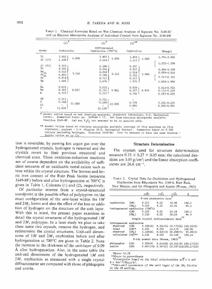
Tent-s l. chemical Formulae Based on wet chemical Analyses of Separate No. 3149-8Fand on Electron Microprobe Analyses of Individual crystals from Separate No. 3149-8w
At oms
(1)
oxyblotlte
( 2 )
HydrogenatedOxyb lo t i te (7000C)
(3) **
oxyblotite (Range)
s i41 (rv)
A1 (vr)TiI "h^ .
! e
Mg
B aN a
K
0 . 1630 . 3 4 20. 0100 . 8 5 50 . 0137 . 6 6 6
o . 0 2 20 . t 620 . 7 7 3
0 . 2 L 20 . 1 6 8
rL.620
3 .049
0 . 9 5 7
12 . 000
3 . 112
0 . 961
r 2 . 0 0 0
t . L47
0. 101o .32 r0 . 0 1 8o . 1 4 r0 . 0 1 17 . 8 7 4
0 . 0 2 00 . 1 5 70 . 7 5 6
i .?i; l 4.ooo i i l l l , *, 4 .000 2 .794 -2 .900
3 . 0 6 6
t . 222 -L .288
0 . 30 5-0. 3380 . 009 -0 .020
o . 7 4 2 - 0 . 7 6 2
1 . 850-1. 906
0 . 013 -0 .0220 . 933 0 .152 -0 . 168
0 . 692 -0 . 802
0 . 1 0 1 - 0 . 1 8 20 . 002-0. 004
0 . 186 I0 . 3 4 4 I0 .010 [o. ras (o . 7 u Ir . 6 7 6 I
OHFc10
0 . 0 2 20 . 1 6 2o . 7 7 7
0 . 7 1 1' 0 . 1 6 9
1 1 . 1 2 0
0 . 1380. 004
* A ton ic ra t ios based on wet chemica l -ana lys is , chen ls t ry labora tory , U .S. Geo log lca lsurvey' sumation based on: ofF+oH = 12. Bao fron electron nrcioprobe analysls,Fractlon 3149-8F. CaO and prO. are assuned !o be in apatlte only.
** Atonic ratios based on electron nicroprobe analysls (average of f lve analyses on fivecrys ta ls ) , ana lys t - L .B . h l tgg lns (U.S. ceo log lca l Survey) , Sumt ion based on 8 .000catlons ucluding hydrogen. Fractlon 3149-8I.I. lron 1s assumed to have the same ferrous -fe r r i c ra t los es 14 (1 ) .
t ion is reversible; by passing hot argon gas over thehydrogenated crystals, hydrogen is removed and thecrystals revert to their previous structural andchemical state. These oxidation-reduction reactionsare of course dependent on the availability of suffi-cient amounts of an oxidizable metal cation such asiron within the crystal structure. The ferrous and fer-ric iron content of the Ruiz Peak biotite (separate3149-8F) before and after hydrogenation at 700.C isgiven in Table l, Columns (l) and (2), respectively.
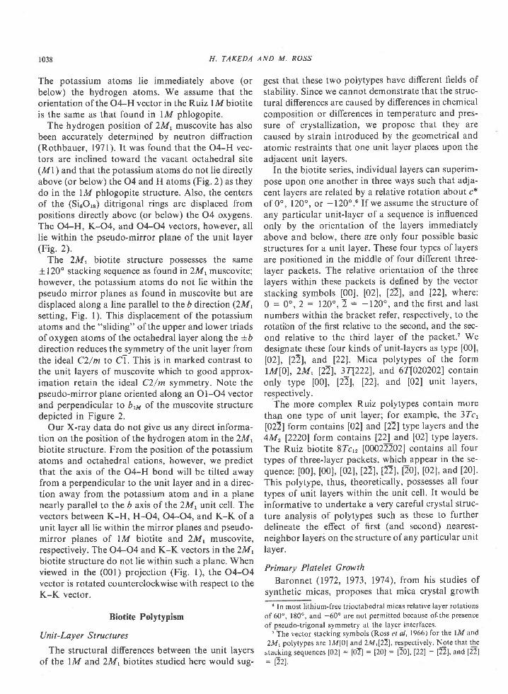
Of particular interest from a crystal-structuralstandpoint is the possible effect of polytypism on theexact configuration of the unit-layer within the lMand2Mt forms and also the effect of the loss or addi-tion of hydrogen on the structure of the unit layer.With this in mind, the present paper examines indetail the crystal structures of the hydrogenated lMand 2Ml pofytypes. In a later study we plan to takethese same two crystals, remove the hydrogen, andredetermine the crystal structures. Unit-cell dimen-sions of lM and 2M, oxybiotites before and afterhydrogenation at 700oC are given in Table 2. Notethe increase in the thickness of the unit-layer of 0.09A after hydrogenation. Also, in the sami table theunit-cell dimensions of the hydrogenated lM and2M, oxybiotites as measured with a single crystaldiffractometer are compared with those of phlogopiteand annite.
Structure Determination
The crystals used for structure determinationmeasure 0.23 X 0.27 x 0.05 mm; the calculated den-sities are 3.05 g/cm3; and the linear absorption coeffi-cients are 26.6 cm-t.
TesLr 2. Crystal Data for Oxybiotites and HydrogenatedOxybiotites from Rhyodacite No. 3149-8, Ruiz Peak,
New Mexico, and for Phlogopite and Annite (Wones, 1963)
" <il a <i.l f <.&l sX-ray precesslon dataT
oxyb lo t i te (1M) 5 .3L7 9 .22 10 .09 L0O.2( 2 1 t 1 1 5 . 3 1 s 9 . 2 2 1 9 . 9 5 9 5 . I
hydrogenated oxyblotlte (700"C)( 1 M ) 5 . 3 2 2 9 . 2 2 1 0 . 1 8 1 0 0 . 2( 2 M 1 ) s . 3 3 5 9 . 2 2 2 0 . 1 0 9 4 . 9
Slngle crystal dlf fractoneter datatf
hydrogoated oxybloriteobaerved (u { ) s .331(2) 9 ,23L(4) 10 .173(4) 100.16(3)ldea l ( t l t l * 5 .331 9 ,234 10 .170 100,06observed (21 , [ ) 5 .329(2) 9 .234(3) 20 .098(7) 9s .09(3)ca lcu la ted (1M)** 5 .331 9 ,23L 10 .168r_ 100.14
X-ray porder data (Wonee, 1953)"
ph losopt te (1M) 5 .326(6) 9 .210(9) 10 .311(9) 100.17(15)ann i te (1M) 5 .401(10) 9 .347(5) 10 .297(10)1OO. t7(15)
+E?"o? \0-2%ItEvror in parentheees
*CaLalation baeed on the ideal relationships a/T= b attdB = tan-1(aaooz/a).**Ilrtt-cell puarete?s of the unit layer of tLe 2M1 biotLte
tn the 7M eetbing,

Intensity measurements were made with a Picker-FAcs-l computer-controlled four-circle, single-crystaldiffractometer, using MoKa radiation with a graphitemonochromator and a scintillation detector. Inten-sities of 1202 reflections for the lM form and 3539reflections for the 2M, form were measured in the4"-60o 20 range using cir-2d scans. Each reflection wasscanned through 1.2o, using dispersion factors ofthesystem for a, and a2 separation at a scanning rate ofone degree per minute. The background was obtainedby counting for 20 seconds at both sides of the peak.Integrated intensities were corrected for Lorentz andpolarization effects and were reduced to structure fac-tors lF,l . The absorption correction was made byBurnham's (1966) subroutine with a linear absorp-tion coefficient of 26.6 cm-l for MoKa radiation. Theestimated standard deviation of an observation wascomputed on the basis of the counting statistics(Burnham et al, l97l).
Of the intensities measured, 649 nonequivalentreflections of the lM biotite, with the indices hkl andhkl, and 875 reflections o[ the 2Mrbiorite,with the in-dices hftl and Ekl, were utilized in the final refine-ments. The space groups C2/m for lM and C2/c for2Mrwere assumed and, on refinement, turned out tobe correct.
The full matrix least-squares program, RntNn(Finger, 1969) which includes the provision for siteoccupancy refinement, was used for the lM and2M,biotite crystal structure determinations. The weightsused in refinement are l/o(Fo)2, where o(Fo) is thestandard deviation of lFol . The atomic-scattering fac-tors used with RrINr were taken from Cromer andMann (1968), the oxygen atoms being consideredhalf-ionized (O'* ), and all cations completely ionized.Anomalous dispersion corrections were made byusing the coefficients of Cromer (1965).
Least-squares refinement of the lM structure wasinitiated by using atomic coordinates of a lMphlogopite (Joswig, 1972). The trial structure modelof the 2M, form was derived from the atomic coor-dinates of the above lM phlogopite using a program(Twuc) written for the Hluc 5020E computer(Takeda et al, l97l). The hydrogen atoms were notincluded in these refinements. The following cationswere fixed during refinement: K i Na in the interlayersi tes, Si + AlIv distr ibuted equal ly over thetetrahedral sites. and Ti + AlvI * Mn distributedequally over the octahedral sites. The relative amountof Fe * Mg in the two octahedral sites was allowed tovary during refinement, During the site refinement,the occupancy factor of Fe in the M2 site was derived
1033
from the total amount of Fe in the chemical formula,less that in Ml. The occupancy of Mg in Ml and M2is fixed by the value (l-Ti-Alvr-Mn-Fe) for each ofthese sites.
In the last stages of refinement, reflections forwhich lFol < l5 and | | Fol - | F" l l /o(Fo)>5 were re-jected. After six cycles of refinement, during whichthe scale factor, the atomic coordinates, and theanisotropic temperature factors Bij wete varied, nofurther change of parameters took place. Theobserved and calculated structure factors fot the lMand 2M, polytypes are listed in Tables 3a and 3b,respectively.3 The final conventional unweightedresiduals for structural parameters given in Tables 4and 5 are R:0.044 for 649 observable reflections ofthe lM form and R:0.056 for 875 reflections for the2M, form.
The various bond lengths (Tables 6,7, 8), selectedbond angles (Table 9), and associated standard devia-tions were computed from the atomic positions givenin Table 5. The variance-covariance matrices (Finger,1969) of the refinements and the standard deviationsof the unit-cell parameters were used for these com-putations. The observed lM coordinates are com-pared in Table 5a with the observed 2Mt coordinatesin the lM setting. The latter coordinates are those ofthe unit layer of the 2Mt polytype in the crystal-lographic orientation of the lM form and are derivedby a matrix rotation of 120" about c* of coordinatesof atoms in a 2M, unit layer (Table 5b) and thentranslation of the origin to that of the lM polytype.
Comparison of Structures
Chemical Similarity
The similarity of the unit-cell dimensions and bond
lengths of the lM and 2Mt crystals indicate that thd
crystals are very close in chemical composition. In
Table 2 the measured unit-cell dimensions of the lM
form are compared with the calculated unit-cell
dimensions of the 2M' polytype in the lM setting'The agreement between these two sets of unit-cell
dimensions is within one standard deviation for all
four parameters. Likewise, the mean tetrahedral, the
octahedral , and the inner and outer sets ofpotassium-oxygen bond lengths are essentially iden-
tical for both structure types. The comparative values
for average bond lengths (Tables 6, 7, 8) for the lM
3 To obtain a copy of Table 3a and 3b, order document AM-75-
008 from the Business Office, Mineralogical Society of America,
1909 K Street , N.W., Washington, D.C. 20006. 'Please remit
S1.00 for the microfiche.
DISSIMILARITIES IN COEXISTING 1M AND 2M, BIOTITE

r034 H. TAKEDA AND M. ROSS
Trslr 4 Site Occupancy Factors for CoexistingBiotites after Hydrogcnation at 700'C+
TlnI-s 5b. Atomic Coordinates of the HydrogenatedRuiz Peak 2M, Biotite in the 2M, Setting
ML M2x*
MgFe
T l + t " l n + A l
FeT l + l , I n + A 1
l-iV Polytype
0 . 5 3 70 .2900 . 1 7 3
2M, Polytype
0 . 5680 .2590 . 1 7 3
0 . 7 50 . 2 4 0 6 ( 3 )0 . 00 . 4 6 2 r ( 3 )0 . 9 6 3 5 ( 3 )0 . 7 4 1 0 ( 8 )0 . 2 4 3 0 ( 8 )0 . 4 3 3 7 ( 8 )o .43L4 (7 ' )0 . 9 3 7 5 ( 7 )0 . 9 3 4 8 ( 7 )
z
0 . 0-0 . 00004 (8 )
0 , 2 50 . 1 3 7 9 8 ( 9 )o . L 3 7 e 9 ( 9 )0 . 1 6 6 2 ( 3 )v . L o t z \ 5 )
o . ]-666 (2)u . u f , c ) ( 2 ,
o . 0 5 4 7 ( 2 )0 . 0 5 0 8 ( 2 )
o . 5 4 60 . 2 8 1o . r 7 3
0 . 5 3 10 .2960 . 1 7 3
Atom*
o . 2 50 . 0 8 0 2 ( 2 )0 . 0 8 4 0 ( 3 )o . 249e (21o . 4169 (2 )0 . 3 r 4 o ( s )0 . 3 5 3 2 ( 5 )0 . 0 8 4 0 ( s )0 . 2 4 0 6 ( 5 )0 . 4 0 9 0 ( 6 )0 . 0 7 3 9 ( 5 )
M1M2KTIr td l 1d)1u 2 z
031o3204* During Le as t- s quate s re finenent, te tz.ahe dtal si te e
uez'e fi.aed at Si=0.71, 41=0.29 qnd the intexLayen siteat Na=0.22, K=0.78. TotaL cati.ons in 141+172 aye no?-nalized to 3.000 atons per fonmtla unit uith Mg=1.630,Fe=a.852, and Ti+l4n+AL=o.519. Anotmt of Ti+I,ln+AL ineach site i.s fired at 0.173, The oaLues fon t"l1+142differ slightly fron tVnt g"ioen in Tohle 1, coLwnn (2)af ter nomnal izat ion to 3.000 (vIz. Mg=1.617, Fe=0.864,ytd Ti+I tn+AL=0.521).
*xTln occupancy of Mg in t42 ia derLued by 0.826-0.5r(occupartcy of Mg in Yl7). Eryon in octahedral occupanci,esuith regatd to Mg and Fe nag be as Lange as 10% con-sidering the possible errors in the chemical analgses.
Tenlr 5a. Atomic Coordinates of the HydrogenatedlM and 2M, Biotites from Ruiz Peak, New Mexico*
* Standard deviations are given in parentheses and areexpressed in w i ts o f las t d ig i t s ta ted .
and 2M1forms, respectively, are: tetrahedral (1.659,1.659 A), octahedral-M I (2.086, 2.086), octahedral-M2(2.068, 2.06'7), inner K-O (2.972, 2.972), and outerK-O (3.318, 3.321) . The s i te occupancy factors(Table 4) are also in good agreement for the twopolytypes even though the errors are large.
The tetrahedral rotation angle, oL, which reflects thedegree of deformation of the oxygen ring from hex-agonal to ditrigonal symmetry, is 7.61' in Lhe lMform and 7.63" in the 2M, form (Table 9), thesevalues being identical within the accuracy of therefinements. This similarity of rotation angles is ex-pected if the composition of the octahedral and
Test-e 5c. Equivalent Isotropic Temperature Factors Band Anisotropic Temperature Factors+
Bii X 104 of Ruiz Peak Biotites
Atom** (Fo rm) x )L
l.{1M1
tr2M2
(1M) 0(2M1) 0 .0002
(1M) 0(2q ) 0 .0002
( IM)(2M1)
( IM ) 0 .0745 (2 )(2M1 ) 0 .A742(2M i ) o .o74 l
( lM ) 0 .016s (7 )(21.{1) 0. 0168
( lM) 0 .3240 ( s )(2q ) o .324L(2M i ) o .32s3
(1M) 0 .1310 (4 )(2M1 ) 0 .1179(2M i ) 0 .1173
( rM) 0 .1312 (6 )(2M1) 0 .1180
0 . 3 3 9 2 ( 1 ) 0 . s0 . 3 3 9 8 0 . 5
00 . 0002
00
00
KK
0 . 5u . )
0 . 1 6 7 1 ( 1 )0 . 1 6 7 30 . 1 5 7 0
00 . 0005
0 . 2 3 1 0 ( 3 )0 . 2 3 0 00 . 2303
0 . 1684 (3 )0 . 1 7 3 30 . 1639
u . )0 . 4957
0 . 2 2 4 2 ( r )0 . 2 2 4 00 .2240
0 . L 6 7 4 ( 4 )0 .L67 6
0 . 1660 (3 )0 . 1 6 5 60 . 1668
0 . 3 9 0 6 ( 2 )0 . 3 9 1 00 . 3906
0 . 398s (3 )0 . 3984
lM biot i te
- " 1 - - - - - - -
5 0 ( 2 ) o4 r ( 1 ) 07 e 1 2 ) 03 0 . e ( 8 ) 0 ( 1 )4 r ( 4 ) 0s o ( 3 ) - 7 7 ( 4 )3 3 ( 2 ) 3 ( 4 )3 l ( 3 ) 0
23(2) 01 2 ( r ) o2 8 ( 3 ) 01 3 ( 1 ) - o . s ( e )1 3 ( 6 ) 01 1 ( 4 ) - 6 ( 2 )r 5 ( 3 ) o ( 2 )8 ( s ) 0
3 ( 1 ) o2 ( 1 ) 2 ( 1 )3 ( 2 ) or ( r ) 1 ( 1 )2 ( 1 ) - 2 ( 1 )0 ( 4 ) r ( 2 10 ( 4 ) - 2 ( 2 )
4 ( 3 ) - 0 ( 3 )3 ( 3 ) 4 ( 3 )2 ( 3 t 5 ( 3 )0 ( 3 ) - 0 ( 3 )
Aton B t zBzz- 1 1TT1T2
04o4
01011
0 2021o22
03031n 2 ,
M l L I 7 ( 4 J 8 5 ( s ) 7 9 ( 2 )
M 2 L . L 1 1 4 ) 6 4 ( 3 ) 3 3 ( r )
K 2 . 2 9 1 4 ) 1 8 0 ( 7 ) s 3 ( 2 )
r 0 . 7 5 ( 2 ) 5 5 ( 3 ) 1 2 . 1 I e )
0 1 1 . 4 6 ( 6 ) r 7 2 ( r 3 ) 2 4 ( 4 )
0 2 1 . s 9 ( s ) r 1 5 ( 8 ) 4 4 ( 3 )
0 3 0 , 9 9 ( 4 ) 1 4 \ 1 ) 1 8 ( 2 )
0 4 r . 0 3 ( 5 ) 7 9 ( 1 1 ) 2 8 1 4 )
' t Bo th fo r re a re L ls ted in the LM se t t lng to fac i l i ta tecomparlson. The atonic coordinates for the 2M. polytypein the 2M1 setEing are given in Table 5b.
** Standard deviations for the lM foms are glven inparentheses; those of the 2I4. form are l isted in Table
M l o . 8 5 ( 4 )r 1 t 2 I . 0 5 ( 3 )K 2 . 2 I 1 4 )1 1 0 . 7 0 ( 3 )1 2 0 . 7 r ( 2 )0 1 1 1 . s 4 ( 8 )o 2 f 1 . 5 3 ( 8 )o 2 2 1 . 4 8 ( 7 )0 3 r 0 . 5 7 ( 6 )o 3 2 0 . 6 9 ( 6 )0 4 0 . 8 0 ( 6 )
s s ( 7 ) 2 5 ( 2 )1 r 0 ( s ) 2 6 ( 2 )r 8 2 ( 8 ) 6 5 ( 3 )
5 2 ( s ) 2 3 1 2 )5 3 ( 5 ) 2 2 ( 2 )
1 3 6 ( r 7 ) 5 4 ( 6 )r 0 8 ( 1 6 ) 6 0 ( 6 )1 e l ( 1 6 ) 3 8 ( 5 )
4 ' t t72) 7 14)7O ( r2 ) 14 14)8 8 ( 1 3 ) 1 1 ( 4 )
6 . 9 ( 5 ) 06 . 3 ( 3 ) 2 0 ( 4 )
1 4 . 6 \ 7 ) o4 . 7 ( 4 \ 3 ( 3 )5 . 0 ( 4 ) - 6 ( 3 )
8 ( r ) - l e ( 8 )I ( r ) 2 5 ( 8 )7 G ) 3 ( 9 )6 ( 1 ) I ( 7 )s ( 1 ) r r ( 8 )6 ( 1 ) e ( 8 )
* 8i i is in the expression!*p-(Brr!2 + B2zk2 + B.rL2 * Brrzvt * Brrrl! + Brz2lu '

DISSIM ILARITIES IN COEXISTING
TesLs 6. Bond Lengths for Tetrahedral Sites ofRuiz Peak Biotites
IM AND 2M, BIOTITE
T,nnt-s 8. Bond Lengths for Interlayer Sites of
Ruiz Peak Biotites
r035
IM biot i te 2Ml biot i te l M b i o t i t e 2Ml biot i te
Bond Length ( i ) Lenqth (E) Lenqth ( f l )Lenqth (.&) Lenqth ( i )
T-0r L .65 '7 (21r - 0 2 1 . 6 5 5 ( 3 )r -o2 ' r . 65? ( 3 )r - 0 3 1 . 6 5 7 ( 3 )Mean 1 .659
0 1 - 0 2 2 . 6 8 6 ( 3 )o l -o2 ' 2 .69r (3 )o 2 - o 2 2 . 6 8 8 ( I )Mean 2.644
0 3 - 0 1 2 . 7 3 L ( 4 )0 3 - 0 2 2 . 7 2 4 1 4 )o l - o z ' 2 . 7 2 a 1 : )Mean 2 .729
f l - 0 1 1 r . 6 4 9 ( 4 ) T 2 - 0 1 1 r . 5 5 e ( 4 )r 1 - 0 2 . 1 1 - 6 5 6 ( 4 ) T 2 - O 2 L r . 6 5 6 ( 4 )T 1 - O 2 2 1 . 6 4 4 ( 5 ) T 2 - O 2 2 1 . 6 6 2 ( s )
1 1 - 0 3 r r . 6 ' 7 5 1 4 ) r 2 - A 3 2 1 . 6 ' 7 4 1 4 )M e a n 1 . 6 5 6 M e a n 1 . 6 6 3
( M e a n 1 . 6 5 9 )
0 1 1 - 0 2 1 2 . 6 A 2 ( 6 ) 0 1 1 - 0 2 1 2 . 6 9 5 ( 6 )
o t 7 - o 2 2 2 . 6 1 9 1 6 ) O r r - 0 2 2 2 . 1 0 0 ( 6 1
o 2 r - o 2 2 2 . 6 7 9 ( 6 ) O 2 l - O 2 2 2 . 6 9 9 1 6 )M e a n 2 . 6 8 0 M e a n 2 . 6 9 4
0 3 1 - 0 1 1 2 . 1 5 r ( 6 ) 0 3 2 - 0 1 1 2 . 1 O 2 1 6 1
0 3 1 - 0 2 1 2 . 7 6 1 1 6 ) 0 3 2 - O 2 r 2 . 1 L 6 1 6 )
0 3 1 - 0 2 2 2 . 6 ' 1 3 ( 6 ) 0 3 2 - 0 2 2 2 . 7 1 9 \ 6 ' , )M e a n 2 . 7 2 a M e a n 2
" 7 3 2
2MI biotite4 . 2 6 L ( 4 ) x 4
4 . 2 6 5 ( 6 \ x 24 . 2 6 2
/ c h ^ r f a c t
3 . 3 8 0 ( 7 ) x 43 . 3 4 8 ( 5 ) x 2
ot1-021011-02 2o2) . -022Mean
2 . 9 7 5 ( 5 ) x 22 . 9 7 1 ( 5 ) x ? .2 . 9 7 I ( 4 ) x 22 . 9 1 2
3 . 3 2 9 ( 5 ) x 23 . 3 1 3 ( 5 ) x 23 . 3 2 2 1 4 ) x 23 . 3 2 r
4 . 2 6 0 ( 6 ) x 24 . 2 5 7 ( 6 1 x 24 .26]. (61 x24 .259
K-01K-02
Mean
K-01K-02
Mean
0 1-02
o 2 - o 2 ' ,
Mean
01-02
0 2 - 0 2 'Mean
01-01o 2 - o 2
( inner )2 , 9 7 L ( 4 ) x 22 .913 (3 ] -x4
2 . 9 7 2
(ou le r )3 . 3 3 1 ( 4 ) x 23 . 3 1 2 ( 3 ) x 4
3 . 3 1 8
( la te ra l )4 .144 (41 x4
4 . 1 4 3 ( 5 ) x 2
4 . L 4 4
K-011K-021
Mean
K-0 l lK-021K-o22Mean
011-01t ' 4 . ] .66 (9 )
O I I - O 2 2 4 . I 4 8 ( 6 ) x 2O 2 ! - O 2 2 4 . L 4 5 ( 6 ) x 2o 2 L - O 2 r ' 4 . r 2 9 ( 9 ' )Mean 4 .147Tnslt 7. Bond Lengths for Octahedral Sites of
Ruiz Peak Biotites(basa1 )
lM biotite
Length (i) EOnO Length (A)
M1 octahedron interlayer lengths)0 1 1 - 0 2 1 3 . 3 6 4 ( 6 ) x 4O 2 2 - O 2 2 3 . 3 5 5 ( 8 ) x 2
Ml -03
MI-04Mean
U J - U J
o3-04
Mean
o3-0 303-04
Mean
M2-03
M2-03 '
t42-O4
Mean
0 3 - 0 3
0 3 - 0 3 '04-03
04-04Mean
03-03
04-03
0 4 - 0 3 '
Mean
2 . IO2 (2 ) x4
2 . o 5 3 ( 3 ) x 2
2 . 8 2 e ( 5 1 x 22 .769 (4 ' , x4
2 . 7 8 9
3 . 1 0 9 ( 5 ) x 23 . 0 9 8 ( 4 ) x 4
3 . 1 0 2
2 . 1 1 8 ( 3 ) x 2
2 . O A 3 ( 2 ' ) x 2
2 . 0 0 4 ( 3 ) x 2
2 . 0 6 8
2 . 7 9 9 ( 5 ) x 2
2 . 8 2 8 ( 5 ) x l2 . 7 6 9 ( 4 ) x 2
2 . 6 9 ! ( 7 | x L2 . 7 ' 1 6
3 . 0 6 2 ( 2 ) x 2
3 . o 7 6 ( 4 ) x 2
3 . 0 6 2 ( 3 ) x 2
Ml -032MI-031Ml -04Mean
(Shared edges)o3r -032o32-O4031-04Mean
(Unshared edges)o3 l -o32031- 04o32-O4Mean
M2 Octahedron
M2-031rrl2-o32M2-031,vt2-032'ti2-04M2-04 'Mean
a c h r r a ^ o i d a c )
031 -031 'o32 -O32 'o3r-03204 -032 '04-031 '
o4-o4Mean
(Unshared edges)0 3 1 - 0 3 2 'o 3 1 ' - O 3 2o4-032 ' ,0 4 ' - o 3 1 '04- 03r0 4 ' - 0 3 2
Mean
LO4 .A2 (17 ) x21 0 4 . 5 3 ( 1 0 ) x 4
LO4.69
1 3 5 . 1 8 ( 1 9 ) x 21 3 5 . 0 8 ( I 7 ) x 4
1 3 5 . t r
7 . 6 r
0 2 1 ' , - 0 1 1 ' - 0 2 2011-o2r -02 2o r r - o 2 2 - o 2 r '
Mean
o2!-oIL-022'otL'-o2r'-o221o L r ' - o 2 2 - o 2 r
Mean
q
2 . O 3 4 ( 4 ' t x 22 . L O 4 ( 4 ) x 22 . L 2 o ( 4 ) x 22 . 0 4 6
2 . 7 4 9
tiltine of the tetrahedra, resulting in the corrugationi.'rlii?i:t ofl thi tetrahedral sheets so characteristic of the 2Mt2 'a6'7 ('7'tx2 dioctahedral micas' is not pronounced in either of the
tetrahedral sheets of each polytype is similar. The
biotites.
between all atomic coordinates, except for the x and y
values of the octahedral oxygen atoms 03 and 04.
For example, the displacement of the .r atomic coor-
Tlsre 9. Selected Angles for Computing Tetrahedral
Rotat ion Angles a*
I t ' t b iot i te 2MI biot i te
A n q l e , a l e q . A n g l e , d e g .
A comparison of the atomic coordinates of the lM
i:i19lilX3 structure with those of a unit layer of rhe 2Mt struc-3.osl (s)x2 ture in the lM setting reveals a remarkable agreement3 . 1 0 3
2 . O s 4 ( s )2 . r 24 (4 )2 . ] - 46 (4 )2 , L 4 ' t ( 5 )1 . 9 3 4 ( 5 )1 . 9 9 e ( 4 )
2 . 7 9 2 ( 7 )2 , 895 (8 )2 .73O (6 )2 . ' 7 7 0 ( 5 )2 .86 ' t ( 7 )2 . s 9 2 ( a l2 . 7 7 4
3 . 064 ( s )
3 . 0 7 3 ( 5 )3 . 0 7 5 ( 5 )3 . O 5 8 ( 5 )3 . 0 6 s ( s )3 . 0 6 6
o2 ' , - o \ ' - o2 "0 l - 02 -02 '
Mean
o2-or " -02 "02 -02 ' - 01 ,
Mean
o
\ o 4 . 6 9 ( 2 0 \r 0 4 . 7 1 ( r 8 )1 0 4 . 7 6 ( 1 9 )L O A . 7 2
1 3 5 . 3 0 ( 2 2 )I 3 5 . 2 0 ( 2 2 \L35 .27 (20',11 3 5 . 2 6
* s = 1 1 2 0 " - m e a n a n g l e s l x 0 . 5 .

t036 H. TAKEDA AND M. ROSS
dinate of oxygen atom O32 of the 2M, structure fromthe x coordinate of O3 of the lM structure is 0.0137,which is l9 times the standard deviation of the O32 xcoordinate (0.0007). The average difference in the xand y parameters of the 03 and 04 oxygens in thetwo crystal structures is l4 standard deviations. Theaverage difference in the relativE x, l, and z positionsof all other atoms, including z of 03 and 04, is onestandard deviation.
Deformation of 2 M, O ctahedra
Previous crystal structure studies of 2M, micashave shown that displacement of the 03 and 04 ox-ygen atoms from the ideal octahedral arrangementcauses two types of octahedral deformation: (l) flat-tening.of the octahedra along the pseudo 3 axes (com-mon in disordered trioctahedral micas, Donnay et al,1964), and (2) lengthening of the edges sharedbetween Ml and M2 octahedra containingmonovalent ions or vacancies. The latter type ofdeformation is found in partially ordered lithium-bearing trioctahedral micas (Takeda and Burnham,1969) and in dioctahedralmicas (Radoslovich, 1960).
The octahedra of the 2Mrbiotite of this study pos-sess a new mode of deformation. A convenientmethod of depicting this deformation is to project theshared octahedral edges 031-O3l and O32-O4 ontothe (001) plane. These projected edges are inclinedapproximately 6o from the a axis of the 2M, unit cell(Fig. l). The equivalent octahedral edge projectionsof the lM biotite are almost exactly parallel to the aaxis when viewed in the 2M, setting. ln 2M,muscovite, these same projected edges are also almostexactly parallel to c.
The inclination of the projected edges of the oc-tahedra of the 2M, form relative to those of the lMpolytype is presented in Figure 4. Note especially theinclination of the O4-O4 and 03l-O32 projectionswith respect to the equivalent projections of the lMoctahedra (solid and dashed lines, respectively, Fig.4). This distortion of the 2M, octahedra is caused bya "sliding" of the upper triads of oxygen atoms (O31,O32, and 04) as a rigid group parallel to the 6 axis(2M, setting) and a "sliding" of the lower triads ofsymmetrically equivalent oxygen atoms in the op-posite direction (Fig. 4); the z coordinates of theseatoms do not change significantly as a result of thisdeformation.a
The deformation of the 2M, octahedra described4 ln the 2M, setting, this octahedral deformation is reflected only
by a change in the y parameters of 03 and 04; in the lM setting, itis reflected by a change in both x and y (Table 5a, Fig. 4).
.\).o
Fra. l. The structure of 2M, biotite from Ruiz Peak, New Mex-ico, projected on (@l). The tetrahedral ring can be seen below theoctahedral layer. Oxygen atoms are at the apices ofthe polyhedra.Cations are shown as cireles. The K-K stacking vectors are shownas dashed lines. The potassium atom designated K' is in the unitlayer above the one shown. The2M, axial setting is shown as solidlines, the lM setting as dashed lines.
\"Frc. 2. The structure of 2M, muscovite (Rothbauer, l97l).
Notation as given in Figure l. The hydrogen atom positions deter-mined by neutron diffraction are shown as small solid circles. Notethat the potassium positions (large open circles) are shifted slightlyfrom the centers of the tetrahedral rings (c). K' and H' denoteatoms in the unit layer above the one shown.
above causes four of the twelve Ml-O and M2-Obond distances to become shorter, four to becomelonger, and four to remain almost the same relative tothe equivalent bond distances in the lM structure.Also, four of the six edges shared between octahedrachange in the 2M, form with respect to those of thelM structure type (Fig. 4, Table 7): two becomeshorter (O4-O4, O3l-O32), two become longer

Ftc. 3. The structure of lM biotite from Ruiz Peak,ico, projected on(001). Notation as given in Figure I
/ iI r u i
DISSIMILARITIES IN COEXISTING IM AND 2ML BIOTITE t037
(O3l-O4, O32-O32), and two remain about the same(O3l-O31, O32-O4). If we designate the sharededges of the M2 octahedra of the lM form as m-m-m-m-m-m (observed in a clockwise sequence from theupper left of Fig. 4), the sequence of shared edges ofthe M2 octahedra of the 2ML form becomes s-l-m-s-l-m, where / indicates a lengthened edge and s a-b'" shortened edge relative to the m edges of the lMpolytype. The sequence of these shared edges is s-/-s-l-s-l in the 2M, muscovite structure. The unshared oc-tahedral edges have the same lengths in the lM and2M, biotites (Table 7).
The 03 oxygens of the octahedra are also the"apical" oxygens of the tetrahedra. In both the lMand 2M, biotites. the bases of the tetrahedra are
New Mex- nearly parallel to (@l). In the (001) projection of thelM biotite structure (Fig. 3), the tetrahedral cationslie just below the 03 apical oxygen atoms. In the pro-jection of the 2M' biotite structure (Fig. I ), however,the apical oxygens 03l and O32 are displaced awayfrom Il and 72, respectively. This shifting of O3land O32 parallel to b causes a shortening of the
- bzur O3l-O22 distance and a lengthening of the O32-O22distance relative to that found in the lM structure(Table 6).
In the 2M, muscovite structure (Fig. 2), the modeof deformation of the octahedral sheets is quitedifferent from that found in the 2M, biotite. The(001) projections of the octahedral edges 03l-O3land O32-O4 arc parallel to c in 2M, muscovite,whereas in 2M, biotite they are not. In 2MrbiotiIe,the octahedral edges O32-O3l and 03l-O4 are par-allel to D, whereas in 2M, muscovite they are not. Thepseudo mirror planes5 of the unit-layers are essential-ly retained in the 2M, muscovite structure ; in 2M,biotite, they are not.
The structure of the tetrahedral sheet of 2M,muscovite is also quite different from that found in2M, biotite. In muscovite, the oxygens (Ol) areshifted towards the vacant octahedral sites by 0.4 A,causing a distinct tilting of the tetrahedron. In 2M,biotite, the tetrahedra remain essentially untilted.
Orientation of the O4-H Groups
Neutron diffraction study of a lM phlogopite(Joswig, 1972) shows that the axis of the O4-H bondis normal to the unit-layer, the hydrogen atoms beingpositioned exactly above (or below) the O4 oxygens.
o t
Eo
N
cr\ 1 /lO 1
o31
F
o
N
I0zrut
2 M r ( 1 M )
Frc. 4. Projection onto (001 ) of the Ml and M2 octahedral edgesof the lM (dashed lines) and 2M, (solid lines) biotite structures.The metal-oxygen and oxygen-oxygen bond lengths are given,with those of the lM polytype enclosed in parentheses. Explana-tion of the letters ,n, s and / is given in the text. Distortion of the2M, octahedra with respect to those of the lM form is depictedwith arows, which indicate the relative displacement of the O3 and04 oxygen atoms in the tb direction.
o The pseudo mirror planes include the O4-O4 octahedralshared edges and are perpendicular to the b axis of the unit layer inthe lM setting.
f, '.ii
oo
v lI
+l
ooN
N
r+
1038
The potassium atoms lie immediately above (orbelow) the hydrogen atoms. We assume that theorientation of the O4-H vector in the Ruiz lM biotiteis the same as that found in lM phlogopite.
The hydrogen position of 2M, muscovite has alsobeen accurately determined by neutron diffraction(Rothbauer, l97l). It was found that the O4-H vec-tors are inclined toward the vacant octahedral site(Ml) and that the potassium atoms do not lie directlyabove (or below) the 04 and H atoms (Fig.2) as theydo in the lM phlogopite structure. Also, the centersof the (Si6O,E) ditrigonal rings are displaced frompositions directly above (or below) the 04 oxygens.The O4-H, K-O4, and O4-O4 vectors, however, alll ie within the pseudo-mirror plane of the unit layer(Fig. 2).
The 2M, biotite structure possesses the same+ 120' stacking sequence as found in 2Mrmuscovite;however, the potassium atoms do not lie within thepseudo mirror planes as found in muscovite but aredisplaced along a line parallel to the 6 direction(2M'setting, Fig. l). This displacement of the potassiumatoms and the "sliding" of the upper and lower triadsof oxygen atoms of the octahedral layer along the tbdirection reduces the symmetry of the unit layer fromthe ideal C2/m to CT. f nis is in marked contrast tothe unit layers of muscovite which to good approx-imation retain the ideal C2/m symmetry. Note thepseudo-mirror plane oriented along an O1-O4 vectorand perpendicular to bru of the muscovite structuredepicted in Figure 2.
Our X-ray data do not give us any direct informa-tion on the position of the hydrogen atom inthe 2M,biotite structure. From the position of the potassiumatoms and octahedral cations, however, we predictthat the axis of the O4-H bond will be tilted awayfrom a perpendicular to the unit layer and in a direc-tion away from the potassium atom and in a planenearly parallel to the b axis of Lhe 2M, unit cell. Thevectors between K-H, H-O4, O4-O4, and K-K of aunit layer all lie within the mirror planes and pseudo-mirror planes of lM biotite and 2M, muscovite,respectively. The O4-O4 and K-K vectors in the 2M 'biotite structure do not lie within such a plane. Whenviewed in the (001) projection (Fig. l), the O4-O4vector is rotated counterclockwise with respect to theK-K vector.
Biotite Polytypism
Unit-Layer Stntctures
The structural differences between the unit layersof the lM and2M, biotites studied here would sug-
gest that these two polytypes have different fields ofstability. Since we cannot demonstrate that the struc-tural differences are caused by differences in chemicalcomposition or differences in temperature and pres-sure of crystallization, we propose that they arecaused by strain introduced by the geometrical andatomic restraints that one unit layer places upon theadjacent unit layers.
In the biotite series, individual layers can superim-pose upon one another in three ways such that adja-cent layers are related by a relative rotation about c*of 0", 120o, or -120".6 If we assume the structure ofany particular unit-layer of a sequence is influencedonly by the orientation of the layers immediatelyabove and below, there are only four possible basicstructures for a unit layer. These four types of layersare positioned in the middle of four different three-layer packets. The relative orientation of the threelayers within these packets is dgfined by the vectorstacking symbols [00], [02], l2l), and f22), whete:0 : 0o, 2 = l20o,j = -120o, and the first and lastnumbers within the bracket refer, respectively, to therotatibn of the first relative to the second, and the sec-ond relative to the third layer of the packet.? Wedesignate these four kinds of unit-layers as type [00],l12l, 1221, and [22]. Mica polytypes of the formlMl}l,2M, [n], 3T12221, and 6T[020202] containonly type 1001, 1221, 1221, and [02] unit layers,respectively.
The more complex Ruiz polytypes contain morethal one type of unit layer; for example, the 3Tc'
[022]form contains [02] and [22]type layers and the4M, 122201form contains [22] and [02] type layers.The Ruiz biotite \Tc," 10002D021 contains all fourtypes of three-layer packets, which appear in the se-quence: [00], [00], lul,l22),tnl,Eol, [02], and [20].This polytype, thus, theoretically, possesses all fourtypes of unit layers within the unit cell. It would beinformative to undertake a very careful crystal struc-ture analysis of polytypes such as these to furtherdelineate the effect of first (and second) nearest-neighbor layeis on the structure of any particular unitlayer.
Primary Platelet GrowthBaronnet (1972, 1973,1974), from his studies of
synthetic micas, proposes that mica crystal growth6 In most lithium-free trioctahedral micas relative layer rotations
of 60o, 180o, and -60o are not permitted because of.the presence
of-pseudo-trigonal symmetry at the layer interfaces.1 The vector stacking symbols (Ross et al,1966) for the lM and
2M, polytypes are lMl}l and 2M,1221, respectively. Note that the
sracking sequences t02l : I0Zl : [20] = P0l,l22l = PTl,andl22l- t a a t
H, TAKEDA AND M. ROSS

starts as a simple nucleation of one layer uponanother. If there exists in the Ruiz lava biotite nucleione unit-layer thick, a second layer may nucleateupon the first in one of two ways: (l) with a relativelayer rotation of 0o to initiate a lM stacking se-quence, and (2) with a relative layer rotation of l20o(or - 120') to initiate a2Mrstacking sequence. In theRuiz sample these two sequences appear to be equallyprobable, for about one-third of the crystals ex-amined are lM and one-third 2Mb Once a stackingsequence has started, we predict that the atomic andgeometric constraints at the interlayer interface willdistort the unit layers in different ways dependingupon whether the sequence is of the lM or 2Mrtype.Further, we suggest that these interlayer constraintswill tend to propagate an ordered sequence of layers,producing the basic lM[0] or 2M,122) stacking ar-rangements within the primary platelet.
Occasionally, a unit layer will nucleate on a grow-ing platelet in a "faulted" position with respect to theordered lM or 2M, sequence, giving rise to unitlayers of the type [02] or [22]. Continued orderedcrystal growth of platelets containing only type [02]or l22l unit layers would produce a 6T10202021 or3f1222) polytype. No 6Z mica, nor any related to it,have been reported, but a number of polytypesclosely related to the 3?n form&have been found in theRuiz specimen. It would appear from our presentknowledge of the Ruiz biotites, that interlayer struc-tural control during the initial stages of plateletgrowth is very strong for ordered lM[0] and 2ML[22]sequences, moderate for the 3TI222l sequence, andweak or nonexistent for the 6T[020202) sequence.
Spiral Growth
Baronnet has also shown that mica platelets onceformed by nucleation of one layer upon another willoften continue their growth by the Frank spiralmechanism through the introduction of a screw dis-location. The screw dislocation perhaps arises by anedgewise encounter of dendritic arms of a singlecrystal platelet.
In an elegant theoretical treatment of mica poly-typism Baronnet (1975) considered the relation-ship between: (l) the layer sequence of the primaryplatelet, (2) the layer sequence within the spiralsteps which are introduced above and below theplatelet by a screw dislocation, and (3) the type ofpolytype generated by subsequent spiral crystal
" A 3Tl222l muscovite has been carefully studied by Gliven andBurnham (1967). Detection of the 3I form is difficult, for its singlecrystal X-ray pattern is similar to the twinned lM polytype and tothe 37c, form.
1039
growth. The exact sequence of unit layers within thesteps define the polytype. Primary platelets with unitlayers in an ordered lM sequence can develop onlythe lM[0] form through spiral growth. Primaryplatelets with unit layers in an ordered 2M, sequencedevelop by spiral growth polytypes of the formtMl\l, 2M,1221, 3Tc[220]. 5Tcl(22)201,(2n+l)Tcl(22)o01, depending upon the number ofunit layers within the dislocation steps and within theprimary platelet.e Polytypes of the series (n *l)Zc[(22)"0] have not yet been found in the Ruiz specimenbut have been reported in synthetic muscovites byBaronnet, Amouric, and Chabot (in preparation).
Polytypes forming from a platelet having only typel22l unit layers can form three different polytypicseries depending upon whether the dislocation step is3 mod 3, 4 mod 3, or 5 mod 3 unit layers in height.These three types of steps form, respectively, theseries 3Z[222], (3n + |)MIQ22),01, and (3n + 2)Ml(222)"221. The latter two series are representedby the Ruiz polytypes 4M,122201, 8M{(222),221,and I lM,[(222)r22]. Another important polytypicseries found in the Ruiz specimen, as well as in othertypes of micas, has the form (n'12)TcI(0)"221. Ex-amples have been reported with n : | , 2, 6,7 , 12, and2l (Baronnet, 1975, Table 7). Such polytypes canoriginate from a primary platelet that has a disloca-tion step of (n+2) layers and possesses a single"faulted" unit layer of the [22] type.
Much more complex polytypes can arise if theprimary platelet does not possess a simple ordered se-quence of layers. Also, if the exposed upper andlower steps contain different stacking sequences, twodifferent polytypes can form in one crystal. For ex-ample, consider a six-layer mica platelet with a layerstacking sequence [02022] and containing upper andlower steps with the sequences [02] and [Z], respec-tively. Spiral growth will then generate the 3Tcl022land 3T@l polytypes on the upper and lower sur-faces, respectively. Baronnet's theory thus explainsone possible origin of "coalescence" of polytypes.
Summary
l. The crystal structures of the unit layers of lMand 2M, biotite are significantly different and thisdifference does not appear to be related to anychemical differences between polytypes, nor to anydifferences in conditions of crystallization.
" The upper and lower spiral steps within an ordered platelet,which are of equal height and identical structure, generate identicalpolytypes on the (001) and (001) faces. The polytype pairs are,however, in l20o twinned relat ionship (twinning abbout [310] or[310]) if the platelets are 2 mod 2 unirlayers thick.
DISSIMILARITIES IN COEXISTING 1M AND 2M, BIOTITE
1040
2. The structure of the unit layer of the 2MLpolytype is characterizedby a shifting, relative to theunit layer of the lM polytype, of the upper and lowertriads of octahedral oxygen atoms as a unit along the*b directions. This causes a reduction of symmetryof the 2M, unit layer from the ideal C2/m, as foundin the lM form, to CT.
3. It is proposed that the structure ofa particularunit layer of a polytype is directly related to theatomic and geometric constraints imposed on it bythe adjacent unit layers, the constraints varying withthe relative orientation of the adjacent layers. Thus,the unit layer of lM polytype with adjacent layers in0o relative orientation has a structure different fromthat of the unit layer of the 2M, polytype which hasadjacent layers in Ll20o relative orientation.
4. It is further proposed that once interlayer con-straints form between adjacent layers these con-straints will tend to control the orientation of the nextnucleated layer so as to give an ordered stacking se-quence, usually of the lM or 2M, type, more rarelythe 37n type.
5. Once a sequence of layers has formed throughlayer-by-layer nucleation within a growing micaplatelet, further crystal growth often occurs by meansof a spiral growth mechanism, the polytypic form ofthe final crystal being controlled by the sequence oflayers within the primary platelet and within the dis-location step.
Acknowledgments
We wish to thank Dr. David R. Wones (U. S. GeologicalSurvey) for his collaboration in studies of the mica polytypes, Mr.L. B. Wiggins (U.S.G.S.) for making the electron microprobeanalyses, Dr. Robin Brett (U.S.G.S.), Professor R. Sadanaga, andProfessor Y. Tak€uchi (University of Tokyo) for the interest given
us during the course of this work, and Dr..Alain Baronnet for fur-nishing us with preprints of his mica studies.
The computations were performed both at the computer centerof the NASA Manned Spacecraft Center and at the ComputerCenter of the University of Tokyo. Part of the work was carried
out at the NASA Manned Spacecraft Center while H. Takeda wassupported by a National Research Coqncil Senior ResidentAssociateship.
References
Brnormr, A. (1972) Growth mechanisms and polytypism in syn-thet ic hydroxyl-bear ing phlogopi te. Am. Mineral . , 26,t2'72-t293.
- (1973) Sur les origines des dislocations vis et des spirales decroissance dans les micas. ./. Crystal Growth, 19' 193-198.
- (1975) The growth aspect of polymorphism/polytypism insynthetic micas of petrological interest. Fortschr. Mineral. (inpress).
- (1975) Growth spirals and complex polytypism in micasI-polytypic structure generation. Acta Crystallogr. A31,345-355.
BunNHltvt, C. W. (1966) Computation of absorption correctionand the significance of end effect. Am. Mineral.5l' 159-167.
-, Y. OunsHr, S. S. Hrrnrn, eNn D. Vtnco (1971) Cation dis-tribution and atomic thermal vibrations in an iron-richorthopyroxene. Am. Mineral. 56' 850-876.
-, AND E. W. Rloost-ovrcH (1964) Crystal structures of coex-isting muscovite and paragonite. Carnegie Inst. Wash. YearBook, 63,232.
Cnourn, D. T. (1965) Anomalous dispersion corrections com-puted from self-consistent field relativistic Dirac-Slater wavefunctions. Acta Crystallogr. lt, l7-23.
-, AND J. B. MANN (1968) X-ray scattering factors computedfrom numerical Hartree-Fock wave functions. Acta Crystallogr.A24,321-324.
DoNNev, C., J. D. H. DoNNnv, eNo H. Texrpe (1964) Trioc'tahedral one-layer micas. IL Prediction of the structure fromcomposit ion and cel l dimensions. Acta Crystal logr. 17,I 374-1 38 I .
FrNcrn, L. W. (1969) Determination of cation distributions byleast-squares refinement of single-crystal X-ray data. CarnegieInst. Wash Year Book, 67,216-21'1 .
Grusrrrrttr, G., eNo C. Teorxt (1972) The crystal structure of 2Obrittle mica: anandite (abstr.). Acta Crystallogr. A2E' 570-71.
GUvrN, N., AND C. W. BunNHlu (1967) The crystal structure of3T muscovite. Z. Kistallogr. 125, 163-183.
Joswlc, W. (1972) Neutronenbeugungsmessungen an einem lM-phlogopit. Neues Jahrb. Mineral. Monatsh. ln2, L-lL.
RADosLovIcH, E. W. (1960) The structure of muscovite,KAl,(SisAl )Or0 (OH)2. A c ta C rystallog r. 13, 9 L9 -932.
Ross, M., H. Trrroe, eNo D. R. WoNrs (1966) Mica polytypes:Systematic description and identification. Science, l5l' 19l-193.
RoTHBAUER, VoN R. (1971) Untersuchung eines 2Mr-Muskovitsmit neutronenstrahlen. Neues Jahrb. Mineral. Monatsh. 1911,143-154.
TAKEDA, H. (1971) An improvement on the geometrical modelstructure of trioctahedral micas (abstr.), Geol. Soc. Am. Abstr.Programs, 3, 727-728.
-, AND C. W. Bumtsrt"t (1969) Fluor-polylithionite: a lithiummica with nearly hexagonal (SLO'f- ing. Mineral. J. 6,102-109.
-, N. Hlcl, luo R. SlolNnce (1971) Structural investiga-tion of polymorphic transition between 2Mz-, lM' lepidolite,and 2M, muscovite. Mineral. J. 6,203-215.
T,rr6ucHr, Y. (1964) Structures of brittle micas. Proc. Nat. Conf.Clays Clay Minerals, 13, l-25.
WoNss, D. R. (1963) Physical properties of synthetic biotite on thejoin phlogopite-annite. Am. Mineral., 48, 1300-1321.
Manuscript receiued, August 13, 1974; acceptedfor publication, June 23, 1975.
H. TAKEDA AND M. ROSS