methylation of histone h3 on lysine 79 associates with a group of
TRANSCRIPT
Methylation of Histone H3 on Lysine 79 Associates with aGroup of Replication Origins and Helps Limit DNAReplication Once per Cell CycleHaiqing Fu1, Alika K. Maunakea2, Melvenia M. Martin1, Liang Huang1, Ya Zhang1, Michael Ryan3,
RyangGuk Kim3, Chii Meil Lin1, Keji Zhao2, Mirit I. Aladjem1*
1 Laboratory of Molecular Pharmacology, Center for Cancer Research, National Cancer Institute, NIH, Bethesda, Maryland, United States of America, 2 Systems Biology
Center, National Heart, Lung, and Blood Institute, NIH, Bethesda, Maryland, United States of America, 3 InSilico Solutions, Fairfax, Virginia, United States of America
Abstract
Mammalian DNA replication starts at distinct chromosomal sites in a tissue-specific pattern coordinated with transcription,but previous studies have not yet identified a chromatin modification that correlates with the initiation of DNA replication atparticular genomic locations. Here we report that a distinct fraction of replication initiation sites in the human genome areassociated with a high frequency of dimethylation of histone H3 lysine K79 (H3K79Me2). H3K79Me2-containing chromatinexhibited the highest genome-wide enrichment for replication initiation events observed for any chromatin modificationexamined thus far (23.39% of H3K79Me2 peaks were detected in regions adjacent to replication initiation events). Theassociation of H3K79Me2 with replication initiation sites was independent and not synergistic with other chromatinmodifications. H3K79 dimethylation exhibited wider distribution on chromatin during S-phase, but only regions with H3K79methylation in G1 and G2 were enriched in replication initiation events. H3K79 was dimethylated in a region containing afunctional replicator (a DNA sequence capable of initiating DNA replication), but the methylation was not evident in amutant replicator that could not initiate replication. Depletion of DOT1L, the sole enzyme responsible for H3K79methylation, triggered limited genomic over-replication although most cells could continue to proliferate and replicate DNAin the absence of methylated H3K79. Thus, prevention of H3K79 methylation might affect regulatory processes thatmodulate the order and timing of DNA replication. These data are consistent with the hypothesis that dimethylated H3K79associates with some replication origins and marks replicated chromatin during S-phase to prevent re-replication andpreserve genomic stability.
Citation: Fu H, Maunakea AK, Martin MM, Huang L, Zhang Y, et al. (2013) Methylation of Histone H3 on Lysine 79 Associates with a Group of Replication Originsand Helps Limit DNA Replication Once per Cell Cycle. PLoS Genet 9(6): e1003542. doi:10.1371/journal.pgen.1003542
Editor: Christopher E. Pearson, The Hospital for Sick Children and University of Toronto, Canada
Received September 14, 2012; Accepted April 19, 2013; Published June 6, 2013
This is an open-access article, free of all copyright, and may be freely reproduced, distributed, transmitted, modified, built upon, or otherwise used by anyone forany lawful purpose. The work is made available under the Creative Commons CC0 public domain dedication.
Funding: This work was funded by the intramural program of the CCR, National Cancer Institute, National Institutes of Health. The funders had no role in studydesign, data collection and analysis, decision to publish, or preparation of the manuscript.
Competing Interests: The authors have declared that no competing interests exist.
* E-mail: [email protected]
Introduction
The ability to turn gene expression on and off is fundamental to
cell cycle progression and metazoan development. Selective gene
expression requires chromatin adjustments, mediated by post-
translational modifications of chromatin-associated proteins such
as histones. In addition to these changes in chromatin condensa-
tion, a complete copy of the entire cellular genome must be
replicated during each cell cycle. Thus, cells must coordinate
replication with chromatin modifications to insure that all genetic
and epigenetic information is accurately transferred to the
daughter cells. It is unclear how replication proceeds along with
chromatin condensation and remodeling while ensuring the
fidelity of the replicated genome. In most somatic cells, DNA
replication starts from consistent multiple initiation sites on each
chromosome and advances in a precise temporal and tissue-
specific order. It is postulated that this temporal and spatial
consistency reflects a tight orchestration of replication initiation
events that is necessary to coordinate replication with other
chromatin transactions such as transcription.
Several lines of evidence suggest that chromatin modifications
play a role in coordinating replication and transcription. Mapping
the locations of replication initiation events show that replication
initiation sites are enriched with transcription factor binding
motifs, CpG islands and G-quartets [1–4]. Replication preferen-
tially starts in transcribed chromatin [5], with the highest
preference observed in moderately transcribed regions [3], and
associates with genomic regions exhibiting DNAse hypersensitivity
and/or containing methylated CpG sequences [3]. Although
many histone modifications were examined, no particular histone
modification examined thus far showed a striking association with
DNA replication. Further evidence for a potential role of
chromatin modifications in DNA replication stems from genetic
studies characterizing the determinants of replication initiation
sites. Distal DNA elements, which do not start replication but are
involved in chromatin remodeling, interact with replicators, which
directly facilitate initiation of DNA replication (for reviews, see
[6,7]). Such interactions are required for initiation of replication at
a number of loci, including a region 40 kb upstream of the human
beta-globin (HBB) replication origin [8], the promoter of the
PLOS Genetics | www.plosgenetics.org 1 June 2013 | Volume 9 | Issue 6 | e1003542
Chinese hamster Dhfr locus [9], and an enhancer of the Th2 locus
[10]. In addition, replicator sequences themselves can affect
chromatin structure. For example, replicators prevent transcrip-
tional silencing [11] by facilitating an interaction between a locus
control region and a chromatin remodeling complex [12]. It is
likely that chromatin modifications play a role in mediating the
distal interactions that determine the locations of replication
initiation events and facilitate the effects of replicators on gene
expression [13], yet whole-genome mapping of replication
initiation sites had not pointed to any particular histone
modification [3,4,14].
Histone H3 exhibits methylation on lysine 79 (H3K79)
catalyzed by the methyltransferase DOT1 (Disruptor of Telomere
silencing 1) enzyme (DOT1-like, or DOT1L in humans) that
facilitates telomeric and Sir mediated silencing [15]. DOT1
promotes the mono-, di- and tri-methylation of H3K79 [16] and
these methylations are involved in transcriptional elongation,
DNA repair, and heterochromatin maintenance. In yeast, cell
cycle regulated genes exhibit differential di- and tri-methylation of
H3K79 [17], and methylation of H3K79 is required for the G1
and intra S-phase checkpoints. H3K79Me2 interacts with CAF-1
and is particularly abundant during late S-phase [18]. In
mammals, methylation of H3K79 is abundant in active genes,
including the murine beta-globin locus [19]. Human DOT1L
methylates H3K79 and associates with a complex that participates
in Wnt signaling [20], which includes beta-catenin, Skp1, and
TRRAP. DOT1L is required for development and plays essential
roles in early erythropoiesis [21] and cellular reprogramming
during development [22]. Proper functioning of DOT1L, in
collaboration with H2B ubiquitination, promotes the DNA
damage checkpoint [23], likely by H3K79Me2 mediated targeting
of 53BP1 to DNA damage lesions [24]. Methylation of H3K79
was implicated as a determinant of global genomic repair [25] and
silencing of tumor suppressor in hematologic malignancies [26].
Aberrant methylation of H3K79 by DOT1L is associated with
MLL rearrangements in leukemia [27–29], possibly by mistarget-
ing of DOT1L activity [30].
The human beta-globin locus (HBB) contains one of the most
intensely studied replication initiation regions (IRs) [31–34] in
mammalian cells. This particular origin is used in both erythroid
and nonerythroid cells, but the timing of replication initiation
differs between these two cell types. In erythroid cells, the HBB
locus initiates DNA replication during the early stages of S-phase,
while initiation in non-erythroid cells typically occurs during late
S-phase [35,36]. The HBB IR contains two independent
replicators (Rep-P and Rep-I), and each can initiate DNA
replication at both native and ectopic sites [31,33]. Detailed
genetic analyses have revealed that both Rep-P and Rep-I contain
an AT-rich sequence and an asymmetric purine:pyrimidine (AG)
sequence, both of which are required for replication initiation
[34]. The HBB IR, therefore, provides an excellent system to study
replicator-binding proteins that both recruit the general replica-
tion machinery to specific chromatin sites and interact with the cell
cycle machinery. Because of the availability of mutants that do not
initiate replication, this system is ideally suited to investigate
whether any particular protein-DNA interaction correlates with
functional replicator activity.
Here, we asked whether methylation of H3K79 is associated
with replication initiation events genome wide and followed those
general observations with functional studies at the well-character-
ized replication initiation site within the human beta-globin locus.
We also studied the function of H3K79 methylation in replication
initiation by depletion of DOT1L. Our results suggest that
H3K79Me2 is associated with initiation of DNA replication
genome wide, that the modification of H3K79 at the beta globin
locus correlates with replicator activity and that H3K79 methyl-
ation might play a role in ensuring that at each locus, replication
would initiate only once per cell cycle.
Results
Chromatin containing H3K79Me2 is enriched inreplication initiation events genome-wide
We have recently mapped the locations of replication initiation
events genome-wide in several human cell lines. Although
replication initiation events were enriched in DNAse hypersensi-
tive sites and in methylated CpG rich regions, previous studies in
our lab and others have not identified chromatin modifications
that exhibited a high enrichment for replication initiation events
[3]. Because replication initiation events tend to be depleted at
transcription start sites and enriched just downstream of those sites
[3], we asked if H3K79 dimethylation, which exhibits a similar
pattern, might mark replication initiation events. We performed
chromatin immunoprecipitation followed by sequencing (ChIP-
Seq) with an antibody specifically directed against H3K79Me2 in
human eryhtroleukemia K562 cells. K562 cells express the gamma
globin in which the beta-globin locus replicates early during S
phase and were used for numerous replication-related studies
including whole genome origin mapping [3]. In addition, ChIP-
Seq data delineating biding sites of many histone modifications are
available for K562 cells, and the cells are amenable to
fractionation according to cell cycle stages using centrifugal
elutriation (see Table S1 for a complete list of cell lines used in
this study, as well as their backgrounds and/or reasons being
used). The genomic locations of chromatin enriched in
H3K79Me2 were identified by massively parallel sequencing
and visualized relative to the locations of replication initiation
events, based on sequencing short, RNA-primed nascent DNAs
isolated from K562 cells [3]. The frequency of initiation events at
individual genomic regions was measured as the ratio between
reads obtained from a nascent strand preparation and reads
Author Summary
Before each cell division, cells must accurately duplicatetheir chromosomes. It is critical that cells coordinate thereplication of DNA with the packaging of DNA intochromosomes to insure that all genetic and epigeneticinformation is accurately transmitted to the next genera-tion. In eukaryotes, replication starts at multiple sites,called ‘‘replication origins,’’ which are distributed through-out the genome and initiate replication in a strict order tomaintain genomic stability and prevent cancer. Previousstudies looked at the effect of chemical modifications onhistone proteins, which affect chromosome packaging, onreplication but no particular histone modifications dis-tinctly associated with replication start sites. Here, we tookadvantage of recent advances in whole genome sequenc-ing to map replication origins and histone modificationsfor the entire DNA in human cancer cells. One of thehistone modifications we tested, methylation of lysine 79on histone H3, was remarkably enriched at a group ofreplication origins. Inhibiting the enzyme that catalyzesthis histone modification caused some DNA to replicatemore than once during a single cell cycle, suggesting thatmethylation of histone H3 on lysine 79 might play animportant role in controlling DNA replication.
H3K79Me2 Associates with Replication Origins
PLOS Genetics | www.plosgenetics.org 2 June 2013 | Volume 9 | Issue 6 | e1003542

obtained from a corresponding control genomic DNA prepara-
tion. The reads were calculated as reads per kilobase (kb) per
million mapped reads (RPKM). The results were visualized using
the Integrative Genome Viewer 2.1 (Broad Institute).
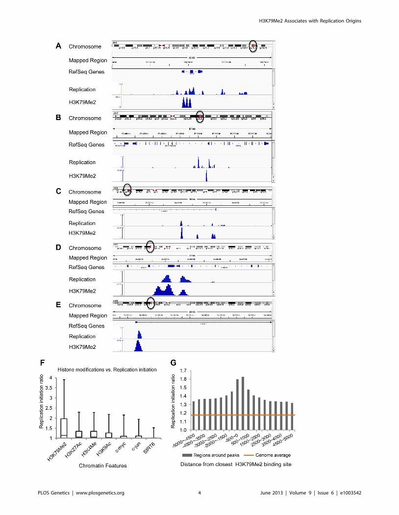
Figure 1A–E shows the replication initiation ratio (nascent
strands vs. genomic RPKM) and H3K79Me2 in sample human
loci. For each image, a chromosome map is shown at the top, and
the region-of-interest is circled. The analyzed region is shown
underneath the ideogram, with map coordinates indicated. Ref-
Seq genes are aligned under the map coordinates. The replication
tracks show the distribution of sequence reads (aligned with the
indicated region) obtained from massively parallel sequencing of
nascent strands from K562 erythroleukemia cells as described [3].
All data are shown as the ratio of reads obtained from a nascent
strand preparation, and reads obtained from a corresponding
control genomic DNA preparation. The y-axis indicates the
nascent strand/genomic DNA ratio. The H3K79Me2 tracks
indicate the distribution of reads from ChIP-Seq analyses with
H3K79Me2 specific antibodies as described in the Methods
section. The data show that chromatin regions exhibiting H3K79
dimethylation exhibited high enrichment for replication initiation
sites, measured by the ratio of reads on nascent strands vs.
genomic DNA controls. When we calculated the enrichment of
genome-wide H3K79Me2 peaks obtained from the ChIP-Seq data
for replication initiation events, overall, 23.39% of the
H3K79Me2 peaks were detected in regions adjacent to replication
initiation events whereas an association with replication initiation
events was expected in only 7.26% of the size-matched random-
ized feature regions (Table 1, ‘‘overall’’). Chromatin regions
exhibiting H3K79 methylation markedly enriched in replication
initiation events, showing an average whole-genome RPKM ratio
of 1.8 (Figure 1F). As shown previously [3], replication initiation
events were also enriched, to a lesser extent, in chromatin regions
exhibiting acetylation of histone H3 on lysines 9 and 27 and
methylated on lysine 4, and in chromatin binding transcription
factors c-Jun and c-Myc. As shown in Figure 1F, the extent of
enrichment for replication initiation events in regions exhibiting
methylation of histone H3 on lysine 79 is markedly higher than in
regions exhibiting other histone modifications. Given the sequenc-
ing depth and the large sample size, an average value of 1.3 (as
calculated for H3K27Ac) represents significant enrichment over
the average whole-genome RPKM ratio (p,1026) despite the
seemingly low numerical values. The statistical significance of
enrichment in H3K79Me2 in replication initiation events is very
high (p,10220). Replication initiation events were markedly
enriched in regions within 500 bp of an H3K79Me2 binding site,
and this is not true for other regions such as JunB binding sites
(Figure 1G and Figure S1, S2). H3K79 methylation exhibited a
similar enrichment with replication initiation events as DNAse
hypersensitivity and CpG methylation, however these traits did
not exhibit any synergy (Figure S3).
To further explore the association between H3K79 dimethyla-
tion and replication initiation, we measured the levels of
H3K79Me2 enrichment during the G1, S, and G2 phases of the
cell cycle in K562 cells fractionated into synchronous cell cycle
populations using centrifugal elutriation (Figure 2, Table 1, rows
G1 through G2, and Table 2) and Western immunoblots. The
overall level of H3K79Me2 modification did not change during
the cell cycle (Figure S4A) and the abundance and distribution of
chromatin regions exhibiting H3K79Me2 was similar in the G1
and G2 phases of the cell cycle. However, the distribution of
H3K79Me2 associated regions expanded in S-phase. Interestingly,
chromatin regions that exhibited H3K79Me2 only during S-phase
did not display a high association with replication initiation events
(Figure 2C and Table 1, ‘‘S only’’). By contrast, another
modification, H3K4Me3, did not exhibit a similar expansion of
peaks in S-phase and displayed a consistent distribution through-
out the cell cycle (Figure 2D and Table 2). These observations
support the conclusion that the H3K79Me2 modification associ-
ates preferentially in a cell cycle specific manner with replication
initiation regions.
H3K79Me2 binds a functional replicator but not a mutantthat cannot initiate replication
To determine if H3K79 was methylated at a replicator
sequence essential for initiation of DNA replication, we performed
chromatin immunoprecipitation (ChIP) analysis with the Rep-P
asymmetric region of the endogenous b-globin locus in K562 cells
(for maps, see Figure 3A and Figure S4B). This region is essential
for initiation of DNA replication from Rep-P, one of the two
adjacent replicators that can initiate DNA synthesis at the b-globin
locus [33,34]. As shown in Figure S4B, probes that colocalized
with the Rep-P replicator (bG59.8, bG61.3, AG) exhibited higher
enrichment in H3K79Me2. We also tested the association between
H3K79Me2 and late-replicating replication initiation site in Jurkat
cells, which are human T-cells that do not express any gene from
the human beta-globin locus and yet initiate replication from Rep-
P late during the S-phase of the cell cycle. The asymmetric region
(probe marked AG) exhibited a high level of H3K79 dimethylation
in both K562 cells that express c-globin and in Jurkat cells, which
do not express any gene within the HBB cluster and replicate the
entire locus late in S-phase.
We then investigated whether DNA sequences required for
initiation of DNA replication were also essential for enrichment of
H3K79Me2 in the Rep-P region. To that end, we introduced
transgene cassettes that included the LCR, an enhanced GFP
expression cassette driven by the beta-globin promoter, and Rep-P
variants into a site termed random locus 4 (RL4) in murine
erythroleukemia (MEL) cells. This site was used previously to
assess the roles of replicator sequences in initiation of DNA
replication and gene silencing [37]. The RL4 site was engineered
to contain an inverted pair of LoxP sites so that any inserted
transgene cassette in that locus could be exchanged with other
cassettes inserted precisely at the same genomic location. This
system can facilitate testing for effects of distinct mutations and
sequences from the murine beta globin locus serve as controls for
human inserted sequences. Previous studies have shown that the
unmodified RL4 site exhibits a heterochromatin conformation and
does not initiate replication, hence initiation activity detected at
this site after insertion of ectopic sequences reflects sequence
information encoded within the inserted transgens [37]. Using this
feature, we tested a transgene cassette that included an intact Rep-
P (Rep-PWT – Figure 3A) and a cassette that included a Rep-P
variant with only 2 nucleotides mutated in AG region of the rep-P
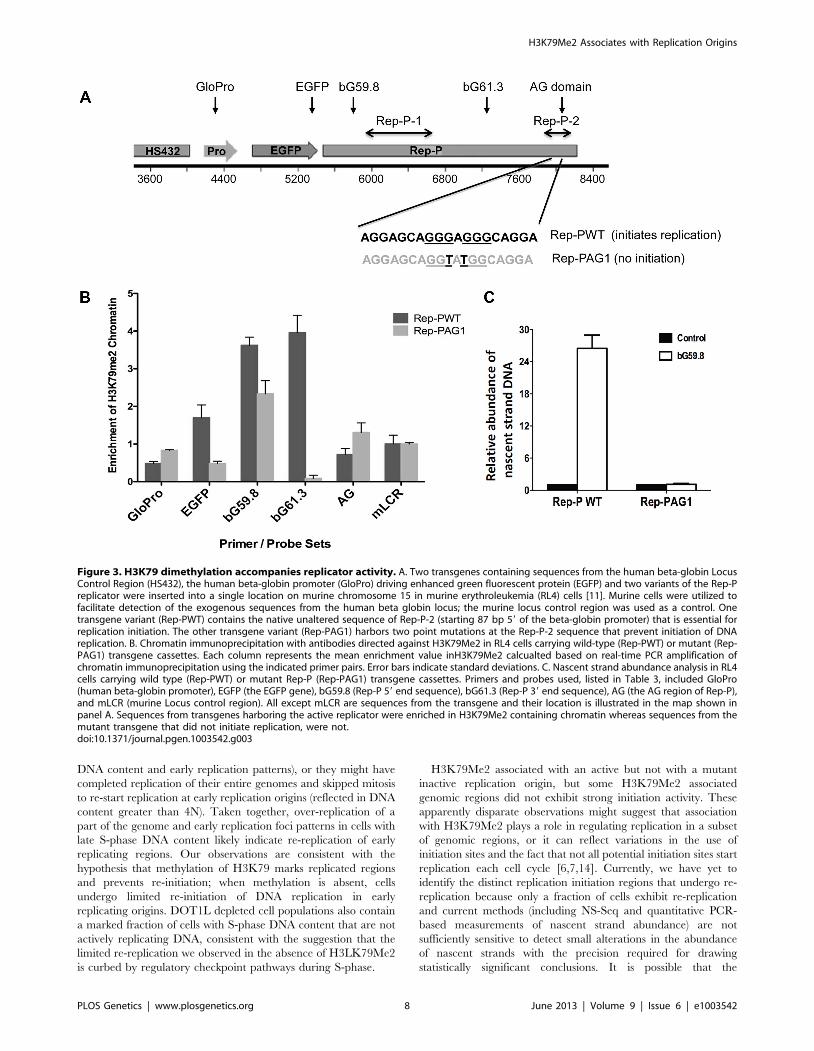
(Rep-PAG1). The intact Rep-P (Rep-PWT) exhibited enrichment
of H3K79Me2 (Figure 3B, probes bG59.8, 61.3 – see Figure S4B
and Table 3 for locations and sequences of primer pairs and
probes) and initiated replication at the ectopic site (Figure 3C). By
contrast, the mutant Rep (Rep-PAG1) did not exhibit enrichment
of H3K79Me2 and did not initiate replication (Figure 3, B and C,
respectively). Although the orientation of the transgene at the RL4
site affects transcriptional silencing [37], analyses of Rep-PAG1
yielded similar results when the transgene was inserted in both
orientations (data not shown). It should be noted that in the RL4
locus the highest enrichment in the intact Rep-P transgene was
observed upstream of the asymmetric region (primer pairs bG59.8
and 61.3) whereas the asymmetric locus exhibited a higher
enrichment at the native locus in K562 cells (Figure S4). This shift
H3K79Me2 Associates with Replication Origins
PLOS Genetics | www.plosgenetics.org 3 June 2013 | Volume 9 | Issue 6 | e1003542
H3K79Me2 Associates with Replication Origins
PLOS Genetics | www.plosgenetics.org 4 June 2013 | Volume 9 | Issue 6 | e1003542

was most likely due to the fact that the inserted transgene
contained only one of the two replicators at the replication
initiation region (Rep-P without Rep-I). Importantly, since of
H3K79Me2 was detected in the active but not the inactive Rep-P,
these observations suggest an association between H3K79
dimethylation and replicator activity.
H3K79Me2 plays a role in restricting replication once percell cycle
That H3K79Me2 is strongly associated with the start of DNA
replication implicates a causal role for H3K79 dimethylation in
regulating the initiation of DNA replication. To address this
question, we depleted DOT1L, the enzyme that methylates
histone H3 on lysine K79. These experiments were performed in
human colon cancer HCT116 cells. HCT116 cells were chosen for
this analysis because these cells exhibit a relatively stable karyotype
and can be easily transfected with siRNA, and are therefore
suitable for analyses aimed to test the effects of siRNA mediated
gene silencing on cell cycle distribution. In HCT116 cells, siRNA
mediated DOT1L depletion prevented H3K79 dimethylation
(Figure 4A). We used those cells to measure the rate of DNA
synthesis in cells with and without DOT1L using dynamic
molecular combing (Figure 4, B–D and Figure S5). As shown in
Figure 4, C and D, depletion of DOT1L did not affect replication
fork velocity, suggesting that H3K79 methylation does not directly
affect the progression of replication forks. DOT1L depletion also
did not affect the frequency of replication initiation events (Figure
S6).
We then asked whether cell populations in which DOT1L was
depleted exhibited changes in cell cycle patterns. The overall
distribution of cells in the G1, S and G2/M phases of the cell cycle
was similar in control and DOT1L depleted cells, but FACS
analyses indicated that DOT1L depleted cells had fewer cells in
the early S-phase and more cells with late S-phase DNA content
than cells in which DOT1L was not depleted (transfected with a
control siRNA; Figure 5A). As shown in Figure 5B, DOT1L
depletion also resulted in an increased frequency of apoptosis
(subG1) and in an increase in the fraction of cells exhibiting DNA
content greater than 4N (.G2/M) or cells that did not synthesized
DNA despite a DNA content between 2N and 4N (S non-
replicating). Similar results were observed in U2OS osteosarcoma
cells (Figure S7 and Table S2). These observations suggested that
although most cells can continue to proliferate and replicate DNA
in the absence of methylated H3K79, prevention of H3K79
methylation might affect regulatory processes that modulate the
order and timing of DNA replication.
Because H3K79 methylation was enriched in replication
initiation sites, we asked whether the order of DNA replication
was altered in cell populations in which DOT1L was depleted. To
measure DNA replication patterns, cells were pulse-labeled with
the nucleotide analog EdU for 60 min. DNA content (DAPI
intensity) was measured, along with the pattern of EdU staining
(Figure 6, A and B). The pattern of replication foci as exhibited by
EdU incorporation was recorded for each nucleus (for examples,
see Figure 6C). In untreated cells, diffuse replication foci patterns
are characteristic of early S-phase (Figure 6C, ES), whereas nuclei
exhibiting a few condensed replication foci are abundant in late S-
phase (Figure 6C, LS). We then tallied the frequency of early and
late S-phase EdU staining patterns in cell populations exhibiting
early and late S-phase DNA content measured by DAPI staining.
As expected we observed that diffuse patterns were indeed
abundant in cells exhibiting early S-phase DNA content, whereas
clustered patterns are frequent in cells exhibiting late S-phase
DNA content (Figure 6A and B). However, cell populations in
which DOT1L was depleted contained a small subset of cells with
DNA content of more than 4N exhibiting diffuse replication
patterns (Figure 6B; examples are shown in Figure 6C, . = G2M).
This cell population might represent cells with late S-phase DNA
content that re-replicated DNA that had already been duplicated
earlier during the same S-phase, thus exhibiting an early S-phase
replication pattern. This pattern is consistent with the observation
that depletion of DOT1L resulted in an increased fraction of cells
with DNA content larger than 4N, representing cells that skipped
mitosis and partially re-replicated their DNA.
To investigate whether the EdU staining patterns we have
observed indeed reflect re-replication of DNA in cells we have
labeled cells with Bromodeoxyuridine (BrdU) for 18 hours that
exceeds the length of a complete S-phase but is shorter than the
Figure 1. H3K79Me2 containing chromatin is associated preferentially with replication initiation sites genome-wide. A–E.Screenshots of replication initiation data visualized with the integrated Genome Viewer (http://www.broadinstitute.org/igv/). A chromosome map isshown at the top, and the region-of-interest is delineated by a circle. The analyzed region is shown underneath the ideogram, with map coordinatesindicated. The Replication panel shows the distribution of replication initiation events (ratio of reads obtained from a nascent strand preparation, andreads obtained from a corresponding control genomic DNA preparation) of the region-of-interest obtained from our published database, see [3] fordetails. Regions abundant in H3K79Me2 immunoprecipitated chromatin from H3K79Me2 ChIP-Seq in K562 cells are shown below the initiation profileas reads per kilobase per million aligned reads (RPKM). Ref-Seq genes are aligned above the experimental data. A. The distribution of H3K79Me2 andreplication initiation events in the MYC locus. B. The distribution of H3K79Me2 and replication initiation events in the DBF4 locus. C. The distributionof H3K79Me2 and replication initiation events in the LCORL locus. D. The distribution of H3K79Me2 and replication initiation events in the BAG1 locus.E. The distribution of H3K79Me2 and replication initiation events in the UBAP1 locus. F. A box plot comparing the relative enrichment of replicationinitiation events in chromatin featuring H3K79Me2 obtained by ChIP-Seq as described in methods with various other chromatin features as reportedin the UCSC genome browser (for details, see[3]. Boxes indicate distributions of the second and third quartiles and whiskers, 95th percentiles. Thehorizontal lines in the boxes represent medians. Values lower than a unit were converted into 1. Methylation of H3K79 exhibits a marked enrichmentin replication initiation events that was higher than any other measured histone modification or transcription factor. G. A histogram showing thereplication enrichment ratio (calculated as in A, B) for genomic regions as a function of their distance from the closest H3K79Me2 interaction sites. Abox plot version of the same histogram is shown as Figure S1.doi:10.1371/journal.pgen.1003542.g001
Table 1. Fraction of H3K79Me2 regions that are within2000 bp from a 15% FDR replication peak.
H3K79Me2 regions
Average from 100 runs withsize-matched randomized featureregions
Overall 23.39 7.26
G1 25.27 7.31
S 20.90 7.29
S-only* 12.25 7.29
G2 27.37 7.31
*Chromatin regions associated with H3K79Me2 solely in S-phase.doi:10.1371/journal.pgen.1003542.t001
H3K79Me2 Associates with Replication Origins
PLOS Genetics | www.plosgenetics.org 5 June 2013 | Volume 9 | Issue 6 | e1003542
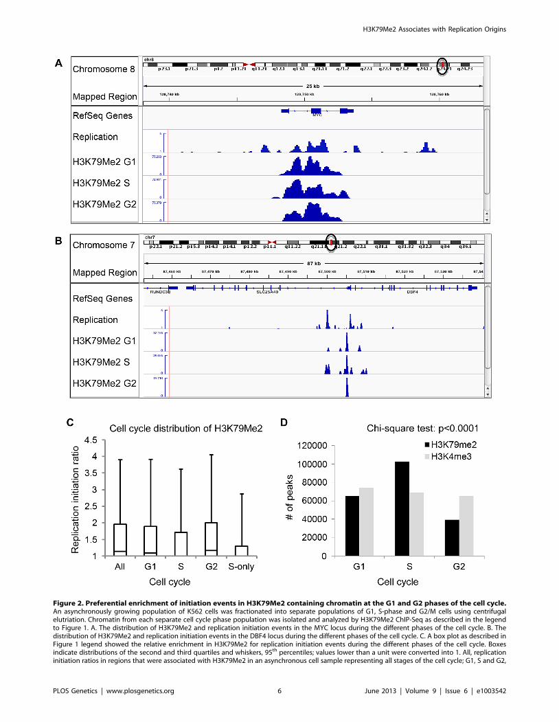
Figure 2. Preferential enrichment of initiation events in H3K79Me2 containing chromatin at the G1 and G2 phases of the cell cycle.An asynchronously growing population of K562 cells was fractionated into separate populations of G1, S-phase and G2/M cells using centrifugalelutriation. Chromatin from each separate cell cycle phase population was isolated and analyzed by H3K79Me2 ChIP-Seq as described in the legendto Figure 1. A. The distribution of H3K79Me2 and replication initiation events in the MYC locus during the different phases of the cell cycle. B. Thedistribution of H3K79Me2 and replication initiation events in the DBF4 locus during the different phases of the cell cycle. C. A box plot as described inFigure 1 legend showed the relative enrichment in H3K79Me2 for replication initiation events during the different phases of the cell cycle. Boxesindicate distributions of the second and third quartiles and whiskers, 95th percentiles; values lower than a unit were converted into 1. All, replicationinitiation ratios in regions that were associated with H3K79Me2 in an asynchronous cell sample representing all stages of the cell cycle; G1, S and G2,
H3K79Me2 Associates with Replication Origins
PLOS Genetics | www.plosgenetics.org 6 June 2013 | Volume 9 | Issue 6 | e1003542

time required for cells to undergo a complete cell cycle. We then
determined the extent of BrdU incorporation into DNA using
density gradients. BrdU substituted DNA is more dense (heavier)
then unsubstituted DNA and the difference between unsubstituted
(LL), semi-substituted (HL) and fully substituted (HH) DNA can be
observed by recording the abundance of DNA in fractionated
CsCl density gradients. BrdU substituted DNA was detected using
anti-BrdU antibodies. As shown in Figure 6D, cells containing
active Dot1L exhibited BrdU substituted DNA in fractions
corresponding to HL (semi-substituted) DNA consistent with the
assumption that those cells have completed a single round of
replication. By contrast, cells in which Dot1L was depleted
exhibited BrdU substituted DNA in both the HL and HH
fractions, consistent with over-replication of a fraction of the DNA
during the labeling period. Importantly, these results imply that
H3K79 methylation plays a role in preventing re-replication
during normal cell cycle progression.
Discussion
The observations reported here demonstrate that H3K79Me2-
containing chromatin was enriched in replication initiation events.
The methylation of histone H3 on histone 79 exhibited the highest
enrichment of replication initiation events observed in any single
chromatin modification that was studied. It is worth noting,
however, that despite the high level of enrichment in replication
initiation events, not all replication initiation sites associated with
H3K79 dimethylation. The association of H3K79Me2 with
replication initiation sites was independent and not synergistic
with other chromatin modifications. H3K79 dimethylation
exhibited a wider distribution on chromatin during S-phase, but
regions of chromatin that only associated with H3K79 dimethyla-
tion during S-phase were not in replication initiation events. We
also observed that H3K79 dimethylation was enriched in
chromatin containing a functional replicator, but was not enriched
in chromatin containing a mutant replicator that could not initiate
replication. Hence, H3K79 dimethylation at the human beta-
globin locus replicator was associated with replicator activity.
Prevention of H3K79 methylation by depletion of DOT1L did not
affect the rate of DNA replication or the inter-origin distance.
Importantly, however, over-replication occurred at a higher
frequency following depletion of Dot1L, the sole enzyme
responsible for H3K79 methylation. Together, these results
demonstrate for the first time that H3K79 methylation is not
required for replication initiation but rather plays a role in
preventing re-replication of DNA once initiated.
Our previous studies showed that methylated CpG regions and
genes undergoing moderate transcription were highly associated
with replication initiation sites [3]. Other studies have also found
that replication initiation sites exhibit enrichment in transcribed
regions [5], G-quartets [4] and methylated CpGs [2]. Studies have
also shown that methylation of H4 lysine 20 is required for
genome-wide DNA replication [38], suggesting a potential
mechanistic involvement of this histone modification in replication
since Orc1, which exhibits an association with replication origins
[39], interacts with H4K20Me2 through its BAH domain [40].
However, these studies have not identified a single histone
modification that is associated with initiation of DNA replication
at distinct genomic sites. Here, we have identified H3K79
methylation as a modification that is not only associated with
initiation but plays a functional role in restricting replication to
once per cell cycle.
Our ChIP-Seq data suggest that H3K79 dimethylation might
occur in regions proximal to replication initiation sites during G1
and then expand to adjacent regions during S-phase. H3K79me2
marks again cluster with initiation sites in G2, suggesting that the
S-phase specific marks, which do not associate with replication
origins, might be erased and the origin-specific marks remain post-
mitosis for the next cell cycle. These results imply the intriguing
possibility for a mechanism to specifically and quickly remove
H3K79 methylation. A precedent for an enzyme that might
remove methylated histones in that way is Rph1/KDM4, which
can specifically demethylate H3K36 in yeast [41]. The observed
restriction of H3K79me2 containing chromatin to replication
origins after S-phase might be mediated by an equivalent
demethylase capable of removing methyl groups from H3K79 in
non-origin regions, or by active removal of H3K79Me2 containing
nucleosomes from chromatin that is not associated with replicator
activity. The mechanism(s) by which potential replication origins
retain H3K79 methylation whereas regions that are not associated
with replication origins lose H3K79 methylation are under
investigation. Regardless of the mechanism, regions of potential
replication origins that can undergo initiation of DNA replication
specifically retain the H3K79 methylation mark during cell
division.
The most likely role played by replication origin associated
DOT1L-mediated H3K79 methylation is to facilitate an interac-
tion that marks origins that had started replication, where such a
mark might prevent replication from initiating a second time.
Consistent with this suggestion, H3K79Me2 associated with active
but not with mutant inactive replication origins and we observed
cells with late S-phase DNA content and early replication foci
patterns following DOT1L depletion. In accordance, direct
measurement of nucleotide incorporation also showed that Dot1
depletion resulted in partial genome over-replication, and cell
cycle profiles of DOT1L depleted cells detected a larger
population of cells with DNA content higher than 4N and cells
with a late-S-phase DNA content. Such cells might have re-started
replication of early-replicating origins without completing the
replication of late-replicating origins (reflected in late S-phase
regions associated with H3K79Me2 in samples from cells at the appropriate cell cycle phase; S-only, regions that were only associated with H3K79Me2in S-phase but not in G1 and G2. D. The number of H3K79Me2 associated peaks on chromatin during subsequent stages of cell cycle progression.H3K4Me3 was used as a control. Chi-square test shows that the distributions of H3K79Me2 and H3K4Me3 in G1, S and G2 cells are statisticallydifferent (p,0.0001). H3K79 dimethylation was more abundant and exhibited a wider distribution during S-phase, but chromatin regions associatedwith H3K79Me2 solely in S-phase were not further enriched in replication initiation events. The association of H3K79Me2 with replication initiationevents was re-established in G2.doi:10.1371/journal.pgen.1003542.g002
Table 2. Number of 100 bp bins associated with histone H3methylation on lysines 79 and 4 during the cell cycle*.
G1 S G2
H3K79me2 65284 102372 39271
H3K4me3 73851 68750 65152
*Data used for Figure 4D.doi:10.1371/journal.pgen.1003542.t002
H3K79Me2 Associates with Replication Origins
PLOS Genetics | www.plosgenetics.org 7 June 2013 | Volume 9 | Issue 6 | e1003542
DNA content and early replication patterns), or they might have
completed replication of their entire genomes and skipped mitosis
to re-start replication at early replication origins (reflected in DNA
content greater than 4N). Taken together, over-replication of a
part of the genome and early replication foci patterns in cells with
late S-phase DNA content likely indicate re-replication of early
replicating regions. Our observations are consistent with the
hypothesis that methylation of H3K79 marks replicated regions
and prevents re-initiation; when methylation is absent, cells
undergo limited re-initiation of DNA replication in early
replicating origins. DOT1L depleted cell populations also contain
a marked fraction of cells with S-phase DNA content that are not
actively replicating DNA, consistent with the suggestion that the
limited re-replication we observed in the absence of H3LK79Me2
is curbed by regulatory checkpoint pathways during S-phase.
H3K79Me2 associated with an active but not with a mutant
inactive replication origin, but some H3K79Me2 associated
genomic regions did not exhibit strong initiation activity. These
apparently disparate observations might suggest that association
with H3K79Me2 plays a role in regulating replication in a subset
of genomic regions, or it can reflect variations in the use of
initiation sites and the fact that not all potential initiation sites start
replication each cell cycle [6,7,14]. Currently, we have yet to
identify the distinct replication initiation regions that undergo re-
replication because only a fraction of cells exhibit re-replication
and current methods (including NS-Seq and quantitative PCR-
based measurements of nascent strand abundance) are not
sufficiently sensitive to detect small alterations in the abundance
of nascent strands with the precision required for drawing
statistically significant conclusions. It is possible that the
Figure 3. H3K79 dimethylation accompanies replicator activity. A. Two transgenes containing sequences from the human beta-globin LocusControl Region (HS432), the human beta-globin promoter (GloPro) driving enhanced green fluorescent protein (EGFP) and two variants of the Rep-Preplicator were inserted into a single location on murine chromosome 15 in murine erythroleukemia (RL4) cells [11]. Murine cells were utilized tofacilitate detection of the exogenous sequences from the human beta globin locus; the murine locus control region was used as a control. Onetransgene variant (Rep-PWT) contains the native unaltered sequence of Rep-P-2 (starting 87 bp 59 of the beta-globin promoter) that is essential forreplication initiation. The other transgene variant (Rep-PAG1) harbors two point mutations at the Rep-P-2 sequence that prevent initiation of DNAreplication. B. Chromatin immunoprecipitation with antibodies directed against H3K79Me2 in RL4 cells carrying wild-type (Rep-PWT) or mutant (Rep-PAG1) transgene cassettes. Each column represents the mean enrichment value inH3K79Me2 calcualted based on real-time PCR amplification ofchromatin immunoprecipitation using the indicated primer pairs. Error bars indicate standard deviations. C. Nascent strand abundance analysis in RL4cells carrying wild type (Rep-PWT) or mutant Rep-P (Rep-PAG1) transgene cassettes. Primers and probes used, listed in Table 3, included GloPro(human beta-globin promoter), EGFP (the EGFP gene), bG59.8 (Rep-P 59 end sequence), bG61.3 (Rep-P 39 end sequence), AG (the AG region of Rep-P),and mLCR (murine Locus control region). All except mLCR are sequences from the transgene and their location is illustrated in the map shown inpanel A. Sequences from transgenes harboring the active replicator were enriched in H3K79Me2 containing chromatin whereas sequences from themutant transgene that did not initiate replication, were not.doi:10.1371/journal.pgen.1003542.g003
H3K79Me2 Associates with Replication Origins
PLOS Genetics | www.plosgenetics.org 8 June 2013 | Volume 9 | Issue 6 | e1003542
H3K79Me2 might mark cryptic replication initiation sites, which
are capable of initiation of DNA replication but only do so under
distinct conditions such as exposure to DNA damaging agents [7].
If replication does occur on such cryptic origins marked by
H3K79Me2, H3K79Me2 might be available to facilitate in an
interaction that will prevent re-replication from cryptic as well as
constitutive origins.
Because limiting replication once per cell cycle is critical in
preventing genome instability, cells employ numerous strategies to
prevent re-replication [42,43]. The mechanisms are diverse and
species-specific, with components of the pre-replication licensing
complex such as Cdt1 and ORC often serving as primary
regulatory targets. Interestingly methylation of Tetrahymena histone
H3K76, which is orthologous to histone H3K79 in mammals, is
required for replication initiation and overexpression of the
Tetrahymena Dot1A methylase results in over-replication [44].
Although this observation suggests that methylation of H3K76 in
Tetrahymena achieves the opposite effect than the methylation of
H3K79 in mammals, and it is yet unclear whether Tetrahymena
H3K76 methylation associates with replication origins, both
enzymes seem to be involved in regulating replication re-initiation
events and thus might both be involved in processes that mark
genomic regions for initiation. Another precedent for a role of
chromatin modifications in regulating replication initiation is
found in plants, in which histone H3 lysine 27 (H3K27)
monomethyltransferases (Arabidopsis Trithorax-Related 5 (ATXR5)
and ATXR6 interact with the two Arabidopsis proliferating cell
nuclear antigen protein [45] and nuclei from atxr5 atxr6 double
mutants exhibit evidence of over-replication associated with
marked decondensation of constitutive heterochromatin at chro-
mocenters [46]. In mammals, methylation of histone H4 on lysine
20 is associated with the onset of replication licensing in G1 and
the dissociation of H4K20Me1 from replication origins in S-phase
is associated with prevention of re-replication [38]. Methylation of
histone H4 on lysine 20 was required for initiation of DNA
replication from replication origins [38], and although genome-
wide analyses of replication initiation events did not exhibit a
marked association with H4K20Me1 [3] it is possible that
H4K20Me1 plays a role in establishing potential replication
initiation patterns during the G1 phase. The temporal pattern of
H3K79Me2, as reported here, is distinct from H4K20Me1. The
increased frequency of apoptotic cells and cells with S-phase DNA
content that do not replicate DNA suggest that the role played by
H3K79Me2 in limiting re-replication is vital for cell survival and
coordinated progression through the cell cycle. It is possible that
the two modifications play distinct roles in regulating replication
patterns and maintenance of genomic stability.
Methods
Cell lines and culture conditionsHuman K562 cells, HCT1116 cells, U2OS cells and murine
erythroleukemia cells (RL4) containing Rep-P variants were grown
at 37uC in a 5% CO2 atmosphere in Dulbecco’s modified Eagle
medium (Invitrogen, Cat. no. 10564-011), supplemented with
10% heat-inactivated fetal calf serum. For RL4 cells containing
Rep-P variants, two transgenes containing sequences from the
human beta-globin Locus Control Region (HS432), the human
beta-globin promoter (GloPro) driving enhanced green fluorescent
protein (EGFP) and two variants of the Rep-P replicator were
inserted into a single location on murine chromosome 15 in
murine erythroleukemia (RL4) cells [11]. We introduced the AG1
mutation by site-directed mutagenesis. Details of the mutagenesis
methods were published previously [12].
Dot1L siRNAHCT116 cells were transfected with 25 nM of control siRNA
(Dharmacon, D-001810-10 or Dot1L siRNA (Dharmacon, L-
014900-01 or Qiagen, GS84444) with DharmaFECT 2 siRNA
(Dharmacon, T-2002) or RNAiMAX Transfection Reagent
(Invitrogen, 13778) according to the manufacturer’s protocol.
Cells were transfected 2 or 3 times with a 2 to 3 days interval and
harvested 72 h after the last transfection. Whole cell lysates
by1XSDS loading buffer were used for western-blot with anti-
Histone H3 (dimethylK79) antibody (Abcam, ab3594 or Millipore,
04-835) to verify the knockdown efficiency of dot1L siRNA.
Nascent-strand DNA analysisWe performed nascent-strand DNA analysis as described
previously [33]. DNA was extracted from asynchronous cells,
denatured by boiling for 10 minutes, incubated on ice for
10 minutes, and fractionated on a neutral sucrose gradient. We
collected 0.5–1 kb DNA fractions, treated them with l exonucle-
ase to remove non-RNA-primed genomic DNA fragments,
purified them, and measured the DNA concentration using a
NanoDrop 1000 (Thermo Scientific). We quantified nascent
strand DNA by real-time polymerase chain reaction (PCR) in an
ABI 7900 thermocycler (primers and probes used for real-time
PCR are listed in Table 3). For whole genome analyses of nascent
strand abundance, DNA from K562 cells was isolated using the
above procedure and processed for massively parallel sequencing
as previously described [3].Three independent biological replicates
were sequenced to the depth of 1.46108 reads. All the data from
mapping replication initiation events in K562 cells were deposited
in GEO, as Series GSE28911.
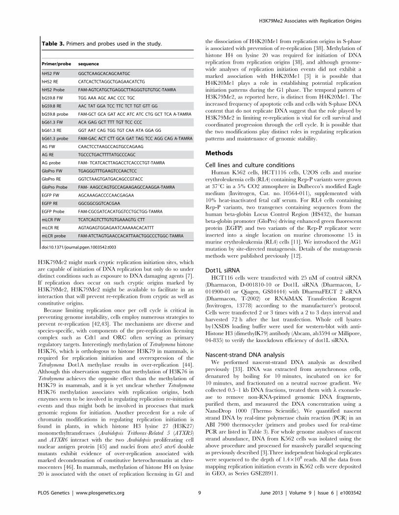
Table 3. Primers and probes used in the study.
Primer/probe sequence
hHS2 FW GGCTCAAGCACAGCAATGC
hHS2 RE CATCACTCTAGGCTGAGAACATCTG
hHS2 Probe FAM-AGTCATGCTGAGGCTTAGGGTGTGTGC-TAMRA
bG59.8 FW TGG AAA AGC AAC CCC TGC
bG59.8 RE AAC TAT GGA TCC TTC TCT TGT GTT GG
bG59.8 probe FAM-GCT GCA GAT ACC ATC ATC CTG GCT TCA A-TAMRA
bG61.3 FW ACA GAG GCT TTT TGT TCC CCC
bG61.3 RE GGT AAT CAG TGG TGT CAA ATA GGA GG
bG61.3 probe FAM-GAC ACT CTT GCA GAT TAG TCC AGG CAG A-TAMRA
AG FW CAACTCCTAAGCCAGTGCCAGAAG
AG RE TGCCCTGACTTTTATGCCCAGC
AG probe FAM- TCATCACTTAGACCTCACCCTGT-TAMRA
GloPro FW TGAGGGTTTGAAGTCCAACTCC
GloPro RE GGTCTAAGTGATGACAGCCGTACC
GloPro Probe FAM- AAGCCAGTGCCAGAAGAGCCAAGGA-TAMRA
EGFP FW AGCAAAGACCCCAACGAGAA
EGFP RE GGCGGCGGTCACGAA
EGFP Probe FAM-CGCGATCACATGGTCCTGCTGG-TAMRA
mLCR FW TCATCAGTCTTGTGTGAAAGTG CTT
mLCR RE AGTAGAGTGGAGAATCAAAAACACATTT
mLCR probe FAM-ATCTAGTGAACCACATTAACTGGCCCTGGC-TAMRA
doi:10.1371/journal.pgen.1003542.t003
H3K79Me2 Associates with Replication Origins
PLOS Genetics | www.plosgenetics.org 9 June 2013 | Volume 9 | Issue 6 | e1003542
Chromatin immunoprecipitation (ChIP) analysisWe performed ChIP analyses with 1% formaldehyde-fixed
K562 and RL4 cells using the Millipore ChIP assay kit (Cat.
no. 17-295) following the manufacturer’s protocol. Anti-
H3K79Me2 antibody was from Abcam (ab3594). We analyzed
ChIP samples by real-time PCR in an ABI 7900 thermocycler
using primers/probes listed in Table 3.
DNA molecular combing analysisDNA combing analyses of replicating DNA were performed
according to previously published methods [47]. Briefly, cells were
pulse-labeled with 20 mM IdU (Sigma, Cat. no. I-7125) for
20 minutes and then with 50 mM CldU (MP biomedical, Cat.
no. 105478) for 20 minutes before harvest. Then the cells were
embedded in low-melting point agarose plugs and lysed in the plug
with proteinase K lysis buffer at 50uC overnight. After agarose was
digested with b-agarase (NewEngland Biolabs), DNA was combed
onto silanized surfaces (Microsurfaces, Inc.) and detected with
anti-IdU (BD, Cat. no. 347580), anti-CldU (Accurate Chemical,
Cat. no. OBT0030), and anti-single strand DNA (Chemicon, Cat.
no. MAB3034) antibodies. Images were captured with the
Attovision software using the epifluorescence microscope Pathway
(Becton Dickinson) and measured the signals using ImageJ (open
source from National Cancer Institute, NIH) with custom-made
macros [48].
EdU staining for cell cycle analysisCells were cultured in 4-well chamber slides, pulse labeled with
10 mM EdU (Invitrogen) for 1 hour before harvest. EdU staining
using the Click-iT EdU kit (Invitrogen) were performed according
to manufacturer’s protocol. Images were captured with the
Attovision software using the epifluorescence microscope Pathway
(Becton Dickinson) and measured with the Attovision software for
DNA content by DAPI for cells with early S-phase and late-S
phase EdU replication patterns.
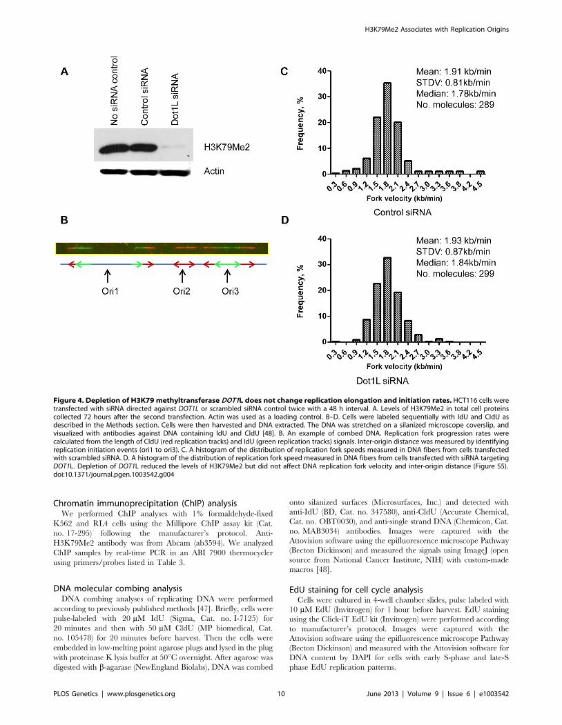
Figure 4. Depletion of H3K79 methyltransferase DOT1L does not change replication elongation and initiation rates. HCT116 cells weretransfected with siRNA directed against DOT1L or scrambled siRNA control twice with a 48 h interval. A. Levels of H3K79Me2 in total cell proteinscollected 72 hours after the second transfection. Actin was used as a loading control. B–D. Cells were labeled sequentially with ldU and CIdU asdescribed in the Methods section. Cells were then harvested and DNA extracted. The DNA was stretched on a silanized microscope coverslip, andvisualized with antibodies against DNA containing ldU and CldU [48]. B. An example of combed DNA. Replication fork progression rates werecalculated from the length of CldU (red replication tracks) and IdU (green replication tracks) signals. Inter-origin distance was measured by identifyingreplication initiation events (ori1 to ori3). C. A histogram of the distribution of replication fork speeds measured in DNA fibers from cells transfectedwith scrambled siRNA. D. A histogram of the distribution of replication fork speed measured in DNA fibers from cells transfected with siRNA targetingDOT1L. Depletion of DOT1L reduced the levels of H3K79Me2 but did not affect DNA replication fork velocity and inter-origin distance (Figure S5).doi:10.1371/journal.pgen.1003542.g004
H3K79Me2 Associates with Replication Origins
PLOS Genetics | www.plosgenetics.org 10 June 2013 | Volume 9 | Issue 6 | e1003542
ChIP-SeqAsynchronous K562 cells or fractionated G1, S and G2M K562
cells by elutriator (see [12] for elutriation details) were used for
ChIP as described above. ChIP-Seq samples were sequenced using
the Illumina GA II platform. The resultant sequences were aligned
to the hg19 (NCBI Build 37) human reference genome using
Bowtie [49]. Alignments were converted to .bam and .tdf format
using SAMtools and igvtools for visualization in Broad’s Integra-
tive Genomics Viewer, http://www.broadinstitute.org/igv/ [50].
Reads per kilobase per million aligned reads (RPKM) values
were calculated for each sample using 100 base genomic bins [51].
A Gaussian smoothing algorithm was applied to the bin values. To
correct for sequencing biases and copy number variation, an
enrichment ratio was defined as the ratio of nascent strand RPKM
to control RPKM, and calculated for each 100 base bin. The
H3K79Me2 and H3K4Me3 ChIP-Seq data are deposited in GEO
Series GSE GSE35294 including 11 sample files from experiments
in unsynchronized and cell cycle fractionated cells.
Cesium chloride gradient for measuring BrdU densityHCT116 cells transfected with control siRNA or Dot1L
siRNA were pulse-labeled with 50 mM of BrdU for 18 hours
before harvest. Genomic DNA were purified and sonicated to
500–4000 bp. Genomic DNA from HCT116 cells with no BrdU
incorporation and BrdU incorporation for 48 hours was used as
control. 100 mg of DNA was fractionated with 6 ml CsCl (1 g/
ml in TE). Samples were spun at 45000 rpm in a Ti75 rotor
(Beckman) for 66 hours. Fractions of 250 ml were collected and
the refractory index was measured to confirm the formation of
the gradient. Same volume samples from each fraction were
loaded to a positive charged nylon membrane by a Slot Blot
Filtration Manifold (PR648, GE Healthcare life sciences).
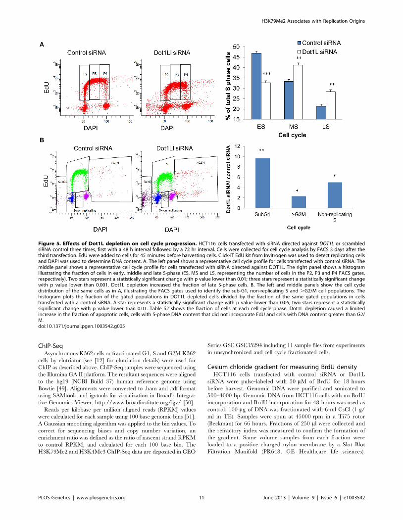
Figure 5. Effects of Dot1L depletion on cell cycle progression. HCT116 cells transfected with siRNA directed against DOT1L or scrambledsiRNA control three times, first with a 48 h interval followed by a 72 hr interval. Cells were collected for cell cycle analysis by FACS 3 days after thethird transfection. EdU were added to cells for 45 minutes before harvesting cells. Click-iT EdU kit from Invitrogen was used to detect replicating cellsand DAPI was used to determine DNA content. A. The left panel shows a representative cell cycle profile for cells transfected with control siRNA. Themiddle panel shows a representative cell cycle profile for cells transfected with siRNA directed against DOT1L. The right panel shows a histogramillustrating the fraction of cells in early, middle and late S-phase (ES, MS and LS, representing the number of cells in the P2, P3 and P4 FACS gates,respectively). Two stars represent a statistically significant change with p value lower than 0.01; three stars represent a statistically significant changewith p value lower than 0.001. Dot1L depletion increased the fraction of late S-phase cells. B. The left and middle panels show the cell cycledistribution of the same cells as in A, illustrating the FACS gates used to identify the sub-G1, non-replicating S and .G2/M cell populations. Thehistogram plots the fraction of the gated populations in DOT1L depleted cells divided by the fraction of the same gated populations in cellstransfected with a control siRNA. A star represents a statistically significant change with p value lower than 0.05; two stars represent a statisticallysignificant change with p value lower than 0.01. Table S2 shows the fraction of cells at each cell cycle phase. Dot1L depletion caused a limitedincrease in the fraction of apoptotic cells, cells with S-phase DNA content that did not incorporate EdU and cells with DNA content greater than G2/M.doi:10.1371/journal.pgen.1003542.g005
H3K79Me2 Associates with Replication Origins
PLOS Genetics | www.plosgenetics.org 11 June 2013 | Volume 9 | Issue 6 | e1003542
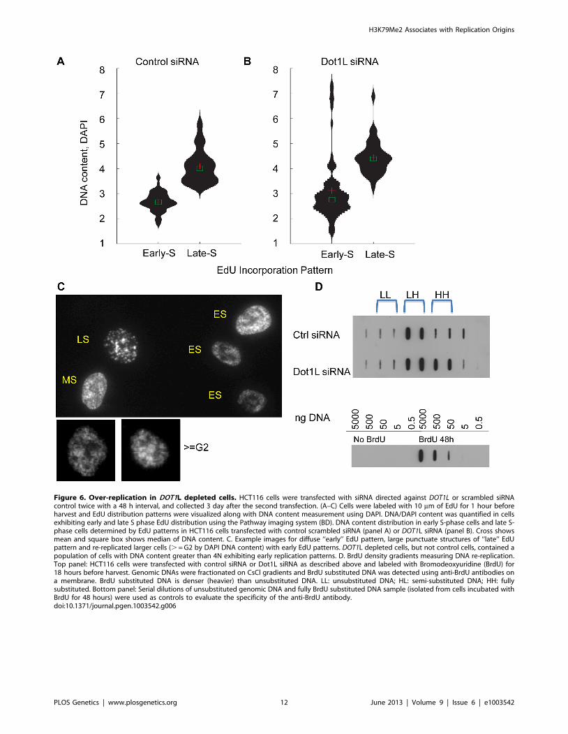
Figure 6. Over-replication in DOT1L depleted cells. HCT116 cells were transfected with siRNA directed against DOT1L or scrambled siRNAcontrol twice with a 48 h interval, and collected 3 day after the second transfection. (A–C) Cells were labeled with 10 mm of EdU for 1 hour beforeharvest and EdU distribution patterns were visualized along with DNA content measurement using DAPI. DNA/DAPI content was quantified in cellsexhibiting early and late S phase EdU distribution using the Pathway imaging system (BD). DNA content distribution in early S-phase cells and late S-phase cells determined by EdU patterns in HCT116 cells transfected with control scrambled siRNA (panel A) or DOT1L siRNA (panel B). Cross showsmean and square box shows median of DNA content. C. Example images for diffuse ‘‘early’’ EdU pattern, large punctuate structures of ‘‘late’’ EdUpattern and re-replicated larger cells (. = G2 by DAPI DNA content) with early EdU patterns. DOT1L depleted cells, but not control cells, contained apopulation of cells with DNA content greater than 4N exhibiting early replication patterns. D. BrdU density gradients measuring DNA re-replication.Top panel: HCT116 cells were transfected with control siRNA or Dot1L siRNA as described above and labeled with Bromodeoxyuridine (BrdU) for18 hours before harvest. Genomic DNAs were fractionated on CsCl gradients and BrdU substituted DNA was detected using anti-BrdU antibodies ona membrane. BrdU substituted DNA is denser (heavier) than unsubstituted DNA. LL: unsubstituted DNA; HL: semi-substituted DNA; HH: fullysubstituted. Bottom panel: Serial dilutions of unsubstituted genomic DNA and fully BrdU substituted DNA sample (isolated from cells incubated withBrdU for 48 hours) were used as controls to evaluate the specificity of the anti-BrdU antibody.doi:10.1371/journal.pgen.1003542.g006
H3K79Me2 Associates with Replication Origins
PLOS Genetics | www.plosgenetics.org 12 June 2013 | Volume 9 | Issue 6 | e1003542
0.5 ng, 5 ng, 50 ng, 500 ng and 5000 ng of total genomic DNA
labeled with BrdU for 48 hours were also loaded to the
membrane as standard for BrdU. BrdU were detected with
anti-BrdU antibody.
Supporting Information
Figure S1 A histogram showing the replication enrichment ratio
(calculated as in Figure 1, A–E) for genomic regions as a function
of their distance from the closest H3K79Me2 interaction sites.
This is a box plot version of Figure 1G.
(PDF)
Figure S2 Replication initiation at JunB binding sites. A
histogram showing the replication enrichment ratio for genomic
regions as a function of their distance from the closest JunB
interaction sites as calculated as in Figure 1G and Figure S2.
(PDF)
Figure S3 Additive effects of DNAse hypersensitivity and CpG
methylation with H3K79Me2. The average replication initiation
enrichment ratios of sequences exhibiting distinct chromatin
features (e.g. DNAse hypersensitivity, specific histone acetylation;
see Martin et al., 2011 for a list of feature tracks) were compared to
the average replication initiation enrichment ratio of sequences
exhibiting those features and methylation of H3K79. For each pair
of chromatin features, a new double-feature track was created that
contained the intersecting regions of the original features
(contiguous regions were treated as one feature region). The
average enrichment ratio of the double-feature track was
compared to that of each of the original single-feature tracks.
(PDF)
Figure S4 A. K562 cells were fractionated by elutriation to G1,
ES, MS, LS and G2M (Representative FACs profiles using
propidium iodide staining before and after elutriation are shown).
Cytosolic and soluble nuclear proteins were removed by hypotonic
buffer with 0.5% NP40 [12]. Chromatin bound proteins were
extracted by suspending the nuclei in 16SDS-PAGE sample buffer
and boiling for 5 minutes. Level of H3K79Me2 and H3K4Me3
were detected by Western blot. Ponceau S staining of Histones was
used as loading controls. There are no cell cycle dependent changes
for both H3K79Me2 and H3K4Me3. B. Validation of ChIP-Seq
data: H3K79 dimethylation is enriched in chromatin containing a
replicator. Chromatin from K562 and Jurkat cells was isolated and
immunoprecipitated with an antibody directed against H3K79Me2
(Abcam, ab 3594). The abundance of sequences from the human
beta-globin locus was analyzed in DNA isolated from immunopre-
cipitated chromatin by real-time PCR as described [12] using
primers/probe combinations listed in Table 3. The locations of
primers/probes in the beta-globin locus are illustrated under the
histogram and the boundaries of the two replicators (Rep-P and
Rep-I) within the initiation region (IR) are shown [33,34].
H3K79Me2 containing chromatin were markedly enriched in
sequences amplified by primers designated bG59.9 and bG61.3,
located within the Rep-P replicator, and the enrichment was very
high in sequences amplified by the primer pair AG, located at an
asymmetric region essential for replicator activity.
(PDF)
Figure S5 Example images of fiber analyses of DNA replication.
Raw images were shown for DNA combing analyses in Figure 5
and Figure S2. Cells were labeled sequentially with ldU and CIdU,
then the DNA was stretched on a silanized microscope coverslip,
and visualized with antibodies against DNA containing ldU (green
replication tracks) and CldU (red replication tracks)(top images in
both A and B). DNA fibers were detected by anti-single strand
antibody (bottom image in both A and B).The white vertical lines
are examples how replication signals are defined and the distance
between them are measured by Image J with a custom-made
macro.
(PDF)
Figure S6 Depletion of H3K79 methytansferase DOT1L does
not change replication initiation rates. HCT116 cell treatments
and DNA combing analyses were described in Figure 4. A. A
histogram of the distribution of inter-origin distance measured in
DNA fibers from cells transfected with scrambled siRNA. B. A
histogram of the distribution of inter-origin distance measured in
DNA fibers from cells transfected with siRNA targeting DOT1L.
(PDF)
Figure S7 Effects of Dot1L depletion on cell cycle progression
inU2OS cells. U2OS cells were transfected with control or DOT1L
siRNA once or twice with a 3 day interval and collected for FACs
3 days after the last transfection. EdU were added to cells for
45 minutes before harvesting cells. Click-iT EdU kit from
Invitrogen was used to detect replicating cells and DAPI was
used to determine DNA content. A. Representative cell cycle
profile for cells transfected with control siRNA or Dot1L siRNA 3
days after the first transfection (top panel) and 3 days after the
second transfection (lower panel). B. A summary histogram of the
cell cycle distribution of U2OS cells 3 days after the second
transfection with control siRNA or Dot1L siRNA.
(PDF)
Table S1 Cell lines used in this work. A list of cell lines and their
backgrounds, as well as reasons being chosen.
(PDF)
Table S2 Fraction of cells at various stages of the cell cycle
following depletion of Dot1L from HCT116 and U2OS cells.
(PDF)
Acknowledgments
We thank Drs. Paul Meltzer, Chiara Conti, HongFang Liu, Elisabeta Leo
and Kurt W. Kohn for helpful discussion. We are grateful to Dr. James
Doroshow and William C. Reinhold for facilitating the bioinformatics
pipeline data analysis and to Dr. Sohyoung Kim for performing the violin
plot analyses. We thank the CCR sequencing facility headed by Bao Tran,
the CCR bioinformatics facility and Louri Chepelev from NHLBI for
expert help in next generation sequencing data acquisition and analysis.
Author Contributions
Conceived and designed the experiments: HF KZ MIA AKM. Performed
the experiments: HF AKM MMM LH YZ CML. Analyzed the data: HF
AKM MMM LH YZ MR RK MIA. Wrote the paper: HF MIA.
References
1. Gomez M, Brockdorff N (2004) Heterochromatin on the inactive X
chromosome delays replication timing without affecting origin usage. Proc Natl
Acad Sci U S A 101: 6923–6928.
2. Antequera F (2004) Genomic specification and epigenetic regulation of
eukaryotic DNA replication origins. EMBO J 23: 4365–4370.
3. Martin MM, Ryan M, Kim R, Zakas AL, Fu H, et al. (2011) Genome-wide
depletion of replication initiation events in highly transcribed regions. Genome
Res 21: 1822–32.
4. Besnard E, Babled A, Lapasset L, Milhavet O, Parrinello H, et al. (2012)
Unraveling cell type-specific and reprogrammable human replication origin
H3K79Me2 Associates with Replication Origins
PLOS Genetics | www.plosgenetics.org 13 June 2013 | Volume 9 | Issue 6 | e1003542
signatures associated with G-quadruplex consensus motifs. Nat Struct Mol Biol
19: 837–844.5. Cayrou C, Coulombe P, Vigneron A, Stanojcic S, Ganier O, et al. (2011)
Genome-scale analysis of metazoan replication origins reveals their organization
in specific but flexible sites defined by conserved features. Genome Res 21:1438–1449.
6. Aladjem MI (2007) Replication in context: dynamic regulation of DNAreplication patterns in metazoans. Nat Rev Genet 8: 588–600.
7. Mechali M (2010) Eukaryotic DNA replication origins: many choices for
appropriate answers. Nat Rev Mol Cell Biol 11: 728–738.8. Aladjem MI, Groudine M, Brody LL, Dieken ES, Fournier RE, et al. (1995)
Participation of the human beta-globin locus control region in initiation of DNAreplication. Science 270: 815–819.
9. Kalejta RF, Li X, Mesner LD, Dijkwel PA, Lin HB, et al. (1998) Distalsequences, but not ori-beta/OBR-1, are essential for initiation of DNA
replication in the Chinese hamster DHFR origin. Molecular Cell 2: 797–806.
10. Hayashida T, Oda M, Ohsawa K, Yamaguchi A, Hosozawa T, et al. (2006)Replication initiation from a novel origin identified in the Th2 cytokine cluster
locus requires a distant conserved noncoding sequence. J Immunol 176: 5446–5454.
11. Feng YQ, Desprat R, Fu H, Olivier E, Lin CM, et al. (2006) DNA methylation
supports intrinsic epigenetic memory in mammalian cells. PLoS Genet 2: e65.12. Huang L, Fu H, Lin CM, Conner AL, Zhang Y, et al. (2011) Prevention of
transcriptional silencing by a replicator-binding complex consisting of SWI/SNF, MeCP1, and hnRNP C1/C2. Mol Cell Biol 31: 3472–3484.
13. Ryba T, Hiratani I, Lu J, Itoh M, Kulik M, et al. (2010) Evolutionarilyconserved replication timing profiles predict long-range chromatin interactions
and distinguish closely related cell types. Genome Res 20: 761–770.
14. Gilbert DM (2012) Replication origins run (ultra) deep. Nat Struct Mol Biol 19:740–742.
15. Lacoste N, Utley RT, Hunter JM, Poirier GG, Cote J (2002) Disruptor oftelomeric silencing-1 is a chromatin-specific histone H3 methyltransferase. J Biol
Chem 277: 30421–30424.
16. Frederiks F, Tzouros M, Oudgenoeg G, van Welsem T, Fornerod M, et al.(2008) Nonprocessive methylation by Dot1 leads to functional redundancy of
histone H3K79 methylation states. Nat Struct Mol Biol 15: 550–557.17. Schulze JM, Jackson J, Nakanishi S, Gardner JM, Hentrich T, et al. (2009)
Linking cell cycle to histone modifications: SBF and H2B monoubiquitinationmachinery and cell-cycle regulation of H3K79 dimethylation. Mol Cell 35: 626–
641.
18. Zhou H, Madden BJ, Muddiman DC, Zhang Z (2006) Chromatin assemblyfactor 1 interacts with histone H3 methylated at lysine 79 in the processes of
epigenetic silencing and DNA repair. Biochemistry 45: 2852–2861.19. Im H, Park C, Feng Q, Johnson KD, Kiekhaefer CM, et al. (2003) Dynamic
regulation of histone H3 methylated at lysine 79 within a tissue-specific
chromatin domain. J Biol Chem 278: 18346–18352.20. Mohan M, Herz HM, Takahashi YH, Lin C, Lai KC, et al. (2010) Linking
H3K79 trimethylation to Wnt signaling through a novel Dot1-containingcomplex (DotCom). Genes Dev 24: 574–589.
21. Jones B, Su H, Bhat A, Lei H, Bajko J, et al. (2008) The histone H3K79methyltransferase Dot1L is essential for mammalian development and
heterochromatin structure. PLoS Genet 4: e1000190.
22. Onder TT, Kara N, Cherry A, Sinha AU, Zhu N, et al. (2012) Chromatin-modifying enzymes as modulators of reprogramming. Nature 483: 598–602.
23. Giannattasio M, Lazzaro F, Plevani P, Muzi-Falconi M (2005) The DNAdamage checkpoint response requires histone H2B ubiquitination by Rad6-Bre1
and H3 methylation by Dot1. J Biol Chem 280: 9879–9886.
24. Huyen Y, Zgheib O, Ditullio RA, Jr., Gorgoulis VG, Zacharatos P, et al. (2004)Methylated lysine 79 of histone H3 targets 53BP1 to DNA double-strand breaks.
Nature 432: 406–411.25. Tatum D, Li S (2011) Evidence that the histone methyltransferase Dot1
mediates global genomic repair by methylating histone H3 on lysine 79. J Biol
Chem 286: 17530–17535.26. Jacinto FV, Ballestar E, Esteller M (2009) Impaired recruitment of the histone
methyltransferase DOT1L contributes to the incomplete reactivation of tumorsuppressor genes upon DNA demethylation. Oncogene 28: 4212–4224.
27. Bernt KM, Zhu N, Sinha AU, Vempati S, Faber J, et al. (2011) MLL-Rearranged Leukemia Is Dependent on Aberrant H3K79 Methylation by
DOT1L. Cancer Cell 20: 66–78.
28. Chang MJ, Wu H, Achille NJ, Reisenauer MR, Chou CW, et al. (2011) Histone
H3 lysine 79 methyltransferase Dot1 is required for immortalization by MLLoncogenes. Cancer Res 70: 10234–10242.
29. Wang P, Lin C, Smith ER, Guo H, Sanderson BW, et al. (2009) Global analysis
of H3K4 methylation defines MLL family member targets and points to a rolefor MLL1-mediated H3K4 methylation in the regulation of transcriptional
initiation by RNA polymerase II. Mol Cell Biol 29: 6074–6085.
30. Okada Y, Feng Q, Lin Y, Jiang Q, Li Y, et al. (2005) hDOT1L links histone
methylation to leukemogenesis. Cell 121: 167–178.
31. Aladjem MI, Rodewald LW, Kolman JL, Wahl GM (1998) Genetic dissection of
a mammalian replicator in the human beta-globin locus. Science 281: 1005–
1009.
32. Kitsberg D, Selig S, Keshet I, Cedar H (1993) Replication structure of the
human beta-globin gene domain. Nature 366: 588–590.
33. Wang L, Lin CM, Brooks S, Cimbora D, Groudine M, et al. (2004) The human
beta-globin replication initiation region consists of two modular independentreplicators. Mol Cell Biol 24: 3373–3386.
34. Wang L, Lin CM, Lopreiato JO, Aladjem MI (2006) Cooperative sequence
modules determine replication initiation sites at the human beta-globin locus.Hum Mol Genet 15: 2613–2622.
35. Forrester WC, Epner E, Driscoll MC, Enver T, Brice M, et al. (1990) A deletionof the human beta-globin locus activation region causes a major alteration in
chromatin structure and replication across the entire beta-globin locus. Genes
Dev 4: 1637–1649.
36. Groudine M, Kohwi-Shigematsu T, Gelinas R, Stamatoyannopoulos G,
Papayannopoulou T (1983) Human fetal to adult hemoglobin switching:changes in chromatin structure of the beta-globin gene locus. Proc Natl Acad
Sci U S A 80: 7551–7555.
37. Fu H, Wang L, Lin CM, Singhania S, Bouhassira EE, et al. (2006) Preventing
gene silencing with human replicators. Nat Biotechnol 24: 572–576.
38. Tardat M, Brustel J, Kirsh O, Lefevbre C, Callanan M, et al. (2010) The histoneH4 Lys 20 methyltransferase PR-Set7 regulates replication origins in
mammalian cells. Nat Cell Biol 12: 1086–1093.
39. Dellino GI, Cittaro D, Piccioni R, Luzi L, Banfi S, et al. (2013) Genome-wide
mapping of human DNA-replication origins: levels of transcription at ORC1sites regulate origin selection and replication timing. Genome Res 23: 1–11.
40. Kuo AJ, Song J, Cheung P, Ishibe-Murakami S, Yamazoe S, et al. (2012) The
BAH domain of ORC1 links H4K20me2 to DNA replication licensing andMeier-Gorlin syndrome. Nature 484: 115–119.
41. Klose RJ, Gardner KE, Liang G, Erdjument-Bromage H, Tempst P, et al.(2007) Demethylation of histone H3K36 and H3K9 by Rph1: a vestige of an
H3K9 methylation system in Saccharomyces cerevisiae? Mol Cell Biol 27: 3951–
3961.
42. Arias EE, Walter JC (2007) Strength in numbers: preventing rereplication via
multiple mechanisms in eukaryotic cells. Genes Dev 21: 497–518.
43. DePamphilis ML, Blow JJ, Ghosh S, Saha T, Noguchi K, et al. (2006)
Regulating the licensing of DNA replication origins in metazoa. Curr Opin CellBiol 18: 231–239.
44. Gassen A, Brechtefeld D, Schandry N, Arteaga-Salas JM, Israel L, et al. (2012)
DOT1A-dependent H3K76 methylation is required for replication regulation inTrypanosoma brucei. Nucleic Acids Res 40: 10302–10311.
45. Raynaud C, Sozzani R, Glab N, Domenichini S, Perennes C, et al. (2006) Twocell-cycle regulated SET-domain proteins interact with proliferating cell nuclear
antigen (PCNA) in Arabidopsis. Plant J 47: 395–407.
46. Jacob Y, Stroud H, Leblanc C, Feng S, Zhuo L, et al. (2010) Regulation of
heterochromatic DNA replication by histone H3 lysine 27 methyltransferases.
Nature 466: 987–991.
47. Conti C, Caburet S, Schurra C, Bensimon A (2001) Molecular combing. Curr
Protoc Cytom Chapter 8: Unit 8 10.
48. Conti C, Leo E, Eichler GS, Sordet O, Martin MM, et al. (2010) Inhibition of
histone deacetylase in cancer cells slows down replication forks, activates
dormant origins, and induces DNA damage. Cancer Res 70: 4470–4480.
49. Langmead B, Schatz MC, Lin J, Pop M, Salzberg SL (2009) Searching for SNPs
with cloud computing. Genome Biol 10: R134.
50. Robinson JT, Thorvaldsdottir H, Winckler W, Guttman M, Lander ES, et al.
(2011) Integrative genomics viewer. Nat Biotechnol 29: 24–26.
51. Mortazavi A, Williams BA, McCue K, Schaeffer L, Wold B (2008) Mapping and
quantifying mammalian transcriptomes by RNA-Seq. Nat Methods 5: 621–628.
H3K79Me2 Associates with Replication Origins
PLOS Genetics | www.plosgenetics.org 14 June 2013 | Volume 9 | Issue 6 | e1003542