methoxyethylamino-numonafide is an efficacious and minimally toxic amonafide derivative in murine...
TRANSCRIPT
Methoxyethylamino-numonafideIs an Efficacious and MinimallyToxic Amonafide Derivative inMurine Models of Human Cancer1
Yanning Liu*,2, John T. Norton†,2, Mark A. Witschi‡,Qun Xu‡, Guohua Lou*, Chen Wang†,Daniel H. Appella‡, Zhi Chen* and Sui Huang†
*State Key Laboratory of Infectious Disease Diagnosis andTreatment, First Affiliated Hospital, College of Medicine,Zhejiang University, Hangzhou, China; †NorthwesternUniversity Medical School, Cell and Molecular Biology,Chicago, IL, USA; ‡Laboratory of Bioorganic Chemistry,National Institute of Diabetes and Digestive and KidneyDiseases, National Institutes of Health, Department ofHealth and Human Services, Bethesda, MD, USA
AbstractAmonafide is a DNA intercalator in clinical development for the treatment of cancer. The drug has a 5-positionamine that is variably acetylated to form a toxic metabolite in humans, increasing adverse effects and complicatingthe dosing of amonafide. Numonafides, 6-amino derivatives of amonafide that avoid the toxic acetylation, alsoshow in vitro anticancer activity, as we have previously described. Here, we report the in vitro and in vivo activitiesof two numonafides, 6-methoxyethylamino-numonafide (MEAN) and 6-amino-numonafide (AN) with comparisonsto amonafide. The in vitro potencies and cellular anticancer mechanisms are similar for the two numonafides andamonafide. Results from several mouse models of human cancer demonstrate that AN and MEAN require slightlyhigher doses than amonafide for equal efficacy in short-term dosing models, but the same dose of all three com-pounds in long-term dosing models are equally efficacious. MEAN is tolerated much better than amonafide and ANat equally efficacious doses based on weight change, activity, stool consistency, and dose tolerance with survivalas the end point. The studies presented here demonstrate that MEAN is much less toxic than amonafide or AN inmouse models of human liver and gastric cancers while being equally efficacious in vivo and inhibiting cancer cellsthrough similar mechanisms. These findings demonstrate that numonafides can be less toxic than amonafide andsupport further preclinical development and novel anticancer agents or as replacements or amonafide.
Neoplasia (2011) 13, 453–460
IntroductionNaphthalamides are a class of anticancer compounds that have beenthe focus of considerable development during the last 25 years [1,2].One naphthalamide drug, amonafide (AMN), which is a DNA in-tercalator and a possible inhibitor of topoisomerase II [1,2], hasproceeded to clinical development for the treatment of neoplastic dis-eases. AMN has shown good activity against advanced breast cancer[3] and as a second-line therapy for AML [4]; however, the 5-positionamine of AMN is acetylated in humans by N -acetyl-transferase 2(NAT2), converting the parental molecule to a toxic 5-amino-acetylmetabolite [1,2]. Polymorphisms in the NAT2 gene cause varying en-zymatic activity of NAT2 among individuals, which thereby requiresphenotyping of acetylation status or genotyping of NAT2 in each
Abbreviations: AMN, amonafide; AN, 6-amino-numonafide; MEAN, 6-methoxy-ethylamino-numonafideAddress all correspondence to: SuiHuang,NorthwesternUniversityMedical School, 303EChicago Ave, Ward 11-240, Chicago, IL 60611. E-mail: [email protected];or Zhi Chen, State Key Laboratory of Infectious Disease Diagnosis and Treatment,First Affiliated Hospital, College of Medicine, Zhejiang University, Hangzhou, China.E-mail: [email protected] authors thank the partial funding from the Robert H. Lurie Comprehensive CancerCenter and grant to S.H. from the National Institutes of Health (R01 GM078555).2These authors contributed equally to this work.Received 16 December 2010; Revised 7 February 2011; Accepted 8 February 2011
Copyright © 2011 Neoplasia Press, Inc. All rights reserved 1522-8002/11/$25.00DOI 10.1593/neo.101738
www.neoplasia.com
Volume 13 Number 5 May 2011 pp. 453–460 453
patient before AMN treatment [1,2], a process that is costly and delaystreatment initiation.We have previously described the synthesis of 6-position amino
derivatives of AMN called numonafides (Figure 1A) [5]. The numo-nafide with a free amine in the 6-position and one with a substitutedamine in the 6-position are not acetylated, whereas the parental com-pound, AMN, is extensively acetylated as determined by an NAT2biochemical assay [5]. Our previous characterization of numonafidesshowed that 6-amino-numonafide (AN) and 6-methoxyethylamino-numonafide (MEAN) (Figure 1A) had the best antitumor propertiesin vitro [5]. In this report, we have further characterized the in vitromechanisms, in vivo antitumor efficacy, and in vivo toxicities of ANand MEAN in murine tumor models of human cancer using AMNas a comparative control throughout.
Materials and Methods
Cell Culture and In Vitro AssaysAll the cells were cultured in Dulbecco modified Eagle medium
(Gibco, Carlsbad, CA) supplemented with 10% fetal bovine serum(Gibco).
MTT AssayBriefly, cells were seeded into 96-well plates and treated with AMN,
AN, or MEAN for 72 hours. The medium was removed, and thecells were incubated with a solution containing 0.5 mg MTT/mlphosphate-buffered saline at 37°C for 4 hours. The MTT solutionwas removed, and the cells were lysed with 100 μl/well dimethyl-sulfoxide (DMSO) for 15 minutes at 37°C. The optical density wasmeasured using a Bio-Rad microplate reader at 570 nm with DMSOas blank. Triplicate wells were assayed for each condition. Data wereanalyzed by GraphPad Prism 5 software (GraphPad, La Jolla, CA) toget the 50% inhibitory concentration (IC50). For DNA content assays,1 × 106 treated cells were collected, stained using Coulter DNA PrepReagents Kit (Beckman Coulter) according to the manufacturer’sprotocol, and then analyzed by FACS (Beckman Coulter FC500MPL). Apoptosis assays were performed on 2 × 10⁵ treated cells stainedwith using annexin V–fluorescein isothiocyanate Kit (BD Biosciences,San Jose, CA), and then FACS was performed.
Gene Expression ArrayRNA was isolated from 106 HepG2 cells with QIAGENs RNeasy
Mini Kit after an overnight treatment with 2 μM of AMN, AN,MEAN, or vehicle (0.2% DMSO). RNA expression analysis wasperformed Illumina Human HT-12 Expression Beadchips, which
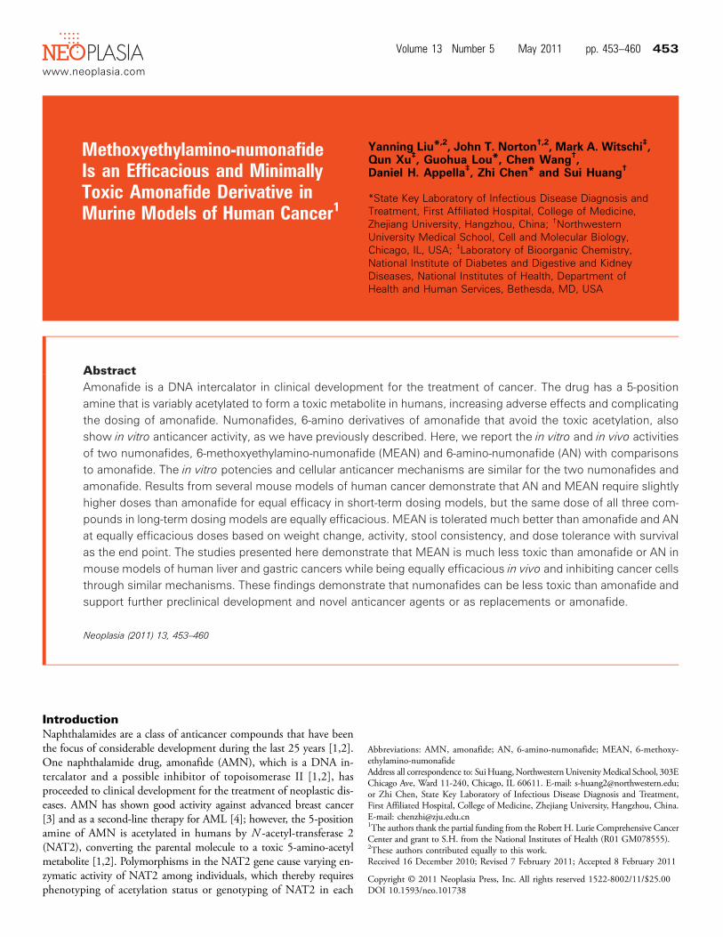
Figure 1. Numonafides behave similar to AMN in vitro. (A) Structures of AMN and the two numonafides examined herein. (B) Concen-trations of AMN, MEAN, and AN that cause 50% growth inhibition in human gastric carcinoma (AGS, MGC, and SGC-7901), leukemia(K562 and Jurkat), breast carcinoma (MDA-MB-435), hepatoma (HepG2, Huh7, and SMMC-7721), lung carcinoma (A549), and coloncarcinoma (HT-29) cell lines as determined by the MTT assay. (C) Cell cycle analysis of AGS gastric cancer cells after 24 hours of treat-ment with AMN, AN, or MEAN as determined by flow cytometry for DNA content (bottom dark gray bars = cells in G1 phase, middlelight gray bars = S phase, and white bars = G2/M phases). (D) Results from flow cytometry analysis of apoptosis in Huh7 hepatoma cellsafter 24 hours of treatment with AMN or numonafides. All error bars = SD from three or more independent experiments and vehicle =1% DMSO.
454 MEAN is Efficacious and Less Toxic than AMN Liu et al. Neoplasia Vol. 13, No. 5, 2011
provides coverage of 48,802 genes and expressed sequence tags. Rawsignal intensities of each probe were obtained using data analysis soft-ware (Beadstudio; Illumina, San Diego, CA) and imported to theLumi package of Bioconductor for data analysis. Before transformationand normalization [6–8], A/P call detection was performed based ondetection of P value. Of 48,802 probes with less than 0.01, 18,678were considered as valid signals. For each pair of five comparisons(AMN and vehicle, AN and vehicle, MEAN and vehicle, MEANand AN, and MEAN and AMN), differentially expressed genes wereidentified using an analysis of variance (ANOVA) model with empiri-cal Bayesian variance estimation [9]. Initially, genes were identified asbeing differentially expressed on the basis of a statistical significance(raw, P < .01; false discovery rate–adjusted, P < .05) and 1.5-foldchange (up or down) in expression level in each comparison.
In Vivo Xenograft Models
Huh7-luc cell line. pGL3-control (Promega, Madison, WI) was firstdigested with XbaI (Takara, Shiga, Japan) and then blunted with DNApolymerase Klenow fragment (Takara). The resulting DNA was thendigested with BglII (Takara) and the DNA fragment (1902 bp) encod-ing luciferase (luc). This fragment was then ligated to the BamHI/SmaI(Takara) digested backbone of pWPXL (Addgene, Cambridge, MA) tocreate pWPXL-luc. Next, 2.5 × 10⁶ of HEK-293T cells were plated in a10-cm diameter plate. The following day, 20 μg of pWPXL-Luc, 15 μgof psPAX2 (Addgene), and 6 μg of pMD2.G (Addgene) were dilutedin 1 ml Hank’s buffered saline with 50 μl of 2.5 M CaCl2 and mixedgently. After 20minutes of incubation at room temperature, the plasmidsolution was added to the HEK-293T (Invitrogen) medium, and after6 hours, the medium was replaced with medium containing no plas-mids. Four days later, the medium was collected, and the lentiviruswas purified with 0.45-μm filters. Then, Huh7 cells were infected withpWPXL-luc lentivirus virus at a multiplicity if infection of 1000:1 inthe presence of polybrene. After 3 days of infection, single cells wereplated into the wells of a 96-well plate and allowed to grow for 3 weeks,at which point the highest expressing clone was expanded and used forthe studies described here.
AGS-luc cell line. Lentiviral vectors expressing firefly luciferase (luc)were generated using a four-plasmid system. Briefly, a lentiviral expres-sion construct encoding luciferase and green fluorescent protein, eachunder the control of an individual CAG-enhanced CMV promoter(pLenti CMV Puro LUC; Addgene), was cotransfected with lentiviralpackaging plasmids (pMD2.G, psPAX2) and a VSV-G envelope ex-pressing plasmid (CVG) into HEK-293T cells using Lipofectamine2000 (Invitrogen, Carlsbad, CA). The medium was collected every24 hours and replaced with fresh media for 3 days. Virus-containingmedium was filtered with 0.45-μm filters, and then the viral particleswere concentrated with sucrose ultracentrifugation. The viral pellet wasresuspended in the medium with polybrene and added to AGS cells for12 hours. After infection, the virus-containing medium was replacedwith fresh medium for 24 hours. Cells expressing high levels of greenfluorescent protein were isolated by fluorescence-activated cell sort-ing, and the pooled population was expanded to create the AGS-luccell line.
Mice and xenografts. Male nu/nu (nude) mice (18-20 g at experi-ment initiation) were maintained at the vivarium of First Affiliated
Hospital, College ofMedicine, in a pathogen-free unit, under a 12-hourlight/dark cycle, and were provided with food and water ad libitum.Mice were inoculated subcutaneously at the right axilla or the perito-neal cavity with HepG2 (106 cells), AGS-luc (106 cells), or Huh7-luccells (106 cells for subcutaneous inoculation and 2 × 10⁶ cells for intra-peritoneal inoculation). For the experiments using AGS-luc and Huh7-luc cells, in vivo bioluminescent imaging was performed with a Luminaimaging system (Nippon Roper, I.C.E., Tokyo, Japan). Fifteen minutesbefore imaging, mice were injected with 150-mg/kg luciferin throughan intraperitoneal route. Images were collected and analyzedwith LivingImage software (Slidebook 4.1, Denver, CO). Vehicle control was20% DMSO.
Results
AMN, AN, and MEAN Share Similar In Vitro GrowthInhibition and Apoptotic PropertiesOur previous studies showed that the numonafides AN and
MEAN (Figure 1A) inhibit the growth of three cancer cell lines withpotencies similar to AMN and demonstrated similar selectivity forgrowth inhibition of cancer cells over normal cells [5]. Here, we sys-tematically investigated the growth inhibition of numonafides andAMN in 11 cell lines derived from various cancers. The results showthat the numonafides, AN and MEAN, inhibit cancer cell growthwith a similar potency as AMN, although AN tends to be slightlyless potent (Figure 1B). Because AN and MEAN are potent inhibi-tors of gastric and liver cancer cell lines and because these cancers areprevalent malignancies with relatively few pharmacologically viabletreatment options, here we evaluate their antitumor properties usingAGS (gastric cancer), Huh7, and HepG2 (hepatomas) cell lines.We evaluated the in vitro effect of these compounds on cell pro-
liferation and apoptosis. First, AGS cells were treated with varyingdoses of AMN, AN, and MEAN and stained to determine theDNA content. AMN, AN, and MEAN all cause AGS cells to increasetheir DNA content in a dose-dependent manner, and all compoundssignificantly (P < .05 by one-way ANOVA with Dunnett test) increaseDNA content at 5 μM, indicating that these compounds cause G2arrest (Figure 1C), which is likely the result of DNA damage throughintercalation or topoisomerase II inhibition by these compounds [10].Next, Huh7 cells were treated with the numonafides and AMN for24 hours then stained to determine the apoptosis index. The resultsshow that AMN, AN, and MEAN all cause significant (P < .05 byone-way ANOVA with Dunnett test) increases in apoptosis at 5 and10 μM with AMN and MEAN being significantly (P < .05 by un-paired t-test with Welch correction) more potent than AN at bothdoses (Figure 1D).
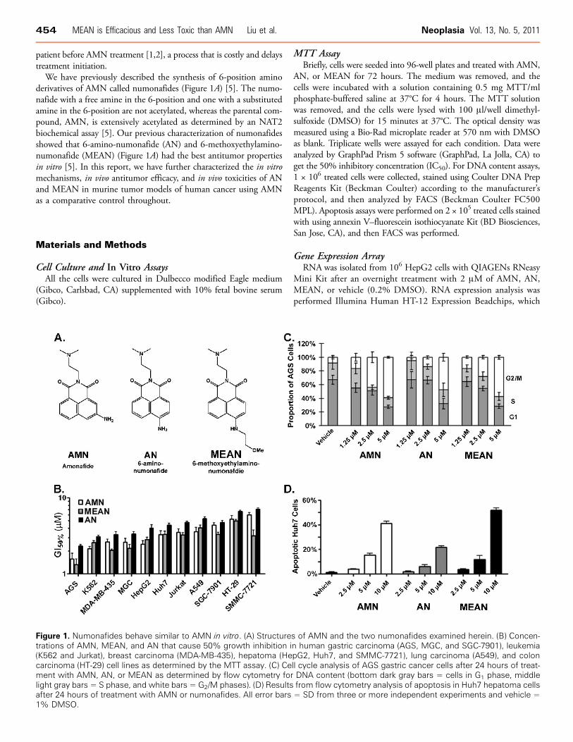
AMN, AN, and MEAN Similarly Influence Gene ExpressionPattern in Cancer CellsGene array analyses on cancer cells treated with numonafides,
MEAN, and AMN were performed using to identify and comparethe molecular mechanism and cellular pathways that are affectedby the treatment of these compounds. HepG2 cells were treated withAMN, MEAN, or AN at 2 μM (a pharmacologically achievable con-centration) overnight, and the changes in the level of approximately25,000 transcripts were determined with the gene array. MEAN,AMN, and AN significantly (P < .05 by t-test) changed the level of347, 199, and 178 transcripts, respectively, by greater than 1.5-fold
Neoplasia Vol. 13, No. 5, 2011 MEAN is Efficacious and Less Toxic than AMN Liu et al. 455
(Figure 2A). The number of transcripts changed is positively correlatedwith the in vitro DNA intercalation potencies of these compounds [5],suggesting that the change in gene expression is due to the differen-tial efficiency of DNA intercalation by each compound in the cellularmilieu. There is a lack of differences in gene expression when eachtreatment group is compared to one another instead of vehicle treat-ment (Figure 2A), indicating that all three compounds change the ex-pression of similar genes and are thereby acting through similarmechanisms. Supporting this theory is the finding that transcriptschanged greater than three-fold are all similarly altered in the threetreatment groups compared to the vehicle group, with a few exceptionswhere AN does not modulate the transcript level to the extent ofAMN and MEAN (Figure 2B), likely due to the lower DNA inter-calation efficiency or cellular potency of AN [5].
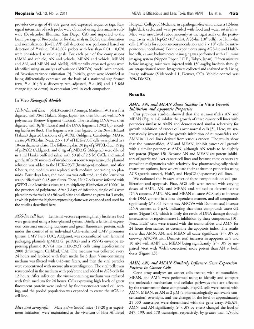
Numonafides Are Efficacious in a Hepatoma Xenograft Model,but MEAN Is Better Tolerated than AMN and ANThe in vivo tolerance and anticancer properties of AMN, AN, or
MEAN were initially tested in a xenograft model, in which nude im-munocompromised mice were implanted with the human HepG2hepatoma cells subcutaneously under the front right axilla (armpit).Mice were treated with vehicle, 50 μmol/kg, or 100 μmol/kg of eachcompound or 200 μmol/kg of MEAN (this dose of AMN or AN rap-idly killed mice). The compounds were administered through the in-traperitoneal route once per day for 14 days, 2 weeks after theimplantation of the xenografts. After treatment, the mice were sacri-ficed, and the tumors were resected and weighed. AMN inhibitedtumor growth most potently at the 50- and 100-μmol/kg doses (Ta-
ble 1). MEAN was less efficacious than AMN at 50 and 100 μmol/kg;however, the 200-μmol/kg dose of MEAN was equally efficacious asthe 100-μmol/kg dose of AMN. This initial end point tumor measure-ment in this study suggested that MEAN and AN are less potent thanAMN, but based on the lack of mice that died in the MEAN-treatedgroups, up to 200-μmol/kg MEAN is tolerated much better thanAMN and AN and can be equally efficacious due to its lower toxicity.
Numonafides Can Inhibit Tumor Growth and Reduce the Sizeof Established TumorsAGSandHuh7 cells expressing luciferase were used to evaluate tumor
inhibition properties of numonafides and AMN in a time-dependentmanner. In this model, mice were imaged every 7 days to quantify tu-mor growth through luminescent output of the tumor. First, mice wereimplanted with the tumors (subcutaneous AGS-luc [Figure 3, B andC] and Huh7-luc [Figure 3A] or intraperitoneal Huh7-luc [Figure 3D])and were treated continuously for 28 days with 50 μmol/kg per dayeach compound and 100 μmol/kg MEAN through intraperitonealadministration [11]. The treatment was initiated 2 weeks after im-plantation of the subcutaneous xenograft and 1 week after the intra-peritoneal xenograft. In all three tumor xenograft models, 50 μmol/kgAN is the least effective and 50 μmol/kg MEAN is slightly more effec-tive than AN, both halting tumor growth. AMN at the 50-μmol/kgdose and MEAN at the 100-μmol/kg dose were equally effective (P >.05 by unpaired t-test at day 21), actually causing a significant (P < .05by unpaired t-test with Welch correction at day 21) decrease in tumorsize from treatment start (Figure 3, A–D).
Table 1. Numonafides Are Efficacious in the HepG2 Human Xenograft Model.
Treatment Dosage (μmol/kg) once a day × 14 d Final Number of Mice (/10) Tumor Weight (g) % Growth Inhibition P, t-Test
Vehicle 0 10 2.185 ± 0.242 — —
AN 100 8 1.455 ± 0.288 33.4 <.01AN 50 10 1.969 ± 0.274 9.9 <.05MEAN 200 10 0.427 ± 0.212 80.5 <.01MEAN 100 10 0.869 ± 0.301 60.2 <.01MEAN 50 10 1.889 ± 0.181 13.5 <.01AMN 100 9 0.509 ± 0.199 76.7 <.01AMN 50 10 1.141 ± 0.216 47.8 <.01
Nude mice were implanted with HepG2 cells in the front axilla and treated through intraperitoneal injection once per day for 14 days, 2 weeks after implantation of tumor. After treatment, tumors wereresected and weighed (each group started with 10 mice). Statistics (unpaired t-test) are for each group compared to vehicle-treated mice.
Figure 2. Amonafide and numonafides alter gene expression with similar patterns. (A) Number of transcripts that are significantly (P <.05 by t-test, n = 3) changed in HepG2 cells determined by Illumina’s BeadArray after an overnight treatment with 2 μM of each com-pound compared to vehicle-treated cells and compared to AMN-treated cells as. (B) Change in levels of transcripts that are significantlyupregulated or downregulated by greater than three-fold in cells treated with AMN or numonafides compared to vehicle (average valuesfrom of three independent experiments and P < .05 for fold changes more than three-fold).
456 MEAN is Efficacious and Less Toxic than AMN Liu et al. Neoplasia Vol. 13, No. 5, 2011
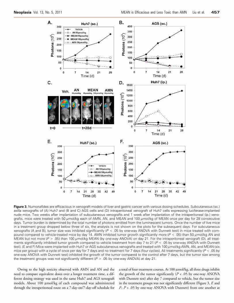
Owing to the high toxicity observed with AMN and AN and theneed to compare equivalent doses over a longer treatment time, a dif-ferent dosing strategy was used in the same Huh7 and AGS xenograftmodels. About 100 μmol/kg of each compound was administeredthrough the intraperitoneal route on a 7-day-on/7-day-off schedule for
a total of four treatment courses. At 100 μmol/kg, all three drugs inhibitthe growth of the tumor significantly (P < .05 by one-way ANOVAwith Dunnett test) after day 7 compared to vehicle, but the tumor sizein the treatment groups was not significantly different (Figure 3, E andF ; P > .05 by one-way ANOVA with Dunnett) from one another at
Figure 3. Numonafides are efficacious in xenograft models of liver and gastric cancer with various dosing schedules. Subcutaneous (sc.)axilla xenografts of (A) Huh7 and (B and C) AGS cells and (D) intraperitoneal xenograft of Huh7 cells expressing luciferase-implantednude mice. Two weeks after implantation of subcutaneous xenografts and 1 week after implantation of the intraperitoneal (ip.) xeno-grafts, mice were treated with 50 μmol/kg each of AMN, AN, and MEAN and 100 μmol/kg of MEAN once per day for 28 consecutivedays. Tumor burden is determined by the total number of photons emitted from the luminescent tumors. Once the number of live micein a treatment group dropped below three of six, the analysis is not shown on the plots for the subsequent days. For subcutaneousxenografts (A and B), tumor size was inhibited significantly (P < .05 by one-way ANOVA with Dunnett test) in mice treated with com-pound compared to vehicle-treated mice by day 14. AMN inhibited tumor growth significantly more (P < .05) than 50 μmol/kg AN andMEAN but not more (P > .05) than 100 μmol/kg MEAN (by one-way ANOVA) on day 21. For the intraperitoneal xenograft (D), all treat-ments significantly inhibited tumor growth compared to vehicle treatment from day 7 to 21 (P < .05 by one-way ANOVA with Dunnetttest). (E and F) Mice were implanted with Huh7 or AGS subcutaneous xenografts and treated with 100 μmol/kg AMN, AN, and MEAN (sixmice per group) with a cycle of once per day for 7 days and no treatment for 7 days (four cycles). All treatments significantly (P < .05 byone-way ANOVA with Dunnett test) inhibited the growth of the tumor compared to the control after 7 days, but the tumor size amongthe treatment groups was not significantly different (P > .05 by one-way ANOVA) at day 21.
Neoplasia Vol. 13, No. 5, 2011 MEAN is Efficacious and Less Toxic than AMN Liu et al. 457
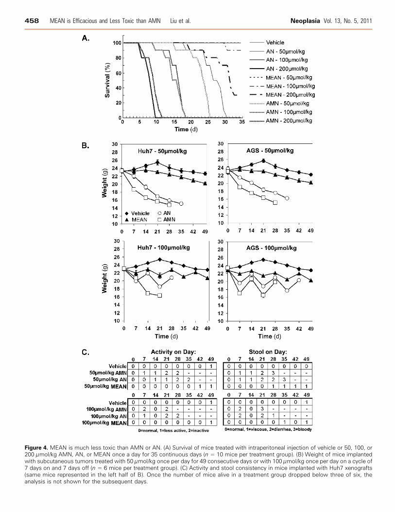
Figure 4. MEAN is much less toxic than AMN or AN. (A) Survival of mice treated with intraperitoneal injection of vehicle or 50, 100, or200 μmol/kg AMN, AN, or MEAN once a day for 35 continuous days (n = 10 mice per treatment group). (B) Weight of mice implantedwith subcutaneous tumors treated with 50 μmol/kg once per day for 49 consecutive days or with 100 μmol/kg once per day on a cycle of7 days on and 7 days off (n = 6 mice per treatment group). (C) Activity and stool consistency in mice implanted with Huh7 xenografts(same mice represented in the left half of B). Once the number of mice alive in a treatment group dropped below three of six, theanalysis is not shown for the subsequent days.
458 MEAN is Efficacious and Less Toxic than AMN Liu et al. Neoplasia Vol. 13, No. 5, 2011

day 21. All three compounds shrank the tumor volume compared to day0 using this dosing schedule.
MEAN Is Less Toxic In Vivo Compared to AMN and ANThe toxicity of these compounds was examined by survival anal-
ysis on mice treated with 50, 100, or 200 μmol/kg of AMN, AN,and MEAN for up to 35 consecutive days. The results show thatAMN and AN were similarly toxic, whereas the MEAN was toleratedmuch better (Figure 4A). Only 1 in 10 mice died by the 35th day atthe 100-μmol/kg dose of MEAN compared with the 100-μmol/kgdose of AMN or AN, which killed all mice by day 20 (Figure 4A). Inaddition, the median survival time for mice treated with 200 μmol/kgMEAN was more than 30 days compared to a median survival of10 days for mice treated with equivalent doses of AMN and AN(Figure 4A).The body weight, activity, and stool consistency were recorded
during the treatments described in Figure 3 to further assess the toxici-ties of these compounds. When dosed at 50 μmol/kg every day forup to 49 days, AMN and AN caused approximately 30% decreasein weight by days 28 and 35, respectively (Figure 4B). In contrast,MEAN caused approximately a 10% decrease in weight at day 49compared to vehicle-treated mice (Figure 4B). On the other hand, allmice in the AMN and AN groups died by day 49 and no mice diedin the MEAN group (not shown). In treatment with 100 μmol/kg on a7-day-on/7-day-off dosing cycles, the final weight of MEAN-treatedmice was only 10% to 15% lower than vehicle-treated mice after fourtreatment courses (Figure 4B). Given the large size of the AGS andHuh7 tumors (Figure 2D), the differences in final weights betweenMEAN- and vehicle-treated mice may be partially attributed to thesmaller tumors in MEAN-treated mice. Furthermore, a rebound to ahealthy weight is observed on removal of MEAN during the 7-day-offportion of the dosing (Figure 4B), and there is a minimal alteration ofactivity and stool consistency (Figure 4C ). In comparison, AN andAMN treatments demonstrated less body weight recovery during the7-day-off in weight and caused decreased activity and worse stool con-sistency. These findings demonstrate that MEAN is much less toxicthan AMN and AN in nude mice, in terms of weight loss, activity lev-els, gastrointestinal toxicities, and survival, suggesting that MEAN isa good candidate to be developed as a novel antigastric and hepaticcancer drug or as a replacement for AMN.
DiscussionHere, we demonstrate that two numonafides, AN and MEAN, in-hibit tumor cell growth, induce G2 arrest, and apoptosis in vitro withpotencies similar to the parental drug, AMN (Figure 1), indicatingthese three compounds inhibit tumor cell growth through similar cel-lular mechanisms. In addition, the numonafides alter the transcrip-tome in cancer cells in a similar pattern to AMN (Figure 2), as hasbeen reported with other derivatives of AMN containing substituted5-position aryl amines [12], further indicating that this class of drugsact on cancer cells with similar mechanisms, independent of alter-ations in aryl amine substitution. Although the association betweenmost transcripts altered over three-fold (Figure 2B) by these com-pounds and cancer is unknown; however, two genes have been asso-ciated with cancer. First, metallothionein 1G, which is upregulatedgreater than six-fold in cells treated by all three compounds, has beendescribed as a tumor suppressor in hepatocellular carcinoma [13] andother carcinomas [14]. This finding suggests that up-regulation of
metallothionein 1G could be a potential anticancer mechanism ofnumonafides and AMN. The second gene, stearoyl-CoA desaturase,downregulated by all three compounds, plays a critical role in fattyacid metabolism that increases cancer cell proliferation and malignanttransformation and decreases apoptosis [15]. The down-regulation ofthis gene by numonafides and AMN may contribute to the growthinhibition and apoptosis induction properties of these compounds.The identification of changes in gene expression patterns by thesecompounds not only helps confirm the common cellular targets be-tween the numonafides and AMN but also provides potentially newmechanisms for tumor cell inhibition by AMN and numonafides,such as the changes in expression of known and unknown genesand noncoding RNA (Figure 2B) and provides potential clinical bio-markers for response. Future studies will explore the additional modeof action for these compounds in cells that contribute to their anti-tumor properties in vitro and in vivo.In three human cancer cell line xenograft models using short-term
daily doses, we found that AN and MEAN are slightly less potentin vivo, but MEAN can be equally efficacious as AMN at higherdoses (Figure 3, A–D). A long-term periodic dosing regimen showedthat all three compounds could be equally efficacious at the samedose, actually shrinking established tumors, in two different xeno-graft models (Figure 3, E and F ). The xenograft models indicate thatnumonafides are efficacious in vivo and that MEAN is more effectivethan AN. Numonafides were developed as potentially less toxic de-rivatives of AMN because they avoid acetylation of the arylamine,which causes toxicities associated with AMN [2]. Mice were injectedwith 50, 100, or 200 μmol/kg AN, MEAN, and AMN once daily forup to 35 days to initially determine the toxicities of numonafides. ANis about equally toxic as AMN in nude mice, suggesting that the freeamine of AN is being metabolized in vivo similar to AMN, butMEAN is much less toxic and better tolerated by mice. MEAN treat-ment at the dose of 200 μmol/kg kill less mice than the 50-μmol/kgdose of AN and AMN (Figure 4A). Further evaluation as judged byweight, activity, and stool consistency in the two different dosing regi-mens used for the tumor efficacy studies confirmed that AN andAMN are equally toxic, whereas MEAN is much less toxic than bothof these compounds (Figure 4, B and C). The similar in vivo potenciesand in vitro mechanisms suggest that these compounds inhibit tumorcell growth by similar mechanisms; however, the large difference intoxicity in vivo between MEAN and AMN/AN may be due to differ-ential pharmacokinetics, biodistribution, metabolism, or a combina-tion thereof. Although this remains to be elucidated, here we haveprovided proof of principle that numonafides can be developed as lesstoxic counterparts to AMN and have identified MEAN as the firstnumonafide for future development as an anticancer drug.
AcknowledgmentThe authors thank the Genomic Core facility at Northwestern Uni-versity for performing the array studies.
References[1] Lv M and Xu H (2009). Overview of naphthalimide analogs as anticancer
agents. Curr Med Chem 16(36), 4797–4813.[2] Ingrassia L, Lefranc F, Kiss R, and Mijatovic T (2009). Naphthalimides and azo-
nafides as promising anti-cancer agents. Curr Med Chem 16(10), 1192–1213.[3] Costanza ME, Berry D, Henderson IC, Ratain MJ, Wu K, Shapiro C, Duggan D,
Kalra J, Berkowitz I, and Lyss AP (1995). Amonafide: an active agent in the
Neoplasia Vol. 13, No. 5, 2011 MEAN is Efficacious and Less Toxic than AMN Liu et al. 459
treatment of previously untreated advanced breast cancer—a Cancer and LeukemiaGroup B study (CALGB 8642). Clin Cancer Res 1(7), 699–704.
[4] Kolitz JE, Lundberg AS, Bennett JM, Capizzi RL, and Budman DR (2010).Phase I trials of amonafide as monotherapy and in combination with cytarabinein patients with poor-risk acute myeloid leukemia. Leuk Res 34(4), 487–491.
[5] Witschi MA, Luong L, Kawamura A, Ghosh S, Stack MS, Sim E, Avram MJ,Appella DH, and Huang S (2008). Synthesis and anticancer activities of 6-aminoamonafide derivatives. Anticancer Drugs 19(1), 23–36.
[6] Du P, Kibbe WA, and Lin SM (2008). lumi: a pipeline for processing Illuminamicroarray. Bioinformatics 24(13), 1547–1548.
[7] Lin SM, Du P, Huber W, and Kibbe WA (2008). Model-based variance-stabilizingtransformation for Illumina microarray data. Nucleic Acids Res 36(2), e11.
[8] Du P, Kibbe WA, and Lin SM (2007). nuID: a universal naming scheme ofoligonucleotides for Illumina, Affymetrix, and other microarrays. Biol Direct2, 16.
[9] Wettenhall JM and Smyth GK (2004). limmaGUI: a graphical user interface forlinear modeling of microarray data. Bioinformatics 20(18), 3705–3706.
[10] Clifford B, Beljin M, Stark GR, and Taylor WR (2003). G2 arrest in response totopoisomerase II inhibitors: the role of p53. Cancer Res 63(14), 4074–4081.
[11] Van Quaquebeke E, Mahieu T, Dumont P, Dewelle J, Ribaucour F, Simon G,Sauvage S, Gaussin JF, Tuti J, El Yazidi M, et al. (2007). 2,2,2-Trichloro-N -({2-[2-(dimethylamino)ethyl]-1,3-dioxo-2,3-dihydro-1H -benzo[de]isoquinolin- 5-yl}carbamoyl)acetamide (UNBS3157), a novel nonhematotoxic naphthalimidederivative with potent antitumor activity. J Med Chem 50(17), 4122–4134.
[12] Mijatovic T, Mahieu T, Bruyere C, De Neve N, Dewelle J, Simon G, DehouxMJ, van der Aar E, Haibe-Kains B, and Bontempi G (2008). UNBS5162, a novelnaphthalimide that decreases CXCL chemokine expression in experimental pros-tate cancers. Neoplasia 10(6), 573–586.
[13] Kanda M, Nomoto S, Okamura Y, Nishikawa Y, Sugimoto H, Kanazumi N,Takeda S, and Nakao A (2009). Detection of metallothionein 1G as a methyl-ated tumor suppressor gene in human hepatocellular carcinoma using a novelmethod of double combination array analysis. Int J Oncol 35(3), 477–483.
[14] Ferrario C, Lavagni P, Gariboldi M, Miranda C, Losa M, Cleris L, Formelli F,Pilotti S, Pierotti MA, and Greco A (2008). Metallothionein 1G acts as an on-cosupressor in papillary thyroid carcinoma. Lab Invest 88(5), 474–481.
[15] Igal RA (2010). Stearoyl-CoA desaturase-1: a novel key player in the mecha-nisms of cell proliferation, programmed cell death and transformation to cancer.Carcinogenesis 31(9), 1509–1515.
460 MEAN is Efficacious and Less Toxic than AMN Liu et al. Neoplasia Vol. 13, No. 5, 2011