methods used for the assessment of lv systolic function ... · methods used for the assessment of...
TRANSCRIPT

Methods used for the assessment of LV systolicfunction: common currency or tower of Babel?Thomas H Marwick
Correspondence toDr Thomas H Marwick,Menzies Research InstituteTasmania, 17 Liverpool St,Hobart T7000, Australia;[email protected]
Received 7 September 2012Revised 27 November 2012Accepted 8 January 2013Published Online First2 February 2013
To cite: Marwick TH. Heart2013;99:1078–1086.
ABSTRACTThe last decade has produced a proliferation oftechniques for the assessment of left ventricular systolicfunction, and there now seems to be more choice thanseems rational for the questions that we need answersto. In some instances, simple estimation is all that isrequired—the risk stratification process is inexact, asemphasised by the variety of modalities used tocharacterise ejection fraction (EF) in studies thatvalidated the efficacy of treatments selected on the basisof EF. Nonetheless, while technical advances often causedisruption and confusion, it would be wrong to dismissthem as lacking benefit. The purpose of this review is totry to provide rational grounds for selecting both testmodality and physiological parameter in various specificclinical situations.
The assessment of global left ventricular (LV) sys-tolic function is a cornerstone of risk evaluationand management in most cardiac diseases. The sim-plest and most widely used parameter for thispurpose has been ejection fraction and regionalwall motion analysis, initially from x-ray contrastventriculography, then nuclear ventriculographyand echocardiography. Over the last 10–15 years,this choice has been supplemented by new tech-nologies including CT and cardiac magnetic reson-ance (CMR), as well as the movement from twodimensional (2D) to three dimensional (3D)imaging. Finally, over the last decade, new para-meters such as strain imaging have become avail-able. In short, all modalities used to image theheart—ultrasound, scintigraphy, x-ray contrast(with and without CT) and CMR can provide thisassessment. Rather than categorise the techniquesby modality, we should categorise them on thebasis of assessing global and regional function,whether they are quantitative versus qualitative,and the clinical setting under which they are used.
INDICATIONS FOR SYSTOLIC FUNCTIONEVALUATIONThe most common indications for LV systolic func-tion evaluation are listed in table 1.The diagnosis and evaluation of heart failure is
probably the most common indication for echocar-diography. Two studies have shown that heartfailure patients who undergo echocardiographyhave a better outcome than those that do not.1 2
Although a variety of biases may account for theseobservations, it seems plausible that performing animaging study identifies individuals with heartfailure and reduced ejection fraction who can thenbe initiated on therapies that are known to have afavourable impact on survival.
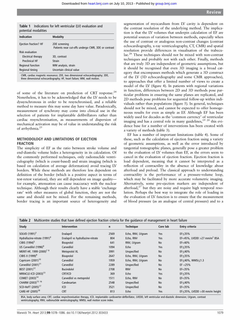
A second (but related) indication for echocardio-graphic evaluation of systolic function relates toindividuals where LV dysfunction is an importantcomponent of risk evaluation for decision-making.In these settings, cut-offs of LV function are surro-gates for the assessment of risk and cut points haveto be acknowledged as arbitrary, although the needfor them is understandable from the standpoint ofdecision-making. However, table 2 illustrates howthe methodologies and cut-offs used in studies thathave defined these ejection fraction criteria for theguidance of management are highly variable, withfew studies using core laboratories.3–18 Recognitionof an ejection fraction of <35% is important indecision-making regarding device therapy, eitherwith implantable defibrillators or cardiac resynchro-nisation therapy (CRT).19 20 Figure 1A emphasiseshow difficult this is to perform accurately withtechniques that require tracing of the LV cavity andcontrasts this with the automated measurement ofglobal strain (figure 1B). Similar to the ejectionfraction criteria for intervention in regurgitantvalve lesions,21 it seems reasonable to conclude thatthese numbers should be considered to be guidesrather than thresholds.A third group relates to asymptomatic subjects at
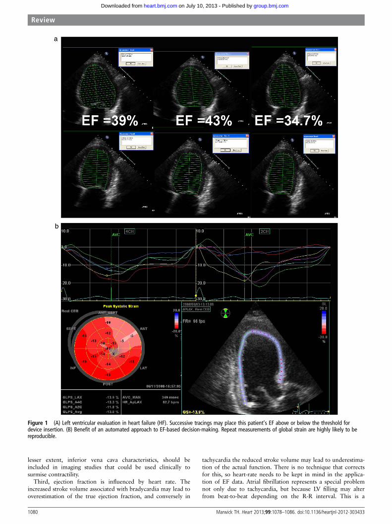
risk for heart failure, in whom the detection ofreduced ejection fraction or subtle structural heartdisease reclassifies the patient from stage A to stageB of heart failure, with resulting managementimplications.22 A specific group of these patientsare those who have previously been administeredcardiotoxic chemotherapy, but it also includes indi-viduals with gene-positive cardiomyopathies, amyl-oidosis or other infiltrative conditions who areexpected to have a subclinical phase to their cardio-myopathy (figure 2).The fourth common indication relates to the
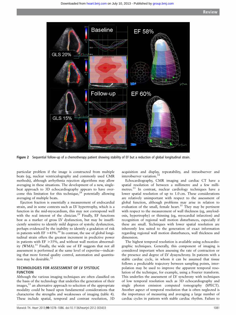
evaluation of regional systolic dysfunction inpatients with chest pain or suspected ischaemicheart disease. There is a strong association of wallmotion abnormalities with ischaemic heart diseaseand Framingham risk score,23 although other con-ditions such as sarcoidosis and myocarditis mayresult in regional changes. The calculation of strainappears to be a reliable and reproducible means ofquantifying regional function at both echocardiog-raphy and CMR (figure 3).Finally, although the temporal analysis of LV con-
traction carries prognostic information, the clinicalapplication of this remains uncertain. There arevery strong prognostic reasons to undertake CRT inpatients with heart failure symptoms, systolic dys-function and left bundle branch block, irrespectiveof the measurement of mechanical synchrony, andthere are reasonable grounds to doubt the reliability
1078 Marwick TH. Heart 2013;99:1078–1086. doi:10.1136/heartjnl-2012-303433
Review
group.bmj.com on July 10, 2013 - Published by heart.bmj.comDownloaded from
of some of the literature on prediction of CRT response.24
Nonetheless, it has to be acknowledged that the LV needs to bedyssynchronous in order to be resynchronised, and a reliablemethod to measure this may some day have value. Paradoxically,measurement of synchrony may come into clinical use in theselection of patients for implantable defibrillators rather thancardiac resynchronisation, as measurements of dispersion ofmechanical activation may be of value in understanding the riskof arrhythmia.25
METHODOLOGY AND LIMITATIONS OF EJECTIONFRACTIONThe simplicity of EF as the ratio between stroke volume andend-diastolic volume hides a heterogeneity in its calculation. Ofthe commonly performed techniques, only radionuclide ventri-culography (which is count-based) and strain imaging (which isbased on calculation of average deformation) avoid tracing LVborders. While these methods are therefore less dependent ondefinition of the border (which is a positive aspect in terms oftest-retest variation), they are still dependent on image quality—for example, attenuation can cause inaccuracy with the nucleartechnique. Although their results clearly have a stable ‘exchangerate’ with other measures of global function, they are not thesame and should not be mixed. For the remaining methods,border tracing is an important source of heterogeneity and
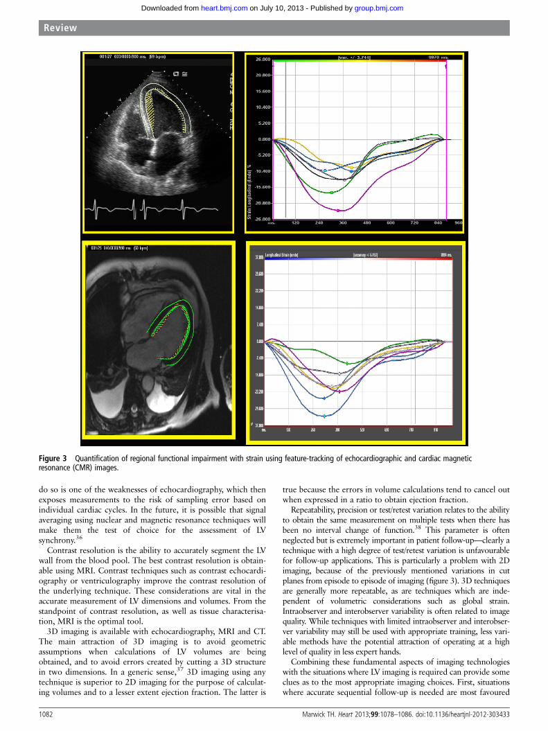
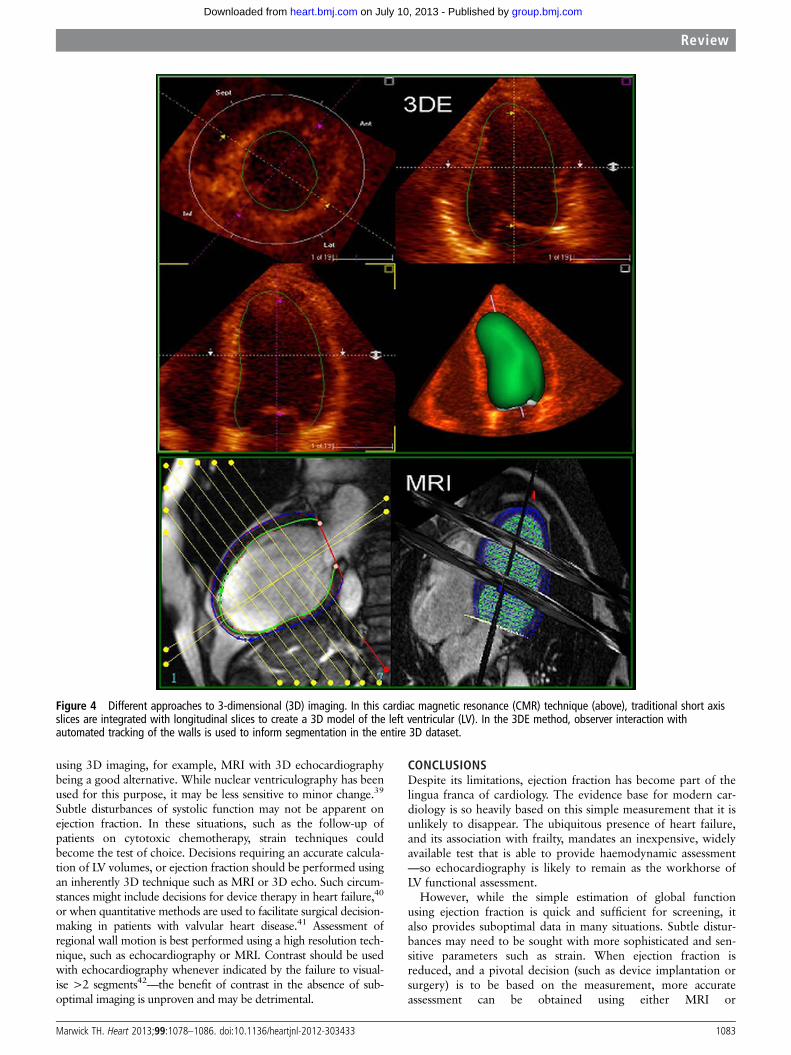
segmentation of myocardium from LV cavity is dependent onthe contrast resolution of the underlying method. The implica-tion is that the LV volumes that underpin calculation of EF arepotential sources of variation between methods, especially whenthe use of contrast or analogous non-contrast changes (contrastechocardiography, x-ray ventriculography, CT, CMR) and spatialresolution provide differences in visualisation of the trabecu-lae.26 These techniques should not be mixed with non-contrasttechniques and probably not with each other. Finally, methodsthat are truly 3D are independent of geometric assumptions, butit should be recognised that even 3D imaging is a broad cat-egory that encompasses methods which generate a 3D constructof the LV (3D echocardiography and some CMR approaches),to approaches that offer a limited number of views to create amodel of the LV (figure 4). In patients with regional variationsin function, differences between 2D and 3D methods pose par-ticular problems in ensuring the same planes are replicated, and2D methods pose problems for sequential follow-up within indi-viduals rather than populations (figure 5). In general, techniquesshould not be mixed, and cannot be expected to offer homoge-neous results for even as simple as EF. Although EF has beenwidely used for decades as the ‘common currency’ of ventricularimaging and has a central role in many guidelines,19 20 this evi-dence base for a number of interventions has been created witha variety of methods (table 3).
EF has a number of important limitations (table 4). Some ofthese, such as the calculation of ejection fraction using a varietyof geometric assumptions, as well as the error introduced bytangential tomographic planes, generally pose a greater problemto the evaluation of LV volumes than EF, as the errors seem tocancel in the evaluation of ejection fraction. Ejection fraction isload dependent, meaning that it cannot be interpreted as areflection of contractility in the absence of knowledge aboutafterload and preload. The classical approach to understandingcontractility is the performance of a pressure-volume loop,which may be facilitated by more accurate volumetric imaging.Alternatively, some pre-ejection markers are independent ofafterload,27 but they are noisy and require high temporal reso-lution. Perhaps the best way to integrate the role of loading inthe evaluation of LV function is to ensure that the measurementof blood pressure (as an analogue of central pressure) and to a
Table 2 Multicentre studies that have defined ejection fraction criteria for the guidance of management in heart failure
Study Intervention n Technique Core lab Entry criteria
SOLVD (1991)3 Enalapril 2569 Echo, RNV, LVgram Yes EF≤35%Hydrallazine-nitrate (1991)4 Enalapril vs hydrallazine-nitrate 804 Echo, RNV Yes EF<45%, LVEDD >27 mm/m2 BSACIBIS (1994)5 Bisoprolol 641 RNV, LVgram No EF<40%US Carvedilol (1996)6 Carvedilol 1094 Echo Yes EF≤35%MERIT-HF, 1999 (2000)7 8 Metoprolol XL 3991 Unspecified No EF≤40%CIBIS II (1999)9 Bisoprolol 2647 Echo, RNV, LVgram No EF≤35%Capricorn (2001)10 Carvedilol 1959 Echo, RNV, LVgram No EF≤40%, WMSI≤1.3Carvedilol (2001)11 Carvedilol 2289 Unspecified No EF <25%BEST (2001)12 Bucindolol 2708 RNV No EF<35%MIRACLE-ICD (2003)13 CRT/ICD 369 Echo Yes EF≤35%COMET (2003)14 Carvedilol vs metoprolol 1511 Echo, RNV No EF<35%CHARM (2003)15 16 Candesartan 2548 Unspecified No EF≤40%SCD-HeFT (2005)17 ICD 3521 Unspecified No EF<35%CARE-HF (2005)18 CRT 813 Echo Yes EF≤35%, LVEDD >30 mm/m height
BSA, body surface area; CRT, cardiac resynchronisation therapy, ICD, implantable cardioverter-defibrillator, LVEDD, left ventricular end-diastolic dimension; LVgram, contrastventriculography; RNV, radionuclide ventriculography; WMSI, wall motion score index.
Table 1 Indications for left ventricular (LV) evaluation andpotential modalities
Indication Modality
Ejection fraction? HF 2DE screeningPatients near cut-offs undergo CMR, 3DE or contrast
Risk evaluationElectrical therapy 3D, contrastPreclinical HF Strain
Regional function WM analysis, strainRegional timing Uncertain clinical value
CMR, cardiac magnetic resonance; 2DE, two dimensional echocardiography; 3DE,three dimensional echocardiography; HF, heart failure; WM, wall motion.
Marwick TH. Heart 2013;99:1078–1086. doi:10.1136/heartjnl-2012-303433 1079
Review
group.bmj.com on July 10, 2013 - Published by heart.bmj.comDownloaded from
lesser extent, inferior vena cava characteristics, should beincluded in imaging studies that could be used clinically tosurmise contractility.
Third, ejection fraction is influenced by heart rate. Theincreased stroke volume associated with bradycardia may lead tooverestimation of the true ejection fraction, and conversely in
tachycardia the reduced stroke volume may lead to underestima-tion of the actual function. There is no technique that correctsfor this, so heart-rate needs to be kept in mind in the applica-tion of EF data. Atrial fibrillation represents a special problemnot only due to tachycardia, but because LV filling may alterfrom beat-to-beat depending on the R-R interval. This is a
Figure 1 (A) Left ventricular evaluation in heart failure (HF). Successive tracings may place this patient’s EF above or below the threshold fordevice insertion. (B) Benefit of an automated approach to EF-based decision-making. Repeat measurements of global strain are highly likely to bereproducible.
1080 Marwick TH. Heart 2013;99:1078–1086. doi:10.1136/heartjnl-2012-303433
Review
group.bmj.com on July 10, 2013 - Published by heart.bmj.comDownloaded from
particular problem if the image is constructed from multiplebeats (eg, nuclear ventriculography and commonly used CMRmethods), although arrhythmia rejection algorithms may allowaveraging in these situations. The development of a new, single-beat approach to 3D echocardiography appears to have over-come this limitation for this technique,28 potentially allowingaveraging of multiple beats.
Ejection fraction is essentially a measurement of endocardialstrain, and in some contexts such as LV hypertrophy, which is afunction in the mid-myocardium, this may not correspond wellwith the real interest of the clinician.29 Finally, EF functionsbest as a marker of gross LV dysfunction, but may be insuffi-ciently sensitive to identify mild degrees of systolic dysfunction,perhaps evidenced by the inability to identify a gradation of riskin patients with EF >45%.30 In contrast, the use of global longi-tudinal strain offers the greatest increment in predictive powerin patients with EF >35%, and without wall motion abnormal-ity (WMA).31 Finally, the wide use of EF suggests that not allassessment is performed at the same level of expertise—indicat-ing that more formal quality control, automation and quantita-tion may be desirable.32
TECHNOLOGIES FOR ASSESSMENT OF LV SYSTOLICFUNCTIONAlthough the various imaging techniques are often classified onthe basis of the technology that underlies the generation of theirimages,33 an alternative approach to selection of the appropriatemodality could be based upon fundamental considerations thatcharacterise the strengths and weaknesses of imaging (table 4).These include spatial, temporal and contrast resolution, 3D
acquisition and display, repeatability, and intraobserver andinterobserver variation.34
Echocardiography, CMR imaging and cardiac CT have aspatial resolution of between a millimetre and a few milli-metres.33 In contrast, nuclear cardiology techniques have alower spatial resolution of up to 1.0 cm. These considerationsare relatively unimportant with respect to the assessment ofglobal function, although problems may arise in relation toevaluation of the small, female heart.35 They may be pertinentwith respect to the measurement of wall thickness (eg, amyloid-osis, hypertrophy) or thinning (eg, myocardial infarction) andrecognition of regional wall motion disturbances, especially ifthese are small. Techniques with lower spatial resolution areinherently less suited to the generation of exact informationregarding regional wall motion disturbances, wall thickness anddimension.
The highest temporal resolution is available using echocardio-graphic techniques. Generally, this component of imaging isconsidered important when assessing the rate of contraction orthe presence and degree of LV dyssynchrony. In patients with astable cardiac cycle, in whom it can be assumed that tissuefollows a predictable trajectory between sampling points, inter-polation may be used to improve the apparent temporal reso-lution of the technique, for example, using a Fourier transform.This underlies the assessment of LV synchrony with techniquesof low temporal resolution such as 3D echocardiography andsingle photon emission computed tomography (SPECT).Another aspect of temporal resolution that is often neglected isthe importance of measuring and averaging a large number ofcardiac cycles in patients with stable cardiac rhythm. Failure to
Figure 2 Sequential follow-up of a chemotherapy patient showing stability of EF but a reduction of global longitudinal strain.
Marwick TH. Heart 2013;99:1078–1086. doi:10.1136/heartjnl-2012-303433 1081
Review
group.bmj.com on July 10, 2013 - Published by heart.bmj.comDownloaded from
do so is one of the weaknesses of echocardiography, which thenexposes measurements to the risk of sampling error based onindividual cardiac cycles. In the future, it is possible that signalaveraging using nuclear and magnetic resonance techniques willmake them the test of choice for the assessment of LVsynchrony.36
Contrast resolution is the ability to accurately segment the LVwall from the blood pool. The best contrast resolution is obtain-able using MRI. Contrast techniques such as contrast echocardi-ography or ventriculography improve the contrast resolution ofthe underlying technique. These considerations are vital in theaccurate measurement of LV dimensions and volumes. From thestandpoint of contrast resolution, as well as tissue characterisa-tion, MRI is the optimal tool.
3D imaging is available with echocardiography, MRI and CT.The main attraction of 3D imaging is to avoid geometricassumptions when calculations of LV volumes are beingobtained, and to avoid errors created by cutting a 3D structurein two dimensions. In a generic sense,37 3D imaging using anytechnique is superior to 2D imaging for the purpose of calculat-ing volumes and to a lesser extent ejection fraction. The latter is
true because the errors in volume calculations tend to cancel outwhen expressed in a ratio to obtain ejection fraction.
Repeatability, precision or test/retest variation relates to the abilityto obtain the same measurement on multiple tests when there hasbeen no interval change of function.38 This parameter is oftenneglected but is extremely important in patient follow-up—clearly atechnique with a high degree of test/retest variation is unfavourablefor follow-up applications. This is particularly a problem with 2Dimaging, because of the previously mentioned variations in cutplanes from episode to episode of imaging (figure 3). 3D techniquesare generally more repeatable, as are techniques which are inde-pendent of volumetric considerations such as global strain.Intraobserver and interobserver variability is often related to imagequality. While techniques with limited intraobserver and interobser-ver variability may still be used with appropriate training, less vari-able methods have the potential attraction of operating at a highlevel of quality in less expert hands.
Combining these fundamental aspects of imaging technologieswith the situations where LV imaging is required can provide someclues as to the most appropriate imaging choices. First, situationswhere accurate sequential follow-up is needed are most favoured
Figure 3 Quantification of regional functional impairment with strain using feature-tracking of echocardiographic and cardiac magneticresonance (CMR) images.
1082 Marwick TH. Heart 2013;99:1078–1086. doi:10.1136/heartjnl-2012-303433
Review
group.bmj.com on July 10, 2013 - Published by heart.bmj.comDownloaded from
using 3D imaging, for example, MRI with 3D echocardiographybeing a good alternative. While nuclear ventriculography has beenused for this purpose, it may be less sensitive to minor change.39
Subtle disturbances of systolic function may not be apparent onejection fraction. In these situations, such as the follow-up ofpatients on cytotoxic chemotherapy, strain techniques couldbecome the test of choice. Decisions requiring an accurate calcula-tion of LV volumes, or ejection fraction should be performed usingan inherently 3D technique such as MRI or 3D echo. Such circum-stances might include decisions for device therapy in heart failure,40
or when quantitative methods are used to facilitate surgical decision-making in patients with valvular heart disease.41 Assessment ofregional wall motion is best performed using a high resolution tech-nique, such as echocardiography or MRI. Contrast should be usedwith echocardiography whenever indicated by the failure to visual-ise >2 segments42—the benefit of contrast in the absence of sub-optimal imaging is unproven and may be detrimental.
CONCLUSIONSDespite its limitations, ejection fraction has become part of thelingua franca of cardiology. The evidence base for modern car-diology is so heavily based on this simple measurement that it isunlikely to disappear. The ubiquitous presence of heart failure,and its association with frailty, mandates an inexpensive, widelyavailable test that is able to provide haemodynamic assessment—so echocardiography is likely to remain as the workhorse ofLV functional assessment.
However, while the simple estimation of global functionusing ejection fraction is quick and sufficient for screening, italso provides suboptimal data in many situations. Subtle distur-bances may need to be sought with more sophisticated and sen-sitive parameters such as strain. When ejection fraction isreduced, and a pivotal decision (such as device implantation orsurgery) is to be based on the measurement, more accurateassessment can be obtained using either MRI or
Figure 4 Different approaches to 3-dimensional (3D) imaging. In this cardiac magnetic resonance (CMR) technique (above), traditional short axisslices are integrated with longitudinal slices to create a 3D model of the left ventricular (LV). In the 3DE method, observer interaction withautomated tracking of the walls is used to inform segmentation in the entire 3D dataset.
Marwick TH. Heart 2013;99:1078–1086. doi:10.1136/heartjnl-2012-303433 1083
Review
group.bmj.com on July 10, 2013 - Published by heart.bmj.comDownloaded from

echocardiography with 3D imaging or echocardiographic con-trast. When volumes are required, as in the assessment of valvu-lar disease, 3D techniques should become mandatory. Finallyfollow-up studies should require a 3D strategy or geometryindependent techniques such as the assessment of global strain.
For regional function, visual assessment is sufficient in manycircumstances. If there is a desire to follow sequentially, or to
Figure 5 Variation between techniques for sequential left ventricular (LV) assessment. In this patient, 2-dimensional imaging suggests increasing LVvolumes with a stable EF (A), but 3-dimensional (confirmed by cardiac magnetic resonance) documents a falling EF (B), end-diastolic volume (EDV) andend-systolic volume (ESV).
Table 3 Limitations of ejection fraction
ProblemCircumstances ofinaccuracy Potential solution
Geometrydependence
LBBB, extensive wallmotion abnormality,off-axis imaging
3D imaging,geometry-independenttechniques
Load dependence Extremes of afterload,mitral regurgitation
Pressure volume loops,pre-ejection markers
High and low HR Heart block, tachycardias(especially atrialfibrillation)
None
Marker ofendocardialshortening
LV hypertrophy Mid-myocardial shortening
Insensitivity tominor change
Prognostic value close toEF 50%
Non-EF techniques forassessing subclinicaldysfunction
Expertise Wide use of EF Quantitation
3D, 3-dimensional; LBBB, left bundle branch block; LV, left ventricular.
Table 4 Selecting the right tool for the job—imagingcharacteristics of various tests
Technique Application
High spatial resolution CMR LV hypertrophy, infiltrationHigh temporal resolution Tissue Doppler, strain LV synchronyHigh contrast resolution CMR, contrast echo LV volumesHigh repeatability CMR, 3D echo Sequential follow-upSensitivity to minor change Strain Subclinical cardiomyopathy
CMR, cardiac magnetic resonance; 3D, 3-dimensional; LV, Left ventricular.
1084 Marwick TH. Heart 2013;99:1078–1086. doi:10.1136/heartjnl-2012-303433
Review
group.bmj.com on July 10, 2013 - Published by heart.bmj.comDownloaded from
improve sensitivity, for example, in the assessment of myocardialviability response to dobutamine, quantitative strain should beconsidered.
Finally, the selection of imaging techniques is sometimesdriven by the measurement of comorbid conditions. Patientswith valvular disease are probably still best studied with echocar-diography. Where the aetiology of heart failure is sought (eg,infiltrative disorders such as amyloidosis), the selection of CMRprovides the potential of tissue characterisation. Patients requir-ing perfusion imaging can have gross ejection fraction distur-bances identified using SPECT.
Competing interests None.
Provenance and peer review Commissioned; externally peer reviewed.
REFERENCES1 Senni M, Rodeheffer RJ, Tribouilloy CM, et al. Use of echocardiography in the
management of congestive heart failure in the community. J Am Coll Cardiol1999;33:164–70.
2 Tribouilloy C, Rusinaru D, Mahjoub H, et al. Impact of echocardiography in patientshospitalized for heart failure: a prospective observational study. Arch Cardiovasc Dis2008;101:465–73.
3 SOLVD investigators. Effect of enalapril on survival in patients with reduced leftventricular ejection fractions and congestive heart failure. The SOLVD Investigators.N Engl J Med 1991;325:293–302.
4 Cohn JN, Johnson G, Ziesche S, et al. A comparison of enalapril withhydralazine-isosorbide dinitrate in the treatment of chronic congestive heart failure.N Engl J Med 1991;325:303–10.
5 CIBIS Investigators and Committees. A randomized trial of beta-blockade in heartfailure. The Cardiac Insufficiency Bisoprolol Study (CIBIS). Circulation1994;90:1765–73.
6 Packer M, Bristow MR, Cohn JN, et al. The effect of carvedilol on morbidity andmortality in patients with chronic heart failure. U.S. Carvedilol Heart Failure StudyGroup. N Engl J Med 1996;334:1349–55.
7 MERIT-HF investigators. Effect of metoprolol CR/XL in chronic heart failure:Metoprolol CR/XL Randomised Intervention Trial in Congestive Heart Failure(MERIT-HF). Lancet 1999;353:2001–7.
8 Hjalmarson A, Goldstein S, Fagerberg B, et al. Effects of controlled-releasemetoprolol on total mortality, hospitalizations, and well-being in patients with heartfailure: the Metoprolol CR/XL Randomized Intervention Trial in congestive heartfailure (MERIT-HF). JAMA 2000;283:1295–302.
9 CIBIS-II investigators. The Cardiac Insufficiency Bisoprolol Study II (CIBIS-II): arandomised trial. Lancet 1999;353:9–13.
10 Dargie HJ. Effect of carvedilol on outcome after myocardial infarction in patientswith left-ventricular dysfunction: the CAPRICORN randomised trial. Lancet2001;357:1385–90.
11 Packer M, Coats AJ, Fowler MB, et al. Effect of carvedilol on survival in severechronic heart failure. N Engl J Med 2001;344:1651–8.
12 Beta-Blocker Evaluation of Survival Trial Investigators. A trial of the beta-blockerbucindolol in patients with advanced chronic heart failure. N Engl J Med2001;344:1659–67.
13 Young JB, Abraham WT, Smith AL, et al. Combined cardiac resynchronization andimplantable cardioversion defibrillation in advanced chronic heart failure: theMIRACLE ICD Trial. JAMA 2003;289:2685–94.
14 Poole-Wilson PA, Swedberg K, Cleland JG, et al. Comparison of carvedilol andmetoprolol on clinical outcomes in patients with chronic heart failure in theCarvedilol Or Metoprolol European Trial (COMET): randomised controlled trial.Lancet 2003;362:7–13.
15 Pfeffer MA, Swedberg K, Granger CB, et al. Effects of candesartan on mortality andmorbidity in patients with chronic heart failure: the CHARM-Overall programme.Lancet 2003;362:759–66.
16 McMurray JJ, Ostergren J, Swedberg K, et al. Effects of candesartan in patients withchronic heart failure and reduced left-ventricular systolic function takingangiotensin-converting-enzyme inhibitors: the CHARM-Added trial. Lancet2003;362:767–71.
17 Bardy GH, Lee KL, Mark DB, et al. Amiodarone or an implantablecardioverter-defibrillator for congestive heart failure. N Engl J Med2005;352:225–37.
18 Cleland JG, Daubert JC, Erdmann E, et al. The effect of cardiac resynchronization onmorbidity and mortality in heart failure. N Engl J Med 2005;352:1539–49.
19 Gregoratos G, Abrams J, Epstein AE, et al. ACC/AHA/NASPE 2002 guideline updatefor implantation of cardiac pacemakers and antiarrhythmia devices—summaryarticle: a report of the American College of Cardiology/American Heart Association
Task Force on Practice Guidelines (ACC/AHA/NASPE Committee to Update the 1998Pacemaker Guidelines). J Am Coll Cardiol 2002;40:1703–19.
20 Vardas PE, Auricchio A, Blanc JJ, et al. Guidelines for cardiac pacing and cardiacresynchronization therapy: the task force for cardiac pacing and cardiacresynchronization therapy of the European Society of Cardiology. Developed incollaboration with the European Heart Rhythm Association. Eur Heart J2007;28:2256–95.
21 Bonow RO, Carabello BA, Chatterjee K, et al. 2008 focused update incorporatedinto the ACC/AHA 2006 guidelines for the management of patients with valvularheart disease: a report of the American College of Cardiology/American HeartAssociation Task Force on Practice Guidelines (Writing Committee to revise the1998 guidelines for the management of patients with valvular heart disease). J AmColl Cardiol 2008;52:e1–142.
22 Hunt SA, Abraham WT, Chin MH, et al. 2009 Focused update incorporated into theACC/AHA 2005 guidelines for the diagnosis and management of heart failure inadults: a report of the American College of Cardiology Foundation/American HeartAssociation Task Force on Practice Guidelines Developed in Collaboration with theInternational Society for Heart and Lung Transplantation. J Am Coll Cardiol2009;53:e1–90.
23 Tsao CW, Gona P, Salton C, et al. Subclinical and clinical correlates of leftventricular wall motion abnormalities in the community. Am J Cardiol2011;107:949–55.
24 Francis DP. How easily can omission of patients, or selection amongstpoorly-reproducible measurements, create artificial correlations? Methods fordetection and implications for observational research design in cardiology. Int JCardiol 2012. doi:10.1016/j.ijcard.2011.12.018.
25 Haugaa KH, Goebel B, Dahlslett T, et al. Risk assessment of ventricular arrhythmiasin patients with nonischemic dilated cardiomyopathy by strain echocardiography.J Am Soc Echocardiogr 2012;25:667–73.
26 Mor-Avi V, Jenkins C, Kühl HP, et al. Real-time 3-dimensional echocardiographicquantification of left ventricular volumes: multicenter study for validation withmagnetic resonance imaging and investigation of sources of error. JACC CardiovascImaging 2008;1:413–23.
27 Vogel M, Schmidt MR, Kristiansen SB, et al. Validation of myocardial accelerationduring isovolumic contraction as a novel noninvasive index of right ventricularcontractility: comparison with ventricular pressure-volume relations in an animalmodel. Circulation 2002;105:1693–9.
28 Thavendiranathan P, Liu S, Verhaert D, et al. Feasibility, accuracy, and reproducibilityof real-time full-volume 3D transthoracic echocardiography to measure LV volumesand systolic function: a fully automated endocardial contouring algorithm in sinusrhythm and atrial fibrillation. JACC Cardiovasc Imaging 2012;5:239–51.
29 Wachtell K, Gerdts E, Palmieri V, et al. In-treatment midwall and endocardialfractional shortening predict cardiovascular outcome in hypertensive patients withpreserved baseline systolic ventricular function: the Losartan Intervention ForEndpoint reduction study. J Hypertens 2010;28:1541–6.
30 Curtis JP, Sokol SI, Wang Y, et al. The association of left ventricular ejectionfraction, mortality, and cause of death in stable outpatients with heart failure. J AmColl Cardiol 2003;42:736–42.
31 Stanton T, Leano R, Marwick TH. Prediction of all-cause mortality from globallongitudinal speckle strain: comparison with ejection fraction and wall motionscoring. Circ—CV Imaging 2009;2:356–64.
32 Johri AM, Picard MH, Newell J, et al. Can a teaching intervention reduceinterobserver variability in LVEF assessment: a quality control exercise in theechocardiography lab. JACC Cardiovasc Imaging 2011;4:821–9.
33 Lang RM, Bierig M, Devereux RB, et al. Recommendations for chamberquantification: a report from the American Society of Echocardiography’s Guidelinesand Standards Committee and the Chamber Quantification Writing Group,developed in conjunction with the European Association of Echocardiography, abranch of the European Society of Cardiology. J Am Soc Echocardiogr2005;18:1440–63.
34 Lin E, Alessio A. What are the basic concepts of temporal, contrast, and spatialresolution in cardiac CT? J Cardiovasc Comput Tomogr 2009;3:403–8.
35 Hansen CL, Crabbe D, Rubin S. Lower diagnostic accuracy of thallium-201 SPECTmyocardial perfusion imaging in women: an effect of smaller chamber size. J AmColl Cardiol 1996;28:1214–19.
36 Chen J, Garcia EV, Bax JJ, et al. SPECT myocardial perfusion imaging for theassessment of left ventricular mechanical dyssynchrony. J Nucl Cardiol2011;18:685–94.
37 Hibberd MG, Chuang ML, Beaudin RA, et al. Accuracy of three-dimensionalechocardiography with unrestricted selection of imaging planes for measurement ofleft ventricular volumes and ejection fraction. Am Heart J 2000;140:469–75.
38 Streiner DL, Norman GR. ‘Precision’ and ‘accuracy’: two terms that are neither. JClin Epidemiol 2006;59:327–30.
39 Streeter RP, Nichols K, Bergmann SR. Stability of right and left ventricular ejectionfractions and volumes after heart transplantation. J Heart Lung Transplant2005;24:815–18.
40 Goldenberg I, Moss AJ, Hall WJ, et al. Predictors of response to cardiacresynchronization therapy in the Multicenter Automatic Defibrillator Implantation
Marwick TH. Heart 2013;99:1078–1086. doi:10.1136/heartjnl-2012-303433 1085
Review
group.bmj.com on July 10, 2013 - Published by heart.bmj.comDownloaded from

Trial with Cardiac Resynchronization Therapy (MADIT-CRT). Circulation2011;124:1527–36.
41 Thavendiranathan P, Liu S, Datta S, et al. Automated quantification of mitral inflowand aortic outflow stroke volumes by three-dimensional real-time volume color-flowDoppler transthoracic echocardiography: comparison with pulsed-wave Doppler and
cardiac magnetic resonance imaging. J Am Soc Echocardiogr2012;25:56–65.
42 Mulvagh SL, Rakowski H, Vannan MA, et al. American society of echocardiographyconsensus statement on the clinical applications of ultrasonic contrast agents inechocardiography. J Am Soc Echocardiogr 2008;21:1179–201.
1086 Marwick TH. Heart 2013;99:1078–1086. doi:10.1136/heartjnl-2012-303433
Review
group.bmj.com on July 10, 2013 - Published by heart.bmj.comDownloaded from

doi: 10.1136/heartjnl-2012-303433 2013 99: 1078-1086 originally published online February 2, 2013Heart
Thomas H Marwick tower of Babel?systolic function: common currency or Methods used for the assessment of LV
http://heart.bmj.com/content/99/15/1078.full.htmlUpdated information and services can be found at:
These include:
References http://heart.bmj.com/content/99/15/1078.full.html#ref-list-1
This article cites 41 articles, 4 of which can be accessed free at:
serviceEmail alerting
the box at the top right corner of the online article.Receive free email alerts when new articles cite this article. Sign up in
Notes
http://group.bmj.com/group/rights-licensing/permissionsTo request permissions go to:
http://journals.bmj.com/cgi/reprintformTo order reprints go to:
http://group.bmj.com/subscribe/To subscribe to BMJ go to:
group.bmj.com on July 10, 2013 - Published by heart.bmj.comDownloaded from