methodology of organ synthesis - dspace.mit.edu filemethodology of organ synthesis a. skin b....
TRANSCRIPT

Methodology of Organ Synthesis
A. SkinB. ConjunctivaC. Peripheral nervesD. Simplest synthetic pathways

The tissue triad in skin and nerves

A. Skin synthesis in vivo or regeneration (Ch. 5)
1. Experimental parameters2. Synthesis of epidermis and BM3. Synthesis of dermis4. Partial synthesis of skin5. Comparative regenerative activity

1. Experimental parameters (skin)
A. Anatomically well-defined defect–Designate experimental volume–Delete nonregenerative tissue(s)–Anatomical bounds–Containment of exudateB. Timescale of observations–Initial state: defect generated–Final state: defect closedNote: Remodeling continues after closure

Image removed due to copyright considerations.See Figure 3.1 in Yannas, I. V. Tissue and Organ Regeneration in Adults. New York: Springer-Verlag, 2001.
Image removed due to copyright considerations.See Figure 3.1 in Yannas, I. V. Tissue and Organ Regeneration in Adults. New York: Springer-Verlag, 2001.

Standardized reactors
• Transected nerve
SKIN
PERIPHERAL NERVE
dermis
epidermis
nerve stumps

1. Experimental parameters (skin) [Cont.]
C. Assays of configuration (final state)-Literature describes several assays,
unrelated to nature of product synthesized (e.g., time of closure by epithelialization, % take of graft, ability to cross histocompatibility barriers).
-Required assays are both qualitative (which tissue was synthesized?) and quantitative (How much?)

Defect closure rule
C + S + R = 100
Assays for C, S and R: see Ch. 4

spontaneous healing of full-thickness skin excision by contraction and scar formation

Measure C
Image removed due to copyright considerations.See Figure 4.1 in [Yannas].Image removed due to copyright considerations.See Figure 4.1 in [Yannas].

Kinetics of change in C
Image removed due to copyright considerations.See Figure 4.2 in [Yannas].Image removed due to copyright considerations.See Figure 4.2 in [Yannas].

Kinetics of change in C
Image removed due to copyright considerations.See Figure 4.3 in [Yannas].Image removed due to copyright considerations.See Figure 4.3 in [Yannas].

Image removed due to copyright considerations.See Figure 4.7 in [Yannas].Image removed due to copyright considerations.See Figure 4.7 in [Yannas].
Measure S (qualitative assay)

Image removed due to copyright considerations.See Table 4.1 in [Yannas].Image removed due to copyright considerations.See Table 4.1 in [Yannas].

2. Synthesis of an epidermis
Structure. Five cell layers (strata); 100 µm thick. Basal layer is closest to BM…stratum corneum is farthest out. Cell maturation gradient (increasing keratin content away from BM). Tissue turns over every 25-50 days.
Function. Protection against dehydration and microorganisms (primarily stratum corneum). Also protection against mechanical, thermal, chemical, UV insults.

Schematic view ofepidermis

2. Synthesis of an epidermis [Cont.]
Synthesis in vitro. Epidermis ⇒ trypsinization⇒ dissociated keratinocytes (KC). Condensation of KC to epidermis requires nondiffusible substrate (e.g., plastic surface) but not growth factors or dermal substrate.
In vivo. Epidermis synthesized spontaneously by KC, originally at the defect edge. KC dissociate spontaneously, migrate over residual dermis toward “center” of defect, synthesize BM and reform epidermis.

2B. Synthesis of BM
Structure. BM structure similar in all organs. 100 nm thick. Egg-carton topology in skin. Layer closest to epidermis is 20-40 nm thick (lamina lucida; mostly laminin). Intermediate layer 40-50 nm thick (lamina densa; type IV collagen). Next to stroma is fibroreticular layer (anchoring fibrils based on type VII collagen) that connects with type I collagen fibers in dermis via anchoring plaques. Hemidesmosomesconnect basal cells to BM (tonofilaments).

injury mode(blister)
through epidermis: reversible healing
Image removed due to copyright considerations.See Figure 2.6 in [Yannas].Image removed due to copyright considerations.See Figure 2.6 in [Yannas]. between epidermis
and dermis:reversible healing
through dermis:irreversible healing

Skin basement membrane Image removed due to copyright considerations.
See Figure 5.1 in [Yannas].Image removed due to copyright considerations.See Figure 5.1 in [Yannas].
LL, lamina lucida
LD, lamina densa
d, dermis

2B. Synthesis of BM [Cont.]
Function. Boundary restricting transfer of cells and molecules; anchorage matrix for epithelial cells; mechanically competent “adhesive” layer binding epithelia to stroma; possibly “scaffold” facilitating repair after injury.

2B. Synthesis of BM [Cont.]
Synthesis in vitro. KC cultures in serum-free medium are transferred to solid surface. BM minus anchoring fibrils is synthesized.
In vivo. KC sheets are grafted on dermis-free defect; synthesize BM minus anchoring fibrils. Complete BM formed when cultured KC sheets are grafted on dermis.

Table 5.1
Image removed due to copyright considerations.See Table 5.1 in [Yannas].Image removed due to copyright considerations.See Table 5.1 in [Yannas].

2B. Synthesis of BM [Cont.]Mechanical failure of dermal-epidermal junction.-- 1952-56 Billingham et al. Epidermal sheets or
KC suspensions grafted on dermis-free surface failed to adhere (“avulsion”).
-- 1977 Rheinwald and Green (RG) achieved KC culture expansion to KC sheets by 10,000X in 3 weeks.
-- 1980-95 Clinical studies of KC sheets prepared by RG method were terminated after completing 105 of them. Problem: avulsion of KC sheets from muscle substrate.
-- 1988-95 Woodley, Grinnell, Carver, Cooper et al. identified source of failure: lack of integration of BM to muscle substrate.

3. Synthesis of dermis
Structure. Consists of two layers: Papillarydermis just below epidermis, comprising loosely packed, thin, type I collagen fibers, as well as dermal papillae with vascular loops and nerve endings. Reticular dermis comprises closely packed, thicker, type I collagen fibers; also elastin fibers.
Mechanically robust tissue comprises two interpenetrating networks of stiff, crystalline collagen fibers and extensible, amorphous, elastin fibers.

3. Synthesis of dermis (Cont.)
Function. Supports epidermis.
-- tough base absorbs mechanical forces.-- rich vascular network supports metabolically the
avascular dermis. -- thermoregulatory control for organism (sweat
glands). -- tactile, pain, hot/cold sensation, “love” nerve
sensations.

3. Synthesis of dermis (Cont.)
Synthesis in vitro. Not observed.
Synthesis in vivo via sequential synthesis of dermis and epidermis. Graft biologically active ECM analog (dermis regeneration template, DRT) on muscle substrate to synthesize dermis. Later, KC from defect margin migrate inside defect and synthesize BM and epidermis. (also via simultaneous synthesis---see below)

4. Partial synthesis of skinStructure. Largest organ (about 18% body
weight). Epidermis bonded to dermis with “rete ridges” (egg-carton topology). Elderly lack rete ridges; their skin peels off easier; mechanical and metabolic role of rete ridges; not present in scar).
Function. 1. Prevents dehydration and invasion of bacteria and viruses. 2. Largest sensory organ, contains receptors for touch, pressure, pain, temperature. 3. Helps thermoregulate body (controls heat transfer). 4. Major source of vitamin D supply.

4. Partial synthesis of skin [Cont.]
Simultaneous synthesis of epidermis and dermis.
-- Uncultured KC seeded into DRT and grafted onto muscle substrate.
-- Contraction arrested and defect perimeter increased.
-- New tissue inside perimeter analyzed for skin (no hair).
Evidence for partial synthesis of skin. Table 5.2

functional propertiesof skinTable 5.2
Comparenormal skin, scar andregenerated skin (guineapig)
epidermisImage removed due to copyright considerations.See Table 5.2 in [Yannas].Image removed due to copyright considerations.See Table 5.2 in [Yannas].
basement membrane
dermis
appendages

Scaffold seeded with epithelial cellsKINETICS
OF SKINSYNTHESISI. Image removed due to copyright considerations.Image removed due to copyright considerations.
Scaffold slowlydegrading
Butler et al., 1998

KINETICS OF SKINSYNTHESISII. Scaffold
degraded; diffuses away
Image removed due to copyright considerations.Image removed due to copyright considerations.
Butler et al., 1998
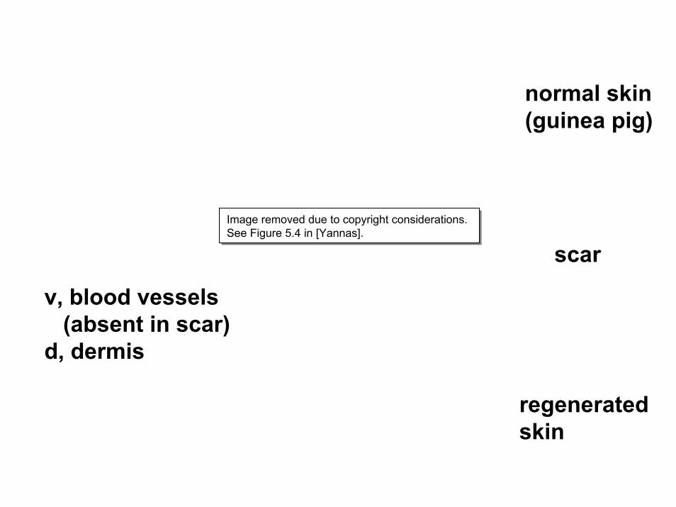
normal skin(guinea pig)
Image removed due to copyright considerations.See Figure 5.4 in [Yannas].Image removed due to copyright considerations.See Figure 5.4 in [Yannas].
scar
v, blood vessels(absent in scar)
d, dermis
regenerated skin

rete ridges withcapillary loopsand vascular plexusunderneath(normal skin)
Image removed due to copyright considerations.See Figure 5.2 (top left) in [Yannas].Image removed due to copyright considerations.See Figure 5.2 (top left) in [Yannas].

Verify basement membrane. I: Immunostaining: Factor VIII for capillary loops
Image removed due to copyright considerations.Image removed due to copyright considerations.
75 µm
Compton et al., 2000

Verify basement membrane. II. Immunostaining: α6β4 Integrin for hemidesmosomes
Image removed due to copyright considerations.Image removed due to copyright considerations.
100 µ m
Compton et al., 2000

Verify basement membrane. III.Immunostaining: Collagen VII for anchoring
fibrils
Image removed due to copyright considerations.Image removed due to copyright considerations.
150 µ m
Compton et al., 2000

Regenerated dermis
polarized light
Image removed due to copyright considerations.See Figure 5.3 in [Yannas].Image removed due to copyright considerations.See Figure 5.3 in [Yannas].
natural light

4. Partial synthesis of skin [Cont.]
In vitro-to-in vivo synthetic routes-- “Composite graft”. KC- and FB-seeded
DRT cultured in vitro and form epidermis, before grafting.
-- In another version, use synthetic polymeric mesh instead of DRT (“living dermal replacement”).
-- FB are cultured inside collagen gel, then KC are seeded, before grafting (“living skin equivalent”).

In vitro or in vivo? Skin synthesis
Image removed due to copyright considerations.See Figure 7.1 in [Yannas].Image removed due to copyright considerations.See Figure 7.1 in [Yannas].

5. Comparative regenerative ability of reactants
See Table 5.3.Growth factors had no effect on final
configuration (use defect closure rule).Pharmacological agents, including steroids,
had no effect.KC sheets were ineffective.Scaffolds, whether seeded or unseeded with
cells (KC and/or FB), were very effective in suppressing contraction and scar synthesis, and inducing regeneration.

Table 5.3Configuration of the final state (skin)
Image removed due to copyright considerations.See Table 5.3 in [Yannas].Image removed due to copyright considerations.See Table 5.3 in [Yannas].

Summary--- Each tissue in skin has been synthesized. --- Partial synthesis of skin has been also achieved.--- Reactants added included KC, FB and scaffolds.--- Epidermis and BM were synthesized in vitro (as
well as in vivo) whereas dermis with rete ridges was synthesized only in vivo.
Questions to be answered:Which are the minimal reactants?What is the difference in conditions between in vitro
and in vivo?