methodological note: neurofeedback: a comprehensive review
TRANSCRIPT

143
Basic and ClinicalApril 2016. Volume 7. Number 2
Hengameh Marzbani1, Hamid Reza Marateb1, Marjan Mansourian2*
Methodological Note: Neurofeedback: A Comprehen-sive Review on System Design, Methodology and Clini-cal Applications
A B S T R A C T
Key Words:Brain diseases, Brain waves, Complementary therapies, Electroencephalography, Neurofeedback
1. Introduction
eurofeedback is not a new concept. It has been the subject of the study of research-ers for several decades. Neurofeedback is a method that assists subjects to control their
brain waves consciously. In fact, the electroencephalog-raphy (EEG) is recorded during the neurofeedback treat-ment. Then, its various components are extracted and fed to subjects using online feedback loop in the form of au-dio, video or their combination. Accordingly, electrophysi-ological components are separately demonstrated. As an N
Article info: Received: 04 April 2015First Revision: 06 May 2015Accepted: 27 July 2015
1. Department of Biomedical Engineering, Faculty of Engineering, University of Isfahan, Isfahan, Iran. 2. Department of Biostatistics and Epidemiology, School of Health, Isfahan University of Medical Sciences, Isfahan, Iran.
* Corresponding Author:Marjan Mansourian, PhDAddress: Department of Biostatistics and Epidemiology, School of Health, Isfahan University of Medical Sciences, Isfahan, Iran.Tel:+98 (31) 37923256 E-mail: [email protected]
Neurofeedback is a kind of biofeedback, which teaches self-control of brain functions to subjects by measuring brain waves and providing a feedback signal. Neurofeedback usually provides the audio and or video feedback. Positive or negative feedback is produced for desirable or undesirable brain activities, respectively. In this review, we provided clinical and technical information about the following issues: (1) Various neurofeedback treatment protocols i.e. alpha, beta, alpha/theta, delta, gamma, and theta; (2) Different EEG electrode placements i.e. standard recording channels in the frontal, temporal, central, and occipital lobes; (3) Electrode montages (unipolar, bipolar); (4) Types of neurofeedback i.e. frequency, power, slow cortical potential, functional magnetic resonance imaging, and so on; (5) Clinical applications of neurofeedback i.e. treatment of attention deficit hyperactivity disorder, anxiety, depression, epilepsy, insomnia, drug addiction, schizophrenia, learning disabilities, dyslexia and dyscalculia, autistic spectrum disorders and so on as well as other applications such as pain management, and the improvement of musical and athletic performance; and (6) Neurofeedback softwares. To date, many studies have been conducted on the neurofeedback therapy and its effectiveness on the treatment of many diseases. Neurofeedback, like other treatments, has its own pros and cons. Although it is a non-invasive procedure, its validity has been questioned in terms of conclusive scientific evidence. For example, it is expensive, time-consuming and its benefits are not long-lasting. Also, it might take months to show the desired improvements. Nevertheless, neurofeedback is known as a complementary and alternative treatment of many brain dysfunctions. However, current research does not support conclusive results about its efficacy.
Citation: Marzbani, H., Marateb, H. R., & Mansourian, M. (2016). Neurofeedback: a comprehensive review on system design, methodology and clinical applications. Basic and Clinical Neuroscience, 7(2), 143-158. http://dx.doi.org/10.15412/J.BCN.03070208
: http://dx.doi.org/10.15412/J.BCN.03070208
CrossMark
Archive
of S
ID
www.SID.ir

144
illustration, the power of a signal in a frequency band can be shown by a varying bar graph. During this procedure, the subject becomes aware of the changes occurring during training and will be able to assess his/her progress in order to achieve optimum performance. For instance, the subject tries to improve the brain patterns based on the changes that occur in the sound or movie. Neurofeedback treatment protocols mainly focus on the alpha, beta, delta, theta, and gamma treatment or a combination of them such as alpha/theta ratio, beta/theta ratio, etc. (Dempster, 2012; Vernon, 2005). However, the most commonly used protocols are alpha, beta, theta, and alpha/theta ratio. In this review pa-per, we discussed various technical and clinical details of different neurofeedback treatment protocols.
2. Various Frequency Components
Activities of cerebral neurons have rich information about neuronal activities. When neurons are activated, they pro-duce electrical pulses. By placing electrodes on the scalp, the electrical activity of the brain, known as EEG, can be recorded. In turn, EEG is generated by a specific type of synchronous activity of neurons which are known as py-ramidal neurons and the electrical output is thus reflected in the following areas of the skin where the electrodes are located. Different patterns of electrical activity, known as brain waves, could be recognized by their amplitudes and frequencies. Frequency indicates how fast the waves oscil-late which is measured by the number of waves per second (Hz), while amplitude represents the power of these waves measured by microvolt (µV).
Different frequency components are categorized into delta (less than 4 Hz), theta (4-8 Hz), alpha (8-13 Hz), beta (13-30 Hz), and gamma (30-100 Hz) where each rep-resents a particular physiological function. In summary, delta waves are observed in the EEG signal when a per-son is asleep, theta waves when a person is sleepy, alpha waves when a person is relaxed and his/her muscles are loose but he/she is awake, beta waves when a person is alert and gamma waves are observed when a person is try-ing to solve a problem (Table 1). However, there are differ-ences in defining the exact range of frequency components in different studies.
These frequency components have subsets. For example, sensorimotor rhythm (SMR) frequency bands (13-15 Hz) are related to the sensorimotor rhythm and entitled as low beta. Some studies claimed that alpha rhythm has two sub-sets: lower alpha in the range of 8-10 Hz and upper alpha in the range of 10-12 Hz. Whereas some studies indicate that the alpha rhythm has 3 subsets. These definitions indi-cate that high and low alpha exhibit different behaviors and performances. It is believed that lower alpha is related to remembering action in semantic memory which is not the case for high alpha (Dempster, 2012).
3. EEG Electrode Placement
Electrodes (placed on the scalp) can record those corti-cal activities of the brain regions that are close to them. Electrode System 10-20 is a method for standardizing areas of the skull and comparing data. The term “10-20” refers to the placement of electrodes over 10% or 20% of
Table 1. Specific brainwaves with their characteristics.
Common brainwave frequency Frequency range (Hz) General characteristics
Delta 1-4 Sleep, repair, complex problem solving, unawareness, deep-unconsciousness
Theta 4-8 Creativity, insight, deep states, unconsciousness, optimal meditative state, depression, anxiety, distractibility
Alpha 8-13 Alertness and peacefulness, readiness, meditation, deeply-relaxed
Lower alpha 8-10 Recalling
Upper alpha 10-13 Optimize cognitive performance
SMR (sensorimotor rhythm) 13-15 Mental alertness, physical relaxation
Beta 15-20 Thinking, focusing, sustained attention, tension, alertness, excitement
High beta 20-32 Intensity, hyperalertness, anxiety
Gamma 32-100 or 40 Learning, cognitive processing, problem solving tasks, mental sharpness, brain activity, organize the brain
Mansourian, M., et al. (2016). Neurofeedback: system design, methodology & clinical applications. Basic and Clinical Neuroscience, 7(2), 143-158.
Archive
of S
ID
www.SID.ir

145
Basic and ClinicalApril 2016. Volume 7. Number 2
the total distance between specified skull locations. Studies have shown that these placements correlate with the cor-responding cerebral cortical regions. Of 21 electrodes, 19 are used for recording cortical areas and 2 other electrodes as reference electrodes (Figure 1). The skull regions are named using letters and numbers. Letters correspond with the brain regions and numbers to the hemisphere of the brain or the locations of this hemisphere. The letters F, P, T, O, and C are related to frontal, parietal, temporal, occipi-tal, and central areas, respectively. Odd/even numbers are associated with the left/right side of the brain region. The letter z is used as PZ suggests that scalp location falls along the central line running between the nasion and the inion. FP1 and FP2 are respectively related to the left and right poles of the forehead. Also A1 and A2 are the left right regions of vestibular (ear) region that are two common sites for the placement of reference and ground electrodes (Figure 1) (Dempster, 2012; Evans & Abarbanel, 1999).
Traditionally, two types of unipolar and bipolar montage are used in the neurofeedback treatment. In unipolar mode, the active electrode is placed on the skull and the recorded signal by the active electrode is compared to the second electrode entitled as the reference electrode. The activity of the active electrode minus the activity of the reference electrode represents the brain activity at the active elec-trode.
On the other hand, in the bipolar mode, two active elec-trodes are used that are separately placed on the skull. The difference between the recorded signals by these 2 elec-trodes, is the basis of the neurofeedback (Demos, 2005; Dempster, 2012). One of the advantages of the bipolar re-cording is the common mode rejection that occurs during the recording procedure. It means that any external artifact occurring at both channels and at the same time, its ampli-tude and phase are subtracted and the spatial selectivity is improved. For example, eye roll and blink artifacts could be reduced in this way (Evans & Abarbanel, 1999).
Neurologists have observed that lesions occurring in specific regions of the brain produce specific symptoms mostly related to these regions. For example, frontal lobes, FP1 , FP2 , FPZ , FZ , F3 , F4 , F7 are responsible for immedi-ate and sustained attention, time management, social skills, emotions, empathy, working memory, executive planning, moral fiber or character. Each region represents a specific feeling or task; Thus identification of these areas provides the best and the most accurate neurofeedback treatment. Parietal lobes, PZ , P3 and P4, solve problems conceptual-ized by the frontal lobes. Complex grammar, naming of the objects, sentence construction, and mathematical pro-cessing are identifiable to the left parietal lobe while map orientation, spatial recognition, and knowing the difference between right and left are entirely functions of the right parietal lobe. Temporal lobes, T3 , T4 , T5 and T6 have various functions. Left hemisphere functions are associated with reading (word recognition), memory, learning and a posi-tive mood, while right hemisphere functions are related to music, anxiety, facial recognition, and sense of direction.
On the other hand, visual memories, accurate reading and traumatic memories accompanying visual flashbacks are usually processed in the occipital lobes, O2 , O1 and . The other functions of this lobe include helping to locate objects in the environment, seeing colors and recognizing drawings and correctly identifying objects, reading, writ-ing, and spelling. Sensory and motor (sensorimotor) cor-tex, CZ , C3 and C4 have functions of conscious control of all skeletal movements such as typing, playing musical in-struments, handwriting, operation of complex machinery, speaking, and the ability to recognize where bodily sensa-tions originate.
Neurologists have mentioned that the motor cortex helps the cerebral cortex to encode both physical and cogni-tive tasks. Therefore, subjects who have trouble seeing the logical sequence of cognitive tasks may benefit from neurofeedback training along the left hemisphere senso-rimotor cortex (C3). Training along the right hemisphere sensorimotor cortex (C4) may invoke feelings, emotions, or calmness. Training at the median or may facilitate a mixed response. The subjects who suffer from epilepsy are usu-ally trained along the sensorimotor cortex (C3) to increase SMR. Also, training along the sensorimotor cortex could be applied for the treatment of stroke, epilepsy, paralysis, ADHD, and disorders of sensory/motor integration (Table 2) (Demos, 2005).
Generally, electrodes are placed in a way that a particular EEG channel is located on one brain side (Bauer & Pllana, 2014). For instance, low beta and beta are trained on the right (C4) and left (C3) brain side, respectively. If they were
Figure 1. The 10-20 electrode placement system and the name of the skull regions.
Mansourian, M., et al. (2016). Neurofeedback: system design, methodology & clinical applications. Basic and Clinical Neuroscience, 7(2), 143-158.
Archive
of S
ID
www.SID.ir

146
Table 2. Brain lobes with their functions and areas (Demos, 2005).
Sites Functions Considerations
Parietal lobes Pz , P3 , P4
LH: Problem solving, math, complexgrammar, attention,
associationRH: Spatial awareness,
Geometry
Dyscalculia sense of direction learning disorders
Frontal lobes FP1, FP2 , FPZ , FZ , F3 , F4 , F7 , F8
LH: Working memory, concentration, Executive planning, positive emotions.
RH: Episodic memory,social awareness
Frontal poles: attention judgment
LH: DepressionRH: Anxiety, fear, executive planning, poor
executive functioning
Temporal lobes T3 , T4 , T5 , T6
LH: Word recognition, reading, language, memory
RH: Object recognition, music, social cues
Facial recognition
Anger, rage, dyslexia, long-term memory, closed head injury
Occipital lobes OZ , O1 , O2
Visual learning,reading, parietal- temporal-occipital
functionsLearning disorders
Sensorimotor cortex CZ , C3 , C4
LH: Attention, mental processing,RH: Calmness, emotion,
EmpathyCombined: Fine motor
skills, manualdexterity, sensory
and motor integrationand processing
Paralysis (stroke), seizure disorder, poor handwriting, ADHD symptoms
Cingulategyrus FPZ , FZ , CZ , PZ , OZ
Mental flexibility, cooperation, attention, motivation,
morals
Obsessions, compulsions, tics, perfection-ism, worry, ADHD symptoms, OCD
& OCD spectrum
Broca’s area F7 , T3 Verbal expression Dyslexia, poor spelling, poor reading
Left hemisphere All odd numbered sites
Logical sequencing, detail oriented, language abilities, word
retrieval,fluency, reading,math, science,
problem solving,verbal memory
Depression(underactivation)
Right hemisphere All even numbered sites
Episodic memory encoding, social awareness, eye
contact, music,humor, empathy,
spatial awareness,art, insight, intuition,non-verbal memory,
seeing the whole picture
Anxiety(overactivation)
Abbreviations: LH, Left hemisphere, RH: Right hemisphere, AHHD: Attention deficit hyperactivity disorder, OCD: Obsessive compulsive disorder.
Mansourian, M., et al. (2016). Neurofeedback: system design, methodology & clinical applications. Basic and Clinical Neuroscience, 7(2), 143-158.
Archive
of S
ID
www.SID.ir
147
Basic and ClinicalApril 2016. Volume 7. Number 2
switched to the opposite brain side, undesirable results could be obtained. For example, training low beta wave on the left side will result in a depletion of mental energy in-stead of improvements in concentration. Thus, the location of the EEG electrodes during the neurofeedback procedure is important (Evans, 2007).
4. Types of Neurofeedback
There are 7 types of Neurofeedback for the treatment of various disorders:
1) The most frequently used neurofeedback is frequency/power neurofeedback. This technique typically includes the use of 2 to 4 surface electrodes, sometimes called “sur-face neurofeedback”. It is used to change the amplitude or speed of specific brain waves in particular brain locations to treat ADHD, anxiety, and insomnia.
2) Slow cortical potential neurofeedback (SCP-NF) im-proves the direction of slow cortical potentials to treat ADHD, epilepsy, and migraines (Christiansen, Reh, Schmidt, & Rief, 2014).
3) Low-energy neurofeedback system (LENS) delivers a weak electromagnetic signal to change the patient’s brain waves while they are motionless with their eyes closed (Zandi-Mehran, Firoozabadi, & Rostami, 2014). This type of neurofeedback has been used to treat traumatic brain injury, ADHD, insomnia, fibromyalgia, restless legs syndrome, anxiety, depression, and anger.
4) Hemoencephalographic (HEG) neurofeedback pro-vides feedback on cerebral blood flow to treat migraine (Dias, Van Deusen, Oda, & Bonfim, 2012).
5) Live Z-score neurofeedback is used to treat insomnia. It introduces the continuous comparison of variables of brain electrical activity to a systematic database to provide continuous feedback (Collura, Guan, Tarrant, Bailey, & Starr, 2010).
6) Low-resolution electromagnetic tomography (LORE-TA) involves the use of 19 electrodes to monitor phase, power, and coherence (Pascual-Marqui, Michel, & Lehm-ann, 1994). This neurofeedback technique is used to treat addictions, depression, and obsessive-compulsive disorder.
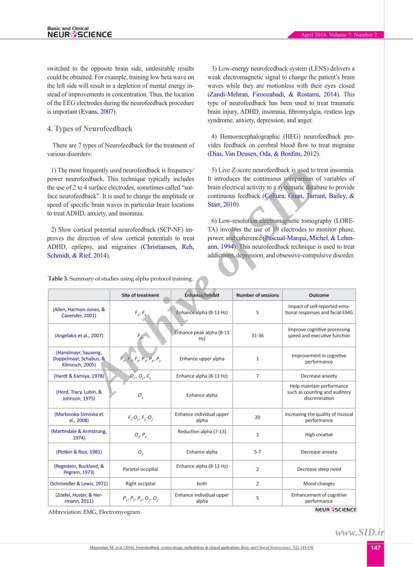
Table 3. Summary of studies using alpha protocol training.
Site of treatment Enhance/inhibit Number of sessions Outcome
(Allen, Harmon-Jones, & Cavender, 2001) F3 , F4 Enhance alpha (8-13 Hz) 5
Impact of self-reported emo-tional responses and facial EMG
(Angelakis et al., 2007) FO3Enhance peak alpha (8-13
Hz) 31-36Improve cognitive processing speed and executive function
(Hanslmayr, Sauseng, Doppelmayr, Schabus, &
Klimesch, 2005)F3 , F4 , FZ , P3 , P4 , PZ Enhance upper alpha 1 Improvement in cognitive
performance
(Hardt & Kamiya, 1978) OZ , O1 , C3 Enhance alpha (8-13 Hz) 7 Decrease anxiety
(Hord, Tracy, Lubin, & Johnson, 1975) O2 Enhance alpha
Help maintain performance such as counting and auditory
discrimination
(Markovska-Simoska et al., 2008) F3-O1 , F4 -O2
Enhance individual upper alpha 20 Increasing the quality of musical
performance
(Martindale & Armstrong, 1974) O2, P4
Reduction alpha (7-13) 1 High creative
(Plotkin & Rice, 1981) OZ Enhance alpha 5-7 Decrease anxiety
(Regestein, Buckland, & Pegram, 1973) Parietal-occipital Enhance alpha (8-13 Hz) 2 Decrease sleep need
(Schmeidler & Lewis, 1971) Right occipital both 2 Mood changes
(Zoefel, Huster, & Her-rmann, 2011) P3 , PZ , P4 , O1 , O2
Enhance individual upper alpha 5 Enhancement of cognitive
performance
Abbreviation: EMG, Electromyogram.
Mansourian, M., et al. (2016). Neurofeedback: system design, methodology & clinical applications. Basic and Clinical Neuroscience, 7(2), 143-158.
Archive
of S
ID
www.SID.ir

148
7) Functional magnetic resonance imaging (fMRI) is the most recent type of neurofeedback to regulate brain activ-ity based on the activity feedback from deep subcortical areas of the brain (Hurt, Arnold, & Lofthouse, 2014; Lévesque, Beauregard, & Mensour, 2006a).
5. Various Treatment Protocols
5.1. Alpha protocol
The alpha wave of the brain is usually associated with alert relaxation (Evans & Abarbanel, 1999). The alpha mood is described as a calm and pleasant situation. All alpha frequencies describe creative activity of the brain, so that it is used in the process of relaxation (relaxing the muscles), which eventually leads to sleep; Such waves emerge and expand rapidly on the skin. The evidence shows that alpha waves increases during meditation.
Alpha training is usually used for the treatment of vari-ous diseases such as pain relief (by 9 Hz simulation), re-ducing stress and anxiety (by 10 and 30 Hz simulation), memory improvement, improving mental performance, and treatment of brain injuries (by 10.2 Hz simulation). Various studies have been performed on the alpha pro-tocol (Table 3). The most common frequency bandwidth
for the alpha treatment is 7-10 Hz frequency range, which is used for meditation, sleep, reducing stress and anxiety. Also frequency of 10 Hz causes deep muscle relaxation, pain reduction, regulating breathing rate, and decreasing heart rate (Dempster, 2012; Vernon, 2005).
5.2 Beta protocol
Beta activity is a good indicator for mental performance and inappropriate beta activity represents mental and physical disorders like depression, ADHD, and insomnia (Egner & Gruzelier, 2004). Beta brain waves are associ-ated with conscious precision, strong focus, and ability to solve problems. Medications that are used to stimulate alertness and concentration such as Ritalin and Adderall also cause the brain to produce beta brainwaves.
Beta training is used to improve focus and attention (simulation of increased beta 12-14 Hz), improve the reading ability (simulation of 7-9 Hz), and introduce positive changes in school performance. It also improves the computational performance, cognitive processing, reduction of worries, over-thinking, obsessive compul-sive disorder (OCD), alcoholism, and insomnia (simula-tion of 14-22 Hz and 12-15 Hz). Meanwhile, this type of neurofeedback improves sleep cognitive performance as
Table 4. Summary of studies using beta protocol training.
Site of treatment Enhance/inhibit Number of sessions Outcome
(Rasey, Lubar, McIntyre, Zoffuto, & Abbott, 1995)
Central-posterior region (CPZ , PCZ )
Enhance beta (16-22 Hz) and inhibit high theta and low alpha 20 Improvement in attentional
performance
(Egner & Gruzelier, 2001)
(12-15 Hz) at right central region (C4) and (15-18 Hz) at the left central region
(C3)
Enhance low beta (12-15 and 15-18 Hz), inhibiting theta (4-7 Hz)
and high beta (22-30 Hz)10 Successful enhancement of
attentional performance
(Vernon et al., 2003) CZ
Enhance low beta (12-15 Hz), inhibiting theta (4-8 Hz) and high
beta (18-23 Hz)15 Enhance cognitive perfor-
mance
(Egner & Gruzelier, 2001) CZ
Enhance SMR (12-15 Hz) and inhibit theta (4-7 Hz) and high
beta (22-30 Hz)10 Improve perceptual
sensitivity
(Egner & Gruzelier, 2001) CZ
Enhance low beta (15-18 Hz), inhibiting theta (4-7 Hz) and high
beta (22-30 Hz )10 Increase cortical arousal
(Vernon et al., 2003) CZ
Enhance SMR (12-15 Hz) and inhibit theta (4-7 Hz) and high
beta (18-22 Hz)8 Increased recall in seman-
tic working memory
(Lubar, Swartwood, Swart-wood, & O’Donnell, 1995) FCZ , CPZ
Enhance beta (16-20 Hz) and inhibit theta 40
Reduction of inatten-tion, hyperactivity and
impulsivity(Fuchs, Birbaumer, Lutzen-berger, Gruzelier, & Kaiser,
2003)C3 , C4
Enhance beta (15-18 Hz) and SMR (12-15), inhibit theta 36 Improvement in attention
and intelligence
(Heinrich, Gevensleben, & Strehl, 2007) C4, CZ Enhance SMR and inhibit theta Treatment epilepsy disor-
der and ADHD(Heinrich, Gevensleben, &
Strehl, 2007) CZ , C3Enhance beta (13-20 Hz) and
inhibit theta Treatment ADHD
Abbreviation: SMR, Sensorimotor rhythm.
Mansourian, M., et al. (2016). Neurofeedback: system design, methodology & clinical applications. Basic and Clinical Neuroscience, 7(2), 143-158.
Archive
of S
ID
www.SID.ir

149
Basic and ClinicalApril 2016. Volume 7. Number 2
well as reducing fatigue and stress (simulation of light and sound of beta) (Table 4). The beta waves in the range of 12-15 Hz (SMR) reduce anxiety, epilepsy, anger and stress (Egner & Gruzelier, 2004; Vernon, 2005).
5.3. Alpha/theta protocol
Alpha/theta is an indicator between awareness and sleep. Alpha/theta training is one of the most popular neurofeed-back trainings for stress reduction (Gruzelier, 2009; Ray-mond, Varney, Parkinson, & Gruzelier, 2005). Also, this treatment is used for deep levels of depression, addiction, anxiety while it increases creativity, relaxation, musical performance, and promotes healing from trauma reactions. The electrodes are usually located on O1 , O2 , CZ and PZ . Alpha/theta frequency range is 7-8.5 Hz with the typical value of 7.8 Hz. This treatment is done under eyes-closed condition that increases the ratio of theta to alpha waves using auditory feedback (Demos, 2005; Egner & Gruze-lier, 2003; Thompson & Thompson, 2003). The summary of the studies using alpha/theta protocol training are pre-sented in Table 5.
5.4. Delta protocol
Delta waves are the slowest brain waves, which are as-sociated with stages 3 and 4 of the sleep (Sürmeli & Er-tem, 2007). They represent increased comfort, reduced pain, and sleep. Thus, they are used to alleviate headaches, traumatic brain injury, learning disorders, and to treatment hard and sharp contraction of muscles (by simulation of 1-3 Hz delta wave). They also reduce concerns and im-prove sleep (Vernon, 2005).
5.5. Gamma protocol
Gamma waves have the highest frequency, and they are associated with cognitive processing and memory (Staufenbiel, Brouwer, Keizer, & Van Wouwe, 2014). Thus, when these waves are faster, the speed of recalling
memory is faster. Gamma waves are fast rhythms that are responsible for the brain’s neural connections and data transfer to the outside world.
They are mainly observed in the hippocampus (an area of the brain which is responsible for converting short-term to long-term memory). Also, these rapid rhythms are ob-served in sudden attacks like seizure and spasm. Hence, gamma training is used for promoting cognition, mental sharpness, brain activity, and problem-solving tasks. It not only improves poor calculation, but also organizes the brain, improves the speed of information processing, short-term memory, and reduces the number of migraine attacks (Hughes, Vernon, 2005).
5.6. Theta protocol
Theta brain waves are related to a number of brain activi-ties such as memory, emotion, creativity, sleep, meditation, and hypnosis. These waves are also associated with the first phase of sleep when the sleep is light and the person easily wakes up. Theta treatment reduces anxiety, depres-sion, day dreaming, distractibility, emotional disorders, and ADHD (Beatty, Greenberg, Deibler, & O’Hanlon, 1974; Vernon, 2005).
5.7. Low frequency versus high frequency training
Basically, there are two classical directions in neurofeed-back training. It is either focusing on low frequencies (al-pha or theta) to strengthen relaxation and focus (Gruzelier, 2009) or emphasizing on high frequencies (low beta, beta, and theta) for reinforcing activation, organizing, and inhib-iting distractibility (Ros et al., 2009).
A suitable comparison between these two directions could be found at Thomas F. Collura (2000), and Kro-potov (2010) studies. For example, in the former strategy eyes are closed while in the later one, eyes are open. Also,
Table 5. Summary of studies using alpha/theta protocol training.
Site of treatment Enhance/inhibit Number of sessions Outcome
(Raymond, Sajid, Parkin-son, & Gruzelier, 2005) P4
Enhance theta (4-7 Hz) over alpha (8-11 Hz) 10 Improvement in artistic
performance
(Egner & Gruzelier, 2003) C4 , C3 , PZEnhance theta (5-8 Hz) over
alpha (8-11 Hz) 10 Improvement of music performance
(Gruzelier, 2009) Enhance theta (4-7 Hz) over alpha ( 8-11 Hz)
Half-hour sessions, twice a week
Enhancement of artistic performance and mood
(Gruzelier, 2009) Enhance theta (4-7 Hz) over alpha ( 8-11 Hz) 10 Enhancement of music
performance
Mansourian, M., et al. (2016). Neurofeedback: system design, methodology & clinical applications. Basic and Clinical Neuroscience, 7(2), 143-158.
Archive
of S
ID
www.SID.ir

150
children are not involved in the first strategy while children and adult could undergo the second training procedure.
6. Clinical Applications of Neurofeedback Training in the Treatment of Diseases and Disorders
Antisocial behavior of individuals, have an undesirable impact on the society. In recent years, with advances in brain science, the cause of abnormal brain function and mental illness has been attributed to the low activity of the anterior brain lobe that presents itself in different types of psychological damages (Gil, Li, & Lee, 2009). The neuro-feedback training has been widely used in the treatment of many diseases and disorders; some of which are mentioned below.
6.1. Attention deficit/hyperactivity disorder
Evidence suggests that the malfunction of the right fron-tal lobe, is the cause of attention deficit/hyperactivity disor-der (ADHD) (Hynd et al., 1991). The resulting symptoms are inattention, distractibility, hyperactivity, and extreme dispassionateness. Neurofeedback therapy is a rehabilita-tion approach for its treatment. Its goal is to normalize the behavior without dependence on medications or behav-ioral therapy. For a long time, such drugs as Ritalin, Con-certa, and Dexedrine have been used for treating ADHD. But, recent research showed that these drugs do not have any effect on the clinical treatment of ADHD on some of
children. Also, these drugs have the side effects such as anxiety, irritability, abdominal pain, decreased appetite, in-somnia, and headache. However, using neurofeedback is associated with their long-term improvement (Yan et al., 2008). Studies showed that people with ADHD disorder have slower brain wave activity (theta) and less beta activ-ity compared to normal people.
In ADHD, the goal is to decrease the brain activity in the theta band and to increase its activity in the beta band (or to decrease theta/beta ratio) at the vertex (electrode) (Heinrich, Gevensleben, & Strehl, 2007). This treatment is effective in reducing hyperactivity; Increasing focus, grades, and parental consent from children’s behavior; and improving indicators of sustained attention (Gnecchi, Her-rera Garcia, & de Dios Ortiz Alvarado, 2007; Karimi, Haghshenas, & Rostami, 2011; Wang & Sourina, 2013). The studies on the neurofeedback treatment of ADHD in children are listed in Table 6. According to this Table, theta/beta protocol and the area for locating the EEG elec-trode are the most commonly used neurofeedback strategy in ADHD treatment.
6.1.1. Schizophrenia
Schizophrenia is known as the most unbearable mental illness (Surmeli, Ertem, Eralp, & Kos, 2012). People with schizophrenia have the illusion of auditory disorders, rest-lessness, non-flexible muscles, confusion, delirium, and depression. Based on several papers on the treatment of schizophrenia, Minnesota Multiphasic Personality Inven-
Table 6. Summary of neurofeedback treatment studies on ADHD.
Site of treat-ment
NeurofeedbackProtocol
Number of sessions
The age range (year) Outcome
(Linden, Habib, & Rado-jevic, 1996) CZ
Enhance beta Inhibit theta 20 5-15
Improvement in mental functions
and accuracy
(Palsson et al., 2001) CZ Theta/beta, SMR 40 9-13 Improvement in effects of ADHD
(Orlandi, 2004) CZ Theta/beta, SMR 40 9-11 Improvement in atten-tion, focus and memory
(Lévesque, Beauregard, & Mensour, 2006b) CZ Theta/beta, SMR 40 8-12
Improving performance of anterior cingulate
cortex
(Leins et al., 2007) CZ Theta/beta 30 8-13Improvement in atten-tion, hyperactivity and
distraction
(Gevensleben et al., 2009) CZ Theta/beta 18 9-12Improvement in com-
bined treatment of neu-rofeedback protocols
(Perreau-Linck, Lessard, Lévesque, & Beauregard,
2010)CZ Theta/SMR 40 8-13 Improvement in the ef-
fects of ADHD
Abbreviations: ADHA: Attention deficit hyperactivity disorder, SMR: Sensorimotor rhythm.
Mansourian, M., et al. (2016). Neurofeedback: system design, methodology & clinical applications. Basic and Clinical Neuroscience, 7(2), 143-158.
Archive
of S
ID
www.SID.ir

151
Basic and ClinicalApril 2016. Volume 7. Number 2
tory (MMPI) and Test of Variables of Attention (TOVA), positive effect of neurofeedback training on the treatment of this disease is expressed in such a way that the person with schizophernia is able to adjust his/her brain activity on specific frequencies (McCarthy-Jones, 2012; Surmeli, Ertem, Eralp, & Kos, 2012; Wenya et al., 2012; Gil, Li, & Lee, 2009).
6.1.2. Insomnia
Insomnia is known as an epidemic disorder. The first change observed in patients, who are treated with neuro-feedback training is the change and improvement in their sleep pattern. Hence, the neurofeedback training is used in the treatment of sleep disorders (Hammer, Colbert, Brown, & Ilioi, 2011). For example, the following pro-cess is used to improve sleep. One electrode is placed on and the treatment is done for 30 minutes at a frequency of 15-18 Hz. This method makes the waking state, alert and active and assist people in waking up faster. The calmness treatment is done at frequencies of 12-15 Hz and in loca-tion. Using neurofeedback helps the people who normally take about an hour in order to prepare their body and mind for sleep, go to sleep faster.
6.1.3. Learning disabilities, dyslexia and dyscalculia
Neurofeedback has created a big change in the treatment of these disorders. These disorders are more common at school age and patients with dyslexia have trouble in read-ing and spelling the characters (Breteler, Arns, Peters, Giepmans, & Verhoeven, 2010). People having dyscal-culia, are unable to understand and solve math problems. These disorders are treated with increased alpha wave ac-tivity using neurofeedback (Wang & Sourina, 2013).
6.1.4. Drug addiction
Studies have shown that neurofeedback training is a good way to quit drug addiction whereas long-term use of the drug has a profound effect on the individual’s EEG. Temp-tation and craving of drugs could be reduced by neurofeed-back in patients addicted to cocaine (Horrell et al., 2010). This treatment can also be used to treat alcoholism and ad-diction to computer games (Moradi et al., 2011).
6.1.5. Enhancing the performance of athletes, artists, and surgeons
Studies have shown that professional athletes have dif-ferent patterns of brain activity compared to those of the beginners. Recognition of the status of the professional’s EEG before and during performance, provides a rationale
for the use of neurofeedback training to create or emulate these patterns and to improve the performance of unprofes-sional individuals (Vernon, 2005). In fact the purpose of neurofeedback on athletes is improving the athlete’s psy-chomotor and self-regulation ability, their confidence, and subsequent performance in important competitions of the year (Edmonds & Tenenbaum, 2011).
6.1.6. Autistic spectrum disorder
Autistic spectrum disorder (ASD) is a neurodevelopmen-tal disorder with challenges that maintain in adulthood. Children with autism have difficulty in functions such as social interaction, verbal and nonverbal communica-tion, behavior and interests. ASD may be associated with emotional problems, mental retardation, or seizure disor-ders. These children may also have extreme sensitivity to sounds and smells. Also, children with autism may show idiosyncratic behaviors, obsessive rumination, poor social interrelatedness, and flat affect. Researchers found out that individuals with autism differ from normative samples with regard to impediments in empathy or theory of mind (TOM) tasks, weak central coherence, and executive func-tioning.
One of the primary symptoms of ASD is a qualitative im-pairment in social interactions related to mutual interest, understanding others’ intentions, empathy, emotional reci-procity, and the underlying concepts of TOM. Empathizing deficits are consistent with problems in reciprocating com-munication, difficulty in predicting thoughts and feelings of others, interpreting abstract emotions of others, and an appearance of social insensitivity. Individuals with autism are also often seen to have interest in system details and pursue careers in engineering, construction, clocks, ma-chines, puzzles, or computers, which are often obsessive interests in ASD (Lucido, 2012).
There are several diagnostic tools designed to show ab-normalities in brain’s function for autism. They are (1) High-beta activity related to anxiety; (2) The high activity of delta/theta corresponding with the slow cortex, lack of attention, impulsivity and hyperactivity; and (3) Abnormal EEG/seizure activity. High beta type is the most common one seen among children with ASD (approximately 50-60% of individuals with ASD) (Coben, Linden, & Myers, 2010; Kouijzer, van Schie, de Moor, Gerrits, & Buite-laar, 2010). The goal of neurofeedback in children with autism is to inhibit theta-alpha ratio while enhancing beta wave. Efficacy of neurofeedback in children diagnosed with autism has been well researched in qualitative case studies summarized in Table 7.
Mansourian, M., et al. (2016). Neurofeedback: system design, methodology & clinical applications. Basic and Clinical Neuroscience, 7(2), 143-158.
Archive
of S
ID
www.SID.ir

152
6.1.8. Epilepsy
In about one-third of patients with epilepsy, medical treat-ment is ineffective. Neurofeedback training was shown to be a good alternative treatment for these patients. Research
has been focused on increasing SMR (12-15 Hz) and syn-chronous or asynchronous reduction of slow rhythms (4-7 Hz) for diagnosing this disorder. Also, observing low-amplitude gamma wave after surgery is a good sign for
Table 7. Summary of neurofeedback treatment studies on autistic spectrum disorder (ASD).
Site of treatment Enhance/inhibit Number of sessions Outcome
(Cowan & Markham, 1994) Parietal and occipital lobes
Enhance (16-20 HZ)Inhibit ( 4-10 HZ) 21 Improvement in focus, atten-
tion, and relax
(Thompson & Thompson, 2003)
Sensorimotor cortex (C2, C4)
Enhance (13-15 Hz)Inhibit (3-10 Hz) 40-100
Improvement in neuro-psychological functioning,
improved educational perfor-mance, decrease anxiety and
impulsivity
(Sichel, Fehmi, & Goldstein, 1995)
Sensorimotor strip and parietal lobe
Enhance SMR (12-15 Hz)Inhibit theta (4-8 Hz) 31
Improvement in sleep, social behaviors
Increase in appropriate eye contact
Reduction in self-simulation
(Othmer, 2007) P4 , T4 , T3 , F2 , FP1 Enhance SMR (12-15 Hz) 28-100Decreased need for special
education services and autism symptoms
(Thompson, Thompson, & Reid, 2010) Central sites
Enhance SMR (12-15 or 13-15 Hz)
Inhibit theta (3-7 Hz) and beta (23-35 Hz)
40-60Improvement in intelligence
testing and psychological assessments
(Cowan & Markham, 1994)Enhance beta (16-20 Hz)Inhibit theta-alpha (4-10
Hz)
Improvement in autistic behaviors, social, academic functioning and attention
Abbreviation: SMR: Sensorimotor rhythm.
Table 8. Summary of neurofeedback treatment studies on epilepsy that the results was the remission.
Neurofeedbackprotocol Measuring results Length of treatment The age range (year)
(Sterman, Macdonald, & Stone, 1974) SMR (11-15 Hz) Seizure frequency,
EEG 6-18 months 6-46
(Kaplan, 1975) SMR The number of seizures per day 20-25 weeks 20-30
(Lubar & Bahler, 1976) SMR The number of seizures 80-260 days 12-29
(Kuhlman & Allison, 1977) SMR (4-9 Hz) The number of seizures, EEG 24 sessions 17-42
(Sterman & Macdonald, 1978) SMR
The number of seizures per month,
EEG12 months 10-40
(Cott, Pavloski, & Black, 1979)
SMR The number of seizures per month 210 days 16-31
(Quy, Hutt, & Forrest, 1979) SMR The number of seizures
per week, EEG 12 months 23-49
(Lubar et al., 1981) SMR Seizure frequency,EEG 10 months 13-52
(Tozzo, Elfner, & May, 1988) SMR The number of seizures 5 weeks 18-29
Abbreviation: EEG, Electroencephalogram, SMR, Sensorimotor rhythm.
Mansourian, M., et al. (2016). Neurofeedback: system design, methodology & clinical applications. Basic and Clinical Neuroscience, 7(2), 143-158.
Archive
of S
ID
www.SID.ir
153
Basic and ClinicalApril 2016. Volume 7. Number 2
the improvement of epilepsy. The results of studies on the treatment of epilepsy by neurofeedback indicated that continuous SMR treatment reduces the rate of seizures in severe and uncontrolled epilepsy (Table 8) (Hughes et al., 2009; Walker, 2010).
6.1.9. Depression
Depression is associated with hypometabolism in the cin-gulate and occasionally in the frontal cortex, insula, ante-rior temporal cortices, amygdala, basal ganglia, and thala-mus. Along with the frontal electrophysiology findings in depression, there seems to be an inverse relationship be-tween frontal alpha asymmetry and parietal asymmetries. More specifically, depressed patients who do not have sig-nificant anxiety, appear to have decreased right parietal ac-tivation (alpha wave at P4). Neurofeedback training is used to increase alpha and theta, while inhibit faster beta fre-quencies, produces significant improvements in depression (Budzynski, 2009a; Hurt, Arnold, & Lofthouse, 2014).
6.1.10. Anxiety
In clinical medicine, anxiety is often defined, at least in part, as high level of muscle tension. Researchers found out that decreasing frontal electromyogram (EMG) levels by EMG biofeedback could alleviate both generalized and specific anxiety patterns. It was believed that anxiety inhib-its alpha waves, so alpha training would relieve the anxiety (Budzynski, 2009a; Demos, 2005; Moore, 2000).
6.1.11. Pain management
Pain is considered a symptom associated with physical damage, purportedly having an objective element connect-ed with the sensation. Neurofeedback methodology pro-poses that by teaching self-regulation, a patient can reduce or even eliminate pain sensations. Studies suggested that brain changes its functional organization at the level of the somatosensory cortex in chronic pain patients. Research-ers recommend the use of biofeedback/neurofeedback for pain management. Biofeedback protocols are designed to address the peripheral correlation of arousal, such as tem-perature, heart rate variability, and muscle tension while neurofeedback directly affects the processing of pain per-ception (Ibric & Dragomirescu, 2009).
6.2. Other uses of neurofeedback
Other applications of neurofeedback include the recov-ery from an injury and stroke problems, improvement of memory by increasing alpha activity (Escolano, Aguilar, & Minguez, 2011; Klimesch, 1999; Vernon, 2005; We-
nya et al., 2012), treatment of headache and migraines (Walker, 2011), distraction, confusion, attention problems, withdrawal (Escolano, Aguilar, & Minguez, 2011; Gnec-chi, Herrera Garcia, & de Dios Ortiz Alvarado, 2007), health promotion (Escolano, Olivan, Lopez-del-Hoyo, Garcia-Campayo, & Minguez, 2012), treatment of men-tal illness (Heinrich, Gevensleben, & Strehl, 2007), eat-ing disorders (Bartholdy, Musiat, Campbell, & Schmidt, 2013) Parkinson disease (Rossi-Izquierdo et al., 2013), fi-bromyalgia, restless legs syndrome (Hurt, Arnold, & Loft-house, 2014), obsessive compulsive disorder (Sürmeli & Ertem, 2011), and obsession (Markovska-Simoska, Pop-Jordanova, & Georgiev, 2008; Surmeli & Ertem, 2011). Meanwhile, artists and surgeons use neurofeedback to im-prove their music performance (Markovska-Simoska et al., 2008) and microsurgical operations (Ros et al., 2009), respectively.
Alpha-EEG/EMG biofeedback is capable of increasing voluntary self-regulation and the quality of musical perfor-mance (Budzynski, 2009b; Markovska-Simoska et al., 2008).
7. Neurofeedback Softwares
Brain-computer interface systems (BCI) are widely used in clinical and research applications. BCI can propose a new aim for playing videogames or interacting with 3D virtual environments (VE). Interaction with VE includes tasks such as navigating to modify the selection and ma-nipulation of virtual objects.
There are several examples of VE feedback games used in sports, puzzles, or trainings. Nowadays, many universi-ties and laboratories are trying to provide more interactions with the virtual world through the BCI. Here, we describe some of the BCI VE feedback software.
Researchers at University College Dublin and Media Lab Europe manufactured Mind Balance videogame that uses BCI to interact with the virtual world. The game was designed to move an animated character in a 3D virtual environment. The purpose is to control the balance of an animated character on a thin rope, based on the EEG sig-nals of a player.
In the other computer game, designed jointly by the University College London and Graz University of Tech-nology, a disabled person in a virtual street controls the movements of the simulated wheelchair (GRAZ-BC). These results indicated that a disabled person sitting in a wheelchair can control his/her movement in the VE using asynchronous BCI based on signal EEG.
Mansourian, M., et al. (2016). Neurofeedback: system design, methodology & clinical applications. Basic and Clinical Neuroscience, 7(2), 143-158.
Archive
of S
ID
www.SID.ir

154
University of Tokyo performed several tests using a “vir-tual joystick” to navigate 3-D VE. Researchers provided two virtual buttons on the left and right sides of the VE. The participants were asked to gaze at either side to move the camera to the other side. The detection enabled the sys-tem to identify the button at which the user gazed.
Researchers at the University of Tokyo also worked on a system to keep the alertness level of car drivers. In this project, the driver’s state of concentration was illustrated when placed in a virtual driving environment. Accordingly, the BCI hearing system actively monitors the state of alert-ness of drivers and warns them when loss of consciousness occurs.
In the field of promotion of neurofeedback in VE, IN-RIA designed several BCI systems. In one of them, called “use-the-force”, subjects were asked to control the launch of a virtual spaceship by using real or imagined foot movements. They studied the response of the subjects in challenging situations (Lecuyer et al., 2008). In another system (Gnecchi, Herrera Garcia, & de Dios Ortiz Al-varado, 2007), neurofeedback was examined in order to diagnose ADHD and hyperactivity disorder. In this system, there are two graphical interfaces.
In the first interface, when the ratio of beta/theta goes higher than a predetermined threshold, dolphins are mov-ing to an area where there are fish. Having maintained the focus, dolphin intercepts a fish. When the number of trapped fish increases, it reflects advances in process of treatment. In the second graphical interface, the speed of a racing car increases when subject’s attention improved. There are various available neurofeedback softwares in the market whose information such as operating systems, developers, and supported devices could be assessed via Wikipedia (“Comparison of neurofeedback software”, April 11, 2015).
8. Conclusion
In this paper, we reviewed the clinical applications of neurofeedback, various protocols of treatment and some of the systems designs by BCI and VR technology.
In neurofeedback, EEG is usually recorded, and various brain-activity components are extracted and feedbacked to subjects. During this procedure, subjects become aware of the changes that occur during training and are able to assess their progress in order to achieve optimal perfor-mance. Electrode placement is performed according to specific brain functions and specific symptoms. Consider-ing information about these skull regions, the entire treat-
ment process is simplified. There are several protocols in neurofeedback training, but alpha, beta, theta, and alpha/theta protocol are the most commonly used ones.
BCI is an EEG-based communication device. VE is a human-computer interface system with which users can virtually move their viewpoint freely in real time. The purpose of using VE is to construct a virtual environment with natural interactivity and to create a real sensation from multimodality. Three-dimensional VR is much more attractive and interesting than most of two-dimensional en-vironments.
To date, many studies have been conducted on the neuro-feedback therapy and its effectiveness on the treatment of many diseases. However, there are some methodological limitations and clinical ambiguities. For example, consid-ering the alpha treatment protocols, there are some issues to deal with such as how many sessions are needed before participants can learn to exert an alert control over their own alpha waves, or how many sessions are needed before such training procedures produce the expected effect on the optimal performance, and how long the desired effects last without feedback (long-term effects). Thus, it is necessary to provide standard protocols to perform neurofeedback.
Similar to other treatments, neurofeedback has its own pros and cons. Although it is a safe and non-invasive pro-cedure that showed improvement in the treatment of many problems and disorders such as ADHD, anxiety, depres-sion, epilepsy, ASD, insomnia, drug addiction, schizophre-nia, learning disabilities, dyslexia and dyscalculia, its va-lidity has been questioned in terms of conclusive scientific evidence of its effectiveness. Moreover, it is an expensive procedure which is not covered by many insurance com-panies. It is also time-consuming and its benefits are not long-lasting. Finally, it might take several months to see the desired improvements (Mauro & Cermak, 2006).
Conflicts of Interest:
None declared.
References
Allen, J. J., Harmon-Jones, E., & Cavender, J. H. (2001). Manipu-lation of frontal EEG asymmetry through biofeedback alters self-reported emotional responses and facial EMG. Psycho-physiology, 38(4), 685-693.
Angelakis, E., Stathopoulou, S., Frymiare, J. L., Green, D. L., Lubar, J. F., & Kounios, J. (2007). EEG neurofeedback: a brief
Mansourian, M., et al. (2016). Neurofeedback: system design, methodology & clinical applications. Basic and Clinical Neuroscience, 7(2), 143-158.
Archive
of S
ID
www.SID.ir

155
Basic and ClinicalApril 2016. Volume 7. Number 2
overview and an example of peak alpha frequency training for cognitive enhancement in the elderly. Clinical Neuropsy-chologist, 21(1), 110-129.
Bartholdy, S., Musiat, P., Campbell, I. C., & Schmidt, U. (2013). The potential of neurofeedback in the treatment of Eating Disorders: A Review of the Literature. European Eating Disorders Review, 21(6), 456-463.
Bauer, H., & Pllana, A. (2014). EEG-based local brain activity feed-back training-tomographic neurofeedback. Frontiers Human Neuroscience, 8, 1005. doi: 10.3389/fnhum.2014.01005.
Beatty, J., Greenberg, A., Deibler, W. P., & O’Hanlon, J. F. (1974). Operant control of occipital theta rhythm affects performance in a radar monitoring task. Science, 183(4127), 871-873.
Breteler, M. H., Arns, M., Peters, S., Giepmans, I., & Verhoeven, L. (2010). Improvements in spelling after QEEG-based neurofeed-back in dyslexia: a randomized controlled treatment study. Ap-plied Psychophysiol Biofeedback, 35(1), 5-11. doi: 10.1007/s10484-009-9105-2.
Budzynski, T. (2009a). Introduction to quantitative EEG and neuro-feedback: Advanced theory and applications (2nd ed.). Amsterdam, Elsevier: Academic Press.
Budzynski, T. (2009b). Introduction to quantitative EEG and neuro-feedback: Advanced theory and applications (2nd ed.). Amsterdam, Elsevier: Academic Press.
Christiansen, H., Reh, V., Schmidt, M. H., & Rief, W. (2014). Slow cortical potential neurofeedback and self-management training in outpatient care for children with ADHD: Study protocol and first preliminary results of a randomized controlled trial. Fron-tiers Human Neuroscience, 8, 943. doi: 10.3389/fnhum.2014.00943.
Coben, R., Linden, M., & Myers, T. E. (2010). Neurofeedback for autistic spectrum disorder: A review of the literature. Applied Psychophysiol Biofeedback, 35(1), 83-105. doi: 10.1007/s10484-009-9117-y.
Collura, T. F. (2000). Practical Issues Concerning EEG Biofeedback Devices, Protocols and Methods (Doctoral Dissertation). Retrieved from http://openeeg.sourceforge.net/arch/att-0944/01-part.
Collura, T. F., Guan, J., Tarrant, J., Bailey, J., & Starr, F. (2010). EEG biofeedback case studies using live Z-score training and a nor-mative database. Journal of Neurotherapy, 14(1), 22-46.
Comparison of neurofeedback software. (2015, April 11). Retrieved from http://en.wikipedia.org/w/index.php?title=Comparison_of_neurofeedback_software&oldid =656032961.
Cott, A., Pavloski, R. P., & Black, A. H. (1979). Reducing epilep-tic seizures through operant conditioning of central nervous system activity: Procedural variables. Science, 203(4375), 73-75.
Cowan, J., & Markham, L. (1994 , March). EEG biofeedback for the attention problems of autism: A case study. In 25th the An-nual Meeting of the Association for applied Psychophysiology and Biofeedback.
Demos, J. N. (2005). Getting started with neurofeedback (1th ed.). New York: W.W. Norton.
Dempster, T. (2012). An investigation into the optimum training para-digm for alpha electroencephalographic biofeedback (PhD Thesis). U.K.: Canterbury Christ Church University.
Dias, A. M., Van Deusen, A. M., Oda, E., & Bonfim, M. R. (2012). Clinical efficacy of a new automated hemoencephalographic neurofeedback protocol. Spanish Journal of Psychology, 15(3), 930-941.
Edmonds, W. A., & Tenenbaum, G. (2011). Case studies in applied psychophysiology: Neurofeedback and biofeedback treatments for ad-vances in human performance. New Jersey: John Wiley & Sons.
Egner, T., & Gruzelier, J. H. (2001). Learned self-regulation of EEG frequency components affects attention and event-relat-ed brain potentials in humans. Neuroreport, 12(18), 4155-4159.
Egner, T., & Gruzelier, J. H. (2003). Ecological validity of neuro-feedback: modulation of slow wave EEG enhances musical per-formance. Neuroreport, 14(9), 1221-1224.
Egner, T., & Gruzelier, J. H. (2004). EEG Biofeedback of low beta band components: frequency-specific effects on variables of at-tention and event-related brain potentials. Clinical Neurophysiol-ogy, 115(1), 131-139. doi: 10.1016/S1388-2457(03)00353-5.
Escolano, C., Aguilar, M., & Minguez, J. (2011). EEG-based up-per alpha neurofeedback training improves working memory performance. International Conference of the IEEE Engineering in Medicine and Biology Society, 2011, 2327-2330. doi: 10.1109/IEMBS.2011.6090651.
Escolano, C., Olivan, B., Lopez-del-Hoyo, Y., Garcia-Campayo, J., & Minguez, J. (2012). Double-blind single-session neuro-feedback training in upper-alpha for cognitive enhancement of healthy subjects. Conference Proceedings of the IEEE Engineering in Medicine and Biology Society, 2012, 4643-4647. doi: 10.1109/EMBC.2012.6347002.
Evans, J. R. (2007). Handbook of neurofeedback: dynamics and clinical applications. New York: Haworth Medical Press.
Evans, J. R., & Abarbanel, A. (1999). Introduction to quantitative EEG and neurofeedback. San Diego, Calif: Academic Press.
Fuchs, T., Birbaumer, N., Lutzenberger, W., Gruzelier, J. H., & Kaiser, J. (2003). Neurofeedback treatment for attention-def-icit/hyperactivity disorder in children: A comparison with methylphenidate. Applied Psychophysiol Biofeedback, 28(1), 1-12.
Gevensleben, H., Holl, B., Albrecht, B., Vogel, C., Schlamp, D., & Kratz, O., et al. (2009). Is neurofeedback an efficacious treatment for ADHD? A randomised controlled clinical trial. Journal of Child Psychology and Psychiatry, 50(7), 780-789. doi: 10.1111/j.1469-7610.2008.02033.
Gil, Y., Li, G., & Lee, J. (2009). Integrated real-time neurofeedback system to raise the frontal lobe activity: Design and Implemen-tation. Institute of Electrical and Electronics Engineers, 2009, 845-848.
Gnecchi, J. A. G., Herrera Garcia, J. C., & de Dios Ortiz Alvarado, J. (2007, Sept). Auxiliary Neurofeedback System for Diagnostic of Attention Deficit Hyperactivity Disorder. Proceedings of the Electronics, Robotics and Automotive Mechanics Conference (pp. 135-138). Mexico: Morelos. doi: 10.1109/CERMA.2007.4367674.
Gruzelier, J. (2009). A theory of alpha/theta neurofeedback, crea-tive performance enhancement, long distance functional con-nectivity and psychological integration. Cognitive Processing, 10(1), 101-109. doi: 10.1007/s10339-008-0248-5.
Hammer, B. U., Colbert, A. P., Brown, K. A., & Ilioi, E. C. (2011). Neurofeedback for insomnia: a pilot study of Z-score SMR
Mansourian, M., et al. (2016). Neurofeedback: system design, methodology & clinical applications. Basic and Clinical Neuroscience, 7(2), 143-158.
Archive
of S
ID
www.SID.ir

156
and individualized protocols. Applied Psychophysiol Biofeedback, 36(4), 251-264. doi: 10.1007/s10484-011-9165-y.
Hanslmayr, S., Sauseng, P., Doppelmayr, M., Schabus, M., & Kli-mesch, W. (2005). Increasing Individual Upper Alpha Power by Neurofeedback Improves Cognitive Performance in Hu-man Subjects. Applied Psychophysiol Biofeedback, 30(1), 1-10. doi: 10.1007/s10484-005-2169-8.
Hardt, J. V., & Kamiya, J. (1978). Anxiety change through electro-encephalographic alpha feedback seen only in high anxiety sub-jects. Science, 201(4350), 79-81.
Heinrich, H., Gevensleben, H., & Strehl, U. (2007). Annotation: Neurofeedback-train your brain to train behaviour. Journal of Child Psychology and Psychiatry, 48(1), 3-16. doi: 10.1111/j.1469-7610.2006.01665.x.
Hord, D. J., Tracy, M. L., Lubin, A., & Johnson, L. (1975). Effect of Self‐Enhanced EEG Alpha on Performance and Mood After Two Nights of Sleep Loss. Psychophysiology, 12(5), 585-590.
Horrell, T., El-Baz, A., Baruth, J., Tasman, A., Sokhadze, G., Stew-art, C., et al. (2010). Neurofeedback Effects on Evoked and In-duced EEG Gamma Band Reactivity to Drug-related Cues in Cocaine Addiction. Journal of Neurotherapy, 14(3), 195-216. doi: 10.1080/10874208.2010.501498.
Hughes, J. R. (2008). Gamma, fast, and ultrafast waves of the brain: Their relationships with epilepsy and behavior. Epilepsy & Be-havior, 13(1), 25-31. doi: 10.1016/j.yebeh.2008.01.011.
Hurt, E., Arnold, L. E., & Lofthouse, N. (2014). Quantitative EEG Neurofeedback for the Treatment of Pediatric Attention-Deficit/Hyperactivity Disorder, Autism Spectrum Disorders, Learning Disorders, and Epilepsy. Child and Adolescent Psychiat-ric Clinics of North America, 23(3), 465-486.
Hynd, G. W., Lorys, A. R., Semrud-Clikeman, M., Nieves, N., Huettner, M. I., & Lahey, B. B. (1991). Attention deficit disorder without hyperactivity: a distinct behavioral and neurocognitive syndrome. Journal of Child Neurology, 6(1), S37-43.
Ibric, V. L., & Dragomirescu, L. G. (2009). Neurofeedback in pain management. In T. H. Budzyknski, H. K. Budzynski, J. R. Evans & A. Abarbanel (Eds.). Introduction to quantitative EEG and neu-rofeedback: Advanced theory and applications (2nd ed.) (pp. 417-451). Amsterdam, Elsevier: Academic Press.
Kaplan, B. J. (1975). Biofeedback in Epileptics: Equivocal Relation-ship of Reinforced EEG Frequency to Seizure Reduction. Epilep-sia, 16(3), 477-485. doi: 10.1111/j.1528-1157.1975.tb06076.x.
Karimi, M., Haghshenas, S., & Rostami, R. (2011). Neurofeedback and autism spectrum: A case study. Procedia-Social and Behavio-ral Sciences, 30, 1472-1475. doi: 10.1016/j.sbspro.2011.10.285.
Klimesch, W. (1999). EEG alpha and theta oscillations reflect cognitive and memory performance: a review and analysis. Brain Research Reviews, 29(2–3), 169-195. doi: 10.1016/S0165-0173(98)00056-3.
Kouijzer, M. E. J., van Schie, H. T., de Moor, J. M. H., Gerrits, B. J. L., & Buitelaar, J. K. (2010). Neurofeedback treatment in autism. Preliminary findings in behavioral, cognitive, and neurophysiological functioning. Research in Autism Spectrum Disorders, 4(3), 386-399. doi: 10.1016/j.rasd.2009.10.007.
Kropotov, J. (2010). Introduction to Quantitative EEG, event-related potentials and neurotherapy. Amsterdam, Elsevier: Academic Press.
Kuhlman, W. N., & Allison, T. (1977). EEG feedback training in the treatment of epilepsy: Some questions and some answers. The Pavlovian Journal of Biological Science, 12(2), 112-122. doi: 10.1007/bf03004498.
Lecuyer, A., Lotte, F., Reilly, R. B., Leeb, R., Hirose, M., & Slater, M. (2008). Brain-Computer Interfaces, Virtual Reality, and Vide-ogames. Computer, 41(10), 66-72. doi: 10.1109/mc.2008.410.
Leins, U., Goth, G., Hinterberger, T., Klinger, C., Rumpf, N., & Strehl, U. (2007). Neurofeedback for Children with ADHD: A Comparison of SCP and Theta/Beta Protocols. Applied Psycho-physiol Biofeedback, 32(2), 73-88. doi: 10.1007/s10484-007-9031-0.
Lévesque, J., Beauregard, M., & Mensour, B. (2006a). Effect of neu-rofeedback training on the neural substrates of selective atten-tion in children with attention-deficit/hyperactivity disorder: A functional magnetic resonance imaging study. Neuroscience Letters, 394(3), 216-221.
Lévesque, J., Beauregard, M., & Mensour, B. (2006b). Effect of neu-rofeedback training on the neural substrates of selective atten-tion in children with attention-deficit/hyperactivity disorder: A functional magnetic resonance imaging study. Neuroscience Let-ters, 394(3), 216-221. doi: 10.1016/j.neulet.2005.10.100.
Linden, M., Habib, T., & Radojevic, V. (1996). A controlled study of the effects of EEG biofeedback on cognition and behavior of children with attention deficit disorder and learning disabilities. Biofeedback & Self Regulation, 21(3), 297. doi: 10.1007/bf02214740.
Lubar, J., & Bahler, W. W. (1976). Behavioral management of epi-leptic seizures following EEG biofeedback training of the senso-rimotor rhythm. Biofeedback and Self-regulation, 1(1), 77-104. doi: 10.1007/bf00998692.
Lubar, J. F., Shabsin, H. S., Natelson, S. E., Holder, G. S., Whitsett, S. F., Pamplin, W. E., et al. (1981). EEG operant conditioning in intractable epileptics. Archives of Neurology, 38(11), 700-704.
Lubar , J. F., Swartwood, M. O., Swartwood, J. N., & O’Donnell, P. H. (1995). Evaluation of the effectiveness of EEG neurofeedback training for ADHD in a clinical setting as measured by changes in TOVA scores, behavioral ratings, and WISC-R performance. Biofeedback and Self-regulation, 20(1), 83-99.
Lucido, M. (2012). College of social and behavioural sciences. Minne-apolis, Minnesota: Walden University.
Markovska-Simoska, S., Pop-Jordanova, N., & Georgiev, D. (2008). Simultaneous EEG and EMG biofeedback for peak performance in musicians. Prilozi, 29(1), 239-252.
Martindale, C., & Armstrong, J. (1974). The relationship of crea-tivity to cortical activation and its operant control. Journal of Genetic Psychology, 124(2), 311-320.
Mauro, T., & Cermak, S. A. (2006). The Everything Parent’s Guide to Sensory Integration Disorder: Get the Right Diagnosis, Understand Treatments, and Advocate for Your Child. USA: Adams Media Cor-poration.
McCarthy-Jones, S. (2012). Taking back the brain: could neuro-feedback training be effective for relieving distressing auditory verbal hallucinations in patients with schizophrenia? Schizo-phrenia Bulletin, 38(4), 678-682. doi: 10.1093/schbul/sbs006.
Moore, N. (2000). A review of EEG biofeedback treatment of anxi-ety disorders. Clinical EEG and Neuroscience (Electroencephalogra-phy), 31(1), 1-6.
Mansourian, M., et al. (2016). Neurofeedback: system design, methodology & clinical applications. Basic and Clinical Neuroscience, 7(2), 143-158.
Archive
of S
ID
www.SID.ir

157
Basic and ClinicalApril 2016. Volume 7. Number 2
Moradi, A., Pouladi, F., Pishva, N., Rezaei, B., Torshabi, M., & Mehrjerdi, Z. A. (2011). Treatment of Anxiety Disorder with Neurofeedback: Case Study. Procedia-Social and Behavioral Sci-ences, 30, 103-107. doi: j.sbspro.2011.10.021.
Orlandi, M. A., & Greco, D. (2004, Aug). A randomized, doubleblind clinical trial of EEG neurofeedback treatment for attention-deficit/hy-peractivity disorder. Paper presented at the meeting of the Inter-national Society for Neuronal Regulation, Fort Lauderdale, FL.
Othmer, S. (2007). Progress in neurofeedback for the autism spectrum. Paper presented at the 38th Annual Meeting of the Association for Applied Psychophysiology & Biofeedback. Monterey, Cana-da, 15-18 February 2007.
Palsson, O. S., Pope, A. T., Ball, J. D., Turner, M. J., Nevin, S., & deBeus, R. (2001). Neurofeedback videogame ADHD technology: Results of the first concept study. Paper presented at the annual meeting of Association for Applied Psychophysiology & Bio-feedback, Research Triangle Park, NC.
Pascual-Marqui, R. D., Michel, C. M., & Lehmann, D. (1994). Low resolution electromagnetic tomography: a new method for lo-calizing electrical activity in the brain. International Journal of Psychophysiology, 18(1), 49-65.
Perreau-Linck, E., Lessard, N., Lévesque, J., & Beauregard, M. (2010). Effects of neurofeedback training on inhibitory capaci-ties in ADHD children: A single-blind, randomized, placebo-controlled study. Journal of Neurotherapy, 14(3), 229-242. doi: 10.1080/10874208.2010.501514.
Plotkin, W. B., & Rice, K. M. (1981). Biofeedback as a placebo: anxiety reduction facilitated by training in either suppression or enhancement of alpha brainwaves. Journal of Consulting and Clinical Psychology, 49(4), 590.
Quy, R. J., Hutt, S. J., & Forrest, S. (1979). Sensorimotor rhythm feedback training and epilepsy: Some methodological and conceptual issues. Biological Psychology, 9(2), 129-149. doi: 10.1016/0301-0511(79)90059-0.
Rasey, H., Lubar, J. F., McIntyre, A., Zoffuto, A., & Abbott, P. L. (1995). EEG biofeedback for the enhancement of attentional pro-cessing in normal college students. Journal of Neurotherapy, 1(3), 15-21. doi: 10.1300/J184v01n03_03.
Raymond, J., Sajid, I., Parkinson, L. A., & Gruzelier, J. H. (2005). Biofeedback and dance performance: A preliminary investiga-tion. Applied Psychophysiology Biofeedback, 30(1), 65-73.
Raymond, J., Varney, C., Parkinson, L. A., & Gruzelier, J. H. (2005). The effects of alpha/theta neurofeedback on personality and mood. Cognitive Brain Research, 23(2), 287-292.
Regestein, Q., Buckland, G., & Pegram, G. (1973). Effect of daytime alpha rhythm maintenance on subsequent sleep. Psychosomatic Medicine, 35(5), 415-418.
Ros, T., Moseley, M. J., Bloom, P. A., Benjamin, L., Parkinson, L. A., & Gruzelier, J. H. (2009). Optimizing microsurgical skills with EEG neurofeedback. BMC Neuroscience, 10(1), 87. doi: 10.1186/1471-2202-10-87.
Rossi-Izquierdo, M., Ernst, A., Soto-Varela, A., Santos-Pérez, S., Faraldo-García, A., Sesar-Ignacio, Á., & Basta, D. (2013). Vi-brotactile neurofeedback balance training in patients with Par-kinson’s disease: Reducing the number of falls. Gait & Posture, 37(2), 195-200.
Schmeidler, G., & Lewis, L. (1971). Mood changes after alpha feed-back training. Perceptual and Motor Skills, 32(3), 709-710.
Sichel, A. G., Fehmi, L. G., & Goldstein, D. M. (1995). Positive out-come with neurofeedback treatment in a case of mild autism. Journal of Neurotherapy, 1(1), 60-64. doi: 10.1300/J184v01n01_08.
Staufenbiel, S., Brouwer, A. M., Keizer, A., & Van Wouwe, N. (2014). Effect of beta and gamma neurofeedback on memory and intelligence in the elderly. Biological Psychology, 95, 74-85.
Sterman, M. B., & Macdonald, L. R. (1978). Effects of central cor-tical EEG feedback training on incidence of poorly controlled seizures. Epilepsia, 19(3), 207-222.
Sterman, M. B., Macdonald, L. R., & Stone, R. K. (1974). Bio-feedback Training of the Sensorimotor Electroencephalogram Rhythm in Man: Effects on Epilepsy. Epilepsia, 15(3), 395-416. doi: 10.1111/j.1528-1157.1974.tb04016.x.
Sürmeli, T., & Ertem, A. (2007). EEG neurofeedback treatment of patients with Down Syndrome. Journal of Neurotherapy, 11(1), 63-68.
Sürmeli, T., & Ertem, A. (2011). Obsessive Compulsive Disorder and the Efficacy of qEEG-Guided Neurofeedback Treatment: A Case Series. Clinical EEG and Neuroscience, 42(3), 195-201. doi: 10.1177/155005941104200310.
Surmeli, T., Ertem, A., Eralp, E., & Kos, I. H. (2012). Schizophrenia and the efficacy of qEEG-guided neurofeedback treatment: a clinical case series. Clinical EEG and Neuroscience, 43(2), 133-144. doi: 10.1177/1550059411429531.
Thompson, L., Thompson, M., & Reid, A. (2010). Neurofeedback outcomes in clients with Asperger’s syndrome. Applied Psycho-physiol Biofeedback, 35(1), 63-81.
Thompson, M., & Thompson, L. (2003). The neurofeedback book: An introduction to basic concepts in applied psychophysiology. Wheat Ridge, CO: Association for Applied Psychophysiology and Bio-feedback.
Tozzo, C. A., Elfner, L. F., & May, J. G. (1988). EEG biofeedback and relaxation training in the control of epileptic seizures. Inter-national Journal of Psychophysiology, 6(3), 185-194.
Vernon, D., Egner, T., Cooper, N., Compton, T., Neilands, C., Sheri, A., et al. (2003). The effect of training distinct neurofeed-back protocols on aspects of cognitive performance. Internation-al Journal of Psychophysiology, 47(1), 75-85. doi: 10.1016/S0167-8760(02)00091-0.
Vernon, D. J. (2005). Can neurofeedback training enhance per-formance? An evaluation of the evidence with implications for future research. Applied Psychophysiol Biofeedback, 30(4), 347-364. doi: 10.1007/s10484-005-8421-4.
Walker, J. E. (2010). Using QEEG-guided neurofeedback for epi-lepsy versus standardized protocols: Enhanced effectiveness? Applied Psychophysiol Biofeedback, 35(1), 29-30. doi: 10.1007/s10484-009-9123-0.
Walker, J. E. (2011). QEEG-guided neurofeedback for recurrent migraine headaches. Clinical EEG and Neuroscience, 42(1), 59-61.
Wang, Q., & Sourina, O. (2013). Real-time mental arithmetic task recognition from EEG signals. IEEE Transactions on Neural Sys-tems and Rehabilitation Engineering, 21(2), 225-232. doi: 10.1109/TNSRE.2012.2236576.
Mansourian, M., et al. (2016). Neurofeedback: system design, methodology & clinical applications. Basic and Clinical Neuroscience, 7(2), 143-158.
Archive
of S
ID
www.SID.ir

158
Wenya, N., Lanshin, C., Rodrigues, J. P., Feng, W., Peng Un, M., Pui-In, M., et al. (2012). Neurofeedback for the treatment of schizo-phrenia: Case study. Paper presented at the Virtual Environ-ments Human-Computer Interfaces and Measurement Systems (VECIMS), 2012 IEEE International Conference on, Tianjin, China, 2-4 July 2012.
Yan, N., Wang, J., Liu, M., Zong, L., Jiao, Y., & Yue, J., et al. (2008). Designing a Brain-computer Interface Device for Neurofeed-back Using Virtual Environments. Medical and Biological Engi-neering, 28(3), 167-172.
Zandi-Mehran, Y., Firoozabadi, M., & Rostami, R. (2014). Im-provement of Neurofeedback Therapy for Improved At-tention Through Facilitation of Brain Activity Using Lo-cal Sinusoidal Extremely Low Frequency Magnetic Field Exposure. Clinical EEG and Neuroscience, 46(2), 100-12. doi: 10.1177/1550059414524403.
Zoefel, B., Huster, R. J., & Herrmann, C. S. (2011). Neurofeedback training of the upper alpha frequency band in EEG improves cognitive performance. NeuroImage, 54(2), 1427-1431. doi: 10.1016/j.neuroimage.2010.08.078.
Mansourian, M., et al. (2016). Neurofeedback: system design, methodology & clinical applications. Basic and Clinical Neuroscience, 7(2), 143-158.
Archive
of S
ID
www.SID.ir