metabolism of hemoglobin-vesicles (artificial oxygen carriers) and their influence on organ...
TRANSCRIPT
Biomaterials 25 (2004) 4317–4325
ARTICLE IN PRESS
*Correspondin
4740.
E-mail addres
0142-9612/$ - see
doi:10.1016/j.bio
Metabolism of hemoglobin-vesicles (artificial oxygen carriers) andtheir influence on organ functions in a rat model
Hiromi Sakaia, Hirohisa Horinouchib, Yohei Masadaa, Shinji Takeokaa, Eiji Ikedac,Masuhiko Takaorid, Koichi Kobayashib, Eishun Tsuchidaa,*
aAdvanced Research Institute for Science and Engineering, Waseda University, Okubo 3-4-1, Shinjukuku, Tokyo 169-8555, JapanbDepartment of Surgery, School of Medicine, Keio University, Tokyo 160-8582, Japan
cDepartment of Pathology, School of Medicine, Keio University, Tokyo 160-8582, JapandEast Takarazuka Satoh Hospital, Takarazuka 665-0873, Japan
Received 25 August 2003; accepted 8 November 2003
Abstract
Phospholipid vesicles encapsulating Hb (Hb-vesicles: HbV) have been developed for use as artificial O2 carriers (250 nmf). As one
of the safety evaluations, we analyzed the influence of HbV on the organ functions by laboratory tests of plasma on a total of 29
analytes. The HbV suspension ([Hb]=10 g/dl) was intravenously infused into male Wistar rats (20ml/kg; whole blood = 56ml/kg).
The blood was withdrawn at 8 h, and 1, 2, 3, and 7 days after infusion, and the plasma was ultracentrifuged to remove HbV in order
to avoid its interference effect on the analytes. Enzyme concentrations, AST, ALT, ALP, and LAP showed significant, but minor
changes, and did not show a sign of a deteriorative damage to the liver that was one of the main organs for the HbV entrapment and
the succeeding metabolism. The amylase and lipase activities showed reversible changes, however, there was no morphological
changes in pancreas. Plasma bilirubin and iron did not increase in spite of the fact that a large amount of Hb was metabolized in the
macrophages. Cholesterols, phospholipids, and b-lipoprotein transiently increased showing the maximum at 1 or 2 days, and
returned to the control level at 7 days. They should be derived from the membrane components of HbV that are liberated from
macrophages entrapping HbV. Together with the previous report of the prompt metabolism of HbV in the reticuloendothelial
system by histopathological examination, it can be concluded that HbV infusion transiently modified the values of the analytes
without any irreversible damage to the corresponding organs at the bolus infusion rate of 20ml/kg.
r 2003 Elsevier Ltd. All rights reserved.
Keywords: Biomimetic material; Blood; Drug delivery; In vivo test; Liposome; Nanoparticle
1. Introduction
Liposomes or phospholipid vesicles have been exten-sively studied for the application of drug deliverysystem, and some are now approved for a clinical useas antifungal or anticancer therapies [1]. Anotherpromising application is to use vesicles for encapsulatinga concentrated human Hb. The resulting Hb-vesicle(HbV) can serve as an O2 carrier with ability compar-able to red blood cells (RBC) [2–4]. The advantages ofthe Hb-based O2 carriers (HBOCs) are the absence ofblood-type antigens and transmission of known and
g author. Tel.:+81-3-5286-3120; fax: +81-3-3205-
s: [email protected] (E. Tsuchida).
front matter r 2003 Elsevier Ltd. All rights reserved.
materials.2003.11.005
unknown blood-borne disease, the possibility to im-prove the rheological properties of blood flow accordingto the needs of patients, and stability for long-termstorage. These characteristics will make it possible to usethe HBOCs both in elective and emergency situations[5,6]. In this sense, the infusion of HBOCs becomessuperior to the conventional blood transfusion that stillhas the potential of mismatching, infection such as HIVand hepatitis virus, and the problems of only 2–3 weekpreservation period. The acellular Hb modificationsincluding polymerized Hb and polymer-conjugated Hbare now undergoing the final stages of clinical trials[7,8]. However, the cellular structure of HbV (particlediameter, ca. 250 nm) most closely mimics the char-acteristics of natural RBC such as the cell membranefunction of physically preventing direct contact of Hb
ARTICLE IN PRESSH. Sakai et al. / Biomaterials 25 (2004) 4317–43254318
with the components of blood and vasculature duringcirculation [9]. In comparison with some acellular Hbmodifications, the Hb encapsulation in vesicles sup-presses hypertension induced by vasoconstriction, atheory that is suggested to be due to the high affinity ofHb with nitric oxide and carbon monoxide as vasor-elaxation factors [10,11]. Moreover, the surface mod-ification of HbV with polyethylene glycol (PEG) chainsnot only prolongs the circulation half-life [12] but alsoprevents the intervesicular aggregation and guaranteesthe homogeneous dispersion in the plasma phase thatprovides a prompt blood flow in the microcirculationand the resulting sufficient tissue oxygenation [13,14].
According to the clinical conditions HbVs aresupposed to be applied for, the organism is faced withthe metabolism of a large amount of both Hb and lipids,because the dose rate of HbV is significantly large. TheHbV particles, as well as phospholipid vesicles, infusedin the blood stream are finally captured by phagocytes inthe reticuloendothelial system (RES, or mononuclearphagocytic system, MPS) [4,15]. In a previous report, weclarified by the histopathological studies of rats receiv-ing 20ml/kg of HbV infusion that the HbV particleswere captured and metabolized within 7 days in RESmainly in the spleen and liver [16]. Transmissionelectron microscopy provided a clear image of theHbV particles in the phagosomes 1 day after infusion,but they disappeared within 7 days. Staining with theanti-human Hb antibody, Berlin blue, and hematoxylin/eosin showed prompt metabolism of Hb molecules withno morphological changes in the liver and spleen. Thephagocytic activity decreased and then transientlyincreased, but tended to return to the original level.From these studies, we did not see any irreversibledamage to the organs.
Serum laboratory tests are the most commondiagnostic tools to monitor organ functions clinically.However, both the PEG-modified HbV particles and thechemically modified Hb solutions remained in theplasma even after usual centrifugation to removeRBC, showing significant interference effects due tothe light absorption by Hb and light scattering by theparticles. These interference effects hindered the accu-rate evaluation of plasma laboratory tests and have beenregarded as a serious issue for the development ofHBOCs [17,18]. However, quite recently we haveclarified by an in vitro experiment that the simpleremoval of PEG-modified HbV as a precipitate byultracentrifugation (50,000 g, 20min) or by conventionalcentrifugation in the presence of a high-molecular-weight dextran diminished most of the interferenceeffects [19]. Using this simple procedure, we aimed toevaluate the safety of HbV by the laboratory tests ofplasma after bolus intravenous infusion of HbV at a rateof 20ml/kg, the same experimental model as in theprevious study [16].
2. Materials and methods
2.1. Preparation of PEG-modified HbV
The PEG-modified HbV was prepared in a sterilecondition as previously reported in the literature [10,20–22]. Hb was purified from outdated donated bloodprovided by the Hokkaido Red Cross Blood Center(Sapporo, Japan) and the Society of Red Cross, Japan(Tokyo, Japan). The encapsulated purified Hb (38 g/dl)contained 14.7mm of pyridoxal 50-phosphate (PLP,Sigma) as an allosteric effector at a molar ratio ofPLP/Hb=2.5. The lipid bilayer was composed of amixture of 1,2-dipalmitoyl-sn-glycero-3-phosphatidyl-choline, cholesterol, and 1,5-bis-O-hexadecyl-N-succi-nyl-l-glutamate at a molar ratio of 5/5/1 (Nippon FineChem. Co., Osaka, Japan), and 1,2-distearoyl-sn-gly-cero-3-phosphatidylethanolamine-N-poly(ethylene gly-col) (NOF Co., Tokyo, Japan, 0.3mol% of the totallipid). The HbCO solution and the lipid powder weremixed and stirred for 12 h at 4�C. The resultingmultilamellar vesicles were extruded through mem-brane filters with a final filter pore size of 0.22 mm.Thus prepared PEG-modified HbV was suspendedin saline at the Hb concentration of 10 g/dl, andfiltrated (pore size: 0.45 mm). The physicochemi-cal parameters of the HbV are as follows: particlediameter, 251780 nm; [Hb], 10 g/dl; [metHb], o3%;[HbCO], o2%; phospholipids, 4.0 g/dl; cholesterol,1.7 g/dl; and oxygen affinity (P50), 30 Torr. The endo-toxin content was precisely measured by modifiedLimulus Amebocyte lysate gel-clotting analysis that hasbeen developed by our group recently, and confirmedthat the endotoxin content was less than 0.1 endotoxinunit/ml [23].
2.2. HbV infusion and procedure for the plasma
laboratory tests
All animal studies were approved by the AnimalSubject Committee of Keio University School ofMedicine and performed according to NIH guidelinesfor the care and use of laboratory animals (NIHpublication #85-23 Rev. 1985). The experiments werecarried out using 40 male Wistar rats (200–210 g,Saitama Experimental Animals, Kawagoe, Japan). Theywere anesthetized with diethylether inhalation, and theHbV suspension was infused into the tail vein at a doserate of 20ml/kg (n ¼ 5 for every time point). Ten ratswere used to obtain the control values. All the rats werehoused in cages and provided with food and water ad
libitum in a temperature controlled room on a 12 h dark/light cycle.
After 8 h, and 1, 2, 3, and 7 days, the rats wereanesthetized with 1.5% sevoflurene inhalation (MaruishiPharm. Co., Osaka, Japan) using a vaporizer (Model
ARTICLE IN PRESSH. Sakai et al. / Biomaterials 25 (2004) 4317–4325 4319
TK-4 Biomachinery, Kimura Med., Tokyo). Polyethy-lene tubes (PE-50, Natsume Co., Tokyo) were implantedin the carotid artery for withdrawing blood intoheparinized syringes for the Hct, HbV concentration,and plasma laboratory tests. The animals were finallylaparotomized and sacrificed with acute bleedingfrom the abdominal aorta and the liver and spleenwere obtained for weight measurements. Thecontrol rats received the same procedure for themeasurements.
A part of the withdrawn blood (6ml) was centrifugedto obtain plasma which was turbid and red/browncolored due to the presence of PEG-modified HbVparticles especially in the samples taken at 8 h, 1 and 2days after infusion. The plasma was ultracentrifuged(50,000 g, 20min) to remove the HbV particles. Theobtained transparent plasma specimens were stored at�80�C until the laboratory tests at BML, Inc. (Kawa-goe, Japan). The selected analytes were total protein,albumin, total bilirubin, aspartate aminotransferase(AST), alanine aminotransferase (ALT), lactate dehy-drogenase (LDH), g -glutamyltransferase (g-GTP) alka-line phosphatase (ALP), cholinesterase (ChE), leucinamino peptidase (LAP), creatine phosphokinase (CPK),amylase, lipase, total cholesterol (Total-Chol.), choles-teryl ester (Chol.Ester), free cholesterol (Free-Chol.),HDL-cholesterol (HDL-Chol.), b-lipoprotein, triglycer-ide (TG), free fatty acid (FFA), phospholipids, totallipids, uric acid (UA), blood urea nitrogen (BUN),creatinine (CRE), K+, Ca2+, inorganic phosphate (IP),and Fe3+. In our previous study, it was confirmed thatthe concentrations of the plasma components in termsof the above analytes did not change after theultracentrifugation at 50,000 g for 20min [19]. Sincerat albumin is slightly insensitive to the bromcresolgreen method, the values were corrected according toTakano et al. [24].
2.3. Histopathological examination of pancreas
After sacrificing the animals by acute bleedingfrom the abdominal aorta, the pancreas was resectedfor a histopathological study. The organs were fixedin a 10% formalin neutral buffer solution (WakoChem. Co., Tokyo) immediately after the resection,and the paraffin sections were stained with hematoxylin/eosin.
2.4. Data analysis
Differences between the control and a treatmentgroup were analyzed using a one-way ANOVA followedby Fisher’s protected least-significant difference (PLSD)test. The changes were considered statistically significantif po0:05:
3. Results
All the rats receiving the bolus infusion of HbV at adose rate of 20ml/kg tolerated the infusion and surviveduntil intentional sacrifice. There was no noticeablechange in appearance such as piloerection.
3.1. Hct and circulation persistence of HbV
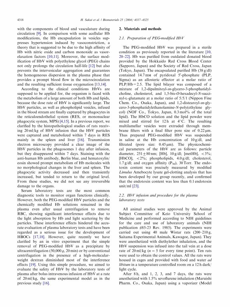
The control Hct was 4271%, and it decreasedslightly to 4071% at 1 day after HbV infusion. Theestimated Hb concentration of HbV in plasma just afterinfusion was about 6 g/dl, and it gradually decreased to4.470.3 g/dl at 8 h, 1.970.2 g/dl at 1 day, 1.370.1 g/dlat 2 days, and 0.870.01 g/dl at 3 days (Fig. 1). At 7days, HbV was not detected at all in the plasma phase.
3.2. Spleen and liver weights
The changes in the spleen and liver weights wereexpressed as percents of the body weight (Fig. 1). Theliver weight ratio (control, 4.8170.17%) showed asignificant increase 1 day after the infusion(5.2970.27%, po0:01), and then it returned to theoriginal level at 2 days. Spleen weight ratio significantlyincreased from 0.3270.05% to 0.6670.06% 3 daysafter the infusion (po0:01), however, it was reduced to0.4170.02% at 7 days.
3.3. Plasma laboratory tests
The plasma fraction after centrifugation of the bloodsample for 3 days after the HbV infusion was turbid dueto the presence of PEG-modified HbV. However,ultracentrifugation of the plasma produced transparentand light-yellow plasma phase and PEG-modified HbVwas precipitated at the bottom in a tube. There was nosign of the presence of Hb in the supernatant, indicatingthat there was no hemolysis of both RBC and HbV.
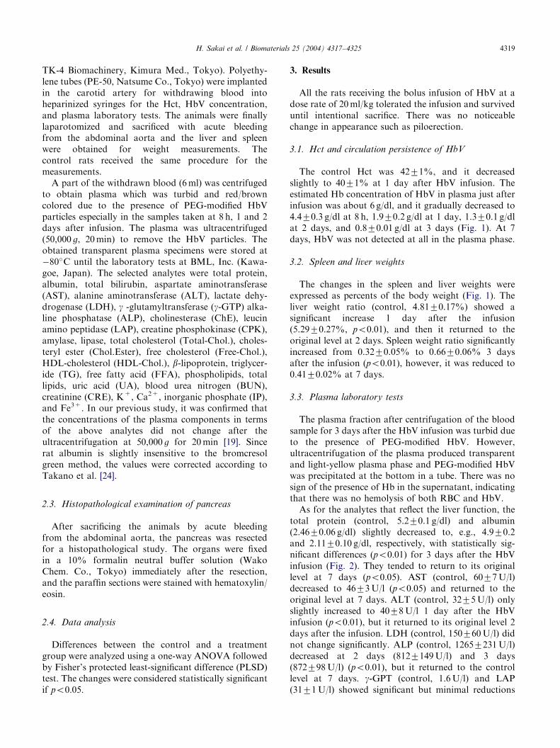
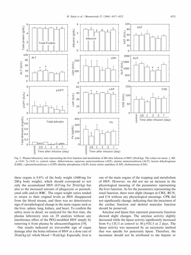
As for the analytes that reflect the liver function, thetotal protein (control, 5.270.1 g/dl) and albumin(2.4670.06 g/dl) slightly decreased to, e.g., 4.970.2and 2.1170.10 g/dl, respectively, with statistically sig-nificant differences (po0:01) for 3 days after the HbVinfusion (Fig. 2). They tended to return to its originallevel at 7 days (po0:05). AST (control, 6077U/l)decreased to 4673U/l (po0:05) and returned to theoriginal level at 7 days. ALT (control, 3275U/l) onlyslightly increased to 4078U/l 1 day after the HbVinfusion (po0:01), but it returned to its original level 2days after the infusion. LDH (control, 150760U/l) didnot change significantly. ALP (control, 12657231U/l)decreased at 2 days (8127149U/l) and 3 days(872798U/l) (po0:01), but it returned to the controllevel at 7 days. g-GPT (control, 1.6U/l) and LAP(3171U/l) showed significant but minimal reductions
ARTICLE IN PRESS
0
0.2
0.4
0.6
0.8
Sp
lee
nw
eig
ht
ratio
(%)
0
1
2
3
4
5
6
Liv
er
we
igh
tra
tio(%
)
Spleen weightLiver weight
Hct
0
10
20
30
40
50
Hct
(%)
0
2
4
6
[Hb
V]
(Hb
,g
/dL
) [HbV] in plasma
zero
con
tro
l
1/3 1 2 3 7
Time after infusion (day)
#
con
tro
l
1/3 1 2 3 7
Time after infusion (day)
Fig. 1. Changes in hematocrit, concentration of HbV in plasma, and spleen and liver weights after infusion of HbV (20ml/kg). The values are
mean7SD. �po0.01; #po0.05 vs. control values. The control value of [HbV] is the estimated concentration of HbV immediately after the infusion
and expressed as with a dashed line. The spleen and liver weights are expressed as the ratio to the body weight (%).
H. Sakai et al. / Biomaterials 25 (2004) 4317–43254320
(po0:05). ChE (control, 76718U/l) did not show anoticeable change. Plasma total bilirubin (p0.1mg/dl)and Fe3+ showed some reductions but were maintainedat a low level for 7 days in spite of the metabolism of alarge amount of Hb.
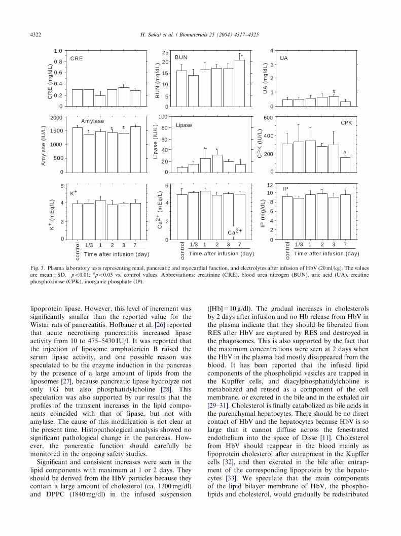
CRE (control, 0.3mg/dl) was maintained at a lowlevel for 7 days. BUN (control, 1673mg/dl) showed aslight increase at 7 days (2173mg/dl) (Fig. 3). UA(control, 0.4770.19mg/dl) increased to 0.7070.16mg/dl at 3 days, however, it returned to a non significantlevel at 7 days. Amylase (control, 1613774U/l)significantly decreased for 3 days after the infusion(po0:01), but returned to its original level at 7 days.Lipase (control, 971U/l) showed significant increases(po0:01) after the HbV infusion, and it tended todecrease after 3 days, and was reduced to a non-significant level at 7 days. CPK (control, 3047116U/l)decreased at 7 days (po0:05), but did not show anoticeable increase during the experiment. As for theelectrolyte concentrations, K+, Ca2+, and IP did notshow any significant changes.
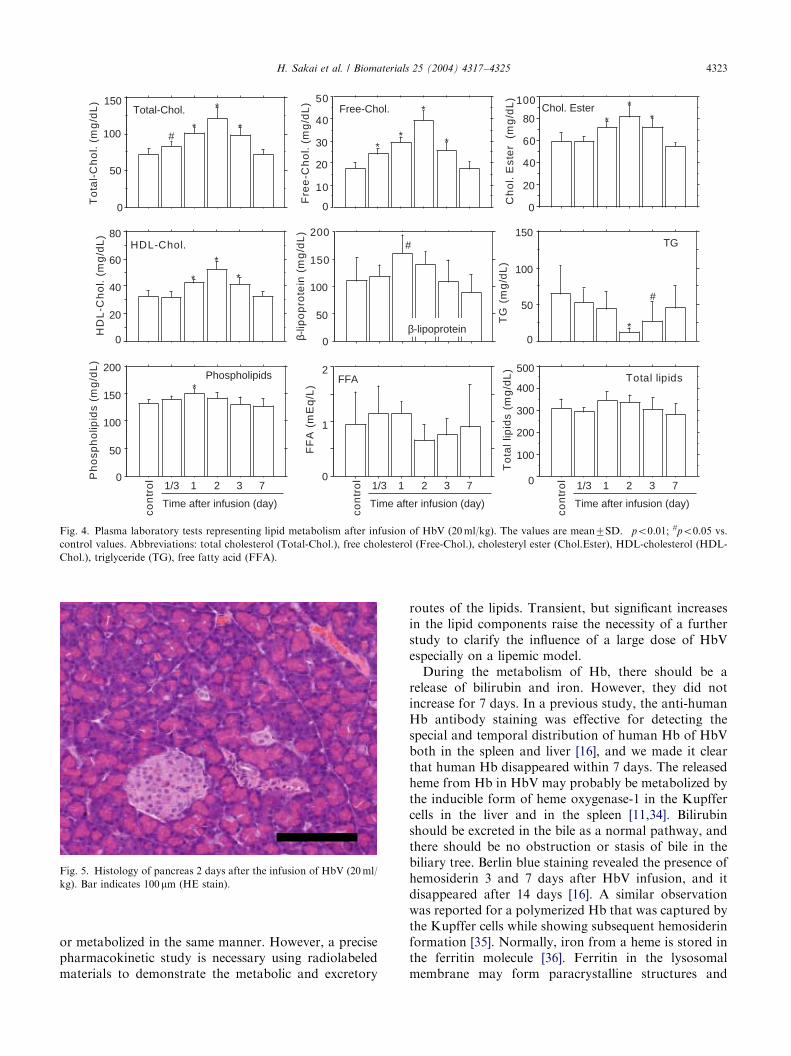
The most consistent changes were seen in the lipidcomponents (Fig. 4). Total-Chol. (control, 7377mg/dl),Free-Chol. (1872mg/dl), Chol.Ester (5978mg/dl), andHDL-Chol. (3274mg/dl) showed significant increasesand maximum values at 2 days (po0:01). Free-Chol.increased to 3974mg/dl, about twice the control value.However, it tended to decrease at 3 days, and returnedto its control level at 7 days. b-Lipoprotein (control,110742mg/dl) slightly increased at 1 day (160733mg/dl), but returned to its original level at 3 days. TG(control, 64.4mg/dl) significantly decreased to 12.4mg/dl at 2 days (po0:01), but tended to increase to its
original level at 7 days. Phospholipid (control,13278mg/dl) significantly increased to 15079mg/dlat 1 day (po0:01), and then returned to the original levelat 3 days.
3.4. Histopathological examination of pancreas
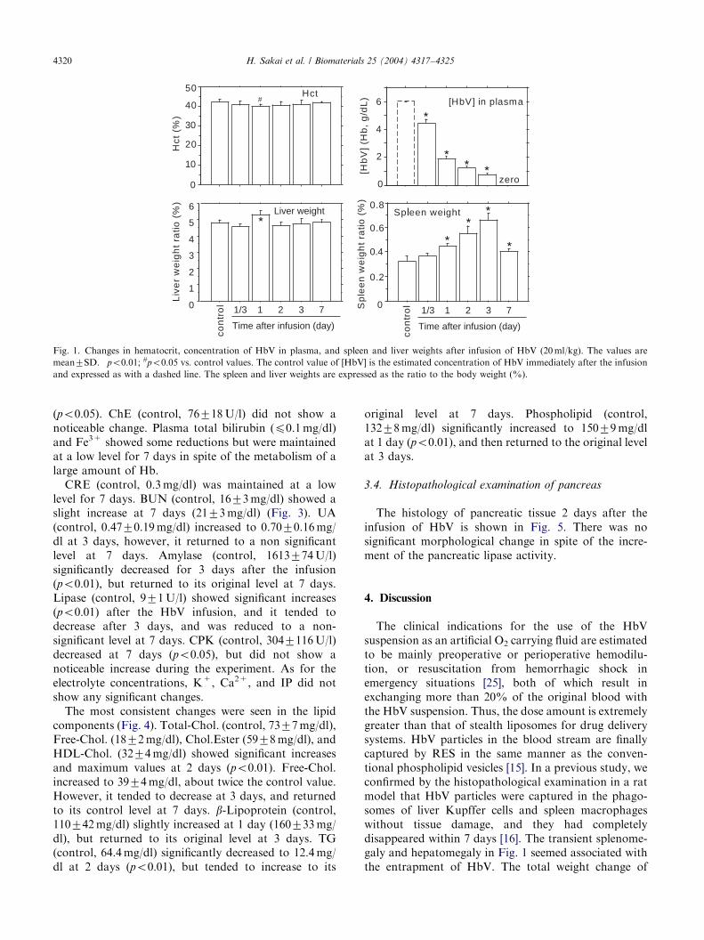
The histology of pancreatic tissue 2 days after theinfusion of HbV is shown in Fig. 5. There was nosignificant morphological change in spite of the incre-ment of the pancreatic lipase activity.
4. Discussion
The clinical indications for the use of the HbVsuspension as an artificial O2 carrying fluid are estimatedto be mainly preoperative or perioperative hemodilu-tion, or resuscitation from hemorrhagic shock inemergency situations [25], both of which result inexchanging more than 20% of the original blood withthe HbV suspension. Thus, the dose amount is extremelygreater than that of stealth liposomes for drug deliverysystems. HbV particles in the blood stream are finallycaptured by RES in the same manner as the conven-tional phospholipid vesicles [15]. In a previous study, weconfirmed by the histopathological examination in a ratmodel that HbV particles were captured in the phago-somes of liver Kupffer cells and spleen macrophageswithout tissue damage, and they had completelydisappeared within 7 days [16]. The transient splenome-galy and hepatomegaly in Fig. 1 seemed associated withthe entrapment of HbV. The total weight change of
ARTICLE IN PRESS
0
2
4
6T
ota
lpro
tein
(g/d
L)
0
20
40
60
80
100
AS
T(I
U/L
)
LD
H(I
U/L
)
0
500
1000
1500
2000
AL
P(I
U/L
)
γ-G
PT
(IU
/L)
0
10
20
30
40
LA
P(I
U/L
)
0
50
100
150
Ch
E(I
U/L
)
0
0.2
0.4
0.6
0.8
1
To
tal-
Bili
rub
in(m
g/d
L)
Total Protein
AST
0
100
200LDH ALP
LAP ChE
Total-bilirubin
0
100
200
300
400
Fe
(µg
/dL
)
Fe
zero zero
γ-GPT
0
2
4
6
0
1
2
Alb
um
in(g
/dL
)
3
Albumin
con
tro
l
1/3 1 2 3 7
Time after infusion (day)
con
tro
l
1/3 1 2 3 7
Time after infusion (day)
con
tro
l
1/3 1 2 3 7
Time after infusion (day)
# # # #
#
# # ##
#
#
0
20
40
60
80
100
#
AL
T (
IU/L
)
ALT
Fig. 2. Plasma laboratory tests representing the liver function and metabolism of Hb after infusion of HbV (20ml/kg). The values are mean 7 SD.�po0.01; #po0.05 vs. control values. Abbreviations: aspartate aminotransferase (AST), alanine aminotransferase (ALT), lactate dehydrogenase
(LDH), g-glutamyltransferase (g-GTP) alkaline phosphatase (ALP), leucin amino peptidase (LAP), cholinesterase (ChE).
H. Sakai et al. / Biomaterials 25 (2004) 4317–4325 4321
these organs is 0.8% of the body weight (1600mg for200 g body weight), which should correspond to notonly the accumulated HbV (635mg for 20ml/kg) butalso to the increased amount of phagocytic or parench-ymal cells and/or RBC. The organ weight ratios tendedto return to their original levels as HbV disappearedfrom the blood stream, and there was no deteriorativesign of morphological change in the main organs such asthe liver, spleen, lung, kidney, and heart. To confirm thesafety more in detail, we analyzed for the first time, theplasma laboratory tests on 29 analytes without anyinterference effect of the PEG-modified HbV simply byremoving it from plasma by ultracentrifugation [19].
Our results indicated no irreversible sign of organdamage after the bolus infusion of HbV at a dose rate of20ml/kg (cf. whole blood=56ml/kg). Especially, liver is
one of the main organs of the trapping and metabolismof HbV. However, we did not see an increase in thephysiological meaning of the parameters representingthe liver function. As for the parameters representing therenal function, there were slight changes in CRE, BUN,and UA without any physiological meanings. CPK didnot significantly change, indicating that the intactness ofthe cardiac function and skeletal muscular functionshould be preserved.
Amylase and lipase that represent pancreatic functionshowed slight changes. The amylase activity slightlydecreased while the lipase activity significantly increasedfrom 971 IU/l at control to 3079 IU/l at 2 days. Thelipase activity was measured by an enzymatic methodthat was specific for pancreatic lipase. Therefore, theincrement should not be attributed to the hepatic or
ARTICLE IN PRESS
Am
yla
se(I
U/L
)
BU
N(m
g/d
L)
0
500
1000
1500
2000
0
200
400
600
CP
K(I
U/L
)
CR
E(m
g/d
L)
0
5
10
15
20
25BUN
UA
(mg
/dL
)
Amylase CPK
0
2
4
6
K+
(mE
q/L
)
0
2
4
6
8
1012
IP(m
g/d
L)
K+
0
2
4
6
Ca
2+
(mE
q/L
)
Ca2+
IP
* * *
UA
0
1
2
3
4
CRE
0
0.2
0.4
0.6
0.8
1.0*
Lipase
20
80
100
Lip
ase
(IU
/L)
0
* *40
60
con
tro
l
1/3 1 2 3 7
Time after infusion (day)
con
tro
l
1/3 1 2 3 7
Time after infusion (day)
con
tro
l
1/3 1 2 3 7
Time after infusion (day)
#
#
Fig. 3. Plasma laboratory tests representing renal, pancreatic and myocardial function, and electrolytes after infusion of HbV (20ml/kg). The values
are mean7SD. �po0.01; #po0.05 vs. control values. Abbreviations: creatinine (CRE), blood urea nitrogen (BUN), uric acid (UA), creatine
phosphokinase (CPK), inorganic phosphate (IP).
H. Sakai et al. / Biomaterials 25 (2004) 4317–43254322
lipoprotein lipase. However, this level of increment wassignificantly smaller than the reported value for theWistar rats of pancreatitis. Hofbauer et al. [26] reportedthat acute necrotising pancreatitis increased lipaseactivity from 10 to 475–5430 IU/l. It was reported thatthe injection of liposome amphotericin B raised theserum lipase activity, and one possible reason wasspeculated to be the enzyme induction in the pancreasby the presence of a large amount of lipids from theliposomes [27], because pancreatic lipase hydrolyze notonly TG but also phosphatidylcholine [28]. Thisspeculation was also supported by our results that theprofiles of the transient increases in the lipid compo-nents coincided with that of lipase, but not withamylase. The cause of this modification is not clear atthe present time. Histopathological analysis showed nosignificant pathological change in the pancreas. How-ever, the pancreatic function should carefully bemonitored in the ongoing safety studies.
Significant and consistent increases were seen in thelipid components with maximum at 1 or 2 days. Theyshould be derived from the HbV particles because theycontain a large amount of cholesterol (ca. 1200mg/dl)and DPPC (1840mg/dl) in the infused suspension
([Hb]=10 g/dl). The gradual increases in cholesterolsby 2 days after infusion and no Hb release from HbV inthe plasma indicate that they should be liberated fromRES after HbV are captured by RES and destroyed inthe phagosomes. This is also supported by the fact thatthe maximum concentrations were seen at 2 days whenthe HbV in the plasma had mostly disappeared from theblood. It has been reported that the infused lipidcomponents of the phospholipid vesicles are trapped inthe Kupffer cells, and diacylphosphatidylcholine ismetabolized and reused as a component of the cellmembrane, or excreted in the bile and in the exhaled air[29–31]. Cholesterol is finally catabolized as bile acids inthe parenchymal hepatocytes. There should be no directcontact of HbV and the hepatocytes because HbV is solarge that it cannot diffuse across the fenestratedendothelium into the space of Disse [11]. Cholesterolfrom HbV should reappear in the blood mainly aslipoprotein cholesterol after entrapment in the Kupffercells [32], and then excreted in the bile after entrap-ment of the corresponding lipoprotein by the hepato-cytes [33]. We speculate that the main componentsof the lipid bilayer membrane of HbV, the phospho-lipids and cholesterol, would gradually be redistributed
ARTICLE IN PRESS
0
50
100
150T
ota
l-C
ho
l.(m
g/d
L)
0
10
20
30
40
50
Fre
e-C
ho
l.(m
g/d
L)
0
20
40
60
80
100
Ch
ol.
Est
er
(mg
/dL
)
0
50
100
150
200
β-li
po
pro
tein
(mg
/dL
)
0
20
40
60
80
HD
L-C
ho
l.(m
g/d
L)
0
50
100
150
TG
(mg
/dL
)
0
100
200
300
400
500
To
tall
ipid
s(m
g/d
L)
0
1
2
FF
A(m
Eq
/L)
0
50
100
150
200
Ph
osp
ho
lipid
s(m
g/d
L)
Total-Chol. Free-Chol. Chol. Ester
β-lipoprotein
HDL-Chol. TG
Total lipidsFFAPhospholipids
*
*
*
**
*
*
*
**
*
*
*
*
con
tro
l
1/3 1 2 3 7
Time after infusion (day)
con
tro
l
1/3 1 2 3 7
Time after infusion (day)
con
tro
l
1/3 1 2 3 7
Time after infusion (day)
#
#
#
*
Fig. 4. Plasma laboratory tests representing lipid metabolism after infusion of HbV (20ml/kg). The values are mean7SD. �po0.01; #po0.05 vs.
control values. Abbreviations: total cholesterol (Total-Chol.), free cholesterol (Free-Chol.), cholesteryl ester (Chol.Ester), HDL-cholesterol (HDL-
Chol.), triglyceride (TG), free fatty acid (FFA).
Fig. 5. Histology of pancreas 2 days after the infusion of HbV (20ml/
kg). Bar indicates 100mm (HE stain).
H. Sakai et al. / Biomaterials 25 (2004) 4317–4325 4323
or metabolized in the same manner. However, a precisepharmacokinetic study is necessary using radiolabeledmaterials to demonstrate the metabolic and excretory
routes of the lipids. Transient, but significant increasesin the lipid components raise the necessity of a furtherstudy to clarify the influence of a large dose of HbVespecially on a lipemic model.
During the metabolism of Hb, there should be arelease of bilirubin and iron. However, they did notincrease for 7 days. In a previous study, the anti-humanHb antibody staining was effective for detecting thespecial and temporal distribution of human Hb of HbVboth in the spleen and liver [16], and we made it clearthat human Hb disappeared within 7 days. The releasedheme from Hb in HbV may probably be metabolized bythe inducible form of heme oxygenase-1 in the Kupffercells in the liver and in the spleen [11,34]. Bilirubinshould be excreted in the bile as a normal pathway, andthere should be no obstruction or stasis of bile in thebiliary tree. Berlin blue staining revealed the presence ofhemosiderin 3 and 7 days after HbV infusion, and itdisappeared after 14 days [16]. A similar observationwas reported for a polymerized Hb that was captured bythe Kupffer cells while showing subsequent hemosiderinformation [35]. Normally, iron from a heme is stored inthe ferritin molecule [36]. Ferritin in the lysosomalmembrane may form paracrystalline structures and

ARTICLE IN PRESSH. Sakai et al. / Biomaterials 25 (2004) 4317–43254324
eventually aggregate in mass with an iron content ashigh as 50%. These are hemosiderins composed ofdegraded protein and coalesced iron. Both ferritin andhemosiderin release iron molecules, and they areanticipated to induce hydroxyl radical production andsucceeding lipid peroxidation [37,38]. However, ironrelease from hemosiderin is substantially less than thatfrom ferritin, thus iron molecules in hemosiderin arerelatively inert [39]. Plasma iron, mostly bound totransferrin, remained constant after HbV infusion. Theiron concentration should be coordinately regulatedthrough the ‘‘iron regulatory proteins’’ that sense thelevels of iron for hematopoiesis and metabolic needs[40], and the excess amount of iron should be stored inan insoluble and less toxic form as hemosiderin.Together with the time course of the histopathologicalchanges, the results of the plasma laboratory testsindicate that the metabolism of heme and the recyclingor excretion of iron molecule is within the physiologicalcapacity and suggested to be on the physiologicalpathway that has been well characterized for themetabolism of senescent RBC [41].
5. Conclusion
In this study, the plasma laboratory tests after theinfusion of HbV (20ml/kg) did not demonstrate anirreversible sign for a deteriorative damage to theorgans. Plasma bilirubin and iron, which were consid-ered to be released during the metabolism of the Hbmolecule, did not increase during the observationperiod. This may be due to the moderate rate of Hbmetabolism in RES after the entrapment of HbV with amoderate length of circulation time. The lipid compo-nents significantly increased at 2 or 3 days after infusion.These may be derived from the membrane component ofHbV entrapped in RES. The complete normalization ofthe lipid components indicates that they are metabolizedin a normal metabolic and/or recycling pathway. Theprecise biodistribution and fate of the componentsshould be confirmed by a radioisotope technique. Ourresults have demonstrated the safety of HbV using onlyhealthy rats, while rats in hemorrhagic shock, septicshock, or lipemia have to be tested in the ongoing safetystudies. It should also be emphasized that the datacannot be extrapolated to large animals or humans,which may react differently to such a large dose of HbV.
Acknowledgements
The authors gratefully acknowledge Dr. N. Hirose(Department of Gerontology, School of Medicine, KeioUniversity) for the discussion on the results. This workwas supported in part by Health Sciences Research
Grants (H15-Research on Pharmaceutical and MedicalSafety, Artificial Blood Project, -011, -016), the Ministryof Health, Labor and Welfare, Japan, Grants in Aid forScientific Research from the Japan Society for thePromotion of Science (B12480268), and 21 COE‘‘Practical Nano-Chemistry’’ from MEXT, Japan.
References
[1] Lian T, Ho RJY. Trends and developments in liposome drug
delivery system. J Pharm Sci 1991;90:667–80.
[2] Djordjevich L, Mayoral J, Miller IF, Ivankovich AD. Cardior-
espiratory effects of exchanging transfusions with synthetic
erythrocytes in rats. Crit Care Med 1987;15:318–23.
[3] Izumi Y, Sakai H, Hamada K, Takeoka S, Yamahata T, Kato R,
Nishide H, Tsuchida E, Kobayashi K. Physiologic responses to
exchange transfusion with hemoglobin vesicels as an artificial
oxygen carrier in anesthetized rats: changes in mean arterial
pressure and renal cortical tissue oxygen tension. Crit Care Med
1996;24:1869–73.
[4] Rudolph AS, Cliff RO, Spargo BJ, Spielberg H. Transient
changes in the mononuclear phagocyte system following admin-
istration of the blood subsitute liposome-encapsulated haemoglo-
bin. Biomaterials 1994;15:796–804.
[5] Tsuchida E. Blood substitutes: present and future perspectives.
Amsterdam: Elsevier Science; 1998.
[6] Chang TMS. Blood substitutes: principles, methods, products,
and clinical trials. Basel: Karger; 1997.
[7] Levy JH, Goodnough LT, Greilich PE, Parr GV, Stewart RW,
Gratz I, Wahr J, Williams J, Comunale ME, Doblar D, Silvay G,
Cohen M, Jahr JS, Vlahakes GJ. Polymerized bovine hemoglobin
solution as a replacement for allogeneic red blood cell transfusion
after cardiac surgery: results of a randomized, double-blind trial.
J Thorac Cardiovasc Surg 2002;124:35–42.
[8] Johnson JL, Moore EE, Offner PJ, Partrick DA, Tamura DY,
Zallen G, Silliman CC. Resuscitation with a blood substitute
abrogates pathologic postinjury neutrophil cytotoxic function.
J Trauma 2001;50:449–55.
[9] Takeoka S, Teramura Y, Atoji T, Tsuchida E. Effect of
Hb-encapsulation with vesicles on H2O2 reaction and lipid
peroxidation. Bioconjugate Chem 2002;13:1302–8.
[10] Sakai H, Hara H, Yuasa M, Tsai AG, Takeoka S, Tsuchida E,
Intaglietta M. Molecular dimensions of Hb-based O2 carriers
determine constriction of resistance arteries and hypertension in
conscious hamster model. Am J Physiol Heart Circ Physiol
2000;279:908–15.
[11] Goda N, Suzuki K, Naito S, Takeoka S, Tsuchida E, Ishimura Y,
Tamatani T, Suematsu M. Distribution of heme oxygenase
isoform in rat liver: topographic basis for carbon mon-
oxide-mediated microvascular relaxation. J Clin Invest 1998;101:
604–12.
[12] Phillips WT, Klipper RW, Awasthi VD, Rudolph AS, Cliff R,
Kwasiborski V, Goins BA. Polyethylene glycol-modified lipo-
some-encapsulated hemoglobin: a long circulating red cell
substitute. J Pharmacol Exp Ther 1999;288:665–70.
[13] Sakai H, Takeoka S, Park SI, Kose T, Izumi Y, Yoshizu A,
Nishide H, Kobayashi K, Tsuchida E. Surface-modification of
hemoglobin vesicles with polyethyleneglycol and effects on
aggregation, viscosity, and blood flow during 90%-exchange
transfusion in anesthetized rats. Bioconjugate Chem 1997;8:
15–22.
[14] Sakai H, Tsai AG, Kerger H, Park SI, Takeoka S, Nishide H,
Tsuchida E, Intaglietta M. Subcutaneous microvascular responses
to hemodilution with a red cell substitute consisting of
ARTICLE IN PRESSH. Sakai et al. / Biomaterials 25 (2004) 4317–4325 4325
polyethyleneglycol-modified vesicles encapsulating hemoglobin.
J Biomed Mater Res 1998;40:66–78.
[15] Rudolph AS, Klipper RW, Goins B, Phillips WT. In vivo
biodistribution of a radiolabeled blood substitute: 99mTc-labeled
liposome-encapsulated hemoglobin in an anesthetized rabbit.
Proc Natl Acad Sci USA 1991;88:10976–80.
[16] Sakai H, Horinouchi H, Tomiyama K, Iikeda E, Takeoka S,
Kobayashi K, Tsuchida E. Hemoglobin-vesicles as oxygen
carriers: influence on phagocytic activity and histopathological
changes in metabolism. Am J Pathol 2001;159:1079–88.
[17] Glick MR, Ryder KW. Double trouble: hemolysis and stabilized
hemoglobins (so you think you’re seeing red now? ). Clin Chem
2003;39:1761–3.
[18] Kazmierczak SC, Catrou PG, Best AE, Sullivan SW, Briley KP.
Multiple regression analysis of interference effects from a
hemoglobin-based oxygen carrier solution. Clin Chem Lab Med
1999;37:453–64.
[19] Sakai H, Tomiyama K, Masada Y, Takeoka S, Horinouchi H,
Kobayashi K, Tsuchida E. Pretreatment of serum containing
Hb-vesicels (oxygen carriers) to avoid interference in clinical
laboratory tests. Clin Chem Lab Med 2003;41:222–31.
[20] Takeoka S, Ohgushi T, Terase K, Ohmori T, Tsuchida E. Layer-
controlled hemoglobin vesicles by interaction of hemoglobin with
a phospholipid assembly. Langmuir 1996;12:1755–9.
[21] Sakai H, Yuasa M, Onuma H, Takeoka S, Tsuchida E. Synthesis
and physicochemical characterization of a series of hemoglobin-
based oxygen carriers: objective comparison between cellular and
acellular types. Bioconjugate Chem 2000;11:56–64.
[22] Sou K, Endo T, Naito Y, Takeoka S, Tsuchida E. Effective
encapsulation of proteins into size-controlled phospholipid
vesicles using freeze-thawing and extrusion. Biotechnol Progr
2003;19:1547–52.
[23] Sakai H, Hisamoto S, Fukutomi I, Sou K, Takeoka S, Tsuchida
E. Detection of Lipopolysaccharide in hemoglobin-vesicles by
Limulus amebocyte lysate test with kinetic-turbidimetric gell
clotting analysis and pretreatment with a surfactant. J Pharm Sci
2004;93:310–21.
[24] Takano S, Komiyama H, Nomura M. A study on the correction
of assay variation caused by the reagents in the measurement of
rat albumin. Rinshokagaku (Clin Chem) 2001;30:164–6.
[25] Sakai H, Takeoka S, Wettstein R, Tsai AG, Intaglietta M,
Tsuchida E. Systemic and microvascular responses to the
hemorrhagic shock and resuscitation with Hb-vesicles. Am
J Physiol Heart Circ Physiol 2002;283:H1191–9.
[26] Hofbauer B, Friess H, Weber A, Baczako, Kisling P, Schilling M,
Uhl W, Dervenis C, Buchler MW. Hyperlipaemia intensifies the
course of acute oedematous and acute necrotising pancreatitis in
the rat. Gut 1996;38:753–8.
[27] Stuecklin-Utsch A, Hasan C, Bode U, Fleischhack G. Pancreatic
toxicity after liposomal amphotericin B. Mycoses 2002;45:170–3.
[28] Rowland RN, Woodley JF. The stability of liposomes in vivo to
pH, bile salts and pancreatic lipase. Biochim Biophys Acta
1980;620:400–9.
[29] Verkade HJ, Derksen JT, Gerding A, Scherphof GL, Vonk RJ,
Kuipers F. Differential hepatic processing and biliary secretion of
head-group and acyl chains of liposomal phosphatidylcholines.
Biochem J 1991;275:139–44.
[30] Dijkstra J, van Galen M, Regts D, Scherphof G. Uptake and
processing of liposomal phospholipids by Kuppfer cells in vitro.
Eur J Biochem 1985;148:391–7.
[31] Matsushita Y, Eshima K, Shindo T, Yamamoto Y, Hasegawa E,
Nishide H, Tsuchida E. Clearance of tissue distribution of
functionalized polymeric liposomes from the blood stream of
rats. Biochim Biophys Acta 1987;901:166–71.
[32] Schwartz K, Lawn RM, Wade DP. ABC1 gene expression and
apoA-1-mediated cholesterol efflux are regulated by LXR.
Biochem Biophys Res Commun 2000;274:794–802.
[33] Kuipers F, Spanjer HH, Havinga R, Scherphof GL, Vonk RJ.
Lipoproteins and liposomes as in vivo cholesterol vehicles in the
rat: preferential use of cholesterol carried by small unilamellar
liposomes for the formation of muricholic acids. Biochim Biophys
Acta 1986;876:559–66.
[34] Braggins PE, Trakshel GM, Kutty RK, Maines MD. Character-
ization of two heme oxygenase isoforms in rat spleen: comparison
with the hematin-induced and constitutive isoforms of the liver.
Biochem Biophys Res Commun 1986;141:528–33.
[35] Lenz G, Junger H, Schneider M, Kothe N, Lissner R, Prince AM.
Elimination of pyridoxalated polyhemoglobin after partial
exchange transfusion in chimpanzees. Biomater Artif Cells
Immobilization Biotechnol 1991;19:699–708.
[36] Finch CA, Huebers H. Perspectives in iron metabolism. New Engl
J Med 1982;306:1520–8.
[37] O’Connell MJ, Peters TJ. Ferritin haemosiderin in free radical
generation, lipid peroxidation and protein damage. Chem Phys
Lipids 1987;45:241–9.
[38] Grady JK, Chen Y, Chasteen ND, Harris DC. Hydroxyl radical
production during oxidative deposition of iron in ferritin. J Biol
Chem 1989;264:20224–9.
[39] O’Connell MJ, Ward RJ, Baum H, Peters TJ. Iron release from
haemosiderin and ferritin by therapeutic and physiological
chelators. Biochem J 1989;260:903–7.
[40] Ponka P, Beaumont C, Richardson DR. Function and regulation
of transferrin and ferritin. Seminar Hematol 1998;35:35–54.
[41] Bennett GD, Kay MM. Homeostatic removal of senescent murine
erythrocytes by splenic macrophages. Exp Hematol 1981;9:
297–307.