mesh skin grafting and new skin grafting efficiencies that take up very little room and often fit...
TRANSCRIPT

Mesh Skin Grafting and New Skin Grafting Efficiencies
Mesh Skin Grafting andNew Skin Grafting Efficiencies
Introduction
This white paper examines the practices and procedures of how donor skin is perforated or fenestrated in preparation for grafting, balances the strengths and weakness of current methodologies, and evaluates new alternative procedures.
Strengths of current methods
Most donor skin preparation takes place at skin banks, in hospital burn units, cosmetic surgery clinics, and veterinary clinics.
At present, donor skin is mostly prepared by meshing, also called fenestration. Small slits or cuts in the donor skin allow the donor skin to stretch, thereby increasing the surface area of the donor skin and reducing the amount of donor skin needed to reconstruct the dermis damaged by the wound. In autografts, this reduces the trauma and scarring in the donor area, and allows the donor area to heal more quickly than it would if more skin were harvested.
Fenestration also helps the wounded area heal because the stretched slits and small “windows” increase the amount of skin perimeter available for capillary pass-through and connection with other vessels. These same windows allow fluids produced by the wound to drain and not get captured between the graft and the wound itself, where infection may breed.
Mesher and scalpel methodologies
Donor skin is most often fenestrated through the use of either a number 11 scalpel or by passing the donor skin through a mechanical mesher, using rollers to flatten and perforate the donor skin. This is what prepares the skin for stretching.
The biggest advantages of the current method of using a mesher to fenestrate donor skin include being able to stretch one unit of donor skin up to 3.0, and in some cases even 4.0 units of skin available for grafting, and perforated donor skin usually heals better than donor skin that has not been passed through a mesher.
In the case of temporary allografts and xenografts, stretched and fenestrated donor skin can reduce the likelihood of rejection until an autograft or close-matching allograft can be performed.
Meshers are sturdy and convenient. Paraprofessionals are familiar and comfortable with their operation.
by BIOCUT SYSTEMS

Mesh Skin Grafting and New Skin Grafting Efficiencies
Current Weaknesses
Current meshing machines resemble old fashioned laundry wringers, and operate on a similar principle. They treat donor skin roughly, and even when the harvested skin is perforated and grafted successfully, the healed skin may remain heavily pocked, making additional,cosmetic surgery necessary.
In the process of making the slits, the machine may grab and entangle donor skin. When the skin is wasted, it reduces the efficiency of the procedure. That is, if the donor portion of an autograft can cover a wounded area three times its spread, but the donor skin is damaged and made unusable by the mesher, then a second harvest is necessary, and the real graft efficiency is not 1:3, but 1:1.5.
Also, small particles of skin may get caught in the meshers, making them difficult to clean in an environment that requires clean room levels of sterilization.
Using a scalpel to perforate the donor skin can be tedious. It requires intense concentration, and the process is deliberate and slow. Even the best professional is prone to imprecision and sometimes accident, making safety an issue. The scalpel procedure is costly because it usually requires the presence of a paraprofessional or professional assistant.
A New Alternative
While there have been small, gradual improvements in use of meshers to fenestrate, new procedures make skin preparation simpler, less prone to imperfection, less prone to scalpel accident, and more efficient by reducing the amount of professional and paraprofessional hours needed to make a perfect and sterile cut.
The cutting board, the cutting tool, the skin, and how it all works
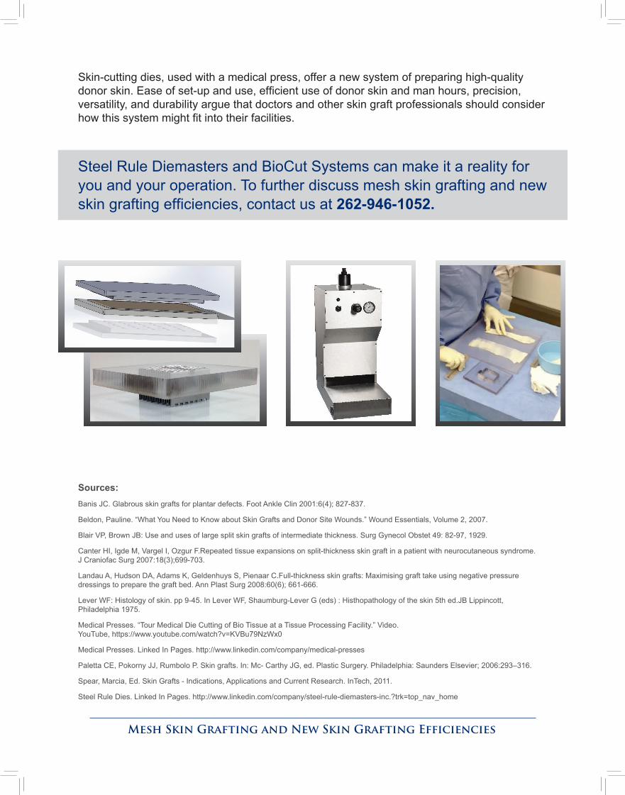
This alternative to the mechanical mesher and the number 11 scalpel offers a new method to perforate and fenestrate donor skin by means of a die cutting process. This new method places the donor skin in between a perfectly flat, stainless steel cutting pad beneath it, and a precision cutting tool on top. The cutting tool is custom designed to put precise slits and “windows” in the donor skin, rendering it fenestrated. No wringers, no scalpels.
The donor skin is fenestrated when pneumatic pressure drives the cutting tool, or die, through the donor skin. The cut is clean, precise to within one hundredth of an inch, and takes but a few seconds. The machine then lifts the cutting dies, and the fenestrated skin sits cleanly on the flat bed cutting pad ready for medical use. If your facility does not have pressurized air faucets, the press works equally well with air tanks that take up very little room and often fit under the machine itself.

Mesh Skin Grafting and New Skin Grafting Efficiencies
Ease of Use
There is no need for an assistant, and one-person operation is easy. With an hour or less of training, an operator will be ready to perform the few, simple steps displayed on the press machine’s screen: The skin is laid on the cutting pad. The dies are positioned on the skin. This “sandwich” of board, skin and cutting die is moved into the cutting area under the pressure plate. With a push of a button, the skin is cut and perforated. The process is virtually foolproof, and the skin is perfectly fenestrated with less handling and without the twists, kinks, and irregularities common to mesher procedures.
Versatility
With easy-to-change tooling, this system of skin-cutting die and cutting pad affords more versatility than any other fenestration method. The dies are capable of many different sizes of meshing and piece patterns, the better to meet the specific needs of surgeon and patient. The dies can cut in strips, shapes or small squares to accommodate Meek Grafts, Strip Grafts, and Micro Grafts with precision.
Easy to clean and sterilize
Another advantage is easy cleaning. These pneumatic skin-cutting die presses respond well to medical grade cleaning products, and easily withstand autoclaving and heat convection to meet the highest clean room requirements.
Durability, efficiency, and mobility
The skin-cutting dies are made with medically-compatible materials with the durability to make many dozens of identical cuts without any measurable degradation. The plates that drive the dies through the skin are easy to adjust and allow precise depth control, and the dies can easily be positioned to cut grafts across the entire donor skin surface, minimizing waste.
Conclusion
Ever since Sir Harold Gillies performed the first modern day skin graft surgery in 1917, medical professionals have made great strides in using fenestrated donor skin to heal wounds. But most skin graft professionals agree that right now some areas of this science remain crude, less effective than desired, inefficient, and in need of innovation.
Perforated donor skin is almost always the best option. Autografts, allografts, or even xenografts performed in burn centers, cosmetic surgery clinics, or veterinary clinics all rely on quality perforated dermis. Current methods of meshing machines and scalpel cuts can sometimes mangle the donor skin, make imprecise cuts, and require many man hours.
Mesh Skin Grafting and New Skin Grafting Efficiencies
Skin-cutting dies, used with a medical press, offer a new system of preparing high-quality donor skin. Ease of set-up and use, efficient use of donor skin and man hours, precision, versatility, and durability argue that doctors and other skin graft professionals should consider how this system might fit into their facilities.
Steel Rule Diemasters and BioCut Systems can make it a reality for you and your operation. To further discuss mesh skin grafting and new skin grafting efficiencies, contact us at 262-946-1052.
Sources:Banis JC. Glabrous skin grafts for plantar defects. Foot Ankle Clin 2001:6(4); 827-837.
Beldon, Pauline. “What You Need to Know about Skin Grafts and Donor Site Wounds.” Wound Essentials, Volume 2, 2007.
Blair VP, Brown JB: Use and uses of large split skin grafts of intermediate thickness. Surg Gynecol Obstet 49: 82-97, 1929.
Canter HI, Igde M, Vargel I, Ozgur F.Repeated tissue expansions on split-thickness skin graft in a patient with neurocutaneous syndrome. J Craniofac Surg 2007:18(3);699-703.
Landau A, Hudson DA, Adams K, Geldenhuys S, Pienaar C.Full-thickness skin grafts: Maximising graft take using negative pressure dressings to prepare the graft bed. Ann Plast Surg 2008:60(6); 661-666.
Lever WF: Histology of skin. pp 9-45. In Lever WF, Shaumburg-Lever G (eds) : Histhopathology of the skin 5th ed.JB Lippincott, Philadelphia 1975.
Medical Presses. “Tour Medical Die Cutting of Bio Tissue at a Tissue Processing Facility.” Video. YouTube, https://www.youtube.com/watch?v=KVBu79NzWx0
Medical Presses. Linked In Pages. http://www.linkedin.com/company/medical-presses
Paletta CE, Pokorny JJ, Rumbolo P. Skin grafts. In: Mc- Carthy JG, ed. Plastic Surgery. Philadelphia: Saunders Elsevier; 2006:293–316.
Spear, Marcia, Ed. Skin Grafts - Indications, Applications and Current Research. InTech, 2011.
Steel Rule Dies. Linked In Pages. http://www.linkedin.com/company/steel-rule-diemasters-inc.?trk=top_nav_home