membrane intercalation-enhanced photodynamic inactivation
TRANSCRIPT
Membrane intercalation-enhanced photodynamicinactivation of bacteria by a metallacycle andTAT-decorated virus coat proteinSijia Gaoa,b,1, Xuzhou Yanc,1,2, Guocheng Xiea,b, Meng Zhua, Xiaoyan Jua,b, Peter J. Stangd,2, Ye Tiana,2,and Zhongwei Niua,e
aKey Laboratory of Photochemical Conversion and Optoelectronic Materials, Technical Institute of Physics and Chemistry, Chinese Academy of Sciences,100190 Beijing, China; bUniversity of Chinese Academy of Sciences, Shijingshan District, 100049 Beijing, China; cSchool of Chemistry and ChemicalEngineering, Shanghai Jiao Tong University, 200240 Shanghai, China; dDepartment of Chemistry, University of Utah, Salt Lake City, UT 84112; and eSchool ofFuture Technology, University of Chinese Academy of Sciences, Shijingshan District, 100049 Beijing, China
Contributed by Peter J. Stang, October 12, 2019 (sent for review July 11, 2019; reviewed by Brian M. Hoffman, David W. C. MacMillan, and Jeffrey S. Moore)
Antibiotic resistance has become one of the major threats toglobal health. Photodynamic inactivation (PDI) develops littleantibiotic resistance; thus, it becomes a promising strategy in thecontrol of bacterial infection. During a PDI process, light-inducedreactive oxygen species (ROS) damage the membrane compo-nents, leading to the membrane rupture and bacteria death. Dueto the short half-life and reaction radius of ROS, achieving thecell-membrane intercalation of photosensitizers is a key chal-lenge for PDI of bacteria. In this work, a tetraphenylethylene-based discrete organoplatinum(II) metallacycle (1) acts as a photo-sensitizer with aggregation-induced emission. It self-assembles witha transacting activator of transduction (TAT) peptide-decorated viruscoat protein (2) through electrostatic interactions. This assembly (3)exhibits both ROS generation and strong membrane-intercalatingability, resulting in significantly enhanced PDI efficiency againstbacteria. By intercalating in the bacterial cell membrane or enter-ing the bacteria, assembly 3 decreases the survival rate of gram-negative Escherichia coli to nearly zero and that of gram-positiveStaphylococcus aureus to ∼30% upon light irradiation. This studyhas wide implications from the generation of multifunctional nano-materials to the control of bacterial infection, especially for gram-negative bacteria.
photodynamic inactivation | bacteria | membrane-intercalating |virus coat protein | aggregation-induced emission
Bacterial infections pose a major threat to global health (1, 2)and have become increasingly serious with increasing anti-
biotic resistance (3, 4). Despite efforts to find new antibacterialagents, the development of new drugs still lags far behind theevolution of antibiotic resistance (5–8). The newly emergingphotodynamic inactivation (PDI) strategy has attracted atten-tion because it involves minimal invasiveness, exhibits specificspatiotemporal selectivity, and is subject to limited antibioticresistance (9–12). In the presence of light and O2, photosensitizerscan generate reactive oxygen species (ROS), such as singlet oxygen(1O2) and hydroxyl radical (•OH), that damage membrane lipids,DNA, or proteins and consequently cause irreversible bacterialdeath (13–16). The main concern with a traditional photosensitizeris its aggregation-caused quenching behavior, which results in self-quenched fluorescence and reduced ROS generation in the ag-gregated state. However, aggregation-induced emission (AIE)active photosensitizers can maintain their ROS production ability,even in the aggregated state, and are thus better PDI agents (17).There are 2 factors that should be taken into consideration to
achieve a high PDI efficiency: 1) The bacterial membrane caneffectively protect bacteria from foreign substances, includingphotosensitizers (7); and 2) the half-life and reaction radius ofROS are relatively limited (18). To address these problems, sci-entists have conjugated a membrane-intercalating moiety to thephotosensitizer to enhance its membrane-intercalating capacity.
Tang and coworkers (19) modified AIE luminogens with amphi-philic molecules consisting of alkyl chains and a positive charge tofacilitate bacterial membrane intercalation of the PDI. Bazan andcoworkers (20) designed a membrane-intercalating conjugatedoligoelectrolyte to achieve high PDI activity against gram-negativebacteria. In addition to chemical conjugation, self-assembly throughphysical interactions also provides a general strategy to preparephotosensitizers with multiple functions, which have already beenwidely adopted for tumor-targeting photodynamic therapy in cancertreatment (21–24).Herein, we report a self-assembly strategy to obtain a
membrane intercalation-enhanced PDI system for the treatmentof bacterial infections. Specifically, a tetraphenylethylene-based discrete organoplatinum(II) metallacycle (1) was self-assembled with tobacco mosaic virus coat protein, which wasdecorated with a transacting activator of transduction (TAT)peptide (2). Metallacycle 1, with the AIE property (25–27), wasused as the photosensitizer. The heavy atoms in this molecule,platinum, can promote intersystem crossing from a singlet stateto a triplet state to enhance ROS generation (28–30). In 2, the
Significance
Photodynamic inactivation (PDI), which has led to little antibi-otic resistance, plays a promising role in the control of bacte-rial infection. Its main mechanism is the damage of membranecomponents by reactive oxygen species (ROS). However,achieving bacterial membrane intercalation of the photosensi-tizers remains a challenge. Here, we report the self-assembly ofan aggregation-induced emission active photosensitizer with acell-penetrating peptide-decorated virus coat protein. This as-sembly exhibits both ROS generation and a strong membrane-intercalating capacity, resulting in significantly enhanced PDIefficiency against bacteria. Especially for Escherichia coli pos-sessing outer membrane, this assembly decreases the survivalrate to nearly zero upon light irradiation. This study has impli-cations from the control of bacterial infection to the generationof multifunctional nanomaterials.
Author contributions: S.G., X.Y., P.J.S., Y.T., and Z.N. designed research; G.X., M.Z., X.J.,and P.J.S. analyzed data; and S.G., X.Y., P.J.S., Y.T., and Z.N. wrote the paper.
Reviewers: B.M.H., Northwestern University; D.W.C.M., Princeton University; and J.S.M.,University of Illinois at Urbana–Champaign.
The authors declare no competing interest.
Published under the PNAS license.1S.G. and X.Y. contributed equally to this work.2To whom correspondence may be addressed. Email: [email protected], [email protected], or [email protected].
This article contains supporting information online at www.pnas.org/lookup/suppl/doi:10.1073/pnas.1911869116/-/DCSupplemental.
First published November 4, 2019.
www.pnas.org/cgi/doi/10.1073/pnas.1911869116 PNAS | November 19, 2019 | vol. 116 | no. 47 | 23437–23443
CHEM
ISTR
Y
Dow
nloa
ded
by g
uest
on
Feb
ruar
y 6,
202
2
negatively charged protein moiety tends to self-assemble with thepositively charged 1 (31), and the TAT moiety provides a strongmembrane-intercalating capacity (32, 33). The resulting assembly(3) shows both ROS generation and membrane-intercalating be-havior and may be a good candidate for PDI in bacterial inhibition,especially for gram-negative bacteria possessing outer membranes.
Results and DiscussionPreparation and Self-Assembly of Metallacycle 1 and TAT-DecoratedProtein 2. Metallacycle 1 was obtained according to a reportedmethod. Its AIE and self-assembly properties were investigatedin detail in previous studies (25–27). Protein 2 was preparedvia copper (I)-catalyzed azide-alkyne cycloaddition between 5-azidopentanoic acid-YGRKKRRQRRR (TAT-N3) and the alkyne-modified tobacco mosaic virus coat protein (2′) at a specific site(SI Appendix, Fig. S1). Sodium dodecyl sulfate/polyacrylamide gel
electrophoresis (SDS/PAGE) analysis (SI Appendix, Fig. S2)revealed the emergence of a new protein band with higher mo-lecular mass in the lane of protein 2. This new protein band wasdigested in-gel by trypsin and analyzed by matrix-assisted laserdesorption/ionization–time-of-flight (MALDI-TOF) mass spec-trometry (SI Appendix, Fig. S3). During the trypsin-digestionprocess, the protein was cut into peptide segments at the car-boxyl side of arginine (R). According to the amino acid sequenceof protein 2′ and TAT (34), the theoretical molecular mass of peptidesegment in which TAT conjugated (sequence shown in SI Ap-pendix, Fig. S3D) was 1,401 Da, which is consistent with the massspectra results. The SDS/PAGE and MALDI-TOF mass resultsproved that TAT was successfully decorated on protein 2′ (35).Based on our previous work on the self-assembly of the posi-tively charged metallacycle 1 with negatively charged protein-based nanoparticles (31), protein 2 tends to self-assemble with
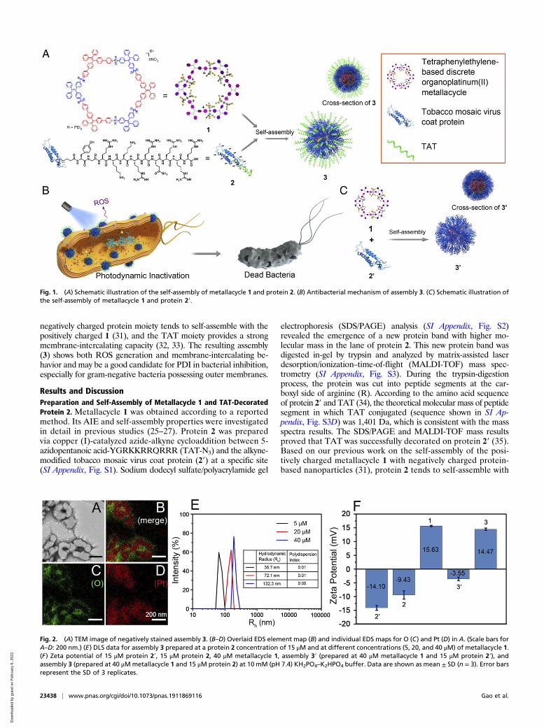
Fig. 1. (A) Schematic illustration of the self-assembly of metallacycle 1 and protein 2. (B) Antibacterial mechanism of assembly 3. (C) Schematic illustration ofthe self-assembly of metallacycle 1 and protein 2′.
Fig. 2. (A) TEM image of negatively stained assembly 3. (B–D) Overlaid EDS element map (B) and individual EDS maps for O (C) and Pt (D) in A. (Scale bars forA–D: 200 nm.) (E) DLS data for assembly 3 prepared at a protein 2 concentration of 15 μM and at different concentrations (5, 20, and 40 μM) of metallacycle 1.(F) Zeta potential of 15 μM protein 2′, 15 μM protein 2, 40 μM metallacycle 1, assembly 3′ (prepared at 40 μM metallacycle 1 and 15 μM protein 2′), andassembly 3 (prepared at 40 μMmetallacycle 1 and 15 μM protein 2) at 10 mM (pH 7.4) KH2PO4–K2HPO4 buffer. Data are shown as mean ± SD (n = 3). Error barsrepresent the SD of 3 replicates.
23438 | www.pnas.org/cgi/doi/10.1073/pnas.1911869116 Gao et al.
Dow
nloa
ded
by g
uest
on
Feb
ruar
y 6,
202
2
1 through electrostatic interactions (Fig. 1A). Transmission elec-tron microscopy (TEM) images (Fig. 2A) and energy-dispersiveX-ray spectroscopy (EDS) mapping (Fig. 2 B–D) showed theformation of assembly 3, in which metallacycle 1 (shown by Pt) wasin the core and protected by a protein 2 shell (shown by O). MoreTEM and EDS mapping images are supplied in SI Appendix, Fig.S4. Free 1 or 2 showed no regular structure (SI Appendix, Fig. S5).Dynamic light scattering (DLS) analysis in 10 mM (pH 7.4)KH2PO4–K2HPO4 buffer showed a broad distribution for metalla-cycle 1 due to its aggregation status in aqueous solution (SIAppendix, Fig. S6), while assembly 3 with different proportionsexhibited a narrow particle size distribution (Fig. 2E). At aprotein 2 concentration of 15 μM, the size of assembly 3 increasedwith the concentration of 1, confirming the 1 in 2 structure.Zeta potential was examined to confirm the self-assembly
mechanism and the composition of assembly 3. As shown in Fig.2F, protein 2′ was negatively charged (−14.10 mV) in 10 mM (pH7.4) KH2PO4–K2HPO4 buffer. Decoration with the positivelycharged peptide TAT slightly decreased its negative charge, butthe entire protein 2 remained negatively charged (−9.43 mV). Inthe absence of TAT, the assembly of 1 and 2′ (denoted as 3′)showed a negatively charged surface, demonstrating that 1 was inthe core and protein 2′ was in the shell (Fig. 1C). With TATdecoration, assembly 3 exhibited a positive charge (14.47 mV) atpH 7.4, demonstrating that the positively charged TAT peptide wasexposed outside the surface of 3. This specific structure (shown inFig. 1A) with the TAT peptide on the exterior surface will bebeneficial to cell-membrane intercalation (shown in Fig. 1B).
ROS Generation of Assembly 3. Electron spin-resonance spec-trometry (ESR) with 2,2,6,6-tetramethylpiperidine (TEMP) as aspin trap is frequently used for the detection of 1O2, which is animportant ROS generated from the type II photochemical reac-tion. For both metallacycle 1 and assembly 3, there was a strongincrease in ESR intensity upon 405-nm irradiation (Fig. 3A), in-dicating significant 1O2 generation by energy transfer from theexcited triplet state of metallacycle 1 to oxygen. The 9,10-anthracenediyl-bis(methylene)dimalonic acid (ABDA) is anothercommonly used probe for 1O2. With the generation of 1O2, theabsorbance of ABDA at 360, 380, and 400 nm will gradually de-crease. We first tested the absorption spectra of 1 and 3 in theabsence of ABDA under light irradiation to eliminate interferencefrom photoinstability. As shown in SI Appendix, Fig. S7 A and B,the absorption intensity at 400 nm did not show a significant de-crease, and the absorption peak at 400 nm of ABDA was thereforeapplied to identify 1O2 in this work. SI Appendix, Fig. S7C and Fig.
3B show that the absorbance of ABDA at 400 nm significantlydecreased upon light irradiation of 1 and 3, indicating 1O2 gen-eration. By eliminating the interference of photoinstability andcalculating the relative absorption intensity, we acquired the nor-malized absorption intensity of ABDA for comparison (SI Ap-pendix, Fig. S7D). From both the ABDA test (SI Appendix, Fig.S7D) and the ESR test (Fig. 3A), assembly 3 did not generate asmuch 1O2 as free metallacycle 1. This may come from 2 factors: 1)Protein 2 may compete with the spin-trap TEMP or probe ABDAto react with 1O2 (36); and 2) because the photosensitizer is
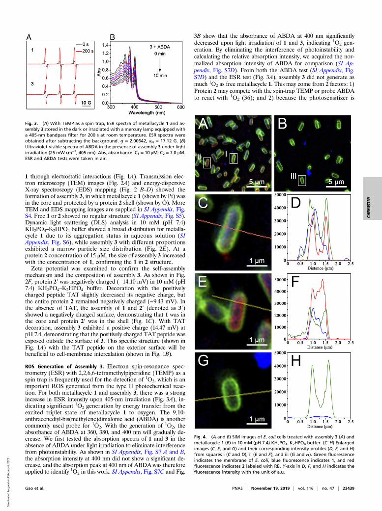
Fig. 3. (A) With TEMP as a spin trap, ESR spectra of metallacycle 1 and as-sembly 3 stored in the dark or irradiated with a mercury lamp equipped witha 405-nm bandpass filter for 200 s at room temperature. ESR spectra wereobtained after subtracting the background. g = 2.00642, αN = 17.12 G. (B)Ultraviolet-visible spectra of ABDA in the presence of assembly 3 under lightirradiation (25 mW cm−2, 405 nm). Abs, absorbance. C1 = 10 μM; C2 = 7.0 μM.ESR and ABDA tests were taken in air.
Fig. 4. (A and B) SIM images of E. coli cells treated with assembly 3 (A) andmetallacycle 1 (B) in 10 mM (pH 7.4) KH2PO4–K2HPO4 buffer. (C–H) Enlargedimages (C, E, and G) and their corresponding intensity profiles (D, F, and H)from squares i (C and D), ii (E and F), and iii (G and H). Green fluorescenceindicates the membrane of E. coli, blue fluorescence indicates 1, and redfluorescence indicates 2 labeled with RB. Y-axis in D, F, and H indicates thefluorescence intensity with the unit of a.u.
Gao et al. PNAS | November 19, 2019 | vol. 116 | no. 47 | 23439
CHEM
ISTR
Y
Dow
nloa
ded
by g
uest
on
Feb
ruar
y 6,
202
2
protected by the protein shell, the ROS generation from assembly3 may be more sensitive to the consumption of ambient O2.
Intracellular Distribution of Assembly 3 in Gram-Negative Bacteria. Ina PDI process for bacterial inhibition, due to the short half-lifeand limited reaction radius of ROS, ROS generation and bac-terial accumulation are both required for the photosensitizers.Here, we labeled protein 2 with rhodamine B (RB) (red) and theEscherichia coli (E. coli) cell membrane with FM 1-43FX mem-brane stain (green). The intracellular distribution of assembly 3could then be monitored by using structured illumination fluo-rescence microscopy (SIM) (Fig. 4). The fluorescence intensityprofiles for the enlarged images were further calculated to ana-lyze the colocalization in detail. As shown in Fig. 4, assembly 3intercalated in the cell membrane of E. coli, causing the greenand red fluorescence in the intensity profiles to coincide closely(Fig. 4 A, C, and D). Some assembly 3 even entered the bacteriaby disturbing the integrity of the membrane, as shown by a “red-in-green” fluorescence distribution in the intensity profiles (Fig. 4 A,E, and F). The high fluorescence intensity of blue in Fig. 4 D and Fprovided evidence of abundant uptake of photosensitizer. Incontrast to assembly 3, metallacycle 1 without membrane-intercalating properties just accumulated only outside the bac-teria (Fig. 4 B andG). There was little blue fluorescence in the E.coli cells (see the fluorescence intensity profiles in Fig. 4H). Tobetter quantify the intracellular distribution, Pearson’s correla-tion for Fig. 4 and more SIM images were analyzed through
Nikon software (SI Appendix, Fig. S8). The high values ofPearson’s correlation (0.69 for green and red; 0.60 for green andblue) indicated that, through self-assembly, assembly 3 achieved asignificantly enhanced membrane-intercalating property (Fig. 1B),which is beneficial for the PDI process.
Antibacterial Activity on E. coli. We investigated the antibacterialeffect of assembly 3 on E. coli in the dark and upon 420-nm irra-diation for 15 min. Metallacycle 1 and assembly 3′ were applied forcomparison. A standard plate count that judged the bacte-rial proliferative capacity was employed to assess the antibacterialactivity. In Fig. 5, assembly 3 and metallacycle 1, which were posi-tively charged (Fig. 2F), created higher dark toxicity than assembly3′ because of the negative charge on the bacterial membrane. Uponlight irradiation, the survival rate of E. coli treated with assembly 3decreased from ∼55% to nearly 0% at metallacycle 1 concentrationsof 20 and 40 μM (Fig. 5A). However, E. coli incubated with freemetallacycle 1 at the same concentrations showed little change insurvival rate after 420-nm irradiation for 15 min compared to that inthe dark (Fig. 5B). The PDI efficiency of assembly 3 was significantlyhigher than that of 1, which benefited from the enhanced bacterialaccumulation capacity of 3. We then calculated the total PDI effi-ciency of protein 2 (15.3%) and metallacycle 1 (46.3%) at a con-centration of 40 μM. This value (61.6%) was much lower than thePDI efficiency of assembly 3 (96.3%) at the same concentration,demonstrating a synergistic antibacterial effect of this system. Toexplore the importance of the membrane-intercalating property of
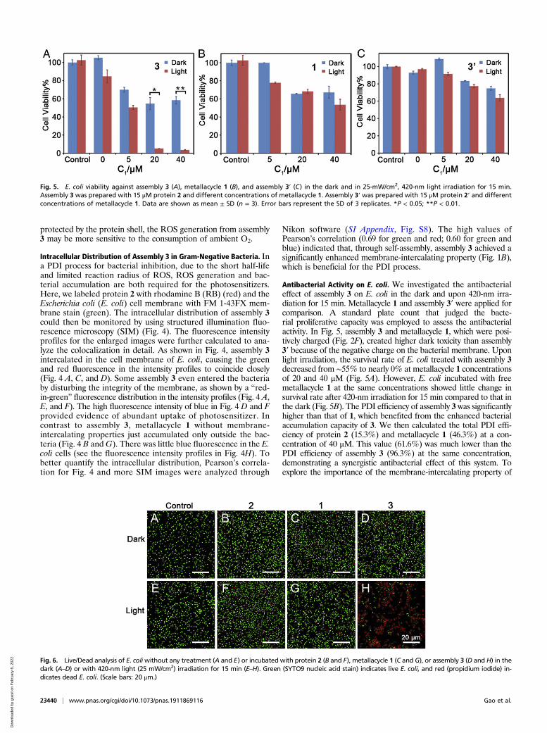
Fig. 5. E. coli viability against assembly 3 (A), metallacycle 1 (B), and assembly 3′ (C) in the dark and in 25-mW/cm2, 420-nm light irradiation for 15 min.Assembly 3 was prepared with 15 μM protein 2 and different concentrations of metallacycle 1. Assembly 3′ was prepared with 15 μM protein 2′ and differentconcentrations of metallacycle 1. Data are shown as mean ± SD (n = 3). Error bars represent the SD of 3 replicates. *P < 0.05; **P < 0.01.
Fig. 6. Live/Dead analysis of E. coliwithout any treatment (A and E) or incubated with protein 2 (B and F), metallacycle 1 (C and G), or assembly 3 (D and H) in thedark (A–D) or with 420-nm light (25 mW/cm2) irradiation for 15 min (E–H). Green (SYTO9 nucleic acid stain) indicates live E. coli, and red (propidium iodide) in-dicates dead E. coli. (Scale bars: 20 μm.)
23440 | www.pnas.org/cgi/doi/10.1073/pnas.1911869116 Gao et al.
Dow
nloa
ded
by g
uest
on
Feb
ruar
y 6,
202
2
protein 2 in this PDI system, we further assessed the PDI efficiencyof assembly 3′ without TAT decoration for comparison. As shown inFig. 5C, at a metallacycle 1 concentration of 40 μM, the survival rateof E. coli was reduced only from 74% (in the dark) to 63% uponlight irradiation. This result confirmed the important contribution ofthe membrane-intercalating capacity to the PDI efficiency.The antibacterial effect was further investigated by the Live/
Dead assay. E. coli cells adsorbed on lysine-embedded coverslipswere incubated with protein 2 (15 μM), metallacycle 1 (40 μM), orassembly 3 at the same concentration, then kept in the dark orirradiated with 420-nm light (25 mW/cm2) for 15 min. Live/Deadkit staining and confocal laser-scanning microscopy (CLSM) ob-servation (Fig. 6) showed a similar trend in PDI efficiency to thatobtained by the standard plate-count method (Fig. 5). Only as-sembly 3 together with light irradiation led to significant bacterialdeath (Fig. 6H). Only 16.0% of bacterial cells were alive (SI Ap-pendix, Fig. S9). Both metallacycle 1 without cell-penetrating ca-pacity and assembly 3 without light irradiation could induce only asmall amount of death (Fig. 6 C, G, and D). More than 90.0% ofbacterial cells were still alive (SI Appendix, Fig. S9). This resultindicates that the photosensitizer, light, and membrane-intercalatingmoiety are all indispensable in this PDI system.Cytotoxicity of assembly 3 toward mammalian cells was assessed
with L929 and HeLa cells (SI Appendix, Fig. S10). The cells weretreated with assembly 3 for 2 h, irradiated or kept in the dark for15 min, and then incubated for another 12 h. The cell viabilities ofL929 and HeLa cells were above 60%, confirming that assembly 3has good biocompatibility toward mammalian cells.
Antibacterial Mechanism. To explore the antibacterial mechanismof assembly 3, we observed the morphology of bacteria by scanning
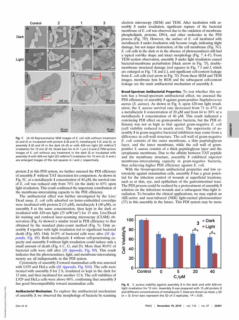
electron microscopy (SEM) and TEM. After incubation with as-sembly 3 under irradiation, significant rupture of the bacterialmembrane of E. coli was observed due to the oxidation of membranephospholipids, proteins, DNA, and other molecules in the PDIprocess (Fig. 7H). However, the surface of E. coli incubated withmetallacycle 1 under irradiation only became rough, indicating slightdamage, but not major destruction, of the cell membrane (Fig. 7G).E. coli cells in the dark or in the absence of photosensitizers still hada typical rod-like shape and intact morphology (Fig. 7 A–F). FromTEM section observation, assembly 3 under light irradiation causedbacterial-membrane perturbation (black arrow in Fig. 7J), double-membrane structural damage (red squares in Fig. 7 I and J, whichare enlarged as Fig. 7 K and L), and significant cell-content leakagefrom E. coli cells (red arrow in Fig. 7J). From these SEM and TEMimages, membrane lysis by ROS and the subsequent cell-contentleakage are the main antibacterial mechanism of assembly 3.
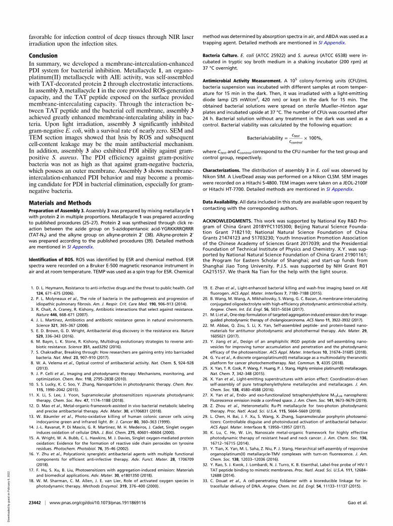
Broad-Spectrum Antibacterial Properties. To test whether this sys-tem has a broad-spectrum antibacterial effect, we assessed thePDI efficiency of assembly 3 against gram-positive Staphylococcusaureus (S. aureus). As shown in Fig. 8, upon 420-nm light irradi-ation, the S. aureus survival rate decreased from 71 to 47% ata metallacycle 1 concentration of 20 μM and from 64 to 30% at ametallacycle 1 concentration of 40 μM. This result indicated aconvincing PDI effect on gram-positive bacteria, but the PDI ef-ficiency was not as high as that against gram-negative E. coli(cell viability reduced to nearly zero). The superiority of as-sembly 3 in gram-negative bacterial inhibition may come from adifference in cell-wall structure. The cell wall of gram-negativeE. coli consists of the outer membrane, a thin peptidoglycanlayer, and the inner membrane, while the cell wall of gram-positive S. aureus consists of a thick peptidoglycan layer and thecytoplasmic membrane. Due to the affinity between TAT peptideand the membrane structure, assembly 3 exhibited superiormembrane-intercalating capacity in gram-negative bacteria,thus achieving higher PDI efficiency against E. coli.With the broad-spectrum antibacterial properties and low cy-
totoxicity against mammalian cells, assembly 3 has a great poten-tial for the infection control of wounds at superficial locations,such as at skin, eye, and epithelium of the gastrointestinal tract.The PDI process could be realized by a pretreatment of assembly 3solution on the infectious wounds and a subsequent blue-light ir-radiation. To broaden the clinical applications, we may exploit anAIE-active and near-infrared (NIR) light-excited photosensitizer(37) in this assembly in the future. This PDI system may be more
Fig. 7. (A–H) Representative SEM images of E. coli cells without treatment(A and E) or incubated with protein 2 (B and F), metallacycle 1 (C and G), orassembly 3 (D and H) in the dark (A–D) or with 420-nm light (25 mW/cm2)irradiation for 15 min (E–H). (Scale bars for A–H: 1 μm.) (I and J) TEM sectionimages of E. coli without any treatment in the dark (I) or incubated withassembly 3 with 420-nm light (25 mW/cm2) irradiation for 15 min (J). K and Lare enlarged images of the red squares in I and J, respectively.
Fig. 8. S. aureus viability against assembly 3 in the dark and with 420-nmlight irradiation for 15 min. Assembly 3 was prepared with 15 μM protein 2and different concentrations of metallacycle 1. Data are shown as mean ± SD(n = 3). Error bars represent the SD of 3 replicates. *P < 0.05.
Gao et al. PNAS | November 19, 2019 | vol. 116 | no. 47 | 23441
CHEM
ISTR
Y
Dow
nloa
ded
by g
uest
on
Feb
ruar
y 6,
202
2
favorable for infection control of deep tissues through NIR laserirradiation upon the infection sites.
ConclusionIn summary, we developed a membrane-intercalation-enhancedPDI system for bacterial inhibition. Metallacycle 1, an organo-platinum(II) metallacycle with AIE activity, was self-assembledwith TAT-decorated protein 2 through electrostatic interactions.In assembly 3, metallacycle 1 in the core provided ROS-generationcapacity, and the TAT peptide exposed on the surface providedmembrane-intercalating capacity. Through the interaction be-tween TAT peptide and the bacterial cell membrane, assembly 3achieved greatly enhanced membrane-intercalating ability in bac-teria. Upon light irradiation, assembly 3 significantly inhibitedgram-negative E. coli, with a survival rate of nearly zero. SEM andTEM section images showed that lysis by ROS and subsequentcell-content leakage may be the main antibacterial mechanism.In addition, assembly 3 also exhibited PDI ability against gram-positive S. aureus. The PDI efficiency against gram-positivebacteria was not as high as that against gram-negative bacteria,which possess an outer membrane. Assembly 3 shows membrane-intercalation-enhanced PDI behavior and may become a promis-ing candidate for PDI in bacterial elimination, especially for gram-negative bacteria.
Materials and MethodsPreparation of Assembly 3. Assembly 3was prepared by mixing metallacycle 1with protein 2 in multiple proportions. Metallacycle 1 was prepared accordingto published procedures (25–27). Protein 2 was synthesized through click re-action between the azide group on 5-azidopentanoic acid-YGRKKRRQRRR(TAT-N3) and the alkyne group on alkyne-protein 2′ (38). Alkyne-protein 2′was prepared according to the published procedures (39). Detailed methodsare mentioned in SI Appendix.
Identification of ROS. ROS was identified by ESR and chemical method. ESRspectra were recorded on a Bruker E-500 magnetic resonance instrument inair and at room temperature. TEMP was used as a spin trap for ESR. Chemical
methodwas determined by absorption spectra in air, and ABDAwas used as atrapping agent. Detailed methods are mentioned in SI Appendix.
Bacteria Culture. E. coli (ATCC 25922) and S. aureus (ATCC 6538) were in-cubated in tryptic soy broth medium in a shaking incubator (200 rpm) at37 °C overnight.
Antimicrobial Activity Measurement. A 105 colony-forming units (CFU)/mLbacteria suspension was incubated with different samples at room temper-ature for 15 min in the dark. Then, it was irradiated with a light-emittingdiode lamp (25 mW/cm2, 420 nm) or kept in the dark for 15 min. Theobtained bacterial solutions were spread on sterile Mueller–Hinton agarplates and incubated upside at 37 °C. The number of CFUs was counted after24 h. Bacterial solution without any treatment in the dark was used as acontrol. Bacterial viability was calculated by the following equation:
Bacterialviability =ctest
ccontrol× 100%,
where Ctest and Ccontrol correspond to the CFU number for the test group andcontrol group, respectively.
Characterizations. The distribution of assembly 3 in E. coli was observed byNikon SIM. A Live/Dead assay was performed on a Nikon CLSM. SEM imageswere recorded on a Hitachi S-4800. TEM images were taken on a JEOL-2100For Hitachi HT-7700. Detailed methods are mentioned in SI Appendix.
Data Availability. All data included in this study are available upon request bycontacting with the corresponding authors.
ACKNOWLEDGMENTS. This work was supported by National Key R&D Pro-gram of China Grant 2018YFC1105300; Beijing Natural Science Founda-tion Grant 7182110; National Natural Science Foundation of ChinaGrants 21474123 and 51703230; Youth Innovation Promotion Associationof the Chinese Academy of Sciences Grant 2017039; and the PresidentialFoundation of Technical Institute of Physics and Chemistry. X.Y. was sup-ported by National Natural Science Foundation of China Grant 21901161;the Program for Eastern Scholar of Shanghai; and start-up funds fromShanghai Jiao Tong University. P.J.S. was supported by NIH Grant R01CA215157. We thank Na Tian for the help with the light source.
1. D. L. Heymann, Resistance to anti-infective drugs and the threat to public health. Cell124, 671–675 (2006).
2. P. L. Molyneaux et al., The role of bacteria in the pathogenesis and progression ofidiopathic pulmonary fibrosis. Am. J. Respir. Crit. Care Med. 190, 906–913 (2014).
3. R. Chait, A. Craney, R. Kishony, Antibiotic interactions that select against resistance.Nature 446, 668–671 (2007).
4. J. L. Martínez, Antibiotics and antibiotic resistance genes in natural environments.Science 321, 365–367 (2008).
5. E. D. Brown, G. D. Wright, Antibacterial drug discovery in the resistance era. Nature529, 336–343 (2016).
6. M. Baym, L. K. Stone, R. Kishony, Multidrug evolutionary strategies to reverse anti-biotic resistance. Science 351, aad3292 (2016).
7. S. Chakradhar, Breaking through: How researchers are gaining entry into barricadedbacteria. Nat. Med. 23, 907–910 (2017).
8. W. A. Velema et al., Optical control of antibacterial activity. Nat. Chem. 5, 924–928(2013).
9. J. P. Celli et al., Imaging and photodynamic therapy: Mechanisms, monitoring, andoptimization. Chem. Rev. 110, 2795–2838 (2010).
10. S. S. Lucky, K. C. Soo, Y. Zhang, Nanoparticles in photodynamic therapy. Chem. Rev.115, 1990–2042 (2015).
11. X. Li, S. Lee, J. Yoon, Supramolecular photosensitizers rejuvenate photodynamictherapy. Chem. Soc. Rev. 47, 1174–1188 (2018).
12. D. Mao et al., Metal-organic-framework-assisted in vivo bacterial metabolic labelingand precise antibacterial therapy. Adv. Mater. 30, e1706831 (2018).
13. W. Bäumler et al., Photo-oxidative killing of human colonic cancer cells usingindocyanine green and infrared light. Br. J. Cancer 80, 360–363 (1999).
14. J.-L. Ravanat, P. Di Mascio, G. R. Martinez, M. H. Medeiros, J. Cadet, Singlet oxygeninduces oxidation of cellular DNA. J. Biol. Chem. 275, 40601–40604 (2000).
15. A. Wright, W. A. Bubb, C. L. Hawkins, M. J. Davies, Singlet oxygen-mediated proteinoxidation: Evidence for the formation of reactive side chain peroxides on tyrosineresidues. Photochem. Photobiol. 76, 35–46 (2002).
16. Y. Zhu et al., Polycationic synergistic antibacterial agents with multiple functionalcomponents for efficient anti-infective therapy. Adv. Funct. Mater. 28, 1706709(2018).
17. F. Hu, S. Xu, B. Liu, Photosensitizers with aggregation-induced emission: Materialsand biomedical applications. Adv. Mater. 30, e1801350 (2018).
18. W. M. Sharman, C. M. Allen, J. E. van Lier, Role of activated oxygen species inphotodynamic therapy. Methods Enzymol. 319, 376–400 (2000).
19. E. Zhao et al., Light-enhanced bacterial killing and wash-free imaging based on AIEfluorogen. ACS Appl. Mater. Interfaces 7, 7180–7188 (2015).
20. B. Wang, M. Wang, A. Mikhailovsky, S. Wang, G. C. Bazan, A membrane-intercalatingconjugated oligoelectrolyte with high-efficiency photodynamic antimicrobial activity.Angew. Chem. Int. Ed. Engl. 56, 5031–5034 (2017).
21. M. Li et al., One-step formulation of targeted aggregation-induced emission dots for image-guided photodynamic therapy of cholangiocarcinoma. ACS Nano 11, 3922–3932 (2017).
22. M. Abbas, Q. Zou, S. Li, X. Yan, Self-assembled peptide- and protein-based nano-materials for antitumor photodynamic and photothermal therapy. Adv. Mater. 29,1605021 (2017).
23. Y. Jiang et al., Design of an amphiphilic iRGD peptide and self-assembling nano-vesicles for improving tumor accumulation and penetration and the photodynamicefficacy of the photosensitizer. ACS Appl. Mater. Interfaces 10, 31674–31685 (2018).
24. G. Yu et al., A discrete organoplatinum(II) metallacage as a multimodality theranosticplatform for cancer photochemotherapy. Nat. Commun. 9, 4335 (2018).
25. X. Yan, T. R. Cook, P. Wang, F. Huang, P. J. Stang, Highly emissive platinum(II) metallacages.Nat. Chem. 7, 342–348 (2015).
26. X. Yan et al., Light-emitting superstructures with anion effect: Coordination-drivenself-assembly of pure tetraphenylethylene metallacycles and metallacages. J. Am.Chem. Soc. 138, 4580–4588 (2016).
27. X. Yan et al., Endo- and exo-functionalized tetraphenylethylene M12L24 nanospheres:Fluorescence emission inside a confined space. J. Am. Chem. Soc. 141, 9673–9679 (2019).
28. Z. Zhou et al., Heterometallic Ru-Pt metallacycle for two-photon photodynamictherapy. Proc. Natl. Acad. Sci. U.S.A. 115, 5664–5669 (2018).
29. L. Chen, H. Bai, J. F. Xu, S. Wang, X. Zhang, Supramolecular porphyrin photosensi-tizers: Controllable disguise and photoinduced activation of antibacterial behavior.ACS Appl. Mater. Interfaces 9, 13950–13957 (2017).
30. K. Lu, C. He, W. Lin, Nanoscale metal-organic framework for highly effectivephotodynamic therapy of resistant head and neck cancer. J. Am. Chem. Soc. 136,16712–16715 (2014).
31. Y. Tian, X. Yan, M. L. Saha, Z. Niu, P. J. Stang, Hierarchical self-assembly of responsiveorganoplatinum(II) metallacycle-TMV complexes with turn-on fluorescence. J. Am.Chem. Soc. 138, 12033–12036 (2016).
32. Y. Rao, S. J. Kwok, J. Lombardi, N. J. Turro, K. B. Eisenthal, Label-free probe of HIV-1TAT peptide binding to mimetic membranes. Proc. Natl. Acad. Sci. U.S.A. 111, 12684–12688 (2014).
33. C. Douat et al., A cell-penetrating foldamer with a bioreducible linkage for in-tracellular delivery of DNA. Angew. Chem. Int. Ed. Engl. 54, 11133–11137 (2015).
23442 | www.pnas.org/cgi/doi/10.1073/pnas.1911869116 Gao et al.
Dow
nloa
ded
by g
uest
on
Feb
ruar
y 6,
202
2
34. J. M. Alonso, M. L. Górzny, A. M. Bittner, The physics of tobacco mosaic virus andvirus-based devices in biotechnology. Trends Biotechnol. 31, 530–538 (2013).
35. Y. Tian et al., Integration of cell-penetrating peptides with rod-like bionanoparticles:Virus-inspired gene-silencing technology. Nano Lett. 18, 5453–5460 (2018).
36. G. Leshem et al., Photoactive chlorin e6 is a multifunctional modulator of amyloid-βaggregation and toxicity via specific interactions with its histidine residues. Chem. Sci.10, 208–217 (2018).
37. E. R. Trivedi et al., Chiral porphyrazine near-IR optical imaging agent exhibitingpreferential tumor accumulation. Proc. Natl. Acad. Sci. U.S.A. 107, 1284–1288(2010).
38. T. L. Schlick, Z. Ding, E. W. Kovacs, M. B. Francis, Dual-surface modification of thetobacco mosaic virus. J. Am. Chem. Soc. 127, 3718–3723 (2005).
39. H. Fraenkel-Conrat, Degradation of tobacco mosaic virus with acetic acid. Virology 4,1–4 (1957).
Gao et al. PNAS | November 19, 2019 | vol. 116 | no. 47 | 23443
CHEM
ISTR
Y
Dow
nloa
ded
by g
uest
on
Feb
ruar
y 6,
202
2