members of myxomycota are commonly referred to as (slime molds). class : myxomycetes: members of...
TRANSCRIPT
•Members of Myxomycota are commonly referred to as (slime molds).
Class : Myxomycetes:•Members of this division are referred to as( the acellular slime molds).
• General Characters :1.There are approximately 500 species of
Myxomycetes.
2.They are found on moist soil, decaying wood, and dung.
3. Not true fungi (lack a cell wall).
4.They posses characters of both plant and animals.
*Like plant (reproduce by spores with a definite cell wall).
*Like animals (vegetative structure plasmodium : slimy , naked , multinucleat mass of protoplasm).
5. They are heterotrophic (lack chlorophyll). Most are saprophytes (absorb food from decaying wood or ingest food, by phagocytosis : but during sexual reproduction).

• The typical life cycle :
1. Once a spore is released from the fruiting body it's dispersed, either by insects, animals, and rain or air movement. On landing on a suitable location with appropriate moisture and temperature, one to four protoplasts are germinated.
2. The protoplasts once released from the spore's wall through either a pore or fissure will be either a flagellated swarm cell if conditions are wet, or a non-flagellated myxamoebae cell in dryer conditions.
3. If conditions for growth are not suitable, the cells can become microcysts to survive long periods of time.

4. - A diploid zygote is formed when two compatible myxamoebae or swarm cells fuse. This is known as plasmogamy and karyogamy.
5. After a time of feeding and growing, the zygote develops into a single celled multinucleate structure known as a plasmodium.
6. If environmental conditions are not suitable, then the plasmodium can change into another dormant state known as the sclerotium.
7. When the conditions are right, the mature plasmodium produces one to many fruiting bodies containing spores depentding on species.
Notes :
1.Not all slime moulds will follow this exactly.
2.Spores of Myxomycetes are normally globose , uninucleat , haploid (1n) , the spores surface may be composed primarily of cellulose.
3.The myxoamoeba moves by amoeboid motion and ingest food by phagocytosis.
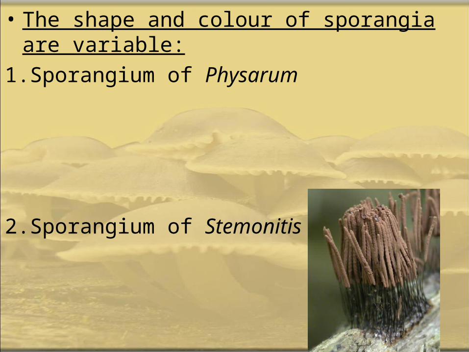
• The shape and colour of sporangia are variable:
1.Sporangium of Physarum
2.Sporangium of Stemonitis
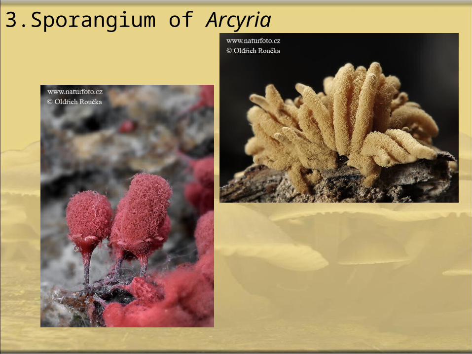
3.Sporangium of Arcyria

A. Plasmodiophora brassicaeCause (club root of cabbage/ crucifer) : it causes the
roots to produce abnormal swellings andmalformations through.
• Notes :
1.Hypertrophy : enlargement of cell size.
2.Hyperplasia : uncontrolled cell divisions (increase in cell divisions).
3.The infect cells are filled with the resting spores of the fungus.
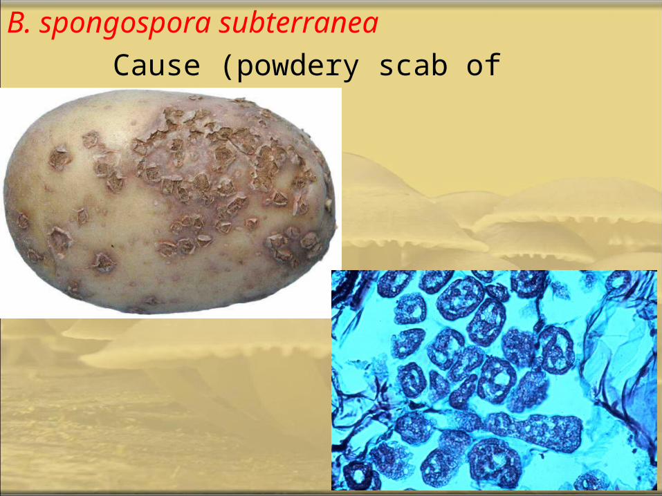
B. spongospora subterranea
Cause (powdery scab of potatoes).
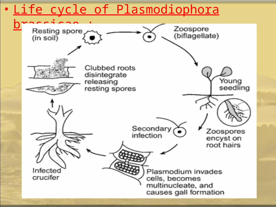
• Life cycle of Plasmodiophora brassicae :
• Plasmodiophora brassicae is an obligate parasite. It survives in the soil only as dormant cysts.
• Primary zoospores released from germinating cysts infect host root hairs by encysting on the root surface and entering through developing epidermal cells in the form of an amoeba like cell. Older roots can also be infected if wounding is present to provide an entrance to the pathogen.
• In the root hairs, amoeboid cells of the pathogen join together to form a multinucleate plasmodium. This plasmodium divides and forms multiple secondary zoospores, which are released into the soil. Secondary zoospores infect healthy parts of the initial host or infect nearby plants. These zoospores also enter through the host root hairs, but the infecting amoeboid cells migrate into the cortical cells of the host.
• Once in the cortex, the amoeboid pathogen infects one host cortical cell where it may multiply or join with other amoeboid cells to form a plasmodium. As the plasmodium develops, it releases plant hormones (IAA) which cause the host cells to enlarge up to 20 times of its normal size. As the plasmodium grows, it divides and infects neighboring cells causing them to enlarge. Clusters of these enlarged cells are responsible for the clubbing on the roots and are referred to as ‘Kankheitsherd’. These Kankheitsherd are diagnostic of P. brassicae and can be observed in cross sections of infected roots.
• Plasmodium in all host cells eventually undergo meiosis and develop into resting cysts. These new cysts will be released into the soil as other soil microorganisms decompose the club root.
Chytridiomycota: • Members of the division have unicellular redumentary to
mycelial thalli. • Their cell wall composition is mostly chitin. • Flagellated spores and gametes are produced. Gametes
and zoospores have a single, posterior whiplash flagellum.• Sexual reproduction is variable and may be isogamous,
anisogamous or oogamous.• The ultrastructure of zoospore is a definitve characteristic
of Chytridiomycota.
* The division has a single class, Chytridiomycetes, and three orders : Chytridiales , Blastocadiales, and Monoblepharidales
Order: Chytridiales:
• the most primitive members of the Chytridiomycota.
• commonly referred to as "chytrids".• Habitat : Fresh water.• The thallus is commonly unicellular and
may have limited hyphal growth, but is not considered to be mycelial. Hyphal cells are coenocytic except where there are reproductive structures.
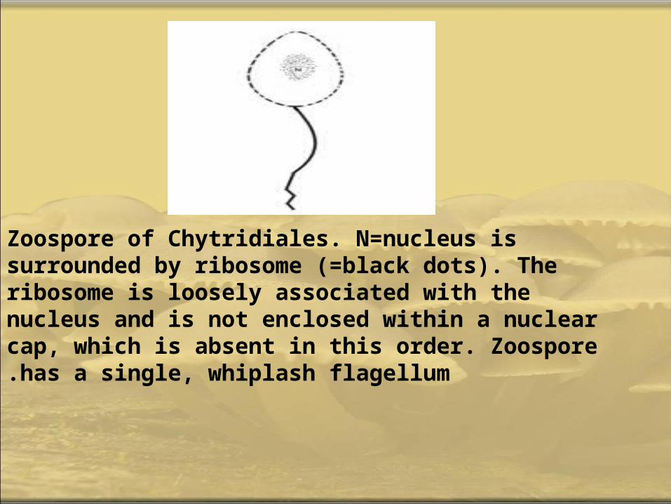
Zoospore of Chytridiales. N=nucleus is surrounded by ribosome (=black dots). The ribosome is loosely associated with the nucleus and is not enclosed within a nuclear cap, which is absent in this order. Zoospore has a single, whiplash flagellum.

• Rhizophydium sphaerotheca : Four visible zoosporangia growing on a pine pollen (one is out of the plane of focus and is behind one of the more visible sporangia). Zoospores can be seen in
• Two of the three visible zoosporangia. The third has released all of its zoospores. Rhizoids (not visible) anchor the zoosporangia to the substrate.
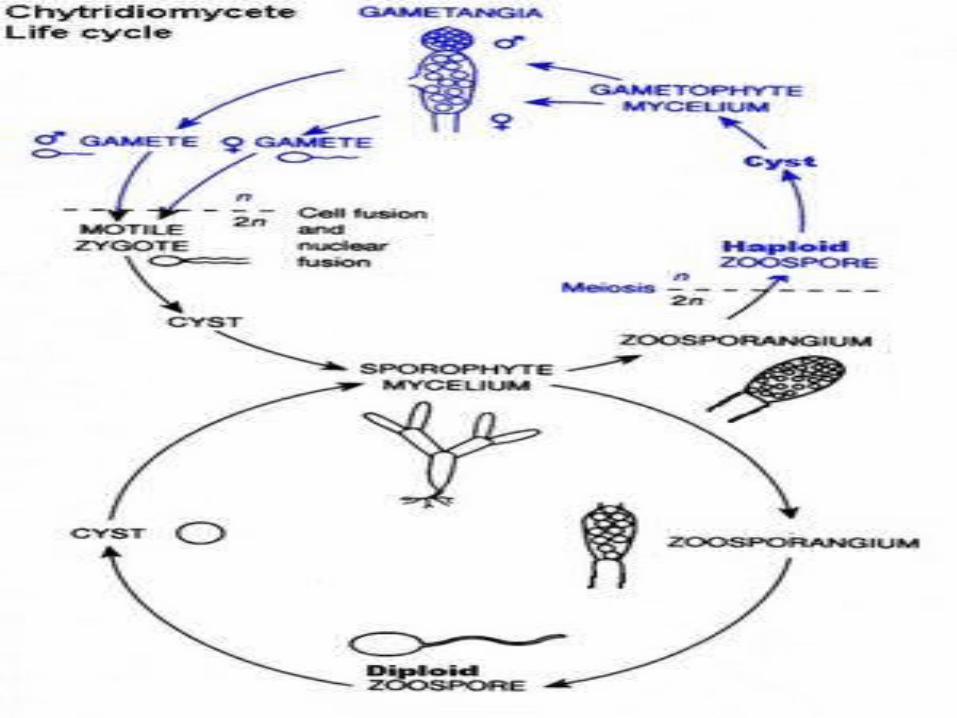
1. Asexually, Chytridiomycota reproduce through the use of zoospores.
2. In asexual reproduction, zoospores will swim until a desireable substrate is located.
3. The zoospore attaches itself, feeds off its host; the cytoplasm grows, meiotic divisions occur, and a cell wall forms around the original zoospore.
4. Protoplasm increases as the cell continues to develop. Finally, cleavage of the protoplasm occurs, which produces individual zoospores that are released through a pore.
5. Sexual reproduction is haploid dominant. It also depends on the isomorphic alternation of generations. The haploid thallus, called the gametothallus, produces female and male gametes
6. These occur in pairs and are terminal and subterminal. Male gametes are orange-colored, while female gametes are colorless.
7. In addition, female gametes are much larger than male gametes.
8. Males are attracted to females when they produce the hormone sirenin, and females are attracted to males when they produce the hormone parisin..
9. The diploid thallus is called the sporothallus.
10. The sporothallus produces two types of zoosporgia: zoosporgangium (meitosporangium) and resistant sporangium (meiosporangium).

11.Zoosporangia produce diploid zoospores, which can function as a means of asexual reproduction.
12.Sexual reproduction may be isogamous, anisogamous, or oogamous. One species, Allomyces macrogynus, has a sporic life cycle, something that occurs in plants but is rare to fungi.
13.Allomyces macrogynus is an example of an anisogamous species.