melanoma cell adhesion and spreading activities of a … · the journal of biological chemistry 0...
TRANSCRIPT
THE JOURNAL OF BIOLOGICAL CHEMISTRY 0 1993 by The American Society for Biochemistry and Molecular Biology, Inc
Vol. 268, No. 19. Issue of July 5, pp. 14153-14160, 1993 Printed in U.S.A.
Melanoma Cell Adhesion and Spreading Activities of a Synthetic 124-Residue Triple-helical “Mini-collagen”*
(Received for publication, January 4, 1993, and in revised form, February 23, 1993)
Cynthia G. Fields, Daniel J. Mickelson, Sandra L. Drake, James B. McCarthy, and Gregg B. Fields$ From the DeDartment of Laboratow Medicine and Pathology and The Biomedical Engineering Center, University of Minnesota, Minneapolis, Minnesota 55455
A great variety of cells, such as melanoma cells, fibroblasts, platelets, keratinocytes, and epithelial cells, adhere to and migrate on specific regions within the triple-helical domains of types I, 111, and IV colla- gen. The relative importance of collagen primary, sec- ondary, and tertiary structures on these cellular ac- tivities has not been ascertained, as no general syn- thetic methodology exists to allow for the study of peptides incorporating biologically active sequences in triple-helical conformation. We have thus developed a novel, generally applicable solid-phase branching methodology for the synthesis of aligned, triple-helical collagen-model polypeptides (Le. “mini-collagens”). Three nascent peptide chains are carboxyl-terminally linked through one Nu-amino and two Ilf-amino groups of Lys, while repeating Gly-Pro-Hyp triplets induce triple helicity. A homotrimeric triple-helical polypep- tide (THP) of 124 amino acids, incorporating residues 1263-1277 of al(IV) collagen, was synthesized. Highly metastatic mouse melanoma cells showed a profound preference for adhesion to this THP as compared with a single-stranded peptide (SSP) incorporating the same type IV collagen sequence or a branched peptide con- taining eight repeats of Gly-Pro-Hyp (designated GPP*). Specifically, 50% cell adhesion occurred at a THP concentration of 1.12 FM, while comparable levels of adhesion required [SSP] = 170 pM or [GPP*] > 100 PM. Melanoma cells also spread on the THP to a greater extent than on the SSP or GPP*. These results are the first direct demonstrations of the significance of triple helicity for cell adhesion to and spreading on a specific collagen sequence and support earlier conclusions of conformational dependency for cell adhesion to and migration on types I and IV collagen. In addition, the melanoma cell THP activities support the concept that tumor cell adhesion and spreading on type IV collagen involves multiple, distinct domains in triple-helical conformation. The triple-helical peptide synthetic pro- tocol developed here will allow eventually for the study of both structure and biological activity of specific,
* This work was supported by National Institutes of Health and Diabetes and Digestive and Kidney Diseases Grants KD 44494 (to G. B. F.) and CA 43924 and CA 54263 (to J. B. M.), Minnesota Medical Foundation Grant CRF-152-91, American Cancer Society Grant 13- 32-6, the Leukemia Task Force, University of Minnesota Grant-in- Aid of Research, Artistry, and Scholarship, and the Millipore Corp. The costs of publication of this article were defrayed in part by the payment of page charges. This article must therefore be hereby marked “aduertisement” in accordance with 18 U.S.C. Section 1734 solely to indicate this fact.
$ Recipient of a McKnight-Land Grant Professorship. To whom correspondence and reprint requests should be addressed Dept. of Laboratory Medicine and Pathology, Box 107, 420 Delaware St. S.E., University of Minnesota, Minneapolis, MN 55455. Tel.: 612-626-2446; Fax: 612-625-1121.
glycosylated collagen sequences in homotrimeric and heterotrimeric forms.
Tumor cell invasion and metastasis involves the adhesion and motility of tumor cells on extracellular matrix compo- nents, such as fibronectin and collagen. Collagens are distin- guished structurally from other extracellular matrix proteins by their composition of three a chains of primarily repeating Gly-X- Y triplets, which induces each a chain to adopt a left- handed poly-Pro I1 helix. Three left-handed chains then intertwine to form a right-handed superhelix. Triple-helical conformation is conducive for adhesion of a variety of cells to types I and IV collagen (Rubin et al., 1981; Santoro, 1986; Aumailley and Timpl, 1986; Vandenberg et al., 1991; Gold- man et al., 1992; Gullberg et al., 1992) and the migration of keratinocytes on type I collagen (Scharffetter-Kochanek et al., 1992) and has been implicated as an essential feature for interstitial collagen catabolism (Fields, 1991). Several se- quences within the triple-helical domains of types I, 111, and IV collagen have been identified as cell adhesion sites. The adhesion of Chinese hamster ovary cells to type I collagen is inhibited by the 757-791 sequence of the rat al(I) chain (Kleinman et al., 1978). The cy2& integrin from human fibro- blasts and platelets adheres to a peptide incorporating resi- dues 430-442 of the rat al(I) chain (Staatz et d . , 1991), while human platelet aggregation is inhibited by a peptide incor- porating residues 76-84 of the bovine al(III) chain (Legrand et al., 1980). Residues 531-543 of the human al(IV) chain promote human keratinocyte adhesion and rabbit corneal epithelial cell adhesion and migration (Wilke and Furcht, 1990; Cameron et al., 1991). A peptide incorporating residues 1263-1277 from the cyI chain of human type IV collagen supports adhesion, spreading, and motility of mouse and human melanoma and rat glioma and neuroblastoma cell lines (Chelberg et al., 1990).
Prior studies on cell adhesion to triple-helical collagen sequences have utilized only single-stranded peptides, even though disruptions of collagen secondary and tertiary struc- tures have been shown to be detrimental for adhesion. For example, the initial rate of rat hepatocyte adhesion was more rapid to native, triple-helical collagen compared with dena- tured collagen due to conformationally-dependent alp1 inte- grin binding sites (Rubin et al., 1981; Gullberg et al., 1992), while platelets adhered effectively to native, but not dena- tured, type I collagen (Santoro, 1986). Human fibrosarcoma (HT 1080) cells adhered to the major triple-helical domain and cyanogen bromide-derived CB3 fragment (residues 291- 576) of type IV collagen, but not to the denatured domain or fragment (Aumailley and Timpl, 1986; Vandenberg et al., 1991). Cell migration and aggregation is also influenced by triple helicity, as chemotaxis of human keratinocytes was twice as great on native uersus denatured type I collagen
14153

14154 Melanoma-adhesive Mini-collagen
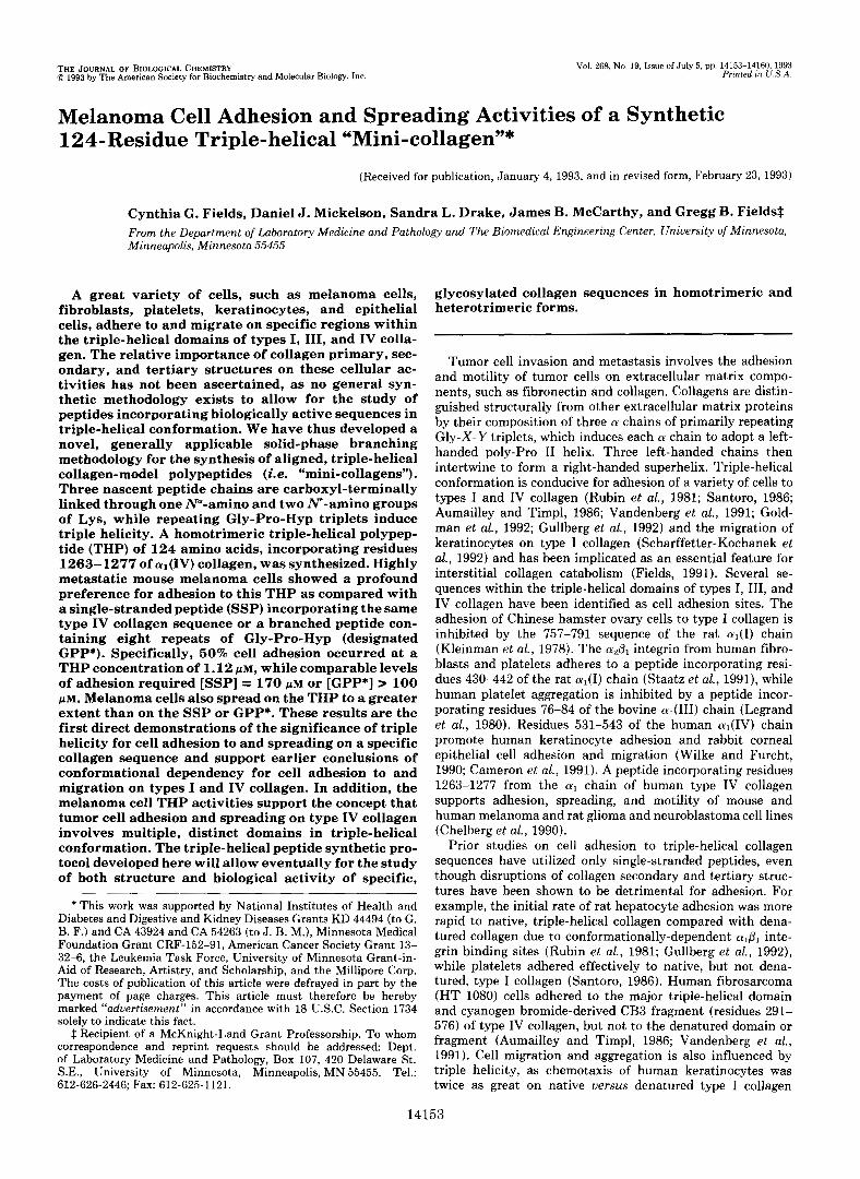
(Gly-Pro-Hyp)8-Gly-Val-Lys-Gly-Asp-Lys-Gly-Asn-Pro-Gly-Trp-Pro-Gly-Ala-Pro-AHA
(Gly-Pro-Hyp)8-Gly-Val-Lys-Gly-Asp-Lys-Gly-Asn-~o-Gly-Trp-Pro-Gly-Ala-Pro-AHA-Lys-L 1 s-Tyr-Gly
(Gly-Pro-Hyp)8-Gly-Val-Lys-Gly-Asp-Lys-Gly-A~-~o-Gly-Trp-Pro-Gly-Ala-Pr~A~ 1
(Gly-Pro-Hyp)s-AHA
FIG. 1. Sequences of THP (top), SSP (middle), and GPP* (bottom). T h e T H P is composed of a carboxyl-terminal branch generated from 2 Lys residues, 1 al(I) 1263-1277 sequence per chain, and 8 Gly-Pro-Hyp repeats per chain. The SSP is composed of the a1(I) 1263- 1277 sequence. GPP* is composed of the carboxyl-terminal branch and 8 Gly-Pro-Hyp repeats per chain.
(Scharffetter-Kochanek et al., 1992), while platelet aggrega- tion was induced by only triple-helical, not denatured, cyan- ogen bromide-derived fragments of types I and I11 collagen (Zijenah and Barnes, 1990). These results suggest that a general synthetic peptide methodology needs to be developed by which the importance of collagen triple-helical structure on cellular activities can be ascertained. Additionally, this unique methodology should allow for the incorporation of post-translationally modified amino acids, as both of the cell adhesion sequences within type IV collagen contain O-glyco- sylated Hyl residues (Babel and Glanville, 1984). Cell-surface galactosyltransferase has been shown to mediate fibroblast adhesion to type IV collagen via glycosylated residues (Ba- biarz and Cullen, 1992). We describe presently the three- dimensional orthogonal solid-phase synthesis and character- ization of a triple-helical polypeptide (THP)’ incorporating
The abbreviations used are: THP, triple-helical peptide; AHA, 6- aminohexanoic acid; AI, allyl; allyl linker, 4-trityloxy-Z-but-2-enylox- yacetic acid dicyclohexylamine salt; Boc, tert-butoxycarbonyl; DBU, 1,8-diazabicyclo[5.4.0]undec-7-ene; DCM, dichlorometbane; DIEA, N,N-diisopropylethylamine; DIPCDI, N,N”diisopropylcarbodiimide; DMF, N,N-dimethylformamide; EDT, 1,2-etbanedithiol; FDAA, 1- fluoro-2,4-dinitrophenyl-5-~-alaninamide; Fmoc, N-fluoren-g-ylme- thoxycarbonyl; GPP*, [N-tris[(Gly-Pro-Hyp),-AHA]-Lys-Lysl-Tyr- Gly; HBTU, 2-(1H-benzotriazole-l-yl)-l,l,3,3-tetramethyluronium hexafluoropbosphate; HMP, 4-hydroxymethylphenoxy; HOBt, l-hy- droxybenzotriazole; HPLC, high performance liquid chromatography; MBHA, 4-methylbenzhydrylamine; Nle, norleucine; NMP, l-methyl- 2-pyrrolidinone; PBS, phosphate-buffered saline solution; (Ph,P),Pd, tetrakis(triphenylphosphine)palladium(O); resin, copoly(styrene-1%- divinylbenzene); PAGE, polyacrylamide gel electrophoresis; SSP, sin- gle-stranded peptide; TFA, trifluoroacetic acid.
residues 1263-1277 from the a1 chain of type IV collagen (Fig. 1). A covalent branching scheme was developed for the syn- thesis of the THP, in similar fashion to branching schemes utilized for the syntheses of a-helical bundle proteins (Hahn et al., 1990; Mutter et al., 1992) and multiple antigen peptide systems (Tam, 1988). Also synthesized were a single-stranded peptide (SSP) incorporating residues 1263-1277 from the a1 chain of type IV collagen and a branched peptide containing eight repeats of Gly-Pro-Hyp (designated GPP*) (Fig. 1). The adhesion and spreading activities of highly metastatic mela- noma cells were quantitated as a function of THP, SSP, or GPP* concentration.
EXPERIMENTAL PROCEDURES
Materials-All standard peptide synthesis chemicals were analyt- ical reagent grade or better and purchased from Applied Biosystems, Inc. (Foster City, CA) or Fisher. DBU, EDT, and (Ph3PLPd were from Aldrich, ~ - H y p and FDAA from Sigma, Gly-Pro-Hyp and MBHA resin (substitution level = 0.80 mmol/g) from Bachem Bios- ciences (Philadelphia, PA), Fmoc-Gly-HMP resin (substitution level = 0.43 mmol/g) from Millipore Corp., Fmoc-Tyr(A1) and allyl linker from Propeptide (Vert-Le-Petit, France), HBTU from Richelieu Bio- technologies (St.-Hyacinthe, Quebec), Fmoc-AHA and Fmoc-Nle from Advanced ChemTech (Louisville, KY), and Fmoc-Hyp(tBu) from Novabiochem (La Jolla, CA). All other Fmoc-amino acids were from Bachem Biosciences or Millipore. Amino acids are of the L- configuration (except for Gly) except where noted. The synthesis, purification, and characterization of the SSP have been described (Chelberg et al., 1990), while that of GPP* will he described else- where.’
* C. G. Fields and G. B. Fields, manuscript in preparation.
Melanoma-adhesive Mini-collagen 14155
Preparation of Fmoc-Gly-Pro-Hyp-Fmoc-Gly-Pro-Hyp was syn- thesized from Gly-Pro-Hyp using an established protocol for Fmoc- amino acids (Fields et al., 1989). A typical preparation was as follows. 3.0 g of Gly-Pro-Hyp (10.5 mmol) was dissolved in 54 ml of Na2C03/ water (1:9) and stored at 4 "C. 4.05 g of N-fluoren-9-ylmethyl succi- nimidyl carbonate (12.0 mmol) was dissolved in 45 ml of dimethoxy- ethane and stirred at 4 "C. The aqueous Na2C03 solution was added slowly to the dimethoxyethane solution, and the reaction proceeded for 2.5 h at 4 "C and 21 h at room temperature. The solution was filtered, and 360 ml of water was added to the filtrate. The aqueous layer was extracted with 300 ml of diethyl ether, acidified to pH 2 with concentrated HC1, reduced to half-volume at 80 "C under re- duced pressure, and stored at 4 "C for 24 h. The aqueous layer was decanted from the oily precipitate, reduced to -30 ml at 81 "C under reduced pressure, and stored at 4 "C for 24 h. The aqueous layer was decanted from the oily precipitate. Both oily precipitates were dis- solved in a total of 20 ml of methanol, then 250 ml of ethyl acetate was added. A white residue was recovered by evaporation at 73 "C for 1 h under reduced pressure; yield 3.23 g (6.39 mmol, 60.6%). The identity of the product as Fmoc-Gly-Pro-Hyp and its homogeneity was verified by thin layer chromatography (chloroform/methanol/ acetic acid (95:20:3)), scanning UV spectroscopy, and Edman degra- dation sequence analysis.
Preparation of Fmoc-Gly-Allyl Resin-0.848 g of Fmoc-Nle (2.4 mmol) was coupled to 2.0 g of MBHA-resin (1.6 mmol) with 0.367 g of HOBt (2.4 mmol) and 0.373 ml of DIPCDI (2.4 mmol) in 20 ml of DCM/DMF (1:l) for 2.3 h. The resin was washed three times with DMF, deprotected with 20 ml of piperidine/DMF (1:l) for 30 min, and washed three times with DMF. 1.82 g of allyl linker (3.2 mmol) was coupled to the resin with 0.489 g of HOBt (3.2 mmol) and 0.497 ml of DIPCDI (3.2 mmol) in 20 ml of DMF for 18.5 h. The resin was washed once with DMF and twice with DCM, deprotected twice with 20 ml of TFA-DCM (9:1), first for 20 min, then for 10 min, washed three times with DCM, neutralized with 20 ml of DIEA/DCM (1:9) for 15 min, and washed once with DCM and twice with DMF. 1.43 g of Fmoc-Gly (4.8 mmol) was esterified to the allyl resin with 0.735 g of HOBt (4.8 mmol), 0.746 ml of DIPCDI (4.8 mmol), and 0.059 g of 4-(dimethy1amino)pyridine (0.48 mmol) in 20 ml of DMF for 6.5 h. The resin was washed once with DMF and twice with DCM and stored under vacuum overnight.
Preparation of [N-tris[Fmoc-AHA]-Lys-Lys]-Tyr(Al)-Gly-Allyl Resin-Fmoc-Tyr(Al), Fmoc-Lys(Boc), and Fmoc-Lys(Boc) were coupled to Fmoc-Gly-allyl resin with 1.40 mmol of Fmoc-amino acid, 0.215 g of HOBt (1.40 mmol), and 0.218 ml of DIPCDI (1.40 mmol) in 20 ml of DMF for 2-4 h. Both Fmoc-Lys(Boc) residues were double-coupled. Fmoc removal was by 20 ml of piperidine/DMF (1:1) for 0.5 h. The peptide-resin was washed three times with DMF after each coupling and deprotection, then once with DCM prior to removal of the Boc groups. The N-amino Boc groups were removed by treatment of Fmoc-Lys(Boc)-Lys(Boc)-Tyr(A1)-Gly-allyl resin with 20 ml of TFA/DCM (1:l) for 0.5 h. The peptide-resin was washed three times with DCM, neutralized with 20 ml of DIEA/DCM (1:9) for 0.5 h, washed twice with DCM and once with DMF, and Fmoc- deprotected as described above. Fmoc-AHA was double-coupled for 2.5-3 h as described above using 4.10 mmol of Fmoc-AHA (1.45 g), HOBt (0.630 g), and DIPCDI (0.638 ml). The substitution level was determined by fulvene-piperidine analysis (Fields et al., 199213) to be 0.181 mmol/g.
Peptide Synthesis and Purification-Incorporation of individual
-Lys I -2 -Gly +-" I
linker
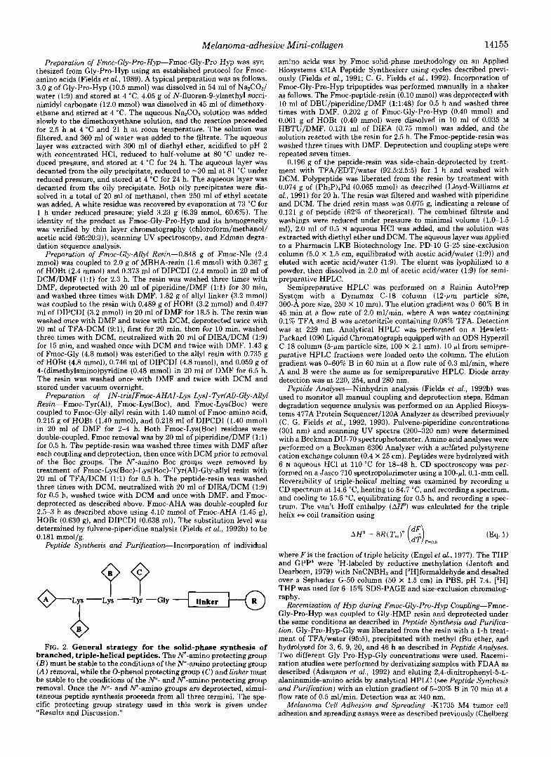
FIG. 2. General strategy for the solid-phase synthesis of branched, triple-helical peptides. The "amino protecting group ( B ) must be stable to the conditions of the N"-amino protecting group ( A ) removal, while the 0-phenol protecting group (C) and linker must be stable to the conditions of the N"- and N-amino protecting group removal. Once the N"- and "amino groups are deprotected, simul- taneous peptide synthesis proceeds from all three termini. The spe- cific protecting group strategy used in this work is given under "Results and Discussion."
amino acids was by Fmoc solid-phase methodology on an Applied Biosystems 431A Peptide Synthesizer using cycles described previ- ously (Fields et al., 1991; C. G. Fields et al., 1992). Incorporation of Fmoc-Gly-Pro-Hyp tripeptides was performed manually in a shaker as follows. The Fmoc-peptide-resin (0.10 mmol) was deprotected with 10 ml of DBU/piperidine/DMF (1:1:48) for 0.5 h and washed three times with DMF. 0.202 g of Fmoc-Gly-Pro-Hyp (0.40 mmol) and 0.061 g of HOBt (0.40 mmol) were dissolved in 10 ml of 0.035 M HBTU/DMF. 0.131 ml of DIEA (0.75 mmol) was added, and the solution reacted with the resin for 2.5 h. The Fmoc-peptide-resin was washed three times with DMF. Deprotection and coupling steps were repeated seven times.
0.196 g of the peptide-resin was side-chain-deprotected by treat- ment with TFA/EDT/water (92.5:2.5:5) for 1 h and washed with DCM. Polypeptide was liberated from the resin by treatment with 0.074 g of (Ph3P),Pd (0.065 mmol) as described (Lloyd-Williams et al., 1991) for 20 h. The resin was filtered and washed with piperidine and DCM. The dried resin mass was 0.075 g, indicating a release of 0.121 g of peptide (62% of theoretical). The combined filtrate and washings were reduced under pressure to minimal volume (1.0-1.5 ml), 2.0 ml of 0.5 N aqueous HCl was added, and the solution was extracted with diethyl ether and DCM. The aqueous layer was applied to a Pharmacia LKB Biotechnology Inc. PD-10 G-25 size-exclusion column (5.0 X 1.5 cm, equilibrated with acetic acid/water (1:9)) and eluted with acetic acid/water (1:9). The eluent was lyophilized to a powder, then dissolved in 2.0 ml of acetic acid/water (1:9) for semi- preparative HPLC.
Semipreparative HPLC was performed on a Rainin AutoPrep System with a Dynamax C-18 column (12-pm particle size, 300-A pore size, 250 X 10 mm). The elution gradient was 0-60% B in 45 min at a flow rate of 2.0 ml/min, where A was water containing 0.1% TFA and B was acetonitrile containing 0.08% TFA. Detection was at 229 nm. Analytical HPLC was performed on a Hewlett- Packard 1090 Liquid Chromatograph equipped with an ODS Hypersil C-18 column (5-pm particle size, 100 X 2.1 mm). 10 pl from semipre- parative HPLC fractions were loaded onto the column. The elution gradient was 0-60% B in 60 min at a flow rate of 0.3 ml/min, where A and B were the same as for semipreparative HPLC. Diode array detection was at 220, 254, and 280 nm.
Peptide Analyses-Ninhydrin analysis (Fields et al., 1992b) was used to monitor all manual coupling and deprotection steps. Edman degradation sequence analysis was performed on an Applied Biosys- tems 477A Protein Sequencer/l20A Analyzer as described previously (C. G. Fields et al., 1992, 1993). Fulvene-piperidine concentrations (301 nm) and scanning UV spectra (200-320 nm) were determined with a Beckman DU-70 spectrophotometer. Amino acid analyses were performed on a Beckman 6300 Analyzer with a sulfated polystyrene cation exchange column (0.4 X 25 cm). Peptides were hydrolyzed with 6 N aqueous HCI at 110 "C for 18-48 h. CD spectroscopy was per- formed on a Jasco 710 spectropolarimeter using a 100-pl, 0.1-mm cell. Reversibility of triple-helical melting was examined by recording a CD spectrum at 14.6 "C, heating to 84.7 "C, and recording a spectrum, and cooling to 15.6 "C, equilibrating for 0.5 h, and recording a spec- trum. The van't Hoff enthalpy (&) was calculated for the triple helix ej coil transition using
where F is the fraction of triple helicity (Engel et al., 1977). The THP and GPP* were 3H-labeled by reductive methylation (Jentoft and Dearborn, 1979) with NaCNBH3 and [3H]formaldehyde and desalted over a Sephadex G-50 column (50 X 1.5 cm) in PBS, p H 7.4. [3H] THP was used for 6-15% SDS-PAGE and size-exclusion chromatog- raphy.
Racemization of Hyp during Fmoc-Gly-Pro-Hyp Coupling-Fmoc- Gly-Pro-Hyp was coupled to Gly-HMP resin and deprotected under the same conditions as described in Peptide Synthesis and Purijica- tion. Gly-Pro-Hyp-Gly was liberated from the resin with a 1-h treat- ment of TFA/water (95:5), precipitated with methyl tBu ether, and hydrolyzed for 3, 6, 9, 20, and 46 h as described in Peptide Analyses. Two different Gly-Pro-Hyp-Gly concentrations were used. Racemi- zation studies were performed by derivatizing samples with FDAA as described (Adamson et al., 1992) and eluting 2,4-dinitrophenyl-5-~- alaninamide-amino acids by analytical HPLC (see Peptide Synthesis and Purification) with an elution gradient of 5-20% B in 70 min at a flow rate of 0.5 ml/min. Detection was at 340 nm.
Melanoma Cell Adhesion and Spreading"K1735 M4 tumor cell adhesion and spreading assays were as described previously (Chelberg

14156 Melanoma-adhesive Mini-collagen
i 1800
1400
1200 FIG. 3. Analytical HPLC elution
profile of the purified THP. The 3 1000 THP elution time was 30.9 min. Condi- tions are given under “Experimental Procedures.”
J+ 1 0 2 0 3 0 4 0
Time (min.)
0
-10
-20
-30
et al., 1990) with minor alterations. Peptides were dissolved in PBS and adsorbed directly onto 96-well polystyrene Immulon 1 plates (Dynatech Laboratories Inc., Chantilly, VA) overnight a t 37 “C. Non- specific binding sites were blocked with 5 mg/ml bovine serum albu- min in adhesion media (Dulbecco’s modified Eagle’s medium contain- ing 20 mM HEPES) for 2 h a t 37 “C. Tumor cells were released from tissue culture flasks with 37 “C PBS containing 10 mM EDTA and washed several times with adhesion media. Cells were labeled over- night with 1 pCi/ml 13H]thymidine (Du Pont-New England Nuclear) for adhesion assays. Cells were added to the plate wells a t a density of 50,000 cells/ml in a total volume of 100 pl and adhered for 1 h a t 37 “C. For adhesion assays, wells were washed several times with adhesion media, the remaining cells were lysed, and radioactivity was determined as described (Chelberg et al., 1990). For spreading assays, wells were fixed and stained using DiffQuik reagents (Baxter) and photographed with a Nikon MF-15 camera mounted on a Nikon Diaphot inverted microscope at 2 0 0 ~ magnification. Cell spreading was quantitated by an Optomax System IV image analyzer equipped with a Hitachi Monitor. The efficiency of peptide adsorption to the
FIG. 4. Circular dichroism spec- tra of the THP in acetic acid/water (1:19), pH 2.4. Spectra were recorded at [THP] = 83.5 pM by accumulating 5 scans at 0.5 nm intervals (response of 1 s). The spectrum at 14.6 “C is character- istic of an intact triple-helix, while the spectrum at 84.7 “C indicates no triple- helix is present (see “Results and Dis- cussion”).
Immulon plates was determined as described (Chelberg et al., 1990) using 3H- or ‘2”I-labeled peptide.
RESULTS AND DISCUSSION
Design, Synthesis, and Characterization of the Triple-helical Polypeptide-To ensure proper alignment of three peptide strands in a triple-helix, a branching protocol was developed based on the liquid-phase synthetic scheme of Heidemann and co-workers (Roth et al., 1979). The branch was introduced at the carboxyl terminus of the synthetic peptide, consistent with the natural nucleation of collagen triple-helices from the carboxyl to amino terminus (Engel and Prockop, 1991). Branching of three peptide strands from one initial chain required three different protecting group strategies (Fig. 2): W-amino protection ( A ) , Lys side chain protection ( B ) , which must be stable to the N”-amino protecting group re-

Melanoma-adhesive Mini-collagen 14157
FIG. 5. Thermal transition curve of the THP in acetic acidlwater (1:19), pH 2.4. Molar ellipticities ( [ H I ) were recorded at X = 225 nm while the temperature was increased from 15 "C to 85 "C. The triple-helix was somewhat de- stabilized by aggregation of the THP from 15-30 "C. The triple-helix was sta- ble from 30-48 "C, then melted with T,,, = 58.5 "C.
0
A
.o -
.o -
0 -
.o -
-
I I I I I I I I I
I I
I I I I I I I I I I I I I I I I I I
, I 1 1 I I , 1
20 30 40 50 60 70
2 0 4 0 6 0 8 0 1 0 0 0 2 0 4 0 Fraction Number
moval conditions, and C"-carboxyl protection (linker), which must be stable to the N"-amino and Lys side chain protecting group removal conditions. As eventual incorporation of gly- cosylated Hyl is desired, the synthetic scheme could not use repetitive acidolysis for W-amino protecting group removal or strong acidolysis or alkali for C-carboxyl protecting group removal (cleavage of the peptide-linker). This desired syn- thetic scheme was achieved with a mild three-dimensional orthogonal3 protecting group strategy. N"-Amino protection
~ ~ ~ . ~ ~ ~ ~ . ~ ~ ~ _ _ _ _ _ _ _ . ~ ~ . ~
An orthogonal system has been defined as a set of completely independent classes of protecting groups, such that each class of
60 &I 100 120
FIG. 6. Elution profile from Seph- adex G-50-80 (100 X 1.0 cm) size- exclusion chromatography of [3H] THP at 4 "C ( A ) or 35 "C ( B ) . Sam- ples were run in PBS containing 1% Triton at a flow rate of 5.0 ml/h. The column, buffer, and samples were equil- ibrated at 4 "C or 35 "C for 12 h. Sample volumes were 750 pl, collected fractions 1.5 ml, and collection time 20 h. Fraction radioactivity was quantitated by a Beck- man LS 3801 Liquid Scintillation Coun- ter. THP estimated molecular mass = 11.6 kDa at 4 "C and 36.0 kDa at 35 "C.
( A ) was the Fmoc group, which is base-labile and stable to acidolysis and (Ph3P)rPd-catalyzed nucleophilic transfer (Kunz and Dombo, 1988; Fields et al., 1992b). Lys side chain protection ( B ) was the Boc group, which is moderate acid- labile and stable to base and (Ph3P).,Pd-catalyzed nucleophilic transfer (Blankemeyer-Menge and Frank, 1988; Fields et al., 199213). (?"-Carboxyl protection was the allyl linker, which is labile to (Ph3P),Pd-catalyzed nucleophilic transfer and acid- and base-stable (Guibi et al., 1989; Kunz et al., 1989; Lloyd-
group can be removed in any order and in the presence of all other classes (Barany and Merrifield, 1977; Barany and Albericio, 1985).

14158 Melanoma-adhesive Mini-collagen
Frc. 7. Adhesion of melanoma cells as a function of THP, SSP, or GPP* concentration. Cells were al- lowed to adhere to peptide-coated Im- mulon plates for 1 h at 37 "C. Melanoma cell adhesion was most specific for the THP, where 50% adhesion occurred at a concentration of 1.12 pM, Half-maximal adhesion to SSP and GPP' occurred at concentrations of 170 and >lo0 pM, re- spectively. Conditions are given under "Experimental Procedures."
Williams et al., 1991). Allyl-based side chain protection was used for Tyr (C).
Branching was achieved by synthesizing Fmoc-Lys(Boc)- Lys(Boc)-Tyr(A1)-Gly-allyl resin and deprotecting the Ne- and "amino groups. Fmoc-AHA was then incorporated onto all three amino termini to provide a flexible spacer, The specific sequence from the a1 chain of type IV collagen (resi- dues 1263-1277) was assembled with Fmoc-amino acids. Fmoc-Gly-Pro-Hyp triplets were then incorporated following the specific type IV collagen sequence, as repeating Gly-Pro- Hyp sequences form stable triple-helices at relatively short chain lengths (Sakakibara et al., 1973; Engel et al., 1977). Since the desired THP was 124 residues, rapid coupling and deprotection reagents were used to ensure high synthetic efficiency. Coupling of Fmoc-amino acids and Fmoc-Gly-Pro- Hyp was achieved with HBTU, which is highly reactive (Fields et al., 1991) and utilized with optimal peptide-resin solvation conditions (Fields and Fields, 1991). Fragment con- densation of Fmoc-Gly-Pro-Hyp tripeptides proceeded smoothly, with less than 0.5% ~ - H y p per Fmoc-Gly-Pro-Hyp incorporated. Rapid removal of the Fmoc group from Fmoc- Gly-Pro-Hyp was achieved with 2% DBU plus 2% piperidine (for scavenging dibenzofulvene) in DMF. DBU removal of the Fmoc group is rapid even in "difficult" sequences (Wade et al., 1991). Solid-phase sequence analysis gave the desired sequence [ (Gly-Pro-Hyp),-Gly-Val-Lys-Gly-Asp-Lys-Gly- Asn-Pro-Gly-Trp-Pro-Gly-Ala-Pro] and 3% cumulative pre- view, indicating that the synthesis of the homotrimeric pep- tide was highly efficient. Polypeptide-resin mass following synthesis was 0.694 g (91% of theoretical). Following side chain deprotection and cleavage of the polypeptide-resin, removal of the majority of the (PhaP)rPd was achieved by dissolving the (Ph,P),Pd-polypeptide complex in 0.5 N HCl, extracting with diethyl ether and DCM, and chromatograph- ing on a G-25 column. Semipreparative HPLC purification yielded a homogeneous polypeptide, as determined by analyt- ical HPLC (Fig. 3), SDS-PAGE, and size-exclusion chroma- tography. The apparent molecular mass of the purified THP was 11.6 kDa by size-exclusion chromatography at 4 "C (cal- culated molecular mass = 11,205 Da). Mass of the purified THP was 6.7 mg, 4.7% of the overall theoretical yield. Non- covalent sequence analysis gave the sequence (Gly-Pro-Hyp)a- Gly-Val-Lys. Amino acid identification could not proceed after Lys-27 due to gradual peptide loss from the filter and/ or inefficient amino acid yield per cycle. Amino acid analysis
Lo9 [PePtw (PM)
ratios were Gly 40 (40 expected), Pro 30.2 (31), Hyp 25.8 (24), Asx 6.4 (6) , Ala 3.2 (3), Val 2.8 (3), Tyr 1.3 ( l ) , Lys 8.8 (8), and AHA 3.4 (3). The integrity of the Trp residues was established by scanning UV spectroscopy.
The THP CD spectrum at 14.6 "C exhibited a large negative [@I200 and a positive [8]225 (Fig. 4), distinguishing features of a coiled-coil triple-helix (Heidemann and Roth, 1982). The CD spectrum at 84.7 "C (Fig. 4) was indicative of a melted triple- helix, as [8]225 was negative (Heidemann and Roth, 1982). Melting o f the triple-helix was reversible. To determine the triple-helix melting temperature (triple-helix - coil transi- tion), [8]225 was monitored as a function of temperature. Two transitions were seen, one with a midpoint at 23.5 "C and one with a midpoint of 58.5 "C (Fig. 5 ) . Size-exclusion chromatog- raphy (Fig. 6) gave apparent THP molecular masses of 11.6 kDa at 4 "C and 36.0 kDa at 35 "C, indicating the first transition was an aggregation phenomenon. Aggregation has been observed previously by x-ray diffraction and electron microscopy studies of triple-helical (Pro-Pro-Gly)zo and (Gly- Pro-Hyp), polypeptides (Andreeva et al., 1963; Millionova et al., 1963; Olsen et al., 1971). Although the large number of Hyp residues in the Y position of the THP could promote aggregation via intermolecular hydrogen bonding (Ramachan- dran et al., 1973; Nkmethy and Scheraga, 1986), aggregation induced by increasing temperature from 15-30 "C is indicative of a hydrophobically driven process (Fields et al., 1992a). The second THP transition, a triple-helical melt at 58.5 "C, was of a similar value to T,,, = 57.5-61 "C for (Pro-Hyp-Gly),, in 0.6-10% acetic acid/water (Sakakibara et al., 1973; Engel et al., 1977; Brodsky et al., 1992; Long et al., 1992). The van't Hoff enthalpy ( A P ) for the THP melt was -97 kcal/mol, similar to LW = -90 kcal/mol (Engel et al., 1977) and -124 kcal/mol (Long et al., 1992) for the triple-helix w coil tran- sition of (Pro-Hyp-Cly)lo.
Melanoma Cell Activities of the Triple-helical Polypeptide- Melanoma cell adhesion was compared for the THP, SSP, and GPP* (sequences given in Fig. 1) over a coated peptide concentration range of 0.1-300 pM. For the SSP concentra- tion, a molecular mass of 4797 Da was used, where 1 mol of SSP accounted for three peptide chains. This conversion allowed for 1 mol of THP, SSP, or GPP* to represent three potential peptide active sites. The coating efficiencies for the three peptides were comparable, ensuring that cell adhesion results would not be a reflection of differential peptide ad- sorption to the plates. Half-maximal melanoma cell adhesion

Melanoma-adhesive Mini-collagen 14159
FIG. 8. Comparison of spreading of melanoma cells on THP ( A ) , SSP ( B ) , and GPP* (C). Photographs shown are representa- tive examples of spreading on peptide-coated Immulon plates a t [THP] = 0.9 p ~ , [SSP] = 2.1 p ~ , and [GPP*] = 1.4 p ~ . Cells adhered and spread for 1 h a t 37 "C, then were fixed and stained with DiffQuik reagents. Conditions are given under "Experimental Procedures."
occurred at [THP] = 1.12 p ~ , [SSP] = 170 p ~ , and [GPP*] > 100 pM (Fig. 7). Thus, triple-helical conformation in com- bination with the aI(IV) 1263-1277 sequence resulted in a 100-fold increase in melanoma cell adhesion activity com- pared with the aI(IV) 1263-1277 sequence alone. This result
is the first direct demonstration of the significance of triple helicity for cell adhesion to a specific collagen sequence. Prior studies had concluded that alP1 and a& integrin-mediated cell adhesion to types I and IV collagen was conformation- dependent, as denaturing collagen resulted in little or no integrin-mediated adhesion (Santoro, 1986; Vandenberg et al., 1991; Gullberg et al., 1992). The level of melanoma cell adhe- sion to GPP* was similar to that reported previously for rat hepatocyte adhesion to (Pro-Pro-Gly)lo and (Pro-Hyp-Gly), (Rubin et al., 1981) and platelet adhesion to (Pro-Gly-Pro), (Zijenah and Barnes, 1990). Intact triple-helical structure may, independent of sequence, serve as a low level recognition feature for cells, as platelet adhesion to types I or I11 collagen was not inhibited by (Pro-Gly-Pro), (Zijenah and Barnes, 1990). Structural elements other than triple-helices, such as polyproline, do not appear to direct collagen-mediated cell adhesion (Rubin et al., 1981).
Melanoma cell spreading was compared for the THP, SSP, and GPP* at [THP] = 0.9 pM, [SSP] = 2.1 p ~ , and [GPP*] = 1.4 p ~ . Cell spreading was more extensive on the THP (Fig. 8A) than on either the SSP (Fig. 8 B ) or GPP* (Fig. 8C). Cell areas averaged 0.048, 0.012, and 0.017 mm*/pM peptide in response to THP, SSP, and GPP*, respectively. As in the case of melanoma cell adhesion, cell spreading was most efficient when triple-helicity was combined with the al(IV) 1263-1277 sequence. This result is consistent with conclu- sions from prior studies that cell migration or motility on collagen or collagen sequences can be conformation-depend- ent. Human keratinocyte migration was twice as great on triple-helical uersus denatured type I collagen (Scharffetter- Kochanek et al., 1992). A single-stranded peptide model of the al(IV) 1263-1277 sequence showed substantially de- creased melanoma spreading activity when the three imino acids (Pro'*'', and Pro'?'') were replaced by Ala (Chel- berg et al., 1990). This reduced activity was correlated by two- dimensional 'H NMR to the absence of a double P-turn structure in the carboxyl-terminal region of the peptide (Mayo et al., 1991). Although triple helicity per se was not a factor in these single-stranded peptides, the relative importance of secondary structure for melanoma cell spreading on a colla- gen-model sequence was clearly demonstrated.
The combined melanoma cell T H P adhesion and spreading activities support the concept that tumor cell adhesion and spreading on type IV collagen involves multiple, distinct domains, as a t least two domains within type IV collagen in triple-helical conformation are tumor cell adhesion sites (Chel- berg et al., 1989; Vandenberg et al., 1991). In addition, the enhancement of cellular activities due to triple helicity con- firms the aI(IV) 1263-1277 sequence as a specific melanoma cell adhesion and spreading site, as this sequence in its native conformation has greater activity than the isolated sequence and implies that basement membrane type IV collagen is a site for tumor cell invasion based on collagen primary, sec- ondary, and tertiary structures. Other activities that are col- lagen-mediated, such as cell adhesion to sites in types I, 111, and IV collagen and matrix metalloproteinase cleavage of types I-IV collagen at specific sites, are accessible to study by the synthetic method described here. We have demonstrated that homotrimeric mini-collagens can be synthesized without repetitive or strong acidolysis and used to study the relative influences of collagen primary, secondary, and tertiary on cellular behavior. By utilizing further dimensions of chemical orthogonality, heterotrimeric mini-collagens will be synthe- sized, and thus all structural features of native collagens will be incorporated into studies of collagen-mediated biological activities.
Acknowledgments-We thank Celeste Hymel for excellent techni-
14160 Melanoma-adhesive Mini-collagen
cal assistance, Dr. M o r t e n Meldal for helpful suggestions regarding Fmoc removal conditions, Marc Fe r re r for ass i s tance wi th the CD studies, Dr. Sal ly Palm for ass is tance with cel l spreading quant i ta t ion, and Dr. Albert Loffet for the generous donat ion of Fmoc-Tyr(A1).
REFERENCES Adamson, J. G., Hoang, T., Crivici, A,, and Lajoie, G. A. (1992) Anal. Biochem.
Andreeva, N. S., Millionova, M. I., and Chirgadze, Y. N. (1963) in Aspects of 2 0 2 , 210-214
Protein Structure (Ramachandran, G. N., ed) pp. 137-144, Academic Press, London
Aumailley, M., and Timpl, R. (1986) J. Cell Bid. 103 , 1569-1575 Babel, W., and Glanville, R. W. (1984) Eur. J. Biochem. 143,545-556 Babiarz, B., and Cullen, E. (1992) Exp. Cell Res. 2 0 3 , 276-279
Barany, G., and Merrifield, R. B. (1977) J . Am. Chem. SOC. 9 9 , 7363-7365 Barany, G., and Albencio, F. (1985) J. Am. Chem. SOC. 107,4936-4942
Blankemeyer-Menge, B., and Frank, R. (1988) Tetrahedron Lett. 2 9 , 5871- KR7A
Brodsky, B., Li, M.-H., Long, C. G., Apigo, J., and Baum, J. (1992) Biopolymers
Cameron. J. D.. Skubitz. A. P. N.. and Furcht. L. T. (1991) Inuest. ODhthalmol.
" " I -
32,447-451
Vis. Sci. 32,'2766-2773
Cancer Res. 4 9 . 4796-4802
. .
Chelberg, M. K., Tsilibary, E. C., Hauser, A. R., and McCarthy, J. B. (1989)
Chelberg, M. K., McCachy, J. B., Skubitz, A. P. N., Furcht, L. T., andTsilibary,
Engel, J., and Prockop, D. J. (1991) Annu. Reu. Biophys. Biophys. Chem. 2 0 , E. C. (1990) J. Cell Blol. 111,261-270
Engel, J., Chen, H.-T., Prockop, D. J., and Klump, H. (1977) Biopolymers 16 ,
Fields. C. G.. Fields. G. B.. Noble. R. L.. and Cross. T. A. (1989) Int. J. Peotide
137-152
601-622
Protein Res. 33 , 298-303
(1991) Peptide Res. 4,95-101
, , . .
Fields, C. G., Lloyd, D. H., Macdonald, R. L., Otteson, K. M., and Noble, R. L.
Fields, C. G.. Loffet, A., Kates, S. A.. and Fields. G. B. (1992) Anal. Biochem. 2 0 3 , 245-251
. .
Fields, C. G., VanDrisse, V. L., and Fields, G. B. (1993) Peptide Res. 6 , 39-47 Fields, G. B. (1991) J. Theoret. Bcol. 153,585-602 and references cited therein Fields, G. B., and Fields, C. G. (1991) J. Am. Chem. Soc. 1 1 3 , 4202-4207 Fields, G. B., Alonso, D. 0. V., Stigter, D., and Dill, K. A. (1992a) J . Phys.
Chem. 96.3974-3981 Fields, G. B:, Tian, Z., and Barany, G. (1992b) Synthetic Peptides: A User's
Goldman, R., Harvey, J., and Hogg, N. (1992) Eur. J. Immunol. 22,1109-1114 Guide (Grant, G. A,, ed) pp. 77-183, W. H. Freeman and Co., New York
Guibe, F., Dangles, O., Balavoine, G., and Loffet, A. (1989) Tetrahedron Lett.
Gullberg, D., Gehlsen, K. R., Turner, D. C., Ahlen, K., Zijenah, L. S., Barnes,
Hahn, K. W., Klis, W. A., and Stewart, J. M. (1990) Science 2 4 8 , 1544-1547
Jentoft, N., and Dearborn, D. G. (1979) J. Biol. Chem. 254,4359-4365 Heidemann, E., and Roth, W. (1982) Adu. Polym. Sci. 43,143-203
Kleinman, H. K., McGoodwin, E. B., Martin, G. R., Klebe, R. J.. Fietzek, P.
Kunz, H., and Dombo, B. (1988) Angew. Chem. Int. Ed. Engl. 27, 711-713 Kunz, H., Dombo, B., and Kosch, W. (1989) in Peptides 1988 (Jung, G., and
Legrand, Y. J., Karniguian, A., Le Francier, P., Fauvel, F., and Caen, J. P.
Lloyd-Williams, P., Jou, G., Albericio, F., and Giralt, E. (1991) Tetrahedron
Long, C. G., Li, M. H., Baum, J., and Brodsky, B. (1992) J . Mol. Biol. 225 , 1-
30 , 2641-2644
M. J., and Rubin, K. (1992) EMBOJ. 11,3865-3873
P., and Woolley, D. E. (1978) J. B i d . Chem. 253,5642-5646
Bayer, E., eds) pp. 154-156, Walter de Gruyter & Co., Berlin
(1980) Biochem. Biophys. Res. Commun. 96,1579-1585
Lett. 32,4207-4210
Mayo, K. H., Parra-Diaz, D., McCarthy, J. B., and Chelberg, M. (1991) 4
Millionova, M. I., Andreeva, N. S., and Lebedev, L. A. (1963) Biophysics 8, Biochemistry 30,8251-8267
Mutter, M., Tuchscherer, G. G., Miller, C., Altmann, K.-H., Carey, R. I., Wyss, 478-481
D. F., Labhardt, A. M., and Rivier, J. E. (1992) J. Am. Chem. SOC. 114 , 1463-1470
Nbmethy, G., and S c h e r a p H. ,A. (1986) Biochemistry 2 5 , 3184-3188 Olsen, B. R., Berg, R. A,, akakibara, S., Kishida, Y., and Prockop, D. J. (1971)
Ramachandran, G. N., Bansal, M., and Bhatnagar, R. S. (1973) Biochim.
Roth, d,'Heppenheimer, K., and Heidemann, E. R. (1979) Makromol. Chem.
J. Mol. Biol. 5 7 , 589-595
Bioph s Acta 3 2 2 , 166-171
180,905-917 Rubin, K., Hook, M., Obrink, B., and Timpl, R. (1981) Cell 24,463-470 Sakakibara, S., Inouye, K., Shudo, K., Kishida, Y., Kobayashi, Y., and Prockop,
Santoro S. A. (1986) Cell 46,913-920 Scharffdtter-Kochanek, K., Klein, C. E., Heinen, G., Mauch, C., Schaefer, T.,
Adelmann-Grill, B. C., Goerz, G., Fusenig, N. E., Krieg, T. M., and Plewig, G. (1992) J. Inuest. Dermatol. 98,3-11
Staatz, W. D., Fok, K. F., Zutter, M. M., Adams, S. P., Rodriguez, B. A., and Santoro, S. A. (1991) J. Biol. Chem. 2 6 6 , 7363-7367
Tam, J. P. (1988) Proc. Natl. Acad. Sci. U. S. A. 85,5409-5413 Vandenberg, P., Kern, A,, Ries, A., Luckenbill-Edds, L., Mann, K., and Kuhn,
Wade, J. D., Bedford, J., Sheppard, R. C., and Tregear, G. W. (1991) Peptide
D. J. (1973) Biochim. Biophys. Acta 303,198-202
K. (1991) J. Cell Bid. 113 , 1475-1483
Wilke, M. S., and Furcht, L. T. (1990) J. Inuest. Dermatol. 9 5 , 264-270 Zijenah, L. S., and Barnes, M. J. (1990) Thromb. Res. 5 9 , 553-566
Res. 4,194-199