meditation, slow wave sleep and ecstatic seizures
TRANSCRIPT

Theoretical
MEDITATION SLOW WAVE SLEEP AND ECSTATIC SEIZURES The Etiology ofKundalini Visions
Philip Nicholson
ABSTRACT
This paper describes phosphene images observed by a medical writer during the onset and evolution of a partial seizure with an ecsratic emotional accompaniment This seizure was inadvertendy induced by rhe authors attempt to practice mediration during the early morning hours while in a sleep-deprived condition A neurological workup did nor find evidence of epileptic lesions or interictal activiry The phosphene sequence matches descriptions of light visions in the ancient Vedic scriptures and in yoga meditarion rexts of rhe Hindu and Tibetan Buddhist traditions suggesting the possibiliry of a common etiology Analysis of the phosphene spatiotemporal characteristics in light of recem research ill the neuroscience of sleep vision and epilepsy suggests that the images were generated by the following sequence of neural events (1) acrivation of slow wave sleep rhythm oscillarors in conicorhalamocortical circuits (CTC) (2) destabilization of sleep rhythm oscillators triggering emergence of hypersynchronous spike-waves and fast runs in CTC circuits (3) a huild-up of rhythmical activity in the right hippocampus (H) due to the synergistic interaction of synchronous sharp waves high-frequency ripples and affcrem visual stimuli (4) an outbreak of paroxysmal dischatges in the comralareral left H and (6) precipitation of a bilateral mesotemporal seizure
KEYWORDS Hippocampus hippocampal commissures lateral geniculate nucleus phosphenes religion and medicine temporal lobe epilepsy sleep vision
Subtle Energies 6- Energy lvfedicine bull Volume 12 bull Number 3 bull Page 183
INTRODUCTION
The concept of a subtle energy called kundalini a relatively late developshyment in the evolution of religious mysticism on the Indian subcontishynent is based primarily on metaphysical interpretations of visual experishy
ences In this introduction we summarize the ideas that gave rise to the concept of kundalini and its contemporary manifestations based on scholarly analyses of the ancient Vedic scripturesI-3 yoga meditation texts in the Hindu mainsrream4-6 and esoteric Tantric traditions-9 histories of Buddhismlo yoga meditation texts in the Tibetan Buddhist traditionY-14 and comparative analyses of meditation strategies in Hindu and Buddhist traditions 15
The ancient Vedic scriptures refer to a fourth state of consciousness different from waking sleeping or dreaming a state called turiyll 3 During the practice of meditation humans enter turiya where they see the ceaseless transformations of a radiant energy that creates and sustains the cosmos the liltimate Reality or Bmhmtlrl In the Rg Veth 14 a collection of hymns chanted to the Vedic gods compiled in written form between 1300 and 1000 BC the vision of Truth (Rtll) is described as flowing into the world in the wake of the dhitllyah the flameshyarrows and lightnings of Agni God of Fire and Light These flame-arrows assemble like streams of water into holes (RV 10 254) swirling around a central locus then disappearing into it In the Uptlrlishads or Commentaries on the Vedas written between 800 and 500 BC more elaborate metaphors are used to describe the cosmic lights Fog smoke sun wind fire fireflies lightning crystal moon these are the preliminary forms which produce the manifestation of Brahman in Yoga (Svetasvatllrtl U II 11 p 721)3 Or this a small lotus flower within it is a small space What is within-that should be sought for that assuredly is what one should desire to understand (Chllndogya U VIII 1 1 p 491)3 Also an important distinction is made more clear Verily indeed of the Brahma light there are these two forms one the rranquil and the other the bounding (Maitri U VI 36 p 849)
In Vedic metaphysics each human is endowed with a small portion of the cosmic energy the atrrltln or purusha (person or self) Cultivation of the inner light is considered to be the highest goal in life By practicing selfshydiscipline and accumulating spiritual merit a yogin nurtures and strengthens the atman to the point that it penetrates the barriers that confine it within the
Subtle Energies 6- Energy A1edicine bull Volume 12 bull Number 3 bull Page 184
physical body and merges in blissful union with the bright primordial radiance of Brahman This merger of atman and Brahman is called by various names in Hindu traditions-shyaloneness (kailJalpa) release (moksha) union (yoga) and Opening of the Divine Eye called the caksus to differentiate this inner spiritual eye from the physical eye
N ew Hindu sects began to appear in the Indian subcontinent between
500 and 1000 AD These sects taught esoteric ideas and practices collectively called the Tantras
The yoga meditation texts of these Tantric sects added more detail to the descriptions of the cosmic lights in turiya479 The lights are Figure 1 COlllwltional representfltion of a described as having shapes like ofmbte centers (ciJakrasj
pamllel with physical body8wheels (chakras) and colors that change in predictable ways as a yogin makes progress on the path to enlightenment The progression of colored chakras was envisioned as a hierarchy of subtle energy wheels (also called chakras) aligned in parallel with the physical body as shown in Figure 1 The chakras are connected by three subtle channels (ida pinghafa and sushumna) through which the cosmic energy can flow The cosmic energy rests in potential form in a reservoir at the base of the trunk (kunda or bowl) Once ignited by the practice of yogic meditation the newly-awakened kundalini energy rises up through the hierarchy of subtle energy centers activating each chakra in succession-and the yogin in turiya sees a progression of light visions that glow with the colors associated with a particular chakra The kundalini hierarchy is sometimes
Subtle Energies 0 Energy Medicine bull Volume 12 bull Number3 bull Page 185
analogized to a cobra rearing its head When you strike a snake with a rod it draws itself up as still as a rod This is how you must perceive [kundalini] when she is aroused by the Guru8(p57) While Tantric texts can differ about the colors of the chakras that appear during the earliest stages of kundalini rising there is general agreement that the final common pathway is as follows (1) a green chakra (2) a dark blue or purple chakra (3) a brachmarandhra (the Egg or Aperture of Brahman) and (4) Enlightenment that is merger with the bright flood of white light that is the primordial radiance of Brahman
I n Buddhist traditions an opening of the Divine Eye is described as nirvana (extinction) rather than as a merger or union This usage probably reflects the experience of the original Buddha Gotama
Sakyamuni He was a Hindu yogi who had withdrawn from society and spent seven years in the forest practicing meditation and severe austerities When these efforts were unavailing he fell into despair left the forest collapsed beneath a tree and vowed to remain there in a meditative state until he either succeeded in finding release or died in the attempt After meditating during the entire first night Gotama saw the Divine Eye open just before dawn His description of this event as preserved in a Buddhist canon (Samyuda Nikaya) is Coming to be Coming to be At this thought brethren there arose in me brethren vision of things not taught unless the Divine Eye opens there arose in me knowledge insight wisdom Iightl The fact that Gotama later proclaimed a doctrine of no-atman (anatman) suggests that either he did not see a vision of something resembling the atman as hed been led to expect or alternatively that he did see such a vision but that he considered it to be just as apparitional transient and unreliable as the rest of the phenomenal world As Buddhist traditions evolved the cultivation of light visions as a spiritual path survived only in Tibetan Buddhism where it received primary emphasis
In this paper we compare the descriptions of light visions in the ancient Vedic scriptures and in yoga meditation texts in the Hindu Tantric and TibetanshyBuddhist religious traditions with detailed systematic and empirically-orienred descriptions of phosphene images observed by the author We will also analyze the phosphene spatiotemporal characteristics in light of recenr research on the neuroscience of vision sleep and epilepsy in order to identifY ad hoc the kinds of neurophysiological events that would have to take place contemporaneously in the visual pathways in order for these images to appear in the visual field
Subtle Energies amp Energy Medicine bull Volume 12 bull Number 3 bull Page 186
CASE HISTORY
The author is a medical writer with no history of drug or alcohol abuse no family or personal history of epileptic symptoms and no sectarian affiliation In graduate school he learned how to hypnotize himself using a combination of progressive muscle relaxation16 silent instructions like let yourself relax or concentrate on the breathing adapted from autogenic training17 and mental images of floating or drifting--a technique that resembles the generic relaxation response popularized by Benson ls He used self-hypnosis for various purposes-to relax to sleep to generate new ideas by free association to dissipate muscle tension headaches and to divert attention during minor surgical or dental procedures This image-based approach did not generate phosphenes After 15 years practicing this technique on sporadic occasions the author attended an evening course in Buddhist meditation and learned how to meditate without mental imagery The phosphene images began to appear during this class
T he details of the authors phosphene induction technique which he was using at the time of the paroxysmal events is as follows he lies on his back closes his eyes takes slow deep rhythmic breaths keeps his
eyes converged and slightly depressed and keeps his attention fixated on the center of the visual field The eye convergence is sustained with enough forcefulness to elicit a sensation of fullness or pressure in the eyeball and the fixation of attention is forceful enough to evoke a sensation of locking in often accompanied by a characteristic tinnitus cerebri-a buzzing that is part sound part vibration and that feels as if it were radiating upward from a site located inside the skull and behind both ears (This buzzing soundvibration can also be generated by staring intently at the tips of the fingers of the fully extended arm which suggests that it may signal activation of brain circuits involved in grasping for something just out of reach) To keep his level of arousal low and his mental field of distraction he maintains a passive indifferent attitude allowing stimuli that are potentially distracting to drift in and out of consciousness without any attempt at suppression During this behavioral state of deep calm and inwardly-focused attention the author begins to see waves of brightly-colored phosphene that follow a predictable sequence The same phosphene sequence appears spontaneously at sleep onset if the author fixes his attention on the center of the visual field
Subtle lcnergies amp Energy A1edicine bull Volume 12 bull Number 3 bull Page 187
At the time of the paroxysmal episode the author was 45 years old and receiving psychotherapy for atypical dysthymic depression secondary to chronic posttraushymatic stress disorder (PTSD) rhe presence of depression is important because depression is associated with cortical instability and bilateral activation of the mesotemporal cortices and the thalamus 1 9-22 Also a diagnosis of chronic PTSD is associated with an increased risk of sclerosis in the right H a condition
25which if present would predispose to hippocampal seizure23- In this case a neurological exam performed several months after the seizure did not find evidence of organic lesions or epileptiform events but as we note in the discusshysion the presence of pre-existing damage in the right H would explain why hypersynchronous activity emerged in that structure
Circumstanrial factors other than depression may have contributed to a lowering of the authors seizure threshold on the morning of the seizure first having just flown east across three time zones on the previous day
he was suffering from jet lag Second as a result of these circadian disturshybances he had slept only four of the 44 hours preceding the seizure an amount
34of sleep loss that is associated with a lowered seizure threshold27- This amount of sleep loss is also associated with a decrease in the latency of stage 2 slow wave sleep a rebound effect that is present in this case35 Finally the effects of sleep
22loss are enhanced by emotional stress including depression 19shy
On the morning of the seizure feeling fatigued from loss of sleep but otherwise alert at 4 am the author forced himself to go to bed and tried to use his familiar technique of phosphene induction to put himself to sleep When he closed his eyes he was surprised to see that without his having to employ the usual calming and focusing behaviors the familiar phosphene images began to
appear almost immediately as if they had been activated spontaneously The display moved rapidly through the familiar sequence but then began to evolve into images hed never seen before as described below
OBSERVATIONS
The full progression of phosphene images is illustrated in schematic drawings in Figures 2 through 4 (Detailed descriptions of the images are presented in the figure legends so they are readily available for comparison with the graphic
Subtle Energies amp Energy Aledicine bull Volume 12 bull Number 3 bull Page 188
Figure 2 Sequence of phosphene images induced by meditation or activated spontashyneollsly at sleep onset A One
of a receding annuli sequence Initially the author sees a dark birely-perceptible wave~-tI senstltion of movement-that (low) inward )Tom the 3600 perimeter of vision then sees tl bright yellowish-green phosphene annulus illumirUlte in the listal field at tlbollt 80deg of isoeccentricity The annulus continues to shrink in ditlmeter itt tI constant rate preserving its symmetry throughout and disappears into the center uision tlfter 4 seconds (liS
estimated by the IIl1thors COlint of 100 l J002 The shrinking generates In ilu)iMJ that the annulw is receding ill 3D space A new aiJrllllw apperrs every 5 seconds (02 Hz) until the sequence terminates tlutomatimlly a total of4 to 5 cycles Abollt halfWay through the trajectory the annltlus fills in with it phosphene disk During the years ofphosphene inductioll the color of the was a brighter more opaque green than the mflllius itself but after releral years ofphosphene induction tbe color of the jill-diJk changed to dark bllte B Emmples of amorpholls WillJeS of expanding and contracting phosphene with a mist-like texture The first row shows an amorphow waul ofyellowish-green phosphene---ditrk blue tifter the noted aboue-which sometimes hm II defined crescent shape IS shown here The amorphow wtl[e illuminates upon reaching ofisoeccentricity like the annuli The waves enterfiom either the right or the left perimeter and sweep across the visual field with an expanding and eliNloping motion lvieanwhile behind the leading the phosphene begins to dissipate so that the r(1r of the Wl1e iJ shrinking inward at wme time that the forward edge continues to expand into i1J yet untouched regions v7ilhin flw seconds all of the remaining phosphene shrinks into the center of viJion like the receding annuli After a prolonged session ofphosphene induction the amorphous expanding clouds often laJt longer and develop tI brighter more jinely-gmined opaque and irridescent phosphene at the core This bright central core keeps ebbing back Fom thefixtltion point and then jilling back in producing (II image reJembles iI diJembodied eye with a bright iris and I dark inner pupil On thl morning the seizure the central eye-like phosphene gradually condensed imo a tiny rtar-like duster ofthin filaments of white and blue phosphene
illustration) Note that these drawings are unable to reproduce the elusive amorphous ever-changing smoke ring quality of the phosphenes that makes it so difficult to render them in static two-dimensional artwork they do however provide an approximation of the actual phenomena that highlights the relevant spatiotemporal characteristics
Subtle Energies amp Energy Medicine bull Volume 12 bull Number 3 bull Page 189
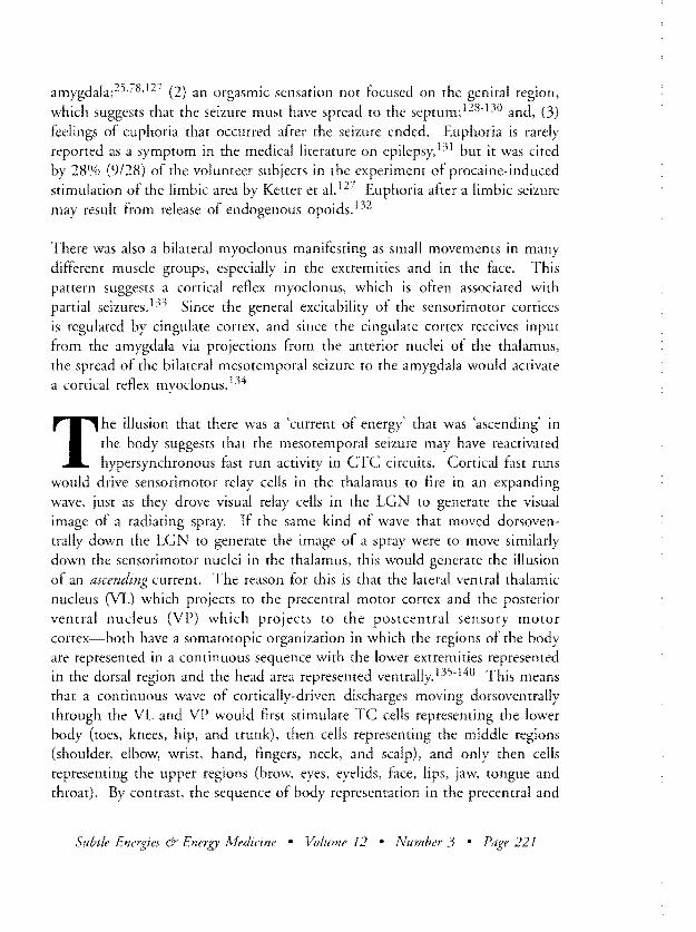
rlgure 3 Transitional phosphene images A The star-like of thin flashing filaments of white and blue phosphene B A stream ofdark barely-perceptible receding annuii that entered the visual field at a rate ofmore than one per second P- 2 Hz) nevel illuminating as colored phosphene The influx ofdark waves generated an illusion of m01ement through a dark tunnel-like 3D space After a few seconds the flood of dark annuli stopped abruptly edipsed by onset of the next 1isual image C A Iltdiating spray of phosphene mist interspened with mrmy beige-colored flecks replaced the tunnel of dark annuli The spray seemed to radiate toward the 1iewer along a conical trajectory through 3D space and to strike the forehead so thm the subject fett compelled to pull his head back against the pillow and
arch his back There were also muscle tremors in the fue and extremities D A gmdual brightening and bluing of the 1imal field This effict slowly but steadily obscured more and more ofthe mdiating spray until it was the only visual senation present The approxshyimate hue of blue based on the authors comparison ofcolor swatches from a ClvYK Process Color (hart is 40 Cyan without any lVagenta Yellow or Black~a color similar to the blue of the sky on a clem~ dry day in the summer or autumn E The appearance of a btllbom hollow translucent white phosphene that seemed to protrude through the bright blue visual field m if there were 3D depth in tbe 1isllal field At first the bulbous image appeared as a fb)llI glow but when the authorfocused his attentioll on it the glow became brighter re1ealing a bulbous nose As the phosphene glowed even more brighty it revealed more caudal extension This change created an illusion that the figure had just moved forward in 3D space and conversely ([S auemion waned the phosphene dimmed so that its caudal extension shortened making it seem to recede Beneath the anterior pole of the bulbolls image wm a thin ine or shadow shaped like an inverted crescentc After many successive protrusions and recessions the bulbous image suddenly glowed brighty-so much so that it looked (tS if it the bulb had lurched toward the vertical meridian-and in the same moment it disappeared alld its former site wm occupied by a cluster of three thin white phosphene rays (see Figure 4) The motion and speed ofthis change created an illusion that the bulb had somehow ruptured and released the phophene rays
Figure 2 describes the sequence of phosphene images usually observed during self-hypnosis (or during a normal drowsy transition to NREMS) including threshold images of receding annuli and the subsequent images of amorphous expanding waves Figures 3 and 4 describe the new phosphenes that inaugu-
Subtle Energies 6middot Energy Medicine bull Volume 12 bull Number3 bull Page 190
Figure 4 Paroxysll1al phosphenes A The white phosphene bulbous image B Vhen the bulbolls image disappeared it W(J replaced instantly by three thin white phosphene rays tlnd tit the same time the bright blue bckgrollnd disappeared lewing the rays silhouetted Igainst the normal charcoal-colored uisual fIeld (eigengmu) In the first present~tion the three white rays extended less than
to the perimeter of vision and the my had a distinctive 90 bend to the left ilt the tip One second later the mys reappeared now in a new realigned version in which the rays had doubled in number (fiwn 3 to 6) had lengthened 50 tIS to extend till the way to the peripheral rim of vision and had fonned farther ilpart at the tops In the next second the iluthor obserled a third transforshymation he saw the fim much farther apart 1 movement that resembled the petals on a flower wilting ill heat In this third displllY the btlSes of the nIys btu tiny shard-like triangles ofbright opaque white phosphene superimposed The third displtty of the mys WtlS the final one and it persisted in the viSlft11 field for about 1 () to 12 secolids There were no auditOlY or sensorimotor symptoms accompanying any of the transformations of the mys C Serialflashes (explosions J ofdull white phosphene appearing in either the right or left hmllfield Single flashes neler occupied more than IIbout a third of the visual field and seemed to alternate between the and lefi side ill a non-rhythmical pattern The experience filt like being inside a storm cloud illuminated fom within by flmhes of sheet lightning The photopilroxysm tutls rlccompanied by IOlid cmcHing sounds sensorishymotor sensations of bilmer([l polymyoclollltJ that seemed to inwlve an ascending current energy an orgflSmic sensuion diffused throughout the lInd psychic symptoms euphoria md ofawe mixed tuith fiar
rate a transition to paroxysmal experiences and that eventually culminate In a simple partial seizure
The photoparoxysm was accompanied by non-visual symptoms (1) a loud crackling and buzzing sound reminiscent of an electric circuit shorring out (2) psychic symptoms that included euphoria and also a blend of awe and fear (3) an orgasmic sensation that was not localized in the genitals but rather diffused throughout the body and (4) motor symptoms of bilateral polymyshyoclonus which seemed to flow through the body in one direction creating the illusion of a current of energy flowing into the lower body (toes lower legs and perineal area) then flowing up the trunk activating myoclonus in the arms and fingers and to the facial muscles and the back of the neck
Subtle Energies 6-- Energy A1edicine bull Volume 12 bull Number 3 bull Page 191
Figure 5 Postictal uisual symptoms For seueral weeks after the photoshyparoxysm when the subject went to bed and closed his eyes a white glow
appetlred in the same place where the bulb and rays appeared earlier When attention was fOCIsed on this glow it began to expand as ifbillowing out toward the dewer in 3D space The surfoce ofthe expanding phosphene presented a ftactal-like pattern resembling the swfoce ofa caulifower a billowing cumulus cloud or a ftoth ofsoap bubbles After a flw days the glow still appeared but now it expanded with a sur(ace that presented an undifforenshytiated fog-like whiteness Once the wave of white phosphene expanded the entire uisual field appeared white as if the uiewer were enleloped by fog or a snow whiteout If the author kept his attention focused the field remained white for a relatiue(y long amount of time sometimeJfor more than seleral seconds While this whiteness was present the Il1lthor experienced il weaker uersion of the seizure-related sensorimotor and psychic symptoms
For many nights after the seizure when the author closed his eyes at night he saw a faint white glow at the spot in the upper right quadrant of the visual field where the bulbous phosphene and the rising rays had once appeared as shown in Figure 5 If he fixed his attention on this faint glow it became brighter and began to expand as if it were radiating toward him in 3D space The outer surface of the white expanding cloud had a cauliflowered texture like the agitated surface of a billowing storm cloud or a foam of soap bubbles This expansion brought a reactivation of a less intense version of the same psychic and sensorimotor symptoms that had accompanied the earlier seizure
DISCUSSION
PARr I PHOSPHENES COMPARED WITH KUNDAUNI LIGHT VISIONS
The similarities between the phosphenes and written descriptions oflight visions in authoritative texts in the Hindu and Tibetan Buddhist traditions are illustrated in Tables I and II (The source texts for excerpts are listed in the Table legends) Despite the diversity of source documents there is a remarkshyable match between the phosphene images described by the author and the descriptions from source texts whether one reads across the horizontal rows matching descriptions of a single image type or one reads down the vertical
Subtle Energies 1amp Energy vledicine bull Volume 12 bull Number 3 bull Page 192
columns to see how a particular text describes the sequence of images There are sizable gaps in some sequences which are most often attributable to our not being able to be sure that the reference was to a vision of light or to our failure to find a clear concise unequivocal description that would not require an extended hermeneutic analysis to demonstrate that a light vision was involved
We acknowledge that any comparison of excerpts drawn from works written in different historical epochs by different authors who lived in different cultures and practiced religious traditions could be challenged on the grounds that all cultural concepts and values are relative so that it is not intellectually justifishyable to compare material drawn from different cultures against some purportshyedly universal standard as we do here not to mention the complication interposed by the indeterminacy inherent in all language which makes it problematical to say how accurately words communicate the underlying
37experience 36 These are important challenges to a work like ours but given constraints on space we do not address those issues in this article
PART II PHOSPHENE IMAGES OF SLOW WAVE SLEEP RHYTHMS
Origins of the Receding Annuli
T he threshold images of receding annuli have the same timing as a type of synchronous slow wave that normally occurs at the onset of the stage 2 transition to non-rapid-eye-movement sleep (NREMS) The transishy
tion from waking to NREMS is governed by the complex interaction of three brain rhythms in the reciprocal projections that link the cortex and thalamus
42in a corticothalamocortical (CTC) circuit38- The transition to NREMS begins with a drowsy waking state (stage 1 ) At this time large populations of cortical cells begin to oscillate in synchrony with a very slow rhythm of less than one wave per second laquo 1 Hertz [Hz]) This cortical slow rhythm referred to the thalamus lowers the polarization (hyperpolarization) of thalamic cell membranes including those of the thalamic reticular nucleus (RTN) a thin sheet of GABAergic cells that forms an outer cover over large portions of the lateral posterior thalamus The hyperpolarization causes the RTN to stop firing the single spikes associated with the processing of sensory signals during waking
Subtle Energies amp Energy A1edicine bull Volume 12 bull Number3 bull Page 193
~ Ishyi
tr oltS ~
~
~ ~
5 ~
s
-
~ shyll
lt~ ~ I)J
bull p ~ shy
~
ANNULI
MISTS OR CLOUDS
STARS
RADIAL SPRAY
SPARKS
BRIGHT BLUENESS
BULBOUS IMAGE
Table I Excerpts of Light Visions from Vedic and Hindu Yoga Meditation Texts
THE RG VEGA
When the visions that are concealed begin ro glow The Seers begin to glow by rhe stream of rta (868)
The flame-arrows of Agni assemble like streams of water into holes (10254)
the divine radiance [Asurias) (IX 71)
The Soma that the Brahmans know-that no one drinks (X85)
[IJn a cloth like to a cloud (IX 69)
Soma storm cloud imbued with life Navel of the Rta (IX 74)
SELECTED UPAMSHADS
smoke sun these are the preliminary forms (SvetlsLJp 1111)
l small lotus Hower within it a small space (Chandogup VllI 1 1)
fireflies are forms If
11 )
That which hangs down hetween the palates like a nipple that is the birthplace of I ndra I 6 1)
the size of a
THE YOGASUTRAS
everything is compassed the seer as well as
the seen (423)
One sees countless bright speckles Keep watching when the whirlin fvini-vrttihl ends
That bending-down hears the
Aloneness [KaiMlpa behind it (4 26)
V
~ ~ ~
lt+ h
I RAYS ~ ~ it ~ bull -
~
shy~
I BRIGHT
IWHITE~ FLASHFS ~
bull p ~ shy~ VI
seer in heavens navel IS in the woolen filter
The Seers milk the bullshySoma (IX 85)
He [Somal sloughs off the divine radiance abandons his envelope and goes to rendevous wirh the
(IX 71)
The filrer of the burning has been spread Its dazzling mesh spread atlr (IX IB)
In jets the pressed Soma is clarified (IX 72)
caksus is the sun (Ix 10)
procure feJ[ LIS the bright substance which excels in worth the outside which procures brilliant light which shines powerfidly 0 thou art born of the rta (2 U15)
H~ is of the measure of a thumb or appcaralKC like the SlIn th~ atman seems to be of the size of the point of a goad
V 13)
The bird of golden hue a swan fhwWlj of radiance
middot lik~ a wheel of fire of the color of the sun (MaitrUp VI 24)
middot the ocean of light In it worshippers hecome dissolved like lit (MaitrUp Vl 16)
middot like lightnings from within the douds 36)
That tearing sec-it will (4 27)
th~ vision of ultimate discernment bursts forth like a storm cloud of cosmic dimensions utlnndliJ (4 2))
~ ltshy
Iigtshy
~ ~ 5 C
~ ~ ~ ~
~ li shy ~ ~ ltshy
~
~ ~
shy
Table II Visions from Tantric Hindu and Tibetan-Buddhist Meditation Texts
TANTRIC TEXTS
In the middle of the vault of the like the tapering flame of a candle the fiery effulgences shine continuously (GA pIO-11)
Above this energy [the ajna chakra midway between the cyesl dwells the dot bindu (GA p 10- 11)
~-~--------------~ ~---
RADIAL SPRAYS
SPARKS
BRIGHT BLUENESS
BULBOUS IMAGE
When the bindu explodes and shatters it (GA p 10-11)
a continuous whirling movement there appear dazzling sparks just as the (AT 5 101 107 111 KS II 3)
rises
Vhen the bindu explodes and shatters it expands and forms the Jrltlsttlka rthe Egg of
Brahman] similar to the triangular fruit of the water chestnut (GA p 10-11)
that resetn bles the stomach of Concentrate on the
a fish and contractinel (TA 5 57shy61)
TIBETAN-BUDDHIST TEXTS
Meditate on the f()ur wheels each like an umbrella or like the whed of a chariot (I ii 62)
a fltnm reHected
The formine of thoughts ceases and phenomena smoke mirage (I ii 98)
Phenomena appearing like fireflies (I ii 98)
The Flaring will appear as a yellow radiance (I ii 25)
a vision of the form of the Buddha outlined a cloudless sky like the moons reflected
Or ol1e sees as mirror the lInobsclired radiant IPlile (ll ii 19-20)
The Pure illusory Body springs f(1rth from the State of the Clear Light like a fish leaping forth from water or like the form of the Bnddha which
--
~ lti-shyshy
~ ~ s
~ ~
~ t--)
bull
~ ~ ~ w
bull ~ ~
~
RAYS
BRIGHT WHITE FLASHES
the of the skull the womb I raised it to the surf1Ce of the bmhmrldrll1d similar to
an ivory tusk the conch (silnkhini) releases its flow Inside the rusk there is hut one orifice the mOllth of the conch known as the tenth door (GA p 10)
The swan hilmsal of dazzling whireness (AT 136)
When the energy with five modalities draws herself and enters Brahmans seat she Hashes forth
such is the so-called serpent 21j8-2) I)
flses [moves] as one does upon waking from sleep (YCS IV iii 34)
the invisible psych ic on the crown of the hltad When the becomes filled with the viral force of the transmuted seminal fluid one realizes the Srare of the Great Vfljra-Dhflm [Wielder of the Thunderhold ( ii 144-14raquo)
Simultaneously with this realization the white fluid How upward to the crown of the head (Iii 144-14raquo)
One mastery of the Very (IV iii Yi)
the State of the Grear Vrljm-Dhrlrtl [Wielder of the Thundcrholtl ([ ii 44-14 i)
Table I References (l) The Vetil oral verses transcribed beTWeen 1300 and 1000 BC inm the worlds (lrst written scriptures (2) ne SllSttlWtlttlm CbtlndolJtl amp Mditri Uptlmhrld (Commentaries) written by anonymous amhors between 800 and )00 (3) The Yogmutrtls attributed to Patafijali and written between 0 and 200 AD
Table II References TIIIllmokrl (AT) ca 1014 ADH (2) Coraksanathas AmtlrtughtlStJ(rlfltl (GA) CL 1200 AD8 () (KS) ca 1000 ADH (4) The Tibetan Buddhist v~rsion of Naropas nil
Six Abridgfd Doctrines compiled monks of tbe Tibetan Kargyu~rpa School aboll( 1050 AD 1l-14
and instead the RTN fires short 1 to 3 second bursts of 7-10 Hz spikes that wax and wane (like ltspindles) and recur at intervals of 3 to 10 seconds (03 01 Hz) By the time the RTN has fired three to seven bursts (478 plusmn 162) the thalamic cell membranes have been further hyperpolarized which causes the RTN to stop firing spindles43 At this time the next stage of NREMS (stage 3) begins but since a very different kind of sleep rhythm is involved there we will delay discussing this until after we see how spindles can generate epiphenomenal phosphene images
The phosphene images of receding annuli enter the visual field every 5 seconds (02 Hz) This timing falls within the range of RTN spindle burst emissions (01 to 03 Hz) Also receding annuli terminate
automatically after three to seven cycles just like spindle bursts There is to
our knowledge no other brain rhythm that operates in the visual pathways that shares these same temporal characteristics Therefore based on timing alone it is likely that the receding annuli are generated by RTN spindle bursts
This hypothesis can be strengthened by showing that the symmetrical annular shape of this phosphene image and its centripetal trajectory can be explained by reference to spindle waves moving through the RTN matrices where they interact with cells in the visual relay of the thalamus Light receptors in the retina send projections that terminate in the lateral geniculate nucleus (LGN) of the thalamus The retinal axons form one-to-one synapses with the thalamshyocortical (TC) neurons that relay visual signals to targets in the primary visual cortex As shown in Figure 6 LGN is composed of six thin sheets (laminae) folded over a central hilus These drawings adapted from arrwork by Le Gros Clark44 and from anatomical studies by Connolly and Van Essen45 and Malpelli and Baker46 show how the largest and most dorsal lamina-lamina 6--can be removed flattened converted from a 3D to a 20 structure then adjusted to accept a grid of retinotopic grid coordinates Since each LGN contains TC cells representing the contralateral half of the visual field the perception of a single unitary field results from a fusion of neural events taking place in both the right and left LGN
The relay of afferent retinal signals by TC cells in the LGN is controlled by inhibitory neurons arrayed in a thin sheet-the RTN-that covers the outer surface of the posterior thalamus including the LGN When TC cell axons
Subtle Energies Energy Medicine bull Volume 12 bull Number 3 bull Page 198
Figure 6 Functional anatomy ofthe primate lateral geniculate nucleus (LGN) A Saggital silhouette ofthe monkev LGN showing six layer 0 sheets (laminae) folded over a central hylus Affirent (Iisua signals from the retina arrive l)ia the optic trade enter the lGN through the rostrodorJal surface and form one-to-one synapses with thalamocortical (TC) lision-relay cells in spec~fic laminae The genicushylate TC cell axOItS exit through the l)entrocaudal surfoces ofthe
B )
LGN lsoggttal VieW LGN lomenor view) GN lamina 6 In 3)
Catro~
D E
LGN iamino 6 In 2D LGN lamina 6 Mth re~~otopiC grid
laminae and are intercepted by RTN neurons that impose inhibitory synapses on the TC cell axom B Drawing of the anterior surfoce of the rostrodorsallamina 6 adapted ftom an anatomical study by Le GrOJ Clark 4bull The human LGN has less of a hilar arch than the monkey LGN but fimctionshyally the two structures are similar C-D Schematic drawings showing how a lamina 6 ftom a monkey LGN was remoued mtd conuerted ftom a 3D anatomical Jtructure into a 2D topographic map by Connolly and Van Essen 45 The triangular shape ofthe 2D drawing of lamina 6 adjusted to accommodate a grid that shows lines ofequidistallce (isoeccentricishytie) ftom the center of the retintI adapted ftom Connolly and Van Essen45 who based the grid on data published by A1alpelli and Baker 46 This 2D map of lamina 6 shows that TC cells representing peripheral vision (Ire distributed in the more ventral regiom of the lamina while TC cells representing central vision are located nearer the dorsal pole Note that the cells in the dorsal half of amina 6 that subserve central vision occupy a relatiuely large proportion of the total area of the lamina
leave the LGN they must pass through the RTN where they receive synapses that subject the axons to RTN regulation During a waking state the inhibishytion imposed by RTN cells is selective so that individual sensory signals are efficiency relayed to the cortices but at sleep onset when the RTN shifts to firing spindle bursts this synchronous activity interferes with normal signalshyprocessing When a spindle burst is fired a wave of excitation moves through the RTN matrices imposing a wave of inhibition on all the TC cell axons it passes Once the wave passes and the inhibition is removed the TC cells usually fire rebound spikes47 In Figure 7 we use the 2D retinotopic map of lamina 6 to show how spindle wave moving simultaneously through the RTN matrices in both hemispheres would generate a pattern of TC cell rebound firing that can account fOf the phosphene image of a receding annulus The
Subtle Energies amp Energy Medicine bull Volume 12 bull Number 3 bull Page 199
A
LGN
LGN
25
5
10
20 40 60 60
RTN
Figure 7 Origins of the receding annuli A The spatiotemporal characteristitgt of receding annuli can be explained in terms ofspindle bursts moving vemrodorsally through the RTN network in a relatively thin coherent spatial wave As it passes the spindle wave inhibits a narrow band of TC cells in the geniculate lamina 6 generating a dark annulus which blocks all other afferent visual signaiJ including the random metabolic discharge of the retinal receptors that generates the normal charcoal or eigengrau
background In the wake ofthe spindle wave when TC cells are released from inhibition man) fire rebound spikes Since each LGN represents half of the visual field a walJe of TC cel rebound spikes moving along lamina 6from the more ventml regions (representing peripheral vision) toward the dorsal pole (representing central vision) will register in the visual cortices ItS a hemi-annulus that shrinks in diameter pindle waves are fired simultashyneously in both RTNs so their passing will reieltSe simultaneous wtwes of TC cell rebound spikes in both LGNs in the liisual field this pairing ofTC cell rebound spikes will generate two complementary (rellerse-image) phosphene hemi-annul annuli with tips fosed to form a single annulus that shrinks in diameter B The same mechanism can explain the ddrk fost-paced rtnnuli If the RTN were to fire spindle waves at a rate 20 Hz-ten times the normal rate of 02 Hz-then the rapidly repeating waves of inhibitioll will not allow TC cells in lamina 6 enough time to raouel and fire rebound spikes The author observed an influx of dark annuli arriving at intervals of more than one per second P- 20 Hz which is egtplained in the text as cortical spike-wave (SW complexes driving the RTiV to fire spindle bursts at this accelerated rate
annulus shrinks steadily in diameter as the wave of TC cell rebound spikes moves up from the ventral regions of the LGN (where TC cells represent the periphery of the visual field) toward the dorsal pole (where TC cells represent the center of the visual field)
The same mechanism can explain the dark fast-paced annuli If the RTN were driven by cortical influences to fire spindle bursts at a rate of 22Hz-an acceleration that does in fact occur during the emergence of hypersynchronous absence-like seizures as we discuss below-then there is not enough time
Subtle Energies amp Energy Medicine bull Volume 12 bull Number3 bull Page 200
between the successive waves of spindle burst inhibition for the TC cells to recover enough to fire rebound spikes The result will be a stream of thin dark receding annuli that enrer the visual field at this hypersynchronous rate of 2 2 Hz This stream of dark annuli generates a sensation of optic flow that is an illusion of motion through a dark tunnel-like space even though the subject is not moving as described by Steinmetz er al Motion in the visual periphery elicits in the stationary observer an illusion of self-motion (vection) indistinguishable from real motion The illusion of vection is compelling for it dominates contradictory proprioceptive signals For example subjects presented with optic flow consistenr with backward self-motion perceive backward motion even if they are actually walking forward 48(p189)
T his analysis suggests that the functional anatomy of the RTN matrix can in some circumstances constitute a resonating circuit that when activated can generate an epiphenomenonal visual image of a slowshy
moving phosphene annulus or of a tunnel of dark fast-moving annuli This hypothesis of a resonating circuit is supported by the authors observation described in an earlier article that when he was given a magnetic resonance imaging (MRl) text and thus exposed to a radio beam every 2 seconds (05 Hz which is the interval that maximizes recapture of energy released by atomic precession) he saw a stream of phosphene receding annuli that appeared at the same rate (05 Hz) instead of the normal rate of 02 Hz and in this MRlshydriven variant the receding annuli did not stop automatically after a normal volley of four but continued ror as long as the MRl test was underway49 Since the MRI beams energy into the body at two second intervals and then recaptures the energy given off by atoms as they return to their normal alignments the vision of a continuous stream of receding annuli at 05 Hz can be explained as the flow of physiological energy released by atoms in the body through a resonating structure in the thalamus-the RTN matrix The hypothshyesis of a resonating circuit has also been proposed by Max Knoll and colleagues based on studies in which they applied electrical stimuli in the EEG range to the heads of subjects although they were unable to identifY the neutal mechanisms that constituted the resonator 5051
Two alternative hypotheses that might be put forward to explain the receding annuli can be easily dismissed A mechanical discharge of retinal receptors generated by torsion-induced ddormation of the converged eyeball might be
Subtle Energies amp Energy A1edicine bull Voltlme 12 bull Number 3 bull pfzge 201
proposed as a competing hypothesis to explain the receding annuli but if such a discharge were to occur it is difficult to envision how it would selectively and simultaneously discharge the receptors situated along the full 3600
perimeter of the retina-nor is it likely that the initial symmetry of such an annulus were it to be initiated would be preserved as the wave of depolarshyization of retinal receptors flowed inward from the periphery toward the central fovea Also there is no reason to suppose that RTN sleep spindles are referred to the retina which would make it hard to explain the timing of the discharges Another hypothesis might be that attention fixated on the center of the visual field would strongly facilitate certain visual neurons in the posterior parietal cortices specifically area pc which is known to have large bilateral receptive fields and a center-surround macula-sparing spatial structure 52 This hypothshyesis can also be rejected first because there is no evidence that visual signals are ever initiated in this region and second because the centers of the receding annuli fill in with disk shapes midway through the trajectory an observation that cannot be explained by activation of macula-sparing receptive fields in area PG (For a discussion of the filling-in of a disk shape see Nicholson49)
Origins of the Amorphous Expanding Clouds
During the transition from a waking state to NREMS the synchronous spindle volleys of stage 2 induce a further drop in the polarization of thalamic cell membranes Once a threshold value of hyperpolarization
is reached after three to five spindle bursts the RTN cells stop firing spindle bursts43 At the same time TC cells begin firing low-threshold calcium spikes The TC cell calcium spikes are not released randomly but rather as groups of spikes fired in response to the receipt in the thalamus of the synchronous pulsations of cortical cells oscillating with cortical slow 1 Hz) wave activity The interaction of the synchronous cortical slow wave and the TC cell calcium spikes generates waves in the delta (1 - 4 Hz) frequency band which is characshyteristic of NREMS stages 3 and 438-42
One important due about the mechanisms responsible for generating the amorphous phosphene waves is that the propagation patterns observed are similar to those manifested by the cortical slow laquo 1 Hz) rhythm The cortical slow wave is one example of a generic type of periodic spontaneous expanding
Subtle Energies amp Energy Medicine bull Volume 12 bull Number 3 bull Page 202
wave that is often observed in large networks of locally-connected neurons Maeda and colleagues have studied these periodic spontaneous expanding waves using matured cultures of dissociated cortical neurons 53 They found that periodic synchronized bursts originate every 10 to 20 seconds at random locations in the sheet of locally-connected cells and spread with unpredictable asymmetrical patterns at a relatively slow speed averaging 50 mmlsecond and propagating sequentially from electrode to electrode as each local group of neurons charges up its neighboring nonrefractory areas
The amorphous expanding phosphene clouds exhibit these same spatiotemporal characteristics they begin at unpredictable locations in the visual field usually in the periphery and sweep across the visual field activating new regions at the same time that passed over regions go dark again with a duration that lasts several seconds-a relatively long time in terms of neural events
PART IT PHOSPHENE 1MAGF5 OF HypERSYNCHRONY IN erc CIRCUITS
Research on the Emergence of Hypersynchronous Seizures At Sleep Onset
Research in animal models shows that hypersynchronous seizures can emerge spontaneously in erc circuits as a result of smail incremental changes in the operation of the same cellular mechanisms that govern
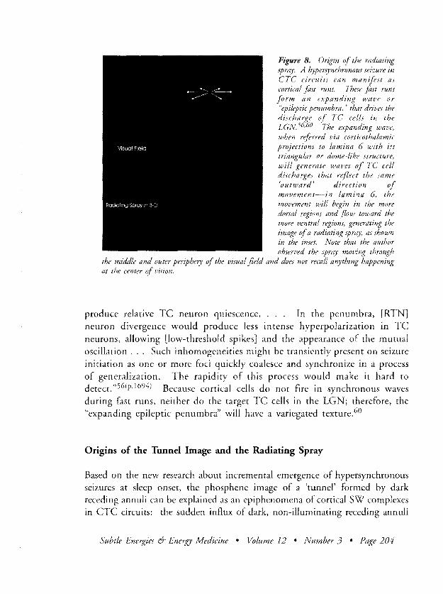
a normal transition to NREMS54-60 For this to happen cortical cells must already be abnormally excitable at the time the transition to NREMS begins The incremental emergence of a hypersynchronous seizure involves an alternashytion between two kinds of paroxysmal activity (1) hypersynchronous spikeshywave (SW) complexes at 15 to 2 Hz intervals fired by cortical cells which then drive thalamic RTN cells to fire spindle bursts at the same rate or (2) fast runs of 10 to 15 Hz spike bursts fired by cortical cells which then drive TC cells to discharge in an expanding wave pattern as shown in Figure 854shy60 This expanding epileptic penumbra was first observed in computer simulashytions by Lytton et al then later confirmed in animal studies5657 The pattern results when cortical fast runs drive paroxysmal discharges in about 40 of TC cells distributed in a periphery around a locus of non-participating TC cells which are too inhibited by corticothalamic signals to discharge At the focus Lytton explains intense cortical and [RTN] neuron activity would
Subtle Energies Energy Medicine bull Volume 12 bull Number 3 bull Page 203
Figure 8 Origin of the radiating spray A hypersynchronous seizure in eTC circuitJ can manifest as cortical fost runs These fost runs form an expanding walle or epileptic penumbra that drives the discharge of TC cells in the LGN5660 The expanding wave when referred viti corticothalamic projections to lamina 6 with its triangular or dome-like structure will generate waves of TC cell discharges that reflect the same outward direction of movement--in lamina 6 the movement will begin in the more dorsa regiom alld flow toward the more ventral regiom generating the image ofa radiating spray as shown in the inset Note that the author observed the spray moving through
the middle and outer periphery of the visual field and does not recall anything happening at the center of vision
produce relative TC neuron quiescence In the penumbra [RTN] neuron divergence would produce less intense hyperpolarization in TC neurons allowing [low-threshold spikes] and the appearance of the mutual oscillation Such inhomogeneities might be transiently present on seizure initiation as one or more foci quickly coalesce and synchronize in a process of generalization The rapidity of this process would make it hard to detect56(p1694) Because cortical cells do not fire in synchronous waves during fast runs neither do the target TC cells in the LGN therefore the expanding epileptic penumbra will have a variegated texture60
Origins of the Tunnel Image and the Radiating Spray
Based on the new research about incremental emergence of hypersynchronous seizures at sleep onset the phosphene image of a tunnel formed by dark receding annuli can be explained as an epiphenomena of cortical SW complexes in CTC circuits the sudden influx of dark non-illuminating receding annuli
Subtle Energies Energy Medicine bull Volume J2 bull Number J bull Page 204
that enter the periphery of vision at a rate of ~ 2 per second is consistent with cortical SW complexes driving RTN cells to fire spindle bursts at ~ 2 Hz intervals ten times faster than the normal rate of 02 Hz Spindle bursts driven at this rate would impose successive waves of inhibition on TC cells in the LGN which does not allow time for them to recover and fire rebound spikes
The shift from an influx of dark annuli to a radiating spray of phosphene flecks signals that cortical cells have shifted from firing SW complexes to firing the other kind of hypersynchronous discharges-cortical fast runs that form an expanding epileptic penumbra The variegated texture of the radiating spray is consistent with cortical fast run activity because this drives large numbers of TC cells to discharge their spikes independently generating a pointillist or mist-like spray that appears to radiate out toward the viewer for the reasons explained in Figure 8 above6o The tiny bright pellet-like flecks amid the mist probably result from the simultaneous discharge of small clusters of contiguous TC cells
T he researchers who documented the cellular processes involved in the emergence of sleep-onset seizures were surprised by the high incidence of seizures that occurred spontaneously an observation that suggested
to them the possibility that many spontaneous electrographic seizures in normal subjects are unrecognized and that those sleeping individuals pass in and out of seizures during their slow sleep oscillation as we showed here for cats 58(p1476) This suggests that meditators who are inducing sleep rhythm phosphenes might also make the transition in and out of hypersynchronous seizures with relative ease as the authors experience in the present article would indicate In this regard it is inreresting to note that Persinger found SW complexes in an EEC recording of a subject who was practicing glossolalia at the time and who felt a sense of unity with the cosmos during this event61
PART III PHOSPHENE IMAGES AsSOCIATED WITH LIMBIC SEIZURE
Origins of the Uniform Brightening Effect
Two recent reviews surveying the basic mechanisms of epilepsy point out that in patients with mesotemporal epilepsy the initial event during the transition to partial seizure is usually a gradual increase in interictal-like rhythmical
Subtle Energies 6- Energy Medicine bull Volume 12 bull Number 3 bull Page 205
discharges in the H6263 This is illustrated in a recent study by Bragin et al64 where seizures were induced in rats by injecting kainic acid in one H the researchers found that a rhythmic build-up began 10 seconds before the seizures started first with irregular interictial spiking that increased in rate from 028 Hz to 082 Hz then with a shift to rhythmical spike-bursts that occurred at intervals of gt 1 Hz A similar pattern has been reported in earlier studies by Pare et a165 and Barbarosie and Avoli66 The build-up of rhythmical activity in field CA3 of one H usually does not trigger an outbreak of paroxysmal discharges for this to occur the rhythmical firing must usually be augmented by local recruiting waves6263 Once recruiting waves are active paroxysmal discharges usually break out in the contralateral H not the one engulfed by rhythmic activity6263
A gradual build-up of rhythmical activity in the H can explain two phosphene images observed by the author-the gradually brightening of the visual field and the appearance of a bulbous white image amid
the bright blue Our hypothesis about how the build-up of rhythmical activity in one H might generate a brightening effect in both halves of the visual field is shown in Figure 9 The basic idea is that output from the right H where the build-up of rhythmical activity must occur (see below) is relayed through the dorsal hippocampal commissure to the left EC These signals which are intense but still subparoxysmal signals and which do not have specific spatial patterns are relayed by the EC into the left H Thus the output of both the right and left H will include these signals even though there is no rhythmical activity taking place in the left H There are a number of complicated mechanisms that contribute to this process that we discuss below
It is generally accepted that the neural mechanisms that cause meso temporal seizures are different from those that cause hypersynchronous seizures in CTC circuits (which include absence seizures and also generalized seizures in the penicillin epilepsy model)6263 This distinction was reaffirmed in a recent study by Kandel et a1 67 who showed that stimulating the cortical cells of rars to the point of including SW seizures did nor drive neuron activity in the H Based on this information we can infer that it is not likely that a build-up of rhythmical activity in the H would be driven by the presence of hypersynshychronous activity in CTC circuits It is however importanr to note that the study by Kandel et al was performed while the rats were awake and physically
Subtle Energies rr Energy Medicine bull Volume 12 bull Number 3 bull Page 206
Figure 9 Origin of the uniform brightening and bluing This schematic drawing adapted from a series ofdrawings in an anatomical atlas by Duvenoy81 and from analyses ofhippocampal fimction by Cloor5 represents the zuious subfields of the unfolded hippocampal formation aligned in the order that afferent sensory signals are processed Using this schemfuic drawing we can show how a build-up of rhythmical spiking in the right H (fields 013 and CAl) is capable ofgenerating a brightening in both halves of the visual field not just the right ha(f
AHenlion Circul let ECmiddotH Ccu~ Rigit EC-H Circuit
KEY
PFC
SEPT0M
No Seizure
SubporOxysma
EC i------=~
This happens because the output of the right H which consists of excitatory signals that are stronger than normal but stil subparoxysmal will be relayed to retrohippocampal regions but also relayed contralaterally via the dorsal hippocampal commissure (DHC) to the left H as shown here Among the regions which receive DHC projections and thus receive the excitatory signals from the right H is the left EC it then relays this excitation into the DC and fields 013 md CAl This means the increased output from the right H which is generated by rhythmical spiking will also affect the left H and as a result the same signals will register in the visual cortices in both hemispheres These signals genertlted by rhythmical actillity will not halle any spatially-specific patterns so the increase of excitatory signals will register in the visual cortices as a uniform brightening ofboth hailles of the visual field Two other important items are also included in this dmwing An arrow labeled VHC that runs from field CM of the right H to the DCICA3 border of the left H identifies a structure called the llentral hippocampal commissure (VHC) a direct monosynaptic interhipshypocampal projection that is present in monkeys but sfil disputed in humans The VHC is in a position to add just enough excitatory input to trigger an outbreak ofparoxysmal actillity in a small number ofgranule cells at the hippocampal pole the region receilling the most intense conllergence of excitatOlY sigrias (see Figure 10) Also note the position of the attention-driven septohippocampal circuit shown here only in relation to the left EC-H loop When attention is mobilized as it commonly is during meditation cells in the prefrontal cortices (PFC) stimulate the septum and the septum in turn stimulates cells in the JUperjicial layers of the extreme rostral pole of the H to releme acetylcholine The acetylcholine incretlSes the excitability ofpyramidal celLI in CA3 and granule cells Figure 10)
Subtle Energies 6- Energy Afedicine bull Volume 12 bull Number 3 bull Page 207
active so its findings do not exclude the possibility that the outcome might have been different if the rats were in a NREMS state at the time the stimulashytion was delivered
During the behavioral states of resting immobility or NREMS pyramidal cells in field CA3 of the H fire synchronous sharp waves (SPWs) at intermittent intervals that range between 002 to 3 Hz These SPWs
enhance the excitability of neurons in all but one subregion of the EC-H 69circuit68 Granule cells in the DG are stimulated by SPWs arriving via
feedback projections from CA3Jo In response to SPWS neuron excitability in the EC-H loop can increase by as much as 200 to 500-or even 900 if as in this case the target cells in the EC-H loop are also being excited by
69receipt of afferent sensory stimuli at the time the SPWs arrive68 The generashytion of intrinsic synchronous SPWs during NREMS might make the H more vulnerable to destabilizing influences than during a waking state
One potential source of destabilization is compromise of the barrier to propagashytion of excitatory SPWs This barrier is normally interposed at the superficial layers of the EC When SPWs reach the deep layers of the EC the cells located there send signals through their ascending intra-entorhinal projections that inhibit cells in the superficial layers of the EC70 If it were not for this barrier SPWs would be relayed back into field CA3 where they originated thereby establishing a reverberating loop of positive feedforward excitation with obvious epileptogenic potential But the superficial layers of the EC are also the sole path through which afferent sensory signals are channeled into the H If afferent sensory signals-or in this case visual signals generated by a seizure in CTC circuits-are bombarding the superficial layers of the EC with excitatory signals that have the rhythmical pattern of cortical fast runs this might compromise the normal dampening effect that the EC exertS on SPWs As noted by Steriade et al the rhythms of fast runs are similar to those found in the mesotemporal regions during partial seizures The seizure epoch characterized by fast runs
resembles the stereotyped fast rhythm (~1 0-20 Hz) reported in human temporal lobe epilepsy that may spread to perihippocampal structures and cingulate cortex 58(p1477) Other studies have also reported similar rhythms64-66n The barrage of afferent visual signals relayed into the H from the EC might also interfere with another process that dampens the excitatoty effect of SPWs during a NREMS state The SPWs alternate with dentate
Subtle Energies amp Energy Medicine bull Volume 12 bull Number 3 bull Page 208
spikes which are large-amplitude short-duration field potentials in DG granule cells that temporarily inhibit the pyramidal cells in CA3 and CA34 that fire the SPWs so that the tiring of dentate spikes produces a transient decrease in H output647172 The normal dampening effect of dentate spikes could be compromised by the receipt of afferent visual signals from CTC circuits
A nother wholly-independent mechanism that might generate rhythmical activity in the H during NREMS is activation of high-frequency ripples73 Siapas and Wilson have shown that high-frequency ripples
in field CAl of the H co-occur in close temporal synchrony with stage 2 NREMS spindles recorded in the cortex which implies that this co-occurrence of spindle-ripple episodes is driven by some common external factor74 Siapas and Wilson speculate that the common external driver of this coshyoccurrence might be the brainstem reticular formation (or some kind of interacshytion between the prefrontal cortices and the H) The importance of this tlnding of spindle-ripple co-occurrence for our purposes is that it raises the possibility that an accelerated barrage of high-frequency ripples might be generated in the H in synchrony with the accelerated tiring of spindle bursts by the RTN in response to driving by cortical SW complexes Thus a co-occurrence of accelershyated spindles and accelerated ripples might have occurred in this case at the point when the author observed the dark fast-paced annuli-and as noted above it would have then been superceded by cortical fast runs and the stereoshytyped fast rhythm (~10-20 Hz) reponed in human temporal lobe epilepsy58
The build-up of interictal spiking in the right H would also have the paradoxshyical effect of suppressing paroxysmal discharges in the H where it arises5-77 This is consistent with the observation mentioned earlier that outbreaks of paroxysmal tiring most often begin in the contralateral H626375-77 This
explains why after the author observed a gradual brightening of the visual tleld this effect persisted for so long before the appearance of the next phosphene images associated with an outbreak of paroxysmal activity
Origins of the Bulbous Phosphene
We can reasonably infer that the build-up of rhythmical firing took place in the right H even though the destabilizing processes discussed above would be
Subtle Energies amp Energy ivfedicine bull Volume 12 bull Number 3 bull Page 209
occurring in both hemispheres This inference is based on the observation that the bulbous image appeared in the right half of the visual field Since the right half of the visual field is represented by neural assemblies in the left hemisphere the processes that generated the bulbous image must have occurred there To generate an object-like image signals must contain contain spatially-specific information but all spatially-specific signals will be obliterated in fields CA3 and CA 1 of the right H which is consumed by rhythmical firing If we conclude that the spatially-specific signals generating the bulbous image must come from the left H then the rhythmical activity must occur in the right H
T his analysis of hippocampal processing of spatially-specific and spatiallyshydiffuse signals is based on a theory of entorhinal-hippocampal (ECshyH) dialogue proposed by Buzsaki and colleagues 7879 It is important
to understand the main ideas in their theory because these will turn out to be crucial for understanding how a spatially-specific image like the bulbous phosphene can be generated inside the H In this theory hippocampal output is produced by the interaction of information processed by two very different kinds of circuits that together compose the EC-H loop There is on one hand an extensive network of pyramidal neurons in field CA3 each of which is linked by many local projections because of the local interaction the CA3 circuit cannot preserve spatially-specific signals received from the EC There is however a spatially-specific circuit which is constituted by the reciprocal projections that form a direct link between the EC and field CA 1 The function of the spatially-specific EC-CAI circuit is to implement a matching process that reimposes the spatial information contained in the afferent sensory signals that originally entered the EC When the EC sends its signals into both CA3 and CAl the spatially-specific signals are lost in CA3 but preserved by keeping the spatially-specific signals oscillating back and forth in the reciprocal projecshytions that link CA 1 and the EC The output of cells in field CA3 which has lost the spatially-specific information is referred downstream to field CAl where it is reintegrated with the spatially-specific patterns still active in the ECshyCAl circuit Thus the original spatial patterns of afferent sensory signals are preserved intact In the discussion that follows we consider what would happen if there were no afferent visual signals arriving from the retina and instead there was a differential activation of cell groups within the H that generated a facsimile of a spatial pattern Before addressing that issue we need to find out more about the bulbous image
Subtle Energies amp Energy Medicine bull Volume 12 bull Number 3 bull Page 210
Figure 10 Origins of the bulbous image This schematic drawing of the human H adapted from a series ofdrawings in an anatorniCtlL atlas by Duvenov81 and lom analyses hippocmpal function GLoor 75
shows how the mediaL bend of the H (genu ) points the anterior
BulbOus Phosphene(uncus) back posterior H At the rostral pole of the uncal
81H is a snub-nosed cone is polar region is called the gyrus intralimbicus (GI)78 The GI contains a subgroup of cells ftom field CA3 and ftom the hilar region (CA314j Its associated CAl subjield called the gyrus uncinatus has been pulled out ofnormal alignment with CA3 by having been stretched around the genu Focusing attention during meditashytion will have the effict of selectively enhancing nmron activity at the G pole relative to other regions of the H The attention-driven septohippocampal projections terminate primarily in the superficial layers the GI pole-the alveus and the stratum oriellS When the terminals are stimlilated acetylcholine is released The acetylcholine inhibits the intemeurom that inhibit 013 pyramidal cells and granlile cells so the result in both cases i an increase in cell The regioll of maximum acetylcholine release-and thus (if maimum all excitability-has the same geometrical shape as the hollow bulbous phosphene described kY the author (See the text for an discussion of how Ihi selective activation ttt the Gf pole results in the generation of a visual image)
The unusual shape of the bulbous image closely resembles the peculiar thumbshylike shape of the extreme rostral pole of the anterior (uncal) H called the gyrus intralimbicus (GI) as shown in Figure 10158182 To our knowledge there is no other anatomical structure in the visual pathways that has this same shape The GI contains a small subgroup of field CA3 and CA34 (hilar) neurons which has been physically separated from the rest of field CA3 by the medial bend of the H (genu) The field CAl neurons associated with that CA3 subfield in the are located nearby in the gyrus uncinatus Interposed between this CA3 subfield in the GI and the associated CAl subfield is the uncal extension of the DG called the Ligature of Giacomini (LG)58182
The Gl pole can be activated by the exercise of attention a potentially important point since meditation involves a strong inward focus of attention This focus mobilizes neuron assemblies in the prefrontal cortex which then
Subtle Energies amp Energy Medicine bull Volume 12 bull Number 3 bull Page 211
stimulate neurons in the medial septum that send excitatory signals to the GI pole The septohippocampal terminals are densely packed in the most superfishyciallayers of the GI-that is in the alvear sheet and in the stratum oriens-and also in the anterior extension of the DC the Ligature of Giacomini758182 As shown in Figure 10 this distribution of septohippocampal terminals forms a hollow bulb-like cone
A ttention-driven septohippocampal stimulation of the GI triggers a release of acetylcholine in this bulb-like conical structure which produces two important effects (1) the acetylcholine inhibits the
interneurons that inhibit CA3 cells which increases their excitability and (2) it excites the muscarinic receptors of GABAergic interneutons in the Ligature of Giacomini which enhances the excitability of the granule cells closest to the GI pole8o83-87 In normal circumstances the function of this attention-driven septohippocampal stimulation would be to facilitate the processing of afferent sensory signals but in this case without any external signals to process the effect of acetylcholine release will be selective disinhibition of the field CA3 neurons at the GI pole and the adjacent granule cells in the Ligature of Giacomini87 There will not be a comparable disinhibition in the rest of the field CA3 neurons located in the middle and posterior regions of the H because there are few if any septohippocampal terminals located there Nor will there be a disinhibition of granule cells in the rest of the DC We propose that this selective increase in the excitability of CA3 and DG cells in the GI pole when referred downstream to field CAl and processed in the EC-CAI circuit will emerge in a form that once it registers in visual awareness will be decoded as the spatially-specific pattern of a hollow thumb-shaped bulb
These signals generated inside the H do not have the same kind of spatiallyshyspecific coordinates as would be generated during normal retinal-based percepshytion and this presents a problem how does this information get registered in visual awareness In retina-based perception an external object triggers retinal light receptors that map position and direction using an egocentric topography that is using coordinates that map the object from the point-of-view of the perceiving subject But when visual signals are processed inside the H they are mapped in a different way instead of recording events using egocentric coordinates the H processes visual signals using an allocentric map-one that records position and direction as coordinates and vectors on an abstract
Subtle Energies amp Energy lvfedicine bull Volume 12 bull Number 3 bull Page 212
geometric grid with no egocentric reference88-94 Therefore the signals that generate the bulbous phosphene will initially be recorded in allocenrric coordishynates-and no image will appear in the egocentric visual field until the signals have been referred back to the visual cortices and registered there This can happen in two ways (1) field CAl sends signals directly to the parahippocampal cortices (PHC) and the perirhinal cortices (PRC) which then relay the signals to neocortical areas via an extensive network of back-projections or (2) if field CAl sends its output to the EC which sends it on to the PHC and PRC for relay backward This means there is a co-activadon of cells in field CAl in the EC in the PHC and PRC regions and in the neocortical areas88-94 This coactivation is thought to be one of the mechanisms involved in the laying down of long-term memories It is also thought to become active during recall of a memory where the cells that contributed to the original experience (and to the laying down of the long-term memory) are reactivated9394
Several new studies point to ways that allocentric spatial information generated inside the H could be relayed back to the visual cortices via the PHC or PRe There is now new evidence that the visual cortices
can acquire new information after visual signals have been processed by the H and returned via the PHCPRC back-projections information that was not part of any afferent visual signaL but rather is incorporated as an added value to the original visual signals92 Recent research also shows that when subjects recall a visual memory the neurons in the H EC PHC and visual cortices that are reacdvated in concert may have the capacity to tune in selectively to particular categories of content in the visual scene being presented95 This is an unexpected finding--that vision-related cells in these regions of the mesotemporal cortex can be category-selective--and it has at least two important implications One is that the hippocampus more than just relational or spatial information (although the hippocampus had the largest proportion of cells selective for the category of spatial scenes) A related implication is that the hippocampus has more than just a modulation or consolshyidation effect on cortex instead it carries complex visual information96(p856)
These recent research findings challenge the conventional view that H processing of visual signals does not generate any value added when the H signals are returned via back-projections to the visual cortices This suggests that it might be possible-and indeed given the authors observations that it
Subtle Energies amp Energy A1edicine bull Volume 12 bull Number 3 bull Page 213
dearly can happen-that a pattern of signals which has spatially-specific allocenshytric coordinates acquired in the H can be preserved when sent via back-projecshytions to the visual cortices and that receipt of this new set of spatially-specific signals in the visual cortices would be registered in awareness as if it were an external object perceived by the retinal receptors and mapped in the egocenshytric coordinates of normal perception
A nother anatomical factor to be considered is that output from CAl fields in the uncal H (and thus from the GI pole) is segregated from the output of CAl fields in the middle and posterior H when it is
sent to the PHC and PRC for relay to the neocortex signals from the GI pole are sent to area TH in the PHC while signals from the middle Hare sent to area TF and signals from the posterior H to area TEav97 This segregashytion of CAl signals might explain why the intense flring of cells at the GI pole appears to be segregated as a locus of intense activation amid a much larger fleld with no spatially-speciflc signals present-thus producing the epipheshynomenal image of a bulbous glow floating in a large expanse of bright blue
The Origin of the Phosphene Rays
Since the phosphene rays appear at the same place in the upper right quadrant of the visual field as the bulbous image we can infer that the rays were also generated by neural events taking place in the EC-H loop of the contralateral (left) hemisphere The initial appearance of a cluster of three rays rising less than halfway to the perimeter of vision and the subsequent realignment of the rays in which they double in number extend to the perimeter of vision and fan farther apart can all be explained based on the anatomy of intrahipshypocampal circuits and on computer simulations of signal propagation in a model of intrahippocampal circuitry9S-102
Earlier we explained how the left EC-H loop received a stream of excitatory signals referred contralaterally from the right H but it is highly unlikely that the image of three thin discrete phosphene rays was triggered by this kind of excitatory stimulation received in the left EC Projections from the EC to the DC have been shown to diverge spatially so that the terminals are widely distribshyuted along the longitudinal axis of the DG9 Also Yeckel and Berger have
Subtle Energies 6 Energy Medicine bull Volume 12 bull Number3 bull Page 214
shown that when granule cells discharge in response to excitatory signals relayed by the EC there is a frequency facilitation that is a progressive increase in the number of active granule cells distributed over a wider spatial extent of the dentate gyrus103 A similar spatial dispersion occurs when granule cells are discharged antidromically by feedback from CA3 cells 104 Therefore we can conclude that the stream of subparoxysmal spatially-diffuse signals relayed to
the left EC from the right H were not sufficient in this case to stimulate an outbreak of paroxysmal discharges limited to only three granule cells Some additional factor would have had to be added into the mix-something that would limit the paroxysmal discharge to those three granule cells
Stimulation studies in rats show that applying an electrode to the fibers of the ventral hippocampal commissure (VHC) never discharges more than three granule cells no matter how strong a stimulus is applied5IOS-107
This would suggest that the human equivalent of the VHC might have added the extra stimulation that triggered the outbreak of paroxysmal discharges in three granule cells in this case This scenario is also consistent with the general principle that during the transition to a partial seizure in patients with mesotemporal epilepsy the initial outbreak of paroxysmal activity occurs most often in the H contralateral to where the build-up of rhythmical activity occurs626j There is however a major problem with the proposition that a VHC signal determined the pattern of paroxysmal outbreak in this case while the presence of a functioning VHC has been demonstrated in monkeys no one has been able to provide convincing evidence that there is such a structure in humans
The Phosphene Rays Imply Existence of a Functioning VHC in Humans
In rats the fibers of the VHC originate from pyramidal cells in the most proximal part of field CA3 (ie that part closest to the DG designated by CA3c) and from the related CA34 cells in the hilar region the fibers terminate in the inner third of the molecular layer of the contralateral DG where their arrival has an excitatoryeffect105-lOS The reason why stimulation of the VHC in rats discharges a maximum of three granule cells is that VHC terminals innervate only a very narrow zone of cells that extends only 50 microns (pm) into the inner molecular layer of the DG and another 50 pm in the subgranular layer 108
Subtle Energies amp vledicine bull Volume 12 bull Number 3 bull Page 215
I n monkeys the VHC is proportionally much smaller than in rats and it connects only the anterior (uncal) regions of the H not the entire longitushydinal extension in other respects the VHC connections in rats and
monkeys are similarJ282I08-11l The strongest evidence in support of a functioning VHC in humans comes from a depth electrode study of epileptic patients awaiting surgery by Spencer et al who found that 20 of spontashyneous seizures that originated in a H in one hemisphere spread first to the contralateral H before activating any of the ipsalareral temporal cortices 112
This pattern is consistent with propagation through a conduit analogous to the monkey VHC bur not consistent with spread through the alternative structure the dorsal hippocampal commissure (DHC) DHC is a bundle of many fibers that originate in several different rerrohippocampal regions and project to the contralateral homologues Some DHC fibers also project to the contralateral EC (which is how the rhythmical activity in the right H gets relayed to the left EC-H loop) But for seizure discharges to propagate contralaterally from one H to the other via the DHC the ipsalateral temporal cortices would first have to be activated This activation did not appear in 20 of the seizures in the study by Spencer et al
The conclusion that a subgroup of seizures spread contralaterally via a VHC projection has been challenged by several researchers Studies of EEG coherence and phase relationships did not find evidence of linear relationships between seizure activity occurring in the right and left mesotemporal regions as might be expected if there were a direct interhippocampal projection 113-1 19 Also in depth electrode studies performed in epileptic patients awaiting surgery Wilson et al stimulated the anterior H (where VHC fibers would be) with a single shock but were not able to evoke discharges in the contralateral H120-122 In the synopsis of their findings Wilson et al did add an important qualification to their conclusion that there was no longer a functional VHC in humans if there were a VHC projection in humans they noted and if this fiber were to
follow the same course as in the monkey it would be so long that action potentials moving along the fiber might become so attenuated by the time they reached the contralateral H that the resulting field potential would be of such a long duration and such low amplitude that it could not be detected Indeed such an input might be below the threshold for local synaptic activashytion of a response unless other factors intervened such as temporal summation occurring with repetitive stimulation122(p185l Finally in an anatomical study
Subtle Energies amp Energy Medicine bull Volume 12 bull Number 3 bull Page 216
of the human brain undertaken to resolve the controversy about the existence of the VHC Gloor et aL found a few thinly-myelinated fibers which crossed the midline at the same place where the VHC fibers cross in monkeys but being unable to trace the fibers to the sites of origin or termination and having noted the oblique trajectory of the fibers Gloor et al concluded that the fibers were more likely part of the H decussion than a remnant of the human VHc 123
They did however acknowledge that they could only guess about the functions of these fibers It remains possible that a small remnant of a ventral hippocampal commissure exists at this level in the human brain 123(pl253)
T he phosphene rays offer new evidence in support of the hypothesis that humans must have at least some functioning remnant of the VHC projecshytion As we discussed earlier the unusual pattern in which only three
granule cells participated in the initial outbreak of paroxysmal activity cannot be explained by the build-up of rhythmical activity in the right H since excitatory stimulation relayed into the left H via the EC would have triggered a large number of granule cells-but the rhythmical activity in CA3 of the right H could have stimulated the cells that gave rise to a VHC projection to send a constant stream of excitatory signals Moreover we know that these VHC signals would arrive in the uncal region of the contralateral H and stimulate granule cells near the GI pole (in the Ligature of Giacomini)-a region that was also being stimulated so strongly by being the focus of attention that it was generating a bulbous phosphene glow in the visual field Based on the rat stimulation studies we can reasonably inter that stimulation of a VHC fiber would not more than three granule cells therefore we conclude that VHC input must have been the crucial stimulus in this case Note that this kind of temporal summation of excitatory signals converging on the apical dendrites of granule cells near the VHC terminal is precisely the type of contingency anticipated by Wilson et al when they wondered if a VHC input might be below the threshold for local synaptic activation of a response unless other factors intervened such as temporal summation occurring with repetitive srimularionI22(p185)
Transformations in the Phosphene Rays and Intrahippocampal Circuitry
Studies by Acsady et al have shown that discharge of a single granule cell is capable of discharging a single pyramidal cell in field CA3 even though as
Subtle Energies amp Energy Medicine bull Volume 12 bull Number 3 bull Page 217
weve mentioned earlier in this article cells in field CA3 are linked by many local interconnections 124 The one-to-one discharge can occur because granule cell mossy fibers innervate many more inhibitory interneurons in CA3 than pyramidal cells in fact there are often several mossy fibers that converge on a single interneuron This circuitry creates a strong feed forward inhibition of pyramidal cells in CA3 an inhibition which can efficiently suppress recurrent excitation in the CA3 collaterals but allow for the selective discharge of a few CA3 pyramidal cells 124(p3399) If there is a selective discharge of three granule
cells by VHC input as we propose what will happen to the visual image of three rays as the action potentials set in motion by the granule cell discharges propagate via mossy fibers to field CA3 then propagate from CA3 to CAl via Schaffer collateral projections and then get relayed back to the visual cortices
Figure 11 presents our hypothesis for how the initial display of phosphene rays could be generated by the registration of granule cell discharges in the allocentric spatial coordinates of field CAl in the left H and also
shows how these initial factors also explain the further transformations in the rays-the doubling in number the extensions in length and the fanning further apart The information shown here is adapted from a paper by Bernard et al that describes the results of simulations performed using a computer model of intrahippocampal circuitryl02 This model incorporates the findings reported by several anatomical studies and in our drawings we have added information about the distribution of longitudinal association strips in CA3 and CAl adapted from Ishizuka et al and Deadwyler and Hampson98-100 basic argument which is presented in detail in the legend to Figure 11 is that the discharge of one granule cell discharges one pyramidal cell in each subsection of field CA3 The action potential set in motion by the discharge of that CA3 cell moves in two directions first it activates other CA3 cells that are interconnected in a longitudinal association strip and second all of these activated CA3 cells send signals via their axons (called Schaffer collaterals) to target cells in CAL Receipt of these signals in CAl activates cells in the longitudinal association strip in CAl which has a reverse mirror-image diagonal to the strip in CA3 Because Schaffer collaterals differ in length the signals from CA3 arrive at different times in different subflelds of CAl which means over time different longitudinal associshyation strips get activated As we explain in the Figure 11 legend this differenshytial activation can account for the changes in the length of the phosphene rays for their doubling in number in the second display and for their fanning apart
Subtle Energies amp Energy Medicine bull Volume 12 bull Number 3 bull Page 218
Figure 11 TransfOrmations B A schematic
AAC b ltI c
Subtle Energies 6 Energy Medicine bull Volume 12 bull Number 3 bull Page 219
The phosphene image of six rays fully fanned apart remained in the visual field for about ten to twelve seconds which is consistent with the time that would be required for propagation of ictal discharges from the left H to the right H-a mean interval of 87 seconds with a range of 1 to 51 seconds 124125
PART IV SYMPTOMS OF BIlATERAL MESOTEMPORAL SEIZURE
Origin of the Lightning-Like Flashes
T he dull-white spatially-diffuse phosphene flashes covered less than half of the visual field and seemed to erupt in either the right or left side of the visual field at irregular intervals The timing seemed to vary
from near-simultaneity of bursts in opposite halves of the visual field on one hand to delays between successive bursts that lasted a full second or longer on the other hand These patterns can be explained as paroxysmal population discharges of granule cells in which the bursts occur independently in the right and left DG The paroxysmal population discharges will be followed by after hyperpolarizations that can vary in duration but last a minimum of a halfshysecond the time required for granule cells to recover their capacity to fire 126
Given these intervals population discharges of granule cells that occur indepenshydently in the two hemispheres could generate a succession of flashes with the timing observed that is from near-simultaneity in both halves of the visual field to intervals in which no flash appear for almost a full second
Auditory Psychic and Sensorimotor Symptoms
The photoparoxym was accompanied by a loud buzzing and crackling sound reminiscent of an electrical circuit shorting out Similar symptoms were reported in a study by Ketter et al of procaine-induced stimulation of neuron hyperexcitability in the anterior limbic region of volunteer subjects where 28 (932) of subjects reported seeing unformed visual hallucinations (lights or colors reported as intense or very intense) and 90 (2932) described hallucishynations of unformed buzzing ringing or electronic sounds127
Psychic symptoms reported by the author include (1) a feeling of awe mixed with fear which suggests that the seizure probably spread to the
Subtle Energies amp Energy Medicine bull Volume 12 bull Number 3 bull Page 220
amygdalati 78127 (2) an orgasmic sensation not focused on the genital region which suggests that the seizure must have spread to the septum128-130 and (3)
feelings of euphoria that occurred after the seizure ended Euphoria is rarely reported as a symptom in the medical literature on epilepsy 131 but it was cited by 28 (928) of the volunteer subjects in the experiment of procaine-induced stimulation of the limbic area by Ketter et aL 127 Euphoria after a limbic seizure may resuIL from release of endogenous opoids 132
There was also a bilateral myoclonus manifesting as small movements in many different muscle groups especially in the extremities and in the face This pattern suggests a cortical reflex myoclonus which is often associated with partial seizures 133 Since the general excitability of the sensorimotor cortices is regulated by cingulate cortex and since the cingulate cortex receives input from the amygdala via projections from the anterior nuclei of the thalamus the spread of the bilateral mesotemporal seizure to the amygdala would activate a cortical reflex myoclonus 134
T he illusion that there was a current of that was ascending in the body suggests that the meso temporal seizure may have reactivated hypersynchronous fast run activity in CTC circuits Cortical fast runs
would drive sensorimotor relay cells in the thalamus to fire in an expanding wave just as they drove visual relay cells in the LGN to generate the visual image of a radiating spray If the same kind of wave that moved dorsovenshytrally down the LGN to generate the image of a spray were to move similarly down the sensorimotor nuclei in the thalamus this would generate the illusion of an ascending current The reason for this is that the lateral ventral thalamic nucleus (VL) which projects to the precentral motor cortex and the posterior ventral nucleus (VP) which projects to the postcentral sensory motor cortex-both have a somatotopic organization in which the regions of the body are represented in a continuous sequence with the lower extremities represented in the dorsal region and the head area represented ventrally135-140 This means that a continuous wave of cortically-driven discharges moving dorsoventrally through the VL and VP would first stimulate TC cells representing the lower body (toes knees hip and trunk) then cells representing the middle regions (shoulder elbow wrist hand fingers neck and scalp) and only then cells representing the upper regions (brow eyes eyelids face lips jaw tongue and throat) By contrast the sequence of body representation in the precentral and
Subtle Energies Energy A1edicine bull Volume 12 bull Numb(r 3 bull Page 221
bull bull bull
postcentral sensorimotor cortices becomes discontinuous at the extreme lateral convexity where the representation of the face begins135 so the wave that creates the illusion of an ascending current cannot originate in the sensorimotor cortices This explanation of the illusory current as a cortically-driven discharge of TC cells in the VL and VP nuclei is consistent with the finding that VL metabolism is elevated in both focal and in generalized epilepsy and also with the finding that there is an elevation of metabolism is in the specific thalamic nuclei which are functionally connected with cortices engulfed by hypersynchronous seizures which would apply to both the VL and VP in the authors case given that involuntary bilateral myoclonus was clearly present 141 142
CORRESPONDENCE Philip T Nicholson bull 52 Norfolk Road bull Chestnut Hill MA 02467 -1832 bull pnicholsonearthlinknet
REFERENCES amp NOTES
I J Gonda The Vision of the Vedic Poets (Mouton amp Co The Hague Netherlands 1963)
2 R G Wasson Soma Divine v1ushroom of Immortality (Harcourt Brace Jovanovich New York NY 1971)
3 S Radhakrishnan Ed The Principal Upanishads (Humanities Press Inrernarional Atlantic Highiands NJ 1992)
4 D R Kinsley Hinduism A Cultural Perspective 2nd Edition (Prentice Hall Englewood Cliffs NJ 1993
5 R S Mishra The Textbook of Yoga Psychology The Definitive Translation and Interpretation of Patanjalis Yogamtras (Crown Publishers New York NY 1987)
6 G Feuerstein The Yogasutras of Patanjali (Inner Tradirions International Rochester VT 1989) G Krishna Kundalini The Evolutionary Energy in v1an (Shambhala Boulder CO 1967)
8 L Silburn Kundalini Energy of the Depths (Stare University of New York Press Albany NY 1988)
9 S Mukrananda Play of Consciousness (SYDA Foundation South Fallsburg NY 1978) 10 E Conze Buddhism Its Essence and Development (Harper amp Row New York NY
1959) 11 D Cozon HigheJt Yoga Tantra An Introduction to the Eoteric Buddhism of Tibet (Snow
Lion Publications Ithaca NY 1986) 12 R A F Thurman Essential Tibetan Buddhism (Harper Collins New York NY 1995) 13 G H Mullin Trans amp Ed Tsollgkhapas Six Yogas ofNaropa (Snow Lion Publications
Ithaca NY 1996) 14 W Y Evans-Wentz Trans amp Ed Tibelrm and Secret Doctrines (Oxford
University Press London 958)
Subtle amp Energy Medicine bull Volume 12 bull Number 3 bull Page 222
15 D P Brown The Stages of Meditation in Cross-Cultural Perspective In Transformations 0 Consciousness (K Wilber 1 Engler amp D Brown Eds Shambhala Press Bosron MA 1986)
16 E Jacobson Progresshe Relaxation (University of Chicago Press Chicago [L 1938) 17 J H Schultz amp W Luthe Autogenic Trlining A Physiological Approach to Psychotherapy
(Grune amp Stratton New York NY 1969) 18 H Benson The Relaxtltion Response (Morrow New York NY 1975) 19 M L Frus amp M Lund Stress Convulsions ArchilJes oNeurology 31 (1974) pp 155shy
159 20 M L Friis Stress Convulsions [n Comprehensil)c Epileptology (M Dam amp L Gram
Eds Ravens Press New York NY 1990) pp 247-250 21 M S George T A Ketter B A Parekh B Horwitz P Herscovirch amp R POSt Brain
Activity During Transient Sadness and Happiness in Healthy Women American Journal 0Psychiatry 152 (1995) pp 341-351
22 P Loiseau Seizure Precipitants In Epilepsy A Comprehensive Textook Vol 1 O Jf amp TA Pedley Eds Lippincott-Raven Publishers Philadelphia PA 1997) pp 97
23 M S Myslobodsky Pro- and And-convulsant Eflects of Stress The Role of Neuroactive Steroids Neurosciece and Bioehallioml Reviews 17 (1993) pp 129-139
24 I D Bremner P Randall T M Scot R A Bronen J P Seibyl S M Sourhwick R middotc Delaney G McCarthy D S Charney amp R B Innis MRI-Based Measurement of Hippo Campal Volume in Patients with Combat Related Posmaumatic Stress Disorder American Journal of Psychiatry 152 (1995) pp 975-981
25 S 1 Rauch B A van der Kolk R E Fisler N M Alpert S P Orr C R Savage A J Fischman M A Jenike amp R K Pitman A Symptom Provocation Study of Posmaumaric Srress Disorder Using Posirron Emission Tomography and Script-Driven Imagery Archilles 0 General pychiatry 53 (996) pp 387-590
26 A M Magarifios J M G Verdugo amp B S IvlcEwen Chronic Srress Alters Synaptic T ermina Structure in Hippocampus Proceedings 0 the National Amdemy USA (Neurobiology) 94 (1997) pp 14002-14008
27 C H Gunderson p B Dunne amp T 1 Fcyer Sleep Deprivarion Seizures Neurology 23 (1975) pp 679-686
28 P Rajna amp ] Veres Correlations Between Night Sleep Duration and Seizure Frequency in Temporal Lobe Epile[)sy Epilepsi345 (1995) pp 574-579
29 I Lorenzo J Ramos C Arce M A Guevara amp M Corsi-Cabrera Effect of Total Deprivation on Reaction Time and Waking EEG Activity in Man SLeep 185
pp 546-554 30 L Bettendorf M Sallanon-Moulin M Touret P Wins I Margineanu amp F
Schofteniels Paradoxical Sleep Deprivation Increases Contenr of Glutamate and Glutamine in Rat Cerebral Cortex Sleep 191 (1996) pp 65-71
31 D P Btunner D J Dijk amp A A Borbety Repeated Partial Sleep Deprivation Progressively Changes the EEG During Sleep and Wakefulness Sleep 162 (1995) pp 100-113
52 D C Hesdorffer amp C M Verity Risk Factors In Epilepsy A Comprehensive Textbook Vol 10 Engel Jr amp T A Pedley Eds Lippincott-Raven Publishers Philadelphia 1997) pp 59-67
53 R Leproult G Copinschi O Buxton amp E Van Cauter Sleep Loss Results in an Elevation of Cortisol Levels the Next Evening SLeep 2010 (1997) pp 865-870
Subtle Energies 6- Energy Medicine bull Volume 12 bull Number 3 bull Page 223
34 P Wolt~ Isolated Seizures In] Engel Jr amp 1 A Pedley Eds Epilepsy A Comprehensive Textbook Vol 1 (Lippincott-Raven Publishers Philadelphia PA 1997) pp 2475-2481
35 A Daurat A Aguirre J Foret amp O Benoit Disruption of Sleep Recovety After 36 Hours of Exposure to Moderately Bright Light Sleep 205 (1997) pp 352-358
36 S 1 Katz Ed Mysticism and Language (Oxford University Press Oxford 1992) 37 R K C Forman Ed The Problem of Pure Consciousness (Oxford University Press
Oxford 1990) 38 M Steriade amp R W McCarley Brainstem Control oWakefulness and Sleep (Plenum
Press New York NY 1990) 39 D A McCormick amp H C Pape Properties of a Hyperpolarization-Activated Cation
Current and Its Role in Rhythmic Oscillation in Thalamocortical Relay Keurons jotlrnal of Physiology (London) 431 (I990) pp 291-318
40 M Steriade D Contreras R Curro Dossi amp A Nunez The Slow laquo 1 Hz) Oscillation in Reticular Thalamic and Thalamocortical Keurons Scenario of Sleep Rhythm Generation in Inreracting Thalamic and Neocortical Networks journal of Neuroscience 138 (1993) pp 3284-3299
41 M von Krosigk T Bal amp D A McCormick Cellular Mechanisms of a Synchronized Oscillation in the Thalamus Science 291 (993) pp 361-364
42 M Steriade D A McCormick amp T Sejnowski Thalamocortical Oscillations in the Sleeping and Aroused Brain Science 262 (1993) pp 679-685
43 S Uchida Y Atsumi T Kojima Dynamic Relationships Between Sleep Spindles and Delta Waves During a NREM Period Brain Researcb Bulletin 33 (1994) pp 351-355
44 W E Le Gros Clark The Laminar Organization and Cell Content of the Lateral Geniculate Body in the Monkey journal ofAnatorny 75 (1940-941) pp 419-433
45 M Connolly amp D Van Essen Representation of the Visual Field in Parvicellular and Magnocellular Layers of the Lateral Geniculate Nucleus in the Macaque Monkey journal of Comparative Neurology 226 (1984) pp 544-564
46 J G Malpelli amp R H Baker The Representation of the Visual Field in the Lateral Geniculate Nucleus of the Macaca v1ulana journal of Comparative Neurology 161 (1975) pp 569-594
47 D A Coulter Thalamocortical Anatomy and Physiology In Epilepsy A Comprehensive Textbook VoL I O Engel Jr amp T A Pedley Eds Lippincott-Raven Philadelphia PA 1997) pp 341-351
48 M A Steinmetz B C Motrer C J Dufry amp V B Mountcastle Functional Properties of Parietal Visual Neurons Radial Organization of Directionalities Within the Visual Field journal of Neuroscience 71 (1987) pp 177-191
49 P 1 Nicholson Phosphene Images of Thalamic Sleep Rhythms Induced by SeU Hypnosis Subtle Energies and Energy lvedicine 72 (J996) pp 111-148
50 M Knoll Subjective Light Panerns Spectroscopy in the Encephalographic Frequency Range Nature 184 (1959) pp 110-126
5I M Knoll J Kugler O Hofer amp S D Lawder Eftects of Chemical Stimulation of Electrically-Induced Phosphenes on Their Bandwidth Shape Number and Intensity Confinia Neuologica 23 (1963) pp 201-236
52 B C Motter M A Steinmerz C J DuffY amp v B Mountcastle Functional Properties of Parietal Visual Neurons Mechanisms of Directionality Along a Single Axis Journal ofNeuroscience 71 (1987) pp 154-176
Subtle Energies 6- Energy Medicine bull Volume 12 bull Number 3 bull Page 224
53 E Maeda H P C Robinson amp A Kwana The Mechanisms of Generation and Propagation of Synchronous Bursting in Developing Nerworks of Corrical Neurons JournaL of Neuroscience 1510 (1995) pp 6834-6845
54 M Steriade amp D Contreras Relations Between Conical and Thalamic Cellular Events During Transition from Sleep Patterns to Paroxysmal Activity JournaL of Neuroscience 151 (1995) pp 623-642
55 D Contreras amp M Steriade Cellular Basis of EEG Slow Rhythms A Study of Dynamic Corticothalamic Relationships JournaL of Neuroscience 151 (1995) pp 604shy622
56 V W Lvtton D Contreras A Destexhe A amp M Stetiade M Dvnamic Interactions Determine Partial Thalamic Quiescence in a Computer Network Model of Spike-and-Wave Seizures JournaL NeurophysioLogy 77 (1997) pp 1679-1696
57 M Steriade amp D Contreras Spike-Wave Complexes and Fast Components of Cortically Generated Seizures I Role of the -Jeocorrex JournaL of NeurophyfioLogy 803 (I998) pp 1439-1455
58 M Steriade F Arnzica D Neckelmann amp I Timofeev Spike-Wave Complexes and Fast Components of Cortically Generated Seizures II Extracellular Parterns JournaL of NeurophysioLogy 803 (1998) pp1456-1479
59 D -Jeckelmann F Amzica amp M Steriade Spike-Wave Complexes and Fast Components of Cortically Generated Seizures III Synchronizing Mechanisms journtd of NeurophysioLogy 803 (1998) pp 1480-1494
60 I Timofeev F Grenier amp M Sreriade Spike-Wave Complexes and Fast Components of Cortically Generated Seizures IV Paroxysmal Fast Runs in Cortical and Thalamic Neurons JournaL ofNeurophysioLolY 803 (1998) pp 1495-1513
61 M A Persinger Suiking EEG Profiles from Single Episodes of Glossolalia and Transcendental Meditation PerceptuaL and Motor SkiLLs 581 (1984) pp 127-133
62 J Engel Jr M A Dichter amp P A Schwarrzkroin Basic Mechanisms of Human Epilepsy In EpiLepsy A Comprehensive Textbook Vol I (] Engel Jr amp T A Pedley Eds Lippincott-Raven Philadelphia PA 1997) pp 499-512
63 P 0 Williamson amp J Engel Jr Complex Partial Seizures In J Engel Jr amp T A Pedley (Eds) Epilepsy A Comprehensile Textbook Vol I (Lippincott-Raven Philadelphia PA 1997) pp 557-566
64 A Bragin G Jando Z Nadasdy M van Landeghen amp G Buzsaki Dentate EEG Spikes and Associated Interneuronal Population Bursts in the Hippocampal Hilar Region of the Rat journal of NeurophysioLogy 734 (1995) pp 1691-1705
65 D Pare M deCunis amp R Uinas Role of the Hippocampal-Elltorhinal Loop in Temporal Lobe Epilepsy Extra- and Intra-cellular Study in the Isolated Guinea Pig Brain In Vitro journal of NeuroLogy 125 (1992) pp 1867-1881
66 M Barbarosie amp M Avoli M CA3-Driven Hippocampal-Enrorhinal Loop Controls Rather Than Sustains In Vitro Limbic Seizures JournaL of Neuroscience 1723 (1997) pp9308-9314
67 A Kandel A Bragin D Carpi amp G Buzsaki Lack of Hippocampal Involvement in a Rat Model of Petit Mal Epilepsy Epilepsy Research 23 (19) Pl 123-127
68 G Buzsaki G Generation of Hippocampal EEG Patterns In Issacson and Pribham The Hippocampus Vol j (Plenum Press New York NY 1986) pp 137-167
69 J J Chrobak amp G BU7saki Selective Activation of Deep Layer (V - VI) Renohippocampal Conical Neurons During rIippocampal Sharp Waves in the Behaving Rat JournaL ofNeuroscimte 1410 (1994) Pl 6160-6170
Subtle Energies amp Energy Medicine bull Volume 12 bull Number 3 bull Page 225
70 M Penttonen A Kamondi A Sik L Acsady L amp G Buzsaki G bull Feed-Forward and Feed-Back Activation of the Dentate Gyrus In Vivo During Dentate Spikes and Sharp Wave Bursts Hippocampus 7 (1997) pp 437-450
71 A Kamondi L Acsady amp G Buzsaki G Dendritic Spikes are Enhanced by Cooperative Netvork Activity in the Intact Hippocampus Journal of Neuroscience 1810 (1998) pp 3919-3928
72 J L Stringer J M Williamson amp EW Lothman Induction of Paroxysmal Discharges in the Dentate Gyrus Frequency Dependence and Relationship to Afterdischarge Production Journal of Neuroplzysiology 62 I ( 989) pp 126-135
73 A Bragin J Engel Jr c L Wilson I Fried amp G Buzsaki G bull High-Frequency Oscillations in Human Brain Hippocampus 9 (1999) pp 137-142
74 A G Siapas amp M A Wilson Coordinated Interactions Betveen Hippocampal Ripples and Cortical Spindles During Slow-Wave Sleep Neuron 21 (1998) pp 1123-1128
75 P Gloor The Temporal Lobe and Limbic System (Oxford University Press London 1997) p 865
76 B W Colder R C Ftysinger C L Wilson R M Harper amp J Engel Jr Decreased Neuronal Burst Discharge Near Site of Seizure Onset in Epileptic Human Temporal Lobes tpilepsia 372 (1996a) pp 113-121
77 B W Colder C L Wilson R C Ftysinger L C Chao R M Harper amp J Engel Jr Neuronal Synchrony in Relation to Burst Discharge in Epileptic Human Temporal Lobes Journal of Neurophysiology 756 0996b) pp 2496-2508
78 G Buzsaki M Pennoncn A Bragin Z Nadasdy amp J ] Chrobak Possible Physiological Role of the Perforant Path-CAl Projection Hippocampus 5 (1995) pp 141-146
79 G Buzsaki The Hippocampal-Neocortical Dialogue Cerebral Lortex 6 (19) pp 81shy92
80 C Buhusi amp N A Schmajuk Attention Configuration and Hippocampal Function Hippocampus 6 (1996) pp 621-642
81 H M Duvernoy The Human Hippocampus An Atlas of Applied Anatomy O F Bergmann Verlag Munich 1988) p 159
82 D L Rosene amp G W Van Hoesen The Hippocampal Formation of the Primate Brain In Cerebral Cortex Vol6 Further Aspects of Cortical Function Including Hippocampus (E G Jones amp A Peters A Eds Plenum Press New York NY 1987) pp 345-457
83 D G Amaral amp J Kutz An Analysis of the Origins of rhe Cholinergic and Noncholinergic Septal Projections to the Hippocampal Formation of the Rat Journal of Comparative Neurology 240 (1985) pp 37-59
84 A I Gulyas L Seress K T 6th L Acsady M Anral amp T F Freund Septal GABAergic Neurons Innervate Inhibitoty Interneurons in the Hippocampus of the Macaque -10nkey Neuroscience 4123 (1991) pp 381-390
85 L) Reece amp P A Schwartzkroin Effects of Cholinergic Agonists on Two NonshyPyramidal Cell Types in Rat Hippocampal Slices Brain Research 566 ( 991) pp 115shy126
86 P A Schwarrzkroin amp D C McIntyre Limbic Anatomy and Physiology In Epilepsy A Comprehensive Textbook Vol I O Engel )r amp T A Pedley Eds Lippincott-Raven Philadelphia PA 1997) pp 323-337
87 N Hajos E c S Papp L Acsady A I Levey amp T F Freund Distinct Interneuron Types Express M2 1I1uscarinic Receptor ImmunoreaCtivity on Their Dendrites or Axon Terminals in the Hippocampus Neuroscience 822 (1998) pp 355-376
Subtle Energies 6- Energy Medicine bull Volume 12 bull Number 3 bull Page 226
88 W A Suzuki amp D G Amaral Topographic Organization of the Reciprocal Connections Berween the Monkey Entorhinal Cortex and the Perirhinal and Parahippocampal Cortices Journal ofNeuroscience 143 (I 994) pp 1856-1877
89 G J Blan amp D L Rosene Organization of Direct Hippocampal Efferent Projections to the Cerebral Cortex of the Rhesus lvfonkey Projections from CA 1 Prosubiculum and Subiculum to the Temporal Lobe journal of Comparatiue Neurology 392 (1998) pp92-114
90 R D Burwell amp D G Amaral Perirhinal and Posrrhinal Cordees of the Rat Interconnectivity and Connecdons wirh the Entorhinal Conex journal of Comparatilc Neurology 391 (1998) pp 293-321
91 R D Burrell The Parahippoeampal Region Corricocortical Connectivity Annals of the New York Academy of Science 911 (2000) pp 25-42
92 P Lavenex amp D G Amaral Hippocampal-Neocorrical Interaction A Hierarchy of Associativity Hippocampus 10 (2000) pp 420-430
93 E Save amp B Poucet Hippocampal-Parietal Conical Interactions in Spatial Cognition Hippocampus 10 (2000) pp 491-499
94 E T Rolls Hippocampal-Cortical and Cortico-Cortical Backproiections Hippocampus 10 (2000) pp 380-388
95 G Kreiman C Koch amp L Fried Imagery Neurons in the Human Brain Nature 408 (16 Nov 2(00) pp 357-361
96 C G Gross Coding tor Visual Categories in the Human Brain Nature Neuroscience 39 (Sept 2000) pp 855-856
97 M Y ukie Connections Between the lmiddotfedial Temporal Cortex and the CA I Subfield of the Hippocampal Formation in the Japanese Monkey journal of Comparative Neurology 423 (2000) pp 282-298
98 N Tamamaki amp Y lojyo Crossing Fiber Arrays in the Rat Hippocampus as Demonstrated by Three-Dimensional Reconstruction Journal of Comparative Neurology 303 (1991) pp 435-442
99 N Ishizuka ] Weber amp D G Amaral Organization of Intrahippocampal Projections Originating From CA Pyramidal Cells in the Rat jOllrnd of CompaJLlthe Neurology 295 (l990) pp 580-623
100 X-G Li P Somogyi A Yinen amp G Buzsaki The Hippocampal CA3 Nerwork An In Vivo Intracellular Labeling Study Journal of Comparathle Neurology 339 (1994) pp 181shy208
101 S A Deadvler amp R F Hampson Anatomical Model of Hippocampal Bncoding of Spatial Information Hippocampus 9 (l999) pp 397-412
102 C Bernard R C Cannon Y Ben An amp H V Wheal Model of Spatio-Temporal Propagation of Action Potentials in the Schaffer Collateral Pathway of the CAl Area of the Rat Hippocampus Hippocmnpus 7 (1997) pp 58-72
103 M F Yeckel amp T W Berger Spatial Distribution of Potentiared Synapses in Hippocampus Dependence on Cellular Mechanisms and Network Properties journal of Neuroscience 18 I (1998) pp 438-450
104 K Wu K ] Canning amp L S Leung Functional Interconnections Between CA3 and the Dentate Gyrus Revealed by Cutrent Source Density Analysis Hippocampus 8 (1998) pp217-230
105 S A Deadwyler I R West C W Cotman amp C S Lynch A Neurophysiological Analysis of Commissural Projections to Dentate Gyrus of the Rat journal of Neurophysiology 38 (l975a) pp 167-184
Subtle Energies amp Energy Medicine bull Volume 12 bull Number 3 bull Page 227
lOG F W Lothman Biological Consequences of Repeated Seizures In Epilepsy A Comprehensive Textbook Vol I a Engel Jr amp T A Pedley Eds Lippincott-Raven Philadelphia PA 1997) pp 481-497
107 M W Jung S I Wiener amp B L McNaughton Comparison of Spatial Firing Characteristics of Cnits in Dorsal and Ventral Hippocampus of the Rat Journal of Neuroscience 14 (1994) pp 7347-7356
108 A Deadwyler J R West C W Cotman amp G Lynch Physiological Studies of the Reciprocal Connections Berween the Hippocampus and Bntorhinal Cortex bperimental Neurology 49 (1975b) pp 35-57
109 E W Lothman E H Bertram III amp J L Stringer Functional Anatomy of Hippocampal Seizures Progress in Neurobiology 37 (1991) pp 1-82
110 D G Amaral R Insausti amp W M Cowan The Commissural Connections of the Monkey Hippocampal Formation Journal of Comparative Neurology 225 (1984) pp 307-306
1J1 S Demeter D L Rosene amp G W Van Hoesen Interhemispheric Pathways of the Hippocampal Formation Presubiculum and Entorhinal and Posterior Parahippocampal Cortices in the Rhesus Monkey The Structure and Organization of the Hippocampal Commissures Journal of Comparative Neurology 233 (1985) pp 30-47
112 S S Spencer P D Williamson D D Spencer amp R H Mattson Human Hippocampal Seizure Spread Studied by Depth and Subdural Recording the Hippocampal Commissure Epilepsia 285 (1987) pp 479-489
113 N J I Mars amp F H Lopes da Silva Propagation of Seizure Activity in Kindled Dogs Electroencephalogy and Clinical Neurophysiology 56 (1983) pp 194-209
114 N J I Mars P M Thompson amp R J Wilkens Spread of Epileptic Seizure Activity in Humans Epitepsia 26 (1985) pp 85-94
115 J Gotman Measurement of Small Time Differences Berween EEG Channels Method and Application (0 Epileptic Seizure Propagation Electroencephrllography and Clinical Neurophysiology 56 (1983) pp 501 -514
116 J Gotman Interhemispheric Interactions in Seizures of Focal Onsec Data From Human Intracranial Recordings Electroencephdography and Clinical Neurophysiology 67 (1987) pp 120-133
I J7] P Lieb J Engel Jr amp T L Babb Interhemispheric Propagation Time of Human Hippocampal Seizures I Relationship to Surgical Outcomes Epilepsia 27 (1986a) pp 286-293
118 J P Lieb amp T L Babb Interhemispheric Propagation Time of Human Hippocampal Seizures II Relationship to Pathology and Cell Density Epfkpsia 27 (198Gb) pp 294-300
119 L P Lieb T L Babb ] Engel Jr amp T Darcey T Propagation Pathways of Interhemispheric Seizure Discharges in Human Versus Animal Hippocampal Epilepsy In Fundamental Mechanisms ofHuman Brain Function Opportunities for Direct investigation in Association with the Surgical Treatment ofEpilepsy (J Engel Jr G A Ojemann H O Luders amp P D Williamson Eds Raven Press New York NY 1987) pp 165-170
120 C L Wilson M Isokawa T L Babb J Engle Jr L D Cahan amp P H Crandall A Comparative View of Local and Interhemispheric Limbic Pathways in Humans An Evoked Potential Analysis In Fundamental Mechanisms afHuman Brain Function Opportunities for Direct investigation in Association with the Surgical Treatment ofEpilepsy (I Engel Jr G A Ojemann H O Luders amp P D Williamson Eds Raven Press London 1987) pp 27-38
Subtle Energies 6- Energy Medicine bull Volume 12 bull Number 3 bull Page 228
121 C L Wilson M Isokawa T L Babb amp P H Crandall Functional Connections in the Human Temporal Lobe I Analysis of Limbic System Pathways Using Neuronal Responses Evoked by Electrical Stimulation Experimental Brain Research 82 (1990) pp 279-292
122 C L Wilson M Isokawa T L Babb P H Crandall M F Levesque amp J Engle Jr Functional Connections in the Human Temporal Lobe II Evidence for a Loss of Functional Linkage Between Contralateral Limbic Structures Experimental Brain Research 85 (1991) pp 174-187
123 P Gloor V Salanov A Olivier amp L F Quesney The Human Dorsal Hippocampal Commissure An Anatomically Identifiable and Functional Pathway Brain 116 (1993) pp 1249-1273
124 L Acsady A Kamondi A Sik T Freund amp G Buzsaki G GABAergic Cells Are the Major Postsynaptic Targets of Mossy Fibers in the Rat Hippocampus Journal of Neuroscience 189 (1998) pp 3386-3403
125 S S Spencer amp D D Spencer Entorhinal-Hippocampal Interactions in Medial Temporal Lobe Epilepsy Epilepsia 354 (1994) pp 721-727
126 J L Stringer amp E W Lothman Reverberatory Seizure Discharges in HippocampalshyParahippocampal Circuits Experimental Neurology 116 (1992) pp 198-203
127 T A Ketter P J Andreason M S George C Lee D S Gill P I Parekh M W Willis P Herscovitch amp R M Post Anterior Paralimbic Mediation of Procaine-Induced Emotional and Psychosensory Experiences Archives of General Psychiatry 53 (1996) pp 59-69
128 J R Stevens V H Mark R Erwin P Pacheco amp K Suematsu K Deep Temporal Stimulation in Man Archilles of Neurology 21 (1969) pp 157-169
129 R G Hearh Pleasure and Brain Activity in Man Journal of Nerllous and Mental Disease 1541 (1972) pp 3-18
130 J D Liporace amp M R Sperling Simple Autonomic Seizures In Epilepsy A Comprehensille Textbook Vol 1 a Engel Jr amp T A Pedley(Eds Lippincott-Raven Philadelphia PA 1997) pp 549-555
131 D R Fish Psychic Seizures In Epilepsy A Comprehensille Textbook Vol 1 a Engel J r amp T A Pedley Eds Lippincott-Raven Philadelphia PA 1997) pp 543-548
132 S B Bausch amp C Chavkin c Changes in Hippocampal Circuitry After PilocarbineshyInduced Seizures as Revealed by Opoid Receptor Distribution and Activation Journal of Neuroscience 171 (1997) pp 477-492
133 M Hallet Myoclonus and Myoclonic Syndromes In Epilepsy A Comprehensille Textbook Vol 1 a Engel Jr amp T A Pedley Eds Lippincott-Raven Philadelphia 1997) pp 2717-2723
134 D G Amaral J L Price A Pitkanen amp T S Carmichael Anatomical Organization of the Primate Amydaloid Complex In The Amygdala a P Aggleton Ed Wiley-Liss New York NY 1992) pp 1-66
135 W Penfield amp H Jasper Epilepsy and the Functional Anatomy of the Human Brain (Little Brown amp Company Boston MA 1954)
136 C Darian-Smirh I Darian-Smith amp S S Cheema Thalamic Projections to Sensorimotor Cortex in the Macaque Monkey Use of Multiple Retrograde Fluorescent T racers Journal of Comparative Neurology 299 (1990) pp 17-46
137 W Kahie H Leonhardt amp W Platzer Color AtlasText of Human Anatomy Vol 3 Nerllous System and Sensory Organs (Georg Thieme Verlag Stuttgart 1993) p 376
138 J L Vitek J Ashe M R DeLong amp Y Kaneoke Microstimulation of Primate Motor Thalamus Somatotopic Organization and Differential Distribution of Evoked Responses Among Subnuclei Journal ofNeurophysiology 756 (1996) pp 2486-2495
Subtle Energies amp Energy Medicine bull Volume 12 bull Number 3 bull Page 229
139 P Kotagal amp H O Liiders Simple Motor Seizures In Epilepsy A Comprehensive Textbook Vol 1 O Engel Jr amp T A Pedley Eds Lippincott-Raven Philadelphia PA
pp 525-532 140 P C Van Ness R P Lesser amp M S Duchowny Simple Sensory Seizures In Epilepsy
A ComprehensizJe Textbook Vol 1 O Engel Jr amp T A Pedley Eds Lippincott Raven Philadelphia PA 1997) pp 533-542
141 J A Detre D C Alsop G K Aguirre amp M R Sperling Coupling of Cortical and Thalamic Ictal Activity in Human Partial Epilepsy Demonstration by Functional Magnetic Resonance Imaging Epiepsia 377 (1996) pp 657-661
142 C Redecker C Bruehl G Hagemann O Binus amp O W Witte Coupling of Cortical and Thalamic Metabolism in Experimentally Induced Visual and Somatosensory Focal Epilepsy Epilepsy Research 27 (1997) pp 127-137
00 00 00
Subtle rrltllP amp Energy Medicine bull Volume 12 bull Number 3 bull Page 230

INTRODUCTION
The concept of a subtle energy called kundalini a relatively late developshyment in the evolution of religious mysticism on the Indian subcontishynent is based primarily on metaphysical interpretations of visual experishy
ences In this introduction we summarize the ideas that gave rise to the concept of kundalini and its contemporary manifestations based on scholarly analyses of the ancient Vedic scripturesI-3 yoga meditation texts in the Hindu mainsrream4-6 and esoteric Tantric traditions-9 histories of Buddhismlo yoga meditation texts in the Tibetan Buddhist traditionY-14 and comparative analyses of meditation strategies in Hindu and Buddhist traditions 15
The ancient Vedic scriptures refer to a fourth state of consciousness different from waking sleeping or dreaming a state called turiyll 3 During the practice of meditation humans enter turiya where they see the ceaseless transformations of a radiant energy that creates and sustains the cosmos the liltimate Reality or Bmhmtlrl In the Rg Veth 14 a collection of hymns chanted to the Vedic gods compiled in written form between 1300 and 1000 BC the vision of Truth (Rtll) is described as flowing into the world in the wake of the dhitllyah the flameshyarrows and lightnings of Agni God of Fire and Light These flame-arrows assemble like streams of water into holes (RV 10 254) swirling around a central locus then disappearing into it In the Uptlrlishads or Commentaries on the Vedas written between 800 and 500 BC more elaborate metaphors are used to describe the cosmic lights Fog smoke sun wind fire fireflies lightning crystal moon these are the preliminary forms which produce the manifestation of Brahman in Yoga (Svetasvatllrtl U II 11 p 721)3 Or this a small lotus flower within it is a small space What is within-that should be sought for that assuredly is what one should desire to understand (Chllndogya U VIII 1 1 p 491)3 Also an important distinction is made more clear Verily indeed of the Brahma light there are these two forms one the rranquil and the other the bounding (Maitri U VI 36 p 849)
In Vedic metaphysics each human is endowed with a small portion of the cosmic energy the atrrltln or purusha (person or self) Cultivation of the inner light is considered to be the highest goal in life By practicing selfshydiscipline and accumulating spiritual merit a yogin nurtures and strengthens the atman to the point that it penetrates the barriers that confine it within the
Subtle Energies 6- Energy A1edicine bull Volume 12 bull Number 3 bull Page 184
physical body and merges in blissful union with the bright primordial radiance of Brahman This merger of atman and Brahman is called by various names in Hindu traditions-shyaloneness (kailJalpa) release (moksha) union (yoga) and Opening of the Divine Eye called the caksus to differentiate this inner spiritual eye from the physical eye
N ew Hindu sects began to appear in the Indian subcontinent between
500 and 1000 AD These sects taught esoteric ideas and practices collectively called the Tantras
The yoga meditation texts of these Tantric sects added more detail to the descriptions of the cosmic lights in turiya479 The lights are Figure 1 COlllwltional representfltion of a described as having shapes like ofmbte centers (ciJakrasj
pamllel with physical body8wheels (chakras) and colors that change in predictable ways as a yogin makes progress on the path to enlightenment The progression of colored chakras was envisioned as a hierarchy of subtle energy wheels (also called chakras) aligned in parallel with the physical body as shown in Figure 1 The chakras are connected by three subtle channels (ida pinghafa and sushumna) through which the cosmic energy can flow The cosmic energy rests in potential form in a reservoir at the base of the trunk (kunda or bowl) Once ignited by the practice of yogic meditation the newly-awakened kundalini energy rises up through the hierarchy of subtle energy centers activating each chakra in succession-and the yogin in turiya sees a progression of light visions that glow with the colors associated with a particular chakra The kundalini hierarchy is sometimes
Subtle Energies 0 Energy Medicine bull Volume 12 bull Number3 bull Page 185
analogized to a cobra rearing its head When you strike a snake with a rod it draws itself up as still as a rod This is how you must perceive [kundalini] when she is aroused by the Guru8(p57) While Tantric texts can differ about the colors of the chakras that appear during the earliest stages of kundalini rising there is general agreement that the final common pathway is as follows (1) a green chakra (2) a dark blue or purple chakra (3) a brachmarandhra (the Egg or Aperture of Brahman) and (4) Enlightenment that is merger with the bright flood of white light that is the primordial radiance of Brahman
I n Buddhist traditions an opening of the Divine Eye is described as nirvana (extinction) rather than as a merger or union This usage probably reflects the experience of the original Buddha Gotama
Sakyamuni He was a Hindu yogi who had withdrawn from society and spent seven years in the forest practicing meditation and severe austerities When these efforts were unavailing he fell into despair left the forest collapsed beneath a tree and vowed to remain there in a meditative state until he either succeeded in finding release or died in the attempt After meditating during the entire first night Gotama saw the Divine Eye open just before dawn His description of this event as preserved in a Buddhist canon (Samyuda Nikaya) is Coming to be Coming to be At this thought brethren there arose in me brethren vision of things not taught unless the Divine Eye opens there arose in me knowledge insight wisdom Iightl The fact that Gotama later proclaimed a doctrine of no-atman (anatman) suggests that either he did not see a vision of something resembling the atman as hed been led to expect or alternatively that he did see such a vision but that he considered it to be just as apparitional transient and unreliable as the rest of the phenomenal world As Buddhist traditions evolved the cultivation of light visions as a spiritual path survived only in Tibetan Buddhism where it received primary emphasis
In this paper we compare the descriptions of light visions in the ancient Vedic scriptures and in yoga meditation texts in the Hindu Tantric and TibetanshyBuddhist religious traditions with detailed systematic and empirically-orienred descriptions of phosphene images observed by the author We will also analyze the phosphene spatiotemporal characteristics in light of recenr research on the neuroscience of vision sleep and epilepsy in order to identifY ad hoc the kinds of neurophysiological events that would have to take place contemporaneously in the visual pathways in order for these images to appear in the visual field
Subtle Energies amp Energy Medicine bull Volume 12 bull Number 3 bull Page 186
CASE HISTORY
The author is a medical writer with no history of drug or alcohol abuse no family or personal history of epileptic symptoms and no sectarian affiliation In graduate school he learned how to hypnotize himself using a combination of progressive muscle relaxation16 silent instructions like let yourself relax or concentrate on the breathing adapted from autogenic training17 and mental images of floating or drifting--a technique that resembles the generic relaxation response popularized by Benson ls He used self-hypnosis for various purposes-to relax to sleep to generate new ideas by free association to dissipate muscle tension headaches and to divert attention during minor surgical or dental procedures This image-based approach did not generate phosphenes After 15 years practicing this technique on sporadic occasions the author attended an evening course in Buddhist meditation and learned how to meditate without mental imagery The phosphene images began to appear during this class
T he details of the authors phosphene induction technique which he was using at the time of the paroxysmal events is as follows he lies on his back closes his eyes takes slow deep rhythmic breaths keeps his
eyes converged and slightly depressed and keeps his attention fixated on the center of the visual field The eye convergence is sustained with enough forcefulness to elicit a sensation of fullness or pressure in the eyeball and the fixation of attention is forceful enough to evoke a sensation of locking in often accompanied by a characteristic tinnitus cerebri-a buzzing that is part sound part vibration and that feels as if it were radiating upward from a site located inside the skull and behind both ears (This buzzing soundvibration can also be generated by staring intently at the tips of the fingers of the fully extended arm which suggests that it may signal activation of brain circuits involved in grasping for something just out of reach) To keep his level of arousal low and his mental field of distraction he maintains a passive indifferent attitude allowing stimuli that are potentially distracting to drift in and out of consciousness without any attempt at suppression During this behavioral state of deep calm and inwardly-focused attention the author begins to see waves of brightly-colored phosphene that follow a predictable sequence The same phosphene sequence appears spontaneously at sleep onset if the author fixes his attention on the center of the visual field
Subtle lcnergies amp Energy A1edicine bull Volume 12 bull Number 3 bull Page 187
At the time of the paroxysmal episode the author was 45 years old and receiving psychotherapy for atypical dysthymic depression secondary to chronic posttraushymatic stress disorder (PTSD) rhe presence of depression is important because depression is associated with cortical instability and bilateral activation of the mesotemporal cortices and the thalamus 1 9-22 Also a diagnosis of chronic PTSD is associated with an increased risk of sclerosis in the right H a condition
25which if present would predispose to hippocampal seizure23- In this case a neurological exam performed several months after the seizure did not find evidence of organic lesions or epileptiform events but as we note in the discusshysion the presence of pre-existing damage in the right H would explain why hypersynchronous activity emerged in that structure
Circumstanrial factors other than depression may have contributed to a lowering of the authors seizure threshold on the morning of the seizure first having just flown east across three time zones on the previous day
he was suffering from jet lag Second as a result of these circadian disturshybances he had slept only four of the 44 hours preceding the seizure an amount
34of sleep loss that is associated with a lowered seizure threshold27- This amount of sleep loss is also associated with a decrease in the latency of stage 2 slow wave sleep a rebound effect that is present in this case35 Finally the effects of sleep
22loss are enhanced by emotional stress including depression 19shy
On the morning of the seizure feeling fatigued from loss of sleep but otherwise alert at 4 am the author forced himself to go to bed and tried to use his familiar technique of phosphene induction to put himself to sleep When he closed his eyes he was surprised to see that without his having to employ the usual calming and focusing behaviors the familiar phosphene images began to
appear almost immediately as if they had been activated spontaneously The display moved rapidly through the familiar sequence but then began to evolve into images hed never seen before as described below
OBSERVATIONS
The full progression of phosphene images is illustrated in schematic drawings in Figures 2 through 4 (Detailed descriptions of the images are presented in the figure legends so they are readily available for comparison with the graphic
Subtle Energies amp Energy Aledicine bull Volume 12 bull Number 3 bull Page 188
Figure 2 Sequence of phosphene images induced by meditation or activated spontashyneollsly at sleep onset A One
of a receding annuli sequence Initially the author sees a dark birely-perceptible wave~-tI senstltion of movement-that (low) inward )Tom the 3600 perimeter of vision then sees tl bright yellowish-green phosphene annulus illumirUlte in the listal field at tlbollt 80deg of isoeccentricity The annulus continues to shrink in ditlmeter itt tI constant rate preserving its symmetry throughout and disappears into the center uision tlfter 4 seconds (liS
estimated by the IIl1thors COlint of 100 l J002 The shrinking generates In ilu)iMJ that the annulw is receding ill 3D space A new aiJrllllw apperrs every 5 seconds (02 Hz) until the sequence terminates tlutomatimlly a total of4 to 5 cycles Abollt halfWay through the trajectory the annltlus fills in with it phosphene disk During the years ofphosphene inductioll the color of the was a brighter more opaque green than the mflllius itself but after releral years ofphosphene induction tbe color of the jill-diJk changed to dark bllte B Emmples of amorpholls WillJeS of expanding and contracting phosphene with a mist-like texture The first row shows an amorphow waul ofyellowish-green phosphene---ditrk blue tifter the noted aboue-which sometimes hm II defined crescent shape IS shown here The amorphow wtl[e illuminates upon reaching ofisoeccentricity like the annuli The waves enterfiom either the right or the left perimeter and sweep across the visual field with an expanding and eliNloping motion lvieanwhile behind the leading the phosphene begins to dissipate so that the r(1r of the Wl1e iJ shrinking inward at wme time that the forward edge continues to expand into i1J yet untouched regions v7ilhin flw seconds all of the remaining phosphene shrinks into the center of viJion like the receding annuli After a prolonged session ofphosphene induction the amorphous expanding clouds often laJt longer and develop tI brighter more jinely-gmined opaque and irridescent phosphene at the core This bright central core keeps ebbing back Fom thefixtltion point and then jilling back in producing (II image reJembles iI diJembodied eye with a bright iris and I dark inner pupil On thl morning the seizure the central eye-like phosphene gradually condensed imo a tiny rtar-like duster ofthin filaments of white and blue phosphene
illustration) Note that these drawings are unable to reproduce the elusive amorphous ever-changing smoke ring quality of the phosphenes that makes it so difficult to render them in static two-dimensional artwork they do however provide an approximation of the actual phenomena that highlights the relevant spatiotemporal characteristics
Subtle Energies amp Energy Medicine bull Volume 12 bull Number 3 bull Page 189
rlgure 3 Transitional phosphene images A The star-like of thin flashing filaments of white and blue phosphene B A stream ofdark barely-perceptible receding annuii that entered the visual field at a rate ofmore than one per second P- 2 Hz) nevel illuminating as colored phosphene The influx ofdark waves generated an illusion of m01ement through a dark tunnel-like 3D space After a few seconds the flood of dark annuli stopped abruptly edipsed by onset of the next 1isual image C A Iltdiating spray of phosphene mist interspened with mrmy beige-colored flecks replaced the tunnel of dark annuli The spray seemed to radiate toward the 1iewer along a conical trajectory through 3D space and to strike the forehead so thm the subject fett compelled to pull his head back against the pillow and
arch his back There were also muscle tremors in the fue and extremities D A gmdual brightening and bluing of the 1imal field This effict slowly but steadily obscured more and more ofthe mdiating spray until it was the only visual senation present The approxshyimate hue of blue based on the authors comparison ofcolor swatches from a ClvYK Process Color (hart is 40 Cyan without any lVagenta Yellow or Black~a color similar to the blue of the sky on a clem~ dry day in the summer or autumn E The appearance of a btllbom hollow translucent white phosphene that seemed to protrude through the bright blue visual field m if there were 3D depth in tbe 1isllal field At first the bulbous image appeared as a fb)llI glow but when the authorfocused his attentioll on it the glow became brighter re1ealing a bulbous nose As the phosphene glowed even more brighty it revealed more caudal extension This change created an illusion that the figure had just moved forward in 3D space and conversely ([S auemion waned the phosphene dimmed so that its caudal extension shortened making it seem to recede Beneath the anterior pole of the bulbolls image wm a thin ine or shadow shaped like an inverted crescentc After many successive protrusions and recessions the bulbous image suddenly glowed brighty-so much so that it looked (tS if it the bulb had lurched toward the vertical meridian-and in the same moment it disappeared alld its former site wm occupied by a cluster of three thin white phosphene rays (see Figure 4) The motion and speed ofthis change created an illusion that the bulb had somehow ruptured and released the phophene rays
Figure 2 describes the sequence of phosphene images usually observed during self-hypnosis (or during a normal drowsy transition to NREMS) including threshold images of receding annuli and the subsequent images of amorphous expanding waves Figures 3 and 4 describe the new phosphenes that inaugu-
Subtle Energies 6middot Energy Medicine bull Volume 12 bull Number3 bull Page 190
Figure 4 Paroxysll1al phosphenes A The white phosphene bulbous image B Vhen the bulbolls image disappeared it W(J replaced instantly by three thin white phosphene rays tlnd tit the same time the bright blue bckgrollnd disappeared lewing the rays silhouetted Igainst the normal charcoal-colored uisual fIeld (eigengmu) In the first present~tion the three white rays extended less than
to the perimeter of vision and the my had a distinctive 90 bend to the left ilt the tip One second later the mys reappeared now in a new realigned version in which the rays had doubled in number (fiwn 3 to 6) had lengthened 50 tIS to extend till the way to the peripheral rim of vision and had fonned farther ilpart at the tops In the next second the iluthor obserled a third transforshymation he saw the fim much farther apart 1 movement that resembled the petals on a flower wilting ill heat In this third displllY the btlSes of the nIys btu tiny shard-like triangles ofbright opaque white phosphene superimposed The third displtty of the mys WtlS the final one and it persisted in the viSlft11 field for about 1 () to 12 secolids There were no auditOlY or sensorimotor symptoms accompanying any of the transformations of the mys C Serialflashes (explosions J ofdull white phosphene appearing in either the right or left hmllfield Single flashes neler occupied more than IIbout a third of the visual field and seemed to alternate between the and lefi side ill a non-rhythmical pattern The experience filt like being inside a storm cloud illuminated fom within by flmhes of sheet lightning The photopilroxysm tutls rlccompanied by IOlid cmcHing sounds sensorishymotor sensations of bilmer([l polymyoclollltJ that seemed to inwlve an ascending current energy an orgflSmic sensuion diffused throughout the lInd psychic symptoms euphoria md ofawe mixed tuith fiar
rate a transition to paroxysmal experiences and that eventually culminate In a simple partial seizure
The photoparoxysm was accompanied by non-visual symptoms (1) a loud crackling and buzzing sound reminiscent of an electric circuit shorring out (2) psychic symptoms that included euphoria and also a blend of awe and fear (3) an orgasmic sensation that was not localized in the genitals but rather diffused throughout the body and (4) motor symptoms of bilateral polymyshyoclonus which seemed to flow through the body in one direction creating the illusion of a current of energy flowing into the lower body (toes lower legs and perineal area) then flowing up the trunk activating myoclonus in the arms and fingers and to the facial muscles and the back of the neck
Subtle Energies 6-- Energy A1edicine bull Volume 12 bull Number 3 bull Page 191
Figure 5 Postictal uisual symptoms For seueral weeks after the photoshyparoxysm when the subject went to bed and closed his eyes a white glow
appetlred in the same place where the bulb and rays appeared earlier When attention was fOCIsed on this glow it began to expand as ifbillowing out toward the dewer in 3D space The surfoce ofthe expanding phosphene presented a ftactal-like pattern resembling the swfoce ofa caulifower a billowing cumulus cloud or a ftoth ofsoap bubbles After a flw days the glow still appeared but now it expanded with a sur(ace that presented an undifforenshytiated fog-like whiteness Once the wave of white phosphene expanded the entire uisual field appeared white as if the uiewer were enleloped by fog or a snow whiteout If the author kept his attention focused the field remained white for a relatiue(y long amount of time sometimeJfor more than seleral seconds While this whiteness was present the Il1lthor experienced il weaker uersion of the seizure-related sensorimotor and psychic symptoms
For many nights after the seizure when the author closed his eyes at night he saw a faint white glow at the spot in the upper right quadrant of the visual field where the bulbous phosphene and the rising rays had once appeared as shown in Figure 5 If he fixed his attention on this faint glow it became brighter and began to expand as if it were radiating toward him in 3D space The outer surface of the white expanding cloud had a cauliflowered texture like the agitated surface of a billowing storm cloud or a foam of soap bubbles This expansion brought a reactivation of a less intense version of the same psychic and sensorimotor symptoms that had accompanied the earlier seizure
DISCUSSION
PARr I PHOSPHENES COMPARED WITH KUNDAUNI LIGHT VISIONS
The similarities between the phosphenes and written descriptions oflight visions in authoritative texts in the Hindu and Tibetan Buddhist traditions are illustrated in Tables I and II (The source texts for excerpts are listed in the Table legends) Despite the diversity of source documents there is a remarkshyable match between the phosphene images described by the author and the descriptions from source texts whether one reads across the horizontal rows matching descriptions of a single image type or one reads down the vertical
Subtle Energies 1amp Energy vledicine bull Volume 12 bull Number 3 bull Page 192
columns to see how a particular text describes the sequence of images There are sizable gaps in some sequences which are most often attributable to our not being able to be sure that the reference was to a vision of light or to our failure to find a clear concise unequivocal description that would not require an extended hermeneutic analysis to demonstrate that a light vision was involved
We acknowledge that any comparison of excerpts drawn from works written in different historical epochs by different authors who lived in different cultures and practiced religious traditions could be challenged on the grounds that all cultural concepts and values are relative so that it is not intellectually justifishyable to compare material drawn from different cultures against some purportshyedly universal standard as we do here not to mention the complication interposed by the indeterminacy inherent in all language which makes it problematical to say how accurately words communicate the underlying
37experience 36 These are important challenges to a work like ours but given constraints on space we do not address those issues in this article
PART II PHOSPHENE IMAGES OF SLOW WAVE SLEEP RHYTHMS
Origins of the Receding Annuli
T he threshold images of receding annuli have the same timing as a type of synchronous slow wave that normally occurs at the onset of the stage 2 transition to non-rapid-eye-movement sleep (NREMS) The transishy
tion from waking to NREMS is governed by the complex interaction of three brain rhythms in the reciprocal projections that link the cortex and thalamus
42in a corticothalamocortical (CTC) circuit38- The transition to NREMS begins with a drowsy waking state (stage 1 ) At this time large populations of cortical cells begin to oscillate in synchrony with a very slow rhythm of less than one wave per second laquo 1 Hertz [Hz]) This cortical slow rhythm referred to the thalamus lowers the polarization (hyperpolarization) of thalamic cell membranes including those of the thalamic reticular nucleus (RTN) a thin sheet of GABAergic cells that forms an outer cover over large portions of the lateral posterior thalamus The hyperpolarization causes the RTN to stop firing the single spikes associated with the processing of sensory signals during waking
Subtle Energies amp Energy A1edicine bull Volume 12 bull Number3 bull Page 193
~ Ishyi
tr oltS ~
~
~ ~
5 ~
s
-
~ shyll
lt~ ~ I)J
bull p ~ shy
~
ANNULI
MISTS OR CLOUDS
STARS
RADIAL SPRAY
SPARKS
BRIGHT BLUENESS
BULBOUS IMAGE
Table I Excerpts of Light Visions from Vedic and Hindu Yoga Meditation Texts
THE RG VEGA
When the visions that are concealed begin ro glow The Seers begin to glow by rhe stream of rta (868)
The flame-arrows of Agni assemble like streams of water into holes (10254)
the divine radiance [Asurias) (IX 71)
The Soma that the Brahmans know-that no one drinks (X85)
[IJn a cloth like to a cloud (IX 69)
Soma storm cloud imbued with life Navel of the Rta (IX 74)
SELECTED UPAMSHADS
smoke sun these are the preliminary forms (SvetlsLJp 1111)
l small lotus Hower within it a small space (Chandogup VllI 1 1)
fireflies are forms If
11 )
That which hangs down hetween the palates like a nipple that is the birthplace of I ndra I 6 1)
the size of a
THE YOGASUTRAS
everything is compassed the seer as well as
the seen (423)
One sees countless bright speckles Keep watching when the whirlin fvini-vrttihl ends
That bending-down hears the
Aloneness [KaiMlpa behind it (4 26)
V
~ ~ ~
lt+ h
I RAYS ~ ~ it ~ bull -
~
shy~
I BRIGHT
IWHITE~ FLASHFS ~
bull p ~ shy~ VI
seer in heavens navel IS in the woolen filter
The Seers milk the bullshySoma (IX 85)
He [Somal sloughs off the divine radiance abandons his envelope and goes to rendevous wirh the
(IX 71)
The filrer of the burning has been spread Its dazzling mesh spread atlr (IX IB)
In jets the pressed Soma is clarified (IX 72)
caksus is the sun (Ix 10)
procure feJ[ LIS the bright substance which excels in worth the outside which procures brilliant light which shines powerfidly 0 thou art born of the rta (2 U15)
H~ is of the measure of a thumb or appcaralKC like the SlIn th~ atman seems to be of the size of the point of a goad
V 13)
The bird of golden hue a swan fhwWlj of radiance
middot lik~ a wheel of fire of the color of the sun (MaitrUp VI 24)
middot the ocean of light In it worshippers hecome dissolved like lit (MaitrUp Vl 16)
middot like lightnings from within the douds 36)
That tearing sec-it will (4 27)
th~ vision of ultimate discernment bursts forth like a storm cloud of cosmic dimensions utlnndliJ (4 2))
~ ltshy
Iigtshy
~ ~ 5 C
~ ~ ~ ~
~ li shy ~ ~ ltshy
~
~ ~
shy
Table II Visions from Tantric Hindu and Tibetan-Buddhist Meditation Texts
TANTRIC TEXTS
In the middle of the vault of the like the tapering flame of a candle the fiery effulgences shine continuously (GA pIO-11)
Above this energy [the ajna chakra midway between the cyesl dwells the dot bindu (GA p 10- 11)
~-~--------------~ ~---
RADIAL SPRAYS
SPARKS
BRIGHT BLUENESS
BULBOUS IMAGE
When the bindu explodes and shatters it (GA p 10-11)
a continuous whirling movement there appear dazzling sparks just as the (AT 5 101 107 111 KS II 3)
rises
Vhen the bindu explodes and shatters it expands and forms the Jrltlsttlka rthe Egg of
Brahman] similar to the triangular fruit of the water chestnut (GA p 10-11)
that resetn bles the stomach of Concentrate on the
a fish and contractinel (TA 5 57shy61)
TIBETAN-BUDDHIST TEXTS
Meditate on the f()ur wheels each like an umbrella or like the whed of a chariot (I ii 62)
a fltnm reHected
The formine of thoughts ceases and phenomena smoke mirage (I ii 98)
Phenomena appearing like fireflies (I ii 98)
The Flaring will appear as a yellow radiance (I ii 25)
a vision of the form of the Buddha outlined a cloudless sky like the moons reflected
Or ol1e sees as mirror the lInobsclired radiant IPlile (ll ii 19-20)
The Pure illusory Body springs f(1rth from the State of the Clear Light like a fish leaping forth from water or like the form of the Bnddha which
--
~ lti-shyshy
~ ~ s
~ ~
~ t--)
bull
~ ~ ~ w
bull ~ ~
~
RAYS
BRIGHT WHITE FLASHES
the of the skull the womb I raised it to the surf1Ce of the bmhmrldrll1d similar to
an ivory tusk the conch (silnkhini) releases its flow Inside the rusk there is hut one orifice the mOllth of the conch known as the tenth door (GA p 10)
The swan hilmsal of dazzling whireness (AT 136)
When the energy with five modalities draws herself and enters Brahmans seat she Hashes forth
such is the so-called serpent 21j8-2) I)
flses [moves] as one does upon waking from sleep (YCS IV iii 34)
the invisible psych ic on the crown of the hltad When the becomes filled with the viral force of the transmuted seminal fluid one realizes the Srare of the Great Vfljra-Dhflm [Wielder of the Thunderhold ( ii 144-14raquo)
Simultaneously with this realization the white fluid How upward to the crown of the head (Iii 144-14raquo)
One mastery of the Very (IV iii Yi)
the State of the Grear Vrljm-Dhrlrtl [Wielder of the Thundcrholtl ([ ii 44-14 i)
Table I References (l) The Vetil oral verses transcribed beTWeen 1300 and 1000 BC inm the worlds (lrst written scriptures (2) ne SllSttlWtlttlm CbtlndolJtl amp Mditri Uptlmhrld (Commentaries) written by anonymous amhors between 800 and )00 (3) The Yogmutrtls attributed to Patafijali and written between 0 and 200 AD
Table II References TIIIllmokrl (AT) ca 1014 ADH (2) Coraksanathas AmtlrtughtlStJ(rlfltl (GA) CL 1200 AD8 () (KS) ca 1000 ADH (4) The Tibetan Buddhist v~rsion of Naropas nil
Six Abridgfd Doctrines compiled monks of tbe Tibetan Kargyu~rpa School aboll( 1050 AD 1l-14
and instead the RTN fires short 1 to 3 second bursts of 7-10 Hz spikes that wax and wane (like ltspindles) and recur at intervals of 3 to 10 seconds (03 01 Hz) By the time the RTN has fired three to seven bursts (478 plusmn 162) the thalamic cell membranes have been further hyperpolarized which causes the RTN to stop firing spindles43 At this time the next stage of NREMS (stage 3) begins but since a very different kind of sleep rhythm is involved there we will delay discussing this until after we see how spindles can generate epiphenomenal phosphene images
The phosphene images of receding annuli enter the visual field every 5 seconds (02 Hz) This timing falls within the range of RTN spindle burst emissions (01 to 03 Hz) Also receding annuli terminate
automatically after three to seven cycles just like spindle bursts There is to
our knowledge no other brain rhythm that operates in the visual pathways that shares these same temporal characteristics Therefore based on timing alone it is likely that the receding annuli are generated by RTN spindle bursts
This hypothesis can be strengthened by showing that the symmetrical annular shape of this phosphene image and its centripetal trajectory can be explained by reference to spindle waves moving through the RTN matrices where they interact with cells in the visual relay of the thalamus Light receptors in the retina send projections that terminate in the lateral geniculate nucleus (LGN) of the thalamus The retinal axons form one-to-one synapses with the thalamshyocortical (TC) neurons that relay visual signals to targets in the primary visual cortex As shown in Figure 6 LGN is composed of six thin sheets (laminae) folded over a central hilus These drawings adapted from arrwork by Le Gros Clark44 and from anatomical studies by Connolly and Van Essen45 and Malpelli and Baker46 show how the largest and most dorsal lamina-lamina 6--can be removed flattened converted from a 3D to a 20 structure then adjusted to accept a grid of retinotopic grid coordinates Since each LGN contains TC cells representing the contralateral half of the visual field the perception of a single unitary field results from a fusion of neural events taking place in both the right and left LGN
The relay of afferent retinal signals by TC cells in the LGN is controlled by inhibitory neurons arrayed in a thin sheet-the RTN-that covers the outer surface of the posterior thalamus including the LGN When TC cell axons
Subtle Energies Energy Medicine bull Volume 12 bull Number 3 bull Page 198
Figure 6 Functional anatomy ofthe primate lateral geniculate nucleus (LGN) A Saggital silhouette ofthe monkev LGN showing six layer 0 sheets (laminae) folded over a central hylus Affirent (Iisua signals from the retina arrive l)ia the optic trade enter the lGN through the rostrodorJal surface and form one-to-one synapses with thalamocortical (TC) lision-relay cells in spec~fic laminae The genicushylate TC cell axOItS exit through the l)entrocaudal surfoces ofthe
B )
LGN lsoggttal VieW LGN lomenor view) GN lamina 6 In 3)
Catro~
D E
LGN iamino 6 In 2D LGN lamina 6 Mth re~~otopiC grid
laminae and are intercepted by RTN neurons that impose inhibitory synapses on the TC cell axom B Drawing of the anterior surfoce of the rostrodorsallamina 6 adapted ftom an anatomical study by Le GrOJ Clark 4bull The human LGN has less of a hilar arch than the monkey LGN but fimctionshyally the two structures are similar C-D Schematic drawings showing how a lamina 6 ftom a monkey LGN was remoued mtd conuerted ftom a 3D anatomical Jtructure into a 2D topographic map by Connolly and Van Essen 45 The triangular shape ofthe 2D drawing of lamina 6 adjusted to accommodate a grid that shows lines ofequidistallce (isoeccentricishytie) ftom the center of the retintI adapted ftom Connolly and Van Essen45 who based the grid on data published by A1alpelli and Baker 46 This 2D map of lamina 6 shows that TC cells representing peripheral vision (Ire distributed in the more ventral regiom of the lamina while TC cells representing central vision are located nearer the dorsal pole Note that the cells in the dorsal half of amina 6 that subserve central vision occupy a relatiuely large proportion of the total area of the lamina
leave the LGN they must pass through the RTN where they receive synapses that subject the axons to RTN regulation During a waking state the inhibishytion imposed by RTN cells is selective so that individual sensory signals are efficiency relayed to the cortices but at sleep onset when the RTN shifts to firing spindle bursts this synchronous activity interferes with normal signalshyprocessing When a spindle burst is fired a wave of excitation moves through the RTN matrices imposing a wave of inhibition on all the TC cell axons it passes Once the wave passes and the inhibition is removed the TC cells usually fire rebound spikes47 In Figure 7 we use the 2D retinotopic map of lamina 6 to show how spindle wave moving simultaneously through the RTN matrices in both hemispheres would generate a pattern of TC cell rebound firing that can account fOf the phosphene image of a receding annulus The
Subtle Energies amp Energy Medicine bull Volume 12 bull Number 3 bull Page 199
A
LGN
LGN
25
5
10
20 40 60 60
RTN
Figure 7 Origins of the receding annuli A The spatiotemporal characteristitgt of receding annuli can be explained in terms ofspindle bursts moving vemrodorsally through the RTN network in a relatively thin coherent spatial wave As it passes the spindle wave inhibits a narrow band of TC cells in the geniculate lamina 6 generating a dark annulus which blocks all other afferent visual signaiJ including the random metabolic discharge of the retinal receptors that generates the normal charcoal or eigengrau
background In the wake ofthe spindle wave when TC cells are released from inhibition man) fire rebound spikes Since each LGN represents half of the visual field a walJe of TC cel rebound spikes moving along lamina 6from the more ventml regions (representing peripheral vision) toward the dorsal pole (representing central vision) will register in the visual cortices ItS a hemi-annulus that shrinks in diameter pindle waves are fired simultashyneously in both RTNs so their passing will reieltSe simultaneous wtwes of TC cell rebound spikes in both LGNs in the liisual field this pairing ofTC cell rebound spikes will generate two complementary (rellerse-image) phosphene hemi-annul annuli with tips fosed to form a single annulus that shrinks in diameter B The same mechanism can explain the ddrk fost-paced rtnnuli If the RTN were to fire spindle waves at a rate 20 Hz-ten times the normal rate of 02 Hz-then the rapidly repeating waves of inhibitioll will not allow TC cells in lamina 6 enough time to raouel and fire rebound spikes The author observed an influx of dark annuli arriving at intervals of more than one per second P- 20 Hz which is egtplained in the text as cortical spike-wave (SW complexes driving the RTiV to fire spindle bursts at this accelerated rate
annulus shrinks steadily in diameter as the wave of TC cell rebound spikes moves up from the ventral regions of the LGN (where TC cells represent the periphery of the visual field) toward the dorsal pole (where TC cells represent the center of the visual field)
The same mechanism can explain the dark fast-paced annuli If the RTN were driven by cortical influences to fire spindle bursts at a rate of 22Hz-an acceleration that does in fact occur during the emergence of hypersynchronous absence-like seizures as we discuss below-then there is not enough time
Subtle Energies amp Energy Medicine bull Volume 12 bull Number3 bull Page 200
between the successive waves of spindle burst inhibition for the TC cells to recover enough to fire rebound spikes The result will be a stream of thin dark receding annuli that enrer the visual field at this hypersynchronous rate of 2 2 Hz This stream of dark annuli generates a sensation of optic flow that is an illusion of motion through a dark tunnel-like space even though the subject is not moving as described by Steinmetz er al Motion in the visual periphery elicits in the stationary observer an illusion of self-motion (vection) indistinguishable from real motion The illusion of vection is compelling for it dominates contradictory proprioceptive signals For example subjects presented with optic flow consistenr with backward self-motion perceive backward motion even if they are actually walking forward 48(p189)
T his analysis suggests that the functional anatomy of the RTN matrix can in some circumstances constitute a resonating circuit that when activated can generate an epiphenomenonal visual image of a slowshy
moving phosphene annulus or of a tunnel of dark fast-moving annuli This hypothesis of a resonating circuit is supported by the authors observation described in an earlier article that when he was given a magnetic resonance imaging (MRl) text and thus exposed to a radio beam every 2 seconds (05 Hz which is the interval that maximizes recapture of energy released by atomic precession) he saw a stream of phosphene receding annuli that appeared at the same rate (05 Hz) instead of the normal rate of 02 Hz and in this MRlshydriven variant the receding annuli did not stop automatically after a normal volley of four but continued ror as long as the MRl test was underway49 Since the MRI beams energy into the body at two second intervals and then recaptures the energy given off by atoms as they return to their normal alignments the vision of a continuous stream of receding annuli at 05 Hz can be explained as the flow of physiological energy released by atoms in the body through a resonating structure in the thalamus-the RTN matrix The hypothshyesis of a resonating circuit has also been proposed by Max Knoll and colleagues based on studies in which they applied electrical stimuli in the EEG range to the heads of subjects although they were unable to identifY the neutal mechanisms that constituted the resonator 5051
Two alternative hypotheses that might be put forward to explain the receding annuli can be easily dismissed A mechanical discharge of retinal receptors generated by torsion-induced ddormation of the converged eyeball might be
Subtle Energies amp Energy A1edicine bull Voltlme 12 bull Number 3 bull pfzge 201
proposed as a competing hypothesis to explain the receding annuli but if such a discharge were to occur it is difficult to envision how it would selectively and simultaneously discharge the receptors situated along the full 3600
perimeter of the retina-nor is it likely that the initial symmetry of such an annulus were it to be initiated would be preserved as the wave of depolarshyization of retinal receptors flowed inward from the periphery toward the central fovea Also there is no reason to suppose that RTN sleep spindles are referred to the retina which would make it hard to explain the timing of the discharges Another hypothesis might be that attention fixated on the center of the visual field would strongly facilitate certain visual neurons in the posterior parietal cortices specifically area pc which is known to have large bilateral receptive fields and a center-surround macula-sparing spatial structure 52 This hypothshyesis can also be rejected first because there is no evidence that visual signals are ever initiated in this region and second because the centers of the receding annuli fill in with disk shapes midway through the trajectory an observation that cannot be explained by activation of macula-sparing receptive fields in area PG (For a discussion of the filling-in of a disk shape see Nicholson49)
Origins of the Amorphous Expanding Clouds
During the transition from a waking state to NREMS the synchronous spindle volleys of stage 2 induce a further drop in the polarization of thalamic cell membranes Once a threshold value of hyperpolarization
is reached after three to five spindle bursts the RTN cells stop firing spindle bursts43 At the same time TC cells begin firing low-threshold calcium spikes The TC cell calcium spikes are not released randomly but rather as groups of spikes fired in response to the receipt in the thalamus of the synchronous pulsations of cortical cells oscillating with cortical slow 1 Hz) wave activity The interaction of the synchronous cortical slow wave and the TC cell calcium spikes generates waves in the delta (1 - 4 Hz) frequency band which is characshyteristic of NREMS stages 3 and 438-42
One important due about the mechanisms responsible for generating the amorphous phosphene waves is that the propagation patterns observed are similar to those manifested by the cortical slow laquo 1 Hz) rhythm The cortical slow wave is one example of a generic type of periodic spontaneous expanding
Subtle Energies amp Energy Medicine bull Volume 12 bull Number 3 bull Page 202
wave that is often observed in large networks of locally-connected neurons Maeda and colleagues have studied these periodic spontaneous expanding waves using matured cultures of dissociated cortical neurons 53 They found that periodic synchronized bursts originate every 10 to 20 seconds at random locations in the sheet of locally-connected cells and spread with unpredictable asymmetrical patterns at a relatively slow speed averaging 50 mmlsecond and propagating sequentially from electrode to electrode as each local group of neurons charges up its neighboring nonrefractory areas
The amorphous expanding phosphene clouds exhibit these same spatiotemporal characteristics they begin at unpredictable locations in the visual field usually in the periphery and sweep across the visual field activating new regions at the same time that passed over regions go dark again with a duration that lasts several seconds-a relatively long time in terms of neural events
PART IT PHOSPHENE 1MAGF5 OF HypERSYNCHRONY IN erc CIRCUITS
Research on the Emergence of Hypersynchronous Seizures At Sleep Onset
Research in animal models shows that hypersynchronous seizures can emerge spontaneously in erc circuits as a result of smail incremental changes in the operation of the same cellular mechanisms that govern
a normal transition to NREMS54-60 For this to happen cortical cells must already be abnormally excitable at the time the transition to NREMS begins The incremental emergence of a hypersynchronous seizure involves an alternashytion between two kinds of paroxysmal activity (1) hypersynchronous spikeshywave (SW) complexes at 15 to 2 Hz intervals fired by cortical cells which then drive thalamic RTN cells to fire spindle bursts at the same rate or (2) fast runs of 10 to 15 Hz spike bursts fired by cortical cells which then drive TC cells to discharge in an expanding wave pattern as shown in Figure 854shy60 This expanding epileptic penumbra was first observed in computer simulashytions by Lytton et al then later confirmed in animal studies5657 The pattern results when cortical fast runs drive paroxysmal discharges in about 40 of TC cells distributed in a periphery around a locus of non-participating TC cells which are too inhibited by corticothalamic signals to discharge At the focus Lytton explains intense cortical and [RTN] neuron activity would
Subtle Energies Energy Medicine bull Volume 12 bull Number 3 bull Page 203
Figure 8 Origin of the radiating spray A hypersynchronous seizure in eTC circuitJ can manifest as cortical fost runs These fost runs form an expanding walle or epileptic penumbra that drives the discharge of TC cells in the LGN5660 The expanding wave when referred viti corticothalamic projections to lamina 6 with its triangular or dome-like structure will generate waves of TC cell discharges that reflect the same outward direction of movement--in lamina 6 the movement will begin in the more dorsa regiom alld flow toward the more ventral regiom generating the image ofa radiating spray as shown in the inset Note that the author observed the spray moving through
the middle and outer periphery of the visual field and does not recall anything happening at the center of vision
produce relative TC neuron quiescence In the penumbra [RTN] neuron divergence would produce less intense hyperpolarization in TC neurons allowing [low-threshold spikes] and the appearance of the mutual oscillation Such inhomogeneities might be transiently present on seizure initiation as one or more foci quickly coalesce and synchronize in a process of generalization The rapidity of this process would make it hard to detect56(p1694) Because cortical cells do not fire in synchronous waves during fast runs neither do the target TC cells in the LGN therefore the expanding epileptic penumbra will have a variegated texture60
Origins of the Tunnel Image and the Radiating Spray
Based on the new research about incremental emergence of hypersynchronous seizures at sleep onset the phosphene image of a tunnel formed by dark receding annuli can be explained as an epiphenomena of cortical SW complexes in CTC circuits the sudden influx of dark non-illuminating receding annuli
Subtle Energies Energy Medicine bull Volume J2 bull Number J bull Page 204
that enter the periphery of vision at a rate of ~ 2 per second is consistent with cortical SW complexes driving RTN cells to fire spindle bursts at ~ 2 Hz intervals ten times faster than the normal rate of 02 Hz Spindle bursts driven at this rate would impose successive waves of inhibition on TC cells in the LGN which does not allow time for them to recover and fire rebound spikes
The shift from an influx of dark annuli to a radiating spray of phosphene flecks signals that cortical cells have shifted from firing SW complexes to firing the other kind of hypersynchronous discharges-cortical fast runs that form an expanding epileptic penumbra The variegated texture of the radiating spray is consistent with cortical fast run activity because this drives large numbers of TC cells to discharge their spikes independently generating a pointillist or mist-like spray that appears to radiate out toward the viewer for the reasons explained in Figure 8 above6o The tiny bright pellet-like flecks amid the mist probably result from the simultaneous discharge of small clusters of contiguous TC cells
T he researchers who documented the cellular processes involved in the emergence of sleep-onset seizures were surprised by the high incidence of seizures that occurred spontaneously an observation that suggested
to them the possibility that many spontaneous electrographic seizures in normal subjects are unrecognized and that those sleeping individuals pass in and out of seizures during their slow sleep oscillation as we showed here for cats 58(p1476) This suggests that meditators who are inducing sleep rhythm phosphenes might also make the transition in and out of hypersynchronous seizures with relative ease as the authors experience in the present article would indicate In this regard it is inreresting to note that Persinger found SW complexes in an EEC recording of a subject who was practicing glossolalia at the time and who felt a sense of unity with the cosmos during this event61
PART III PHOSPHENE IMAGES AsSOCIATED WITH LIMBIC SEIZURE
Origins of the Uniform Brightening Effect
Two recent reviews surveying the basic mechanisms of epilepsy point out that in patients with mesotemporal epilepsy the initial event during the transition to partial seizure is usually a gradual increase in interictal-like rhythmical
Subtle Energies 6- Energy Medicine bull Volume 12 bull Number 3 bull Page 205
discharges in the H6263 This is illustrated in a recent study by Bragin et al64 where seizures were induced in rats by injecting kainic acid in one H the researchers found that a rhythmic build-up began 10 seconds before the seizures started first with irregular interictial spiking that increased in rate from 028 Hz to 082 Hz then with a shift to rhythmical spike-bursts that occurred at intervals of gt 1 Hz A similar pattern has been reported in earlier studies by Pare et a165 and Barbarosie and Avoli66 The build-up of rhythmical activity in field CA3 of one H usually does not trigger an outbreak of paroxysmal discharges for this to occur the rhythmical firing must usually be augmented by local recruiting waves6263 Once recruiting waves are active paroxysmal discharges usually break out in the contralateral H not the one engulfed by rhythmic activity6263
A gradual build-up of rhythmical activity in the H can explain two phosphene images observed by the author-the gradually brightening of the visual field and the appearance of a bulbous white image amid
the bright blue Our hypothesis about how the build-up of rhythmical activity in one H might generate a brightening effect in both halves of the visual field is shown in Figure 9 The basic idea is that output from the right H where the build-up of rhythmical activity must occur (see below) is relayed through the dorsal hippocampal commissure to the left EC These signals which are intense but still subparoxysmal signals and which do not have specific spatial patterns are relayed by the EC into the left H Thus the output of both the right and left H will include these signals even though there is no rhythmical activity taking place in the left H There are a number of complicated mechanisms that contribute to this process that we discuss below
It is generally accepted that the neural mechanisms that cause meso temporal seizures are different from those that cause hypersynchronous seizures in CTC circuits (which include absence seizures and also generalized seizures in the penicillin epilepsy model)6263 This distinction was reaffirmed in a recent study by Kandel et a1 67 who showed that stimulating the cortical cells of rars to the point of including SW seizures did nor drive neuron activity in the H Based on this information we can infer that it is not likely that a build-up of rhythmical activity in the H would be driven by the presence of hypersynshychronous activity in CTC circuits It is however importanr to note that the study by Kandel et al was performed while the rats were awake and physically
Subtle Energies rr Energy Medicine bull Volume 12 bull Number 3 bull Page 206
Figure 9 Origin of the uniform brightening and bluing This schematic drawing adapted from a series ofdrawings in an anatomical atlas by Duvenoy81 and from analyses ofhippocampal fimction by Cloor5 represents the zuious subfields of the unfolded hippocampal formation aligned in the order that afferent sensory signals are processed Using this schemfuic drawing we can show how a build-up of rhythmical spiking in the right H (fields 013 and CAl) is capable ofgenerating a brightening in both halves of the visual field not just the right ha(f
AHenlion Circul let ECmiddotH Ccu~ Rigit EC-H Circuit
KEY
PFC
SEPT0M
No Seizure
SubporOxysma
EC i------=~
This happens because the output of the right H which consists of excitatory signals that are stronger than normal but stil subparoxysmal will be relayed to retrohippocampal regions but also relayed contralaterally via the dorsal hippocampal commissure (DHC) to the left H as shown here Among the regions which receive DHC projections and thus receive the excitatory signals from the right H is the left EC it then relays this excitation into the DC and fields 013 md CAl This means the increased output from the right H which is generated by rhythmical spiking will also affect the left H and as a result the same signals will register in the visual cortices in both hemispheres These signals genertlted by rhythmical actillity will not halle any spatially-specific patterns so the increase of excitatory signals will register in the visual cortices as a uniform brightening ofboth hailles of the visual field Two other important items are also included in this dmwing An arrow labeled VHC that runs from field CM of the right H to the DCICA3 border of the left H identifies a structure called the llentral hippocampal commissure (VHC) a direct monosynaptic interhipshypocampal projection that is present in monkeys but sfil disputed in humans The VHC is in a position to add just enough excitatory input to trigger an outbreak ofparoxysmal actillity in a small number ofgranule cells at the hippocampal pole the region receilling the most intense conllergence of excitatOlY sigrias (see Figure 10) Also note the position of the attention-driven septohippocampal circuit shown here only in relation to the left EC-H loop When attention is mobilized as it commonly is during meditation cells in the prefrontal cortices (PFC) stimulate the septum and the septum in turn stimulates cells in the JUperjicial layers of the extreme rostral pole of the H to releme acetylcholine The acetylcholine incretlSes the excitability ofpyramidal celLI in CA3 and granule cells Figure 10)
Subtle Energies 6- Energy Afedicine bull Volume 12 bull Number 3 bull Page 207
active so its findings do not exclude the possibility that the outcome might have been different if the rats were in a NREMS state at the time the stimulashytion was delivered
During the behavioral states of resting immobility or NREMS pyramidal cells in field CA3 of the H fire synchronous sharp waves (SPWs) at intermittent intervals that range between 002 to 3 Hz These SPWs
enhance the excitability of neurons in all but one subregion of the EC-H 69circuit68 Granule cells in the DG are stimulated by SPWs arriving via
feedback projections from CA3Jo In response to SPWS neuron excitability in the EC-H loop can increase by as much as 200 to 500-or even 900 if as in this case the target cells in the EC-H loop are also being excited by
69receipt of afferent sensory stimuli at the time the SPWs arrive68 The generashytion of intrinsic synchronous SPWs during NREMS might make the H more vulnerable to destabilizing influences than during a waking state
One potential source of destabilization is compromise of the barrier to propagashytion of excitatory SPWs This barrier is normally interposed at the superficial layers of the EC When SPWs reach the deep layers of the EC the cells located there send signals through their ascending intra-entorhinal projections that inhibit cells in the superficial layers of the EC70 If it were not for this barrier SPWs would be relayed back into field CA3 where they originated thereby establishing a reverberating loop of positive feedforward excitation with obvious epileptogenic potential But the superficial layers of the EC are also the sole path through which afferent sensory signals are channeled into the H If afferent sensory signals-or in this case visual signals generated by a seizure in CTC circuits-are bombarding the superficial layers of the EC with excitatory signals that have the rhythmical pattern of cortical fast runs this might compromise the normal dampening effect that the EC exertS on SPWs As noted by Steriade et al the rhythms of fast runs are similar to those found in the mesotemporal regions during partial seizures The seizure epoch characterized by fast runs
resembles the stereotyped fast rhythm (~1 0-20 Hz) reported in human temporal lobe epilepsy that may spread to perihippocampal structures and cingulate cortex 58(p1477) Other studies have also reported similar rhythms64-66n The barrage of afferent visual signals relayed into the H from the EC might also interfere with another process that dampens the excitatoty effect of SPWs during a NREMS state The SPWs alternate with dentate
Subtle Energies amp Energy Medicine bull Volume 12 bull Number 3 bull Page 208
spikes which are large-amplitude short-duration field potentials in DG granule cells that temporarily inhibit the pyramidal cells in CA3 and CA34 that fire the SPWs so that the tiring of dentate spikes produces a transient decrease in H output647172 The normal dampening effect of dentate spikes could be compromised by the receipt of afferent visual signals from CTC circuits
A nother wholly-independent mechanism that might generate rhythmical activity in the H during NREMS is activation of high-frequency ripples73 Siapas and Wilson have shown that high-frequency ripples
in field CAl of the H co-occur in close temporal synchrony with stage 2 NREMS spindles recorded in the cortex which implies that this co-occurrence of spindle-ripple episodes is driven by some common external factor74 Siapas and Wilson speculate that the common external driver of this coshyoccurrence might be the brainstem reticular formation (or some kind of interacshytion between the prefrontal cortices and the H) The importance of this tlnding of spindle-ripple co-occurrence for our purposes is that it raises the possibility that an accelerated barrage of high-frequency ripples might be generated in the H in synchrony with the accelerated tiring of spindle bursts by the RTN in response to driving by cortical SW complexes Thus a co-occurrence of accelershyated spindles and accelerated ripples might have occurred in this case at the point when the author observed the dark fast-paced annuli-and as noted above it would have then been superceded by cortical fast runs and the stereoshytyped fast rhythm (~10-20 Hz) reponed in human temporal lobe epilepsy58
The build-up of interictal spiking in the right H would also have the paradoxshyical effect of suppressing paroxysmal discharges in the H where it arises5-77 This is consistent with the observation mentioned earlier that outbreaks of paroxysmal tiring most often begin in the contralateral H626375-77 This
explains why after the author observed a gradual brightening of the visual tleld this effect persisted for so long before the appearance of the next phosphene images associated with an outbreak of paroxysmal activity
Origins of the Bulbous Phosphene
We can reasonably infer that the build-up of rhythmical firing took place in the right H even though the destabilizing processes discussed above would be
Subtle Energies amp Energy ivfedicine bull Volume 12 bull Number 3 bull Page 209
occurring in both hemispheres This inference is based on the observation that the bulbous image appeared in the right half of the visual field Since the right half of the visual field is represented by neural assemblies in the left hemisphere the processes that generated the bulbous image must have occurred there To generate an object-like image signals must contain contain spatially-specific information but all spatially-specific signals will be obliterated in fields CA3 and CA 1 of the right H which is consumed by rhythmical firing If we conclude that the spatially-specific signals generating the bulbous image must come from the left H then the rhythmical activity must occur in the right H
T his analysis of hippocampal processing of spatially-specific and spatiallyshydiffuse signals is based on a theory of entorhinal-hippocampal (ECshyH) dialogue proposed by Buzsaki and colleagues 7879 It is important
to understand the main ideas in their theory because these will turn out to be crucial for understanding how a spatially-specific image like the bulbous phosphene can be generated inside the H In this theory hippocampal output is produced by the interaction of information processed by two very different kinds of circuits that together compose the EC-H loop There is on one hand an extensive network of pyramidal neurons in field CA3 each of which is linked by many local projections because of the local interaction the CA3 circuit cannot preserve spatially-specific signals received from the EC There is however a spatially-specific circuit which is constituted by the reciprocal projections that form a direct link between the EC and field CA 1 The function of the spatially-specific EC-CAI circuit is to implement a matching process that reimposes the spatial information contained in the afferent sensory signals that originally entered the EC When the EC sends its signals into both CA3 and CAl the spatially-specific signals are lost in CA3 but preserved by keeping the spatially-specific signals oscillating back and forth in the reciprocal projecshytions that link CA 1 and the EC The output of cells in field CA3 which has lost the spatially-specific information is referred downstream to field CAl where it is reintegrated with the spatially-specific patterns still active in the ECshyCAl circuit Thus the original spatial patterns of afferent sensory signals are preserved intact In the discussion that follows we consider what would happen if there were no afferent visual signals arriving from the retina and instead there was a differential activation of cell groups within the H that generated a facsimile of a spatial pattern Before addressing that issue we need to find out more about the bulbous image
Subtle Energies amp Energy Medicine bull Volume 12 bull Number 3 bull Page 210
Figure 10 Origins of the bulbous image This schematic drawing of the human H adapted from a series ofdrawings in an anatorniCtlL atlas by Duvenov81 and lom analyses hippocmpal function GLoor 75
shows how the mediaL bend of the H (genu ) points the anterior
BulbOus Phosphene(uncus) back posterior H At the rostral pole of the uncal
81H is a snub-nosed cone is polar region is called the gyrus intralimbicus (GI)78 The GI contains a subgroup of cells ftom field CA3 and ftom the hilar region (CA314j Its associated CAl subjield called the gyrus uncinatus has been pulled out ofnormal alignment with CA3 by having been stretched around the genu Focusing attention during meditashytion will have the effict of selectively enhancing nmron activity at the G pole relative to other regions of the H The attention-driven septohippocampal projections terminate primarily in the superficial layers the GI pole-the alveus and the stratum oriellS When the terminals are stimlilated acetylcholine is released The acetylcholine inhibits the intemeurom that inhibit 013 pyramidal cells and granlile cells so the result in both cases i an increase in cell The regioll of maximum acetylcholine release-and thus (if maimum all excitability-has the same geometrical shape as the hollow bulbous phosphene described kY the author (See the text for an discussion of how Ihi selective activation ttt the Gf pole results in the generation of a visual image)
The unusual shape of the bulbous image closely resembles the peculiar thumbshylike shape of the extreme rostral pole of the anterior (uncal) H called the gyrus intralimbicus (GI) as shown in Figure 10158182 To our knowledge there is no other anatomical structure in the visual pathways that has this same shape The GI contains a small subgroup of field CA3 and CA34 (hilar) neurons which has been physically separated from the rest of field CA3 by the medial bend of the H (genu) The field CAl neurons associated with that CA3 subfield in the are located nearby in the gyrus uncinatus Interposed between this CA3 subfield in the GI and the associated CAl subfield is the uncal extension of the DG called the Ligature of Giacomini (LG)58182
The Gl pole can be activated by the exercise of attention a potentially important point since meditation involves a strong inward focus of attention This focus mobilizes neuron assemblies in the prefrontal cortex which then
Subtle Energies amp Energy Medicine bull Volume 12 bull Number 3 bull Page 211
stimulate neurons in the medial septum that send excitatory signals to the GI pole The septohippocampal terminals are densely packed in the most superfishyciallayers of the GI-that is in the alvear sheet and in the stratum oriens-and also in the anterior extension of the DC the Ligature of Giacomini758182 As shown in Figure 10 this distribution of septohippocampal terminals forms a hollow bulb-like cone
A ttention-driven septohippocampal stimulation of the GI triggers a release of acetylcholine in this bulb-like conical structure which produces two important effects (1) the acetylcholine inhibits the
interneurons that inhibit CA3 cells which increases their excitability and (2) it excites the muscarinic receptors of GABAergic interneutons in the Ligature of Giacomini which enhances the excitability of the granule cells closest to the GI pole8o83-87 In normal circumstances the function of this attention-driven septohippocampal stimulation would be to facilitate the processing of afferent sensory signals but in this case without any external signals to process the effect of acetylcholine release will be selective disinhibition of the field CA3 neurons at the GI pole and the adjacent granule cells in the Ligature of Giacomini87 There will not be a comparable disinhibition in the rest of the field CA3 neurons located in the middle and posterior regions of the H because there are few if any septohippocampal terminals located there Nor will there be a disinhibition of granule cells in the rest of the DC We propose that this selective increase in the excitability of CA3 and DG cells in the GI pole when referred downstream to field CAl and processed in the EC-CAI circuit will emerge in a form that once it registers in visual awareness will be decoded as the spatially-specific pattern of a hollow thumb-shaped bulb
These signals generated inside the H do not have the same kind of spatiallyshyspecific coordinates as would be generated during normal retinal-based percepshytion and this presents a problem how does this information get registered in visual awareness In retina-based perception an external object triggers retinal light receptors that map position and direction using an egocentric topography that is using coordinates that map the object from the point-of-view of the perceiving subject But when visual signals are processed inside the H they are mapped in a different way instead of recording events using egocentric coordinates the H processes visual signals using an allocentric map-one that records position and direction as coordinates and vectors on an abstract
Subtle Energies amp Energy lvfedicine bull Volume 12 bull Number 3 bull Page 212
geometric grid with no egocentric reference88-94 Therefore the signals that generate the bulbous phosphene will initially be recorded in allocenrric coordishynates-and no image will appear in the egocentric visual field until the signals have been referred back to the visual cortices and registered there This can happen in two ways (1) field CAl sends signals directly to the parahippocampal cortices (PHC) and the perirhinal cortices (PRC) which then relay the signals to neocortical areas via an extensive network of back-projections or (2) if field CAl sends its output to the EC which sends it on to the PHC and PRC for relay backward This means there is a co-activadon of cells in field CAl in the EC in the PHC and PRC regions and in the neocortical areas88-94 This coactivation is thought to be one of the mechanisms involved in the laying down of long-term memories It is also thought to become active during recall of a memory where the cells that contributed to the original experience (and to the laying down of the long-term memory) are reactivated9394
Several new studies point to ways that allocentric spatial information generated inside the H could be relayed back to the visual cortices via the PHC or PRe There is now new evidence that the visual cortices
can acquire new information after visual signals have been processed by the H and returned via the PHCPRC back-projections information that was not part of any afferent visual signaL but rather is incorporated as an added value to the original visual signals92 Recent research also shows that when subjects recall a visual memory the neurons in the H EC PHC and visual cortices that are reacdvated in concert may have the capacity to tune in selectively to particular categories of content in the visual scene being presented95 This is an unexpected finding--that vision-related cells in these regions of the mesotemporal cortex can be category-selective--and it has at least two important implications One is that the hippocampus more than just relational or spatial information (although the hippocampus had the largest proportion of cells selective for the category of spatial scenes) A related implication is that the hippocampus has more than just a modulation or consolshyidation effect on cortex instead it carries complex visual information96(p856)
These recent research findings challenge the conventional view that H processing of visual signals does not generate any value added when the H signals are returned via back-projections to the visual cortices This suggests that it might be possible-and indeed given the authors observations that it
Subtle Energies amp Energy A1edicine bull Volume 12 bull Number 3 bull Page 213
dearly can happen-that a pattern of signals which has spatially-specific allocenshytric coordinates acquired in the H can be preserved when sent via back-projecshytions to the visual cortices and that receipt of this new set of spatially-specific signals in the visual cortices would be registered in awareness as if it were an external object perceived by the retinal receptors and mapped in the egocenshytric coordinates of normal perception
A nother anatomical factor to be considered is that output from CAl fields in the uncal H (and thus from the GI pole) is segregated from the output of CAl fields in the middle and posterior H when it is
sent to the PHC and PRC for relay to the neocortex signals from the GI pole are sent to area TH in the PHC while signals from the middle Hare sent to area TF and signals from the posterior H to area TEav97 This segregashytion of CAl signals might explain why the intense flring of cells at the GI pole appears to be segregated as a locus of intense activation amid a much larger fleld with no spatially-speciflc signals present-thus producing the epipheshynomenal image of a bulbous glow floating in a large expanse of bright blue
The Origin of the Phosphene Rays
Since the phosphene rays appear at the same place in the upper right quadrant of the visual field as the bulbous image we can infer that the rays were also generated by neural events taking place in the EC-H loop of the contralateral (left) hemisphere The initial appearance of a cluster of three rays rising less than halfway to the perimeter of vision and the subsequent realignment of the rays in which they double in number extend to the perimeter of vision and fan farther apart can all be explained based on the anatomy of intrahipshypocampal circuits and on computer simulations of signal propagation in a model of intrahippocampal circuitry9S-102
Earlier we explained how the left EC-H loop received a stream of excitatory signals referred contralaterally from the right H but it is highly unlikely that the image of three thin discrete phosphene rays was triggered by this kind of excitatory stimulation received in the left EC Projections from the EC to the DC have been shown to diverge spatially so that the terminals are widely distribshyuted along the longitudinal axis of the DG9 Also Yeckel and Berger have
Subtle Energies 6 Energy Medicine bull Volume 12 bull Number3 bull Page 214
shown that when granule cells discharge in response to excitatory signals relayed by the EC there is a frequency facilitation that is a progressive increase in the number of active granule cells distributed over a wider spatial extent of the dentate gyrus103 A similar spatial dispersion occurs when granule cells are discharged antidromically by feedback from CA3 cells 104 Therefore we can conclude that the stream of subparoxysmal spatially-diffuse signals relayed to
the left EC from the right H were not sufficient in this case to stimulate an outbreak of paroxysmal discharges limited to only three granule cells Some additional factor would have had to be added into the mix-something that would limit the paroxysmal discharge to those three granule cells
Stimulation studies in rats show that applying an electrode to the fibers of the ventral hippocampal commissure (VHC) never discharges more than three granule cells no matter how strong a stimulus is applied5IOS-107
This would suggest that the human equivalent of the VHC might have added the extra stimulation that triggered the outbreak of paroxysmal discharges in three granule cells in this case This scenario is also consistent with the general principle that during the transition to a partial seizure in patients with mesotemporal epilepsy the initial outbreak of paroxysmal activity occurs most often in the H contralateral to where the build-up of rhythmical activity occurs626j There is however a major problem with the proposition that a VHC signal determined the pattern of paroxysmal outbreak in this case while the presence of a functioning VHC has been demonstrated in monkeys no one has been able to provide convincing evidence that there is such a structure in humans
The Phosphene Rays Imply Existence of a Functioning VHC in Humans
In rats the fibers of the VHC originate from pyramidal cells in the most proximal part of field CA3 (ie that part closest to the DG designated by CA3c) and from the related CA34 cells in the hilar region the fibers terminate in the inner third of the molecular layer of the contralateral DG where their arrival has an excitatoryeffect105-lOS The reason why stimulation of the VHC in rats discharges a maximum of three granule cells is that VHC terminals innervate only a very narrow zone of cells that extends only 50 microns (pm) into the inner molecular layer of the DG and another 50 pm in the subgranular layer 108
Subtle Energies amp vledicine bull Volume 12 bull Number 3 bull Page 215
I n monkeys the VHC is proportionally much smaller than in rats and it connects only the anterior (uncal) regions of the H not the entire longitushydinal extension in other respects the VHC connections in rats and
monkeys are similarJ282I08-11l The strongest evidence in support of a functioning VHC in humans comes from a depth electrode study of epileptic patients awaiting surgery by Spencer et al who found that 20 of spontashyneous seizures that originated in a H in one hemisphere spread first to the contralateral H before activating any of the ipsalareral temporal cortices 112
This pattern is consistent with propagation through a conduit analogous to the monkey VHC bur not consistent with spread through the alternative structure the dorsal hippocampal commissure (DHC) DHC is a bundle of many fibers that originate in several different rerrohippocampal regions and project to the contralateral homologues Some DHC fibers also project to the contralateral EC (which is how the rhythmical activity in the right H gets relayed to the left EC-H loop) But for seizure discharges to propagate contralaterally from one H to the other via the DHC the ipsalateral temporal cortices would first have to be activated This activation did not appear in 20 of the seizures in the study by Spencer et al
The conclusion that a subgroup of seizures spread contralaterally via a VHC projection has been challenged by several researchers Studies of EEG coherence and phase relationships did not find evidence of linear relationships between seizure activity occurring in the right and left mesotemporal regions as might be expected if there were a direct interhippocampal projection 113-1 19 Also in depth electrode studies performed in epileptic patients awaiting surgery Wilson et al stimulated the anterior H (where VHC fibers would be) with a single shock but were not able to evoke discharges in the contralateral H120-122 In the synopsis of their findings Wilson et al did add an important qualification to their conclusion that there was no longer a functional VHC in humans if there were a VHC projection in humans they noted and if this fiber were to
follow the same course as in the monkey it would be so long that action potentials moving along the fiber might become so attenuated by the time they reached the contralateral H that the resulting field potential would be of such a long duration and such low amplitude that it could not be detected Indeed such an input might be below the threshold for local synaptic activashytion of a response unless other factors intervened such as temporal summation occurring with repetitive stimulation122(p185l Finally in an anatomical study
Subtle Energies amp Energy Medicine bull Volume 12 bull Number 3 bull Page 216
of the human brain undertaken to resolve the controversy about the existence of the VHC Gloor et aL found a few thinly-myelinated fibers which crossed the midline at the same place where the VHC fibers cross in monkeys but being unable to trace the fibers to the sites of origin or termination and having noted the oblique trajectory of the fibers Gloor et al concluded that the fibers were more likely part of the H decussion than a remnant of the human VHc 123
They did however acknowledge that they could only guess about the functions of these fibers It remains possible that a small remnant of a ventral hippocampal commissure exists at this level in the human brain 123(pl253)
T he phosphene rays offer new evidence in support of the hypothesis that humans must have at least some functioning remnant of the VHC projecshytion As we discussed earlier the unusual pattern in which only three
granule cells participated in the initial outbreak of paroxysmal activity cannot be explained by the build-up of rhythmical activity in the right H since excitatory stimulation relayed into the left H via the EC would have triggered a large number of granule cells-but the rhythmical activity in CA3 of the right H could have stimulated the cells that gave rise to a VHC projection to send a constant stream of excitatory signals Moreover we know that these VHC signals would arrive in the uncal region of the contralateral H and stimulate granule cells near the GI pole (in the Ligature of Giacomini)-a region that was also being stimulated so strongly by being the focus of attention that it was generating a bulbous phosphene glow in the visual field Based on the rat stimulation studies we can reasonably inter that stimulation of a VHC fiber would not more than three granule cells therefore we conclude that VHC input must have been the crucial stimulus in this case Note that this kind of temporal summation of excitatory signals converging on the apical dendrites of granule cells near the VHC terminal is precisely the type of contingency anticipated by Wilson et al when they wondered if a VHC input might be below the threshold for local synaptic activation of a response unless other factors intervened such as temporal summation occurring with repetitive srimularionI22(p185)
Transformations in the Phosphene Rays and Intrahippocampal Circuitry
Studies by Acsady et al have shown that discharge of a single granule cell is capable of discharging a single pyramidal cell in field CA3 even though as
Subtle Energies amp Energy Medicine bull Volume 12 bull Number 3 bull Page 217
weve mentioned earlier in this article cells in field CA3 are linked by many local interconnections 124 The one-to-one discharge can occur because granule cell mossy fibers innervate many more inhibitory interneurons in CA3 than pyramidal cells in fact there are often several mossy fibers that converge on a single interneuron This circuitry creates a strong feed forward inhibition of pyramidal cells in CA3 an inhibition which can efficiently suppress recurrent excitation in the CA3 collaterals but allow for the selective discharge of a few CA3 pyramidal cells 124(p3399) If there is a selective discharge of three granule
cells by VHC input as we propose what will happen to the visual image of three rays as the action potentials set in motion by the granule cell discharges propagate via mossy fibers to field CA3 then propagate from CA3 to CAl via Schaffer collateral projections and then get relayed back to the visual cortices
Figure 11 presents our hypothesis for how the initial display of phosphene rays could be generated by the registration of granule cell discharges in the allocentric spatial coordinates of field CAl in the left H and also
shows how these initial factors also explain the further transformations in the rays-the doubling in number the extensions in length and the fanning further apart The information shown here is adapted from a paper by Bernard et al that describes the results of simulations performed using a computer model of intrahippocampal circuitryl02 This model incorporates the findings reported by several anatomical studies and in our drawings we have added information about the distribution of longitudinal association strips in CA3 and CAl adapted from Ishizuka et al and Deadwyler and Hampson98-100 basic argument which is presented in detail in the legend to Figure 11 is that the discharge of one granule cell discharges one pyramidal cell in each subsection of field CA3 The action potential set in motion by the discharge of that CA3 cell moves in two directions first it activates other CA3 cells that are interconnected in a longitudinal association strip and second all of these activated CA3 cells send signals via their axons (called Schaffer collaterals) to target cells in CAL Receipt of these signals in CAl activates cells in the longitudinal association strip in CAl which has a reverse mirror-image diagonal to the strip in CA3 Because Schaffer collaterals differ in length the signals from CA3 arrive at different times in different subflelds of CAl which means over time different longitudinal associshyation strips get activated As we explain in the Figure 11 legend this differenshytial activation can account for the changes in the length of the phosphene rays for their doubling in number in the second display and for their fanning apart
Subtle Energies amp Energy Medicine bull Volume 12 bull Number 3 bull Page 218
Figure 11 TransfOrmations B A schematic
AAC b ltI c
Subtle Energies 6 Energy Medicine bull Volume 12 bull Number 3 bull Page 219
The phosphene image of six rays fully fanned apart remained in the visual field for about ten to twelve seconds which is consistent with the time that would be required for propagation of ictal discharges from the left H to the right H-a mean interval of 87 seconds with a range of 1 to 51 seconds 124125
PART IV SYMPTOMS OF BIlATERAL MESOTEMPORAL SEIZURE
Origin of the Lightning-Like Flashes
T he dull-white spatially-diffuse phosphene flashes covered less than half of the visual field and seemed to erupt in either the right or left side of the visual field at irregular intervals The timing seemed to vary
from near-simultaneity of bursts in opposite halves of the visual field on one hand to delays between successive bursts that lasted a full second or longer on the other hand These patterns can be explained as paroxysmal population discharges of granule cells in which the bursts occur independently in the right and left DG The paroxysmal population discharges will be followed by after hyperpolarizations that can vary in duration but last a minimum of a halfshysecond the time required for granule cells to recover their capacity to fire 126
Given these intervals population discharges of granule cells that occur indepenshydently in the two hemispheres could generate a succession of flashes with the timing observed that is from near-simultaneity in both halves of the visual field to intervals in which no flash appear for almost a full second
Auditory Psychic and Sensorimotor Symptoms
The photoparoxym was accompanied by a loud buzzing and crackling sound reminiscent of an electrical circuit shorting out Similar symptoms were reported in a study by Ketter et al of procaine-induced stimulation of neuron hyperexcitability in the anterior limbic region of volunteer subjects where 28 (932) of subjects reported seeing unformed visual hallucinations (lights or colors reported as intense or very intense) and 90 (2932) described hallucishynations of unformed buzzing ringing or electronic sounds127
Psychic symptoms reported by the author include (1) a feeling of awe mixed with fear which suggests that the seizure probably spread to the
Subtle Energies amp Energy Medicine bull Volume 12 bull Number 3 bull Page 220
amygdalati 78127 (2) an orgasmic sensation not focused on the genital region which suggests that the seizure must have spread to the septum128-130 and (3)
feelings of euphoria that occurred after the seizure ended Euphoria is rarely reported as a symptom in the medical literature on epilepsy 131 but it was cited by 28 (928) of the volunteer subjects in the experiment of procaine-induced stimulation of the limbic area by Ketter et aL 127 Euphoria after a limbic seizure may resuIL from release of endogenous opoids 132
There was also a bilateral myoclonus manifesting as small movements in many different muscle groups especially in the extremities and in the face This pattern suggests a cortical reflex myoclonus which is often associated with partial seizures 133 Since the general excitability of the sensorimotor cortices is regulated by cingulate cortex and since the cingulate cortex receives input from the amygdala via projections from the anterior nuclei of the thalamus the spread of the bilateral mesotemporal seizure to the amygdala would activate a cortical reflex myoclonus 134
T he illusion that there was a current of that was ascending in the body suggests that the meso temporal seizure may have reactivated hypersynchronous fast run activity in CTC circuits Cortical fast runs
would drive sensorimotor relay cells in the thalamus to fire in an expanding wave just as they drove visual relay cells in the LGN to generate the visual image of a radiating spray If the same kind of wave that moved dorsovenshytrally down the LGN to generate the image of a spray were to move similarly down the sensorimotor nuclei in the thalamus this would generate the illusion of an ascending current The reason for this is that the lateral ventral thalamic nucleus (VL) which projects to the precentral motor cortex and the posterior ventral nucleus (VP) which projects to the postcentral sensory motor cortex-both have a somatotopic organization in which the regions of the body are represented in a continuous sequence with the lower extremities represented in the dorsal region and the head area represented ventrally135-140 This means that a continuous wave of cortically-driven discharges moving dorsoventrally through the VL and VP would first stimulate TC cells representing the lower body (toes knees hip and trunk) then cells representing the middle regions (shoulder elbow wrist hand fingers neck and scalp) and only then cells representing the upper regions (brow eyes eyelids face lips jaw tongue and throat) By contrast the sequence of body representation in the precentral and
Subtle Energies Energy A1edicine bull Volume 12 bull Numb(r 3 bull Page 221
bull bull bull
postcentral sensorimotor cortices becomes discontinuous at the extreme lateral convexity where the representation of the face begins135 so the wave that creates the illusion of an ascending current cannot originate in the sensorimotor cortices This explanation of the illusory current as a cortically-driven discharge of TC cells in the VL and VP nuclei is consistent with the finding that VL metabolism is elevated in both focal and in generalized epilepsy and also with the finding that there is an elevation of metabolism is in the specific thalamic nuclei which are functionally connected with cortices engulfed by hypersynchronous seizures which would apply to both the VL and VP in the authors case given that involuntary bilateral myoclonus was clearly present 141 142
CORRESPONDENCE Philip T Nicholson bull 52 Norfolk Road bull Chestnut Hill MA 02467 -1832 bull pnicholsonearthlinknet
REFERENCES amp NOTES
I J Gonda The Vision of the Vedic Poets (Mouton amp Co The Hague Netherlands 1963)
2 R G Wasson Soma Divine v1ushroom of Immortality (Harcourt Brace Jovanovich New York NY 1971)
3 S Radhakrishnan Ed The Principal Upanishads (Humanities Press Inrernarional Atlantic Highiands NJ 1992)
4 D R Kinsley Hinduism A Cultural Perspective 2nd Edition (Prentice Hall Englewood Cliffs NJ 1993
5 R S Mishra The Textbook of Yoga Psychology The Definitive Translation and Interpretation of Patanjalis Yogamtras (Crown Publishers New York NY 1987)
6 G Feuerstein The Yogasutras of Patanjali (Inner Tradirions International Rochester VT 1989) G Krishna Kundalini The Evolutionary Energy in v1an (Shambhala Boulder CO 1967)
8 L Silburn Kundalini Energy of the Depths (Stare University of New York Press Albany NY 1988)
9 S Mukrananda Play of Consciousness (SYDA Foundation South Fallsburg NY 1978) 10 E Conze Buddhism Its Essence and Development (Harper amp Row New York NY
1959) 11 D Cozon HigheJt Yoga Tantra An Introduction to the Eoteric Buddhism of Tibet (Snow
Lion Publications Ithaca NY 1986) 12 R A F Thurman Essential Tibetan Buddhism (Harper Collins New York NY 1995) 13 G H Mullin Trans amp Ed Tsollgkhapas Six Yogas ofNaropa (Snow Lion Publications
Ithaca NY 1996) 14 W Y Evans-Wentz Trans amp Ed Tibelrm and Secret Doctrines (Oxford
University Press London 958)
Subtle amp Energy Medicine bull Volume 12 bull Number 3 bull Page 222
15 D P Brown The Stages of Meditation in Cross-Cultural Perspective In Transformations 0 Consciousness (K Wilber 1 Engler amp D Brown Eds Shambhala Press Bosron MA 1986)
16 E Jacobson Progresshe Relaxation (University of Chicago Press Chicago [L 1938) 17 J H Schultz amp W Luthe Autogenic Trlining A Physiological Approach to Psychotherapy
(Grune amp Stratton New York NY 1969) 18 H Benson The Relaxtltion Response (Morrow New York NY 1975) 19 M L Frus amp M Lund Stress Convulsions ArchilJes oNeurology 31 (1974) pp 155shy
159 20 M L Friis Stress Convulsions [n Comprehensil)c Epileptology (M Dam amp L Gram
Eds Ravens Press New York NY 1990) pp 247-250 21 M S George T A Ketter B A Parekh B Horwitz P Herscovirch amp R POSt Brain
Activity During Transient Sadness and Happiness in Healthy Women American Journal 0Psychiatry 152 (1995) pp 341-351
22 P Loiseau Seizure Precipitants In Epilepsy A Comprehensive Textook Vol 1 O Jf amp TA Pedley Eds Lippincott-Raven Publishers Philadelphia PA 1997) pp 97
23 M S Myslobodsky Pro- and And-convulsant Eflects of Stress The Role of Neuroactive Steroids Neurosciece and Bioehallioml Reviews 17 (1993) pp 129-139
24 I D Bremner P Randall T M Scot R A Bronen J P Seibyl S M Sourhwick R middotc Delaney G McCarthy D S Charney amp R B Innis MRI-Based Measurement of Hippo Campal Volume in Patients with Combat Related Posmaumatic Stress Disorder American Journal of Psychiatry 152 (1995) pp 975-981
25 S 1 Rauch B A van der Kolk R E Fisler N M Alpert S P Orr C R Savage A J Fischman M A Jenike amp R K Pitman A Symptom Provocation Study of Posmaumaric Srress Disorder Using Posirron Emission Tomography and Script-Driven Imagery Archilles 0 General pychiatry 53 (996) pp 387-590
26 A M Magarifios J M G Verdugo amp B S IvlcEwen Chronic Srress Alters Synaptic T ermina Structure in Hippocampus Proceedings 0 the National Amdemy USA (Neurobiology) 94 (1997) pp 14002-14008
27 C H Gunderson p B Dunne amp T 1 Fcyer Sleep Deprivarion Seizures Neurology 23 (1975) pp 679-686
28 P Rajna amp ] Veres Correlations Between Night Sleep Duration and Seizure Frequency in Temporal Lobe Epile[)sy Epilepsi345 (1995) pp 574-579
29 I Lorenzo J Ramos C Arce M A Guevara amp M Corsi-Cabrera Effect of Total Deprivation on Reaction Time and Waking EEG Activity in Man SLeep 185
pp 546-554 30 L Bettendorf M Sallanon-Moulin M Touret P Wins I Margineanu amp F
Schofteniels Paradoxical Sleep Deprivation Increases Contenr of Glutamate and Glutamine in Rat Cerebral Cortex Sleep 191 (1996) pp 65-71
31 D P Btunner D J Dijk amp A A Borbety Repeated Partial Sleep Deprivation Progressively Changes the EEG During Sleep and Wakefulness Sleep 162 (1995) pp 100-113
52 D C Hesdorffer amp C M Verity Risk Factors In Epilepsy A Comprehensive Textbook Vol 10 Engel Jr amp T A Pedley Eds Lippincott-Raven Publishers Philadelphia 1997) pp 59-67
53 R Leproult G Copinschi O Buxton amp E Van Cauter Sleep Loss Results in an Elevation of Cortisol Levels the Next Evening SLeep 2010 (1997) pp 865-870
Subtle Energies 6- Energy Medicine bull Volume 12 bull Number 3 bull Page 223
34 P Wolt~ Isolated Seizures In] Engel Jr amp 1 A Pedley Eds Epilepsy A Comprehensive Textbook Vol 1 (Lippincott-Raven Publishers Philadelphia PA 1997) pp 2475-2481
35 A Daurat A Aguirre J Foret amp O Benoit Disruption of Sleep Recovety After 36 Hours of Exposure to Moderately Bright Light Sleep 205 (1997) pp 352-358
36 S 1 Katz Ed Mysticism and Language (Oxford University Press Oxford 1992) 37 R K C Forman Ed The Problem of Pure Consciousness (Oxford University Press
Oxford 1990) 38 M Steriade amp R W McCarley Brainstem Control oWakefulness and Sleep (Plenum
Press New York NY 1990) 39 D A McCormick amp H C Pape Properties of a Hyperpolarization-Activated Cation
Current and Its Role in Rhythmic Oscillation in Thalamocortical Relay Keurons jotlrnal of Physiology (London) 431 (I990) pp 291-318
40 M Steriade D Contreras R Curro Dossi amp A Nunez The Slow laquo 1 Hz) Oscillation in Reticular Thalamic and Thalamocortical Keurons Scenario of Sleep Rhythm Generation in Inreracting Thalamic and Neocortical Networks journal of Neuroscience 138 (1993) pp 3284-3299
41 M von Krosigk T Bal amp D A McCormick Cellular Mechanisms of a Synchronized Oscillation in the Thalamus Science 291 (993) pp 361-364
42 M Steriade D A McCormick amp T Sejnowski Thalamocortical Oscillations in the Sleeping and Aroused Brain Science 262 (1993) pp 679-685
43 S Uchida Y Atsumi T Kojima Dynamic Relationships Between Sleep Spindles and Delta Waves During a NREM Period Brain Researcb Bulletin 33 (1994) pp 351-355
44 W E Le Gros Clark The Laminar Organization and Cell Content of the Lateral Geniculate Body in the Monkey journal ofAnatorny 75 (1940-941) pp 419-433
45 M Connolly amp D Van Essen Representation of the Visual Field in Parvicellular and Magnocellular Layers of the Lateral Geniculate Nucleus in the Macaque Monkey journal of Comparative Neurology 226 (1984) pp 544-564
46 J G Malpelli amp R H Baker The Representation of the Visual Field in the Lateral Geniculate Nucleus of the Macaca v1ulana journal of Comparative Neurology 161 (1975) pp 569-594
47 D A Coulter Thalamocortical Anatomy and Physiology In Epilepsy A Comprehensive Textbook VoL I O Engel Jr amp T A Pedley Eds Lippincott-Raven Philadelphia PA 1997) pp 341-351
48 M A Steinmetz B C Motrer C J Dufry amp V B Mountcastle Functional Properties of Parietal Visual Neurons Radial Organization of Directionalities Within the Visual Field journal of Neuroscience 71 (1987) pp 177-191
49 P 1 Nicholson Phosphene Images of Thalamic Sleep Rhythms Induced by SeU Hypnosis Subtle Energies and Energy lvedicine 72 (J996) pp 111-148
50 M Knoll Subjective Light Panerns Spectroscopy in the Encephalographic Frequency Range Nature 184 (1959) pp 110-126
5I M Knoll J Kugler O Hofer amp S D Lawder Eftects of Chemical Stimulation of Electrically-Induced Phosphenes on Their Bandwidth Shape Number and Intensity Confinia Neuologica 23 (1963) pp 201-236
52 B C Motter M A Steinmerz C J DuffY amp v B Mountcastle Functional Properties of Parietal Visual Neurons Mechanisms of Directionality Along a Single Axis Journal ofNeuroscience 71 (1987) pp 154-176
Subtle Energies 6- Energy Medicine bull Volume 12 bull Number 3 bull Page 224
53 E Maeda H P C Robinson amp A Kwana The Mechanisms of Generation and Propagation of Synchronous Bursting in Developing Nerworks of Corrical Neurons JournaL of Neuroscience 1510 (1995) pp 6834-6845
54 M Steriade amp D Contreras Relations Between Conical and Thalamic Cellular Events During Transition from Sleep Patterns to Paroxysmal Activity JournaL of Neuroscience 151 (1995) pp 623-642
55 D Contreras amp M Steriade Cellular Basis of EEG Slow Rhythms A Study of Dynamic Corticothalamic Relationships JournaL of Neuroscience 151 (1995) pp 604shy622
56 V W Lvtton D Contreras A Destexhe A amp M Stetiade M Dvnamic Interactions Determine Partial Thalamic Quiescence in a Computer Network Model of Spike-and-Wave Seizures JournaL NeurophysioLogy 77 (1997) pp 1679-1696
57 M Steriade amp D Contreras Spike-Wave Complexes and Fast Components of Cortically Generated Seizures I Role of the -Jeocorrex JournaL of NeurophyfioLogy 803 (I998) pp 1439-1455
58 M Steriade F Arnzica D Neckelmann amp I Timofeev Spike-Wave Complexes and Fast Components of Cortically Generated Seizures II Extracellular Parterns JournaL of NeurophysioLogy 803 (1998) pp1456-1479
59 D -Jeckelmann F Amzica amp M Steriade Spike-Wave Complexes and Fast Components of Cortically Generated Seizures III Synchronizing Mechanisms journtd of NeurophysioLogy 803 (1998) pp 1480-1494
60 I Timofeev F Grenier amp M Sreriade Spike-Wave Complexes and Fast Components of Cortically Generated Seizures IV Paroxysmal Fast Runs in Cortical and Thalamic Neurons JournaL ofNeurophysioLolY 803 (1998) pp 1495-1513
61 M A Persinger Suiking EEG Profiles from Single Episodes of Glossolalia and Transcendental Meditation PerceptuaL and Motor SkiLLs 581 (1984) pp 127-133
62 J Engel Jr M A Dichter amp P A Schwarrzkroin Basic Mechanisms of Human Epilepsy In EpiLepsy A Comprehensive Textbook Vol I (] Engel Jr amp T A Pedley Eds Lippincott-Raven Philadelphia PA 1997) pp 499-512
63 P 0 Williamson amp J Engel Jr Complex Partial Seizures In J Engel Jr amp T A Pedley (Eds) Epilepsy A Comprehensile Textbook Vol I (Lippincott-Raven Philadelphia PA 1997) pp 557-566
64 A Bragin G Jando Z Nadasdy M van Landeghen amp G Buzsaki Dentate EEG Spikes and Associated Interneuronal Population Bursts in the Hippocampal Hilar Region of the Rat journal of NeurophysioLogy 734 (1995) pp 1691-1705
65 D Pare M deCunis amp R Uinas Role of the Hippocampal-Elltorhinal Loop in Temporal Lobe Epilepsy Extra- and Intra-cellular Study in the Isolated Guinea Pig Brain In Vitro journal of NeuroLogy 125 (1992) pp 1867-1881
66 M Barbarosie amp M Avoli M CA3-Driven Hippocampal-Enrorhinal Loop Controls Rather Than Sustains In Vitro Limbic Seizures JournaL of Neuroscience 1723 (1997) pp9308-9314
67 A Kandel A Bragin D Carpi amp G Buzsaki Lack of Hippocampal Involvement in a Rat Model of Petit Mal Epilepsy Epilepsy Research 23 (19) Pl 123-127
68 G Buzsaki G Generation of Hippocampal EEG Patterns In Issacson and Pribham The Hippocampus Vol j (Plenum Press New York NY 1986) pp 137-167
69 J J Chrobak amp G BU7saki Selective Activation of Deep Layer (V - VI) Renohippocampal Conical Neurons During rIippocampal Sharp Waves in the Behaving Rat JournaL ofNeuroscimte 1410 (1994) Pl 6160-6170
Subtle Energies amp Energy Medicine bull Volume 12 bull Number 3 bull Page 225
70 M Penttonen A Kamondi A Sik L Acsady L amp G Buzsaki G bull Feed-Forward and Feed-Back Activation of the Dentate Gyrus In Vivo During Dentate Spikes and Sharp Wave Bursts Hippocampus 7 (1997) pp 437-450
71 A Kamondi L Acsady amp G Buzsaki G Dendritic Spikes are Enhanced by Cooperative Netvork Activity in the Intact Hippocampus Journal of Neuroscience 1810 (1998) pp 3919-3928
72 J L Stringer J M Williamson amp EW Lothman Induction of Paroxysmal Discharges in the Dentate Gyrus Frequency Dependence and Relationship to Afterdischarge Production Journal of Neuroplzysiology 62 I ( 989) pp 126-135
73 A Bragin J Engel Jr c L Wilson I Fried amp G Buzsaki G bull High-Frequency Oscillations in Human Brain Hippocampus 9 (1999) pp 137-142
74 A G Siapas amp M A Wilson Coordinated Interactions Betveen Hippocampal Ripples and Cortical Spindles During Slow-Wave Sleep Neuron 21 (1998) pp 1123-1128
75 P Gloor The Temporal Lobe and Limbic System (Oxford University Press London 1997) p 865
76 B W Colder R C Ftysinger C L Wilson R M Harper amp J Engel Jr Decreased Neuronal Burst Discharge Near Site of Seizure Onset in Epileptic Human Temporal Lobes tpilepsia 372 (1996a) pp 113-121
77 B W Colder C L Wilson R C Ftysinger L C Chao R M Harper amp J Engel Jr Neuronal Synchrony in Relation to Burst Discharge in Epileptic Human Temporal Lobes Journal of Neurophysiology 756 0996b) pp 2496-2508
78 G Buzsaki M Pennoncn A Bragin Z Nadasdy amp J ] Chrobak Possible Physiological Role of the Perforant Path-CAl Projection Hippocampus 5 (1995) pp 141-146
79 G Buzsaki The Hippocampal-Neocortical Dialogue Cerebral Lortex 6 (19) pp 81shy92
80 C Buhusi amp N A Schmajuk Attention Configuration and Hippocampal Function Hippocampus 6 (1996) pp 621-642
81 H M Duvernoy The Human Hippocampus An Atlas of Applied Anatomy O F Bergmann Verlag Munich 1988) p 159
82 D L Rosene amp G W Van Hoesen The Hippocampal Formation of the Primate Brain In Cerebral Cortex Vol6 Further Aspects of Cortical Function Including Hippocampus (E G Jones amp A Peters A Eds Plenum Press New York NY 1987) pp 345-457
83 D G Amaral amp J Kutz An Analysis of the Origins of rhe Cholinergic and Noncholinergic Septal Projections to the Hippocampal Formation of the Rat Journal of Comparative Neurology 240 (1985) pp 37-59
84 A I Gulyas L Seress K T 6th L Acsady M Anral amp T F Freund Septal GABAergic Neurons Innervate Inhibitoty Interneurons in the Hippocampus of the Macaque -10nkey Neuroscience 4123 (1991) pp 381-390
85 L) Reece amp P A Schwartzkroin Effects of Cholinergic Agonists on Two NonshyPyramidal Cell Types in Rat Hippocampal Slices Brain Research 566 ( 991) pp 115shy126
86 P A Schwarrzkroin amp D C McIntyre Limbic Anatomy and Physiology In Epilepsy A Comprehensive Textbook Vol I O Engel )r amp T A Pedley Eds Lippincott-Raven Philadelphia PA 1997) pp 323-337
87 N Hajos E c S Papp L Acsady A I Levey amp T F Freund Distinct Interneuron Types Express M2 1I1uscarinic Receptor ImmunoreaCtivity on Their Dendrites or Axon Terminals in the Hippocampus Neuroscience 822 (1998) pp 355-376
Subtle Energies 6- Energy Medicine bull Volume 12 bull Number 3 bull Page 226
88 W A Suzuki amp D G Amaral Topographic Organization of the Reciprocal Connections Berween the Monkey Entorhinal Cortex and the Perirhinal and Parahippocampal Cortices Journal ofNeuroscience 143 (I 994) pp 1856-1877
89 G J Blan amp D L Rosene Organization of Direct Hippocampal Efferent Projections to the Cerebral Cortex of the Rhesus lvfonkey Projections from CA 1 Prosubiculum and Subiculum to the Temporal Lobe journal of Comparatiue Neurology 392 (1998) pp92-114
90 R D Burwell amp D G Amaral Perirhinal and Posrrhinal Cordees of the Rat Interconnectivity and Connecdons wirh the Entorhinal Conex journal of Comparatilc Neurology 391 (1998) pp 293-321
91 R D Burrell The Parahippoeampal Region Corricocortical Connectivity Annals of the New York Academy of Science 911 (2000) pp 25-42
92 P Lavenex amp D G Amaral Hippocampal-Neocorrical Interaction A Hierarchy of Associativity Hippocampus 10 (2000) pp 420-430
93 E Save amp B Poucet Hippocampal-Parietal Conical Interactions in Spatial Cognition Hippocampus 10 (2000) pp 491-499
94 E T Rolls Hippocampal-Cortical and Cortico-Cortical Backproiections Hippocampus 10 (2000) pp 380-388
95 G Kreiman C Koch amp L Fried Imagery Neurons in the Human Brain Nature 408 (16 Nov 2(00) pp 357-361
96 C G Gross Coding tor Visual Categories in the Human Brain Nature Neuroscience 39 (Sept 2000) pp 855-856
97 M Y ukie Connections Between the lmiddotfedial Temporal Cortex and the CA I Subfield of the Hippocampal Formation in the Japanese Monkey journal of Comparative Neurology 423 (2000) pp 282-298
98 N Tamamaki amp Y lojyo Crossing Fiber Arrays in the Rat Hippocampus as Demonstrated by Three-Dimensional Reconstruction Journal of Comparative Neurology 303 (1991) pp 435-442
99 N Ishizuka ] Weber amp D G Amaral Organization of Intrahippocampal Projections Originating From CA Pyramidal Cells in the Rat jOllrnd of CompaJLlthe Neurology 295 (l990) pp 580-623
100 X-G Li P Somogyi A Yinen amp G Buzsaki The Hippocampal CA3 Nerwork An In Vivo Intracellular Labeling Study Journal of Comparathle Neurology 339 (1994) pp 181shy208
101 S A Deadvler amp R F Hampson Anatomical Model of Hippocampal Bncoding of Spatial Information Hippocampus 9 (l999) pp 397-412
102 C Bernard R C Cannon Y Ben An amp H V Wheal Model of Spatio-Temporal Propagation of Action Potentials in the Schaffer Collateral Pathway of the CAl Area of the Rat Hippocampus Hippocmnpus 7 (1997) pp 58-72
103 M F Yeckel amp T W Berger Spatial Distribution of Potentiared Synapses in Hippocampus Dependence on Cellular Mechanisms and Network Properties journal of Neuroscience 18 I (1998) pp 438-450
104 K Wu K ] Canning amp L S Leung Functional Interconnections Between CA3 and the Dentate Gyrus Revealed by Cutrent Source Density Analysis Hippocampus 8 (1998) pp217-230
105 S A Deadwyler I R West C W Cotman amp C S Lynch A Neurophysiological Analysis of Commissural Projections to Dentate Gyrus of the Rat journal of Neurophysiology 38 (l975a) pp 167-184
Subtle Energies amp Energy Medicine bull Volume 12 bull Number 3 bull Page 227
lOG F W Lothman Biological Consequences of Repeated Seizures In Epilepsy A Comprehensive Textbook Vol I a Engel Jr amp T A Pedley Eds Lippincott-Raven Philadelphia PA 1997) pp 481-497
107 M W Jung S I Wiener amp B L McNaughton Comparison of Spatial Firing Characteristics of Cnits in Dorsal and Ventral Hippocampus of the Rat Journal of Neuroscience 14 (1994) pp 7347-7356
108 A Deadwyler J R West C W Cotman amp G Lynch Physiological Studies of the Reciprocal Connections Berween the Hippocampus and Bntorhinal Cortex bperimental Neurology 49 (1975b) pp 35-57
109 E W Lothman E H Bertram III amp J L Stringer Functional Anatomy of Hippocampal Seizures Progress in Neurobiology 37 (1991) pp 1-82
110 D G Amaral R Insausti amp W M Cowan The Commissural Connections of the Monkey Hippocampal Formation Journal of Comparative Neurology 225 (1984) pp 307-306
1J1 S Demeter D L Rosene amp G W Van Hoesen Interhemispheric Pathways of the Hippocampal Formation Presubiculum and Entorhinal and Posterior Parahippocampal Cortices in the Rhesus Monkey The Structure and Organization of the Hippocampal Commissures Journal of Comparative Neurology 233 (1985) pp 30-47
112 S S Spencer P D Williamson D D Spencer amp R H Mattson Human Hippocampal Seizure Spread Studied by Depth and Subdural Recording the Hippocampal Commissure Epilepsia 285 (1987) pp 479-489
113 N J I Mars amp F H Lopes da Silva Propagation of Seizure Activity in Kindled Dogs Electroencephalogy and Clinical Neurophysiology 56 (1983) pp 194-209
114 N J I Mars P M Thompson amp R J Wilkens Spread of Epileptic Seizure Activity in Humans Epitepsia 26 (1985) pp 85-94
115 J Gotman Measurement of Small Time Differences Berween EEG Channels Method and Application (0 Epileptic Seizure Propagation Electroencephrllography and Clinical Neurophysiology 56 (1983) pp 501 -514
116 J Gotman Interhemispheric Interactions in Seizures of Focal Onsec Data From Human Intracranial Recordings Electroencephdography and Clinical Neurophysiology 67 (1987) pp 120-133
I J7] P Lieb J Engel Jr amp T L Babb Interhemispheric Propagation Time of Human Hippocampal Seizures I Relationship to Surgical Outcomes Epilepsia 27 (1986a) pp 286-293
118 J P Lieb amp T L Babb Interhemispheric Propagation Time of Human Hippocampal Seizures II Relationship to Pathology and Cell Density Epfkpsia 27 (198Gb) pp 294-300
119 L P Lieb T L Babb ] Engel Jr amp T Darcey T Propagation Pathways of Interhemispheric Seizure Discharges in Human Versus Animal Hippocampal Epilepsy In Fundamental Mechanisms ofHuman Brain Function Opportunities for Direct investigation in Association with the Surgical Treatment ofEpilepsy (J Engel Jr G A Ojemann H O Luders amp P D Williamson Eds Raven Press New York NY 1987) pp 165-170
120 C L Wilson M Isokawa T L Babb J Engle Jr L D Cahan amp P H Crandall A Comparative View of Local and Interhemispheric Limbic Pathways in Humans An Evoked Potential Analysis In Fundamental Mechanisms afHuman Brain Function Opportunities for Direct investigation in Association with the Surgical Treatment ofEpilepsy (I Engel Jr G A Ojemann H O Luders amp P D Williamson Eds Raven Press London 1987) pp 27-38
Subtle Energies 6- Energy Medicine bull Volume 12 bull Number 3 bull Page 228
121 C L Wilson M Isokawa T L Babb amp P H Crandall Functional Connections in the Human Temporal Lobe I Analysis of Limbic System Pathways Using Neuronal Responses Evoked by Electrical Stimulation Experimental Brain Research 82 (1990) pp 279-292
122 C L Wilson M Isokawa T L Babb P H Crandall M F Levesque amp J Engle Jr Functional Connections in the Human Temporal Lobe II Evidence for a Loss of Functional Linkage Between Contralateral Limbic Structures Experimental Brain Research 85 (1991) pp 174-187
123 P Gloor V Salanov A Olivier amp L F Quesney The Human Dorsal Hippocampal Commissure An Anatomically Identifiable and Functional Pathway Brain 116 (1993) pp 1249-1273
124 L Acsady A Kamondi A Sik T Freund amp G Buzsaki G GABAergic Cells Are the Major Postsynaptic Targets of Mossy Fibers in the Rat Hippocampus Journal of Neuroscience 189 (1998) pp 3386-3403
125 S S Spencer amp D D Spencer Entorhinal-Hippocampal Interactions in Medial Temporal Lobe Epilepsy Epilepsia 354 (1994) pp 721-727
126 J L Stringer amp E W Lothman Reverberatory Seizure Discharges in HippocampalshyParahippocampal Circuits Experimental Neurology 116 (1992) pp 198-203
127 T A Ketter P J Andreason M S George C Lee D S Gill P I Parekh M W Willis P Herscovitch amp R M Post Anterior Paralimbic Mediation of Procaine-Induced Emotional and Psychosensory Experiences Archives of General Psychiatry 53 (1996) pp 59-69
128 J R Stevens V H Mark R Erwin P Pacheco amp K Suematsu K Deep Temporal Stimulation in Man Archilles of Neurology 21 (1969) pp 157-169
129 R G Hearh Pleasure and Brain Activity in Man Journal of Nerllous and Mental Disease 1541 (1972) pp 3-18
130 J D Liporace amp M R Sperling Simple Autonomic Seizures In Epilepsy A Comprehensille Textbook Vol 1 a Engel Jr amp T A Pedley(Eds Lippincott-Raven Philadelphia PA 1997) pp 549-555
131 D R Fish Psychic Seizures In Epilepsy A Comprehensille Textbook Vol 1 a Engel J r amp T A Pedley Eds Lippincott-Raven Philadelphia PA 1997) pp 543-548
132 S B Bausch amp C Chavkin c Changes in Hippocampal Circuitry After PilocarbineshyInduced Seizures as Revealed by Opoid Receptor Distribution and Activation Journal of Neuroscience 171 (1997) pp 477-492
133 M Hallet Myoclonus and Myoclonic Syndromes In Epilepsy A Comprehensille Textbook Vol 1 a Engel Jr amp T A Pedley Eds Lippincott-Raven Philadelphia 1997) pp 2717-2723
134 D G Amaral J L Price A Pitkanen amp T S Carmichael Anatomical Organization of the Primate Amydaloid Complex In The Amygdala a P Aggleton Ed Wiley-Liss New York NY 1992) pp 1-66
135 W Penfield amp H Jasper Epilepsy and the Functional Anatomy of the Human Brain (Little Brown amp Company Boston MA 1954)
136 C Darian-Smirh I Darian-Smith amp S S Cheema Thalamic Projections to Sensorimotor Cortex in the Macaque Monkey Use of Multiple Retrograde Fluorescent T racers Journal of Comparative Neurology 299 (1990) pp 17-46
137 W Kahie H Leonhardt amp W Platzer Color AtlasText of Human Anatomy Vol 3 Nerllous System and Sensory Organs (Georg Thieme Verlag Stuttgart 1993) p 376
138 J L Vitek J Ashe M R DeLong amp Y Kaneoke Microstimulation of Primate Motor Thalamus Somatotopic Organization and Differential Distribution of Evoked Responses Among Subnuclei Journal ofNeurophysiology 756 (1996) pp 2486-2495
Subtle Energies amp Energy Medicine bull Volume 12 bull Number 3 bull Page 229
139 P Kotagal amp H O Liiders Simple Motor Seizures In Epilepsy A Comprehensive Textbook Vol 1 O Engel Jr amp T A Pedley Eds Lippincott-Raven Philadelphia PA
pp 525-532 140 P C Van Ness R P Lesser amp M S Duchowny Simple Sensory Seizures In Epilepsy
A ComprehensizJe Textbook Vol 1 O Engel Jr amp T A Pedley Eds Lippincott Raven Philadelphia PA 1997) pp 533-542
141 J A Detre D C Alsop G K Aguirre amp M R Sperling Coupling of Cortical and Thalamic Ictal Activity in Human Partial Epilepsy Demonstration by Functional Magnetic Resonance Imaging Epiepsia 377 (1996) pp 657-661
142 C Redecker C Bruehl G Hagemann O Binus amp O W Witte Coupling of Cortical and Thalamic Metabolism in Experimentally Induced Visual and Somatosensory Focal Epilepsy Epilepsy Research 27 (1997) pp 127-137
00 00 00
Subtle rrltllP amp Energy Medicine bull Volume 12 bull Number 3 bull Page 230
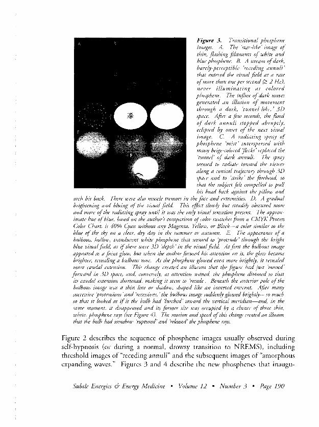
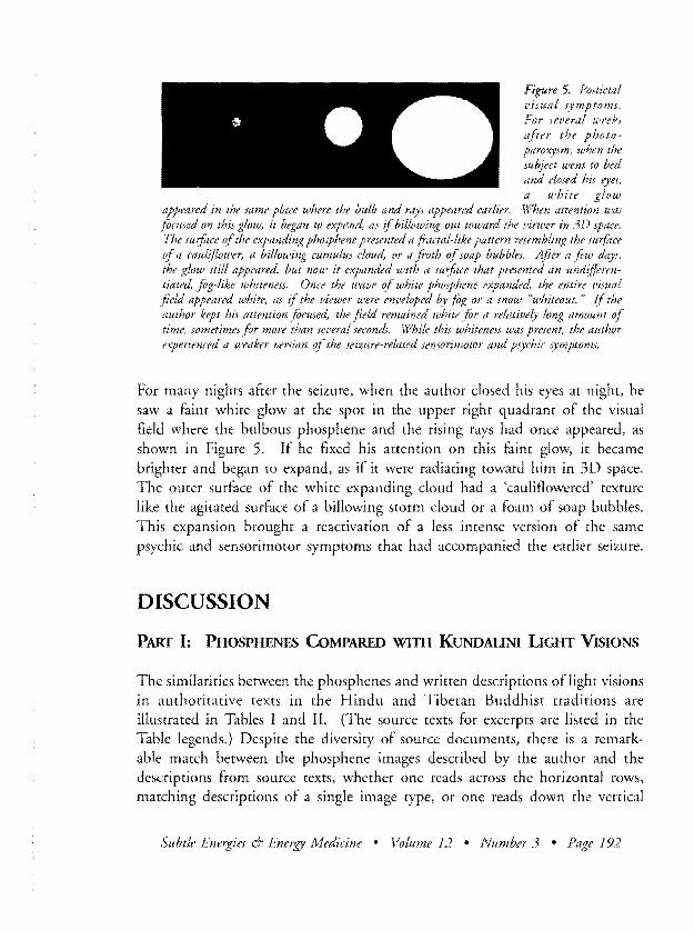
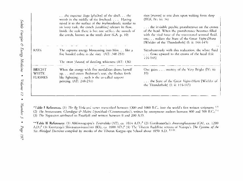
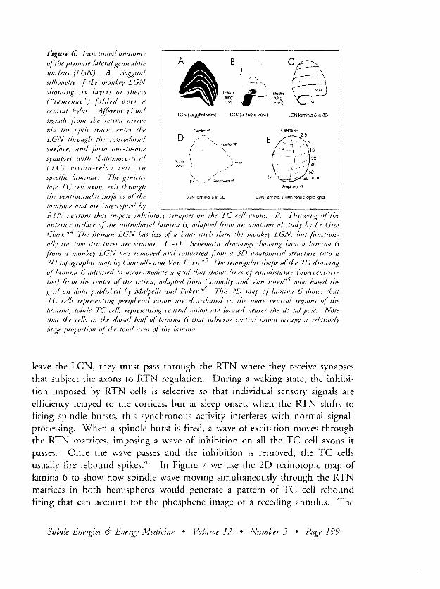
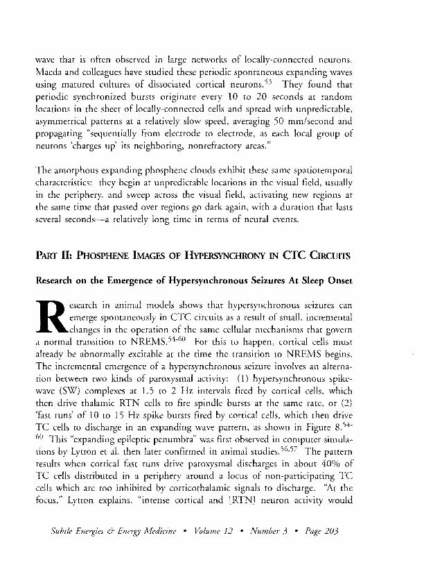