medicine care
TRANSCRIPT

P1Protective role of autophagy in mouse cecal ligation and puncture-induced sepsis modelW Takahashi1, H Hatano2, H Hirasawa1, S Oda1
1Graduate School of Medicine, Chiba University, Chiba, Japan; 2Biomedical Research Center, Chiba University, Chiba, JapanCritical Care 2013, 17(Suppl 2):P1 (doi: 10.1186/cc11939)
Introduction Autophagy is well known as one of the biogenic responses against various stresses, which possesses the benefi cial roles for survival, but little is known about the dynamics and its signifi cance during the septic condition. We hypothesized that autophagy is induced during the septic condition, and contributes to protect from tissue damage which subsequently leads to organ dysfunction. We confi rm whether the autophagic process is accelerated or sustained in an acute phase of sepsis and we also determine its physiological role.Methods Sepsis was induced by cecal ligation and puncture (CLP) in mice. We examined the kinetics of autophagosome and auto lysosome formation which may explain the status of autophagy by western blotting, immunohistochemistry, and electron microscopy. To investigate a precise role of autophagy in CLP-induced sepsis, chloroquine, an autophagy inhibitor, was administered to the CLP-operated mice, and blood chemistry, pathology of the liver and survival were evaluated.Results Autophagy demonstrated by the ratio of LC3-II/LC3-I was induced over the time course up to 24 hours after CLP. The ratio was particularly increased in the liver, heart and spleen. Autophagosome formation became maximal at 6 hours and declined by 24 hours after CLP. Autolysosome formation as evaluated by both fusion of GFP-LC3 dots with LAMP1 immunohistochemistry and electron microscopy was also increased after the procedure. Furthermore, inhibition of autophagy by chloroquine during the CLP procedure resulted in elevation of serum AST levels, and signifi cantly increased mortality in mice.Conclusion Autophagy was induced in several organs over the time course of the CLP sepsis model and then the process was gradually completed to degradation of the components. Our data suggest autophagy plays a protective role in organ dysfunction in sepsis.
P2Reversible depressive eff ect of TNFα on a model of isolated perfused rat heartBV Nguyen1, M Guillouet2, MA Giroux-Metges2, G Gueret3, M Ould-Ahmed1, JP Pennec2
1Hôpital d’Instruction des Armées Clermont Tonnerre, Brest, France; 2Laboratory of Physiology, Faculty of Medicine, Brest, France; 3University Hospital, Brest, FranceCritical Care 2013, 17(Suppl 2):P2 (doi: 10.1186/cc11940)
Introduction Acute myocardial depression in septic shock is common [1]. Myocardial depression is mediated by circulating depressant substances, which until now have been incompletely characterized [2].
The aim of our study was to observe the eff ects of TNFα on the model of perfused rat heart.Methods After profound anesthesia with pentothal, the Wistar rats were killed by exsanguination. After sternotomy, the heart was taken and connected to the Langendorf column. The apex of the heart was hooked to a strength sensor. Biopac student laboratory software was used to record and analyse heart contractions. Contractions were recorded every 5 minutes during periods of 20 minutes. Control measurements were fi rst recorded. We measured four parameters: heart rate, contraction force, speeds of contraction and relaxation for control, during TNFα (20 ng/ml) exposure and after removal of TNFα. We express the variations of parameters as percentage of the control ± SEM. A paired t test was used to compare heart rate, contraction amplitude, speeds of contraction and relaxation with TNFα and control measurements and after removal of TNFα.Results Eight rat hearts Wistar (weight = 325 ± 23 g) were studied. See Table 1.
Table 1 (abstract P2)
TNFα Removal of TNFα
Heart rate 78 ± 6* 91 ± 5
Contractile 62 ± 8* 91 ± 4
Speed of contraction 72 ± 6* 93 ± 2
Speed of relaxation 53 ± 10* 89 ± 4
Results expressed as percentage of control ± SEM. *P <0.05.
Conclusion TNFα decreases signifi cantly the heart rate, contractile force, speeds of contraction and relaxation on isolated perfused rat heart. TNFα probably plays a role in the pathophysiology of cardiomyopathy during septic shock. The partial reversibility of these eff ects could explain why left ventricular hypokinesia in patients with septic shock is reversible.References1. Vieillard-Baron A, et al.: Actual incidence of global left ventricular
hypokinesia in adult septic shock. Crit Care Med 2008, 36:1701-1706.2. Hunter JD, et al.: Sepsis and the heart. Br J Anaesth 2010, 104:3-11.
P3Eff ect of cdp-choline on microcirculatory alterations during endotoxemiaK Schmidt1, M Doerr1, T Brenner1, S Hofer1, A Walther2
1Universitätsklinikum Heidelberg, Germany; 2Klinikum Stuttgart, Klinik für Anästhesiologie u. operative Intensivmedizin, Stuttgart, GermanyCritical Care 2013, 17(Suppl 2):P3 (doi: 10.1186/cc11941)
Introduction The cholinergic anti-infl ammatory pathway (CAP) is a physiological mechanism that inhibits cytokine production and minimizes tissue injury during infl ammation. CAP-mediated anti-infl ammatory signals in vagal eff erent nerve fi bers result in the release of acetylcholine, which interacts with innate immune cells that express the nicotinic acetylcholine receptor subunit α7 (α7nAChR). © 2010 BioMed Central Ltd
33rd International Symposium on Intensive Care and Emergency MedicineBrussels, Belgium, 19-22 March 2013
Published: 19 March 2013
M E E T I N G A B S T R AC T S
Critical Care 2013, Volume 17 Suppl 2 http://ccforum.com/supplements/17/S2
© 2013 BioMed Central Ltd

Endothelial dysfunction during sepsis is responsible for increased endothelial permeability, leukocyte–endothelial interaction and functional breakdown of microvascular perfusion. Endotoxemia-induced endothelial dysfunction can be reduced by cholinergic CAP activation [1]. The aim of this study was to determine the eff ects of the α7nAChR-agonist cdp-choline on microcirculatory alterations during experimental endotoxemia.Methods Using fl uorescent intravital microscopy, we determined venular wall shear rate, macromolecular effl ux and leukocyte adhesion in mesenteric postcapillary venules of male Wistar rats. Endotoxemia was induced over 120 minutes by intravenous infusion of lipopolysaccharide (LPS). Control groups received an equivalent volume of saline. Cdp-choline was applied as an i.v. bolus in treatment groups. Animals received either (i) saline alone, (ii) cdp-choline 10 minutes prior to saline administration, (iii) cdp-choline 10 minutes prior to LPS administration, (iv) cdp-choline 30 minutes after LPS administration or (v) LPS alone.Results There were no signifi cant diff erences in venular wall shear rate between the groups after 120 minutes. There was no signifi cant diff erence in the number of adhering leukocytes between the cdp-choline/LPS groups (iii, iv) and the LPS group after 120 minutes. Macromolecular effl ux signifi cantly increased in all groups over 120 minutes. All groups (i, ii, iii, iv) showed a signifi cantly reduced macromolecular effl ux compared with the LPS group after 120 minutes.Conclusion Cdp-choline has no eff ect on leukocyte–endothelial interaction and microhemodynamic alterations during endotoxemia. By activating the CAP, cdp-choline reduces capillary leakage. Thus cdp-choline might have a prophylactic and therapeutic anti-infl ammatory eff ect on LPS-induced endothelial permeability. These fi ndings identify the endothelium as a target of anti-infl ammatory cholinergic mediators and cdp-choline as a potential therapeutic substance in sepsis treatment.Reference1. Peter C, et al.: Shock 2010, 33:405-411.
P4Immune response after stimulation with wall components of Gram-positive bacteriaS Aloizos1, E Tsigou2, P Myrianthefs2, S Gourgiotis1, A Tsakris3, G Baltopoulos2
1NIMTS Hospital, Athens, Greece; 2A. Anargiroi Hospital, Athens, Greece; 3Medical School, Athens, GreeceCritical Care 2013, 17(Suppl 2):P4 (doi: 10.1186/cc11942)
Introduction The purpose of this study was to evaluate the immune response of patients susceptible to infection by Gram-positive bacteria after ex vivo provocation with lipoteichoic acid (LTA) and to compare the reaction with the one of healthy adults.Methods Blood sample was obtained from 10 healthy volunteers, 10 hemodialysis patients with end-stage chronic renal failure (CRF), 10 patients with type II diabetes mellitus (DM) and 10 ICU patients on the second day of hospitalization, who suff ered nonseptic SIRS and had an APACHE II score >25. After suitable treatment the samples were incubated with 1 mg LTA for 8 hours and maintained at –20°C until the measurement of cytokines TNFα, IL-6, IL-1β, and IL-10, using the ELISA method. The results are presented as mean values ± SEM. Graph Pad 4.0 was used, applying a t test to test the variation of each cytokine in each group, and ANOVA to assess the diff erences between the four groups.Results Baseline cytokine values in the three groups were increased compared with the control group, but the diff erence was signifi cant only for the ICU group (Table 1, data only for IL-6 and IL-10). The quotient IL-10/IL-6 of baseline values was between 0.23 and 0.96 among healthy, ESRD and DM persons, and 1.32 among ICU patients. In all examined groups the levels of cytokines increased signifi cantly after stimulation with LTA, although ICU patients showed a diff erential
response (a fi vefold to ninefold rise compared with other groups who had an increase of 14-fold to 36-fold).Conclusion Severely ill patients and secondarily hemodialysis and diabetic patients are in a proinfl ammatory state. The response of all examined groups to provocation by LTA was suffi cient, with a diff erential expression of severely ill patients, a fact that refl ects their diff erent immunologic status.
P5Correlation of the oxygen radical activity and antioxidants and severity in critically ill surgical patients: preliminary reportJ Lee1, H Shim2, JY Jang1
1Yonsei University College of Medicine, Seoul, South Korea; 2Wonju Severance Christian Hospital, Yonsei University Wonju College of Medicine, Wonju, KoreaCritical Care 2013, 17(Suppl 2):P5 (doi: 10.1186/cc11943)
Introduction In septic patients, the oxygen radical (OR) showed toxic eff ect to induce infl ammation and antioxidant activity could aff ect organ dysfunction. This study was designed to determine the relationship between antioxidant level and severity of organ dysfunction.Methods The medical records of adult patients managed in a surgical ICU from August 2012 to December 2012 were reviewed prospectively. Abstracted data included age, body weight (with BMI), APACHE II scores, SOFA scores, MODS scores, fl uid intake, fl uid output, nutritional support, shock, antioxidant levels, OR activities, zinc and selenium levels, complication and mortality. In addition, length of stay (LOS) in the ICU and in hospital, and in-hospital mortality were collected. These data were investigated on the fi rst, the third and the seventh day, respectively.Results A total of 13 patients were enrolled. The in-hospital mortality rate was 7.7% and mean LOS in the ICU and hospital was 6.5 and 27.6, respectively. Mean APACHE II score was 20.2. On the fi rst day of ICU, the mean antioxidant level and OR were 1.5 (± 0.5) mmol/l and 1.6 (± 0.5) mmol/l, respectively. At the same time, SOFA and MODS scores were 7.3 and 5.0, respectively, and zinc and selenium were 32.6 μg/dl and 68.4 ng/ml. On the third day, mean antioxidant and OR were 1.5 (± 0.4) and 1.8 (± 0.7) respectively (SOFA 6.6, MODS 4.9, zinc 50.0, selenium 70.7). On the seventh day, mean antioxidant and OR were 1.4 (± 0.5) and 1.9 (± 0.7), respectively (SOFA 4.3, MODS 3.1, zinc 62.8, selenium 77.3). In the correlation analysis, MODS scores and antioxidant level had signifi cant correlations on the fi rst and seventh days of ICU (P = 0.001, P = 0.009).Conclusion Antioxidant level had a correlation with organ dysfunction which might be used as a prognostic factor in critically septic patients. To prove this, large-scale data collection is required.References1. Noveanu M, Mebazaa A, Mueller C: Cardiovascular biomarkers in the ICU.
Curr Opin Crit Care 2009, 15:377-383.2. Piechota M, Banach M, Irzmanski R, Barylski M, Piechota-Urbanska M, Kowalski
J, et al.: Plasma endothelin-1 levels in septic patients. J Intensive Care Med 2007, 22:232-239.
3. Kotsovolis G, Kallaras K: The role of endothelium and endogenous vasoactive substances in sepsis. Hippokratia 2010, 14:88-93.
P6Simultaneous analysis of the expression of CD64 and HLA-DR in the peripheral blood and bronchoalveolar lavage fl uid in sepsisT Skirecki1, M Mikaszewska-Sokolewicz2, G Hoser1, U Zielińska-Borkowska1
1The Centre of Postgraduate Medical Education, Warsaw, Poland; 2 Medical University of Warsaw, PolandCritical Care 2013, 17(Suppl 2):P6 (doi: 10.1186/cc11944)
Introduction The core pathophysiological changes in sepsis involve systemic activation of the immune system followed by the
Table 1 (abstract P4). Levels of cytokines before and after stimulation with LTA
Control baseline LTA ESRD baseline LTA DM baseline LTA ICU baseline LTA
IL-6 8.90 ± 0.76, 245.30 ± 26.68 86.60 ± 45.55, 1,310.00 ± 154.80 15.90 ± 1.89, 252.00 ± 35.52 372.40 ± 120.60, 3,659.00 ± 485.20
IL-10 3.00 ± 1.08, 40.90 ± 7.45 19.20 ± 7.14, 273.10 ± 126.50 15.30 ± 2.08, 350.50 ± 89.42 492.60 ± 66.72, 2,822.00 ± 432.70
Values in pg/ml.
Critical Care 2013, Volume 17 Suppl 2 http://ccforum.com/supplements/17/S2
S2

anti-infl ammatory compensatory response. However, controversies exist regarding the status of the immune system in local tissue compartments during sepsis. The aim of this study was to compare selected markers of activation between the systemic circulation and local lung environment.Methods Twenty patients with severe sepsis were included into this study. Peripheral blood (PB) samples and bronchoalveolar lavage fl uid (BALF) samples were obtained on the day of diagnosis (D1). BALF was collected from 11 patients. Samples were stained with antibodies: CD15/CD64 and CD3/CD14/HLA-DR and isotypic control. Cells were analysed by fl ow cytometry. Expression of markers of activation was analysed as the geometric median of fl uorescence (GMF). All values are expressed as median values. Comparisons between groups were performed using Mann–Whitney and Wilcoxon tests.Results The mortality of sepsis reached 70%. Nonsurvivors had signifi cantly (P = 0.001) elevated expression of CD64 on neutrophils. Expression of HLA-DR was higher in monocytes from BAL than PB GMF (1,032 vs. 342; P = 0.02) and this tendency was present in sepsis originating from both pneumonia and peritonitis. Percentage of HLA-DR-positive T cells was lower in PB than in BAL (2.9% vs. 6.5%; P = 0.07), but the GMF values for HLA-DR were higher in the circulating T cells (1,904 vs. 1,346; P = 0.004). The expression of CD64 on neutrophils was not signifi cantly diff erent in PB and BAL, but there was a trend towards its higher expression in BAL from patients with pneumonia while its expression was higher in PB of patients with peritonitis.Conclusion In this study we noticed that during sepsis some signifi cant diff erences in the status of activation of immune cells exist between peripheral blood and lung resident cells. The lung milieu seems to promote activation of monocytes while neutrophil activation is more dependent on the site of infection. However, these observations require further studies in a larger group of patients.Acknowledgements This study was supported by the Centre of Postgraduate Medical Education grant no 501-01-02-012 and by the sources of the Medical University of Warsaw.
P7Anti-infl ammatory eff ects of Kupff er cells through α7-nicotinic acetylcholine receptorsY Li, X ShiChangzheng Hospital, Second Military Medical University, Shanghai, ChinaCritical Care 2013, 17(Suppl 2):P7 (doi: 10.1186/cc141945)
Introduction Nicotine exerts anti-infl ammatory eff ects in several cell types. α7-nicotinic acetylcholine receptor (α7-nAChR), which has high permeability to calcium, is believed to contribute signifi cantly to nicotinic anti-infl ammatory eff ects. However, the molecular mechanism is largely unknown. Kupff er cells in the liver play an important role in infl ammatory response to pathogens invading, but whether there is α7-nAChR expression in Kupff er cells or cholinergic anti-infl ammatory pathway involved in this process remains unclear.Methods (1) Kupff er cells, isolated by collagenase digestion and diff erential centrifugation from mice and labeled with FITC-aBGT, were observed under laser scanning confocal microscope to test the expression of α7-nAChR. Protein level was also tested by western blotting, with RAW264.7 as positive control; (2) 100 nM LPS was given to Kupff er cells, with or without 1 mM nicotine. TNFα, IL-10 and HMGB-1 were tested at 4 hours, 12 hours or 24 hours, respectively; (3) 100 BALB/c mice were randomly divided into four group: Group I (only lethal dose of LPS was given), Group II (nicotine and LPS were given), Group III (LPS, nicotine and GdCl3 were given), and Group IV (LPS and nicotine were given and the left cervical vagus nerve was cut off ). The mortality of mice was observed for 72 hours.Results (1) Expression of α7-nAChR in Kupff er cells was confi rmed by confocal microscope and western blotting; (2) after nicotine was administered, the level of TNFα and HMGB-1 increased and the level of IL-10 decreased. Given left cervical vagus nerve cut off or aBGT, the eff ect of nicotine was weakened; (3) Group I had the highest mortality rate, while in Group II nicotine did reduce the mortality rate dramatically. After the left cervical vagus nerve was cut off or aBGT was given, the eff ects of nicotine were weakened. Diff erence for the mortality rate between Group III and Group IV was not signifi cant.
Conclusion Kupff er cells played a crucial rule in modulating infl ammation and the anti-infl ammatory eff ect of nicotine was partially weakened after left cervical vagus nerve cut off or aBGT was given. It was verifi ed that left cervical vagus nerve was essential for the anti-infl ammatory eff ect of nicotine and α7 acetylcholine receptors might play a critical role.References1. Wang H, et al.: Nicotinic acetylcholine receptor α7 subunit is an essential
regulator of infl ammation. Nature 2003, 421:384-388.2. Wang H, et al.: Cholinergic agonists inhibit HMGB1 release and improve
survival in experimental sepsis. Nat Med 2004, 10:1216-1221.
P8Prevention of sepsis by correcting apoptosisM Puhtinskaya, V EstrinResearch Institute of Obstetrics and Pediatrics, Rostov-on-Don, RussiaCritical Care 2013, 17(Suppl 2):P8 (doi: 10.1186/cc11946)
Introduction Activation of apoptosis in lymphocytes determines the development of neutropenia and of sepsis [1,2]. We investigated prevention of sepsis and correction of lymphocyte apoptosis by recombinant human granulocyte colony-stimulating factor (hr-GCSF, fi lgrastim) [1,2].Methods With the permission of the ethics committee, a controlled, randomized, blind clinical trial included 69 term newborns on mechanical ventilation, without neutropenia and clinical signs of infection, with a content of lymphocytes in early apoptosis (AnnexinV–FITC+PI–) of >9.59%, and in late (AnnexinV–FITC+PI+) of 0.56%. Lymphocytes in apoptosis were detected using antibodies to AnnexinV and propidium iodide staining method of immunophenotyping (fl ow cytometry; Beckman Coulter Epics XL, USA). The survey was conducted at admission, at 3 to 5 days, and 20 days. The method of random numbers in Group I included 39 newborns who on admission (with written parental consent) received an intravenous infusion of hr-GCSF dose of 10 μg/kg, 3 days. Newborns of Group II (n = 30) did not receive hr-GCSF. Power of the study was 80% (α ≤0.05).Results For 3 to 5 days, Group I signifi cantly decreased apoptosis of lymphocytes in the early from 16.1% to 7.8%, and in late from 1.3% to 0.1%. The development of sepsis and neutropenia have been reported. We observed no clinical or laboratory signs of adverse eff ects of the drug. Fatal outcomes (n = 4) are not associated with hr-GCSF, which was confi rmed postmortem. Decreased duration of mechanical ventilation (P <0.05). In Group II, 27 patients at 3 to 5 days developed neutropenia and increased lymphocytes in apoptosis (P <0.05). Sepsis was diagnosed in 19 children; eight fatal outcomes.Conclusion hr-GCSF reduces the incidence of septic complications and one of the mechanisms of its clinical eff ectiveness is the reduction of apoptotic factors aff ecting the development of neutropenia.References1. Gillan ER, Christensen RD, Suen Y, et al.: A randomized, placebo-controlled
trial of recombinant human granulocyte colony-stimulating factor administration in newborn infants with presumed sepsis: signifi cant induction of peripheral and bone marrow neutrophilia. Blood 1994, 84:1427-1433.
2. Pukhtinskaya MG, Estrin VV, Gulova ES: Clinical and diagnostic value of apoptosis markers in the pathogenesis of neutropenia and bacterial complications in newborns with respiratory distress syndrome. Cytokines Infl amm 2011, 10:66-69.
P9Immune paralysis in trauma patients; implications for prehospital interventionM Kox, K Timmermans, M Vaneker, GJ Scheff er, P PickkersRadboud University Nijmegen Medical Center, Nijmegen, the NetherlandsCritical Care 2013, 17(Suppl 2):P9 (doi: 10.1186/cc11947)
Introduction Multi-trauma is one of the major indications for intensive care admission. Recovery is frequently complicated by post-injury immunological complications, caused by a dysfunctional immune system; for example, sepsis and multiple organ failure. In order to treat
Critical Care 2013, Volume 17 Suppl 2 http://ccforum.com/supplements/17/S2
S3

or prevent this immune paralysis, knowledge on the time course of immune paralysis in vivo and the pathophysiological mechanisms of immune paralysis is essential. The aim of this study is to determine factors that could predict and/or induce immunological complications in these patients to ultimately fi nd a suitable target and timeframe for intervention.Methods Blood was drawn from adult multi-trauma patients (n = 94) admitted to the emergency room (ER) of the Radboud University Nijmegen Medical Center. Blood was drawn at the trauma scene by the helicopter emergency medical services (HEMS), at arrival in the ER and at days 1, 3, 5, 7, 10 and 14 after trauma. Plasma concentrations of TNFα, IL-6, IL-10, IFNγ, IL-8 and MCP-1 were determined by Luminex. Ex vivo 24-hour whole blood stimulations with LPS or pam3cys were performed and produced TNFα, IL-6 and IL-10 was measured using ELISA to determine the level of immune paralysis. Clinical data – for example, Injury Severity Scores, trauma mechanism, medication and survival – were collected from electronic patient fi les.Results The plasma IL-10 concentration at ER was 16.5-fold increased in comparison with time-point HEMS (P <0.01). Similar but less pronounced eff ects were found for IL-8 and MCP-1. A signifi cant correlation (P = 0.03, R = 0.53) was found between injury severity scores and IL-10 plasma concentration at time-point ER. Time-courses of ex vivo produced cytokines suggest that LPS-induced IL-6 and TNFα production is already decreased in the fi rst few hours after trauma and recovering from day 5. Ex vivo IL-10 production shows an inverse pattern.Conclusion Immune paralysis can be established within hours after trauma. Production of anti-infl ammatory IL-10 in the prehospital phase could play a crucial role in the pathogenesis. Patients with a higher injury severity score are more prone to produce excessive IL-10 in this phase. Immune stimulatory strategies applied by the HEMS or early after hospital admission could form a potential future approach to prevent immune paralysis in multitrauma patients in the intensive care ward.
P10Is hemoglobin concentration aff ected by sepsis in the acute phase?G Jansma, H Buter, RT Gerritsen, EC BoermaMedical Centre Leeuwarden, the NetherlandsCritical Care 2013, 17(Suppl 2):P10 (doi: 10.1186/cc11948)
Introduction In the acute phase of sepsis several potential mechanisms may change the hemoglobin (Hb) concentration. On the one hand, endothelial activation may lead to increased vascular permeability and fl uid sequestration to the interstitium, leading to hemoconcentration. On the other hand, degradation of the glycocalyx has been reported [1]. Shedding of this carbohydrate-rich layer with an estimated thickness of 0.2 to 0.5 μm may lead to a substantial increase of the intravascular space, and thus to decrease of Hb concentration [2]. The aim of this study is to determine whether there is a decrease in Hb in the acute phase of sepsis.Methods In this single-center retrospective analysis we identifi ed patients with sepsis as the primary reason for non-elective ICU admission from a standard patient database. Patients who fulfi lled the international criteria of sepsis and organ failure during ICU admission were included in the sepsis group (S-group). The control group was formed by patients with other non-elective reasons for ICU admission (C-group). Exclusion criteria were (recent) bleeding, surgery in the last 6 weeks, chronic renal failure (creat >177 μmol/l, or hemodialysis), untreated chronic anemia, pregnancy, polytrauma, age <18,
hematologic or metastasized malignancies, cardiac arrest, and use of bone marrow suppressive drugs. Laboratory data were collected from blood samples, prior to in-hospital i.v. fl uid therapy. In order to detect a diff erence in Hb concentration of 0.2 mmol/l, we anticipated a sample size of 283 per group, based on a standard deviation (SD) of 1.2, α = 0.05 and β = 0.8. Data are expressed as mean ± SD.Results We included 296 patients in the S-group and 320 in the C-group. The diff erence in Hb between the S-group and C-group was not signifi cant (8.76 ± 1.18 mmol/l vs. 8.93 ± 1.16 mmol/l, P = 0.07). After correction for a number of confounders, using a multivariate regression analysis, we observed a signifi cant diff erence in Hb of –0.23 mmol/l in the S-group in comparison with the C-group (P = 0.01).Conclusion At fi rst presentation, prior to in-hospital i.v. fl uid therapy, Hb concentration in patients with sepsis is signifi cantly lower in comparison with controls; however, the diff erence is very small, without the existence of anemia.References1. Steppan J, et al.: Sepsis and major abdominal surgery lead to fl aking of the
endothelial glycocalyx. J Surg Res 2011, 165:136-141.2. van den Berg BM, et al.: The endothelial glycocalyx protects against
myocardial edema. Circ Res 2003, 92:592-594.
P11Do changes in red blood cell deformability in patients with septic shock correlate with changes in SOFA scores?T Clark1, S Jewell2, M Sair1, P Petrov2, P Winlove2
1Derriford Hospital, Plymouth, UK; 2University of Exeter, UKCritical Care 2013, 17(Suppl 2):P11 (doi: 10.1186/cc11949)
Introduction Traditional whole blood experiments suggest that sepsis causes abnormal red blood cell (RBC) deformability. To investigate this at the cellular level, we employed a novel biophysical method to observe individual RBC membrane mechanics in patients with septic shock.Methods We collected blood samples from patients with septic shock until either death or day 5 of admission. Thermal fl uctuations of individual RBCs were recorded allowing a complete analysis of RBC shape variation over time. Mean elasticity of the cell membrane was then quantifi ed for each sample collected.Results We recruited nine patients with septic shock. Table 1 shows mean RBC thermal fl uctuation and SOFA scores.Conclusion RBC thermal fl uctuation analysis allows variations in RBC elasticity during sepsis to be quantifi ed at a cellular level. We could not identify any specifi c trend between sepsis severity and erythrocyte elasticity. Cells demonstrated both increases and decreases in fl uctua-tion independent of SOFA score. This is contrary to current evidence that suggests RBC deformability is reduced during sepsis.Reference1. Piagnerelli M, et al.: Intensive Care Med 2003, 29:1052-1061.
P12Do erythrocytes subjected to cardiopulmonary bypass exhibit changes in their membrane mechanical properties?T Clark1, S Jewell2, M Sair1, P Petrov2, P Winlove2
1Derriford Hospital, Plymouth, UK; 2University of Exeter, UKCritical Care 2013, 17(Suppl 2):P12 (doi: 10.1186/cc11950)
Introduction Whole blood experiments suggest that cardiopulmonary bypass (CPB) causes red blood cell (RBC) trauma and changes in deformability that may contribute to postoperative microcirculatory
Table 1 (abstract P11). Mean RBC fl uctuation (daily SOFA score)
Day A B C D E F G H I
1 4.8 (10) 5.2 (9) – 4.8 (12) 4.6 (16) 4.9 (13) 5.1 (16) 4.6 (18) 5.1 (15)
2 4.9 (9) 5.0 (10) 4.6 (13) 5.1 (11) 4.8 (17) 5.1 (13) 5.0 (16) 4.9 (19) – (16)
3 4.0 (6) 5.1 (9) 4.7 (12) 4.8 (11) 4.9 (18) 4.8 (13) 5.0 (16) 4.7 (21) 5.3 (16)
4 – 4.8 (7) 4.6 (11) 5.0 (9) 5.9 (19) – – (18) – 5.0 (15)
5 – – – 5.1 (6) 5.2 (18) – – (19) – 5.0 (10)
Critical Care 2013, Volume 17 Suppl 2 http://ccforum.com/supplements/17/S2
S4

dysfunction. We used a novel fl uctuation microscopy technique to quantify the eff ects of CPB on RBC elasticity at a cellular level.Methods We collected blood samples from elective cardiac surgery patients pre (at induction) and post (immediately, each day until CICU discharge) CPB. Thermal fl uctuations of individual RBCs were recorded using a high-frame-rate camera allowing a complete analysis of RBC shape variation over time. Mean elasticity of the cell membrane was then quantifi ed for each sample collected.Results Fifteen patients were recruited. Table 1 displays the results. RBC thermal fl uctuation is measured relative to pre-bypass values. An increase in RBC fl uctuation marks a decrease in stiff ness. CPB caused two distinct changes in RBC elasticity; pre-fi x A indicates samples where stiff ness increases or shows no change, B those where stiff ness decreases. Data on day 2 were not collected in patients discharged from the CICU. CPB type or time had no apparent impact on RBC response to CPB.Conclusion RBC thermal fl uctuation analysis quantifi es the impact of CPB on erythrocyte membrane elasticity. We clearly identifi ed two separate RBC elasticity responses to CPB. This fi nding is contrary to traditional fl ow measurement techniques that suggest CPB impairs whole blood fl ow and reduces RBC deformability.Reference1. Lindmark et al.: J Thoracic Cardiovasc Surg 2002, 123:381-383.
P13Platelet-associated oxidative stress and ADAMTS-13 levels are inversely associated with a poor prognosis in septic shockL Montini1, G De Pascale1, MA Pennisi1, ES Tanzarella1, SL Cutuli1, A Occhionero1, R De Cristofaro2, M Antonelli11Catholic University School of Medicine, Rome, Italy; 2Haemostasis and Thrombosis Center, Catholic University School of Medicine, Rome, ItalyCritical Care 2013, 17(Suppl 2):P13 (doi: 10.1186/cc11951)
Introduction Sepsis causes widespread microvascular injury and thrombosis. Some hemostatic factors mediate the mechanisms involved in sepsis-related organ ischemia and failure. Oxidative stress is also increased in sepsis and reactive oxygen species (ROS) favor secretion of von Willebrand factor (vWF) multimers from endothelium and inhibit vWF proteolysis by ADAMTS-13. Moreover, the enzyme indoleamine-2,3-dioxygenase, an important immune regulator, is activated in sepsis and, through generation of kynurenins, promotes antioxidative and anti-infective activities. This study evaluated the relative role of ADAMTS-13, vWF and fi brinogen in the morbidity and mortality of patients with septic shock (SS). The above hemostatic factors were measured together with kynurenine and plasma protein carbonyls, marker of oxidative stress.Methods One group of 12 patients with SS, defi ned using standard criteria, was enrolled in the ICU of the ‘A. Gemelli’ Hospital (Rome, Italy). Biochemical, hematologic and hemodynamic parameters were measured on days 1 to 4, 7, 14 and 21. A group of 12 age-matched and gender-matched healthy subjects was used as controls.Results Low ADAMTS-13 activity was observed in SS patients (268 ± 123 ng/ml vs. 760 ± 80 ng/ml in controls). vWF levels (antigen and activity) were increased ~3-fold compared with controls. Likewise, plasma protein carbonyls and kynurenine were globally increased in patients (2.1 ± 1.5 nmol/mg vs. 0.3 ± 02 nmol/mg and 14.4 ± 9.7 μM vs. 2.3 ± 1.3 μM, respectively). Intra-ICU mortality (3 of 15) was strongly and inversely correlated with carbonyl levels (P = 0.04) and platelets (P = 0.022).Conclusion Hence, we hypothesize that, in the SS setting, platelets contribute to oxidative stress that counteracts the organ failure-associated mortality. Thus, low platelet count, irrespective of bleedings, may favor mortality in SS patients by generating lower ROS amounts.
Reference1. Strauss R, et al.: Thrombocytopenia in patients in the medical intensive care
unit: bleeding prevalence, transfusion requirements, and outcome. Crit Care Med 2002, 30:1765-1771.
P14Neutrophil gelatinase-associated lipocalin/lipocalin2, derived from gut crypt cells, exerts intestinal antimicrobial eff ect via bacterial stimulation of Toll-like receptor 4 and 9K Mori, T Igarashi, K Inoue, T Suzuki, H Morisaki, J TakedaKeio University School of Medicine, Tokyo, JapanCritical Care 2013, 17(Suppl 2):P14 (doi: 10.1186/cc11952)
Introduction Neutrophil gelatinase-associated lipocalin (NGAL)/lipocalin2, known as a sensitive biomarker of acute kidney injury, prevents bacterial iron uptake, resulting in the inhibition of its overgrowth [1]. We previously demonstrated that this protein was discharged into gut lumen from crypt cells in septic conditions, and inhibited the growth of Escherichia coli [2]. However, it remains unclear which pathway is associated with the upregulation of NGAL. We therefore designed the present study to reveal whether the pattern-recognition receptor of bacteria, the Toll-like receptor (TLR) family, plays a pivotal role for NGAL secretion from gut crypt cells.Methods With our institutional approval, the ileum and colon of male C57BL/6J mice (6 to 7 weeks) were everted and washed by Ca2+ and Mg2+ free PBS buff er fi ve times. Tissues were incubated with Ca2+ and Mg2+ free PBS containing 30 mM EDTA for 1 hour to isolate crypt cells of gut. The cell suspension was fi ltered through a cell strainer (40 μm) twice, and deposited the crypt cells by centrifugation at 700×g. The isolated crypt cells were resuspended in PBS and stained with 0.25% amido black for labeling paneth cells. The 5×10⁵ crypt cells were resuspended in 50 ml HBSS containing 2.5% fetal bovine serum and 1% penicillin–streptomycin. The crypt cells were incubated at 37°C with or without TLR ligands: lipopolysaccharide (TLR4 ligand, 10 μg/ml) and CpG-DNA (TLR9 ligand, 8 μg/ml). After a 2-hour incubation period, the crypt cells were deposited and eluted mRNA to measure the expression of both NGAL and TLR mRNA using real-time PCR.Results More than 70 to 80% of collected cells were stained by amido black. LPS signifi cantly upregulated the expression of NGAL and TLR4 mRNA in ileum and colon crypt cells (P <0.05). Although the CpG-DNA did not upregulate NGAL and TLR9 mRNA in ileum crypt cells, the apparent expression of NGAL and TLR9 mRNA was found in colon crypt cells (P <0.05).Conclusion Bacterial stimulation of TLR4 and TLR9 pathways plays a pivotal role in the expression of NGAL mRNA in gut, suggesting that NGAL, derived from gut crypt cells, could contribute to the regulation of the intraluminal microfl ora in the critically ill.References1. Nature 2004, 432:917.2. Crit Care Med 2011, 39:46.
P15Lethal infl uenza virus A H1N1 infection in two relatives with autosomal dominant GATA-2 defi ciencyJ Sole-Violan1, I Sologuren1, E Betancor2, S Zhang3, C Pérez1, E Herrera-Ramos1, M Martínez-Saavedra1, M López-Rodríguez1, J Pestano2, J Ruiz-Hernández1, J Ferrer1, F Rodríguez de Castro1, J Casanova3, C Rodríguez-Gallego1
1Hospital GC Dr Negrín, Las Palmas de GC, Spain; 2Universidad Las Palmas de GC, Spain; 3The Rockefeller University, New York, NY, USACritical Care 2013, 17(Suppl 2):P15 (doi: 10.1186/cc11953)
Introduction Most individuals infected with the 2009 pandemic H1N1 infl uenza A virus (IAV) (H1N1pdm) experienced uncomplicated fl u.
Table 1 (abstract P12). Change in RBC thermal fl uctuation relative to baseline: two distinct groups seen
Patient A1 A2 A3 A4 A5 A6 A7 B8 B9 B10 B11 B12 B13 B14 B15
Post CPB +0.1 –0.4 –0.1 –0.5 –0.1 –0.2 –0.1 +0.5 0 +0.4 +0.1 +0.2 0 +0.2 +0.1
Day 1 0 0 +0.1 –0.1 –0.1 +0.1 +0.1 0 +0.5 +0.4 +0.7 +0.4 +0.6 +0.5 +0.7
Day 2 –0.2 +0.1 0 NA NA 0 NA +0.5 –0.1 +0.4 NA +0.1 NA NA NA
Critical Care 2013, Volume 17 Suppl 2 http://ccforum.com/supplements/17/S2
S5

However, in a small subset of patients the infection rapidly progressed to primary viral pneumonia (PVP) and a minority of them developed ARDS. Inherited and acquired variability in host immune responses may infl uence susceptibility and outcome of IAV infection. However, the molecular nature of such human factors remains largely elusive.Methods We report three adult relatives with the autosomal dominant GATA-2 defi ciency. P1 and his son P2 had a history of myelodysplastic syndrome and a few episodes of mild respiratory infections. They developed PVP by H1N1pdm which rapidly evolved to ARDS. They died at the age of 54 and 31, respectively.Results Patients were heterozygous for a novel R396L mutation in GATA2. Like other patients with GATA-2 defi ciency, the three relatives had absence of peripheral NK and B cells and monocytopenia. However a high number of plasma cells, which were found to be pauciclonal, were observed in peripheral blood from P1 during H1N1pdm infection. P1 and P2 had normal levels of immunoglobulins and IgG antibodies against common viruses. Microneutralization test showed that P1 produced normal titers of neutralizing antibodies against H1N1pdm and against the previous annual H1N1 strain. Our results suggest that a few clones of long-living memory B cells against IAV expanded in P1; and that these cells produced cross-reactive antibodies against H1N1pdm, similar to those recently described. During the fl u episode P1 had a strong increase of IFNγ-producing T cells and of IFNγ production. The Th1-related chemokines CXCL10 and CXCL9, as well as IFNγ, MCP-1 and IL-8, were strongly elevated in serum from P1 and P2 in the course of H1N1pdm infection.Conclusion GATA-2 defi ciency is the fi rst described Mendelian inborn error of immunity underlying severe IAV infection. Primary immunodefi ciencies predisposing to severe IAV infections may debut, even in adults without a history of previous severe infections. The massive IFNγ-mediated cytokine storm may explain the fatal course of H1N1pdm infection in our patients.
P16Bacterial translocation primes proinfl ammatory responses and is connected to early death in an experimental model of lethal injuryN Baxevanos, T Tsaganos, A Pistiki, D Droggiti, A Spyridaki, E Giamarellos-BourboulisUniversity of Athens, Medical School, Athens, GreeceCritical Care 2013, 17(Suppl 2):P16 (doi: 10.1186/cc11954)
Introduction Some cases of multiple trauma are rapidly deteriorating; the mechanism was investigated.Methods Forty-one rabbits were assigned into two groups; sham-operated and subject to crush of the right femur. Survival was recorded; peripheral blood was sampled for LPS measurement by the kinetic QCL-1000 LAL assay; quantitative tissue growth was assessed after death. Some rabbits were sacrifi ced at 48 hours; blood was sampled from the portal vein for LPS measurement; splenocytes were
isolated and incubated for 24 hours in the presence of 10 ng/gl LPS of Escherichia coli O55:B5 and of 5 μg/ml phytohemagglutin (PHA); TNFα was measured in supernatants by a bioassay on L929 fi broblasts.Results Fifty percent of rabbits died early; that is, within the fi rst 48 hours. Mean ± SE log10 bacteria in the liver and lung of animals that died early was 2.27 ± 0.62 and 3.16 ± 0.78 cfu/g; respective values of rabbits that started dying late (that is, after 72 hours) were below the limit of detection. Mean circulating LPS at 24 hours was 2.09 EU/ml and 1.99 EU/ml respectively (P = NS). Mean LPS of the portal vein of the sham and of the injury groups were 1.25 and 5.62 EU/ml (P = 0.047). Concentrations of TNFα in splenocyte supernatants are shown in Figure 1.Conclusion Early death after injury is not related to peripheral endotoxemia and sepsis; bacterial translocation priming for enhanced proinfl ammatory responses is a likely explanation.
P17Eff ects of the common 34C>T variant of the AMPD1 gene on immune function, multiorgan dysfunction and mortality in patients with sepsisB Ramakers1, E Giamarellos-Bourboulis2, M Coenen1, M Kox1, J Van der Hoeven1, C Routsi2, A Savva2, I Perdios3, F Diamantea4, D Sinapidis5, P Smits1, N Riksen1, P Pickkers1
1Radboud University Nijmegen Medical Centre, Nijmegen, the Netherlands; 2University of Athens, Medical School, Athens, Greece; 3Nafplion General Hospital, Nafplio, Greece; 4‘G.Gennimatas’ General Hospital, Athens, Greece; 5Alexandra General Hospital, Athens, GreeceCritical Care 2013, 17(Suppl 2):P17 (doi: 10.1186/cc11955)
Introduction Adenosine exerts anti-infl ammatory and tissue protective eff ects during systemic infl ammation. While the anti-infl ammatory properties may induce immunoparalysis and impede bacterial clearance, the tissue protective eff ects might limit organ damage. The eff ects of a common loss-of-function variant of the adenosine monophosphate deaminase 1 gene (AMPD1), which is associated with increased adenosine formation, in patients with sepsis are unknown.Methods In a prospective cohort, genetic-association study, the eff ects of the presence of the AMPD1 gene on immune function, multiorgan dysfunction and mortality in septic patients was studied. Pneumosepsis patients (n = 402) and controls without infection (n = 101) were enrolled.Results In pneumosepsis patients and controls, a similar prevalence of the 34C>T (rs17602729) mutation in the AMPD1 gene was found. Univariate logistic regression analysis showed a tendency of increased mortality in patients with the CT genotype, compared with patients with the CC genotype (OR 1.53; 95% CI 0.95 to 2.5). Moreover, carriers of the CT genotype tended to suff er more from multiorgan dysfunction, OR 1.4 (0.84 to 2.3) and 3.0 (0.66 to 13.8), for CT and TT, respectively (P = 0.07). In septic carriers of the CT genotype, the ex vivo production of TNFα by LPS-stimulated monocytes was attenuated (P = 0.005),
Figure 1 (abstract P16). Stimulation of TNFα from isolated splenocytes. Figure 1 (abstract P17). Kaplan–Meier curve for the 402 sepsis patients.
Critical Care 2013, Volume 17 Suppl 2 http://ccforum.com/supplements/17/S2
S6
indicative for more pronounced immunoparalysis in these patients. See Figure 1.Conclusion The presence the 34C>T variant of the AMPD1gene is not related to infection susceptibility; however, it is associated with more pronounced immunoparalysis in patients with sepsis, and shows a tendency towards increased mortality. Mechanistically, the anti-infl ammatory eff ects of adenosine may account for this and apparently overrule its tissue protective eff ects.
P18Exploring the translational disconnect between the murine and human infl ammatory response: in vitro analysis of the dose–response relationship of LPS and NFκB activation in murine and human immune cellsEP McCarron, I Welters, D Williams, D Antoine, A KiparUniversity of Liverpool, UKCritical Care 2013, 17(Suppl 2):P18 (doi: 10.1186/cc11956)
Introduction Infl ammation, as seen in sepsis and systemic infl ammatory response, is dependent on the activation of the NFκB pathway through Toll-like receptors (TLRs) [1]. Recreating an infl ammatory response using lipopolysaccharide (LPS) can provide results that are diff erent to clinical sepsis [2]. By examining NFκB activation in murine and human cells, a species comparison can be made to investigate diff erences at the cell level that may contribute to the translational disconnect seen in vivo.Methods THP1 human monocytes (passages 9 to 11) and RAW 264.7 murine macrophages (passages 15 to 20) were cultured in RPMI-1640 and DMEM respectively and then challenged with LPS. After settling for 24 hours, cells were dosed with six or seven doses of LPS. After 1 hour, nuclear extraction and proteins were separated by acrylamide gel electrophoresis. Membranes where then immunoblotted for actin and p65, followed by densitometric analysis in order to quantify the amount of p65 that had translocated from the cytoplasm to the nucleus (by subtraction from consistent nuclear actin).Results Murine cells required higher doses of LPS compared with human cells in order to detect p65 (human, 1 pg/ml to 100 ng/ml; murine, 30 pg/ml to 1,000 ng/ml). THP1 cells showed a greater fold increase in the p65:actin ratio compared with RAW 264.7 cells. Human cells responded to lower concentrations of LPS. Murine cells appeared to show a molecular resistance to lower doses, but their response was very sensitive at higher doses. A dose–response relationship of LPS dosing and NFκB activation was observed in both cell lines.Conclusion Immunoblotting for p65 is a reliable and reproducible method to determine NFκB activation in cultured cells. Macrophages are more responsive to LPS than monocytes [3] so diff erences between cell lines would have been expected to be the reverse of what was observed. The species diff erence in response to LPS may contribute to the apparent disconnect between human and murine responses to LPS and may partially explain the diffi culties of translating therapeutic interventions into clinical human sepsis.References1. Bonizzi G, Karin M: Trends Immunol 2004, 25:280-288.2. Remick DG, Ward PA: Shock 2005, 24(Suppl 1):7-11.3. Takashiba S, et al.: Infect Immun 1999, 67(11):5573-5578.
P19Hypogammaglobulinemia in sepsis is not correlated to high circulating angiopoietin-2 levelsU Kovačič1, F Starič2, M Kmet2, M Godnič2, R Kapš2
1Faculty of Medicine, University of Ljubljana, Slovenia; 2General Hospital Novo Mesto, SloveniaCritical Care 2013, 17(Suppl 2):P19 (doi: 10.1186/cc11957)
Introduction Hypogammaglobulinemia has been frequently found in adult patients with severe sepsis and septic shock. Furthermore, it seems that at least a low serum level of IgM is correlated with higher mortality in sepsis. The mechanisms of hypogammaglobulinemia in septic shock have not yet been explained. It has been hypothesized that outfl ow of immunoglobulins into the extravascular space due to increased capillary permeability could reduce immunoglobulin serum concentrations. Angiopoietin-2, which directly disrupts the endothelial barrier, is markedly elevated in sepsis and other infl ammatory states and its serum level has been correlated with microvascular leakage, end-organ dysfunction and death in sepsis.Methods In the prospective, noninterventional study, we assessed the correlation between the capillary leakage marker angiopoetin-2 and serum levels of IgG and IgM in 41 patients with community-acquired severe sepsis or septic shock on admission. Blood samples were obtained during the fi rst 12 hours after admission to hospital.Results Mean age of patients (17 females) was 70 years. Median APACHE II and SOFA scores at admission were 24 and 11, respectively. The mortality rate was 45%. Thirty-four percent of all patients had level of IgG <650 mg/dl. The median concentration of angiopoietin-2 in the hypo-IgG group was 11,958 pg/ml, which was not statistically diff erent (Mann–Whitney; P >0.05) than in the rest of patients with normal levels of IgG (15,688 pg/ml). The concentration of IgM <40 mg/dl was found in only four patients (10%) and all died. Pearson’s correlation test showed that the correlation between the concentrations of angiopoietin-2 and IgG (correlation coeffi cient 0.191) or IgM (correlation coeffi cient 0.0408), respectively, were not statistically signifi cant (P <0.05).Conclusion At present the hypothesis that increased microvascular leakage is responsible for hypogammaglobulinemia in septic patients could not be accepted. Studies on larger number of patients are needed. In addition, it is necessary to further explore other possible mechanisms, such as increased catabolism and consumption of antibodies or inadequate synthesis of immunoglobulins, which could also be responsible for hypogammaglobulinemia in sepsis.References1. Taccone FS, et al.: Gamma-globulin levels in patients with community-
acquired septic shock. Shock 2009, 32:379-385.2. Werdan K, et al.: Score-based immunoglobulin G therapy of patients with
sepsis: the SBITS study. Crit Care Med 2007, 35:2693-2701.
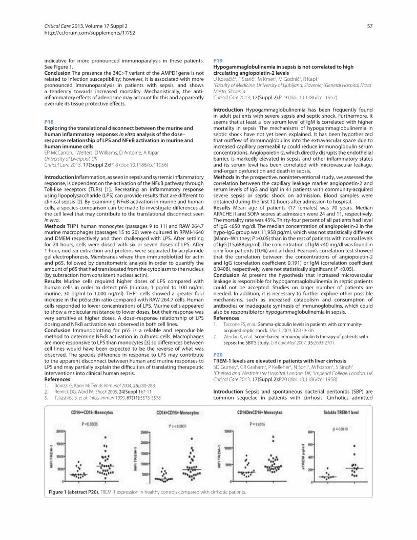
P20TREM-1 levels are elevated in patients with liver cirrhosisSD Gurney1, CR Graham2, P Kelleher2, N Soni1, M Foxton1, S Singh1
1Chelsea and Westminster Hospital, London, UK; 2Imperial College, London, UKCritical Care 2013, 17(Suppl 2):P20 (doi: 10.1186/cc11958)
Introduction Sepsis and spontaneous bacterial peritonitis (SBP) are common sequelae in patients with cirrhosis. Cirrhotics admitted
Figure 1 (abstract P20). TREM-1 expression in healthy controls compared with cirrhotic patients.
Critical Care 2013, Volume 17 Suppl 2 http://ccforum.com/supplements/17/S2
S7

to the ICU have an in-hospital mortality of up to 50% [1]. Microbial translocation (MT) is the pathogenic mechanism implicated in SBP. The triggering receptor expressed by myelocytes-1 (TREM-1) modulates the immune response with resultant production of proinfl ammatory cytokines and has been used as a biomarker in the diagnosis of bacterial infection. We wish to evaluate the role of TREM-1 as a biomarker in cirrhosis.Methods Blood samples were obtained from 18 healthy controls (HC) and 29 cirrhotic patients (CA) as defi ned by clinicoradiological criteria. Disease severity was graded according to Child–Pugh class (median 10, range 5 to 13) and modifi ed end-stage liver disease (MELD) score (median 14, range 6 to 21). Simultaneous ascitic fl uid samples were taken from 10 patients in the CA group. Soluble TREM-1 and CD14 levels (a surrogate marker of MT) were measured by ELISA. Flow cytometry was used to quantify the expression of TREM-1 on monocytes and neutrophils in blood and ascitic fl uid.Results TREM-1 expression is signifi cantly higher in the CA group compared with HC across all monocyte subsets but not neutrophils, even in the absence of sepsis (see Figure 1). There is no correlation between cell surface TREM-1 expression and severity of cirrhosis by Child–Pugh or MELD score. sTREM and sCD14 levels were elevated in the CA group compared with HC (P = 0.0010 and 0.0016 respectively). In addition, plasma sTREM-1 levels correlated with disease severity according to MELD score (R = 0.71, CI = 0.22 to 0.92 P = 0.012) and serum bilirubin (R = 0.78,CI = 0.36 to 0.94, P = 0.004). There was no correlation with either form of TREM-1 with sCD14 levels. There was no diff erence in cell surface or soluble TREM-1 expression between blood and ascitic fl uid monocytes in culture-negative, non-neutrophilic ascites.Conclusion Blood monocyte and soluble TREM-1 are elevated in cirrhotic patients even in the absence of sepsis. Soluble TREM-1 levels correlate with disease severity in cirrhosis. Further studies are ongoing to ascertain the utility of TREM-1 as a biomarker in cirrhosis.Reference1. Olson JC, et al.: Intensive care of the patient with cirrhosis. Hepatology 2011,
54:1864-1872.
P21Eff ects of diff erent doses and serotypes of LPS on blood–brain barrier permeability in Sprague–Dawley ratsE Senturk, F Esen, P Ergin Ozcan, G Orhun, N Orhan, N Arican, M Kucuk, M KayaUniversity of Istanbul, TurkeyCritical Care 2013, 17(Suppl 2):P21 (doi: 10.1186/cc11959)
Introduction The blood–brain barrier (BBB) is highly restrictive of the transport of substances between blood and the central nervous system. Lipopolysaccharide (LPS) from Gram-negative bacteria was reported to aff ect the permeability of the BBB. BBB disruption using a LPS is used as a model of septic encephalopathy in mice. The present study was designed to investigate the eff ects of diff erent doses and serotypes of LPS on BBB integrity in Sprague–Dawley rats.Methods Male Sprague–Dawley rats weighing 200 to 250 g were used in the study. Rats were given two diff erent types of LPS (026:B6-L5543 and 026:B6-L2762) in diff erent doses (3, 5, and 10 mg/kg; i.v.). Rectal temperature and arterial blood pressure measurements were recorded for sepsis severity. The changes in the BBB permeability were measured using the Evans blue (EB) and sodium fl uorescein (NaFl) dye extravasation techniques 24 hours after LPS administration.Results Both LPS serotypes showed comparable arterial blood pressure and rectal temperature recordings and the severity of the disease increased with the increasing doses (5 mg/kg and 10 mg/kg) of LPS and the mortality rates were found to be 29% and 63% respectively. The extravasated contents of EB and NaFl tracers did not signifi cantly increase in brain parenchyma following the administration of diff erent doses of LPS with diff erent serotypes.Conclusion Our results showed no disruption to BBB by two diff erent serotypes of LPS even administered in increasing doses. These result indicate that the BBB integrity of Sprague–Dawley rats are resistant to the eff ects of two diff erent serotypes of LPS.
P22Bioenergetic imbalance and oxidative stress in the pathophysiology of septic encephalopathyJ D’Avila1, R Rodrigues2, H Castro-Faria-Neto1, M Oliveira3, F Bozza4
1Oswaldo Cruz Foundation – FIOCRUZ, Rio de Janeiro, Brazil; 2Federal University of Rio de Janeiro (UFRJ) and D’Or Institute for Research and Education (IDOR), Rio de Janeiro, Brazil; 3UFRJ, Rio de Janeiro, Brazil; 4Oswaldo Cruz Foundation – FIOCRUZ and IDOR, Rio de Janeiro, BrazilCritical Care 2013, 17(Suppl 2):P22 (doi: 10.1186/cc11960)
Introduction Septic encephalopathy is a frequent complication in severe sepsis but its pathogenesis and mechanisms are not fully understood. Oxygen supply and utilization are critical for organ function, especially for the brain, a tissue extremely dependent on oxygen and glucose. Disturbances in oxygen utilization are common in sepsis and a number of mitochondrial dysfunctions have been described in diff erent tissues in septic animals as well as in septic patients. Our group described mitochondrial dysfunctions in the brain during experimental sepsis.Methods Experimental sepsis was induced by endotoxemia (LPS 10 mg/kg i.p.) in Sprague–Dawley rats and by polymicrobial fecal peritonitis in Swiss mice. Brain glucose uptake was observed in vivo in endotoxemic rats using positron emission tomography with [18F]fl uorodeoxyglucose and autoradiography with 2-deoxy-14C-glucose.Results Mice with polymicrobial sepsis present hypoglycemia, hyperlactatemia and long-term cognitive impairment. We observed a rapid increase in the uptake of fl uorescent glucose analog 2-deoxy-2-((7-nitro-2,1,3-benzoxadiazol-4-yl)amino)-D-glucose in brain slices from septic mice in vitro. A similar increase in brain glucose uptake was observed in vivo in endotoxemic rats. Remarkably, the increase in glucose uptake started 2 hours after LPS injection, earlier than other organs. The brains of mice with experimental sepsis presented neuroinfl ammation, mitochondrial dysfunctions and oxidative stress, but mitochondria isolated from septic brains generated less ROS in vitro in the fi rst 24 hours. This led us to investigate the role of NADPH oxidase, an enzyme induced during innate immune response, as a potential source of reactive oxygen species in experimental sepsis. Inhibiting NADPH oxidase with apocynin acutely after sepsis prevented cognitive impairment in mice.Conclusion Our data indicate that a bioenergetic imbalance and oxidative stress is associated with the pathophysiology of septic encephalopathy. We are observing a new metabolic phenotype in the brain during sepsis, characterized by a rapid increase in glucose uptake and mitochondrial dysfunctions that may be secondary to infl ammation and hypoxia.
P23Pathophysiology of sepsis-associated brain dysfunction: an experimental study of cerebral microdialysis and mitochondrial functionP Kurtz1, C Vargas-Lopes1, C Madeira1, I Mello1, R Panizzutti1, L C Azevedo2, F A Bozza1
1Fiocruz, Rio de Janeiro, Brazil; 2Hospital Sirio e Libanes, São Paulo, BrazilCritical Care 2013, 17(Suppl 2):P23 (doi: 10.1186/cc11961)
Introduction Pathophysiology of brain dysfunction associated with sepsis is still poorly understood. Potential mechanisms involve oxidative stress, neuroinfl ammation and blood–brain barrier altera-tions. Our purpose was to study the metabolic alterations and markers of mitochondrial dysfunction in a clinically relevant model of septic shock.Methods Twelve anesthetized (midazolam/fentanyl/pancuronium), invasively monitored, and mechanically ventilated pigs were allocated to a sham procedure (n = 5) or sepsis (n = 7), in which peritonitis was induced by intra-abdominal injection of autologous feces. Animals were studied until spontaneous death or for a maximum of 24 hours. In addition to global hemodynamic and laboratory assessment, intracranial pressure and cerebral microdialysis were assessed at baseline, 6, 12, 18 and 24 hours after sepsis induction. After death, brains were removed and brain homogenates were studied to assess markers of mitochondrial dysfunction.
Critical Care 2013, Volume 17 Suppl 2 http://ccforum.com/supplements/17/S2
S8

Results All septic animals developed a hyperdynamic state associated with lower arterial pressure, fever and organ dysfunction in comparison with control animals. In the septic animals, we observed increased brain dialysate glutamin levels at 12, 18 and 24 hours after sepsis induction, as compared with control animals. Moreover, after analyzing homogenates from the frontal cortex, we found higher concentrations of glutamin and glutamate in septic as compared with control animals (85.67 ± 14.98 vs. 28.77 ± 7.0; P = 0.01 and 132.1 ± 19.72 vs. 53.33 ± 16.83; P = 0.02, respectively). See Figure 1.Conclusion We found higher concentrations of glutamate and glutamin in brain tissues of septic animals as compared with control. Furthermore, glutamin concentrations increased over time in the extracellular space as measured by cerebral microdialysis. These fi ndings suggest an increased excitatory state that is potentially associated with high energy expenditure. However, associations with neuronal injury need further study.
P24Cholinergic modulation of hippocampal activity during septic encephalopathyA Zivkovic1, CP Bengtson2, O Sedlaczek1, R Von Haken1, H Bading2, S Hofer1
1Universitätsklinikum Heidelberg, Germany; 2Universität Heidelberg, GermanyCritical Care 2013, 17(Suppl 2):P24 (doi: 10.1186/cc11962)
Introduction Septic encephalopathy is a sepsis-related brain dys-function with a deterioration of cortical functions. The experimental studies in the rat brain revealed a deranged neurotransmitter profi le during septic encephalopathy. Glutamatergic synapses, essential in learning and memory, undergo use-dependent changes in synaptic strength, referred to as plasticity. Permanent strengthening of synapses after a brief stimulus, termed long-term potentiation (LTP), was discovered in the hippocampus, and here it has been most thoroughly studied. Cholinergic neurotransmission plays an important role in regulating the cognitive functions of the brain. It acts as a signal-to-noise ratio modulator of sensory and cognitive inputs. The irregularities in brain functions give rise to the symptoms of delirium, including disorganized thinking and disturbances of attention and consciousness, which in turn might aff ect learning and memory. Possible mechanisms for cholinergic defi ciency include impairment of synaptic functions of acetylcholine. Imbalances in the cholinergic system during sepsis might therefore play an extensive role in the septic delirium.Methods By using MRI imaging we identifi ed functional changes in the hippocampal region of patients with severe sepsis. This fi nding was further supported by the experimental recordings in the rat brains of lipopolysaccharide (LPS)-treated rats using the electrophysiological patch clamp technique.
Results Critically ill ICU patients diagnosed with septic delirium using the CAM-ICU method underwent diagnostic MRI scans. The serial MRI analysis revealed increased signal intensity in the hippocampal region in diff usion-weighted MRI (DWI). We used endotoxemia model to induce sepsis in the rats. Electrophysiological analysis of the hippocampal neurons in LPS-treated rats showed impaired LTP in the excitatory synapses, as compared with controls. Application of physostigmine, a blood–brain barrier permeable cholinesterase inhibitor, resulted in a partial recovery of LTP in the hippocampal synapses of LPS-treated rats.Conclusion The patients with septic delirium show functional changes in the hippocampus. Furthermore, we show that endotoxemia aff ects synaptic plasticity in the rat hippocampus, suggesting the involvement of this brain region in the pathophysiology of septic delirium. Moreover, the eff ect of the cholinergic neurotransmission onto the induction and maintenance of synaptic plasticity in the rat hippocampus during endotoxemia suggests that cholinergic neurotransmission might play a critical role in septic encephalopathy.
P25Neutrophil–lymphocyte count ratio as a biomarker of severe sepsis in Escherichia coli infections in adultsLR Ljungstrom1, G Jacobsson1, R Andersson2
1Skaraborgs Sjukhus, Skövde, Sweden; 2Gothenburg University, Gothenburg, SwedenCritical Care 2013, 17(Suppl 2):P25 (doi: 10.1186/cc11963)
Introduction The neutrophil–lymphocyte count ratio (NLCR) is an easy to analyse biomarker reacting very early in the course of acute infl ammation. It has previously been reported to correspond to bacteremia and recently to disease severity in community-acquired pneumonia [1]. We have looked at 205 consecutive patients with Escherichia coli infections (ECI) and found the same to be true for ECIs. This may be of great clinical importance since E. coli is the most frequently isolated pathogen in patients with infections requiring in hospital care.Methods This study is part of a 9-month consecutive study of community-acquired severe sepsis and septic shock in adults at Skaraborg Hospital in the western region of Sweden. The hospital serves a population of 256,000 inhabitants and has approximately 60,000 annual visits to the ED. All patients admitted to the hospital receiving intravenous antibiotic treatment within the fi rst 48 hours of admission were evaluated for severe sepsis and septic shock. Upon admission, two sets of blood cultures and other relevant cultures were obtained from each patient as well as sampling for NLCR and venous plasma lactate. The patient records were evaluated by one infectious diseases specialist. Approximately 2,300 patients were diagnosed as having a bacterial infection. From those, an informed consent to participate in the study could be obtained from approximately 1,600 patients.Results Of the 1,600 patients who gave consent to participate in the study, 205 had an ECI. Sixty-four had a positive blood culture for E. coli. Fifty of the patients met one or more criteria for severe sepsis or septic shock. The NLCR was signifi cantly higher (P <0.001) within the severe sepsis group (median = 21.1 with quartiles 11.1 to 42.4) compared with the group with no severe sepsis (median = 11.6 with quartiles 7.6 to 18.9).Conclusion The NLCR can be used as a biomarker of disease severity even in ECIs. The biomarker reacts rapidly, is cheap and needs no extra sampling. The higher the value, the higher the probability for severe sepsis. A high value can even precede the development of severe sepsis or septic shock. However, a low value never excludes neither bacteremia nor severe sepsis. The method cannot be used in patients with disturbances in neutrophil or lymphocyte levels due to other causes than sepsis.Reference1. de Jager C, et al.: The neutrophil–lymphocyte count ratio in patients with
community acquired pneumonia. PLoS ONE 2012, 7:e46561.
Figure 1 (abstract P23).
Critical Care 2013, Volume 17 Suppl 2 http://ccforum.com/supplements/17/S2
S9

P26Obesity and infl ammatory markers in severe sepsisP Simon1, D Thomas-Rüddel2, T Nemes1, K Reinhart2, F Bloos2, UX Kaisers1
1University Hospital of Leipzig, Germany; 2Center for Sepsis Control and Care, Jena, GermanyCritical Care 2013, 17(Suppl 2):P26 (doi: 10.1186/cc11964)
Introduction Chronic infl ammation has recently been recognized as an important factor in the pathophysiology of obesity and associated morbidities [1]. In this clinical study we aimed at identifying possible eff ects of obesity on infl ammatory markers in severe sepsis.Methods With institutional ethical committee approval, 243 consecutive patients treated for severe sepsis or septic shock in the ICUs of two university hospitals over a period of 5 months were studied. Six patients were excluded due to cachexia, syndromal disorders or missing clinical data. Diagnosis of sepsis was made according to SCCM criteria. Serum levels of C-reactive protein (CRP, mg/l) and procalcitonin (PCT, ng/ml) on day 1 of sepsis were compared among fi ve body mass index (BMI) strata according to WHO defi nitions. Two groups (BMI <30, normal weight, and BMI ≥30, obesity) were formed for further analysis, and PCT was logarithmically transformed (LogPCT), resulting in normal distribution. Statistical analysis was performed using a t test.Results Patients with BMI ≥30 had higher values of PCT and CRP (Table 1). The diff erence in LogPCT was of borderline signifi cance (P = 0.052). However, patients with positive blood cultures had signifi cantly higher LogPCT values (P = 0.017) (Figure 1). Diff erence in CRP was not signifi cant (P = 0.09). The trends over all fi ve BMI strata (Table 1) were not signifi cant.Conclusion Obesity with BMI ≥30 seems to be associated with an increase in infl ammatory markers in patients with severe sepsis, particularly in bacteraemia. The role of adipose tissue in severe sepsis should therefore be studied in more detail.Reference1. Wellen K, et al.: Infl ammation, stress, and diabetes. J Clin Invest 2005,
115:1111-1119.
P27Cytokine gene expression can predict infectious complications following severe traumaHD Torrance, K Brohi, CJ Hinds, MJ O’DwyerBart’s Health NHS Trust, London, UKCritical Care 2013, 17(Suppl 2):P27 (doi: 10.1186/cc11965)
Introduction Identifying a group of patients at high risk of developing infectious complications is the fi rst step in the introduction of eff ective pre-emptive therapies in specifi c patient groups. Quantifying cytokine gene expression also furthers our understanding of trauma-induced immunosuppression. Our group has already demonstrated that a predictive immunological signature derived from mRNA expression in elective thoracic surgical patients accurately predicts pneumonia risk [1].Methods In total, 121 ventilated polytrauma patients were recruited. mRNA was extracted from PaxGene tubes collected within 2 hours of the initial insult, at 24 and 72 hours. T-helper cell subtype specifi c cytokines and transcription factors mRNA was quantifi ed using qPCR. Ten healthy controls served as a comparator.Results The Median Injury Severity Score (ISS) was 29. Time 0 bloods demonstrated a reduction in TNFα†, IL-12§, IL-23‡, RORγT* and T bet§, and an increase in IL-10* and IL-4† mRNA levels in comparison with the control group (*P <0.0001, †P <0.001 to 0.0001, ‡P <0.01 to 0.001, §P <0.05 to 0.01). There was a positive correlation between ISS and IL-10‡ whilst both IL-23§
Figure 1 (abstract P26).
Table 1 (abstract P26). PCT and CRP as median (IQR)
BMI n PCT (ng/ml) CRP (mg/l)
18.5 to 24.9 196 4.8 (11.9) 153 (189)
≥30.0 47 7.3 (24.6) 228 (254)
18.5 to 24.9 101 4.9 (11.7) 147 (175)
25 to 29.9 95 4.4 (12.0) 157 (218)
30 to 34.9 32 6.9 (22.6) 212 (118)
35 to 39.9 7 7.4 (38.3) 241 (264)
≥40.0 8 11.4 (32) 278 (272)
Figure 1 (abstract P27). Cytokine mRNA levels in trauma (time 0) and control groups.
Critical Care 2013, Volume 17 Suppl 2 http://ccforum.com/supplements/17/S2
S10

and RORγT‡ were negatively correlated at time 0. TNFα†, IL-10* and IL-27‡ increased and IFNγ†, IL-12*, IL-17A§, RORγT* and T bet* mRNA levels decreased over the initial 24 hours. Subsequent bacteraemia (18/121 patients) was associated with a lower TNFα/IL-10 ratio‡ at baseline. Similarly, higher IL-10‡ and lower T bet‡ mRNA at 24 hours also predicted later bacteraemic episodes. Development of pneumonia followed a similar pattern. A multivariate logistical regression model proved highly accurate in predicting infectious complications from mRNA analysis of early blood samples. See Figure 1.Conclusion Cytokine gene expression patterns indicate an immediate and sustained impairment in Th1, Th17 and innate immunity with concurrent upregulation of the Th2 response following major trauma. The magnitude of this response predicts subsequent infectious complications.Reference1. White M, et al.: Chest 2011, 139:626-632.
P28A cohort study of routinely used sepsis biomarkers and 28-day mortalityM De La Torre-Prados1, A Garcia-De la Torre2, C Trujillano-Férnández1
1Hospital Virgen de la Victoria, Málaga, Spain; 2Hospital Puerto Real, Cádiz, SpainCritical Care 2013, 17(Suppl 2):P28 (doi: 10.1186/cc11966)
Introduction The evaluation of sepsis severity is complicated by the highly variable and nonspecifi c nature of clinical signs and symptoms. We studied routinely used biomarkers together with clinical parameters to compare their prognostic value for severe sepsis and evaluate their usefulness.Methods A cohort study of 150 patients >18 years with severe sepsis according to the Surviving Sepsis Campaign, in an ICU of a university hospital. Demographic, clinical parameters and coagulation, infection and infl ammation parameters during the fi rst 24 hours from severe sepsis or septic shock onset were studied. Descriptive and comparative statistical analysis was performed using SPSS version 15.0 (SPSS Inc., Chicago, IL, USA).Results We analyzed 150 consecutive episodes of severe sepsis (16%) or septic shock (84%) in the ICU. The median age of the patients was 64 (interquartile range, 48.7 to 71) years; the main sources of infection were intra-abdomen (45%) and respiratory (38%); 70.7% had medical diseases. The 28-day mortality was 22.7%. The profi le of death patients were men (64.7%, n =22), with signifi cantly higher average age (63 vs. 57 years; P = 0.049), as well as clinical severity scores, APACHE II (29.8 vs. 24.1; P <0.001) and SOFA (12.1 vs. 8.9; P <0.001) and major dysfunction organ number (4.6 vs. 3.6; P <0.001). Bilirubin was the best predictor of 28-day mortality with the largest AUC (0.71), followed by hemoglobin (0.69) and C3 (0.67). The multivariate logistic regression was adjusted for three risk parameters, hemoglobin (OR: 0.68; 95% CI: 0.51 to 0.94), bilirubin (OR:1.63; 95% CI: 1.08 to 2.45) and white blood cells (OR:1.04; 95% CI: 1.01 to 1.08) and with these parameters a ROC analysis was performed, giving an AUC of 0.77 (0.69 to 0.84).Conclusion The assessment of routine biomarkers (bilirubin, white blood cells and hemoglobin) may be a helpful tool in the decision-making process at the bedside, for the evaluation of early ICU admission of recoverable patients, as indicators of infl ammatory response, organ dysfunction or catabolism level, and their signifi cant predictive value on mortality.Reference1. Glickman SW, Cairns CB, Otero RM, et al.: Disease progression in
hemodynamically stable patients presenting to the emergency department with sepsis. Acad Emerg Med 2010, 17:383-390.
P29Procalcitonin as prognostic marker of mortalityS Zampieri1, P Bettonte2, M Ortolani1, G Frison1, V Schweiger1, L Gottin3, E Polati11Policlinico G.B. Rossi, Verona, Italy; 2Santa Chiara Hospital, Trento, Italy; 3Ospedale Maggiore Civile, Verona, ItalyCritical Care 2013, 17(Suppl 2):P29 (doi: 10.1186/cc11967)
Introduction We analyze procalcitonin (PCT) as a prognostic marker, in order to assess the clinical impact of a daily PCT measure.
Methods From November 2010 to November 2011 we collected clinical data, drug administration, scores and PCT values of 420 consecutive patients during hospitalization. Statistical analysis was made using SPSS software. We calculated ICU mortality, 1-month mortality and 1-year mortality. Median percentage daily variation was calculated as: (PCT day after – PCT of the date value) / PCT of the date value×100. PCT variation in the last 48 hours of hospitalization was calculated as: (PCT at discharge – PCT at 48 hours before discharge) / PCT 48 hours before discharge×100. We compared peak values in dead patients versus alive patients. A logistic regression was performed in order to assess mortality odds ratio.Results Of the 420 patients, 63 (15%) died in the ICU, 12 (2.86%) died 1 month after ICU discharge and 16 (3.80%) died 1 year after ICU discharge. PCT values were higher during the last day of hospitalization in dead patients versus alive patients. PCT percentage variation during the last 48 hours of hospitalization had a slower trend in patients who died than in those who survived; these diff erences are even more marked in patients who had a septic event. A slower descending trend of daily PCT values was found in patients who died than in those who survived. PCT peak levels during the ICU stay were higher in dead patients with respect to alive ones. At logistic regression analysis PCT decrease in the last 48 hours <–30% (OR 3.71), PCT peak higher than 10 ng/ml (OR 2.38), and PCT last day/PCT peak ratio >50% (OR 2.064) were ICU mortality risk factors. PCT values were a higher predictive ICU mortality risk factor than SOFA and APACHE II scores. Other prognostic factors were age and lactate values. Only age was a risk factor in 1-month and 1-year mortality.Conclusion PCT is a good prognostic marker and is strongly correlated to the clinical status and gravity of the patients, so PCT seems to be a useful marker in an intensive care scenario.References1. Jensen JU, Heslet L, Jensen TH, et al.: Procalcitonin increase in early
identifi cation of critically ill patients at high risk of mortality. Crit Care Med 2006, 34:2596-2602.
2. Fritz HG, Brandes H, Bredle DL, et al.: Post-operative hypoalbuminaemia and procalcitonin elevation for prediction of outcome in cardiopulmonary bypass surgery. Acta Anaesthesiol Scand 2003, 47:1276-1283.
P30Changes in circulating procalcitonin versus C-reactive protein in predicting evolution of infectious disease in febrile, critically ill patientsS Hoeboer1,2, J Groeneveld1,2
1VU University Medical Center, Amsterdam, the Netherlands; 2Erasmus Medical Center, Rotterdam, the NetherlandsCritical Care 2013, 17(Suppl 2):P30 (doi: 10.1186/cc11968)
Introduction Although absolute values for C-reactive protein (CRP) and procalcitonin (PCT) are well known to predict sepsis in the critically ill, it remains unclear if and how changes in CRP and PCT predict evolution of infectious disease and how they compare in this respect.Methods In 72 critically ill patients with new-onset fever, CRP and PCT were measured on day 0, 1, 2 and 7 after inclusion, and their clinical course was documented over 1 week with follow-up to day 28. Infection was microbiologically defi ned, as was bloodstream infection; septic shock was defi ned as infection plus shock.Results From peak at day 0 to 2 to day 7, CRP decreases most when (bloodstream) infection and septic shock (day 0 to 2) resolve and increases most when complications such as a new (bloodstream) infection or septic shock (day 3 to 7) supervene (area under the receiver operating characteristic curve 0.70 or higher, P = 0.04 or lower). PCT decreases most when septic shock resolves (AUC 0.72, P = 0.007) and increases most when a new bloodstream infection or septic shock supervenes (AUC 0.82 or higher, P <0.001). The day 7 value of PCT rather than of CRP was predictive for 28-day outcome (AUC 0.70, P = 0.005).Conclusion The data, obtained during ICU-acquired fever and infections, suggest that CRP and PCT changes predict the course of infectious disease and its complications. CRP may be favoured over PCT courses in decisions on appropriateness and duration of antibiotic treatment, whereas PCT rather than CRP courses may help predicting complications such as bloodstream infection, septic shock and mortality.
Critical Care 2013, Volume 17 Suppl 2 http://ccforum.com/supplements/17/S2
S11

P31Procalcitonin-guided antibiotic therapy in patients with congestive heart failure and suspicion of lower respiratory tract infection: results from a randomized trialP Schuetz, E Grolimund, A Kutz, S Haubitz, B MuellerKantonsspital Aarau, SwitzerlandCritical Care 2013, 17(Suppl 2):P31 (doi: 10.1186/cc11969)
Introduction Diff erentiation of acute heart failure from infection in patients with respiratory symptoms and a history of congestive heart failure (CHF) is challenging due to overlap of clinical symptoms and X-ray fi ndings. The BACH study found higher mortality rates if patients presenting with dyspnea were treated with antibiotics and their procalcitonin (PCT) levels were low indicating absence of bacterial infection. Yet the BACH study was observational and causal inference cannot be drawn. Herein, we analyzed the eff ects of PCT-guided antibiotic stewardship in CHF patients from a previous trial (ProHOSP).Methods This is a secondary analysis of a previous randomized trial of adult ED patients with respiratory symptoms and a history of CHF. Patients were randomized to administration of antibiotics based on a PCT algorithm (PCT group) or standard guidelines without knowledge of PCT levels (control group). The primary endpoint of this analysis is the risk of adverse outcome defi ned as death or ICU admission within 30 days after ED admission.Results A total of 233 patients met the inclusion criteria, with 116 in the PCT-guided group and 117 in the control group. In the subgroup of patients with low initial PCT levels <0.25 ng/l (n = 110), PCT-guided patients had a signifi cant reduction in antibiotic exposure (mean 3.7 vs. 6.5 days, diff erence –2.8 (95% CI –4.4, –1.2), P <0.001). Furthermore, PCT-guided patients had a signifi cant lower risk for death and ICU admission (4% vs. 20%, odds ratio 6.0 (1.3, 28.2), P = 0.02). See Figure 1.Conclusion In CHF patients with suspicion of respiratory infection, use of a PCT protocol resulted in a signifi cant decrease of antibiotic exposure and signifi cantly improved outcomes in patients with low PCT levels indicating absence of bacterial infection. Whether inadequate antibiotic therapy in these CHF patients requiring diuretic treatment explains this diff erence in clinical outcomes needs verifi cation.
P32Pro-adrenomedullin as a clinical predictor after cardiac surgeryJ Van Fessem, F De Graaf, J Van Paassen, S ArbousLUMC, Leiden, the NetherlandsCritical Care 2013, 17(Suppl 2):P32 (doi: 10.1186/cc11970)
Introduction Pulmonary complications after cardiac surgery like ARDS are frequent and linked to high mortality [1]. Pro-adrenomedullin (pro-ADM) has a possible role in the development of ARDS [2] and a positive correlation between levels of pro-ADM and infl ammation was found [3]. In this study we investigated whether intraoperative and postoperative pro-ADM transpulmonary gradient could predict postoperative morbidity.
Methods In this prospective cohort study, 39 patients undergoing cardiac surgery using CPB were included. Blood was collected before surgery (T0), after induction of anesthesia (T1), after termination of CPB (T2), at ICU arrival (T3) and 3 hours (T4), 6 hours (T5) and 18 hours (T6) after arrival. Pro-ADM was measured with a sandwich immunoassay. Primary endpoints were length of ICU and hospital stay (ICU-LOS, hospital-LOS).Results An increase of arterial and venous pro-ADM plasma con-centrations was observed after surgery. Immediately after termination of CPB the venous concentration was signifi cantly lower than arterial pro-ADM concentration, but at T6 the venous concentration was signifi cantly higher, indicating a switch from a negative to positive transpulmonary gradient (Figure 1). The pro-ADM venous–arterial diff erence at T5 was a signifi cant predictor of ICU-LOS (P = 0.032) and the diff erence at T3 was a signifi cant predictor of hospital-LOS (P = 0.001).Conclusion We found that the transpulmonary gradient of pro-ADM was a predictor for ICU-LOS and hospital-LOS at T3 and T5, respectively. Pro-ADM might be a promising marker for prediction on outcome of patients undergoing cardiac surgery on CPB. The transpulmonary shift of pro-ADM might be caused by an infl ammatory response.References1. Apostolakis E, et al.: J Card Surg 2010, 25:47-55.2. Kamei M, et al.: Acta Anaesthesiol Scand 2004, 48:980-985.3. Ueda S, et al.: Am J Respir Crit Care Med 1999, 160:132-136.
P33Pro-adrenomedullin as prognostic biomarker in the sepsisM De La Torre-Prados, A Garcia-De la Torre, A Enguix, M Mayor, N Zamboschi, C Trujillano-Fernández, A Garcia-AlcantaraHospital Virgen de la Victoria, Málaga, SpainCritical Care 2013, 17(Suppl 2):P33 (doi: 10.1186/cc11971)
Introduction Measurement of biomarkers is a potential approach to early assessment and prediction of mortality in septic patients. The purpose of this study was to ascertain the prognostic value of pro-adrenomedullin (pADM), measured in all patients admitted to the ICU of our hospital with a diagnosis of severe sepsis or septic shock during 1 year.Methods A cohort study of 117 patients >18 years with severe sepsis according to the Surviving Sepsis Campaign, in an ICU of a university hospital. Demographic, clinical parameters and pADM, C-reactive protein and procalcitonin were studied during 1 year. Descriptive and comparative statistical analysis was performed using the statistical software packages Statistica Stat Soft Inc 7.1 and MedCalc 9.2.1.0.Results We analyzed 117 consecutive episodes of severe sepsis (15%) or septic shock (85%) in the ICU. The median age of the patients was
Figure 1 (abstract P31). Time to adverse outcome according to group allocation.
Figure 1 (abstract P32). Pro-ADM transpulmonary gradient at diff erent time points.
Critical Care 2013, Volume 17 Suppl 2 http://ccforum.com/supplements/17/S2
S12

64 (interquartile range, 53 to 72) years; the main sources of infection were respiratory tract (46%) and intra-abdomen (21%). The 28-day mortality was 32.5%. The profi le of death patients had a signifi cantly higher average age (64.7 vs. 57.6 years; P = 0.024), as well as clinical severity scores, APACHE II (26.6 vs. 23; P = 0.006) and SOFA (11.6 vs. 89.2; P <0.001). Kaplan–Meier survival analysis was signifi cant. P = 0.0017 for patients with pADM <1.2 nmol/l. Cox regression analysis also showed statistical signifi cance (P = 0.0033) and a likelihood ratio = 1.18 per each 1 nmol/l increase in pADM.Conclusion The protein pADM is an important prognostic biomarker of survival when measured on admission of septic patients to the ICU.References1. Pezzilli R, Barassi A, Pigna A, et al.: Time course of proadrenomedullin in the
early phase of septic shock. A comparative study with other proinfl ammatory proteins. Panminerva Med 2012, 54:211-217.
2. Wang RL, Kang FX: Prediction about severity and outcome of sepsis by proatrial natriuretic peptide and pro-adrenomedullin. Chin J Traumatol 2010, 13:152-157.
P34Early IL-6 response in sepsis is correlated with mortality and severity scoreP Srisangthong, A Wongsa, P Kittiworawitkul, A WattanathumPhramongkutklao Hospital, Bangkok, ThailandCritical Care 2013, 17(Suppl 2):P34 (doi: 10.1186/cc11972)
Introduction IL-6, a proinfl ammatory cytokine, is synthesized from fi bro-blasts, T lymphocytes, endothelial cells and monocytes. It serves as an important mediator during the acute phase response to infl ammation in sepsis. We hypothesized that the plasma IL-6 is correlated with mortality and severity scores in critically ill patients with sepsis.Methods We conducted a prospective study of plasma IL-6 level at the initial phase of sepsis and the risk of mortality. A total of 203 patients with sepsis, who were admitted to the medical ICU at Phramongkutklao Hospital, Bangkok during January to December 2011, were analyzed. Serum IL-6, C-reactive protein (CRP), and lactate were measured within the fi rst 24 hours of ICU admission. Severity scores (APACHE II, SAP II, and SOFA scores) were measured. The primary outcome variable was 28-day all-cause mortality.Results We found that the overall 28-day mortality was 46% (93 out of 203 patients). There was a signifi cantly positive correlation between mortality rate and plasma IL-6 (survivors vs. nonsurvivors; 74 (4.4 to 1,718) vs. 206 (19 to 5,000) pg/ml, P <0.05), lactate (survivors vs. nonsurvivors; 1.65 (0.7 to 11.61) vs. 2.47 (0.94 to 19.13) mmol/l, P <0.05), but not CRP levels (P = 0.24). Compared with the patients with plasma IL-6 <100 pg/ml, septic patients with IL-6 levels ≥100 were associated with an increased 28-day mortality with the odd ratio of 2.99 (95% CI 1.42 to 6.29, P <0.05). We also found that plasma IL-6 levels were well correlated with APACHE II (P <0.05), SAPS II (P <0.05), and SOFA (P <0.05) scores.Conclusion The initial phase plasma IL-6 levels were correlated with severity and mortality in critically ill patients with sepsis.References1. Pathan N, et al.: Crit Care Med 2005, 33:1839-1844.2. Harbarth S, et al.: Am J Respir Crit Care Med 2001, 164:396-402.
P35Compared values of presepsin (sCD14-ST) and procalcitonin as early markers of outcome in severe sepsis and septic shock: a preliminary report from the Albumin Italian Outcome Sepsis (ALBIOS) studyP Caironi1, S Masson2, E Spanuth3, R Thomae4, R Fumagalli5, A Pesenti5, M Romero6, G Tognoni6, R Latini2, L Gattinoni11Fondazione IRCCS Ca’ Granda – Ospedale Maggiore Policlinico, Università degli Studi di Milano, Milan, Italy; 2Istituto di Ricerche Farmacologiche Mario Negri, Milan, Italy; 3Diagnostic Engineering & Research GmbH, Heildelberg, Germany; 4Mitsubishi Chemical Europe GmbH, Munich, Germany; 5Ospedale San Gerardo, Monza, Italy; 6Consorzio Mario Negri Sud, Santa Maria Imbaro, ItalyCritical Care 2013, 17(Suppl 2):P35 (doi: 10.1186/cc11973)
Introduction Sepsis results from complex interactions between infecting microorganisms and host responses, often leading to multiple organ failures and death. Over the years, its treatment has
been standardized in early goal-oriented therapies, which may benefi t from circulating biomarkers for early risk stratifi cation. We aimed to evaluate the prognostic value of presepsin (sCD14-ST), a novel marker of bacterial infection.Methods We performed a nested case–control study from the randomized controlled Albumin Italian Outcome Sepsis (ALBIOS) trial, enrolling patients with severe sepsis or septic shock from 100 ICUs in Italy. Fifty survivors and 50 nonsurvivors at ICU discharge were selected, matched for age, sex, center and time of enrollment after inclusion criteria were present. EDTA-plasma samples were collected at days 1, 2 and 7 after enrolment for presepsin (immune-chemiluminescence assay PATHFAST Presepsin, URL 320 pg/ml, CV 5%; Mitsubishi Chemicals) and procalcitonin assay (PCT, Elecsys BRAHMS Cobas® PCT, URL 0.046 ng/ml, CV 8.8%; Roche Diagnostics).Results Clinical characteristics were similar between the two groups, except for a worse SOFA score at day 1 in decedents. Presepsin at day 1 was signifi cantly higher in decedents (2,268 (1,145 to 4,305) pg/ml, median (Q1 to Q3)) than in survivors (1,184 (855 to 2,158) pg/ml, P = 0.001), while PCT did not diff er (18.5 (3.3 to 45.7) vs. 10.8 (2.6 to 46.4) ng/ml, P = 0.31). Presepsin decreased over time in survivors, but remained elevated in decedents (974 (674 to 1,927) vs. 2,551 (1,438 to 5,624) pg/ml at day 7, P = 0.02 for time–survival interaction); PCT decreased similarly in the two groups (P = 0.19). Patients with early elevated presepsin had worse SOFA score, higher number of MOFs, hemodynamic instability (lower mean arterial pressure at baseline and after 6 hours), and mortality rate at 90 days (75% vs. 42%, log-rank P <0.001). The association between presepsin and outcome was more marked in patients with late enrollment (6 to 24 hours), and in septic shock. Early presepsin had better prognostic accuracy than PCT (AUROC 0.69 vs. 0.56, P = 0.07), and improved discrimination over SOFA score, especially in septic shock.Conclusion Early presepsin measurements may provide important prognostic information in patients with severe sepsis or septic shock, and may be of crucial importance for early risk stratifi cation.
P36Usefulness of presepsin in the diagnosis of sepsis in acute kidney injury patientsY Nakamura, H Ishikura, T Nishida, Y Kawanno, R Yuge, R Ichiki, A MuraiFukuoka University, Fukuoka, JapanCritical Care 2013, 17(Suppl 2):P36 (doi: 10.1186/cc11974)
Introduction The level of presepsin is useful for diff erentiating sepsis from noninfectious systemic infl ammatory response syndrome. It has been reported that the presepsin levels in patients with chronic renal failure are abnormally high. However, there are no studies investigating the usefulness of presepsin for diagnosis of sepsis in patients with acute kidney injury (AKI). Our purpose of this study is to clarify the diagnostic accuracy of presepsin in patients with AKI and the relationship between presepsin level and AKI severity.Methods This study was conducted as a single-center retrospective study. Blood samples were collected from patients admitted to the emergency room at Fukuoka University Hospital between June 2010 and October 2012. We enrolled 254 patients with suspected sepsis and other disease patients. We classifi ed the patients into an AKI group according to the RIFLE criteria (Risk n = 52, Injury n = 39, Failure n = 41, Loss of kidney function and End-stage kidney disease n = 7) and a non-AKI group (n = 115). The AKI patient group was further classifi ed into a sepsis group and a nonsepsis group in each AKI stage and we analyzed the diagnostic accuracy of presepsin in patients with sepsis.Results For the non-AKI patients, the median of presepsin in patients with nonsepsis (n = 78) and the sepsis group (n = 37) were 406 pg/ml (range: 86 to 4,374) and 1,065 pg/ml (range 86 to 9,960), respectively (P <0.0001). For the Risk patients, the median of presepsin in patients with nonsepsis (n = 25) and the sepsis group (n = 27) were 299 pg/ml (range: 71.2 to 3,361) and 831 pg/ml (range: 233 to 16,759), respectively (P <0.01). For the Injury patients, the median of presepsin in patients with nonsepsis (n = 12) and the sepsis group (n = 27) were 463 pg/ml (range: 122 to 1,197) and 1,451 pg/ml (range: 237 to 4,200), respectively (P <0.001). For the Failure patients, the median of presepsin in patients
Critical Care 2013, Volume 17 Suppl 2 http://ccforum.com/supplements/17/S2
S13

with nonsepsis (n = 14) and the sepsis group (n = 27) were 1,607 pg/ml (range: 454 to 8,516) and 1,523 pg/ml (range: 293 to 16,764), respectively (P = 0.175). The diagnostic accuracy of presepsin in patients with sepsis was determined by ROC analysis, the area under the curve was 0.789 for the non-AKI patient group, 0.735 for the Risk patient group, 0.855 for the Injury patient group and 0.593 for the Failure patient group.Conclusion In Failure and more progressed AKI patients, the diagnostic accuracy of the presepsin level was lower than the other groups.References1. Endo S, et al.: J Infect Chemother 2012, 18:891-897.
doi:10.1007/s10156-012-0435-22. Bellomo R, et al.: Crit Care 2004, 8:R204-R212. doi:10.1186/cc2872
P37Serum levels of presepsin refl ects the APACHE II and SOFA scores in patients with sepsisR Sato, Y Suzuki, M Sato, G Takahashi, M Kojika, Y Inoue, S EndoIwate Medical University, Morioka, JapanCritical Care 2013, 17(Suppl 2):P37 (doi: 10.1186/cc11975)
Introduction As a method for the diagnosis of sepsis, we previously reported an ELISA method for measuring the serum levels of presepsin. That method, however, took approximately 2 hours to yield results.Methods To resolve this problem, we later developed a simplifi ed assay kit making use of immunochromatography (Point of Care test; POC test), and are currently evaluating the usefulness of this kit for diagnosis of sepsis and evaluation of its severity. In 21 septic patients with sepsis, we analyzed the serum levels of presepsin in relation to APACHE II and SOFA scores.Results APACHE II and SOFA scores at baseline were 28.7 ± 7.5 and 10.8 ± 3.9, respectively, in these patients. These were signifi cant correlations between the serum presepsin levels and the APACHE II score, and also between the serum presepsin levels and the SOFA score.Conclusion Furthermore, there was also a signifi cant correlation between the results of the POC test and the serum presepsin levels. These results indicate that measurement of the serum presepsin might be useful for evaluating the severity of sepsis.
P38Relationship between serum nitrite/nitrate levels in the early phase of septic acute lung injury and prognosisM Sato, Y Suzuki, T Masuda, G Takahashi, M Kojika, Y Inoue, S EndoIwate Medical University, Morioka, JapanCritical Care 2013, 17(Suppl 2):P38 (doi: 10.1186/cc11976)
Introduction Serum nitrite/nitrate (NOx) levels in the early phase of septic acute lung injury (ALI)/acute respiratory distress syndrome (ARDS) have recently been reported to possibly play a key role in pathogenesis of ALI/ARDS.Methods NOx levels in the early phase of septic ALI/ARDS were measured by autoanalyzer (TCI-NOX 1000; Tokyo Kasei Kogyo Co., Ltd, Tokyo, Japan). Cytokine was measured by ELISA (Medogenix, Fleurus, Belgium).Results Both NOx and TNFα levels were signifi cantly higher in the ARDS group than in the ALI group. A negative correlation was found between the PaO2/FIO2 (P/F) ratio and serum NOx levels. In addition, a positive correlation was found between the TNFα and serum NOx levels. The 30-day, 60-day and 90-day mortality rates were 8.7%, 15.2% and 19.6%, respectively, in the patients with ALI/ARDS. There were no diff erences in the P/F ratio, serum NOx levels or TNFα levels in the early phase of ALI/ARDS between the 30-day survival and death groups. On the other hand, the P/F ratio, serum NOx levels and TNFα levels in the early phase of ALI/ARDS were signifi cantly higher in the 60-day and 90-day death groups than in the corresponding survival groups. There were no signifi cant diff erences in the 90-day mortality rates between the ALI and ARDS groups.Conclusion Our fi ndings suggested that NOx may be involved in the pathogenesis of ALI/ARDS.
P39Simple and fast prediction of Legionella sp. in community-acquired pneumonia: validation of a prediction ruleS Haubitz1, F Hitz2, L Graedel2, T Wiemken3, P Peyrani Dicastelnuovo3, J Ramirez3, M Batschwaroff 1, C Fux1, B Mueller1, P Schütz1
1Kantonsspital Aarau, Switzerland; 2University of Basel, Switzerland; 3University of Louisville School of Medicine, Louisville, KY, USACritical Care 2013, 17(Suppl 2):P39 (doi: 10.1186/cc11977)
Introduction Ruling out Legionella sp. in patients presenting with community-acquired pneumonia (CAP) is important due to diff erences in treatment regimens. Yet antigen tests as well as blood cultures have low sensitivity and an important time delay, making empirical broad spectrum coverage necessary particularly in severe cases. Fiumefreddo and colleagues recently proposed a clinical score based on six clinical and laboratory variables (fever, cough, sodium, lactate-dehydrogenase, C-reactive protein, platelet count) which allowed assessing the likelihood of Legionella [1]. Yet these variables need validation in an independent patient cohort before implementation into clinical routine.Methods We analyzed data from a large multinational database of patients with CAP (CAPO) [2] between 2001 and 2012. We performed logistic regression analysis and the area under the receiver operating characteristics (AUC) curve to study the association of these variables with the diagnosis of Legionella.Results Data for 8,278 CAP patients were analysed; the infectious organism was known in 2,321 cases (28%), including a total of 101 patients with urinary antigen-confi rmed Legionnaires’ disease and 983 patients with confi rmed pneumococcal disease. All variables were predictors for Legionella with odds ratios ranging from 1.002 to 5.767. Combining the variables in a joint logistic regression model showed a high predictive accuracy with an AUC of 0.86.Conclusion This analysis validates the Legionella score in an inde-pendent sample and shows high diagnostic accuracy. Interventional trials with adapted antibiotic regimes for non-inferiority in a real live population are warranted.References1. Fiumefreddo R, Zaborsky R, Haeuptle J, et al.: Clinical predictors for
Legionella in patients presenting with community-acquired pneumonia to the emergency department. BMC Pulm Med 2009, 9:4.
2. Ramirez JA: Fostering international multicenter collaborative research: the CAPO Project. Int J Tuberc Lung Dis 2007, 11:1062-1065.
P40Host genetic variants associated with community-acquired pneumoniaLE Salnikova1, TV Smelaya1, VV Moroz1, A Golubev1, AV Rubanovich2
1V.A. Negovsky Research Institute of General Reanimatology, Moscow, Russia; 2N.I. Vavilov Institute of General Genetics, Russian Academy of Sciences, Moscow, RussiaCritical Care 2013, 17(Suppl 2):P40 (doi: 10.1186/cc11978)
Introduction It has been shown that polymorphic variants at some host genes can modify risk of community-acquired pneumonia (CAP), including those critical for the host response to CAP – innate immune system, the lung’s defense against inhaled microorganisms, and inhibition of fi brinolysis and the renin–angiotensin system. The aim of the study was to analyze polymorphisms in genes potentially relevant to CAP pathogenesis mechanisms to reveal novel and confi rm reported genetic risk factors in the general Russian population.Methods Patients with CAP (n = 334), volunteers without a previous history of CAP, control group A (n = 141) and a second control group B (n = 314) were included in the study. Using allele-specifi c tetraprimer PCR, all subjects were genotyped for 13 polymorphic variants in the genes of xenobiotic detoxifi cation CYP1A1 (rs2606345, rs4646903, rs1048943), GSTM1 (Ins/Del), GSTT1 (Ins/Del), ABCB1 (rs1045642); immune and infl ammation response IL-6 (rs1800795), TNFα (rs1800629), MBL2 (rs7096206), CCR5 (rs333), NOS3 (rs1799983), angiotensin-converting enzyme (ACE; rs4340), and occlusive vascular disease/hyperhomocysteinemia MTHFR (rs1801133).
Critical Care 2013, Volume 17 Suppl 2 http://ccforum.com/supplements/17/S2
S14

Results Seven genes CYP1A1 rs2606345T/T, GSTM1 Ins/*, ABCB1 C/C, IL-6 C/C-G/G, NOS3 T/T-G/G, CCR5 Ins/Ins, and ACE Del/Del were associated with CAP. The highest eff ect was detected for the CYP1A1 rs2606345: in comparison with the control A, P = 3.9×10⁻⁵, OR = 2.40, 95% CI: 1.59 to 3.64; and in comparison with the control B, P = 1.4×10⁻⁵, OR = 2.0, 95% CI: 1.46 to 2.74. For the two genes CYP1A1 and GSTM1, associations remained signifi cant after correction for multiple comparisons. Multiple analysis by the number of all risk genotypes showed a highly signifi cant association with CAP (P = 2.4×10⁻⁷, OR = 3.03, 95% CI 1.98 to 4.64) with the threshold for three risk genotypes. Using the ROC analysis, the AUC value for multi-locus model was estimated as 68.38, which is rather high for genetic markers.Conclusion We have provided the fi rst experimental evidence for the associations of genes coding detoxifi cation enzymes with the risk of CAP. Our results also demonstrate that predisposition to CAP is strongly attributed to the eff ects of a number of genes with low penetrance and therefore imply that inter-locus interactions may be regarded as an important component of polygenic and multifactorial factors of susceptibility to CAP.
P41Outcome of severe community-acquired pneumonia: the impact of comorbiditiesJM Pereira1, JA Paiva1, F Froes2, JP Baptista3, J Gonçalves-Pereira4
1Centro Hospitalar S. João, Porto, Portugal; 2Hospital Santa Maria-CHLN, Lisboa, Portugal; 3Hospitais da Universidade de Coimbra – CHUC, Coimbra, Portugal; 4Hospital São Francisco Xavier, Lisboa, PortugalCritical Care 2013, 17(Suppl 2):P41 (doi: 10.1186/cc11979)
Introduction Several comorbidities have been independently asso-ciated with both predisposition to community-acquired pneumonia and a worse outcome. The goal of this study was to evaluate the impact of comorbidities on the outcome of patients with severe community-acquired pneumonia (SCAP).Methods A prospective, multicentre, observational cohort study of all patients with SCAP consecutively admitted to 14 Portuguese ICUs during a 12-month period. Several comorbidities were evaluated: congestive heart failure, cancer, chronic renal failure, chronic respira-tory failure, chronic hepatic disease, alcoholism, diabetes mellitus, neurologic disease, immunosuppression, HIV infection. To evaluate the impact of comorbidities associated with hospital mortality in univariate analysis, a logistic regression analysis adjusted to other variables (clinical relevant or statistically signifi cant in univariate analysis) was performed.Results A total of 536 (14%) of the 3,766 enrolled patients had SCAP. They were mostly male (66%) with median age 59 (29 to 82) years, median SAPS II 44 (21 to 80) and total SOFA score 8 (3 to 16). Thirty-seven per cent of the cases were microbiologically documented (St. pneumoniae – 24%; Enterobacteriaceae – 20%; infl uenza A (H1N1) virus – 18%) and 45% had septic shock. Antibiotic combination was used in 76% of the patients and 61% received a macrolide. Median hospital length of stay was 19 (3 to 70) days and hospital mortality was 35%. Comorbidities were present in 70% of the patients. The most frequent were: diabetes mellitus (21%), chronic respiratory failure (18%) and alcoholism (15%). Median Charlson’s comorbidity index (CCI) was 4 (0 to 13). In univariate analysis, the presence of at least one comorbidity (odds ratio (OR) 2.29; 95% CI 1.49 to 3.52), namely cancer (OR 3.80; 95% CI 2.14 to 6.74; P <0.001), chronic renal failure (OR 3.23; 95% CI 1.53 to 6.82; P = 0.001), immunosuppression (OR 2.12; 95% CI 1.15 to 3.92; P = 0.014) and neurologic disease (OR 1.87; 95% CI 1.10 to 3.17; P = 0.02), increased the chances of dying in the hospital. Median CCI was also signifi cantly higher in nonsurvivors (5 vs. 3; P <0.001; OR per point 1.10 (95% CI: 1.05 to 1.15)). The only independent risk factor for hospital mortality was the presence of at least one comorbidity (OR 2.09; 95% CI 1.13 to 3.85).Conclusion In SCAP, the presence of at least one comorbidity doubles the chances of dying in the hospital and is an independent risk factor for hospital mortality.
P42Prognostic scores and infective endocarditisP Fernandez Ugidos1, R Gomez Lopez1, P Vidal Cortes1, A Ceniceros Barros1, L Seoane Quiroga1, JM Lopez Perez2
1Complejo Hospitalario Universitario Ourense, Spain; 2Complejo Hospitalario Universitario A Coruña, SpainCritical Care 2013, 17(Suppl 2):P42 (doi: 10.1186/cc11980)
Introduction Infective endocarditis (IE) is a high-mortality disease, especially in the early surgery subgroup. The aim of this study was try to identify prognostic factors of IE that will require surgery during the same admission of their diagnosis, including evaluation of surgical severity scores.Methods A retrospective study (5 years) of all patients admitted to a tertiary hospital in northern Spain with diagnosis of IE (modifi ed Duke criteria) who required early surgery. Demographic, clinical and microbiology data were collected. Chi-square and Student t tests. Signifi cance: P <0.05. SPSS17.Results We had 73 patients, 79.5% men, age 65 years. Eighty-two percent had positive blood cultures. Forty-one percent of cases required previous ICU admission. Surgery was urgent in 35%. Fifty-six per cent of patients had postoperative shock and 58% suff ered post-surgery ARF. Hospital mortality was 31.5%. Regarding prognostic scales: the mean EuroSCORE was 11.38 ± 3.93 points. No patient was placed in the low-risk group. Ninety-four per cent of cases were at high risk. The Parsonnet mean score was 27.7 ± 11.6. The mean Beth Israel Medical Center was 37.6 ± 10 points. The mean Ontario scale was 7.09 ± 2.7. The mean Surgical risk scale of Roques was 6.69. And the mean by Pons Scale was 24.02. Dead patients are older, with previous heart disease, require urgent surgery and have previous ICU stay; usually they have MOF and they had received less than 7 days of antibiotic treatment. The etiology did not worsen the prognosis.Conclusion In early surgery IE, it appears to be associated with mortality: age >70 years, previous heart disease, emergency surgery, antibiotics within 7 days before surgery, preoperative MOF, and high scores on scales of Pons and Ontario. The causal agent and echocardiography have no relation with worse prognosis. The general syndrome debut and the Streptococcus spp. etiology seem to have lower mortality. IE has pathologies with high .preoperative severity scores but these are not suffi cient to guide therapeutic decisions.
P43Eff ectiveness of early ureteric stenting for urosepsis associated with urinary tract calculiS Nishiguchi1, Y Tokuda2
1Shonan Kamakura General Hospital, Kamakura, Japan; 2Mito Kyodo General Hospital, Mito, JapanCritical Care 2013, 17(Suppl 2):P43 (doi: 10.1186/cc11981)
Introduction Urosepsis associated with urinary tract calculi is a critical disease. Patients with this condition occasionally require drainage, mostly ureteric stent placement and these patients need longer hospitalization. However, indications for timely ureteric stenting for urosepsis associated with urinary tract calculi have not been clearly determined. The objective of the study was to evaluate whether earlier stent placement might lead to shorter length of stay (LOS) in hospitals.Methods For patients who required ureteric stent procedures for urosepsis associated with urinary tract calculi in our hospital from July 2008 to July 2012, we compared the LOS in our hospital between patients with earlier stenting and those without it. A linear regression model was used for multivariable-adjusted comparison.Results In a total of 30 patients (mean age, 72; 13 males), the mean days from emergency room admission to ureteric stenting was 3.5 days (range, 1 to 14 days) and the overall mean LOS was 36 days (range, 8 to 102 days). The early-stenting group (mean LOS, 21 days) had signifi cantly shorter LOS than the delayed-stenting group (mean LOS, 50 days) with adjusted β coeffi cient of –26 days (95% CI, –46, –6).Conclusion In patients with urosepsis associated with urinary tract calculi, earlier stenting within 2 days of admission may reduce the hospital LOS.References1. Yamamoto Y, et al.: BMC Urol 2012, 12:4.2. Yoshimura K, et al.: J Urol 2005, 173:458-462.
Critical Care 2013, Volume 17 Suppl 2 http://ccforum.com/supplements/17/S2
S15

P44Outcomes in urinary sepsisC Joya-Montosa, E Trujillo-Garcia, MJ Delgado-Amaya, E Curiel-BalseraHospital Regional Carlos Haya, Málaga, SpainCritical Care 2013, 17(Suppl 2):P44 (doi: 10.1186/cc11982)
Introduction Analysis of mortality-related factors in urinary sepsis patients.Methods A retrospective descriptive study of urologic sepsis patients in the ICU from 2008 to 2010. Clinical, epidemiological and outcome variables were analysed. Quantitative variables are expressed as either mean and standard deviation or as median and interquartile range for asymmetric variables. Qualitative variables are expressed as percentages and absolute values. Mann–Whitney’s U test and Fisher’s exact test were applied (α error was 5% in both cases), as well as binary logistic regression for multivariate analysis.Results There was a total number of 44 patients (aged 59.39 ± 17.71; 63.8% females). APACHE II score upon admission was 18 ± 6. Out of these patients, 27.3% showed no underlying disorder and 18.2% (no = 8) showed chronic renal failure; 25% were immunodepressed patients; 31% underwent urinary instrumentation in the previous 15 days, yet only three of them had undergone permanent urine catheterization. Observed mortality was 25%, while sepsis-related mortality was 22.7%. The patients who died were, on average, older that those who survived (67.9 ±7 10.2 vs. 56.8 ± 18.7; P = 0.02). Besides, the former also reported greater delay in turning to the hospital after symptom onset (13.4 ± 6.6 vs. 6.2 ± 4.7 days; P = 0.0001). Immunodepressed patients presented higher mortality rate: OR 8.7 (95% CI 1.7 to 42.3), as well as those who underwent inappropriate initial antibiotic treatment: OR 10.8 (95% CI 2.1 to 54.7). No relation was observed between germ typology or resistance to β-lactam antibiotics and mortality. After adjustment of mortality due to APACHE II score upon admission, delay in the onset of appropriate antibiotic treatment was an independent predictor of mortality in our patients: OR 1.2, 95% CI (1.02 to 1.42).Conclusion Urinary sepsis mortality is associated with late-onset and/or inappropriate antibiotic use, as well as with immunodepression and advanced age.References1. Clec’h C, Schwebel C, Français A: Does catheter-associated urinary tract
infection increase mortality in critically ill patients. Infect Control Hosp Epidemiol 2007, 28:1367-1373.
2. Marx G, Reinhart K: Urosepsis: from the intensive care viewpoint. Int J Antimicrob Agents 2008, 31S:S79-S84.
P45Nosocomial pneumonia in the postoperative period after heart transplantationR Gómez López1, P Fernández Ugidos1, P Vidal Cortes1, M Bouza Vieiro2, J Miñiz2, S Fojon Polanco2, M Paniagua Martin2, R Marzoa Rivas2, E Barge Caballero2, M Crespo Leiro2
1Complexo Hospitalario Universitario de Ourense, Spain; 2Complexo Hospitalaro Universitario de A Coruña, SpainCritical Care 2013, 17(Suppl 2):P45 (doi: 10.1186/cc11983)
Introduction Infections are a major complication during the postoperative period after heart transplantation (HT). In our hospital, nosocomial pneumonia is the most frequent infection in this period. The objective of this study is to determine the epidemiological and microbiological characteristics of this disease in our centre.Methods A descriptive retrospective study of all medical records of HT performed in a single institution from 1991 to 2009 followed until June 2010. Clinical and microbiological variables were considered. Centre for Diseases Control (CDC) criteria were used to defi ne nosocomial infections. Invasive aspergillosis was considered if there were criteria for probable aspergillosis according to IDSA criteria.Results In 594 HTs there were 97 infectious episodes in 75 patients (12.6%). Eighty-fi ve patients (14.3%) died during hospitalization. Infection is the second cause of mortality during the postoperative period (17.9% of dead patients). The most common locations of infections were pneumonia (n = 31, 31.9% of infection episodes), bloodstream (n = 24, 24.7%), urinary tract (n = 14, 14.4%), surgical site
(n = 13, 13.4%) and intraabdominal infections (n = 13, 13.4%). Patients with pneumonia were treated according to knowledge in a specifi c moment, thus diff erent antibiotics were used. The duration of antibiotic therapy was 20 ± 15.5 days. In nine episodes of pneumonia according to the CDC no germ was isolated in the cultures. Six of the episodes were polymicrobial infections. The most frequent microbes isolated were E. coli (n = 7, 22.5% of pneumonia cases), A. fumigatus (n = 7, 22.5%), S. aureus (n = 3, 9.68%), P. aeruginosa (n = 3, 9.68%), P. mirabilis, K. pneumoniae, E. cloacae, E. faecalis, C. glabrata, and S. marcescens (one case each, 3.22%). Pneumonia was suspected but not confi rmed in 75 patients. Despite this, antibiotic treatment was maintained for a media of 17.35 ± 7.01 days: 56 wide-spectrum treatments and 18 targeted therapy after knowing the antibiogram. The length of ICU stay was 38.4 ± 70.8 (3 to 264) days, of hospital stay was 66.2 ± 80.5 (3 to 304) days and of mechanical ventilation was 27.3 ± 50.2 (3 to 264) days. The mortality of patients with pneumonia was 32.3%.Conclusion Nosocomial pneumonia is the most frequent infection in our series. Despite when infection was not confi rmed, antibiotic therapy was maintained in suspect cases. We found a high incidence of aspergillosis. Limitations because of wide duration of this study should be considered.
P46Abdominal infection plays a role in the incidence of ventilator-associated pneumoniaGB Bouroche1, S Ruckly2, B Misset3, JF Timsit4, F Philippart3
1Institut Gustave Roussy, Villejuif, France; 2Institut Albert Bonniot, La Tronche, France; 3Hôpital Saint Joseph, Paris, France; 4CHU Grenoble, FranceCritical Care 2013, 17(Suppl 2):P46 (doi: 10.1186/cc11984)
Introduction Despite many therapeutic interventions, ventilator-acquired pneumonias (VAP) are frequent in the ICU and are associated with major morbidity and mortality. Sepsis causes a time-dependent modifi cation of the infl ammatory response. This reprogramming could promote the occurrence of a secondary infection and worsen the prognosis. In animals, peritonitis is associated with an alteration of pulmonary immunity and an increasing mortality from secondary pneumonia.Methods To investigate, in humans, the potential involvement of previous intra-abdominal infection (IIA) in preventing or promoting VAP, we realized a prospective observational study using data from a multicenter database (OUTCOMEREA), including all patients admitted
Figure 1 (abstract P46). Cumulative incidence of VAP.
Critical Care 2013, Volume 17 Suppl 2 http://ccforum.com/supplements/17/S2
S16

to the ICU for severe sepsis or septic shock who required mechanical ventilation for at least 72 hours.Results In total, 2,623 patients were included, of which 290 had an IIA. A total of 862 patients (33%) developed a VAP, 56 (19%) in the IIA group and 806 (34%) in the non-IIA group (P <0.01). VAP, after sepsis, occurred less frequently and later in patients with IIA. The occurrence of IIA, in comparison with another sepsis, is a protective factor against VAP (HR = 0.643 (0.478 to 0.863), P = 0.003). There is, however, no signifi cant diff erence between the groups in terms of ICU mortality (28% vs. 32%, P = 0.16). See Figures 1 and 2.Conclusion In this study, the presence of an abdominal sepsis, in a context of severe sepsis or septic shock, was associated with a lower incidence of later VAP. These results have to be confi rmed in other studies, especially prospective. They open interesting new research directions.
P47Incidence and risk factors for ventilator-associated pneumonia in Siriraj HospitalO Chaiwat, S NakavirojSiriraj Hospital Mahidol University, Bangkok, ThailandCritical Care 2013, 17(Suppl 2):P47 (doi: 10.1186/cc11985)
Introduction Ventilator-associated pneumonia (VAP) is a serious illness with substantial morbidity and mortality and increases the cost of hospital care. Even when bundles of care to prevent VAP have been implemented, the incidence of VAP was not dramatically improved. This study aims to determine the incidence and risk factors of VAP in the SICU of Siriraj Hospital.Methods During a 1-year period, 228 patients admitted to the SICU were enrolled. All patients required ventilatory support longer than 48 hours. Data were collected by reviewing patient medical records including demographic data, onset of VAP, type of organisms, medication used, number of central venous catheters (CVC) used and blood transfusion. VAP outcomes were also reported.Results VAP occurred in 21 patients (9.21%) or 8.21 per 1,000 ventilator-days. The onset of VAP was late in the majority of patients. The most common organism was A. baumannii (66%), followed by P. aeuruginosa (19%). Compared with non-VAP groups, patients in the VAP group had higher APACHE II score (18 vs. 13, P <0.001), blood transfusion (95% vs. 75%, P = 0.04), numbers of CVC used (3 vs. 1, P <0.001), muscle relaxant used (43% vs. 3%, P <0.001) and steroid used (33% vs. 4%, P <0.001). The VAP group also had a signifi cantly higher number of intubation, reintubation and self-extubation. Multiple logistic regression showed
that numbers of CVC, intubation and surgery, the use of muscle relaxant and steroid were independent risk factors for developing VAP. Ventilator days and ICU length of stay were longer in the VAP group (25 vs. 6 and 25 vs. 7 days, respectively). Lastly, the hospital mortality rate was signifi cantly higher in the VAP group (33% vs. 12%, P = 0.008).Conclusion The incidence of VAP was 9.2% in the SICU of Siriraj Hospital, which was comparable with previous reports. Bundles of care to prevent VAP should include weaning from a ventilator. Muscle relaxant and steroid should be administered according to strong indication. Meticulous care of the airway should be implemented as protocol in order to prevent complications that can result in the development of VAP.Reference1. Werarak P, Kiratisin P, Thamlikitkul V: Hospital acquired pneumonia and
ventilator associated pneumonia in adults at Siriraj hospital, etiology, clinical outcomes and impact of antimicrobial resistance. J Med Assoc Thai 2010, 93(Suppl 1):126-138.
P48Hospital-acquired bloodstream infection: Indian perspectiveR Agrawal, A VarmaFEHI, New Delhi, IndiaCritical Care 2013, 17(Suppl 2):P48 (doi: 10.1186/cc11986)
Introduction This is a 1-year prospective study to determine the incidence, source and etiology of hospital-acquired bloodstream infection (HABSI) in the Indian context. The resistance pattern was also reviewed.Methods A single-centre prospective study in a 35-bed ICU. HABSI was defi ned according to current CDC guidelines. HCAP, catheter-associated UTI (CAUTI) and skin-related infections causing BSI was also defi ned according to recent guidelines and analysed.Results Out of 332 positive samples, 90 samples (n = 45) were HABSI. The microbiological analysis showed 60% were Gram-negative, 6% were candida and 27% were Gram-positive. The commonest isolate was klebsiella and MRSA was commonest in Gram-positive. The source of HABSI showed CRBSI was the commonest cause at 69%, which correlates with international data. Ventilator-associated pneumonia and CAUTI caused 9.5% BSI respectively. The resistance pattern among Gram-negative bacteria showed multidrug-resistant (MDR) and extreme drug-resistant (XDR) isolates were highest. See Tables 1 and 2.
Table 1 (abstract P48). Source
Source Total (%)
CRBSI 69
HCAP 9.5
CAUTI 9.5
Devices 7
Skin 5
Table 2 (abstract P48). Resistance pattern
Resistance Total (%)
Non-MDR 15
MDR 44
XDR 33
PDR 8
Conclusion The incidence of HABSI is 27%. Of this, CRBSI cause 70% and Gram-negative bacteria were commonest with high resistance. This is in contrast to western data where Gram-positive infections are common. Our study highlights need for stringent guidelines for CRBSI prevention.References1. Richard et al.: Crit Care Med 1999, 27:887-892.2. Valles et al. : J Infect 2008, 56:27-34.
Figure 2 (abstract P46). Cumulative occurrence of death.
Critical Care 2013, Volume 17 Suppl 2 http://ccforum.com/supplements/17/S2
S17
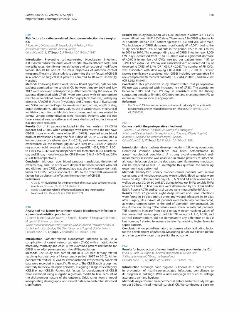
P49Risk factors for catheter-related bloodstream infections in a surgical ICUA Kundakci, O Ozkalayci, P Zeyneloglu, H Arslan, A PiratBaskent University Hospital, Ankara, TurkeyCritical Care 2013, 17(Suppl 2):P49 (doi: 10.1186/cc11987)
Introduction Preventing catheter-related bloodstream infections (CR-BSI) can reduce the duration of hospital stay, healthcare costs, and mortality rates. Identifying the risk factors and correction of modifi able factors should be one of the main objectives of infection control measures. The aim of this study is to determine the risk factors of CR-BSI in a cohort of surgical ICU patients admitted to Baskent University Hospital.Methods Following Institutional Review Board approval, data for 876 patients admitted to the surgical ICU between January 2009 and July 2012 were reviewed retrospectively. After completing the review, 25 patients diagnosed with CR-BSI were compared with 66 appropriate matches who did not have CR-BSI. Demographical features, underlying diseases, APACHE II (Acute Physiology and Chronic Health Evaluation) and SOFA (Sequential Organ Failure Assessment) scores, length of stay, organ dysfunctions, laboratory values, use of vasopressors, mechanical ventilation, nutrition, antibiotics, transfusions, and features related to central venous catheterization were recorded. Patients who did not have a central venous catheter and were discharged within 2 days of ICU stay were excluded.Results Out of 91 patients included in the fi nal analysis, 25 (27%) patients had CR-BSI. When compared with patients who did not have CR-BSI, those who did were older (P = 0.029), required more blood product transfusions during the fi rst 3 days of ICU (P = 0.016), had a longer duration of catheter stay (P = 0.019), and were more frequently catheterized via the internal jugular vein (IJV) (P = 0.022). A logistic regression model revealed that advanced age (OR: 1.037; 95% CI: 1.001 to 1.073; P = 0.042) was an independent risk factor for CR-BSI. Fourteen-day and 28-day mortality rates for CR-BSI were 12% (P = 0.749) and 28% (P = 0.406), respectively.Conclusion Although age, blood product transfusion, duration of catheter stay, and use of IJV were diff erent between patients who did and did not have CRBSI, advanced age was the only independent risk factor for CR-BSI. Early suspicion of CR-BSI by the other well-known risk factors has a substantial eff ect on the treatment of CR-BSI.References1. O’Grady NP: Guidelines for the prevention of intravascular catheter-related
infections. Clin Infect Dis 2011, 52:e162-e193.2. Bouza E: Catheter-related infections: diagnosis and intravascular
treatment. Clin Microbiol Infect 2002, 8:265-274.
P50Analysis of risk factors for catheter-related bloodstream infection in a parenteral nutrition populationI Conrick-Martin1, M McGovern2, K Boner1, J Bourke1, E Fitzgerald1, R Hone1, M Lynch1, D Phelan1, C Walshe3
1Mater Misericordiae University Hospital, Dublin, Ireland; 2Harvard School of Public Health, Cambridge, MA, USA; 3Beaumont Hospital, Dublin, IrelandCritical Care 2013, 17(Suppl 2):P50 (doi: 10.1186/cc11988)
Introduction Catheter-related bloodstream infection (CRBSI) is a complication of central venous catheters (CVCs) with an attributable morbidity, mortality and cost [1]. We examined patient risk factors for CRBSI in an adult parenteral nutrition (PN) population.Methods The study was carried out in a 525-bed tertiary-referral teaching hospital over a 14-year study period (1997 to 2010). All in-patients referred for PN via CVCs were included. Prospectively collected data were recorded in a specifi c PN record. The CRBSI audit group met quarterly to review all sepsis episodes, assigning a diagnostic category (CRBSI or non-CRBSI). Patient risk factors for development of CRBSI were examined using a logistic regression model to take account of the dichotomous nature of the outcome. Odds ratios from a model incorporating demographic and clinical data were tested for statistical signifi cance.
Results The study population was 1,961 patients in whom 3,213 CVCs were utilised over 19,511 CVC days. There were 256 CRBSI episodes in 216 patients. Median (IQR) patient age was 62 (23), and 58% were male. The incidence of CRBSI decreased signifi cantly (P <0.001) during the study period from 16% of patients in the period 1997 to 2003 to 7% in 2004 to 2010. The corresponding rate of CRBSI infection (per 1,000 CVC days) decreased from 18 to 10. There was a signifi cant decrease (P <0.001) in numbers of CVCs inserted per patient (from 1.87 to 1.49). Each extra CVC PN day was associated with an increased risk of developing CRBSI of 3.4% (OR 1.034, P <0.05). The number of PN CVCs was associated with developing CRBSI (OR 1.218, P <0.10). Patient factors signifi cantly associated with CRBSI included perioperative PN use (compared with medical patients) (OR 2.414, P <0.01), and male sex (OR 1.952, P <0.01).Conclusion This prospective study demonstrated that perioperative PN use was associated with increased risk of CRBSI. The association between CRBSI and CVC PN days is consistent with the theory suggesting benefi t to limiting CVC duration and changing from PN to enteral nutrition as soon as appropriate.Reference1. Blot SI, et al.: Clinical and economic outcomes in critically ill patients with
nosocomial catheter-related bloodstream infection. Clin Infect Dis 2005; 41:1591-1598.
P51Can we predict the postoperative infections?T Mózes1, K Gornicsar2, A Grosz3, ZS Domján2, I Buzogány2
1Ministry of Defense Health Centre, Budapest, Hungary; 2Péterfy Hospital, Budapest, Hungary; 3University of Szeged, HungaryCritical Care 2013, 17(Suppl 2):P51 (doi: 10.1186/cc11989)
Introduction Many patients develop infections following operations. Decreased immune competence has been demonstrated in acute neurological conditions. A strong cytokine-mediated anti-infl ammatory response was observed in stroke patients at infection, although infection due to the decreased proinfl ammatory mediators can be expected as well. To investigate this question the following experiment was performed.Methods Twenty-two urinary bladder cancer patients with radical cystectomy and lymphadenectomy were studied. Blood samples were taken on day 0 (before) and days 1, 3, 6, 9 and 14 after operation as well as on days 30, 60, 90 and 270 during follow-up. TNFα, soluble TNFα receptor I and IL-6 levels in sera were determined by HS ELISA and/or ELISA. Plasma ACTH and cortisol values were measured by RIA kits.Results From 22 patients, eight deep wound and urine infections were found in 14 days and six urine and wound infections in 30 days after surgery, all survived. All patients were bacterially contaminated, as wound samples taken at the end of operation demonstrated. On day 0 the circulating TNFα values were lower in infected patients. TNF started to increase from day 3 to day 9, never reaching values of the uneventful healing group. Soluble TNF receptor I, IL-6, ACTH, and cortisol concentrations did not demonstrate any diff erence on day 0 but from day 1 started to increase transiently, reaching higher levels in septic patients.Conclusion A low proinfl ammatory response is a key facilitating factor for the development of infection. Measuring serum TNFα levels before and after operations can thus predict the outcome.
P52Results for introduction of a new hand hygiene program in the ICUP Vos, A Harts-Laurijsen, R Snoeren, R Ramnarain, JA Van OersSt Elisabeth Hospital, Tilburg, the NetherlandsCritical Care 2013, 17(Suppl 2):P52 (doi: 10.1186/cc11990)
Introduction Although hand hygiene is known as a core element in prevention of healthcare-associated infections, compliance to its program is not high. With a new campaign we tried to enlarge awareness on hand hygiene.Methods We performed an experimental, before and after, study design on our 30-bed, mixed medical–surgical ICU. We conducted a baseline
Critical Care 2013, Volume 17 Suppl 2 http://ccforum.com/supplements/17/S2
S18
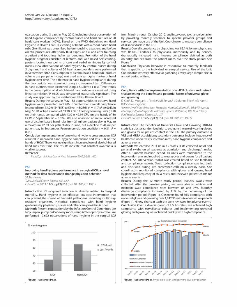
evaluation during 3 days in May 2012 including direct observation of hand hygiene compliance by control nurses and hand cultures of 50 healthcare workers (HCW). Based on the WHO Guidelines on Hand Hygiene in Health Care [1], cleaning of hands with alcohol-based hand rubs (Sterillium) was prescribed before touching a patient and before aseptic procedures, after body fl uid exposure risk and after touching a patient and touching his/her surroundings. Promotion of the hand hygiene program consisted of lectures and web-based self-learning, posters located near points of care and verbal reminders by control nurses. New observations of hand hygiene by control nurses during 3 days and hand cultures of 50 healthcare providers were performed in September 2012. Consumption of alcohol-based hand rub (product volume use per patient-days) was used as a surrogate marker of hand hygiene over time. The diff erence in hand hygiene compliance during the two periods was examined using a chi-squared test. Diff erences in hand cultures were examined using a Student’s t test. Time trends in the consumption of alcohol-based hand rub were examined using linear correlation. P <0.05 was considered statistically signifi cant. The study was approved by the institutional Ethics Review Board.Results During the survey, in May 158 opportunities to observe hand hygiene were presented and 286 in September. Overall compliance improved from 34.2% (54/158) to 51% (146/286), χ2 = 11.7 (P <0.001). In May, 50 HCW had a mean of 63.20 ± 39.37 colony-forming units (CFU) on their hands compared with 43.0 ± 40.19 CFU on the hands of 50 HCW in September (P = 0.024). We also observed an initial increased use of alcohol-based hand rubs from 21 ml per patient-day in May to a maximum 72 ml per patient-day in June, but a decline to 44 ml per patient-day in September, Pearson correlation coeffi cient = 0.31 (P = 0.61).Conclusion Implementation of a new hand hygiene program at our ICU resulted in improved hand hygiene compliance and less CFU on the hands of HCW. There was no signifi cant increased use of alcohol-based hand rubs over time. The results indicate that constant awareness is vital for success.Reference1. Pittet D, et al.: Infect Control Hosp Epidemiol 2009, 30:611-622.
P53Improving hand hygiene performance in a surgical ICU: a novel method for data collection to change physician behaviorSA NasrawayTufts Medical Center, Boston, MA, USACritical Care 2013, 17(Suppl 2):P53 (doi: 10.1186/cc11991)
Introduction ICU-acquired infection is directly related to hospital mortality. Hand hygiene is an eff ective, low-cost intervention that can prevent the spread of bacterial pathogens, including multidrug-resistant organisms. Historical compliance with hand hygiene guidelines by physicians, nurses and other care providers is poor.Methods Present expectations by the Infection Control Committee are to ‘pump in, pump out’ of every room, using 63% isopropyl alcohol. We performed 17,622 observations of hand hygiene in the surgical ICU
from March through October 2012, and intervened to change behavior by providing monthly feedback to specifi c provider groups and services. We made use of the Unit Coordinator to measure compliance of all individuals in the ICU.Results Overall compliance by physicians was 82.1%, for nonphysicians was 84.8%. Feedback to physicians, individually and by service, dramatically increased hand hygiene compliance, defi ned as both on entry and exit from the patient room, over the study period. See Figure 1.Conclusion Physician behavior is responsive to monthly feedback that is specifi c to the individual or surgical service. Use of the Unit Coordinator was very eff ective at gathering a very large sample size in a short period of time.
P54Compliance with the implementation of an ICU cluster-randomized trial assessing the benefi ts and potential harms of universal glove and gowningD Kett1, DJ Morgan2, L Pineles2, MJ Zervos3, LS Munoz-Price1, AD Harris2, BUGG Investigators2
1University of Miami/Jackson Memorial Hospital, Miami, FL, USA; 2University of Maryland/VA Maryland Healthcare System, Baltimore, MD, USA; 3Henry Ford Health System, Detroit, MI, USACritical Care 2013, 17(Suppl 2):P54 (doi: 10.1186/cc11992)
Introduction The Benefi ts of Universal Glove and Gowning (BUGG) study is a cluster-randomized trial to evaluate the use of wearing gloves and gowns for all patient contact in the ICU. The primary outcome is VRE and MRSA acquisitions; secondary outcomes include frequency of healthcare worker visits, infection rates, hand hygiene compliance and adverse events.Methods We enrolled 20 ICUs in 15 states. ICUs collected nasal and perianal swabs on all patients at admission and discharge/transfer. After a 3-month baseline period, 10 units were randomized to the intervention arm and required to wear gloves and gowns for all patient contact. An intervention toolkit was created based on site feedback and compliance reports. Swab collection compliance was fed back and discussed during site conference calls on a weekly basis. Site coordinators monitored compliance with gloves and gowns, hand hygiene and frequency of HCW visits and reviewed patient charts for adverse events.Results During the 12-month study period, 100,210 swabs were collected. After the baseline period, we were able to achieve and maintain swab compliance rates between 85 and 97%. Monthly discharge compliance increased by 21% by the beginning of the intervention period (Figure 1). Observers found 86% compliance with universal glove and gowning over 1,242 30-minute observation periods (Figure 1). Ninety charts at each site were reviewed for adverse events.Conclusion Over a diverse group of US hospitals, we achieved high compliance with surveillance cultures and implementing universal gloving and gowning was achieved quickly with high compliance.
Figure 1 (abstract P53). Figure 1 (abstract P54). Swab collection and gown/glove compliance.
Critical Care 2013, Volume 17 Suppl 2 http://ccforum.com/supplements/17/S2
S19
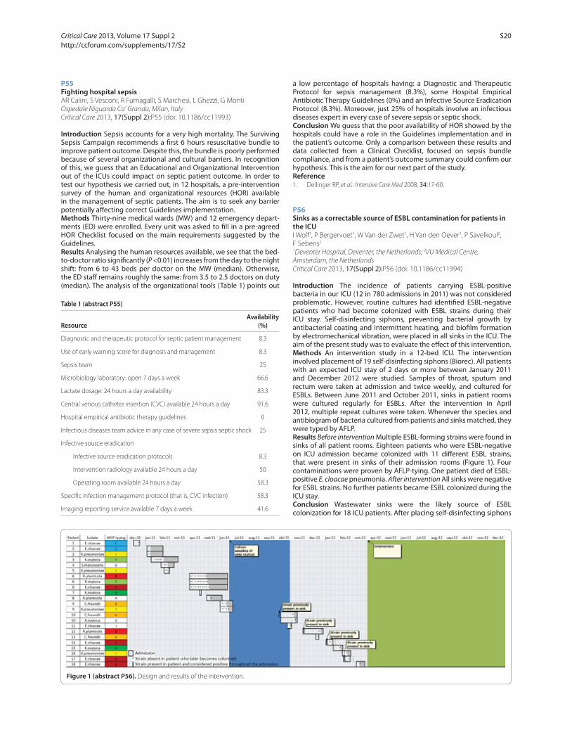
P55Fighting hospital sepsisAR Calini, S Vesconi, R Fumagalli, S Marchesi, L Ghezzi, G MontiOspedale Niguarda Ca’ Granda, Milan, ItalyCritical Care 2013, 17(Suppl 2):P55 (doi: 10.1186/cc11993)
Introduction Sepsis accounts for a very high mortality. The Surviving Sepsis Campaign recommends a fi rst 6 hours resuscitative bundle to improve patient outcome. Despite this, the bundle is poorly performed because of several organizational and cultural barriers. In recognition of this, we guess that an Educational and Organizational Intervention out of the ICUs could impact on septic patient outcome. In order to test our hypothesis we carried out, in 12 hospitals, a pre-intervention survey of the human and organizational resources (HOR) available in the management of septic patients. The aim is to seek any barrier potentially aff ecting correct Guidelines implementation.Methods Thirty-nine medical wards (MW) and 12 emergency depart-ments (ED) were enrolled. Every unit was asked to fi ll in a pre-agreed HOR Checklist focused on the main requirements suggested by the Guidelines.Results Analysing the human resources available, we see that the bed-to-doctor ratio signifi cantly (P <0.01) increases from the day to the night shift: from 6 to 43 beds per doctor on the MW (median). Otherwise, the ED staff remains roughly the same: from 3.5 to 2.5 doctors on duty (median). The analysis of the organizational tools (Table 1) points out
a low percentage of hospitals having: a Diagnostic and Therapeutic Protocol for sepsis management (8.3%), some Hospital Empirical Antibiotic Therapy Guidelines (0%) and an Infective Source Eradication Protocol (8.3%). Moreover, just 25% of hospitals involve an infectious diseases expert in every case of severe sepsis or septic shock.Conclusion We guess that the poor availability of HOR showed by the hospitals could have a role in the Guidelines implementation and in the patient’s outcome. Only a comparison between these results and data collected from a Clinical Checklist, focused on sepsis bundle compliance, and from a patient’s outcome summary could confi rm our hypothesis. This is the aim for our next part of the study.Reference1. Dellinger RP, et al.: Intensive Care Med 2008, 34:17-60.
P56Sinks as a correctable source of ESBL contamination for patients in the ICUI Wolf1, P Bergervoet1, W Van der Zwet1, H Van den Oever1, P Savelkoul2, F Sebens1
1Deventer Hospital, Deventer, the Netherlands; 2VU Medical Centre, Amsterdam, the NetherlandsCritical Care 2013, 17(Suppl 2):P56 (doi: 10.1186/cc11994)
Introduction The incidence of patients carrying ESBL-positive bacteria in our ICU (12 in 780 admissions in 2011) was not considered problematic. However, routine cultures had identifi ed ESBL-negative patients who had become colonized with ESBL strains during their ICU stay. Self-disinfecting siphons, preventing bacterial growth by antibacterial coating and intermittent heating, and biofi lm formation by electromechanical vibration, were placed in all sinks in the ICU. The aim of the present study was to evaluate the eff ect of this intervention.Methods An intervention study in a 12-bed ICU. The intervention involved placement of 19 self-disinfecting siphons (Biorec). All patients with an expected ICU stay of 2 days or more between January 2011 and December 2012 were studied. Samples of throat, sputum and rectum were taken at admission and twice weekly, and cultured for ESBLs. Between June 2011 and October 2011, sinks in patient rooms were cultured regularly for ESBLs. After the intervention in April 2012, multiple repeat cultures were taken. Whenever the species and antibiogram of bacteria cultured from patients and sinks matched, they were typed by AFLP.Results Before intervention Multiple ESBL-forming strains were found in sinks of all patient rooms. Eighteen patients who were ESBL-negative on ICU admission became colonized with 11 diff erent ESBL strains, that were present in sinks of their admission rooms (Figure 1). Four contaminations were proven by AFLP-tying. One patient died of ESBL-positive E. cloacae pneumonia. After intervention All sinks were negative for ESBL strains. No further patients became ESBL colonized during the ICU stay.Conclusion Wastewater sinks were the likely source of ESBL colonization for 18 ICU patients. After placing self-disinfecting siphons
Figure 1 (abstract P56). Design and results of the intervention.
Table 1 (abstract P55)
AvailabilityResource (%)
Diagnostic and therapeutic protocol for septic patient management 8.3
Use of early warning score for diagnosis and management 8.3
Sepsis team 25
Microbiology laboratory: open 7 days a week 66.6
Lactate dosage: 24 hours a day availability 83.3
Central venous catheter insertion (CVC) available 24 hours a day 91.6
Hospital empirical antibiotic therapy guidelines 0
Infectious diseases team advice in any case of severe sepsis septic shock 25
Infective source eradication
Infective source eradication protocols 8.3
Intervention radiology available 24 hours a day 50
Operating room available 24 hours a day 58.3
Specifi c infection management protocol (that is, CVC infection) 58.3
Imaging reporting service available 7 days a week 41.6
Critical Care 2013, Volume 17 Suppl 2 http://ccforum.com/supplements/17/S2
S20

there were no new ESBL colonizations in patients. This coincided with the disappearance of ESBL strains from all sinks.
P57Hemodynamic and mitochondrial eff ects of enalapril in experimental sepsisAJ Pereira, V Jeger, T Corrêa, M Vuda, S Djafarzadeh, J Takala, S JakobUniversity Hospital (Inselspital)/University of Bern, SwitzerlandCritical Care 2013, 17(Suppl 2):P57 (doi: 10.1186/cc11995)
Introduction Sepsis may cause mitochondrial dysfunction. The renin–angiotensin system (RAS) activity is increased in sepsis, and can interfere with the mitochondrial function either directly or by modifying hemodynamics. We studied the eff ects of ACE inhibition by enalapril on hemodynamics and hepatic mitochondrial function in sepsis. Two septic groups without enalapril and with diff erent blood pressure targets served as controls.Methods Sepsis (fecal peritonitis) was induced in 24 anesthetized, mechanically ventilated pigs (40.1 ± 2.1 kg), divided into three groups (n = 8 for each): ENL (enalapril pretreatment for 7 days and at 0.005 to 0.02 mg/kg/hour during the study; MAP target 75 to 85 mmHg), ML (target MAP 50 to 60 mmHg), and MH (target MAP 75 to 85 mmHg). Hemodynamic support with fl uids, norepinephrine (NE; maximum dose 5,000 μg/hour), and antibiotics were started after 12 hours of peritonitis and continued for 48 hours.Results All enalapril pigs received the maximum NE dose without reaching the target MAP. Enalapril resulted in lower MAP, higher CO, and transiently increased regional blood fl ows (Table 1). Fluid administration and urinary output were similar among groups. Liver mitochondrial respiration was reduced (State 3, State 4) in the enalapril group. One animal died in each ML and enalapril group.Conclusion Enalapril enhances cardiac output and early regional blood fl ows in sepsis despite lower MAP, and reduces liver mitochondrial respiration, suggesting that RAS is involved in the hemodynamic and metabolic changes in sepsis.
P58Eff ects of intraperitoneal immunoglobulin therapy on behavior and cognitive functions in CLP-induced sepsis model in ratsF Esen1, E Senturk1, P Ergin Ozcan1, G Orhun1, S Gümrü2, M Kaya1, N Orhan1, N Arıcan1, F Arıcıoglu2
1University of Istanbul, Turkey; 2Marmara University Faculty of Pharmacy, Istanbul, TurkeyCritical Care 2013, 17(Suppl 2):P58 (doi: 10.1186/cc11996)
Introduction The present study investigated the eff ects of a single dose of intraperitoneal (i.p.) IgG and IgGAM administration on various behavioral alterations in a cecal ligation perforation (CLP)-induced sepsis model in rats.Methods Female Wistar albino rats (200 to 250 g) were divided into fi ve groups (n = 8): a naive Control group, a Sham operated group receiving conventional antibiotic treatment, a CLP group receiving CLP procedure and conventional antibiotic treatment, and IgG and IgGAM groups which were also applied 1 g/kg, i.p. IgG and IGAM therapy 5 minutes after the CLP procedure. Ten, 30 and 60 days after the surgery, animals underwent three behavioral tasks: an open fi eld test to evaluate the locomotor activity, an elevated plus maze test to measure the level of anxiety, and a forced swim test to assess the possible depressive state. The results acquired from these tests were used to estimate the eff ect
of immunoglobulin therapy on behavioral changes in CLP-induced sepsis in rats.Results In the open fi eld test, the CLP group showed a signifi cant decrease in total squares passed on days 10 and 30. Similarly, total numbers of rearing and grooming were dramatically decreased in the CLP group in comparison with control and sham groups (P <0.005). In the elevated plus maze test, the number of entries to open arms decreased in the CLP group. In the forced swim test, there was a tendency for increase in immobility time in the CLP group, although the data were statistically insignifi cant. All of these values which were indicating the importance of behavioral alterations were improved on day 60. Immunoglobulin therapy prevented the occurrence of these behavioral changes. Especially, animals in the IgGAM group conserved the values quite near to those of the control group in measured parameters.Conclusion Sepsis, even though it has been treated with conventional antibiotics, caused a negative eff ect on behavioral parameters. In this study, IgG and IgGAM treated animals in the presence of CLP did not show these behavioral changes. Therefore our results suggest that a single dose of i.p. IgG and IgGAM treatment, which was applied immediately after the sepsis procedure, prevents behavioral defects observed following sepsis.
P59Clinical effi cacy of recombinant human soluble thrombomodulin in patients with septic disseminated intravascular coagulationY Suzuki, R Sato, M Sato, S EndoIwate Medical University, Morioka, JapanCritical Care 2013, 17(Suppl 2):P59 (doi: 10.1186/cc11997)
Introduction Thrombomodulin is an endothelial cell cofactor and glycoprotein for thrombin-catalyzed activation of protein C. A recom-binant human soluble thrombomodulin (rhsTM) has been recently developed, and this new agent has a unique amino-terminal structure exhibiting anti-infl ammatory activity including sequestraction and cleavage of high-mobility group box 1(HMGB-1).Methods In this study, 13 patients with septic disseminated intravascular coagulation (DIC) were treated with rhsTM, which is Recomodulin® Inj. 12800 (Asahi Kasei Pharma Co., Tokyo, Japan). Patients with septic DIC were treated with 130 to 380 U/kg/day.Results There were signifi cant results for improvement of APACHE II score and DIC diagnostic criteria score for critically ill patients after treatment using rhsTM (P <0.01). Improvement for platelet count and D-dimer level were also observed in this study (P <0.05). Activation of antithrombin (AT) also was signifi cantly increased after treatment (P <0.05). Hospital mortality was 15.4% in this study.Conclusion The rhsTM might be one of most important endogenous regulators of coagulation, acting as the major inhibitor of thrombin as well as AT III. This new agent may play an important role in treatment for septic DIC.
P60Antithrombin III concentrate therapy may have an effi cacy in sepsisN Saito, M Takeda, T Harada, R Moroi, M Namiki, A YaguchiTokyo Women’s Medical University, Tokyo, JapanCritical Care 2013, 17(Suppl 2):P60 (doi: 10.1186/cc11998)
Introduction Antithrombin III (AT III) has been known to contribute to anti-infl ammatory response as well as its anticoagulation. Our previous
Table 1 (abstract P57). Hemodynamic variables, among times and groups
P BL R0h R12h R24h R36h R48h (time×group)
MAP (ENL/MH/ML) 66 (9)/72 (15)/ 65 (11)/73 (10)/ 57 (4)/82 (6)/ 58 (8)/78 (6)/ 56 (12)/80 (5)/ 56 (11)/79 (9)/ 0.003 65 (9) 81 (16) 72 (13) 67 (12) 61 (6) 56 (13)
CO (ENL/MH/ML) 106 (13)/89 (7)/ 92 (17)/67 (11)/ 169 (16)/95 (19)/ 187 (22)/129 (28)/ 161 (25)/139 (32)/ 157 (37)/140 (21)/ <0.0001 105 (13) 88 (22) 104 (13) 143 (31) 145 (13) 150 (18)
Carotid fl ow (ENL/MH/ML) 6 (2)/7 (2)/7 (2) 5 (1)/4 (1)/5 (2) 10 (3)/5 (1)/6 (2) 10 (3)/8 (1)/9 (2) 8 (2)/8 (2)/8 (2) 8 (3)/8 (2)/8 (2) <0.0001
Femoral fl ow (ENL/MH/ML) 4 (1)/3 (1)/4 (2) 2 (1)/2 (0)/2 (1) 4 (1)/2 (1)/2 (0) 4 (1)/3 (1)/3 (1) 3 (1)/3 (1)/4 (1) 3 (2)/3 (1)/4 (1) 0.004
Critical Care 2013, Volume 17 Suppl 2 http://ccforum.com/supplements/17/S2
S21

study showed AT III defi ciency happened in the early stage of sepsis with no relation to DIC status. However, whether AT III concentrate is a benefi cial therapy or not for septic patients is controversial. Our hypothesis is that AT III concentrate may have effi cacy as anti-infl ammation for sepsis.Methods From January 2009 to December 2011, adult septic patients, whom were given AT III concentrate in our medico-surgical ICU, were included in this study. AT III concentrate was administered 30 to 60 U/kg intravenously every 24 hours for 3 days in the patients with DIC status and AT III defi ciency. Between before and after the AT III concentrate therapy, WBC (/mm3), CRP (mg/dl), platelet (×104/μl), PT (seconds), fi brinogen (mg/dl), FDP (μg/ml), SOFA score and DIC score were evaluated. Values are expressed as mean ± SD. Data were analyzed by Wilcoxon signed-rank test. P <0.05 was considered signifi cant.Results There were 85 patients (52 men, 33 women; age range 19 to 92 years (mean 69.0 ± 16.9)), and the mortality rate was 27.1% and APACHE II score was 20.1 ± 7.6. WBC, CRP, PT and SOFA score were signifi cantly improved after AT III concentrate therapy (13,176 ± 9,400 vs. 11,693 ± 7,089, P = 0.011, 15.9 vs. 13.3, P = 0.0015, 16.5 ± 4.4 vs. 15.9 ± 5.9, P = 0.0045, and 9.7 ± 3.8 vs. 8.6 ± 4.6, P = 0.01, respectively). Platelet was signifi cantly decreased (11.7 ± 9.7 vs. 10.6 ± 9.0, P = 0.028), while there were no signifi cant diff erences in fi brinogen, FDP and DIC score (387.3 ± 202.6 vs. 381.8 ± 163.2, P = 0.088, 36.9 ± 52.7 vs. 28.8 ± 39.9, P = 0.059, and 2.9 ± 1.6 vs. 2.6 ± 1.8, P = 0.25, respectively) after the therapy. One week after the therapy, platelet and DIC score were signifi cantly improved compared with before the therapy (15.6 ± 10.0, P = 0.0036 and 1.8 ± 1.9, P = 0.0041).Conclusion In the patients with septic DIC, WBC, CRP and SOFA score were immediately improved after the AT III concentrate therapy, while platelet and DIC score were improved later. AT III concentrate may also contribute to anti-infl ammatory for septic DIC with anticoagulation.
P61A clinical randomized trial on the use of atorvastatin in patients with sepsis or septic shock: eff ects on endothelial functionK Prado, C Ribeiro, T Furian, R Pinto Ribeiro, D Silvello, L Rohde, N Clausell, L BeckerHospital de Clinicas de Porto Alegre, BrazilCritical Care 2013, 17(Suppl 2):P61 (doi: 10.1186/cc11999)
Introduction Sepsis and septic shock are complex infl ammatory syndromes. Multiple cellular activation processes are involved, and many humoral cascades are triggered. Presumably, endothelial cells play a pivotal rule in the pathogenesis of sepsis, not only because they may infl uence the infl ammatory cascade but also because, upon interaction with excessive amounts of infl ammatory mediators, the function of these cells may become impaired. It is likely that a general dysfunction of the endothelium is a key event in the pathogenesis of sepsis [1]. HMG-CoA-reductase inhibitors have been shown to exhibit pronounced immunomodulatory eff ects independent of lipid lowering. Most of these benefi cial eff ects of statins appear to involve restoring or improving endothelial function [2]. We hypothesize that statins can improve endothelial dysfunction in septic patients.Methods A double-blinded, placebo-controlled, randomized trial was undertaken. We enrolled adult patients within 24 hours of severe sepsis or septic shock diagnosis and randomized them to placebo or atorvastatin 80 mg/day for a short term. Endothelial dysfunction was assessed measuring plasmatic levels of IL-6, ET-1, VCAM-1 by ELISA and measuring fl ow-mediated vasodilatation of the brachial artery at basal, 24 and 72 hours after randomization.Results We studied 47 patients, 24 in the placebo group (mean age 52 ± 20 years, 29.1% male; APACHE II risk score 23.5 ± 7.3) and 23 in the statin group (mean age 49.5 ± 18 years, 53.4% male; APACHE II risk score 23 ± 6.9). The baseline characteristics of the placebo group were similar to statin patients as well as the mean length of stay in the ICU (8.6 ± 7.4 and 9.1 ± 8 days, respectively) and the time on vasopressors (49.3 ± 47.1 and 59 ± 91.1 hours, respectively). No signifi cant diff erence was observed on the temporal variation of biomarker levels (IL-6, VCAM-1, ET-1) between treatment and control groups. The intrahospital mortality rate was 26% in the statin group and 45% in the placebo group (P = 0.17).
Conclusion Our data showed no benefi t with the use of a potent statin acutely in patients with sepsis or septic shock with regards to improvement in endothelial function.References1. Hack EC, et al.: The endothelium in sepsis: source of and a target for
infl ammation. Crit Care Med 2001, 29:S21-S27.2. Davignon J: Benefi cial cardiovascular pleiotropic eff ects of statins.
Circulation 2004, 109(Suppl III):III-39-III-43.
P62A multicenter randomized controlled study of an extracorporeal cytokine hemoadsorption device in septic patientsD Schädler1, C Porzelius2, A Jörres3, G Marx4, A Meier-Hellmann5, C Putensen6, M Quintel7, C Spies8, C Engel2, N Weiler1, M Kuhlmann9
1University Medical Center Schleswig-Holstein, Campus Kiel, Germany; 2University of Leipzig, Germany; 3Charité University Hospital Campus Virchow-Klinikum, Berlin, Germany; 4RWTH University Hospital Aachen, Germany; 5HELIOS Klinikum, Erfurt, Germany; 6University of Bonn, Germany; 7University Hospital Göttingen, Germany; 8Campus Charité Mitte and Campus Charité Virchow-Klinikum, Charité-University Medicine Berlin, Germany; 9Vivantes Klinikum im Friedrichshain, Berlin, GermanyCritical Care 2013, 17(Suppl 2):P62 (doi: 10.1186/cc12000)
Introduction A novel sorbent hemoadsorption device for cytokine removal (CytoSorbents, USA) was developed and successfully tested in animal models of sepsis. The experience in the clinical setting is still limited to case reports. In this fi rst clinical trial, we tested the hypothesis that treatment with sorbent hemoadsorption could safely and eff ectively reduce cytokines in septic patients with acute lung injury (ALI).Methods Ventilated patients fulfi lling the criteria for severe sepsis and ALI were enrolled in this multicenter randomized, controlled, open-label study comparing standard of care with or without hemoperfusion treatment. Primary endpoints were safety and IL-6 reduction. Treated patients underwent hemoperfusion at fl ow rates of ~200 to 300 ml/minute for 6 hours per day for 7 consecutive days. The overall mean reduction in individual plasma cytokines for the control and treatment groups during the treatment period was calculated using a generalized linear model.Results Forty-three patients (18 treated, 25 control) completed the study and were further analyzed. Incidence of organ dysfunction at enrollment (treatment vs. control) was: septic shock (94% vs. 100%, P = 0.42), acute respiratory distress syndrome (67% vs. 56%, P = 0.33), and renal failure (39% vs. 24%, P = 0.54). During 115 treatments no serious device-related adverse events occurred. On average, there were no changes in hematology and other blood parameters except for a modest reduction in platelet count (<10%) and albumin (<5%) with treatment. Hemoperfusion decreased IL-6 blood concentration signifi cantly (–49.1%, P = 0.01), with similar reductions of MCP-1 (–49.5%, P = 0.002), IL-1ra (–36.5%, P = 0.001), and IL-8 (–30.2%, P = 0.002). The 28-day mortality (28% vs. 24% control, P = 0.84) and 60-day mortality (39% vs. 32% control, P = 0.75) did not diff er signifi cantly between the two studied groups.Conclusion In this fi rst clinical study of a novel sorbent hemoadsorption device in patients with severe sepsis and ALI, the device appeared to be safe and decreased the blood concentration of several cytokines. Further research is needed to study the eff ect of the device on the clinical outcome of septic patients.
P63Continuous renal replacement therapy with the adsorbent membrane oXiris in septic patients: a clinical experienceF Turani, F Candidi, R Barchetta, E Grilli, A Belli, E Papi, A di Marzio, M FalcoAurelia Hospital/European Hospital, Rome, ItalyCritical Care 2013, 17(Suppl 2):P63 (doi: 10.1186/cc12001)
Introduction Renal failure is an important complication of sepsis and CRRT with adsorbing membranes may be useful in this clinical setting [1]. The aims of the study in septic/septic shock patients are to evaluate: the safety of a new hemofi lter membrane oXiris with adsorbing properties and anti-endotoxin activity; the renal and hemodynamic
Critical Care 2013, Volume 17 Suppl 2 http://ccforum.com/supplements/17/S2
S22

response; and the changes of endotoxin and proinfl ammatory molecules.Methods Forty septic/septic shock patients with renal failure were enrolled in the study. All patients had preoperative endotoxin >0.6 level/units (EAA Spectral D) and were submitted to high-volume hemodiafi ltration (50 ml/kg/hour, Prismafl ex; Gambro) with a new treated heparin-coated membrane (oXiris; Gambro). At T0 (pre-treatment) and T1 (24 hours) the main clinical and biochemical data were evaluated. All data are expressed as mean ± SD. One-way ANOVA test with Bonferroni correction was used to evaluate the data changes. P <0.05 was considered signifi cant.Results Table 1 presents the main results of this study.Conclusion In septic/septic shock patients with renal failure, CRRT with a new treated heparin-coated membrane (oXiris; Gambro) is clinically feasible, and has a positive eff ect on renal function and hemodynamics. An adsorbing eff ect on proinfl ammatory mediators may have a role in these results. These data and the trend toward a decrease of endotoxin during the treatment warrant further investigation.Reference1. Rimmelé T, et al.: Nephrol Dial Transplant 2009, 24:421-427.
P64Combined use of pumpless extracorporeal lung assist system and continuous renal replacement therapy with citrate anticoagulation in polytrauma patientsHK Atalan, M Dumantepe, TB Denizalti, IA Tarhan, A OzlerAtasehir Memorial Hospital, Istanbul, TurkeyCritical Care 2013, 17(Suppl 2):P64 (doi: 10.1186/cc12002)
Introduction The usefulness of a pumpless extracorporeal lung assist system (pECLA) and continuous renal replacement therapy (CRRT) in critically ill patients has been demonstrated in previous studies [1,2]. The aim of this report was to examine combined use of pECLA and CRRT to improve carbon dioxide and infl ammatory mediator removal, which allows for lung protective ventilation strategies.Methods In our 10 patients with ARDS due to polytrauma and sepsis, pECLA was established by insertion of cannulae to the femoral artery and vein. CRRT cannulae were introduced by venous line of the same vascular access (Figure 1). We preferred regional anticoagulation with trisodium citrate for both CRRT and ILA.
Results Mean SAPS II and APACHE II scores were 55 and 23 respectively. Mean time on mechanical ventilation was 22 days. Mean ICU stay was 30 days for survivors and 38 days for nonsurvivors. When compared with baseline values most relevant parameters were the improvement in tidal volumes, plateau pressures, PaCO2 levels and pH (Figure 2). Four patients survived while six patients died from sepsis-MOF.Conclusion We concluded that pECLA can eff ectively address the impaired gas exchange in ARDS and CRRT is a safe procedure with potential therapeutic value for treating MOF. Citrate anticoagulation was well tolerated and fi lter life was appropriate. The use of the same vascular access for ILA and CRRT may minimize invasive procedures and related side eff ects.References1. Bein T, et al.: Int J Emerg Med 2010, 3:177-179.2. Demetrios J, et al.: Kidney Int 2005, 67:2361-2367.
P65Abstract withdrawn
P66Use of extracorporeal endotoxin elimination therapy for septic shockBA Adamik, JS Smiechowicz, SZ Zielinski, AK KüblerMedical University, Wroclaw, PolandCritical Care 2013, 17(Suppl 2):P66 (doi: 10.1186/cc12004)
Introduction Endotoxin, a component of the outer membrane of Gram-negative bacteria, is considered an important factor in pathogenesis of septic shock [1]. The aim of our study was to determine whether endotoxin elimination treatment added to the standard treatment would improve organ function in patients with septic shock.Methods Adult patients with septic shock who required renal replacement therapy (RRT), with a confi rmed endotoxemia, and suspected Gram-negative infection were consecutively added to the study within the fi rst 24 hours after diagnosis. All patients received full standard treatment for septic shock. Endotoxin elimination was performed using the membrane oXiris (Gambro, Sweden), a medical device for continued RRT with the unique feature of endotoxin adsorbtion. An endotoxin activity assay was used to monitor endotoxin elimination therapy at baseline (T0), 3 hours (T1), 12 hours (T2), 24 hours (T3), 48 hours (T4), and 72 hours (T5). Our key indicators were the improvement in hemodynamics and organ function, and decrease of endotoxin activity (EA) in blood. Continuous variables are presented as mean values with standard deviations.Results High EA level at baseline (0.74 ± 0.14 endotoxin activity units (EAU)) signifi cantly decreased during RRT with oXiris membrane to 0.46 ± 0.02 (T1), 0.34 ± 0.01 (T2), 0.4 ± 0.02 (T3), 0.46 ± 0.04 (T4), 0.35 ± 0.07 (T5) EAU (P <0.05). MAP increased from baseline 72 ± 14 to 81 ± 18, 76 ± 6, 77 ± 7, 83 ± 13, 87 ± 10 mmHg (P <0.05), and the mean norepinephrine use decreased from 0.23 ± 0.04 to 0.19 ± 0.02, 0.11 ± 0.01, 0.09 ± 0.01, 0.04 ± 0.01, 0.0 μg/kg/minute (P <0.05) at T0, T1, T2, T3, T4, T5, respectively. The SOFA score had decreased from 14 ± 4 to 12 ± 2, 9 ± 3, 7 ± 3 points (P <0.05), and the procalcitonin level declined from 107 ± 123 to 45 ± 41, 29 ± 30, 17 ± 157 ± 1 ng/ml (P <0.05) at T0, T3, T4, T5.Conclusion RRT with oXiris membrane resulted in the eff ective elimination of endotoxins from the blood. The therapy was associated with an increase in blood pressure, a reduction of vasopressor requirements, and an improvement of organ function. The application of the endotoxin activity assay was useful for bedside monitoring of endotoxemia in ICU patients.Reference1. Marshall JC, Foster D, Vincent JL, Cook DJ, Cohen J, Dellinger RP, Opal S,
Abraham E, Brett SJ, Smith T, Mehta S, Derzko A: Diagnostic and prognostic implications of endotoxemia in critical illness: results of the MEDIC Study. J Infect Dis 2004, 190:527-534.
Figure 1 (abstract P64). ILA and CRRT connected to the patient.
Table 1 (abstract P63)
Parameter Units T0 T1
Creatinine mg/dl 1.9 ± 0.1 1.18 ± 0.1*
Diuresis ml/24 hours 1,284 ± 78 1,573 ± 98
Norepinephrine μg/kg/minute 0.17 ± 0.2 0.06 ± 0.1*
IL-6 pg/ml 572 ± 78 278 ± 57*
Procalcitonin ng/ml 35 ± 7 15 ± 2*
Endotoxin Level/U 0.64 ± 0.2 0.49 ± 0.1
*P <0.05 vs. T0.
Critical Care 2013, Volume 17 Suppl 2 http://ccforum.com/supplements/17/S2
S23

P67Selective adsorption of lipopolysaccharide in the complex treatment of patients with severe sepsisS Rei, I Aleksandrova, V Kiselev, M Ilynskiy, G Berdnikov, L Marchenkova, G Bulava, N BorovkovaHospital Research Institute for Emergency Medicine named after N.V. Sklifosovsky, Moscow, RussiaCritical Care 2013, 17(Suppl 2):P67 (doi: 10.1186/cc12005)
Introduction Severe sepsis and septic shock remain the most serious problem of critical care medicine with a mortality rate of 30 to 55% [1]. Several studies have demonstrated positive eff ects of selective adsorption of LPS on blood pressure, PaO2/FiO2 ratio, endotoxin removal and mortality [2,3]. The purpose of the study was to evaluate the effi ciency of using the selective adsorption of LPS, Toraymyxin – PMX-F (Toray, Japan) and Alteco® LPS Adsorber (Alteco Medical AB, Sweden), in the complex treatment of patients with severe sepsis.Methods Forty-six patients with Gram-negative sepsis in the postoperative period were enrolled into the study. Toraymyxin – PMX-F was used in the PMX-F group (n = 14), while Alteco LPS adsorption was used in the Alteco LPS group (n = 32). The clinical characteristics are listed in Table 1. The SOFA score, PaO2/FiO2, procalcitonin (PCT), C-reactive protein (CRP), endotoxin activity assay (EAA) was noted before, 24 and 48 hours after the selective adsorption of LPS.Results At 48 hours after PMX-F, signifi cantly decreased PCT from 17.5 (5.0; 40.9) to 7.1 (4.8; 13.0) ng/ml, P = 0.028, decreased CRP from 180
(133; 286) to 132 (68; 155) mg/l, P = 0.015 and SOFA score from 7.0 (3,0; 8.0) to 6.0 (3,0; 7.0), P = 0.007. At 24 hours after Alteco LPS, signifi cantly decreased PCT from 8.7 (3.0; 25.9) to 4.8 (2.1; 10.0) ng/ml. The 28-day mortality rate was 14.2% (n = 2) in the PMX-F group and 31.3% (n = 10) in the Alteco LPS group.
Table 1 (abstract P67). Clinical characteristics of the groups
Characteristic PMX-F Alteco LPS
Sex M/F 9/5 17/15
Age (years) 41.6 ± 17.1 42.1 ± 13.5
Shock 6 (42.9%) 13 (40.6%)
APACHE II 21 (17; 25) 21 (16; 24)
SOFA 7 (3.0; 8) 7 (3.5; 11)
Conclusion The use of the LPS-selective adsorption (particularly PMX-F) in patients with severe sepsis leads to improvement of systemic infl ammation and organ dysfunction.References1. Vincent JL, et al.: Crit Care Med 2006, 34:344-353.2. Cruz DN, et al.: JAMA 2009, 301:2445-2452.3. Yaroustovsky M et al.: Blood Purif 2009, 28:227-233.
Figure 2 (abstract P64). Changes in tidal volume, plateau pressure, pH and PaCO2.
Critical Care 2013, Volume 17 Suppl 2 http://ccforum.com/supplements/17/S2
S24

P68Alteco endotoxin hemoadsorption in Gram-negative septic shock patientsHP Shum, KC Chan, WW YanPamela Youde Nethersole Eastern Hospital, Hong KongCritical Care 2013, 17(Suppl 2):P68 (doi: 10.1186/cc12006)
Introduction Septic shock is a common cause of mortality and morbidity in the ICU. Endotoxin hemoadsorption using a Polymyxin B fi ber column can improve patient outcome [1]. This study investigated the therapeutic eff ect of a new endotoxin hemoadsorption device in Gram-negative septic shock patients.Methods An open, controlled, prospective, randomized, single-centre trial conducted between February 2010 and June 2012. Patients with septic shock due to intra-abdominal sepsis were recruited and randomized to either standard therapy (ST, n = 8) or standard therapy plus two 2 hourly sessions of Alteco endotoxin hemoperfusion (AT, n = 7). Standard therapy included infective source control, appropriate early antibiotic, low-dose steroid, early continuous renal replacement therapy in the presence of acute kidney injury (RIFLE Injury class or more), hemodynamic optimization and lung-protected ventilatory support. Primary outcome was change in organ dysfunction at 48 hours measured by Sequential Organ Failure Assessment (SOFA) scores. Secondary outcomes were change in vasopressor requirement (measured by vasopressor score), PaO2/FiO2 (fraction of inspired oxygen) ratio, total urine output at 72 hours and 28-day mortality.Results This study was terminated early as interim analysis identifi ed no signifi cant clinical benefi t. Baseline characteristics (age/APACHE IV score) were similar between two groups of patients. No signifi cant diff erence was noted between two groups with respect to change in total SOFA score (+1 vs. –5.5 for AT vs. ST, P = 0.382), vasopressor score (–29 vs. –46.6, P = 0.775), PaO2/FiO2 ratio (–26 vs. +163, P = 0.199), total urine output from 0 to 72 hours (3,850 ml vs. 4,570 ml, P = 0.355) and 28-day mortality (14.3% vs. 37.5%, P = 0.569). No signifi cant side eff ect was noted when using this new hemoadsorption device.Conclusion This small study cannot identify any extra clinical benefi t on addition of Alteco endotoxin hemoadsorption to standard therapy in patients suff ering from intra-abdominal sepsis with shock due to Gram-negative bacterial infection.Reference1. Cruz DN, et al.: JAMA 2009, 301:2445-2452.
P69Steroid therapy in septic shock: survey of practice amongst UK critical care physiciansG Rajendran, K Dasari, A DhrampalNorfolk & Norwich University Hospital, Norwich, UKCritical Care 2013, 17(Suppl 2):P69 (doi: 10.1186/cc12007)
Introduction Corticosteroid (CS) therapy in sepsis remains contro-versial and was fi rst introduced in sepsis management for its anti-infl ammatory property. CS has found a role in septic shock amelioration with inconsistent outcomes. The Surviving Sepsis Campaign (SSC) includes CS as a Level 2C recommendation in septic shock [1]. Adapting and practicing SSC guidelines vary between critical care units. Accordingly, a survey was conducted to elucidate the usage of CS for septic shock by UK critical care physicians (CCPs).Methods Following approval by the UK Intensive Care Society (ICS), the survey was publicised on the ICS website and its newsletter.Results A total of 81 intensivists responded to this online survey. Seventy-four (92.5%) CCPs prescribed CS only if the septic shock is poorly responsive to fl uid resuscitation and vasopressor therapy. Six (7.5%) initiated CS at the same time as vasopressor therapy. None initiated CS for patients with severe sepsis. No CS other than hydrocortisone is being used. The most commonly used intravenous regimen is 50 mg 6 hourly (65%) followed by 50 mg 8 hourly (11%). Only 10% of CCPs would prescribe it by infusion. Less commonly used regimens were 100 mg 8 hourly (6%) and 100 mg 6 hourly (5%). Only 5% would consider adding fl udrocortisone. Prior to initiating CS, 5% of CCPs would perform a short synacthen test, while 94% would not. The majority (89%) of CCPs would stop CS after resolution of shock state
or when vasopressor infusion is terminated whilst 11% after a fi xed duration. Withdrawal of CS also diff ered, in that 25% tapered/weaned steroids, 31% stopped it abruptly and 44% of CCPs would base their CS cessation pattern on the clinical context. Only 46% of CCPs believe that CS is benefi cial whereas 44% were unsure of the benefi ts in septic shock. Only 29 (36%) responders indicated that their critical care unit had a written protocol for CS in septic shock.Conclusion The perceptions, usage and cessation of CS in septic shock vary but do appear to have shifted in the last decade. A UK survey in 2003 identifi ed that only 60% of ICUs used CS for septic shock and over 22% perform a short synacthen test [2]. It appears that many intensivists are using CS for septic shock, despite confl icting outcome data. We all strive to practice evidence-based medicine but until we have a robust, reliable and methodical randomised control trial that attempts to resolve the CS debate, practice will remain diverse on this subject, as refl ected by our survey.References1. Dellinger et al.: Crit Care Med 2008, 36:296-327.2. Bourne et al.: Anaesthesia 2003, 58:591-596.
P70Need for restoration of cortisol serum levels for successful antimicrobial therapy in experimental sepsisT Doulias1, A Pistiki2, P Christopoulos1, V Papaziogas1, E Giamarellos-Bourboulis2, I Koutelidakis1
1University of Thessaloniki, Medical School, Thessaloniki, Greece; 2University of Athens, Medical School, Athens, GreeceCritical Care 2013, 17(Suppl 2):P70 (doi: 10.1186/cc12008)
Introduction It is postulated that clinical benefi t of low-dose hydrocortisone in septic shock is related to reversal of relative adrenal insuffi ciency [1]. This was proved in an animal model of sepsis.Methods Sixty-nine Wistar male rats were assigned to the following groups: A, sham-operation; B, sepsis; C, bilateral adrenalectomy and sepsis; D, bilateral adrenalectomy, sepsis and hydrocortisone treatment; E, bilateral adrenalectomy, sepsis and ertapenem treatment; and F, bilateral adrenalectomy, sepsis, hydrocortisone and ertapenem treatment. Sepsis was induced by the i.p. infusion of 1×10⁶ cfu/ml of Escherichia coli after adrenalectomy. Hydrocortisone 10 mg/kg was infused i.v. bid starting 1 hour after bacterial challenge. Ertapenem 5 mg/kg was infused i.v. once daily starting 1 hour after bacterial challenge. Survival was recorded. In a separate set of experiments in 18 rats, animal sacrifi ce was performed to measure the free cortisol concentration.Results Survival is shown in Figure 1. Experiments in each animal were starting at 7:00 am. At 8:00 am, respective mean free cortisol of groups A, C and D was 1.81, 0.55 and 2.05 μg/dl; at 1:00 pm they were 0.92, 0.47 and 1.40 μg/dl.
Figure 1 (abstract P70). Survival of rats.
Critical Care 2013, Volume 17 Suppl 2 http://ccforum.com/supplements/17/S2
S25

Conclusion Even when eff ective antimicrobial treatment is adminis-tered, administration of hydrocortisone at a regimen restoring normal secretion is mandatory for survival.Reference1. Annane D, et al.: JAMA 2002, 288:862-871.
P71Impact of time to initiation of appropriate antibiotic therapy on early mortality of patients with septic shockJP Quenot1, C Binquet2, S Vinault2, A Pavon1
1University Hospital Dijon, France; 2CHU de Dijon, Centre d’Investigation Clinique – Epidémiologie Clinique, Dijon, FranceCritical Care 2013, 17(Suppl 2):P71 (doi: 10.1186/cc12009)
Introduction The impact of appropriate antibiotic therapy on prognosis of patients with septic shock is well established. However, the prognostic infl uence of time to initiation of antibiotics remains debated. We evaluated the eff ect on 7-day mortality of time to initiation of appropriate antibiotic therapy in patients hospitalised in critical care for septic shock.Methods Secondary analysis from the EPISS cohort. We included only patients admitted to the University Hospital Dijon. Septic shock was defi ned as initiation of vasopressors in a patient with suspected or documented infection with at least one criterion of hypoperfusion (metabolic acidosis and/or renal insuffi ciency and/or hepatic dys func-tion). We excluded patients with no available bacteriological data. Anti-biotherapy was considered appropriate if at least one of the antibiotics prescribed was active against the germ identifi ed. Bivariate and multivariate logistic regression analysis was used to assess the impact of time to initiation of appropriate antibiotherapy on 7-day mortality.Results In total, 383 patients were admitted with septic shock, of whom 253 (66%) were included; 231(92%) had appropriate antibiotic therapy, of whom 52 (22.5%) died at 7 days. Average time to initiation of appropriate antibiotic therapy was 9 ± 23 hours. By bivariate analysis, body mass index (BMI) <20, SAPS II ≥56, SOFA score ≥11 and bacteremia were signifi cantly associated with 7-day mortality. Urinary tract infection (UTI) was a protective factor. Age, sex, comorbidities (particularly immunosuppression), Knaus score, nosocomial infection and type of germ had no infl uence on 7-day mortality. By multivariate logistic regression, BMI <20 (OR = 4.87, 95% CI = 1.36 to 17.43, P = 0.01) and SOFA score ≥11 (OR = 7.99, 95% CI = 3.11 to 20.5, P <0.001) were the only factors signifi cantly associated with 7-day mortality. UTI was a signifi cant protective factor (OR = 0.30, 95% CI = 0.10 to 0.88, P = 0.03). Time to initiation of appropriate antibiotherapy was not associated with 7-day mortality (OR = 0.99, 95% CI = 0.99 to 1.00, P = 0.48).Conclusion Prognosis at 7 days of patients with septic shock is largely related to the number of failing organs. The majority received appropriate antibiotic therapy although time to initiation is longer than recommended. Further eff orts are warranted to reduce mortality in patients with septic shock.
P72Comparison of survivors and nonsurvivors in 27 confi rmed injectional anthrax cases from the 2009 outbreak in ScotlandM Booth1, L Donaldson1, C Xizhong2, S Junfeng2, P Eichacker2
1Glasgow Royal Infi rmary, Glasgow, UK; 2National Institutes for Health, Bethesda, MD, USACritical Care 2013, 17(Suppl 2):P72 (doi: 10.1186/cc12010)
Introduction From December 2009 to December 2010, 47 patients in Scotland presented with confi rmed anthrax infection manifested by soft tissue disease related to heroin injection. These cases represent the fi rst known outbreak of a recently recognized form of anthrax, termed injectional anthrax, which appears to be associated with a high mortality rate (28% in confi rmed cases from the UK outbreak). While epidemiologic data from this outbreak have been published, no report has systematically described fi ndings in patients at presentation or compared these fi ndings in nonsurvivors and survivors.Methods To better describe injectional anthrax, we developed a questionnaire and sent it to clinicians who had cared for confi rmed
cases during the outbreak. Completed questionnaires describing 27 patients, 11 nonsurvivors and 16 survivors, were returned.Results In preliminary analysis of categorical data, a signifi cantly (Fisher exact test) greater proportion of patients with compared with without the following fi ndings did not survive; history of alcohol use (P = 0.05); the presence of lethargy (P = 0.01), confusion (P = 0.03), nausea (P = 0.04), abdominal pain (P = 0.02), or the need for vasopressors (P = 0.002), oxygen, mechanical ventilation, or steroids (all P = 0.004) at presentation; and excessive bleeding at surgery (P = 0.01). Initial analysis of continuous data demonstrated that, compared with survivors at presentation, nonsurvivors had signifi cantly (one-way ANOVA) increased respiratory rate, percent neutrophils on complete blood count, hemoglobin, INR, C-reactive protein, and bilirubin and signifi cantly decreased temperature, systolic blood pressure, platelets, sodium, albumin, calcium (corrected for albumin), base excess and bicarbonate (all P ≤0.05).Conclusion The implications of the apparent diff erences noted between nonsurvivors and survivors in this survey of cases from the fi rst known outbreak of injectional anthrax require further study. However, these diff erences might inform the design of research during future outbreaks or of methods to identify patients most in need of anthrax-specifi c therapies such as toxin-directed antibodies.
P73Effi cacy and utility of preemptive anti-Gr(+) coverage in selected critically ill patientsVT Todorova, SM Milanov, G Georgiev, M MilanovEmergency Hospital ‘Pirogov’, Sofi a, BulgariaCritical Care 2013, 17(Suppl 2):P73 (doi: 10.1186/cc12011)
Introduction Based on the results of our previous studies [1] we have identifi ed clinical risk factors for the emergence of Gr(+) infections in our ICU and we have developed a new algorithm for combating them. The choice of the particular antibiotic drug is guided by additional risk factors for severity of illness and data on the infectious focus. The response to therapy and its duration are also stated. The aim of the current study was to evaluate the effi cacy and safety of this preemptive approach.Methods A randomized prospective controlled trial was carried out from September 2010 to September 2012. Patients were submitted to block randomization and stratifi ed on the basis of their initial SAPS II exp score. Antibiotic therapy was started on the day of inclusion in the treatment group and only with proven Gr(+) pathogen in the control group. Initial data were gathered on demographics, diagnosis, proven risk factors for sepsis-related mortality, severity of infl ammatory response, ventilator-associated pneumonia and organ dysfunction. Dynamics of SIRS, CPIS and SOFA scores, subsequent infectious isolates, ventilator-free days, length of ICU stay and outcome were followed for each patient.Results A total of 170 patients were enrolled. No statistically signifi cant diff erences in their basal characteristics were found. The subsequent score values, length of ICU stay and the number of ventilator-free days were also comparable between groups. The majority of Gr(+) pathogens were isolated between 6 and 10 days of inclusion. No diff erences were found regarding the concomitant Gr(–) fl ora and the related antibiotic therapy. The new organ dysfunction severity was similar in both groups (P = 0.37). The in-hospital mortality was 26.2% in the treatment group versus 18.6% in the control group (P = 0.56). Signifi cant diff erences between the Kaplan–Meier estimates of survival were also not found (log-rank test P = 0.81). No major adverse reactions were observed.Conclusion The implementation of this new policy failed to reduce the degree of organ dysfunction severity and was not associated with signifi cant survival benefi t. Moreover, even though it did not reach statistical signifi cance, a second peak of Gr(+) isolates was observed as a possible complication of the preemptive therapy. Whether this approach could lead to vancomycin MIC creep or there could still be a niche for it later in the course of treatment and/or in nontrauma patients remains to be further explored.Reference1. Milanov S, et al.: Gram positive nosocomial infections in a general ICU –
emerging new clues. Crit Care 2011, 15(Suppl 1):P224.
Critical Care 2013, Volume 17 Suppl 2 http://ccforum.com/supplements/17/S2
S26
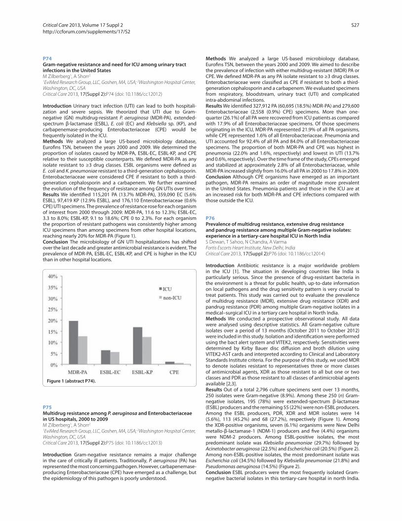
P74Gram-negative resistance and need for ICU among urinary tract infections in the United StatesM Zilberberg1, A Shorr2
1EviMed Research Group, LLC, Goshen, MA, USA; 2Washington Hospital Center, Washington, DC, USACritical Care 2013, 17(Suppl 2):P74 (doi: 10.1186/cc12012)
Introduction Urinary tract infection (UTI) can lead to both hospitali-za tion and severe sepsis. We theorized that UTI due to Gram-negative (GN) multidrug-resistant P. aeruginosa (MDR-PA), extended-spectrum β-lactamase (ESBL), E. coli (EC) and Klebsiella sp. (KP), and carbapenemase-producing Enterobacteriaceae (CPE) would be frequently isolated in the ICU.Methods We analyzed a large US-based microbiology database, Eurofi ns TSN, between the years 2000 and 2009. We determined the proportion of isolates caused by MDR-PA, ESBL-EC, ESBL-KP, and CPE relative to their susceptible counterparts. We defi ned MDR-PA as any isolate resistant to ≥3 drug classes. ESBL organisms were defi ned as E. coli and K. pneumoniae resistant to a third-generation cephalosporin. Enterobacteriaceae were considered CPE if resistant to both a third-generation cephalosporin and a carbapenem. We further examined the evolution of the frequency of resistance among GN UTIs over time.Results We identifi ed 115,201 PA (13.7% MDR-PA), 359,090 EC (5.6% ESBL), 97,419 KP (12.9% ESBL), and 176,110 Enterobacteriaceae (0.6% CPE) UTI specimens. The prevalence of resistance rose for each organism of interest from 2000 through 2009: MDR-PA, 11.6 to 12.3%; ESBL-EC, 3.3 to 8.0%; ESBL-KP, 9.1 to 18.6%; CPE 0 to 2.3%. For each organism the proportion of resistant pathogens was consistently higher among ICU specimens than among specimens from other hospital locations, reaching nearly 20% for MDR-PA (Figure 1).Conclusion The microbiology of GN UTI hospitalizations has shifted over the last decade and greater antimicrobial resistance is evident. The prevalence of MDR-PA, ESBL-EC, ESBL-KP, and CPE is higher in the ICU than in other hospital locations.
P75Multidrug resistance among P. aeruginosa and Enterobacteriaceae in US hospitals, 2000 to 2009M Zilberberg1, A Shorr2
1EviMed Research Group, LLC, Goshen, MA, USA; 2Washington Hospital Center, Washington, DC, USACritical Care 2013, 17(Suppl 2):P75 (doi: 10.1186/cc12013)
Introduction Gram-negative resistance remains a major challenge in the care of critically ill patients. Traditionally, P. aeruginosa (PA) has represented the most concerning pathogen. However, carbapenemase-producing Enterobacteriaceae (CPE) have emerged as a challenge, but the epidemiology of this pathogen is poorly understood.
Methods We analyzed a large US-based microbiology database, Eurofi ns TSN, between the years 2000 and 2009. We aimed to describe the prevalence of infection with either multidrug-resistant (MDR) PA or CPE. We defi ned MDR-PA as any PA isolate resistant to ≥3 drug classes. Enterobacteriaceae were classifi ed as CPE if resistant to both a third-generation cephalosporin and a carbapenem. We evaluated specimens from respiratory, bloodstream, urinary tract (UTI) and complicated intra-abdominal infections.Results We identifi ed 327,912 PA (60,695 (18.5%) MDR-PA) and 279,600 Enterobacteriaceae (2,558 (0.9%) CPE) specimens. More than one-quarter (26.1%) of all PA were recovered from ICU patients as compared with 17.9% of all Enterobacteriaceae specimens. Of those specimens originating in the ICU, MDR-PA represented 21.9% of all PA organisms, while CPE represented 1.6% of all Enterobacteriaceae. Pneumonia and UTI accounted for 92.4% of all PA and 84.0% of all Enterobacteriaceae specimens. The proportion of both MDR-PA and CPE was highest in pneumonia (22.0% and 1.6%, respectively) and lowest in UTI (13.7% and 0.6%, respectively). Over the time frame of the study, CPEs emerged and stabilized at approximately 2.8% of all Enterobacteriaceae, while MDR-PA increased slightly from 16.0% of all PA in 2000 to 17.8% in 2009.Conclusion Although CPE organisms have emerged as an important pathogen, MDR-PA remains an order of magnitude more prevalent in the United States. Pneumonia patients and those in the ICU are at an increased risk for both MDR-PA and CPE infections compared with those outside the ICU.
P76Prevalence of multidrug resistance, extensive drug resistance and pandrug resistance among multiple Gram-negative isolates: experience in a tertiary-care hospital ICU in North IndiaS Dewan, T Sahoo, N Chandra, A VarmaFortis Escorts Heart Institute, New Delhi, IndiaCritical Care 2013, 17(Suppl 2):P76 (doi: 10.1186/cc12014)
Introduction Antibiotic resistance is a major worldwide problem in the ICU [1]. The situation in developing countries like India is particularly serious. Since the presence of drug-resistant bacteria in the environment is a threat for public health, up-to-date information on local pathogens and the drug sensitivity pattern is very crucial to treat patients. This study was carried out to evaluate the prevalence of multidrug resistance (MDR), extensive drug resistance (XDR) and pandrug resistance (PDR) among multiple Gram-negative isolates in a medical–surgical ICU in a tertiary care hospital in North India.Methods We conducted a prospective observational study. All data were analysed using descriptive statistics. All Gram-negative culture isolates over a period of 13 months (October 2011 to October 2012) were included in this study. Isolation and identifi cation were performed using the bact alert system and VITEK2, respectively. Sensitivities were determined by Kirby Bauer disc diff usion and broth dilution using VITEK2-AST cards and interpreted according to Clinical and Laboratory Standards Institute criteria. For the purpose of this study, we used MDR to denote isolates resistant to representatives three or more classes of antimicrobial agents, XDR as those resistant to all but one or two classes and PDR as those resistant to all classes of antimicrobial agents available [2,3].Results Out of a total 2,796 culture specimens sent over 13 months, 250 isolates were Gram-negative (8.9%). Among these 250 (n) Gram-negative isolates, 195 (78%) were extended-spectrum β-lactamase (ESBL) producers and the remaining 55 (22%) were non-ESBL producers. Among the ESBL producers, PDR, XDR and MDR isolates were 14 (5.6%), 113 (45.2%) and 68 (27.2%), respectively (Figure 1). Among the XDR-positive organisms, seven (6.1%) organisms were New Delhi metallo-β-lactamase-1 (NDM-1) producers and fi ve (4.4%) organisms were NDM-2 producers. Among ESBL-positive isolates, the most predominant isolate was Klebsiella pneumoniae (29.7%) followed by Acinetobacter aeruginosa (22.5%) and Escherichia coli (20.5%) (Figure 2). Among non-ESBL-positive isolates, the most predominant isolate was Escherichia coli (34.5%) followed by Klebsiella pneumoniae (21.8%) and Pseudomonas aeruginosa (14.5%) (Figure 2).Conclusion ESBL producers were the most frequently isolated Gram-negative bacterial isolates in this tertiary-care hospital in north India.
Figure 1 (abstract P74).
Critical Care 2013, Volume 17 Suppl 2 http://ccforum.com/supplements/17/S2
S27

Among ESBL producers, XDR organisms were most frequent, followed by MDR and PDR organisms. Few of the XDR isolates were NDM producers, which have propensity to spread to other bacteria. In view of signifi cant prevalence of multidrug resistance amongst Gram-negative organisms in the ICU, regular surveillance of antibiotic susceptibility patterns plays a crucial role for setting orders to guide the clinician in choosing empirical or directed therapy of infected patients.References1. Radji M, et al.:Asian Pac J Trop Biomed 2011, 1:39-42.2. Falagas ME, et al.: Clin Infect Dis 2008, 46:1121-1122.3. Souli M, et al.: Euro Surveill 2008, 13:30-40.
P77Risk factors associated with Acinetobacter baumannii septicemia and its mortality rates in critically ill patientsK Kontopoulou, K Tsepanis, I Sgouropoulos, A Triantafyllidou, D Socratous, P Tassioudis, E Chasou, E Antypa, F Renta, E Antoniadou, K MandraveliGeneral Hospital G. Gennimatas, Thessaloniki, GreeceCritical Care 2013, 17(Suppl 2):P77 (doi: 10.1186/cc12015)
Introduction Acinetobacter baumannii (A. baum) is a leading cause of septicemia of patients hospitalized in the ICU with high mortality rates. The aim of our study is to investigate the risk factors associated with A. baum bacteremia and its mortality rates.Methods A total of 937 patients (457 women and 480 men, median age 59) admitted to the ICU during the period 1 January 2009 to 30 September 2012 were enrolled in our retrospective study. Blood cultures were obtained from all patients. The identifi cation and the antimicrobial susceptibility testing were performed by the automated system VITEK2 (Bio Merieux, France). Data collected included underlying diseases, malignancies and immune suppression (MIS), prognostic factors (APACHE score, adjusted mortality), age, sex, length of ICU stay (LS), recent administration of broad-spectrum β-lactam antibiotics (especially carbapenems; ABL), mechanical ventila-tion (MV), implementation of invasive procedures (central venous catheter and urine catheter; INV.PR) and outcome. At fi rst a univariate statistical model was used with signifi cance level set at P = 0.05. For the multivariate statistical analysis model we used all variables with
P <0.05 from the previous model and those mentioned at recent medical literature as signifi cantly related with A. baum septicemia and its mortality.Results A total of 101 patients (10.78%) developed bloodstream infection caused by A. baum and the mortality rate due to A. baum septicemia was estimated as 49.5% (50/101). Multiple regression analysis revealed adjusted mortality >55% (Exp(B) = 2.01, P = 0.013), MIS (Exp(B) = 1.97, P = 0.017), ABL (Exp(B) = 2.34, P = 0.009), LS >14 days (Exp(B) = 1.34, P = 0.034), MV (Exp(B) = 2.67, P = 0.005) and INV.PR (Exp(B) = 3.27, P = 0.001) as independent risk factors associated with A. baum septicemia.Conclusion The fact that the probability of A. baum bacteremia increases in immunocompromised patients refl ects the opportunistic characteristic of these infections. MV accelerates respiratory A. baum colonization, which is a risk factor for AB bacteremia. Recent INV.PR increased the incidence of A. baum bacteremia and this result is probably related to the severe status in patients with central venous catheter. The administration of carbapenems inhibits the growth of other more susceptible bacteria, allowing the growth of multidrug-resistant A. baum.References1. Wu HS, et al.: Clin Microbiol Infect 2012, 18:E373-E3762. Huang ST, et al.: J Microbiol Immunol Infect 2012, 45:356-362.
P78Prevalence of colonization by multiresistant bacteria on admission to the ICU in the French military hospital in Kabul, AfghanistanJV Schaal, P Pasquier, H Delacour, A Salvadori, A Jarrassier, JR Renner, SM MératMilitary Teaching Hospital Bégin, Saint-Mandé, FranceCritical Care 2013, 17(Suppl 2):P78 (doi: 10.1186/cc12016)
Introduction The French military hospital at the Kaboul International Airport (KaIA) base provides surgical care for International Force and Afghan National Army soldiers, and also local patients. The development of multiresistant bacteria (MRB) nosocomial infections has raised a major problem complicating the care of combat casualties [1]. The aim of this study is to assess the prevalence of MRB carriage on admission to the ICU in this combat support hospital.Methods We used a prospective observation study on patients admitted to the French military ICU in KaIA over 3 months (July to September 2012). All hospitalized patients were assessed for the presence of colonization with MRB: nasal and rectal swabs were performed to identify, respectively, methicillin-resistant Staphylococcus aureus (MRSA) and extended-spectrum β-lactamases bacteria (ESBLB). The following data were recorded for each patient on admission: demographic characteristics, bacteriological results, length of stay, type of previous hospitalization.Results Sixty-three patients were admitted. The mean length of stay (MLS) was 3 ± 3 days, and the mean age was 25 ± 14 (13 patients <15 years). Patients were hospitalized for combat-related trauma (74%), noncombat-related trauma, medical pathologies (10%), and postoperative care (8%). They were Afghans (92%) or westerners (8%). Swabs were not realized for eight patients. Forty-three percent revealed an ESBLB colonization: Escherichia coli (22 patients), Klebsiella pneumoniae (one patient), Acinetobacter baumanii (one patient). No patients were colonized with MRSA. Ten patients (16%) were directly admitted to the ICU, 12 (19%) had been hospitalized before admission, 39 (62%) were transferred after resuscitative and stabilization care in a level 2 unit. For the two last categories, the MLS (for previous hospitalization) was respectively 14 ± 28 days and 8 ± 6 hours. Among patients transferred after care in a level 2 unit, MLS was no diff erent between colonized and noncolonized patients: 8 ± 7 versus 9 ± 6 hours (P = 0.5, Mann–Whitney test).Conclusion In this study, prevalence of colonization with ESBLB at admission is very high, suggesting a high prevalence of MDR colonization in the local population in Afghanistan. It remains important to intensify the prevention policy against MRB cross-transmission in the deployed ICU.Reference1. Whitman TJ: Infection control challenges related to war wound infections
in the ICU setting. J Trauma 2007, 62:S53.
Figure 1 (abstract P76). Prevalence of drug-resistant Gram-negative isolates.
Figure 2 (abstract P76). Prevalence of Gram-negative isolates.
Critical Care 2013, Volume 17 Suppl 2 http://ccforum.com/supplements/17/S2
S28

P79Extended-spectrum β-lactamase Providencia stuartii in a general ICUP Myrianthefs1, E Evodia2, G Fildissis1, G Baltopoulos1
1Athens University, Athens, Greece; 2Iatriko Kentro Athinon, Athens, GreeceCritical Care 2013, 17(Suppl 2):P79 (doi: 10.1186/cc12017)
Introduction Providencia stuartii, a member of the family of Entero-bacteriacea, is a Gram-negative pathogen causing colonization and opportunistic infections in ICU patients.Methods We retrospectively recorded the characteristics of P. stuartii infections in our ICU in a total period of 1 year (six-bed general ICU).Results A total of 116 patients (80 males, 68.9%) were hospitalized in our ICU of mean age 58.5 ± 1.8, mean ICU stay: 23.2 ± 3.3 days, APACHE II: 19.3 ± 0.7, SAPS II: 45.7 ± 1.6, SOFA: 7.9 ± 0.4 and mortality: 18.9%. Admission diagnosis was multiple trauma (29.3%), emergency surgery (37.1%), and medical (33.6%). Of them 21 (18.1%) developed P. stuartii infection that was related to multiple trauma (P = 0.0289), length of ICU stay 69.8 ± 12.6 (median 51.0) versus 12.6 ± 1.7 (median 6.0) (P <0.0001) and illness severity APACHE II (21.7 ± 1.3 vs. 17.5 ± 0.8; P = 0.0056), SAPS II (54.9 ± 2.9 vs. 43.6 ± 1.8; P = 0.0296) and SOFA (10.6 ± 0.5 vs. 7.2 ± 0.4; P <0.0001). There was no statistically signifi cant diff erence regarding sex, age or mortality (P = 0.3789). Mean day of fi rst isolation was 31.1 ± 2.1 (median 28.0). The number of isolations per site were – blood: 11 (median day: 25.0), tracheal aspirates: 9 (median: 28.5), catheter tip: 15 (median: 31.0), urine: 6 (median: 32.0), wound: 12 (median: 25.0), biological fl uids: 4 (median: 29.5), other catheters: 1 (median: 28.0), middle ear: 2 (median: 32.5), and nose: 1 (median: 25.0). Six patients had only one site isolation and the remaining 15 had multiple sites of P. stuartii isolation. We totally recorded 63 isolates of which 82.5% were second-generation and third-generation cephalosporin-resistant, 80.3% aztreonam-resistant and 81% carbapenem-resistant strains expressing an extended-spectrum β-lactamase phenotype. All patients had previously received colistin or meropenem or tygecycline for a median period of 18.5, 15 and 10.5 days, respectively.Conclusion P. stuartii infection/colonization may develop in critically ill patients, especially those with multiple trauma and prolonged ICU stay, and maybe isolated in blood after the 25th day of ICU stay. Also, infections due to extended-spectrum β-lactamase-producing multidrug-resistant P. stuartii are an emerging problem.
P80High-dose tigecycline use in severe infectionsG De Pascale1, L Montini1, T Spanu2, V Bernini1, A Occhionero1, DL Grieco1, M Biancone1, P De Santis1, ES Tanzarella1, SL Cutuli1, MA Pennisi1, MA Antonelli11Sacro Cuore Catholic University, Rome, Italy; 2Institute of Microbiology, Sacro Cuore Catholic University, Rome, ItalyCritical Care 2013, 17(Suppl 2):P80 (doi: 10.1186/cc12018)
Introduction The aim of this study is to describe the clinical and epidemiological profi le of ICU patients receiving tigecycline (TGC) and to evaluate the potential benefi ts of TGC higher doses.Methods All patients admitted to our ICU between 1 June 2009 and 31 May 2012 who received TGC were evaluated. Cases were excluded when infections were not microbiologically confi rmed.Results Over the study period, 100 patients fulfi lled the inclusion criteria: 54 in the SD group (50 mg every 12 hours) and 46 in the HD group (100 mg every 12 hours). The SD group and the HD group were not signifi cantly diff erent in terms of age, severity of disease, duration of TGC therapy, rate of concomitant other active antibiotic use and of inadequate empirical antimicrobial therapy (IIAT) (P = NS). MDR A. baumannii and K. pneumoniae were the main pathogens isolated. The percentage of germs other than A. baumannii and K. pneumoniae was higher in the SD TGC group (P <0.01). Otherwise infections due to less susceptible germs (TGC MIC value ≥1 μg/ml) were mainly treated with TGC higher doses (P <0.01). No signifi cant diff erences were found in terms of ICU mortality (P = 0.8). The rate of abnormal laboratory measures during TGC treatment was similar between the two groups (P = NS). No patients required TGC discontinuation or dose reduction because of suspected adverse events. In the VAP subpopulation
(63 patients: 30 received SD and 33 HD), the clinical cure rate and microbiological eradication percentage were higher when TGC was used at higher doses (57.6% vs. 33.3%; P = 0.08 and 57.1% vs. 30.4%; P = 0.1). Table 1 shows multivariate analysis of clinical cure predictors in the VAP subgroup.
Table 1 (abstract P80). Predictors of clinical cure in patients with VAP (multivariate analysis)
OR (95% CI) P value
HD TGC 5.93 (1.38 to 25.6) 0.02
IIAT 0.23 (0.06 to 0.85) 0.03
SOFA score 0.62 (0.45 to 0.86) <0.01
Conclusion In critically ill patients, HD TGC use seems to be safe and, combined with other active antibiotics, may increase the rate of MDR germ VAP clinical success. IIAT and the severity degree of patients’ clinical condition still remain major determinants of VAP treatment failure.Reference1. Tasina E, et al.: Effi cacy and safety of tigecycline for the treatment of
infectious diseases: a meta-analysis. Lancet Infect Dis 2011, 11:834-844.
P81In vitro effi ciency of Amikacin Inhale, a novel drug-device delivery systemN Kadrichu1, S Boc1, K Corkery1, P Challoner2
1Novartis Pharmaceuticals, San Carlos, CA, USA; 2Nektar Therapeutics, San Francisco, CA, USACritical Care 2013, 17(Suppl 2):P81 (doi: 10.1186/cc12019)
Introduction Amikacin Inhale (NKTR-061, BAY41-6551) is a drug-device combination in clinical development for adjunctive treatment of intubated and mechanically ventilated patients with Gram-negative pneumonia. The product uses a proprietary vibrating mesh nebulizer system (PDDS Clinical) with amikacin sulfate formulated for inhalation (3.2 ml of 125 mg/ml amikacin solution) for a 10-day twice-daily course of therapy. It is designed for use with two delivery systems: one system for intubated patients (On-vent; Figure 1), and a second Handheld (HH) system for patients who are extubated before completing the course of therapy (Figure 2). We investigated in vitro the amikacin lung dose delivered by PDDS Clinical.Methods An estimated lung dose (ELD) for On-vent setting was measured in vitro after collecting aerosolized amikacin from a fi lter at the end of an endotracheal tube during ventilation. The ELD for the HH device was calculated from the fi ne particle fraction (FPF <5 μm) post-mouthpiece, multiplied by the in vitro delivered dose post-mouthpiece. FPF <5 μm refl ects lung deposition observed during phase 2 clinical trials [1]. Eighty-one nebulizers with volume median diameter (VMD)
Figure 1 (abstract P81). On-vent system with brown blinder shell for clinical trials.
Critical Care 2013, Volume 17 Suppl 2 http://ccforum.com/supplements/17/S2
S29

of 4.4 ± 0.5 μm and output rates of 0.23 ± 0.10 ml/minute were tested for each system. Delivered dose data were fi t to the independent variables (that is, VMD and output rates) using a least-squares fi t with 95% confi dence limits.Results Total percentage recoveries for On-vent and HH test runs were between 85% and 115% of the nominal dose. The mean ELDs were 50 ± 9% (On-vent) and 49 ± 11% (HH) of the nominal dose. Nebulizers with longer dosing times and lower VMDs had higher ELD values for both delivery systems.Conclusion The results support the use of the PDDS Clinical with either system to administer aerosolized amikacin with high effi ciency and no dose adjustment is required when switching from the On-vent to the HH system for extubated patients.Reference1. Fink J, et al.: High in vivo amikacin lung deposition after NKTR-061 dosing
correlates with in vitro aerosol characterization. Am J Respir Crit Care Med 2008, 177:A530.
P82Analysis of parameters in critically ill patients infl uencing amikacin peak levelsIM Hollevoet1, SJ Toye2, J De Waele1
1UZ Gent, Belgium; 2Stijn Toye, Gent, BelgiumCritical Care 2013, 17(Suppl 2):P82 (doi: 10.1186/cc12020)
Introduction Recent studies demonstrate that a loading dose of 25 mg/kg (total body weight) of amikacin in septic patients is required to reach a suffi cient peak concentration. This study examines
parameters infl uencing the relation between amikacin dose and peak concentration.Methods In this retrospective study we looked at 47 patients (128 peak levels) between 2003 and 2012. Multivariate linear regression analysis was done for several parameters: administered dose calculated with total body weight, ideal body weight, adjusted body weight, type of intensive care patient, BMI, daily fl uid balance, SOFA score and APACHE score, and patient characteristics were analyzed.Results A linear correlation between dose and amikacin peak level was confi rmed (Figure 1). A total 54.69% of all amikacin administrations did not result in a therapeutic peak level. The multivariate linear regression analysis showed the best linear correlation with adjusted body weight and SOFA score. The comparison of variables between four patient groups, based on the deviation between measured peak level and predicted peak level (according the linear correlation), showed new variables that may infl uence peak level.Conclusion This confi rms that low doses (<18 mg/kg) of amikacin in intensive care patients seldom result in a therapeutic peak level. The proposed loading dose of 25 mg/kg is good for reaching a therapeutic level, although 29.6% remains subtherapeutic. Due to the linear correlation, more therapeutic levels may be reached with higher doses (25 to 30 mg/kg). New variables need further investigation to explain the high variability in achieved peak level.References1. Delattre IK, et al.: Ther Drug Monit 2010, 32:749-756.2. Taccone FS, et al.: Crit Care 2010, 14:R53.3. Gálvez R, et al.: Int J Antimicrobial Agents 2011, 38:146-151.4. Avent ML, et al.: Internal Med J 2011, 41:441-449.5. Fernández de Gatta MM, et al.: J Clin Pharm Ther 1996, 21:417-421.
P83Cost implication of antibiotic-associated diarrhoea and the fi nancial impact of probiotic use in its preventionC Ebm, M Cecconi, C Moran, A Rhodes, T RahmanSt Georges Healthcare Trust, London, UKCritical Care 2013, 17(Suppl 2):P83 (doi: 10.1186/cc12021)
Introduction Antibiotic-associated diarrhoea (AAD) occurs in as many as 30% of patients receiving antibiotics, often leading to increased morbidity, prolonged in-hospital stay and additional healthcare resource utilisation. Age, antibiotics and prolonged postoperative ward and ICU stay have been suggested to be independent risk factors. In such patient populations, probiotics may be used to prevent antibiotic-associated diarrhoea, yet they are not routinely recommended as a component of perioperative care. The aim of this study was to model the long-term costs associated with AAD and to assess the eff ectiveness of probiotics as a preventive strategy.Methods We developed a simulation model to determine clinical costs and outcomes attributable to AAD. To assess the cost-eff ectiveness of probiotics, as part of a perioperative regime, we constructed a decision
Figure 2 (abstract P81). Handheld system with brown blinder shell for clinical trials.
Figure 1 (abstract P82). Correlation between dose of amikacin and peak concentration.
Critical Care 2013, Volume 17 Suppl 2 http://ccforum.com/supplements/17/S2
S30
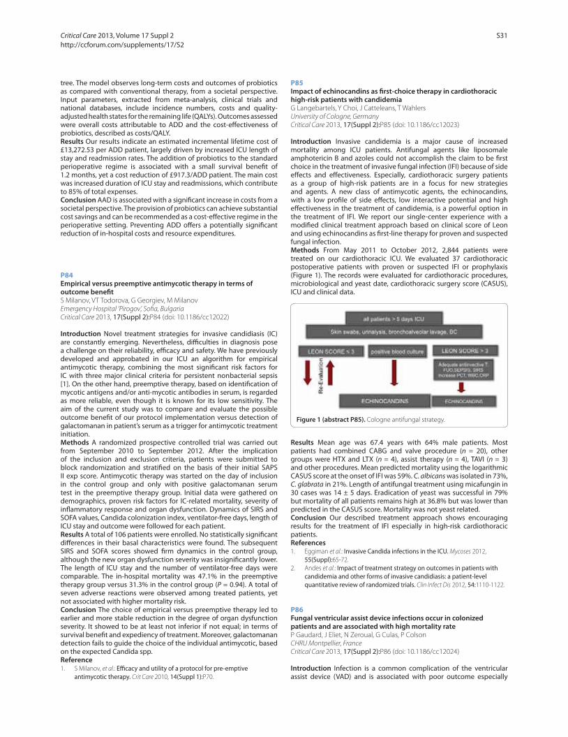
tree. The model observes long-term costs and outcomes of probiotics as compared with conventional therapy, from a societal perspective. Input parameters, extracted from meta-analysis, clinical trials and national databases, include incidence numbers, costs and quality-adjusted health states for the remaining life (QALYs). Outcomes assessed were overall costs attributable to ADD and the cost-eff ectiveness of probiotics, described as costs/QALY.Results Our results indicate an estimated incremental lifetime cost of £13,272.53 per ADD patient, largely driven by increased ICU length of stay and readmission rates. The addition of probiotics to the standard perioperative regime is associated with a small survival benefi t of 1.2 months, yet a cost reduction of £917.3/ADD patient. The main cost was increased duration of ICU stay and readmissions, which contribute to 85% of total expenses.Conclusion AAD is associated with a signifi cant increase in costs from a societal perspective. The provision of probiotics can achieve substantial cost savings and can be recommended as a cost-eff ective regime in the perioperative setting. Preventing ADD off ers a potentially signifi cant reduction of in-hospital costs and resource expenditures.
P84Empirical versus preemptive antimycotic therapy in terms of outcome benefi tS Milanov, VT Todorova, G Georgiev, M MilanovEmergency Hospital ‘Pirogov’, Sofi a, BulgariaCritical Care 2013, 17(Suppl 2):P84 (doi: 10.1186/cc12022)
Introduction Novel treatment strategies for invasive candidiasis (IC) are constantly emerging. Nevertheless, diffi culties in diagnosis pose a challenge on their reliability, effi cacy and safety. We have previously developed and approbated in our ICU an algorithm for empirical antimycotic therapy, combining the most signifi cant risk factors for IC with three major clinical criteria for persistent nonbacterial sepsis [1]. On the other hand, preemptive therapy, based on identifi cation of mycotic antigens and/or anti-mycotic antibodies in serum, is regarded as more reliable, even though it is known for its low sensitivity. The aim of the current study was to compare and evaluate the possible outcome benefi t of our protocol implementation versus detection of galactomanan in patient’s serum as a trigger for antimycotic treatment initiation.Methods A randomized prospective controlled trial was carried out from September 2010 to September 2012. After the implication of the inclusion and exclusion criteria, patients were submitted to block randomization and stratifi ed on the basis of their initial SAPS II exp score. Antimycotic therapy was started on the day of inclusion in the control group and only with positive galactomanan serum test in the preemptive therapy group. Initial data were gathered on demographics, proven risk factors for IC-related mortality, severity of infl ammatory response and organ dysfunction. Dynamics of SIRS and SOFA values, Candida colonization index, ventilator-free days, length of ICU stay and outcome were followed for each patient.Results A total of 106 patients were enrolled. No statistically signifi cant diff erences in their basal characteristics were found. The subsequent SIRS and SOFA scores showed fi rm dynamics in the control group, although the new organ dysfunction severity was insignifi cantly lower. The length of ICU stay and the number of ventilator-free days were comparable. The in-hospital mortality was 47.1% in the preemptive therapy group versus 31.3% in the control group (P = 0.94). A total of seven adverse reactions were observed among treated patients, yet not associated with higher mortality risk.Conclusion The choice of empirical versus preemptive therapy led to earlier and more stable reduction in the degree of organ dysfunction severity. It showed to be at least not inferior if not equal; in terms of survival benefi t and expediency of treatment. Moreover, galactomanan detection fails to guide the choice of the individual antimycotic, based on the expected Candida spp.Reference1. S Milanov, et al.: Effi cacy and utility of a protocol for pre-emptive
antimycotic therapy. Crit Care 2010, 14(Suppl 1):P70.
P85Impact of echinocandins as fi rst-choice therapy in cardiothoracic high-risk patients with candidemiaG Langebartels, Y Choi, J Catteleans, T WahlersUniversity of Cologne, GermanyCritical Care 2013, 17(Suppl 2):P85 (doi: 10.1186/cc12023)
Introduction Invasive candidemia is a major cause of increased mortality among ICU patients. Antifungal agents like liposomale amphotericin B and azoles could not accomplish the claim to be fi rst choice in the treatment of invasive fungal infection (IFI) because of side eff ects and eff ectiveness. Especially, cardiothoracic surgery patients as a group of high-risk patients are in a focus for new strategies and agents. A new class of antimycotic agents, the echinocandins, with a low profi le of side eff ects, low interactive potential and high eff ectiveness in the treatment of candidemia, is a powerful option in the treatment of IFI. We report our single-center experience with a modifi ed clinical treatment approach based on clinical score of Leon and using echinocandins as fi rst-line therapy for proven and suspected fungal infection.Methods From May 2011 to October 2012, 2,844 patients were treated on our cardiothoracic ICU. We evaluated 37 cardiothoracic postoperative patients with proven or suspected IFI or prophylaxis (Figure 1). The records were evaluated for cardiothoracic procedures, microbiological and yeast date, cardiothoracic surgery score (CASUS), ICU and clinical data.
Results Mean age was 67.4 years with 64% male patients. Most patients had combined CABG and valve procedure (n = 20), other groups were HTX and LTX (n = 4), assist therapy (n = 4), TAVI (n = 3) and other procedures. Mean predicted mortality using the logarithmic CASUS score at the onset of IFI was 59%. C. albicans was isolated in 73%, C. glabrata in 21%. Length of antifungal treatment using micafungin in 30 cases was 14 ± 5 days. Eradication of yeast was successful in 79% but mortality of all patients remains high at 36.8% but was lower than predicted in the CASUS score. Mortality was not yeast related.Conclusion Our described treatment approach shows encouraging results for the treatment of IFI especially in high-risk cardiothoracic patients.References1. Eggiman et al.: Invasive Candida infections in the ICU. Mycoses 2012,
55(Suppl):65-72.2. Andes et al.: Impact of treatment strategy on outcomes in patients with
candidemia and other forms of invasive candidiasis: a patient-level quantitative review of randomized trials. Clin Infect Dis 2012, 54:1110-1122.
P86Fungal ventricular assist device infections occur in colonized patients and are associated with high mortality rateP Gaudard, J Eliet, N Zeroual, G Culas, P ColsonCHRU Montpellier, FranceCritical Care 2013, 17(Suppl 2):P86 (doi: 10.1186/cc12024)
Introduction Infection is a common complication of the ventricular assist device (VAD) and is associated with poor outcome especially
Figure 1 (abstract P85). Cologne antifungal strategy.
Critical Care 2013, Volume 17 Suppl 2 http://ccforum.com/supplements/17/S2
S31

with fungi [1]. The relationship between colonization and invasive fungal infection (IFI) in severely ill ICU patients with a VAD support is not described. This study analyzes the incidence and outcome of fungal infection and colonization in VAD patients in bridge to transplantation or in destination therapy.Methods We conducted a retrospective review of all VAD implantations in our surgical ICU between 2007 and 2012. The incidence of fungal colonization, antifungal prophylaxis, bacterial sepsis and the mortality of IFI versus no IFI patients were compared.Results In the study period, 34 patients with severe heart failure or cardiogenic shock were selected for a VAD implantation (nine in destination therapy). The overall mortality rate was 50% during mechanical assistance. Confi rmed (n = 8) and highly suspected (n = 2) IFI occurred during the ICU stay in 29% of patients who were treated with echinocandins, voriconazole and/or liposomal amphotericin B. The isolated fungi were: six Candida albicans, two parapsilosis, one glabrata and one invasive pulmonary aspergillosis. Antifungal prophylaxis with fl uconazole was administered to 18% of patients at mean for 5 days mainly in the more recent implantations. In the no IFI population, 54% (n = 13) had a systemic or VAD bacterial sepsis with a mortality rate about 54%. The mortality without any sepsis was reduced to 18%. Fungal colonization was signifi cantly more present (90% vs. 50%) before IFI in VAD patients. The mortality rate was dramatically higher with IFI (80% vs. 38%) in accordance with the literature [1]. See Table 1.
Table 1 (abstract P86). Fungal colonization (FC), infection and outcome during VAD support
n FC Mortality
No IFI 24 12 9
IFI 10 9 8
P value 0.029 0.024
Conclusion In our center, we observed a high incidence of IFI in ICU patients with VAD that was associated with a mortality rate of 80%. Screening of fungal colonization appears to be very important during the ICU stay for VAD patients. Trials are needed for investigating the use, the drug choice and the timing of antifungal prophylaxis for such high-risk patients.Reference1. Aslam S, et al.: Clin Infect Dis 2010, 50:664-671.
P87Cost-eff ectiveness analysis of anidulafungin in the treatment of candidaemiaG Auzinger1, G Playford2, C Graham3, H Narula3, C Charbonneau4, D Weinstein4, M Kantecki4, H Schlamm5, M Ruhnke6
1King’s College Hospital, London, UK; 2Princess Alexandra Hospital, Brisbane, Australia; 3RTI Health Solutions, Durham, NC, USA; 4Pfi zer International Operations, Paris, France; 5Pfi zer Inc., New York, NY, USA; 6Charité University Medicine, Berlin, GermanyCritical Care 2013, 17(Suppl 2):P87 (doi: 10.1186/cc12025)
Introduction Echinocandins are recommended fi rst-line treatment for candidaemia [1]. A cost-eff ectiveness model developed from a UK perspective examined costs and outcomes of antifungal treatment for candidaemia and other forms of invasive candidiasis based on European clinical guidelines [1].Methods Costs and treatment outcomes with the echinocandin anidula fungin were compared with caspofungin, micafungin, fl uconazole, voriconazole and amphotericin B. The model included non-neutropenic patients aged ≥16 years with confi rmed candidaemia/another form of invasive candidiasis receiving intravenous fi rst-line treatment [2]. Patients were categorised as a clinical success or failure (patients with persistent/breakthrough infection); frequency data for each outcome were taken from a mixed-treatment comparison [3]. Successfully treated patients switched to oral therapy. Clinical failures switched to a diff erent antifungal class. It was assumed that second-line treatment duration was equivalent to that of fi rst-line treatment and only two lines of therapy were required to treat infection. Other inputs
were all-cause 6-week mortality, cost of treatment-related adverse events (AEs) and other medical resource use costs. Life-years were calculated using a published model [4]. Antifungal agent-related AEs were taken from the product label/literature. Resource use was derived from the literature and discussion with clinical experts. Drug acquisition/administration costs were taken from standard UK costing sources.Results First-line anidulafungin for treatment of candidaemia was cost-eff ective per life-year gained versus fl uconazole (incremental cost-eff ectiveness ratio £813). Anidulafungin was cost saving versus caspofungin and micafungin in terms of life-years gained due to lower ICU costs and a higher rate of survival combined with a higher probability of clinical success.Conclusion Anidulafungin was cost-eff ective compared with fl uconazole for treatment of candidaemia and was cost saving versus other echinocandins in the UK. European guidelines recommend echinocandins as fi rst-line treatments for candidaemia [1]; this model indicates that anidulafungin marries clinical eff ectiveness and cost-eff ectiveness.References1. Cornely OA: Clin Microbiol Infect 2012, 18:19-37.2. Reboli AC, et al.: N Engl J Med 2007, 356:2472-2482.3. Mills EJ, et al.: Ann Clin Microbiol Antimicrob 2009, 8:23.4. Sidhu MK, et al.: Curr Med Res Opin 2009, 25:2049-2059
P88Prevalence and impact of invasive fungal infections in intensive careJM Patel, K Couper, T Melody, R O’Brien, D ParekhHeart of England NHS Foundation Trust, Birmingham, UKCritical Care 2013, 17(Suppl 2):P88 (doi: 10.1186/cc12026)
Introduction Invasive fungal infections (IFI) aff ect 1% of ICU patients and are increasing in incidence. IFIs are associated with a poor prognosis, which is further complicated by diffi culties in identifi cation of fungal organisms by traditional culture methods and the emergence of Candida species resistant to triazole therapy [1,2]. This study aimed to assess the prevalence of IFIs, the organisms responsible and outcomes of patients aff ected.Methods Patients admitted to the Heart of England NHS Foundation Trust ICUs who acquired an IFI were identifi ed through the hospital Fungal Infection Risk Evaluation (FIRE) study database. All ICU patients admitted between October 2009 and March 2011 were used as a comparative cohort. Data collected included: baseline demographics, length of stay, ICU and hospital mortality, and nature of IFI. Data were analysed using Student’s t test for continuous data and Fischer’s exact test for categorical data.Results A total of 2,426 patients were admitted to Heart of England NHS Foundation Trusts ICUs during the study period. Of these, 31 patients were identifi ed as having an IFI (1.3%). Baseline demographic data were similar between groups. Patients with IFI had signifi cantly longer ICU length of stay (19 days vs. 5 days, P <0.0001) and required more days of advanced organ support (12 days vs. 3 days, P <0.0001). A trend towards higher hospital mortality (41% vs. 27%, P = 0.08) was observed. Candida albicans was the predominant organism cultured (64%), followed by Candida glabrata (23%), with other Candida species accounting for the remaining 13% of IFIs. Sites of IFIs were blood (45%), intra-abdominal (39%), and pleural (16%). Most patients (52%) who acquired an IFI had had intra-abdominal surgery prior to ICU admission. The majority of patients (71%) were treated with echinocandins, whilst of the nine patients who were initially treated with fl ucanazole, six (67%) required therapy escalation to an echinocandin.Conclusion The results of our study are consistent with other published data, in that whilst IFI prevalence is low, they are associated with increased morbidity in critically ill patients. This study has led to a change in hospital policy regarding antifungal use in the ICU, with echinocandins being fi rst-line in the pre-emptive treatment of IFI. We keenly await the results of the FIRE study, which will provide important insights to identifi cation of patients at risk of IFIs and optimal drug therapy.References1. Allou N, et al.: Curr Infect Dis Rep 2011, 13:426-432.2. Zaragosa R, et al.: Adv Sepsis 2008, 6:90-98.
Critical Care 2013, Volume 17 Suppl 2 http://ccforum.com/supplements/17/S2
S32

P89Beliefs and actual practice of oxygen therapy in the ICUHJ Helmerhorst1, MJ Schultz2, PH Van der Voort3, E De Jonge1, DJ Van Westerloo1
1Leiden University Medical Center, Leiden, the Netherlands; 2Academic Medical Center, Amsterdam, the Netherlands; 3Onze Lieve Vrouwe Gasthuis, Amsterdam, the NetherlandsCritical Care 2013, 17(Suppl 2):P89 (doi: 10.1186/cc12027)
Introduction The aim of this study was to compare self-reported beliefs with actual clinical practice of oxygen therapy in the ICU. Hyperoxia is frequently encountered in ventilated patients and prolonged exposure has repeatedly been shown to induce lung injury and (systemic) toxicity.Methods An online questionnaire for ICU clinicians was conducted to investigate beliefs and motives regarding oxygen therapy for critically ill patients. Furthermore, arterial blood gas (ABG) samples and corresponding ventilator settings were retrieved to retrospectively assess objective oxygenation between 1 April 2011 and 31 March 2012 in the ICUs of three teaching hospitals in the Netherlands.Results Analyzable questionnaire responses were received from 200 ICU physicians and nurses. The majority of respondents believed that oxygen-induced lung injury is a concern, although barotrauma and volutrauma are generally considered to impose a greater risk in mechanical ventilation. Frequently allowed minimal saturation ranges in the questionnaire were 85 to 95% and 7 to 10 kPa (Figure 1). Self-reported FiO2 adjustment in hypothetical patient cases with variable saturation levels was moderately impacted by the underlying clinical condition. To study actual clinical practice, a total of 107,888 ABG samples with corresponding ventilator settings, covering 5,565 patient admissions, were retrieved. Analysis showed a median (IQR) PaO2 of 11.7 kPa (9.9 to 14.3), median FiO2 was 0.4 (0.4 to 0.5), median PEEP was 5 (5 to 8). A total 63.5% of all PaO2 registries were higher than previously suggested oxygenation goals (7.3 to 10.7 kPa) [1]. In 56.8% of cases with PaO2 higher than the target range, neither FiO2 nor PEEP levels had been lowered when the next ABG sample was taken.
Conclusion Most clinicians acknowledge the detrimental eff ects of prolonged exposure to hyperoxia in the ICU and report a low tolerance for high saturation levels. However, the self-reported intention for conservative oxygen therapy is not consistently expressed in our objective data of actual clinical practice and a large proportion of patients was exposed to high and potentially toxic oxygen levels.Reference1. Brower RG, et al.: N Engl J Med 2004, 351:327-336.
P90FiO2/PEEP index: a simple tool for opitimizing ventilator settingsD Trasy, M Nemeth, K Kiss, Z Till, Z MolnarUniversity of Szeged, HungaryCritical Care 2013, 17(Suppl 2):P90 (doi: 10.1186/cc12028)
Introduction During mechanical ventilation, oxygenation can be infl uenced by adjusting FiO2 and positive end-expiratory pressure (PEEP). There have been recommendations for how the FiO2 and PEEP should be set [1]. However, in a recent audit we found that the compliance of doctors of these recommendations is very low [2]. Therefore we invented a simple parameter called the FiO2/PEEP index (FPi) of which the physiologic value is ≤7 (that is, FiO2 = 21%/PEEP = 3 cmH2O), which corresponds to the ARDSNet trial’s minimum FiO2/PEEP settings: 35%/5 cmH2O [2]. The aim of this case–control study was to investigate the impact of an FPi ≤7 targeted protocol on clinical practice.Methods A prospective observational study in 2010 and 2012. Every mechanically ventilated patient was recruited. Demographics, outcome data and Lung Injury Score (LIS) were recorded after admission. Respiratory settings, oxygenation and ventilation parameters were recorded after mechanical ventilation was commenced and the fi rst arterial blood gas sample was taken (T0). Measurements were repeated in 24 hours (T1). Data are presented as mean ± SD, paired-sample and independent-sample t test and chi-square tests were used for statistics.Results In 2010, 75 patients, and in 2012, 130 patients were included. There was no diff erence in demographics, disease severity, but LIS was higher in 2012: 1.34 ± 1.13 versus 0.84 ± 0.98, P = 0.001. There was no signifi cant diff erence in FPi between the two groups at T0: 10.91 ± 4.25 versus 10.26 ± 5.01 (2012 vs. 2010, respectively). At T24 the FPi was signifi cantly lower in 2012 as compared with 2010: 7.28 ± 2.58 versus 8.17 ± 3.3, P = 0.001; which was due to the higher PEEP applied: 7.08 ± 2.87 versus 6.63 ± 2.91, P = 0.014. Although in 2012 signifi cantly more patients, 112 (86%), were ventilated with FiO2 ≥50% at T0 as compared with 2010, 44 (58%) (P = 0.001), by T24 signifi cantly less patients received FiO2 ≥50%, 46 (35%) vs. 34 (45%) (P = 0.011). There was no signifi cant diff erence between the two groups regarding FiO2, PaO2 and PaCO2 at T0 and T24.Conclusion Implementing an FPi ≤7-based algorithm signifi cantly reduced the FiO2 and increased the PEEP applied in mechanically ventilated within the fi rst 24 hours. Whether this has any impact on earlier weaning due to reaching the weaning criteria of FiO2 sooner, and as a result shortening the duration of mechanical ventilation, has to be investigated in the future.References1. ARDSNet: N Engl J Med 2000, 342:1301.2. Kiss K, et al.: Intensive Care Med 2011, 37(Suppl 1):S195.
P91Use of high-fl ow nasal canulae: eff ect on alveolar pressure and its limitationH Hayami, K Mizutani, M Shioda, S Takaki, H Maejima, K Ueno, Y Yamaguchi, T Kariya, T GotohYokohama City University Hospital, Yokohama, JapanCritical Care 2013, 17(Suppl 2):P91 (doi: 10.1186/cc12029)
Introduction High-fl ow nasal canulae (HFNC) deliver high-fl ow humidifi ed gas at up to 60 l/minute. There are two types of respiratory circuit to generate mix gas fl ow, Blender type (typeB) and Venturi type (typeV). The therapy is well established in the pediatric population and HFNC use has been described in the adult population. It has been reported that HFNC provide higher FIO2 compared with low-fl ow canulae, and also create mild positive pharyngeal airway pressure, but the eff ect on alveolar pressure is unknown. We aimed to investigate the eff ect of HFNC on alveolar pressure, by measuring intratracheal pressure in patients with a cricothyrotomy catheter (CTC). At the same time, we measured the actual gas fl ow rate (AGFR) by fl owmeter and compared it with assumed fl ow.Methods Seven patients with a CTC were participated. A tube was connected to the CTC and the tube was then connected to a pressure transducer to measure intratracheal pressure. The HFNC (Optifl ow
Figure 1 (abstract P89).
Critical Care 2013, Volume 17 Suppl 2 http://ccforum.com/supplements/17/S2
S33

system) were applied with the humidifi er to optimize humidication. TypeB was used in three patients and typeV in four patients. The fl ow was started at 10 l/minute. This fl ow rate was titrated upwards to a maximum of 60 l/minute (10, 25, 30, 40, 50, 60 l/minute) and the AGFR was measured. Intratracheal pressure tracing was done over 1 minute. Airway pressure measurement was repeated and the maximal expiratory pressure was measured in mmHg.Results The AGFR in the respiratory circuit was almost same in typeB, but there was obvious decrease in the AGFR in typeV (7.1 ± 1.0, 17.7 ± 0.8, 21.9 ± 0.9, 29.9 ± 3.6, 36.9 ± 2.7, 45.0 ± 5.5 l/minute at assumed fl ow, 10, 25, 30, 40, 50, 60 l/minute, respectively). HFNC signifi cantly increased maximal expiratory pressure in both groups, 1.5 ± 2.1, 2.0 ± 1.0, 3.0 ± 2.8, 4.5 ± 3.5 mmHg for typeV and 2.5 ± 0.7, 5.8 ± 2.4, 6.0 ± 2.8, 8.0 ± 2.8 mmHg (maximum 10 mmHg) for typeB, when AGFR was set at 30, 40, 50, 60 l/minute. Higher AGFRs were found to result in larger increase in maximum expiratory pressure. The data indicate that HFNC are associated with an increase in intratracheal expiratory pressure. Because it was diffi cult to determine end-expiratory pressure, we chose maximal expiratory pressure for a substitute. The reason why AGFR in typeV was lower than assumed fl ow may be the resistance generated by NC. The larger increase in expiratory pressure in our study than previously reported may be due to the eff ect of high respiratory resistance of Japanese who have relatively small airway structure compared with western people.Conclusion HFNC are eff ective in providing higher expiratory pressure. It is important to know the fl ow rate is lower than expected when the Venturi type is used.
P92Relation between PaO2/FiO2 ratio, SpO2/FiO2 ratio, oxygenation index and ventilation ratio in critically ill patientsT Aslanidis, A Myrou, E Chytas, E Anastasiou, E Geka, E Efthimiou, V Ourailoglou, I Soultati, S Primikiri, M Giannakou-PeftoulidouA.H.E.P.A. University Hospital, Thessaloniki, GreeceCritical Care 2013, 17(Suppl 2):P92 (doi: 10.1186/cc12030)
Introduction Many authors have proposed less invasive ways to measure oxygenation in patients with ALI. The aim of the present study is to compare the already popular oxygenation index (OI) and PaO2/FiO2 ratio (PFr) with other recently proposed indices such as SpO2/FiO2 ratio (SFr) and ventilation ratio (VR) in a mixed ICU population.Methods During a 6-month prospective observational study carried out in a polyvalent 10-bed adult ICU, ABGs were obtained from 145 patients. Two independent measurements were taken from each patient under the same mode of ventilation (SIMVPSV). PFr, SFr, VR and OI were calculated. Demographic data (APACHE II score, age, sex,
diagnosis, body weight) were also recorded. Kolmogorov–Smirnov test for normality was calculated followed by bivariate nonparametric analysis (statistical signifi cance: P <0.05).Results A total of 290 measurements were included for further analysis. Mean ± SD values for age and APACHE II score were 61.2 ± 16 years and 15.4 ± 1.8, respectively, while median ± SD values for PFr are 257 ± 135, for SFr 211 ± 58, for OI 4.8 ± 4.46 and for VR 1.19 ± 0.31. Relations between PFr, SFr and OI are displayed in Table 1.
Table 1 (abstract P92). Correlations (Spearman ρ) between VR, PFr, SFr and OI
PFr SFr OI
VR –0.326* –0.325* 0.373*
PFr – 0.759* –0.939*
SFr – – –0.773*
*P <0.01.
Conclusion Our study identifi ed a strong relation between PFr, SFr and OI but not VR. Thus, these markers may be used interchangeably as bedside indices of oxygenation in critically ill patients. Yet larger studies are needed to come to a safer conclusion.References1. Rice TW, et al.: Chest 2007, 132:410-417.2. Sinha P, et al.: Br J Anaesth 2009, 102:692-697.
P93Decreased respiratory complexity during apnea in acute respiratory distress syndromeA Batchinsky1, C Necsoiu1, T Langer2, V Vecchi2, W Baker1, J Salinas1, L Cancio1
1US Army Institute of Surgical Research, Fort Sam Houston, TX, USA; 2University of Milan, ItalyCritical Care 2013, 17(Suppl 2):P93 (doi: 10.1186/cc12031)
Introduction Respiratory complexity (RC) as assessed by sample entropy (SampEn) is lower in patients failing spontaneous breathing trials. We evaluated the role of RC in monitoring subjects with severe acute respiratory distress syndrome (ARDS) treated with the Cardiohelp (Maquet, Rastatt, Germany) extracorporeal life-support system (ECLS). We hypothesized that RC is reduced during apnea.Methods Six sheep sedated with midazolam were connected to the Cardiohelp via a 23F Avalon catheter. Blood fl ow was ~2 l/minute and FiO2 was 0.5. Sheep were on CPAP of 8 cmH2O with FiO2 of 1 via tracheostomy. After ~6 hours of ECLS, ARDS was induced by injection
Table 1 (abstract P93)
Preinjury Preinjury Preinjury Postinjury Postinjury Postinjury BL pre-apnea apnea post-apnea pre-apnea apnea post-apnea
RR (breaths/minute) 22 ± 2 22 ± 3 17 ± 2 23 ± 4 24 ± 7 5 ± 1* 25 ± 7
TV (ml) 388 ± 31 217 ± 32 145 ± 14 205 ± 21 211 ± 51 116 ± 30* 229 ± 40
HR (beats/minute) 156 ± 15 153 ± 21 152 ± 21 166 ± 21 162 ± 22 165 ± 21 151 ± 16
MAP (mmHg) 102 ± 4.12 102 ± 6.13 103 ± 6.91 104 ± 6.08 106 ± 3.76 108 ± 3.86 111 ± 5.69
PFR (unitless) 435 ± 24.80 417 ± 48.06 354 ± 49.52 429 ± 37.87 139 ± 50.90* 92 ± 30.43*# 91 ± 27.25*
IBI (ms) 2,765.1 ± 230.5 3,149.8 ± 378.0 5,151.7 ± 467.7 3,404.7 ± 663.0 2,253.2 ± 427.0 10,334.0 ± 1,739.2*# 2,162.0 ± 387.6
IBI Dev (ms) 771.8 ± 117.49 1,630.0 ± 230.64 4,505.7 ± 785.34 1,598.3 ± 292.45 1,404.8 ± 445.04 25,065.0 ± 11,480.37*# 844.6 ± 236.65
SampEn (unitless) 1.7 ± 0.12 1.6 ± 0.16 1.3 ± 0.13 1.6 ± 0.11 1.2 ± 0.14 0.5 ± 0.25*# 1.3 ± 0.20
FW (%) 29.9 ± 4.60 41.5 ± 7.43 41.8 ± 10.15 34.5 ± 6.10 44.0 ± 8.80 62.2 ± 4.16* 33.6 ± 5.26
BPwEn (unitless) 4.9 ± 0.18 4.3 ± 0.27 4.1 ± 0.37 4.6 ± 0.22 4.1 ± 0.42 2.7 ± 0.38*# 4.4 ± 0.24
DisNEn (unitless) 0.8 ± 0.03 0.7 ± 0.04 0.7 ± 0.06 0.8 ± 0.05 0.7 ± 0.07 0.4 ± 0.06*# 0.7 ± 0.04
StatAv (unitless) 0.42 ± 0.06 0.37 ± 0.09 0.37 ± 0.04 0.41 ± 0.10 0.31 ± 0.05 0.51 ± 0.07 0.42 ± 0.08
*Signifi cant diff erence versus Baseline. #Signifi cant diff erence between Preinjury and Postinjury apnea.
Critical Care 2013, Volume 17 Suppl 2 http://ccforum.com/supplements/17/S2
S34

of 0.1 ml/kg oleic acid. Apneas (defi ned as >60% reduction in minute ventilation) developed both before (Preinjury) and after ARDS (Postinjury). Heart rate (HR), mean arterial pressure (MAP), respiratory rate (RR) and tidal volume (TV) were recorded, and the PaO2:FiO2 ratio (PFR) calculated corresponding to sections of respiratory pressure waveforms containing 200 breaths during Baseline, during Preinjury and Postinjury apnea periods. Similar periods were selected before and after apneas. Inter-breath interval (IBI) and its deviation, SampEn, percentage of forbidden words (FW), bit per-word entropy (BPWEn), symbol distribution entropy (DisNEn), and stationarity (StatAv) were calculated using software. Statistics by one-way ANOVA with adjustment for multiple comparisons.Results See Table 1. SampEn was associated with PFR (r2 = 0.31, P <0.001).Conclusion Respiratory complexity during apnea and ECLS remains unchanged in a healthy state but decreases after ARDS. It has a signifi cant association with PFR and may serve as an index of injury severity.
P94Acute respiratory distress syndrome – the Berlin defi nition: impact on an ICU of a university hospitalA Agrifoglio, J Lopez, J Figueira, M Hernández, L Fernández, M Irazabal, S Yus, M JiménezHospital Universitario La Paz, Madrid, SpainCritical Care 2013, 17(Suppl 2):P94 (doi: 10.1186/cc12032)
Introduction The current defi nition of ARDS was established in 1994 by the AECC. In 2011 emerged the Berlin defi nition. The aim of this study was to fi nd a new approach from the impact of ARDS in an ICU and to analyze APACHE II score, days of mechanical ventilation (MV), duration of ARDS and mortality.Methods We analyzed the database of our ICU collected prospectively from October 2008 to January 2011 and regarded all patients undergoing MV for 48 hours or more and who met the AECC criteria. All patients were ventilated according to the ARDSNet protocol.Results During this period 1,900 patients were admitted, 607 were under MV for at least 48 hours and 104 had ARDS criteria. Moderate ARDS 60.6%: 60% male, median age 53 years, APACHE II mean 22.49 (95% CI = 20.71 to 24.27), mean duration MV 23.84 (95% CI = 18.08 to 29.60), ARDS duration 12.87 (95% CI = 9.83 to 15.90), average stay 29.22, mortality 52.4%. Severe ARDS 39.4%: 63% male, median age 45 years, APACHE II mean score 23.19 (95% CI = 20.62 to 25.75), mean duration MV 18.52 (95% CI = 13.01 to 24.03), ARDS duration 12.15 (95% CI = 8.72 to 15.58), average stay 22.65, mortality 48.8% with no statistically signifi cant diff erence with moderate ARDS (P = 0.72). Leading cause of death in both groups: multiorgan dysfunction (60%). See Tables 1 and 2.Conclusion In this new approach of our ARDS patients we found no statistically signifi cant diff erences in relation to the variables of interest to analyze in the two groups. Patients with severe ARDS who survive
ICU admission have a longer duration of ARDS, which means more days on MV and therefore increased ICU stay.Reference1. Ranieri M, et al.: JAMA 2012, 307:2526-2533.
P95Extravascular lung water, B-type natriuretic peptide and blood volume contraction for diagnosing weaning-induced pulmonary edemaM Dres, JL Teboul, N Anguel, C Richard, X MonnetService de réanimation médicale, Hôpital de Bicêtre, Le Kremlin Bicêtre, FranceCritical Care 2013, 17(Suppl 2):P95 (doi: 10.1186/cc12033)
Introduction We compared three diff erent methods for diagnosing pulmonary edema induced by weaning from mechanical ventilation: the increase in extravascular lung water index (EVLWI), the increase in B-type natriuretic peptide (BNP) and blood volume contraction, refl ected by increases in plasma protein and in hemoglobin concen-trations, all observed during a spontaneous breathing trial (SBT).Methods We included 12 diffi cult-to-wean patients (22 recordings). Before and at the end of a SBT (T tube), we measured pulmonary occlusion arterial pressure (PAOP), EVLWI (PiCCO device), BNP, plasma protein and hemoglobin concentrations. Weaning-induced pulmonary edema was confi rmed if a clinical intolerance to SBT was associated with an increase of PAOP >18 mmHg at the end of SBT.Results A weaning-induced pulmonary edema was diagnosed in 12 instances (PAOP signifi cantly increased from 15.6 ± 0.6 to 25.8 ± 0.9 in these cases). EVLWI, BNP, plasma protein and hemoglobin concentrations signifi cantly increased in these instances (28.3 ± 5.7%, 20.2 ± 7.8%, 9.6 ± 0.8% and 9.3 ± 1.3%, respectively) while they did not signifi cantly changed in cases without weaning-induced pulmonary edema. The increase of EVLWI ≥8.5% (+1.5 ml/kg), an increase in BNP ≥6.7% (+23 pg/ml), an increase in plasma protein concentration ≥5% and in hemoglobin concentration ≥5% exhibited good areas under the ROC curves to predict weaning-induced pulmonary edema (0.97 ± 0.03, 0.80 ± 0.11, 1.0 ± 0.00 and 0.92 ± 0.05, respectively). These areas under the ROC curves were not statistically diff erent. The baseline values of EVLWI, BNP, plasma protein and hemoglobin concentrations did not predict weaning-induced pulmonary edema.Conclusion The increases in EVLWI, in plasma protein and hemoglobin concentration and in BNP are valuable alternatives to the pulmonary artery catheter for diagnosing weaning-induced pulmonary edema.
P96Pressure support ventilation with minimal sedation as the main ventilatory mode in critically ill patients with lung injury: eff ect on mortality and incidence of complicationsA Gomez1, A Leon1, G Fernandez1, G Montenegro1, H Gomez2
1Clinica Palermo, Bogota, Colombia; 2University of Pittsburgh, PA, USACritical Care 2013, 17(Suppl 2):P96 (doi: 10.1186/cc12034)
Introduction The primary aim of this study is to assess the impact of pressure support ventilation (PSV) on the rate of pneumothorax and mortality in critically ill patients with lung injury. The secondary aim is to evaluate pressure–volume (P–V) relationships. Spontaneous modes of ventilation have been associated with lower rates of atelectasis, less muscle atrophy, better airfl ow distribution and importantly lower sedation requirements, which relates to lower mortality. Accordingly, we hypothesized that the use of PSV in patients with moderate/severe lung injury would have rates of pneumothorax and mortality within the standard of care. We further hypothesized that given its spontaneous nature, set pressures (PEEP and PS) but not tidal volume (Vt) would be related to airway pressures.Methods All adult patients admitted to two surgical/medical ICUs subjected to invasive mechanical ventilation (MV) were enrolled. Patients were stratifi ed by Lung Injury Score (LIS) in two groups: <2.5 (LISL); ≥2.5 (LISH). Exclusion criteria included pneumothorax on admission, use of other ventilatory strategies, and inability to trigger ventilation. Patients were ventilated with PSV, and treated only with pro re nata haldol, morphine and clozapine. Airway pressures and
Table 1 (abstract P94). Subgroup analysis: moderate ARDS
Variable Survivors Nonsurvivors P value
APACHE II 20.06 ± 6.96 24.83 ± 6.17 0.006
MV (days) 25.63 ± 29.13 22.21 ± 15.43 0.557
ARDS (days) 10.29 ± 9.07 15.20 ± 12.92 0.107
ICU stay 34.33 ± 33.26 24.57 ± 16.91 0.142
Table 2 (abstract P94). Subgroup analysis: severe ARDS
Variable Survivors Nonsurvivors P value
APACHE II 20.82 ± 8.89 25.31 ± 5.63 0.761
MV (days) 26.95 ± 18.03 10.10 ± 11.64 0.001
ARDS (days) 15.8 ± 11.60 8.5 ± 8.6 0.031
ICU stay 33.52 ± 18.25 11.25 ± 11.80 0.001
Critical Care 2013, Volume 17 Suppl 2 http://ccforum.com/supplements/17/S2
S35

ventilatory mechanics were measured twice daily. Data are presented as mean ± 95% CI. *P > 0.05.Results A total of 166 patients with mean age 55 (52 to 58) years, LIS of 2.27 (2.16 to 2.37) and MODS of 3.13 (2.9 to 3.3) were enrolled and stratifi ed by LISL (65.8%) and LISH (34.2%). Mortality, and incidence of pneumothorax and atelectasis were 21% (14.9 to 28.2), 1.6% (0.8 to 3.1) and 6.4 (3.2 to 11.2). Duration of MV was 6 days (4.83 to 7.16). Pneumothorax in LISL and LISH was 1% (0 to 5.2) versus 2.4% (0.7 to 6.1)*, and mortality 11.4% (3.8 to 24.6) versus 25% (9.8 to 46.7)*. Mean parameters were: Vt 10.3 ml/kg (10.1 to 10.6); PEEP 10.6 (9.9 to 11); PS 16.6 (15.9 to 17.1); plateau pressure (Ppl) 25.7 (25.1 to 26.2). Ppl was >26.2 only in 2.5%. PEEP and PS (P <0.0001), and MODS were associated with Pplt, but not Vt* or LIS. Only lower Vt was associated with barotrauma (OR = 0.996, P = 0.02).Conclusion We demonstrate that PSV in minimally sedated patients with severe lung injury is safe as it is associated with low incidence of barotrauma, atelectasis and mortality, and with Ppl and duration of MV within standard of care. We also demonstrate in PSV that P–V relationships may diff er and that in this setting higher Vt may not be deleterious.
P97Comparison of a fully automatic ARDSNet protocol and a feedback-controlled open lung management conceptA Pomprapa1, D Schwaiberger2, B Lachmann2, S Leonhardt1
1RWTH Aachen University, Aachen, Germany; 2Charite Berlin, GermanyCritical Care 2013, 17(Suppl 2):P97 (doi: 10.1186/cc12035)
Introduction The aim of this study is to compare two ventilation strategies, the ARDSNet protocol and open lung management, using computer control for 6 hours. The standard therapy for patients with ARDS does typically apply a mechanical ventilator to support breathing. The cost of therapy is high and it requires much attention from physicians to adjust the proper ventilation settings in a timely manner. A closed-loop ventilation concept has therefore been developed and tested with two induced ARDS pigs.Methods The hardware system is composed of a ventilator (Servo 300), a spectrophotometry (CEVOX), a capnography device (CO2SMO+), an electrical impedance tomography device (GOE MF II) and a patient monitor (Sirecust). The software is developed with Labview 7.1. With approval from the ethical committee, two 27 kg pigs were exposed to surfactant depletion with a warm saline washout to induce ARDS (PaO2/FiO2 <200 mmHg). One pig model was ventilated with an automatic ARDSNet protocol and another was automatically ventilated with open lung management. Blood gas analysis (BGA) was carried out every half an hour.Results Artifi cial ventilation using the auto ARDSNet protocol success-fully stabilized oxygenation, minimized plateau pressure (<30 cmH2O), and controlled the pH value for acidosis and alkalosis management. On
the other hand, auto open lung management off ers a distinctive result of ventilation. A signifi cant improvement of oxygenation and lung compliance was observed within a few breaths after the recruitment maneuvers. Both subjects were ventilated at the same tidal volume of 6 ml/kg and the comparative results of automatic ventilation settings and BGA are provided in Table 1 for every 2 hours.Conclusion The auto open lung management concept gave much better gas exchange than the auto ARDSNet protocol. These preliminary results showed a necessity to evaluate the two diff erent ventilation strategies. Therefore, further experiments with pig models will be implemented in the near future to obtain results with statistical signifi cance and to ensure the safety of automation in a mechanical ventilation system.
P98Comparison of an entirely automated ventilation mode, Intellivent-ASV, with conventional ventilation in ARDS patients: a 48-hour studyE Bialais1, L Vignaux2, X Wittebole1, D Novotni2, J Meyer2, M Wysocki2, T Sottiaux3, G Reychler1, J Roeseler1, P Laterre1, P Hantson1
1Cliniques Universitaires Saint Luc, Brusells, Belgium; 2Hamilton Medical AG, Bonaduz, Switzerland; 3Clinique Notre-Dame de Grâce, Gosselies, BelgiumCritical Care 2013, 17(Suppl 2):P98 (doi: 10.1186/cc120236)
Introduction Intellivent-ASV has been developed to provide fully closed loop mechanical ventilation using a ventilation controller keep-ing EtCO2 and SpO2 within expert-based ranges. Ventilation of ARDS patients focuses on delivering adequate oxygenation and allowing elimination of CO2 while protecting the lung. The objectives were to compare Intellivent-ASV with conventional ventilation on safety and effi cacy, and to compare the number of manual adjustments between the two ventilatory modalities.Methods A randomized, controlled study including all consecutive patients receiving mechanical ventilation for at least 48 hours. Patients were randomly ventilated either with Intellivent-ASV or conventional ventilation, with a S1 (Hamilton, Bonaduz, Switzerland). Parameters were adjusted by the clinician in charge of the patient. Ventilatory and oxygenation parameters were recorded cycle by cycle during 48 hours and blood gases were performed every 6 hours.Results Twenty-four patients with ARDS were included, 10 female, 14 male, median age 58 (46 to 63) years, APACHE II score 22 (17 to 29), PaO2/FiO2 at inclusion 136 (107 to 154). Eleven were ventilated in the conventional group and 13 in the Intellivent-ASV group. The study was stopped for one patient from the Intellivent-ASV group because of a pneumothorax not caused by ventilation. The delivered Vt was slightly higher during Intellivent-ASV (7.9 (7.5 to 8.5) vs. 7.2 (6.8 to 7.8) ml/kg, P = 0.013). The time spent by the various parameters in the suboptimal zone (safety) is the same for the two ventilation modes. The time spent in the optimal zone (effi cacy) is the same for the two ventilation modes,
Table 1 (abstract P97). Comparative results between auto ARDSNet protocol and auto open lung management
Before auto ventilation After 2 hours After 4 hours After 6 hours
Auto ARDSNet Auto OLM Auto ARDSNet Auto OLM Auto ARDSNet Auto OLM Auto ARDSNet Auto OLM
FiO2 1.0 1.0 0.40 0.25 0.40 0.25 0.50 0.25
RR (bpm) 22 40 23 38 23 39 23 35
I:E ratio 1:2 1:1 1:2 1:1 1:2 1:1 1:2 1:1
VT (ml/kg) 6 6 6 6 6 6 6 6
Pplat /PIP (mmHg) 22 22 18 20.6 15.8 17.7 14.6 15.4
PEEP (cmH2O) 5 10 8 15 2 13 10 11
pH 7.34 7.30 7.32 7.52 7.37 7.60 7.38 7.51
pO2/FiO2 (mmHg) 59 59 180 380.8 155 387.2 122 404
pCO2 (mmHg) 52 64 56 39.6 58 31.5 54 41
OLM, open lung management.
Critical Care 2013, Volume 17 Suppl 2 http://ccforum.com/supplements/17/S2
S36

expect for SpO2 (78% (75 to 89) with Intellivent-ASV vs. 44% (41 to 58), P = 0.001). Intellivent-ASV required less manual adjustments during 48 hours (2 (0 to 7) vs. 17 (10 to 27), P <0.001). The total number of adjustments during 48 hours, including automatic regulation, was higher in the Intellivent-ASV group (379 (274 to 493) vs. 17 (10 to 27), P <0.001).Conclusion Over 48 hours, Intellivent-ASV allowed safe ventilation for ARDS patients, with better oxygenation and an identical ventilation effi cacy when compared with conventional ventilation, with less manual adjustments.
P99Diff erence in pulmonary permeability between indirect and direct acute respiratory distress syndrome assessed by the transpulmonary thermodilution techniqueK Morisawa1, Y Taira1, M Yanai1, Y Takamatu1, S Kushimoto2, S Fujitani31St Marianna University, Kawasaki-Shi, Japan; 2Tohoku University Graduate School of Medicine, Sendai-shi, Japan; 3Tokyo Bay Urayasu/Ichikawa Medical Center, Urayasu-shi, JapanCritical Care 2013, 17(Suppl 2):P99 (doi: 10.1186/cc12037)
Introduction ARDS is characterized by increased pulmonary capillary permeability secondary to diff use alveolar infl ammation and injury. Common risk factors can be classifi ed into two groups: extrapulmonary causes (indirect etiologies: ARDSexp) or pulmonary causes (direct etiologies: ARDSp). There are few quantitative methods to distinguish between the diff erences in these two ARDS categories.Methods A subanalysis of the trial by the PiCCO Pulmonary Edema Study (prospective, observational, multi-institutional study) in 23 ICUs of academic tertiary referral hospitals in Japan. All consecutive adult patients requiring mechanical ventilation with the diagnosis of ARDS were monitored by the transpulmonary thermodilution technique system (PiCCO; Pulsion Medical Systems, Munich, Germany) for 3 days: day 0, day 1 and day 2. The pulmonary vascular permeability index (PVPI), extravascular lung water index and intrathoracic blood volume index were measured concurrently. Three experts retrospectively determined the pathophysiological mechanism causing ARDS by patient history, clinical presentation, chest computed tomography and radiography. Patients were classifi ed into two groups: patients with ARDS triggered by ARDSexp and ARDSp.Results During the study period from March 2009 to August 2011, a total of 173 patients were assessed including 56 ARDSexp patients and 117 ARDSp patients, with the most common cause of ARDSexp secondary to sepsis (71%) and of ARDSp pneumonia (80%). The measure ment of PVPI was signifi cantly elevated in the ARDSp group on all days. There were no signifi cant diff erences in mortality at 28 days, mechanical ventilation days, and hospital length-of-stay between the two groups, while the ARDSexp group seemed to be associated with prolonged mechanical ventilation days and hospital length-of-stay.Conclusion This study suggests the existence of several diff erences in pathogenetic pathways and the degree of pulmonary permeability between patients with ARDSexp and ARDSp. We therefore believe that using the PVPI may provide us with timely quantitative information to make clinical decisions.
P100Biomarkers for early stage of acute respiratory distress syndrome in septic patients: surfactant protein D and Clara cell proteinA Kuzovlev1, V Moroz1, A Goloubev1, S Polovnikov2
1V.A. Negovsky Scientifi c Research Insitute of General Reanimatology RAMS, Moscow, Russia; 2N.N. Burdenko Main Military Hospital, Moscow, RussiaCritical Care 2013, 17(Suppl 2):P100 (doi: 10.1186/cc12038)
Introduction The injury of alveolar epithelium and endothelium is the basis of pathogenesis for the early stage of acute respiratory distress syndrome (ARDS). A prompt detection of this injury will provide us with a possibility for early ARDS diagnosis and treatment. Currently no biomarkers of alveolar epithelial injury are clinically available. The purpose of our study was to investigate the role of surfactant protein D (SPD) and Clara cell protein (CCP) as biomarkers of early ARDS.
Methods This observational study in ICU mechanically ventilated septic patients with intra-abdominal surgical infections was carried out at the V.A. Negovsky Research Institute of General Reanimatology in 2010 to 2012. ARDS was diagnosed and staged according to the research institute criteria [1] and the American–European criteria. Plasma concentrations of SPD and CCP were measured on ARDS diagnosis (day 0) and days 3, 5, and 7 by immunoenzyme essay (Bio Vendor, USA). Data were statistically analyzed by STATISTICA 7.0, ANOVA method, and presented as mean ± σ, ng/ml. P <0.05 was considered statistically signifi cant. Areas under the receiver operating curves (ROC) were calculated.Results Fifty-fi ve patients (out of 250 screened) were enrolled according to the inclusion/exclusion criteria. Patients were assigned into groups: ARDS (n = 30; subgroups – ARDS stage 1 (n = 15), stage 2 (n = 15)) and noARDS (n = 25). In the ARDS group SPD was higher at all points than in the noARDS group (day 0 – 352.5 ± 287.8 vs. 141.0 ± 103.4; day 3 – 278.6 ± 204.8 vs. 151.8 ± 125.9; day 5 – 339.9 ± 331.4 vs. 162.3 ± 138.8; day 7 – 339.9 ± 300.1 vs. 169.8 ± 154.5; P <0.05). SPD levels were lower at ARDS stage 1 in comparison with stage 2 (day 0 – 154.6 ± 125.2 vs. 451.8 ± 299.2; day 3 – 129.3 ± 74.9 vs. 362.9 ± 202.0; day 5 – 167.8 ± 120.4 vs. 428.5 ± 372.4; day 7 – 186.2 ± 127.1 vs. 415.5 ± 337.0; P <0.05). Plasma SPD had a good diagnostic capacity for stage 1 ARDS: SPD on day 0 ≤253.0 ng/ml yielded a sensitivity of 90% and specifi city of 74% (AUC = 0.83; 95% CI = 0.631 to 0.951; P = 0.0001). No such diff erences for CCP were detected.Conclusion Plasma SPD level ≤253.0 ng/ml is a sensitive and specifi c biomarker of the early stage of ARDS in septic patients.Reference1. Kuzovlev A, et al.: Semin Cardiothorac Vasc Anesth 2010, 14:231-241.
P101Intravenous perfl uorocarbons for prevention of ventilator-associated ARDSA Scultetus1, A Haque1, F Arnaud1, G McNamee2, C Auker1, R McCarron1, P McKay2, R Mahon1
1Naval Medical Research Center, Silver Spring, MD, USA; 2Uniformed Services University of the Health Sciences, Bethesda, MD, USACritical Care 2013, 17(Suppl 2):P101 (doi: 10.1186/cc12039)
Introduction Emulsifi ed perfl uorocarbons (PFC) are synthetic hydro-carbons that can carry 50 times more oxygen than human plasma. Their properties may be advantageous in applications requiring preservation of tissue viability in oxygen-deprived states [1,2], making them a potential candidate for combat and civilian prehospital resuscitation. Our hypothesis is that an intravenous dose of PFC increases vital organ tissue oxygenation, improves survival after hemorrhagic shock (HS) and may reduce or prevent the development of ventilator-associated ARDS. Here we report data from the fi rst part (HS only) of a multiphase swine study to study the benefi ts of PFC in treating HS and preventing ARDS. This initial study was designed to ensure safe use of PFC in traumatized animals.Methods Anesthetized Yorkshire swine were hemorrhaged 55% of their estimated blood volume (Time 0 (T0)) over 15 minutes. At T15 minutes, pigs received a bolus of the PFC Oxycyte or 10% hydroxyethyl starch (HES). At T60 animals underwent continuous hemorrhage (0.5 cm3/kg/minute) until death. Time to death and physiological parameters were primary endpoints.Results Average survival time after onset of second hemorrhage was 60 minutes in PFC-treated animals versus 65 minutes in the HES group. Mean arterial pressure (MAP) was similar between T0 and T60, thereafter PFC-treated animals had lower MAP, mean pulmonary artery pressure (MPAP), heart rate and cardiac output (P >0.05).Conclusion There was no signifi cant diff erence in survival time, MAP and MPAP in the PFC group compared with HES control. These data suggest that it is safe to administer in this HS model. Regulatory approval of hemoglobin-based oxygen carriers has been halted due to possible side eff ects related to vasoconstriction. In this model, and with this class of oxygen-carrying drugs (PFCs), we did not observe evidence of vasoconstriction. Using PFCs did not result in a survival advantage here; however, there was also no observation of adverse events. Based
Critical Care 2013, Volume 17 Suppl 2 http://ccforum.com/supplements/17/S2
S37

on these data we will continue to the next phase of this project and test PFC in the prevention of ARDS alone, and in combination with HS.References1. Keipert PE, et al.: Enhanced oxygen delivery by perfl ubron emulsion during
acute hemodilution. Artif Cells Blood Substit Immobil Biotechnol 1994, 22:1161-1167.
2. Habler O, et al.: IV perfl ubron emulsion versus autologous transfusion in severe normovolemic anemia: eff ects on left ventricular perfusion and function. Res Exp Med (Berl) 1998, 197:301-318.
P102Eff ect of angiotensin-converting enzyme gene I/D polymorphism and its expression on clinical outcome in acute respiratory distress syndromeI Tsangaris, A Tsantes, P Kopterides, G Tsaknis, S Kokkori, I Karampela, D Konstantonis, S Karabi, E Vrigkou, A Pappas, S Orfanos, A ArmaganidisUniversity Hospital ‘Attikon’, University of Athens Medical School, Athens, GreeceCritical Care 2013, 17(Suppl 2):P102 (doi: 10.1186/cc12040)
Introduction The role of the D allele of the angiotensin-converting enzyme (ACE) gene I/D polymorphism in the clinical outcomes of patients with acute lung injury and acute respiratory distress syndrome (ALI/ARDS) remains controversial. We assessed simultaneously the eff ect of the ACE I/D polymorphisms as well as the serum and BALF ACE levels on prognosis of ARDS patients.Methods We recruited 69 mechanically ventilated ALI/ARDS patients. ACE activity levels in serum and BALF were assessed by chemical methods. Patients were genotyped for ACE I/D polymorphisms. Time-to-event analysis evaluated the variables associated with the 28-day and 90-day mortality.Results In the multivariable model, age, lung compliance, serum lactate and serum ACE levels were signifi cantly associated with both 28-day and 90-day mortality. No signifi cant correlation was found between serum and BALF ACE levels (Spearman’s ρ = 0.054; P = 0.66). Serum ACE concentrations were signifi cantly higher (P = 0.046) in patients with D/D genotype versus the two other groups combined (I/D and I/I genotypes). A meta-analysis of six studies (including ours) provided evidence that the D allele is signifi cantly associated with increased mortality in ALI/ARDS patients, yielding a per-allele odds ratio of 1.76 (95% CI: 1.19 to 2.59). See Figure 1 and Table 1.Conclusion Serum ACE levels appear to be aff ected by the I/D poly-morphism and are correlated with prognosis in patients with ALI/ARDS, indicating that further investigation of the clinical signifi cance of the ACE in ARDS might be of value.
P103End-expiratory esophageal pressure versus lower infl ection point in acute lung injuryA Yaroshetskiy1, D Protsenko1, E Larin2, O Ignatenko1, B Gelfand1
1Russian National Research Medical University, Moscow, Russia; 2City Hospital #7, Moscow, RussiaCritical Care 2013, 17(Suppl 2):P103 (doi: 10.1186/cc12041)
Introduction No recommendations available concerning protocols of static PV loop and esophageal pressure measurements use set positive end-expiratory pressure (PEEP). The aim of the study was evaluation of the signifi cance of the lower infl ection point (LIP) and esophageal pressure monitoring for PEEP adjustment in ALI and ARDS.Methods A prospective study performed in one general ICU. We include 72 patients who received mechanical ventilation before evaluation and met ARDS criteria by AECC (1994) – acute onset, PaO2/FiO2 lower than 250 Torr, bilateral infi ltrates on chest X-ray. Exclusion criteria were age <15 years and pregnancy. We drew a static pressure–volume loop with sustained infl ation 40×30 (PV loop) for all patients using a low-fl ow technique (Hamilton G5) and measured the esophageal pressure (Avea) in 36 of 72 patients. After that PEEP was set according to zero end-expiratory transpulmonary pressure. We compare PV loop data with esophageal pressure measurements.Results The low infl ection point median was 8 (95% CI = 5 to 10.5) mbar, which does not correspond to the empirically set optimal PEEP of 13 (95% CI = 12 to 15) mbar (P <0.001, Wilcoxon test). End-expiratory esophageal pressure (EEEP) median was 14 (95% CI = 12 to 18) mbar, the correlation between LIP and EEEP was poor (ρ = 0.279, P = 0.049). We fi nd signifi cant correlation between static compliance and EEEP (ρ = –0.421, P = 0.005). Sustained infl ation did not lead to improved oxygenation (P >0.05). PEEP adjustment by EEEP led to an increase in PaO2/FiO2 – median 107 mmHg (95% CI = 18 to 147, P <0.001). EEEP was similar to empirically set PEEP (P >0.05).Conclusion LIP has poor correlation with EEEP. PEEP adjustment by esophageal pressure was close to empirically set PEEP and can improve oxygenation.
Figure 1 (abstract P102).
Table 1 (abstract P102). Characteristics of the study population (n = 69)
Age (years) 64.4 ± 17.9; 69 (28 to 89)
Sex (males, %) 43 (62.3%)
APACHE II score 22.1 ± 6.2; 21 (14 to 35)
SOFA score 8.4 ± 3.2; 8 (4 to 16)
Lung compliance (ml/cmH2O) 29.4 ± 7.2; 30 (17 to 40)
PaO2/FiO2 139, 1 ± 47.1; 140 (72 to 223)
PEEP 8.1 ± 2.9; 8 (5 to 15)
Lung Injury Score 2.5 ± 0.6; 2.5 (1.75 to 3.50)
Blood lactate (mmol/l) 1.7 ± 1.7; 1.3 (0.5 to 5.2)
Septic status 39 (56.5%)
Serum ACE (U/l) 16.5 ± 10.8; 13.6 (4.8 to 39.8)
BALF (U/l) 2.3 ± 1.4; 2.0 (0.3 to 4.7)
ACE I/D polymorphism:
D/D 27 (39.1%)
I/D 28 (40.6%)
I/I 14 (20.3%)
28-day mortality 34/69 (49.3%)
90-day mortality 43/69 (62.3%)
Ventilator-free days 5.1 ± 7.8; (0 to 23)
Days w/o cardiovascular failure 14.0 ± 10.2; 16 (0 to 27)
Days w/o renal failure 15.4 ± 10.6; 16 (0 to 28)
Critical Care 2013, Volume 17 Suppl 2 http://ccforum.com/supplements/17/S2
S38

P104Lung recruitment induced by Sigh in hypoxemic intubated critically ill patientsTM Mauri1, G Bellani1, A Coppadoro1, P Tagliabue2, A Barletta1, V Meroni2, M Teggia Droghi1, N Patroniti1, A Pesenti11University of Milan-Bicocca, Monza, Italy; 2San Gerardo Hospital, Monza, ItalyCritical Care 2013, 17(Suppl 2):P104 (doi: 10.1186/cc12042)
Introduction In intubated critically ill patients, cyclic short recruitment manoeuvres (Sigh) introduced during pressure support ventilation (PSV) improve oxygenation, probably by increasing end-expiratory lung volume (EELV). We assessed Sigh eff ects on regional distribution of EELV by electrical impedance tomography (EIT): a bedside, radiation-free lung imaging technique. Moreover, we investigated baseline characteristics correlated with response to Sigh.Methods We enrolled 20 intubated critically ill patients undergoing PSV with PaO2/FiO2 ≤300 mmHg and PEEP ≥5 cmH2O. We applied on each patient’s thorax a 16-electrode belt connected to an EIT monitor (PulmoVista 500®; Dräger Medical GmbH, Lübeck, Germany). Sigh was introduced as 35 cmH2O continuous positive airway pressure phase lasting 3 to 4 seconds at diff erent rates (0, 0.5, 1, 2 per minute, random order) for 20 minutes. From raw EIT data, we calculated global changes in EELV (ΔEELVgl), calculated as changes in end-expiratory lung impedance calibrated versus tidal volume to estimate changes in ΔEELV and considering 0 Sigh/minute as baseline; and ΔEELV of nondependent and dependent lung regions (ΔEELVnondep and ΔEELVdep). Together, we collected ventilation parameters, hemodynamics and arterial blood gases.Results Patients were 65 ± 13 years old, ventilation days were 9 ± 10 and PaO2/FiO2 was 194 ± 49 mmHg at PEEP 8 ± 2 cmH2O. Introduction of Sigh improved PaO2/FiO2 (P <0.001) and ΔEELVgl (P <0.001). The increase in EELV was diff use across all lung regions as both ΔEELVnondep and ΔEELVdep changed (P <0.01 and P = 0.06). minute ventilation, arterial blood pH and hemodynamics were not signifi cantly aff ected by introduction of Sigh, while mean airway pressure increased (P <0.001), albeit only at the highest Sigh rate and by a limited extent (~2 cmH2O). Patients with mean ΔEELVgl over the three Sigh phases >200 ml (that is, Sigh-responders) had signifi cantly lower PaO2/FiO2 values at baseline (that is, at Sigh rate = 0/minute) than Sigh-nonresponders (163 ± 25 mmHg vs. 222 ± 39 mmHg, P <0.01).Conclusion Introduction of Sigh in intubated critically ill patients undergoing PSV improves gas exchange by inducing alveolar recruitment across all lung regions with minimal increase of airway pressure. Patients with more severe gas exchange impairment present the highest degree of potentially recruitable lung.
P105Quantifi cation of stress raisers in ARDSM Cressoni1, M Amini1, P Cadringher2, C Chiurazzi1, D Febres1, E Gallazzi1, A Marino1, M Brioni1, F Menga1, I Cigada1, E Carlesso1, D Chiumello2, L Gattinoni21Università degli Studi di Milano, Milan, Italy; 2Fondazione IRCCS Ca’ Granda-Ospedale Maggiore Policlinico, Milan, ItalyCritical Care 2013, 17(Suppl 2):P105 (doi: 10.1186/cc12043)
Introduction Ventilator-induced lung injury (VILI) is a well-known side eff ect of mechanical ventilation. The pressures and volumes needed to induce VILI in healthy animals are far greater than pressure and volumes applied in clinical practice [1]. A possible explanation may be the presence of local pressure multipliers (stress raisers).Methods We retrospectively analyzed CT scans of 147 patients with ARDS and CT scans of 100 healthy subjects. A homogeneous lung would have the same gas/tissue ratio in all its regions. If a lung region expands less than the neighbour regions these will be more strained to vicariate the non/less expanding region. We measured the stress raisers by computing the ratio between the gas fraction of the region of interest and the neighbouring regions: if the infl ation would be the same (homogeneity), the ratio will be equal to one; if the infl ation of the surrounding regions would be greater than the region of interest (that is, more strained), the ratio between the two will be greater than one
and was taken as a measure of stress raiser. We considered pathological stress raisers as the regions showing infl ation ratio greater than the 95th percentile of the control group (1.61) and defi ned as the extent of the stress raisers the fraction of lung volume above this threshold.Results The extent of stress raisers increased with the severity of ARDS (14 ± 5, 18 ± 8, 23 ± 1% of lung parenchyma in mild, moderate and severe ARDS, P <0.0001). The extent of stress raisers correlated with the dead space fraction (r2 = 0.34, P <0.001), with the fraction of poorly aerated tissue (r2 = 0.36, P <0.0001) and also has a negative correlation with the fraction of well infl ated tissue (r2 = 0.47, P <0.0001). The response to PEEP, passing from 5 to 45 cmH2O is minimal (average decrease of stress raiser extent 6 ± 5%) and inter-individual variability is great (in 11 patients, stress raisers increased passing from PEEP 5 to PEEP 45). Stress raisers turn out to be greater in nonsurvivor patients than in survivor patients (17 ± 7 vs. 20 ± 9% of lung volume, P = 0.03).Conclusion Stress raisers correlate with the severity of disease; lungs with greater stress raisers are subjected to higher local pressures and probably those patients are more susceptible to VILI. The application of PEEP has little eff ect on stress raisers.Reference1. Protti A: Am J Respir Crit Care Med 2011, 183:1354-1362.
P106Recruited lung tissue does not resume normal mechanical propertiesM Cressoni1, C Chiurazzi1, M Amini1, D Febres1, E Gallazzi1, E Carlesso1, P Cadringher1, T Langer1, D Chiumello1, L Gattinoni21Università degli Studi di Milano, Milan, Italy; 2Fondazione IRCCS Ca’ Granda – Ospedale Maggiore Policlinico, Milan, ItalyCritical Care 2013, 17(Suppl 2):P106 (doi: 10.1186/cc12044)
Introduction Positive end-expiratory pressure (PEEP) is commonly used in patients with ARDS to prevent end-expiratory collapse. The increase in gas volume and/or change in mechanical properties of the lung are used at the bedside to estimate lung recruitment and individualize PEEP selection.Methods Retrospective analysis of patients who underwent whole-lung CT scan at 5 and 15 cmH2O PEEP. Every chest CT was divided both into 10 apex-base levels of equal height and into 10 sterno-vertebral levels, yielding 100 units per scanned lung (200 units per patient); each unit was classifi ed according to its average CT number (<–100 not infl ated, –100 to –500 poorly infl ated, –500 to –900 well infl ated and –900 overinfl ated). Lung regions defi ned as not infl ated at PEEP 5 cmH2O and infl ated (poorly, well or over) at PEEP 15 cmH2O were classifi ed as recruited. Moreover, a surrogate specifi c lung compliance (CLsp) was determined on the expiratory limb of the pressure–volume curve (15 to 5 cmH2O PEEP at CT) and was defi ned as Δgas/cmH2O PEEP/gram of tissue.Results We included in our study the CT scans of 89 patients (24 mild, 55 moderate and 10 severe ARDS, age 58 ± 17 years, BMI 25.6 ± 5.4, PaO2/FiO2 165 ± 66, PaCO2 42 ± 9, 60 discharged alive from ICU (67%)). At PEEP 5 cmH2O 39 ± 16% of the lung parenchyma was not infl ated, 33 ± 13% poorly infl ated, 28 ± 15% well infl ated and 0 ± 1% overinfl ated. The median lung recruitability between 5 and 15 cmH2O PEEP was 6% (3 to 10%) of lung parenchyma (71 g (26 to 182 g)). Mean EELV at PEEP 5 cmH2O was 1,233 ± 709 while it increased to 1,784 ± 848 at PEEP 15 cmH2O (ΔEELV = 551 ± 407 ml). More than one-half (52% (31 to 75%)) of the increase in end-expiratory lung volume (EELV) was due to an increase in infl ation of the already well-aerated tissue; 38% (23 to 43%) was due to the infl ation of poorly aerated tissue, while only 4% (1 to 8%) to the recruitment of lung tissue. The CLsp of the well, poorly and recruited tissue was 0.07 (0.031 to 0.13), 0.03 (0.01 to 0.06) and 0.02 (0.01 to 0.04) ml/cmH2O/g, respectively (P <0.0001).Conclusion Most of the PEEP-related EELV increase is due to an increased infl ation of already well aerated lung tissue. In agreement with the baby lung theory [1], the well-aerated tissue showed near to normal mechanical properties.Reference1. Gattinoni L, et al.: Int Care Med 2005. 31:776-784.
Critical Care 2013, Volume 17 Suppl 2 http://ccforum.com/supplements/17/S2
S39

P107Intraoperative management of hypoxemia with recruitment maneuvers: are the benefi ts worth the costs?V Grosomanidis, B Fyntanidou, K Karakoulas, K Kotzampasi, E Oloktsidou, C Nouris, C SkourtisAristotle Medical School, Thessaloniki, GreeceCritical Care 2013, 17(Suppl 2):P107 (doi: 10.1186/cc12045)
Introduction Several strategies can be applied for the prevention and management of anesthesia-related hypoxemia. The aim of our study was to investigate the cardiovascular eff ects of a specifi c recruitment maneuver (RM) used for the management of intraoperative hypoxemia.Methods Forty-nine patients (29 male/20 female) of mean age 57.4 ± 9.9 years, mean weight 80.2 ± 13.6 kg and ASA-PS classifi cation I to IV (I: 2, II: 27, III: 16, IV: 6), undergoing general surgery procedures, who developed intraoperative hypoxemia (PaO2/FiO2 <200), were enrolled in our study. For the management of hypoxemia a total of 67 RMs have been applied, which consisted of a manual increase of the airway pressure to 40 cmH2O. This rise has been maintained for 10 seconds and was followed by an increase of positive end-expiratory pressure from 5 to 10 cmH2O. Before RM application an oesophageal Doppler monitor probe was inserted into the patients for measuring stroke volume (SV), cardiac output (CO), peak velocity (PV) and fl ow corrected time (FTc). Standard monitoring also included ECG, IBP, ETCO2 and SpO2. Heart rate (HR), systemic arterial pressure (SAP), SV, CO, PV and FTc were recorded directly before, during, right after and 5 minutes after RM application (T1 to T4). Kolmogorov–Smirnoff was used to test normal distribution of data and ANOVA was used for the statistical analysis. P <0.05 was considered statistically signifi cant.Results HR, SAP, SV, CO, PV and FTc showed a statistically signifi cant decrease during and right after RM compared with the baseline values but they were gradually restored to control values after RM discontinuation (Table 1).
Table 1 (abstract P107)
T1 T2 T3 T3
HR (beats/minute) 69.8±7.1 60.2±10.6* 67.1±7.4*^ 68.4±6.9^
SAP mean (mmHg) 89.8±7.7 53.4±5.7* 81.5±7.7*^ 87.6±6.7^
SV (ml) 82.2±21.2 22.5±15.5* 75.4±21.7*^ 79.6±6.4^
CO (l/minute) 5.75±1.7 1.4±0.9* 5.1±1.7*^ 5.5±1.5^
PV (cm/second) 72.6±17.1 26±15.9* 68±16.8*^ 70.7±15.9^
FTc (seconds) 0.4±0.06 0.19±0.08* 0.36±0.06*^ 0.36±0.06^
*P <0.05 versus T1. ^P <0.05 versus T2.
Conclusion According to our results, RM application causes a profound impairment of the cardiovascular system. This can be attributed to the increase of the airway pressures, which results in preload decrease and afterload increase of the right ventricle and cardiac contractility attenuation. Nevertheless, these eff ects are transient and reversible and RM application is a safe technique for the management of intraoperative hypoxemia, provided that adequate cardiac preload is ensured and the anesthesiologist is alert to discontinue the RM if it exceeds cardiovascular reserves.
P108Haemodynamic changes during alveolar recruitment manoeuvre in patients with diastolic dysfunctionA Rubino, E Marini, B Ferro, M Collareta, F Forfori, F Guarracino, F GiuntaAzienda Ospedaliero Universitaria Pisana, Pisa, ItalyCritical Care 2013, 17(Suppl 2):P108 (doi: 10.1186/cc12046)
Introduction To investigate the haemodynamic eff ects of an alveolar recruitment manoeuvre in intubated patients with diastolic dysfunction compared with patients with normal diastolic function in a mixed ICU.Methods Between October 2011 and February 2012, 16 mechanically ventilated patients admitted to the ICU with normal systolic function
and without inotropic support were enrolled. A transthoracic echo-cardiography was performed to assess diastolic function: E/A, isovolumic relaxation time (IVRT), deceleration time (DT), E/e’ were assessed. Eight patients (fi ve with poor relaxation pattern, three with decreased left ventricle compliance) were identifi ed in diastolic dysfunction group (Group 1) and eight were included in the normal diastolic function group (Group 2). A recruitment manoeuvre was performed by applying a PEEP of 40 cmH2O for 30 seconds. Haemodynamic parameters of cardiac output (CO), stroke volume (SV), heart rate (HR), pulse pressure variation (PPV), and cardiac cycle effi ciency (CCE) were continuously recorded during the manoeuvre using the MostCare pulse contour method.Results No signifi cant haemodynamic changes happened in the fi rst part of the recruitment (t0 to t15 seconds). A signifi cant decrease in CO (P <0.05), SV (P <0.01), systolic pressure (P <0.04) and PPV (P <0.04) occurred in Group 1 during the remaining part of the manoeuvre. No signifi cant decrease in the same haemodynamic parameters were noted when only E/A was used to discriminate the diastolic dysfunction. See Table 1.
Table 1 (abstract P108). CO diff erence associated with diastolic dysfunction parameters
E/A <1 E/A >1 P value
CO 15 to 30 3.9 4.8 0.20
E/e’ <1 E/e’ >1
CO 15 to 30 3.92 5.6 0.02
Conclusion The recruitment manoeuvre can compromise left diastolic fi lling by increasing the transpulmonary pressure. In patients with diastolic dysfunction a signifi cant decrease in SV can be observed during the recruitment manoeuvre with no compensatory mechanisms evoked determining a signifi cant decrease in CO. The E/A ratio is not able to discriminate between the two groups, confi rming the power of tissue Doppler imaging to recognize the correct diastolic pattern.Reference1. Gernoth C, et al.: Respiratory and haemodynamic changes during
decremental open lung positive end-expiratory pressure titration in patients with acute respiratory distress syndrome. Crit Care 2009, 13:R59.
P109Alveolar Recruitment for ARDS Trial: preliminary resultsAB Cavalcanti1, EA Suzumura1, M Abreu1, GF Ribeiro1, A Kodama1, F Moreira1, HP Guimarães1, E Romano1, MB Amato2, O Berwanger1, CR Carvalho2, ART Investigators1
1Hospital do Coração – HCor, São Paulo, Brazil; 2Universidade de São Paulo, BrazilCritical Care 2013, 17(Suppl 2):P109 (doi: 10.1186/cc12047)
Introduction The aim of the ongoing Alveolar Recruitment for ARDS Trial (ART) is to evaluate whether a maximum stepwise alveolar recruit-ment maneuver (MSARM) followed by ventilation at optimal PEEP may decrease 28-day mortality in patients with moderate to severe ARDS compared with ventilation with the ARDSNet strategy (NCT01374022). Here we report the results of a planned a priori preliminary analysis involving 100 patients to assess feasibility, physiologic variables and safety outcomes.Methods ART is an event-driven multicenter randomized controlled trial planned to last until 520 deaths within 28 days are observed. Patients assigned to the experimental group receive a MSARM achieving PEEP of 45 cmH2O and plateau pressure of 60 cmH2O plus PEEP titrated according to the static compliance of the respiratory system (ART strategy). The target tidal volume is 4 to 6 ml/kg predicted body weight (PBW) and plateau pressure ≤30 cmH2O in both groups.Results We randomized 101 patients between November 2011 and October 2012 in 51 centers. Considering 124 active sites in July 2013 and the current recruitment rate, we will fi nish enrollment by November 2014. MSARM was complete in 34/47 (72%) of patients allocated to the
Critical Care 2013, Volume 17 Suppl 2 http://ccforum.com/supplements/17/S2
S40

ART group. The main reason for MSARM interruption was hypotension (8/9 (90%)). Mean titrated PEEP in the ART group was 16.1 ± 3.5 versus 12.9 ± 3.4 cmH2O in ARDSNet (P <0.001). One hour after randomization, tidal volume was similar between the ART and ARDSNet groups (5.1 ± 0.8 vs. 5.3 ± 0.7 ml/kg PBW, respectively; P = 0.08). Mean values remained below 6 ml/kg PBW up to day 3. Few patients had plateau pressure >30 cmH2O 1 hour after randomization in the ART and ARDSNet groups (3/43 (7.0%) vs. 4/47 (8.5%), respectively; P = 1.00) and on subsequent days. PaO2/FiO2 was signifi cant higher in the ART group (179.5 ± 84.5 vs. 143.3 ± 46.8, P = 0.01) and increased over time up to 7 days after randomization (272.3 ± 136.5 vs. 192.6 ± 72.3, P = 0.003). The ART strategy did not increase the risk of barotrauma (relative risk (RR) = 0.78, 95% CI = 0.19 to 3.30) in the fi rst 7 days after randomization or the need to initiate or increase vasopressors or mean arterial pressure <65 mmHg (RR = 1.14, 95% CI = 0.65 to 2.02, P = 0.67) 1 hour after randomization. However, the ART strategy increased the risk for severe acidosis (pH <7.10) 1 hour after randomization (RR = 3.20, 95% CI = 1.12 to 9.20, P = 0.03).Conclusion ART is feasible. The incidence of adverse events was similar between groups except for severe acidosis 1 hour after randomization. Hence we adjusted the study protocol, increasing the respiratory rate (from 10 to 15/minute) during MSARM.
P110Intensive alveolar recruitment protocol reduces pulmonary complications and intensive care permanence after cardiac surgeryA Leme1, L Hajjar1, E Nozawa1, C Hashizume1, J Almeida1, J Fukushima1, J Auler Jr1, R Santiago1, R Ianotti1, M Amato2, E Osawa1, M Feltrim1, F Galas1
1Heart Institute, São Paulo, Brazil; 2Faculdade de Medicina da Universidade de São Paulo, BrazilCritical Care 2013, 17(Suppl 2):P110 (doi: 10.1186/cc12048)
Introduction Cardiac surgical procedures are associated with a high incidence of postoperative complications, increasing costs and mortality. The purpose of this study is to evaluate prospectively the impact of two protective mechanical ventilation strategies, both using low-tidal volume ventilation (6 ml/kg/ibw) after cardiac surgery.Methods We prospectively evaluated 270 patients immediately after cardiac surgery, presenting hypoxemia and PaO2/FiO2 <250. Patients were randomized to an intensive alveolar recruitment maneuver (ARM) or a moderate ARM strategy. Intensive ARM group: recruitment with an inspiratory pressure amplitude of 15 cmH2O and PEEP of 30 cmH2O, followed by ventilation with PEEP = 13 cmH2O, during 4 hours of protective mechanical ventilation with VT = 6 ml/kg/pbw. Moderate ARM group: recruitment with opening pressures of 20 cmH2O in the airways, followed by ventilation with PEEP = 8 cmH2O, during 4 hours of protective mechanical ventilation with VT = 6 ml/kg/pbw. The primary outcome was a composite endpoint of severe pulmonary complications in the postoperative period defi ned as intra-hospital death, need for mechanical ventilation for more than 48 hours after surgery, pulmonary infection or after reintubation within 28 days after randomization. The secondary outcome was the incidence of nonpulmonary complications as postoperative myocardial ischemia, acute renal failure (RIFLE-R), respiratory mechanics and blood gas analysis after ARM, ICU length of stay, hospital length of stay and 30-day mortality.Results The intensive ARM group compared with the moderate ARM group had lower incidence of the primary outcome, mainly due to the reduced rate of pulmonary infection (2.3% vs. 10.1%, P = 0.009). Moreover, the intensive ARM group presented higher lung compliance (68 ± 19 vs. 51 ± 17 ml/cmH2O, P <0.001) and PaO2/FiO2 ratio (360 ± 68 vs. 240 ± 74, P <0.001) after intervention when compared with the moderate group. Also, the intensive ARM presented a lower length of ICU stay (3 days vs. 4 days, P = 0.027) than the moderate ARM. There are no diff erences regarding severe nonpulmonary complications and 30-day mortality between groups.Conclusion An intensive ARM strategy reduces postoperative pulmo-nary complications, reduces hypoxemia, increases lung compliances and decreases the length of ICU stay after cardiac surgery.Reference1. Amato MB, et al.: N Engl J Med 1998, 338:347-354.
P111Reliability of pressure measurements via balloon catheters is high: an evaluation of six esophageal cathetersS Walterspacher, L Isaak, HJ Kabitz, J Guttmann, S SchumannUniversity Medical Center Freiburg, GermanyCritical Care 2013, 17(Suppl 2):P111 (doi: 10.1186/cc12049)
Introduction Reliable measurement of esophageal pressure is a prerequisite for analysis of respiratory system mechanics in spontaneous breathing patients. For that purpose, various types of balloon catheters exist that diff er in material, size and shape. In physical models we studied the quality of pressure measurement via six diff erent balloon catheters, three of them containing a second balloon for measurement of gastric pressure.Methods Nine balloons of six esophageal catheters were investigated in three conditions: measurement of balloon pressure during initial infl ations immediately after unpacking; measurement of static pressures at diff erent fi lling volumes; and compliance estimation in physical models (27, 54, 90 ml/cmH2O) at diff erent levels of superimposed pressure.Results During the initial infl ation most catheters showed pressure artifacts resulting from material adhesion. Those artifacts disappeared during following infl ations. Static pressure measurements could be performed with an error below 1 cmH2O if the balloon was fi lled appropriately. Overfi lling of the balloon resulted in larger errors only in two catheters. Compliance estimations resulted in errors below 1 ml/cmH2O. Superimposed pressure had no relevant eff ect on compliance estimation.Conclusion The reliability of pressure measurements and also of compliance estimation via the tested catheters is high. Only in two catheters was the fi lling volume a critical point for a precise measurement of pressure or for estimation of compliance. Immediately after unpacking, adhesion of the balloon material might prevent reliable pressure measurement, therefore before the fi rst measurement overfi lling of the balloon and retention of the excess gas seems strongly recommended.
P112Eff ect of protective ventilation on organ-specifi c cytokine production in an experimental postoperative sepsis modelJ Sperber1, M Lipcsey2, A Larsson3, A Larsson2, J Sjölin3, M Castegren1
1Centre for Clinical Research Sörmland, Uppsala University, Uppsala, Sweden; 2Surgical Sciences, Uppsala University, Uppsala, Sweden; 3Medical Sciences, Uppsala University, Uppsala, SwedenCritical Care 2013, 17(Suppl 2):P112 (doi: 10.1186/cc12050)
Introduction Low tidal volume (VT) ventilation in intensive care patients without lung injury attenuates the systemic infl ammatory response [1]. The contribution of the specifi c organ infl ammatory responses to the systemic picture remains to be elucidated. We investigated the eff ect of low VT ventilation compared with medium high VT on hepatic, splanchnic and cerebral cytokine responses in an experimental large animal postoperative sepsis model.Methods Twenty pigs, group Protective Ventilation (PV), were ventilated with low VT (6 ml/kg) and PEEP 10 cmH2O while 10 pigs, group Control (C), were ventilated with a VT of 10 ml/kg and PEEP 5 cmH2O. Catheters were introduced into an artery, the jugular bulb, the hepatic vein and the portal vein. Laparotomy for 2 hours simulated a surgical procedure after which baseline ensued and a continuous endotoxin infusion was started at 0.25 μg/kg/hour for 5 hours. Diff erences were analyzed with ANOVA for repeated measures.Results TNFα levels were higher in the hepatic vein than in the artery, the jugular bulb and the portal vein. IL-6 levels were higher in the artery and the jugular bulb compared with the portal and hepatic veins. IL-10 levels were higher in the portal vein compared with the jugular bulb and hepatic vein. The organ-specifi c IL-10 concentrations were all higher than the arterial concentration. Comparison between the ventilation groups showed that TNFα, IL-6 and IL-10 in the hepatic vein were higher in group C compared with group PV at the end of the experiment. Peak concentrations of TNFα and IL-6 in the portal vein were higher in group C compared with group PV.
Critical Care 2013, Volume 17 Suppl 2 http://ccforum.com/supplements/17/S2
S41

Conclusion In this experiment TNFα was mainly generated in the liver while the results point to signifi cant nonhepatic IL-6 and IL-10 production. Ventilation with low VT and medium-high PEEP attenuated hepatic and splanchnic cytokine production compared with medium-high VT and lower PEEP.Reference1. Determann et al.: Crit Care 2010, 14:R1. doi:10.1186/cc8230
P113Time required for gas exchange equilibration after a change of positive end-expiratory pressure in acute respiratory distress syndromeS Coppola1, D Chiumello1, F Menga2, M Brioni2, I Cigada2, S Froio1
1Fondazione IRCCS Ca’ Granda – Ospedale Maggiore Policlinico, Milan, Italy; 2Università degli Studi di Milano, Milan, ItalyCritical Care 2013, 17(Suppl 2):P113 (doi: 10.1186/cc12051)
Introduction Positive end-expiratory pressure (PEEP) is fundamental to prevent lung collapse in ARDS patients. A common method to titrate PEEP is to perform a PEEP test, recording the variation of cardio-respiratory parameters after a PEEP change [1]. The aim of this study was to evaluate the time-course changes of gas exchanges following a PEEP test.
Methods Mechanically ventilated patients (PEEP 10 cmH2O and TV 7 ml/kg – Baseline) were randomized to two groups: in the PEEP 15 group, PEEP was increased from 10 to 15 cmH2O; while in the PEEP 5 group, PEEP was decreased from 10 to 5 cmH2O. Arterial gas analyses were performed in both groups after 5, 15, 30 and 60 minutes from the change of PEEP.Results We enrolled 44 ARDS patients: 23 in the PEEP 15 group and 21 in the PEEP 5 group. At Baseline, PaO2/FiO2 (P/F) and PaCO2 were similar in both groups (P/F 169.5 ± 78.8 vs. 165.4 ± 80.6; PaCO2 45.6 ± 8.5 vs. 41.7 ± 5.0 mmHg, PEEP 15 vs. PEEP 5, respectively). In the PEEP 15 group, P/F signifi cantly continuously increased over time. In PEEP 5, P/F signifi cantly decreased after 5 minutes and remained stable over time. In the PEEP 15 group, PaCO2 did not change within 60 minutes after PEEP increase. When PEEP was reduced (PEEP 5) PaCO2 remained stable for the fi rst two steps, while at 30 and 60 minutes PaCO2 was signifi cantly higher than at Baseline (Figures 1 and 2).Conclusion Our data indicate that it is important in critically ill patients to allow suffi cient time for the full eff ect of PEEP increase on oxygenation and to prevent excessive delay when P/F decrease occurs following the application of a lower level of PEEP.Reference1. N Engl J Med 2006, 354:1775-1786.
P114Comparison between the standard and low-dose chest CT scans on the lung quantitative analysis in critically ill patients.S Froio1, D Chiumello1, I Cigada2, M Brioni2, S Coppola1, F Menga2, L Gattinoni21Fondazione IRCCS Ca’ Granda – Ospedale Maggiore Policlinico, Milan, Italy; 2Università degli Studi di Milano, Milan, ItalyCritical Care 2013, 17(Suppl 2):P114 (doi: 10.1186/cc12052)
Introduction Quantitative analysis of a lung CT scan is considered the reference method to study lung recruitability to optimize the ventilatory strategy in ARDS patients. However, CT implies a risk of radiation exposure, especially when CT analysis is necessary to monitor the evolution of ARDS. The aim of this study was to evaluate the impact of a lower radiation dose on lung CT quantitative analysis compared with analysis computed by standard CT.Methods Sedated and paralyzed patients underwent two chest CT scans: a Standard CT (120 kV, 110 mAs, pitch 1.2, collimation 0.6 mm; Care Dose Technology) and a CT performed by a lower radiation dose (Lowdose: 120 kV, 60 mAs). CTs were performed during the same inspiratory or expiratory hold, for diff erent values of airway pressure. Each CT image was manually delineated excluding pleural eff usions and mediastinal structures. We analyzed Standard and Lowdose CT images with dedicated software to quantify the tissue weight of lung regions with diff erent degrees of infl ation. Lung quantitative data computed by Standard and by Lowdose scans were compared according to Bland–Altmann analysis.
Figure 2 (abstract P113). Two-way ANOVA RM. P <0.05, #versus 5 minutes.
Figure 1 (abstract P113). Two-way ANOVA RM. P <0.05, *versus Time 0, #versus 5 minutes, °versus 15 minutes.
Figure 1 (abstract P114). Total tissue weight.
Critical Care 2013, Volume 17 Suppl 2 http://ccforum.com/supplements/17/S2
S42
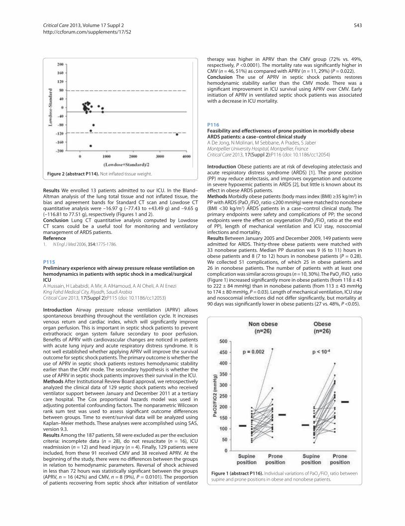
Results We enrolled 13 patients admitted to our ICU. In the Bland–Altman analysis of the lung total tissue and not infl ated tissue, the bias and agreement bands for Standard CT scan and Lowdose CT quantitative analysis were –16.97 g (–77.43 to +43.49 g) and –9.65 g (–116.81 to 77.51 g), respectively (Figures 1 and 2).Conclusion Lung CT quantitative analysis computed by Lowdose CT scans could be a useful tool for monitoring and ventilatory management of ARDS patients.Reference1. N Engl J Med 2006, 354:1775-1786.
P115Preliminary experience with airway pressure release ventilation on hemodynamics in patients with septic shock in a medical/surgical ICUA Hussain, H Lababidi, A Mir, A AlHamoud, A Al Oheli, A Al EneziKing Fahd Medical City, Riyadh, Saudi ArabiaCritical Care 2013, 17(Suppl 2):P115 (doi: 10.1186/cc12053)
Introduction Airway pressure release ventilation (APRV) allows spontaneous breathing throughout the ventilation cycle. It increases venous return and cardiac index, which will signifi cantly improve organ perfusion. This is important in septic shock patients to prevent extrathoracic organ system failure secondary to poor perfusion. Benefi ts of APRV with cardiovascular changes are noticed in patients with acute lung injury and acute respiratory distress syndrome. It is not well established whether applying APRV will improve the survival outcome for septic shock patients. The primary outcome is whether the use of APRV in septic shock patients restores hemodynamic stability earlier than the CMV mode. The secondary hypothesis is whether the use of APRV in septic shock patients improves their survival in the ICU.Methods After Institutional Review Board approval, we retrospectively analyzed the clinical data of 129 septic shock patients who received ventilator support between January and December 2011 at a tertiary care hospital. The Cox proportional hazards model was used in adjusting potential confounding factors. The nonparametric Wilcoxon rank sum test was used to assess signifi cant outcome diff erences between groups. Time to event/survival data will be analyzed using Kaplan–Meier methods. These analyses were accomplished using SAS, version 9.3.Results Among the 187 patients, 58 were excluded as per the exclusion criteria: incomplete data (n = 28), do not resuscitate (n = 16), ICU readmission (n = 12) and head injury (n = 4). Finally, 129 patients were included, from these 91 received CMV and 38 received APRV. At the beginning of the study, there were no diff erences between the groups in relation to hemodynamic parameters. Reversal of shock achieved in less than 72 hours was statistically signifi cant between the groups (APRV, n = 16 (42%) and CMV, n = 8 (9%), P = 0.0101). The proportion of patients recovering from septic shock after initiation of ventilator
therapy was higher in APRV than the CMV group (72% vs. 49%, respectively, P <0.0001). The mortality rate was signifi cantly higher in CMV (n = 46, 51%) as compared with APRV (n = 11, 29%) (P = 0.022).Conclusion The use of APRV in septic shock patients restores hemodynamic stability earlier than the CMV mode. There was a signifi cant improvement in ICU survival using APRV over CMV. Early initiation of APRV in ventilated septic shock patients was associated with a decrease in ICU mortality.
P116Feasibility and eff ectiveness of prone position in morbidly obese ARDS patients: a case–control clinical studyA De Jong, N Molinari, M Sebbane, A Prades, S JaberMontpellier University Hospital, Montpellier, FranceCritical Care 2013, 17(Suppl 2):P116 (doi: 10.1186/cc12054)
Introduction Obese patients are at risk of developing atelectasis and acute respiratory distress syndrome (ARDS) [1]. The prone position (PP) may reduce atelectasis, and improves oxygenation and outcome in severe hypoxemic patients in ARDS [2], but little is known about its eff ect in obese ARDS patients.Methods Morbidly obese patients (body mass index (BMI) ≥35 kg/m2) in PP with ARDS (PaO2/FiO2 ratio ≤200 mmHg) were matched to nonobese (BMI <30 kg/m2) ARDS patients in a case–control clinical study. The primary endpoints were safety and complications of PP; the second endpoints were the eff ect on oxygenation (PaO2/FiO2 ratio at the end of PP), length of mechanical ventilation and ICU stay, nosocomial infections and mortality.Results Between January 2005 and December 2009, 149 patients were admitted for ARDS. Thirty-three obese patients were matched with 33 nonobese patients. Median PP duration was 9 (6 to 11) hours in obese patients and 8 (7 to 12) hours in nonobese patients (P = 0.28). We collected 51 complications, of which 25 in obese patients and 26 in nonobese patients. The number of patients with at least one complication was similar across groups (n = 10, 30%). The PaO2/FiO2 ratio (Figure 1) increased signifi cantly more in obese patients (from 118 ± 43 to 222 ± 84 mmHg) than in nonobese patients (from 113 ± 43 mmHg to 174 ± 80 mmHg, P = 0.03). Length of mechanical ventilation, ICU stay and nosocomial infections did not diff er signifi cantly, but mortality at 90 days was signifi cantly lower in obese patients (27 vs. 48%, P <0.05).
Figure 2 (abstract P114). Not infl ated tissue weight.
Figure 1 (abstract P116). Individual variations of PaO2/FiO2 ratio between supine and prone positions in obese and nonobese patients.
Critical Care 2013, Volume 17 Suppl 2 http://ccforum.com/supplements/17/S2
S43
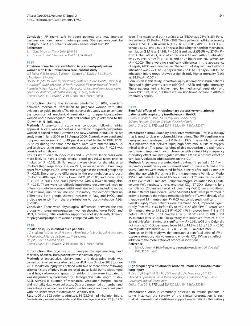
Conclusion PP seems safe in obese patients and may improve oxygenation more than in nonobese patients. Obese patients could be a subgroup of ARDS patients who may benefi t most from PP.References1. Gong MN, et al.: Thorax 2010, 65:44-50.2. Charron C, et al.: Intensive Care Med 2011, 37:785-790.
P117Provision of mechanical ventilation to pregnant/postpartum women with H1N1 infl uenza: a case–control studyWE Pollock1, R Bellomo2, S Webb3, I Seppelt4, A Davies5, E Sullivan6, S Morrison7, B Howe7
1Mercy Hospital for Women, Heidelburg, Australia; 2Austin Health, Heidelberg, Australia; 3Royal Perth Hospital, Perth, Australia; 4Nepean Hospital, Penrith, Australia; 5Alfred Hospital, Prahran, Australia; 6University of New South Wales, Randwick, Australia; 7Monash University, Prahran, AustraliaCritical Care 2013, 17(Suppl 2):P117 (doi: 10.1186/cc12055)
Introduction During the infl uenza pandemic of 2009, clinicians delivered mechanical ventilation to pregnant women with little evidence to guide practice. The objective of this study was to compare the provision of mechanical ventilation to pregnant/postpartum women and a nonpregnant matched control group admitted to the ICU with H1N1 infl uenza.Methods A case–control study was conducted following ethics approval. A case was defi ned as a ventilated pregnant/postpartum woman reported to the Australian and New Zealand INFINITE H1N1 09 study from 1 June 2009 to 31 August 2009. Controls were ventilated nonpregnant women (15 to 49 years) reported to the INFINITE H1N1 09 study during the same time frame. Data were entered into SPSS and analysed using nonparametric statistics; two-tailed P <0.05 was considered signifi cant.Results We studied 36 index cases and 38 controls. Index cases were more likely to have a single arterial blood gas (ABG) taken prior to intubation (P <0.05). Similar reasons were given for the trigger to intubate (high respiratory rate, low PaO2, increased work of breathing) apart from a high PaCO2, which was a trigger in the control group only (P <0.05). There were no diff erences in the pre-intubation and post-intubation ABGs apart from a lower PaCO2 (P <0.05) and lower HCO3 (P <0.05) in cases, and cases presented with a lower haemoglobin (P <0.05). There were six diffi cult intubations documented with no diff erences between groups. Initial ventilator settings including mode, tidal volume, minute volume and respiratory rate demonstrated no diff erences. Both groups showed increases in PaO2 and PaCO2, and a decrease in pH from the pre-intubation to post-intubation ABGs (P <0.05).Conclusion There were physiological diff erences between the two groups with pregnant/postpartum women showing lower PaCO2 and HCO3. However, initial ventilator support was not signifi cantly diff erent for pregnant/postpartum women compared with controls.
P118Inhalation injury in critical burn patientsL Cachafeiro, M Sanchez, E Herrero, L Fernández, M Irazabal, M Hernandez, A Agrifoglio, A Garcia de Lorenzo, M LendinezHospital La Paz, Madrid, SpainCritical Care 2013, 17(Suppl 2):P118 (doi: 10.1186/cc12056)
Introduction The objective is to analyze the epidemiology and mortality of critical burn patients with inhalation injury.Methods A prospective, observational and descriptive study was carried out in all patients admitted to an ICU from October 2008 to June 2011. Inhalation injury was defi ned with two or more of the following criteria: history of injury in an enclosed space, facial burns with singed nasal hair, carbonaceus sputum or stridor. If they were intubated it was diagnosed by bronchoscopy. Demographic data, length of stay, ABSI, APACHE II, duration of mechanical ventilation, hospital course and mortality data were collected. Data are presented as number and percentage or as median and interquartile range and were analyzed with the Fisher exact test and Mann–Whitney test.Results Of the 362 patients admitted, 84 (23.2%) had inhalation injury. Seventy-six percent were male and the average age was 52 (± 17.5)
years. The mean total burn surface area (TBSA) was 28% (± 25). Forty-fi ve patients (53.5%) had TBSA >20%. These patients had higher severity scores: ABSI 8 (± 2.8) versus 6 (± 2.2) (P = 0.0001), APACHE II 13 (± 6.9) versus 7 (± 6.7) (P = 0.0001). They also had a higher need for mechanical ventilation (88.1% vs. 34.9%, P = 0.001) and shock (59.5% vs. 27.0%, P = 0.001). The PaO2/FiO2 ratio at admission with and without inhalation was 245 versus 359 (P = 0.003), and at 72 hours was 207 versus 306 (P = 0.002). There were no signifi cant diff erences in the appearance of sepsis, ARDS and renal failure. The length of stay with and without inhalation was 25.2 (1 to 95) days versus 22.5 (1 to 92) days (P = 0.4). The inhalation injury group showed a signifi cantly higher mortality (9.0% vs. 28.9%, P = 0.001).Conclusion In this study, inhalation injury is common in burn patients. They had higher severity scores (APACHE II, ABSI) and higher mortality. These patients had a higher need for mechanical ventilation and lower PaO2/FiO2 ratio, but there was no signifi cant increase in ARDS or respiratory sepsis.
P119Benefi cial eff ects of intrapulmonary percussive ventilation in patients with respiratory insuffi ciency in the ICUI Blum, R Janssen-Dean, A Overdijk van, B SpeelbergSt Anna Hospital Geldrop, Geldrop, the NetherlandsCritical Care 2013, 17(Suppl 2):P119 (doi: 10.1186/cc12057)
Introduction Intrapulmonary percussive ventilation (IPV) is a therapy that is used to clear endobronchial secretions. The IPV ventilator was designed and developed by FM Bird in 1979. The ventilator consists of a phasitron that delivers rapid, high-fl ow, mini bursts of oxygen, mixed with air. The potential aims of this mechanism are pulmonary recruitment, improved mucus clearance with a direct high-frequency oscillatory eff ect. We investigated whether IPV has a positive eff ect on ventilatory values in adult patients on the ICU.Methods All patients presenting during a 4-month period in 2011 with respiratory insuffi ciency on our mixed adult ICU were included in this study. Patients were monitored before, directly after and 15 minutes after therapy with IPV using a Bird Intrapulmonary Ventilator Model IPV-2C. All patients received IPV for a period of 20 minutes consisting of two cycles of 10 minutes. Peripheral oxygen saturation (SpO2), tidal volume (Vt), respiratory rate, end-tidal CO2 (ET-CO2), dynamic lung compliance (C-dyn) and work of breathing (WOB) were monitored at the diff erent time points. Paired Student t tests were performed in order to compare the values immediately before IPV, with directly after therapy and 15 minutes later. P <0.05 was considered signifi cant.Results Eighty-three patients were examined. SpO2 improved signifi -cantly from 93.7 ± 3.7 before IPV to 95.7 ± 2.8 after IPV (P <0.001) and 15 minutes later to 95.2 ± 2.8 (P <0.001). Vt improved from 418 ± 111 before IPV to 476 ± 102 directly after (P <0.001) and to 480 ± 131 15 minutes later (P <0.01). Respiratory rate improved from 24 ± 6 to 23 ± 6 only after 15 minutes signifi cantly (P <0.01). WOB and C-dyn did not change. ET-CO2 decreased from 34.9 ± 14.8 to 33.3 ± 13.3 (P <0.05) directly after IPV and to 32.1 ± 12.8 (P <0.01) 15 minutes later.Conclusion In this study we demonstrated a benefi cial eff ect of IPV on oxygen saturation, tidal volume and end-tidal CO2. IPV has this eff ect in addition to the mobilization of bronchial secretions.Reference1. Salim A, Martin M: High-frequency percussive ventilation. Crit Care Med
2005, 33:S241-S245.
P120High-frequency ventilation for acute traumatic and nontraumatic lung injuryR Varutti1, R Bigai1, M Fiorillo1, D Tomasello1, W Mercante1, G Trillò2
1Azienda Ospedaliera Santa Maria degli Angeli, Pordenone, Italy; 2Udine University Hospital, Udine, ItalyCritical Care 2013, 17(Suppl 2):P120 (doi: 10.1186/cc12058)
Introduction ARDS is commonly observed in trauma patients. In some instances the severity of the clinical presentation is such that all conventional ventilatory support mode fails. In this setting,
Critical Care 2013, Volume 17 Suppl 2 http://ccforum.com/supplements/17/S2
S44

high-frequency oscillatory ventilation (HFOV) was considered mostly a rescue therapy.Methods Fifteen adult patients admitted to our ICU for acute traumatic and nontraumatic lung injury were submitted to HFOV when conventional mechanical ventilation failed.Results Clinical and demographic data are shown in Table 1. Figure 1 shows the trend of gas parameters during the recovery. At baseline PaO2 was 94 ± 28 mmHg; after 6 hours of HFOV: 135 ± 41 mmHg, P <0.01. At baseline PaO2/FiO2 was 182 ± 97 mmHg; after 6 hours of HFOV: 264 ± 101 mmHg, P <0.01. The benefi ts are maintained when returned to conventional ventilation.Conclusion HFOV may therefore be anticipated to improve end-organ perfusion and gas exchange; it should be considered in severe traumatic and nontraumatic respiratory failure [1].Reference1. BMJ 2010, 340:c2327.
P121Improvement in oxygenation with high-frequency oscillatory ventilation combined with tracheal gas insuffl ation is correlated to the extravascular lung water indexCS Vrettou, S Malachias, SG Zakynthinos, SD MentzelopoulosEvaggelismos General Hospital, Athens, GreeceCritical Care 2013, 17(Suppl 2):P121 (doi: 10.1186/cc12059)
Introduction High-frequency oscillatory ventilation combined with tracheal gas insuffl ation (HFO-TGI) can signifi cantly improve oxygenation in patients with ARDS. It has been demonstrated that oxygenation in patients with ARDS has a better response to HFO when extravascular lung water is >15 ml/kg body weight (BW). Our aim is to examine whether the extravascular lung water index (ELWI) correlates with changes in PaO2/FiO2 in patients ventilated with HFO-TGI.Methods Data from 18 sessions of HFO-TGI in six patients were included in the analysis. HFO frequency, oscillatory pressure amplitude, and bias
fl ow were 3.5 Hz, 85 to 95 cmH2O, and 40 l/minute, respectively; a 2.5 to 3.5 cmH2O tracheal tube cuff leak was used. HFO mean airway pressure (mPaw) exceeded preceding conventional ventilation (CV)-mPaw by 7 to 13 cmH2O. PaO2/FiO2, lung mechanics, and hemodynamics were documented during lung-protective CV (baseline) and 1 hour following the initiation of HFO-TGI ventilation. PULSION PICCOplus v7.0 was used for hemodynamic measurements including the ELWI.Results Oxygenation (PaO2/FiO2) improved signifi cantly with HFO-TGI compared with CMV (125.5 ± 54.7 vs. 195.6 ± 108.7, P <0.001). Changes in PaO2/FiO2 were positively correlated with ELWI at baseline (Spearman’s ρ = 0.56, P = 0.016). See Figure 1. There were no signifi cant changes in patients’ fl uid balance and hemodynamics including the ELWI.Conclusion Estimation of the ELWI can help to predict the oxygenation response of ARDS patients considered for HFO-TGI ventilation. The possibility that HFO-TGI exerts an eff ect on pulmonary oedema needs further investigation.Acknowledgement This research has been cofi nanced by the European Union and Greek national funds through the operating programme ‘Education and Lifelong Learning’ – Research Funding Programme Heracleitus II.Reference1. Zeravik J, et al.: Acta Anaesthesiol Scand 1989, 33(Suppl 90):149-152.
Figure 1 (abstract P120). Mean PaO2, PaCO2, PaO2/FiO2 at baseline and during HFOV (P, percussionator; C, conventional). *P <0.05 versus baseline, †P <0.01 versus baseline.
Table 1 (abstract P120). Clinical and demographic data
Mean
Age (years) 64
Male/female 11/4
Dead 4
Trauma 3
SAPS II 45/40
APACHE 23/8
Figure 1 (abstract P121). Scatter plot and fi t line of ELWI versus dPaO2/FiO2.
Critical Care 2013, Volume 17 Suppl 2 http://ccforum.com/supplements/17/S2
S45

P122Physiologic eff ects and regional ventilation of high-frequency positive-pressure ventilation using a conventional ventilator in a severe ARDS animal model associated with an inspiratory pause or recruitment maneuversRL Cordioli1, M Park2, M Amato3, S Gomes3, E Leite2, L Azevedo2
1Hopitaux Universitaire de Geneve, Geneva, Switzerland; 2Hospital Sirio Libanes IEP, São Paulo, Brazil; 3Hospital das Clinicas LIM 09, São Paulo, BrazilCritical Care 2013, 17(Suppl 2):P122 (doi: 10.1186/cc12060)
Introduction Protective mechanical ventilation (MV) in ARDS is based on reduced stretch of pulmonary tissue, sometimes resulting in severe hypoventilation that can be avoided when using high respiratory rate. High-frequency positive-pressure ventilation (HFPPV) has not been fully explored, especially when associated with other strategies aiming to avoid hypercapnia.Methods We induced ARDS in eight pigs by lung lavage with saline plus 3 hours of injurious MV with low PEEP and high driving pressure (DP). We then performed a recruitment maneuver (RM) followed by PEEP titration using the amount of alveolar collapse in electrical impedance tomography (EIT). Then stabilization during 1 hours with tidal volume (VT) at 6 ml/kg, respiratory rate (RR) 35 breaths/minute and PEEP selected with the PEEP-FiO2 table (ARMA study), which was kept constant during two steps of HFPPV with a RR 60: one without an inspiratory pause (HFPPV-60), and one with a pause of 30% of inspiratory time (HFPPV-60 w/P30%). In another HFPPV step, we used PEEP titrated with EIT after RM (HFPPV-60 w/RM). During each HFPPV step, VT was set to reach a PaCO2 of 60 ± 3 mmHg. Distribution of regional ventilation was analyzed using EIT. Equilibrium was considered if PaCO2 was stable (<5% of variation) for >30 minutes.Results HFPPV allowed reduction in PaCO2 levels: 81 (77, 94) versus 60 (58, 61), 59 (58, 60), 60 (58, 61) mmHg, besides using lower VT: 5.2 (5.0, 5.9), 5.1 (4.5, 6.0), 4.7 (4.2, 5.7) and 4.8 (4.5, 5.6) ml/kg during stabilization, HFPPV-60, HFPPV-60 w/P30% and HFPPV-60 w/RM, respectively. It had no signifi cant diff erent results comparing HFPPV-60 with and without an inspiratory pause. HFPPV-60 w/RM allowed a better alveolar homogenization and improvement in oxygenation, shunt, dead space and DP compared with the other steps. See Table 1.
Table 1 (abstract P122). Physiological variables
Variable VT = 6, stabilization HFPPV-60 HFPPV-60 w/RM
DP (cmH2O) 16 ± 2 16 ± 3 12 ± 2*
P/F ratio 95 ± 13 149 ± 60 246 ± 99*
*Tukey’s post-hoc analysis, P < 0.05 versus others.
Conclusion HFPPV with a conventional mechanical ventilator is able to maintain stable PaCO2 in clinically acceptable values, allowing reductions in VT. HFPPV-60 w/RM and PEEP titration using EIT allowed further physiologic benefi ts in a severe ARDS model.
P123High-frequency percussive ventilation at altitude: study in a hypobaric chamber with a mechanical test lungAC Cirodde1, S Montmerle2, ND Donat1, CB Bourhillon2, P Jault1, L Bargues1, T Leclerc1
1Military Hospital Percy, Clamart, France; 2IRBA, Brétigny, FranceCritical Care 2013, 17(Suppl 2):P123 (doi: 10.1186/cc12061)
Introduction High-frequency percussive ventilation (HFPV) is a rescue technique for most severe acute lung injury/acute respiratory distress syndrome (ARDS) patients [1], especially with smoke inhalation or respiratory burns [2]. This study aimed at characterizing HFPV as delivered by Percussionnaire VDR4® and at evaluating how hypobarism interferes with HFPV, in order to assess its usability at altitude.Methods Using a mechanical test lung mimicking ARDS (compliance 17 ml/cmH2O) with two resistance levels (5 and 15 cmH2O/l/second) and ventilated with VDR4® in a hypobaric chamber, ascents/descents between 0 and 5,000 and then 0 and 8,000 ft were performed. Adjustable
VDR4® parameters were modifi ed one at a time at each altitude. Besides these parameters (cross-measured with standalone hardware), oxygen consumption of the respirator and three calculated parameters were studied: low-frequency tidal volume (Vt, integrated from instantaneous fl ows measured with a Fleisch pneumotachograph), end-inspiratory (PmEI) and end-expiratory (PmEE) mean pressures. PmEI and PmEE in HFPV refl ect plateau pressure and positive end-expiratory pressure in conventional ventilation. The correction of altitude-induced off set with the modifi cation of working pressure was also tested.Results Data displayed by VDR4® overestimated pulmonary pressures by more than 10%, but were reliable for other parameters. During ascent, an off set appeared for all respiratory parameters: Vt increased by 59% and PmEI by 53% between 0 and 8,000 ft. During descent, the off set was reversely directed with a 39% decrease in Vt and a 28% decrease in PmEE between 8,000 and 0 ft. Modifying working pressure adequately corrected PmEI and PmEE, but not Vt. In all cases, manually correcting VDR4® parameters to their 0 ft level also corrected these off sets. Multivariate analysis further established that, adjusting for other parameters, Vt, PmEI and PmEE did practically not depend on altitude. Oxygen consumption of the respirator was high, 25 l/minute at 0 ft, and stable with altitude. It was reduced with percussive rate and with FiO2.Conclusion HFPV can be safely used at altitude, provided that VDR4®-displayed parameters are used to manually adjust settings in order to avoid exposing patients to volutrauma or barotrauma during ascent, and to major hypoventilation and alveolar collapse during descent. The high oxygen consumption is currently the main limit to its use for long-range aeromedical evacuations.References1. Velmahos GC, et al.: Chest 1999, 116:440-446.2. Chung KK, et al.: Crit Care Med 2010, 38:1970-1977.
P124Recruitable volume is comparable in acute respiratory distress syndrome and in healthy lungsCA Stahl1, K Moeller2, D Steinmann1, D Henzler3, S Lundin4, O Stenqvist4
1University Medical Center Freiburg, Germany; 2Biomedical Engineering, Villingen-Schwenningen, Germany; 3Klinikum Herford, Germany; 4Sahlgrenska University Hospital, Gothenburg, SwedenCritical Care 2013, 17(Suppl 2):P124 (doi: 10.1186/cc12062)
Introduction The application of PEEP is commonly used in acute respiratory distress syndrome (ARDS) and has been shown to improve oxygenation. To identify patients that most benefi t from the application of PEEP, the discrimination of recruiters and nonrecruiters has been postulated by Gattinoni and colleagues [1]. Recently, Dellamonica and colleagues [2] presented a method to predict alveolar recruitment. We hypothesised that the amount of recruitable volume allows the discrimination between ARDS patients and patients with healthy lungs (HL).Methods We recalculated the recruited volume (RV) in 25 patients with ARDS [3] according to the method proposed by Dellamonica and colleagues during an incremental PEEP manoeuvre (PEEP increased until the plateau pressure reached 45 cmH2O). RV was calculated as the change in end-expiratory lung volume minus total respiratory system compliance times the PEEP change (RV = ΔEELV – CTOT×ΔPEEP). For comparison, 12 patients with HL undergoing elective surgery in general anaesthesia were measured using the same protocol.Results Both ARDS and HL patients exhibited typical P–V curves and stepwise recruitment (Figure 1). By raising PEEP from 0 to 12 cmH2O, ARDS patients recruited 331 ± 195 ml (mean ± SD) and HL patients 435 ± 43 ml. There was a strong correlation (R2 = 0.88) of the total RV with the end-inspiratory volume at a plateau pressure of 45 cmH2O in both groups; that is, recruitment was found to the same extent in both groups (Figure 2).Conclusion The relative contribution of RV to lung volume gain is similar in ARDS and in patients with healthy lungs. Our results question the relevance of recruitability as defi ned by Dellamonica and colleagues as a typical phenomenon of ARDS, but support the baby lung concept, as the recruited volume was closely related to the size of the lung.
Critical Care 2013, Volume 17 Suppl 2 http://ccforum.com/supplements/17/S2
S46

References1. Gattinoni et al.: N Engl J Med 2006, 354:1775.2. Dellamonica et al.: Intensive Care Med 2011, 37:1595.3. Stahl et al.: Crit Care Med 2006, 34:2090.
P125Nosocomial bloodstream infection and venovenous extracorporeal membrane oxygenation: a retrospective cohort studyB Serra de Oliveira, S Mendes Fernandes, C FrançaHospital de Santa Maria CHLN, Lisboa, PortugalCritical Care 2013, 17(Suppl 2):P125 (doi: 10.1186/cc12063)
Introduction Venovenous extracorporeal membrane oxygenation (VV-ECMO) for respiratory failure in the ICU is used in a variety of clinical situations and has been demonstrated to signifi cantly improve survival without disability in adult respiratory distress syndrome [1]. ECMO has been presented as a risk factor for bloodstream infection although recently published data do not support this view or the use of antibiotic prophylaxis [2]. We aimed to examine VV-ECMO as a risk factor for nosocomial bloodstream infection.
Methods A retrospective cohort study from patients admitted to our ICU between April 2009 and June 2012. We compared incidence rates of nosocomial bacteremia using the Hospitals in Europe Link for Infection Control through Surveillance (HELICS) between general ICU and VV-ECMO patients and used multiple logistic regression analysis to control for possible confounders.Results During the study period 1,146 patients were admitted and 16 received VV-ECMO. The incidence of bloodstream infection in patients with ECMO was 19/1,000 exposure-days versus 4,9/1,000 exposure-days in general ICU patients (incidence rate ratio of 3.82; 95% CI: 2.0 to 7.3; P <0.001). Bacteremia was mostly due to Gram-negative agents (65%). The patients with bloodstream infections under ECMO (n = 10) had a nonsignifi cant younger age (P = 0.08) and a lower SAPS II score (P = 0.03) compared with non-ECMO patients (n = 25). VV-ECMO patients had a signifi cantly higher risk of primary bacteremia than non-ECMO patients (P = 0.04). Patients with bloodstream infections in the VV-ECMO group had a lower crude mortality rate (OR: 0.1, P = 0.04), not confi rmed in the adjusted analysis. There were no crude or adjusted diff erences in the time to bacteremia or infections due to multiple drug-resistant microorganisms (OR: 0.26; P = 0.085) between groups.Conclusion This study suggests that VV-ECMO patients have a signifi cantly higher risk for primary nosocomial bloodstream infection. A larger study is needed to confi rm such fi ndings and to assess the need for specifi c intervention, namely routine antibiotic prophylaxis.References1. Peek GJ, et al.: Effi cacy and economic assessment of conventional
ventilatory support versus extracorporeal membrane oxygenation for severe adult respiratory failure (CESAR): a multicentre randomised controlled trial. Lancet 2009, 374:1351-1363.
2. Conrick-Martin I, et al.: Nosocomial infections in a cohort of extracorporeal life support patients. Crit Care Resusc 2012, 14:198-201.
P126Monitoring anticoagulation during extracorporeal membrane oxygenation in patients with acute respiratory failureM Panigada1, C Mietto2, F Pagan2, L Bogno2, V Berto2, L Gattinoni21Fondazione IRCCS Ca’ Granda Ospedale Maggiore Policlinico, Milan, Italy; 2IRCCS Ca’ Granda Maggiore Policlinico Hospital Foundation, Milan, ItalyCritical Care 2013, 17(Suppl 2):P126 (doi: 10.1186/cc12064)
Introduction aPTT is a common tool for anticoagulation monitor ing during extracorporeal membrane oxygenation (ECMO). Thrombo-elasto graphy (TEG) is another available option in this setting.Methods A prospective observational study on 12 consecutive patients during venovenous ECMO. Anticoagulation was provided
Figure 1 (abstract P124).
Figure 2 (abstract P124).
Critical Care 2013, Volume 17 Suppl 2 http://ccforum.com/supplements/17/S2
S47
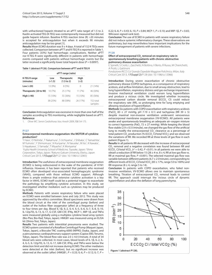
with unfractioned heparin titrated to an aPTT ratio target of 1.5 to 2. Kaolin-activated TEG (K-TEG) was contemporarily measured but did not guide heparin infusion. Baseline K-TEG reaction time (R) >20 minutes is accepted for anticoagulation but when it exceeds 90 minutes anticoagulation may be too great [1].Results Mean ECMO duration was 9 ± 4 days. A total of 152 K-TEGs were collected. Comparison between aPTT and K-TEG R is reported in Table 1. Four patients (33%) had hemorrhagic complications. Neither aPTT nor K-TEG R were signifi cantly diff erent in patients with hemorrhagic events compared with patients without hemorrhagic events but the latter received a signifi cantly lower total heparin dose (P = 0.0097).
Table 1 (abstract P126). Comparison between aPTT and K-TEG R
aPTT range (ratio)
K-TEG R range Low Therapeutic High(minutes) (<1.5) (1.5 to 2) (>2)
Low (<20) 13 (9%) 8 (5%) 0 21 (14%)
Therapeutic (20 to 90) 10 (7%) 25 (17%) 11 (7%) 46 (30%)
High (>90) 15 (10%) 55 (36%) 15 (10%) 85 (56%)
38 (25%) 88 (58%) 26 (17%) 152 total samples
Conclusion Anticoagulation was excessive in more than one-half of the samples according to TEG monitoring, while negligible based on aPTT.Reference1. Oliver WC: Semin Cardiothorac Vasc Anesth 2009, 13:154-175.
P127Extracorporeal membrane oxygenation: the MOTOR of cytokine production?Y Hara1, O Nishida1, T Nakamura1, S Uchiyama1, J Shibata1, C Yamashita1, M Yumoto1, Y Shimomura1, N Kuriyama1, N Yasuoka1, M Ito1, K Kawata1, S Hayakawa1, S Yamada2, T Miyasho3, K Moriyama1
1Fujita Health University School of Medicine, Toyoake, Japan; 2Shino Corporation, Tokyo, Japan; 3Rakuno Gakuen University, Ebetsu, JapanCritical Care 2013, 17(Suppl 2):P127 (doi: 10.1186/cc12065)
Introduction The usefulness of extracorporeal membrane oxygenation (ECMO) is being rediscovered in the wake of the pandemic of H1N1 infl uenza. However, it has been reported that patients who received ECMO often developed virus-associated hemophagocytic syndrome (VAHS), compared with those without ECMO support. Although there is ample evidence that extensive cytokine activation is a key factor in VAHS, ECMO itself could be a potential trigger to exacerbate the pathology by amplifying cytokine activation. In this study, we investigated whether mediators such as cytokines may be produced by ECMO.Methods Patients with severe respiratory failure who were placed on ECMO were enrolled between June and July 2012. This study was approved by the ethics committee. Blood specimens were drawn from the blood circuit at the inlet of the centrifugal pump (before) and outlet of the hollow fi ber oxygenator (after) at a frequency of three to four times per day. Blood IL-1β, IL-2, IL-4, IL-5, IL-6, IL-7, IL-8, IL-10, IL-12(p70), IL-13, IL-17, G-CSF, GM-CSF, IFNγ, MCP-1, MIP-1β, and TNFα were measured globally using a multiplex cytokine bead array system (Bio-Plex; Bio-Rad, Tokyo, Japan). HMGB1 was measured using an ELISA kit (Shino-Test, Tokyo, Japan).Results Two patients with interstitial pneumonia were studied. The ECMO system consisted of a Rotafl ow Centrifugal Pump (Maquet Japan, Tokyo, Japan), a Biocube TNC coating 6000 (NIPRO, Osaka, Japan), and a percutaneous cardiopulmonary support system (Capiox EBS; Terumo, Tokyo, Japan). The blood fl ow rate was 2.0 ± 4.0 l/minute. A total of 34 blood sets were collected. In most cases, blood levels of IL-1β, IL-2, IL-4, IL-5, IL-12(p70), IL-13, IL-17, GM-CSF, IFNγ, and TNFα were below the detection limit and did not increase during ECMO. The other mediators were detected at the inlet (before), but no signifi cant increase was observed at the outlet (after) (HMGB1, P = 0.33; IL-6, P = 0.12; IL-7, P =
0.22; IL-8, P = 0.43; IL-10, P = 0.84; MCP-1, P = 0.10; and MIP-1β, P = 0.65; Wilcoxon signed-rank test).Conclusion The use of ECMO in patients with severe respiratory failure did not induce systemic infl ammatory changes. These observations are preliminary, but may nevertheless have important implications for the future management of patients with severe infections.
P128Eff ect of extracorporeal CO2 removal on respiratory rate in spontaneously breathing patients with chronic obstructive pulmonary disease exacerbationE Spinelli, S Crotti, L Zacchetti, N Bottino, V Berto, R Russo, M Chierichetti, A Protti, L GattinoniFondazione IRCCS Ca’ Granda Ospedale Maggiore Policlinico, Milan, ItalyCritical Care 2013, 17(Suppl 2):P128 (doi: 10.1186/cc12066)
Introduction During severe exacerbation of chronic obstructive pulmonary disease (COPD) tachypnea, as a consequence of respiratory acidosis, and airfl ow limitation, due to small airway obstruction, lead to lung hyperinfl ation, respiratory distress and gas exchange impairment. Invasive mechanical ventilation could worsen lung hyperinfl ation and produce a vicious circle. We investigated whether increasing extracorporeal carbon dioxide removal (ECCO2 Cl) could reduce the respiratory rate (RR), so prolonging time for lung emptying and allowing resolution of hyperinfl ation.Methods Six patients with COPD exacerbation with respiratory acidosis (PaCO2 83 ± 27 mmHg, pH 7.19 ± 0.1) and tachypnea (RR 39 ± 5) despite maximal non-invasive ventilation underwent venovenous extracorporeal membrane oxygenation (VV-ECMO). All patients were awake and spontaneously breathing an adequate air–oxygen mixture to correct hypoxemia (PaO2 72 ± 27 mmHg). While keeping the blood fl ow stable (2.9 ± 0.5 l/minute), we changed the gas fl ow of the artifi cial lung to modify the extracorporeal CO2 clearance as a percentage of total patient CO2 production (% ECCO2 Cl/total VCO2) and we observed the variations of RR. We recorded RR at three levels of gas fl ow in each patient (Figure 1).Results In all patients RR decreased with the increase of extracorporeal CO2 removal and a negative correlation was found between RR and ECCO2 Cl/total VCO2 (r2 = 0.42, P <0.01). In all patients we were able to obtain a reduction of RR below 15 (28 ± 4 vs. 8 ± 4, RR at low gas fl ow vs. RR at maximal gas fl ow, P <0.001). The selected maximal gas fl ow was variable between diff erent patients (6.7 ± 2 l/minute), corresponding to diff erent levels of ECCO2 Cl/total VCO2 (83 ± 17%, range 53 to 100%) and RR response (8 ± 4, range 5 to 14).Conclusion In patients with COPD exacerbation, who failed non-invasive ventilation, VV-ECMO allows one to maintain spontaneous breathing. Titration of extracorporeal CO2 removal leads to control RR. This approach could interrupt the vicious circle of dynamic hyperinfl ation and allow the defl ation of lung parenchyma.
Figure 1 (abstract P128).
Critical Care 2013, Volume 17 Suppl 2 http://ccforum.com/supplements/17/S2
S48

P129ECCO2 removal with a phosphorylcholine-coated membrane oxygenator in diffi cult respiratory weaning patientsF Turani, S Martini, A Marinelli, M Falco, R Barchetta, F Candidi, F Gargano1Aurelia Hospital/European Hospital, Rome, ItalyCritical Care 2013, 17(Suppl 2):P129 (doi: 10.1186/cc12067)
Introduction ECCO2 removal may be a useful support in patients with diffi cult respiratory weaning. The aim of this study is to evaluate a new phosphorylcholine-coated EECO2 removal system with no thrombogenic activity to assess the clinical safety of the system, the changes of main cardiorespiratory indices and CO2 removal by the system.Methods Ten patients were enrolled. Before starting with ECCO2 removal all patients were ventilated with TV <6 ml/kg, peak pressure >35 cmH2O and pH <7.25. ECCO2 removal was initiated using a modifi ed continuous venovenous hemofi ltration system with a membrane oxygenator (ABYLCAP; Bellco, Mirandola, Italy; membrane surface area: 0.67 m2, blood fl ow 280 to 350 ml/minute, phosphorylcholine coated). Femoral vein cannulation with a double-lumen catheter was used to connect the patients to the extracorporeal system. Heparin was infused to maintain ACT <190 to 240 seconds. All patients had ECCO2 for 4 days. Every 12 hours the pH, PaCO2, peak pressure and PaO2 were evaluated. VCO2 was determined by indirect calorimetry using a gas analyser equipped on an Engstrom Carestation Ventilator. During the ECCO2 removal the patients were ventilated with TV ≤6 ml/kg and peak pressure <30 cmH2O. All data are expressed as mean ± SD. One-way ANOVA was used to compare the changes of parameters. P <0.05 was considered signifi cant.Results Table 1 presents the main results. The CO2 removal by mem-brane oxygenator ranged from 56 to 37 ml/minute. All patients survived to the treatment and 7/10 were weaned from the ventilator at the end of ECCO2 removal. Only one oxygenator was used for every patient without clotting of the circuit or any major bleeding problem.
Table 1 (abstract P129)
Day 0 Day 2 Day 4
pH 7.24 ± 0.06 7.38 ± 0.1 7.41 ± 0.07*
PaCO2 (mmHg) 70 ± 5 57 ± 8 52 ± 3*
PP (cmH2O) 48 ± 4 28 ± 4* 25 ± 4
VCO2 (ml/minute) 220 ± 15 201 ± 14 180 ± 13*
*P <0.05 versus T0.
Conclusion ECCO2 removal with a phosphorylcholine-coated mem-brane oxygenator is clinically safe, avoids clotting of the oxygenator circuit and allows adequate CO2 removal.Reference1. Livigni S, et al.: Crit Care 2006, 10:R151.
P130Lung elastance can be determined without esophageal pressure measurementsC Grivans, S Lundin, O StenqvistSahlgrenska University Hospital, Gothenburg, SwedenCritical Care 2013, 17(Suppl 2):P130 (doi: 10.1186/cc12068)
Introduction We have previously shown, in an ex vivo porcine model, that lung elastance calculated as the PEEP change divided by lung volume increase (ΔPEEP/ΔEELV) is closely correlated to conventionally measured lung elastance using oesophageal pressure [1]. In this study we hypothesize that the successive change in lung volume during a PEEP-step manoeuvre could be predicted from ΔPEEP and lung elastance as ΔPEEP/EL. The objective of the study was to validate this hypothesis in patients with acute respiratory failure (ARF).Methods Thirteen patients with ARF were studied during an incremental PEEP trial, 0–4–8–12–16 cmH2O. ΔEELV was determined as the change in expiratory tidal volume following each PEEP step. Conventional calculation of lung elastance was obtained from tidal variation in airway
pressure minus tidal variation in oesophageal pressure divided by tidal volume. Position of the oesophageal catheter was verifi ed according to Baydur [2]. The measured change in end-expiratory lung volume during the PEEP-step manoeuvre using spirometry was compared with the end-expiratory lung volume change calculated from EL and stepwise changes in PEEP as ΔPEEP/EL.Results There was a close correlation between the measured build-up of end-expiratory lung volume during a PEEP-step manoeuvre and ΔPEEP/EL where EL was conventionally determined using oesophageal pressure measurements (see Figure 1).Conclusion Esophageal pressure measurements are diffi cult to perform [3] and rarely used in routine clinical practice. Our fi ndings indicate that a change in PEEP together with measurements of the resulting change in end-expiratory volume by spirometry in the ventilator could be used to determine lung elastance separately, the relation between lung and chest wall elastance as well as the transpulmonary pressure.References1. Stenqvist O, et al.: Acta Anaesthesiol Scand 2012, 56:738-747.2. Baydur A: Chest 2002, 121:324-326.3. Hedenstierna G: Minerva Anestesiol 2012, 78:959-966.
P131Feasibility of minimally invasive stimulation of the phrenic nerves for supporting ventilation in fully anesthetized swineJ Bijwadia1, M Karamanoglu2
1Regions Hospital, St Paul, MN, USA; 2Respithera, Bloomington, MN, USACritical Care 2013, 17(Suppl 2):P131 (doi: 10.1186/cc12069)
Introduction Long-term use of mechanical ventilators may lead to ventilator-induced diaphragmatic dysfunction (VIDD) and increase the duration of weaning from MV [1]. It was hypothesized that stimulating the diaphragm during MV may prevent VIDD and may lead to early weaning [2]. In this study, the feasibility of generating coordinated contraction of both diaphragms was investigated using a novel transvenous diaphragmatic pacing system.Methods Two juvenile pigs were anesthetized with propofol (150 to 250 μg/kg/minute) and ventilated (VENT) with an assist control mode MV (Nellcor Puritan Bennett 840). Using fl uoroscopy, a novel multipolar neurostimulation catheter (Inspirx RL PICC53; Respithera, Bloomington, MN, USA) was threaded into the left internal jugular vein and advanced to the junction of right atrium and the superior vena cava using a
Figure 1 (abstract P130). Correlation between spirometrically measured and calculated increase in EELV.
Critical Care 2013, Volume 17 Suppl 2 http://ccforum.com/supplements/17/S2
S49

modifi ed Seldinger technique. The successful capture of the right and left phrenic nerves was confi rmed by fl uoroscopic visualization. Peak airway pressures (PAWP) and blood gases were determined after 10 minutes MV (MV), MV and stimulation applied together (MV+STIM) and stimulation only (STIM).Results No animal–ventilator dyssynchrony during stimulation (MV+STIM) was noted while peak airway pressures were reduced. During STIM there was no discernible paradoxical movement of the diaphragm. In addition, PCO2 and PO2 confi rmed that adequate ventilation and oxygenation can be provided by the system, while PAWP could be reduced (Table 1).
Table 1 (abstract P131). Ventilation parameters
MV MV+STIM STIM
PAWP (cmH2O) 20 16 9
PO2 (mmHg) 89 101 95
PCO2 (mmHg) 32 26 26
Conclusion It was possible to capture and stimulate both phrenic nerves using a minimally invasive approach to support respiration and sustain blood gases at physiological levels. This development could help wean MV-dependent ICU patients earlier. Further long-term studies are needed to assess the full potential of this novel system.References1. Vassilakopoulos T, Petrof BJ: Am J Respir Crit Care Med 2004, 169:336-341.2. Sassoon CS, Zhu E, Caiozzo VJ: Am J Respir Crit Care Med 2004, 170:626-632.
P132Prehospital management of COPD patients in respiratory failure and short-term outcomeG Campagne, J Cuny, P Gosselin, P Goldstein, N Assez, E WielLille University Hospital Center, Lille Cedex, FranceCritical Care 2013, 17(Suppl 2):P132 (doi: 10.1186/cc12070)
Introduction Respiratory failure in COPD patients is a frequent call in French emergency dispatching centers. We have evaluated the prehospital management of COPD patients and severity signs, and analyse outcome in the emergency department or ICU.Methods We conducted an observational, descriptive, retrospective, single-center study during a 4-month period. All COPD patients with respiratory failure and prehospital care were included. Diff erent data were recorded.Results Ninety patients were included (77% male, 23% female). Mean age 69 years (± 11.88). Fifty-fi ve percent were smokers, 52% had arterial hypertension, 39% received long-term oxygenotherapy, 18% received antibiotics in the 7 days before, 18% corticosteroids, and 14% were on long-term NIV support at home. An emergency medical ambulance was immediately sent for 86% of patients. Ninety-two percent had normal consciousness (Glasgow Coma Scale 15), 78% had bronchospasm, 71% had signs of respiratory struggle, and 12% were unable to speak. The mean respiratory rate was 31.4 cycles/minute (± 8.18), the average cardiac pulse was 103.6 beats/minute (± 23.14). Nasal EtCO2 44.92 mmHg (± 16.38), pulse oximetry with air was 83.48% (± 12.09), and the average fl ow rate of oxygen delivered was 5.69 l/minute (± 2.93). None of the patients had fever. Eighty-fi ve percent were supported on spontaneous ventilation, 22% received prehospital non-invasive ventilation, they all showed signs of severity and 3% need tracheal intubation. Seventy-fi ve percent of patients received β2-agonist and anticholinergic nebulization, 45% intravenous corticosteroids. Seventy-one percent were admitted to the emergency room, 29% to the ICU.Conclusion Most of the patients had signs of severity and bronchospasm. The absence of fever and antibiotics allows us to think that the cause of decompensation is not pneumonia. Although most of them were hypoxic and hypercapnic, they seem to be good candidates for NIV support in prehospital care. Very few studies report the use of NIV in cases of COPD respiratory failure in the fi rst care delivered at home.
References1. Antoine G: The epidemiology and outcome of medical emergency team
call patients treated with non invasive ventilation. Resuscitation 2011, 82:1218-1223.
2. Schmidbauer W: Early prehospital use of non invasive ventilation improves acute respiratory failure in acute exacerbation of chronic obstructive pulmonary disease. Emerg Med J 2011, 28:626-627.
P133N-terminal pro-brain natriuretic peptide as a prognostic marker in patients with exacerbation of chronic obstructive pulmonary diseaseP Stamatis, H Michalopoulou, D StamatisMetaxa Hospital, Athens, GreeceCritical Care 2013, 17(Suppl 2):P133 (doi: 10.1186/cc12071)
Introduction Plasma N-terminal pro-brain natriuretic peptide (NT-pro-BNP) levels are elevated in patients with pulmonary disease especially in those with concomitant right ventricular dysfunction. The aim of the present study was to investigate the use of plasma NT-pro-BNP levels as a prognostic tool in patients with exacerbation of chronic obstructive pulmonary disease (COPD).Methods A retrospective medical records analysis of all patients hospitalized between June 2009 and June 2012 with the fi nal diagnosis of acute exacerbation of COPD who had undergone NT-pro-BNP measurements at admission followed by echocardiogram.Results Seventy-two patients (mean age 69 years, 58% male) with COPD exacerbation but without clinical or echocardiography-oriented evidence of acute cardiac disease were enrolled. The mean forced expiratory volume in 1 second (FEV 1)/forced vital capacity ratio was 44.2% and the mean FEV 1 was 0.82 l. Median NT-pro-BNP levels at admission were 424.4 pg/ml and the tertile limits were 262.1 and 930 pg/ml. NT-pro-BNP levels signifi cantly predicted 30-day mortality (OR: 7.2, 95% CI: 3.9 to 15.2; P <0.001), (OR: 25, 95% CI: 16.3 to 37.3; P <0.001) and (OR: 44, 95% CI: 44.5 to 76.2) for each tertile. These associations persisted after adjusting for arterial CO2 pressure, body mass index, age, gender and systolic pulmonary artery pressure.Conclusion Among patients with acute exacerbation of chronic pulmonary disease, NT-pro-BNP could be a useful marker for severity and poor prognosis.
P134Novel biomarkers for prediction of mortality after acute exacerbation of chronic obstructive pulmonary diseaseH Michalopoulou, H Michalopoulou, P Stamatis, F Kattis, D StamatisMetaxa Hospital, Athens, GreeceCritical Care 2013, 17(Suppl 2):P134 (doi: 10.1186/cc12072)
Introduction Retrospective studies suggest that cardiac troponin levels are often elevated in patients with acute exacerbation of chronic obstructive pulmonary disease (AECOPD) indicating a poor survival. Novel high-sensitivity cardiac troponin (hs-cTnT) assays have better analytical precision than standard troponin (cTnT) assays. We elaborated a prospective cohort study to investigate the prognostic value of this novel biomarker in patients with AECOPD.Methods Fifty-six patients (mean age 64 years, 68% male) with the fi nal diagnosis of AECOPD were enrolled. Those who were diagnosed with acute coronary syndromes were excluded. We measured cardiac troponin T with a standard fourth-generation assay and a high-sensitivity assay. Clinical, electrocardiographic and echocardiographic data were collected at admission and the primary prognostic endpoint was death during 30 days of follow-up.Results Mean hs-cTnT levels at admission were 34 ng/l. During the follow-up period seven patients (12%) died. Thirty-eight percent of patients had hs-cTnT above the range of 14 ng/l. Prognostic accuracy of hs-cTnT for death was signifi cantly higher, with area under the ROC curve (AUC) of 0.83 (95% CI: 0.72 to 0.86), than that of cTnT (AUC: 0.63, 95% CI: 0.56 to 0.74; P <0.001). After adjustment for age, gender, creatinine levels, heart rate, left ventricular ejection fraction, arterial O2 pressure and systolic pulmonary artery pressure, hs-cTnT above the
Critical Care 2013, Volume 17 Suppl 2 http://ccforum.com/supplements/17/S2
S50

99th percentile was associated with a hazard ratio for death of 3.2 (95% CI: 1.5 to 6.7).Conclusion In AECOPD, novel biomarkers such as hs-cTnT appear to be positively associated with COPD severity and could be a determinant of mortality.
P135High incidence of respiratory viruses in critically ill adult patients with respiratory failureM Sietses1, TE Faber1, L Bont2, H Buter1, EC Boerma1
1Medical Center Leeuwarden, the Netherlands; 2University Medical Center Utrecht, the NetherlandsCritical Care 2013, 17(Suppl 2):P135 (doi: 10.1186/cc12073)
Introduction Laboratory testing for viral infections is not routinely performed in adult patients admitted to the ICU. However, reports indicate that viruses may contribute to signifi cant morbidity and mortality and that viral–bacterial co-infection is associated with poor outcome in this particular patient population. The aim of the study was to document the incidence of respiratory virus infections in critically ill adult patients admitted to the ICU for acute respiratory failure.Methods The prospective, observational study took place during two consecutive winter seasons in a mixed 16-bed ICU at the Medical Center Leeuwarden, the Netherlands. Adult patients admitted to the ICU, suspected of respiratory failure due to community-acquired pneumonia, were included. After informed consent brushed nasopharyngeal swab (Copan®) samples were taken within 24 hours after admission and analyzed by PCR assays for the presence of respiratory syncytial virus (RSV), infl uenza virus, metapneumovirus (MPV), parainfl uenza virus, adenovirus, rhinovirus and coronavirus. Data are presented as median (IQR).Results Thirty-four patients were enrolled. Median age was 66 (54 to 71) years; 65% of the patients were male; median APACHE IV score on admission was 76 (61 to 96). Median ICU stay was 8 (5 to 17) days, with an overall hospital mortality of 12%. Thirteen patients (38%) tested positive for a respiratory virus. The most frequently found virus was infl uenza (37%), followed by RSV (15%), rhinovirus (15%), MPV (15%), corona virus (9%) and parainfl uenza virus (9%). Two of the patients (6%) had a bacterial–viral co-infection (blood culture and PCR positive for Streptococcus pneumonia and rhinovirus in one patient and for Haemophilus infl uenza and infl uenza virus in the second patient). Length of stay in the ICU was signifi cantly longer in PCR-negative patients, in comparison with PCR-positive patients (11 (6 to 20) vs. 7 (2 to 8) days, respectively, P <0.05).Conclusion Respiratory viruses, and particularly infl uenza virus, are frequently found in adult patients with respiratory failure admitted to the ICU.References1. Carrat F, et al.: Intensive Care Med 2006, 2:156-159.2. Choi SH, et al.: Am J Respir Crit Care Med 2012, 186:325-332.3. Miggins M, et al.: PLoS One 2011, 6:18890.
P136Role of bronchoscopy during non-invasive ventilation in hypercapnic respiratory failureW Rady, A Abouelela, A Aly, W YoussefAlexandria University, Alexandria, EgyptCritical Care 2013, 17(Suppl 2):P136 (doi: 10.1186/cc12074)
Introduction Non-invasive positive-pressure ventilation (NIPPV) is the fi rst-line treatment for hypercapnic acute respiratory failure (ARF) secondary to COPD exacerbation in selected patients. Limited data exist supporting the use of fi beroptic bronchoscopy (FOB) during this clinical setting. The aim of this study is to assess the role of FOB during NIPPV in patients with decompensated COPD acute exacerbation.Methods This study is a randomized prospective case–control pilot study carried out on 50 patients admitted to critical care units at Alexandria University Hospital, Egypt suff ering from hypercapnic ARF secondary to COPD exacerbation with Kelly Matthay Score from 2 to 4. All patients received NIPPV. Patients were divided randomly into two
equal groups: group I (cases, 25 patients) were subjected to additional intervention, early FOB during the fi rst 6 to 12 hours from admission; while group II (control, 25 patients) received the conventional treatment and NIPPV only. Outcome parameters measured were changes in ABG data, duration of NIPPV, rate of its success, ICU stay and mortality as well as the safety of FOB and possible complications.Results No signifi cant diff erence was detected between the two groups regarding the baseline characteristics. No serious complications happened from FOB, oxygen desaturation occurred in 4/25 patients (16%), tachycardia in 2/25 patients (8%). In group I, 23 patients (92%) were successfully weaned from NIPPV versus 16 patients (64%) in group II (P = 0.037). Total duration of NIPPV was 28.52 hours in group I versus 56.25 hours in group II (P = 0.001). Length of ICU stay was 4.84 days in group I versus 8.68 days in group II (P = 0.001). Only one patient died in group I versus three patients in group II (P = 0.609).Conclusion The early application of FOB during NIPPV in patients with ARF due to COPD exacerbation was shown to be safe. Signifi cant improvement in the outcome of patients who underwent FOB was noticed in terms of improved ABG data, shorter duration of NIPPV, higher percentage of success and shorter ICU stay while no signifi cant diff erence was detected in mortality.References1. Scala R, et al.: Crit Care 2010, 14:R80.2. Ambrosino N, et al.: Eur Respir J 2008, 31:874-886.3. Heunks LM, et al.: Intensive Care Med 2010, 36:143-147.
P137SMART-COP score for patients admitted with community-acquired pneumonia (CAP) to an ICU in a district general hospital: a smarter way of identifying patients with severe CAP?M Pachucki1, F Kelly2, A Padkin2
1Frenchay Hospital, Bristol, UK; 2RUH, Bath, UKCritical Care 2013, 17(Suppl 2):P137 (doi: 10.1186/cc12075)
Introduction British Thoracic Society guidelines on community-acquired pneumonia (CAP) advocate ICU referral for patients with CURB65 score of 4 and 5. A recently developed scoring system, SMART-COP, designed to identify patients at need of intensive respiratory or vasopressor support (IRVS), has been validated in a variety of settings. It predicts the need for ICU admission (defi ned as need for IRVS) with greater accuracy than CURB65, but is not used routinely in our UK institution.Methods We retrospectively analysed critical care admissions of patients with a diagnosis of CAP in a UK district general hospital – ICNARC-coded diagnoses of pneumonia (bacterial, viral, no organisms isolated) over a 7-month period (August 2011 to January 2012). We ascertained the CURB65 and SMART-COP scores on referral to the ICU and matched them in relation to the need for IRVS, length of inotropic and ventilatory support and ICU length of stay.Results Our search revealed 28 potential matches. Five patients were excluded (not CAP) and the notes for seven patients were not available for analysis. We analysed the notes of 16 patients matching our criteria. In this small sample, there was a strong association between increasing SMART-COP score and the need for IRVS (correlation coeffi cient r = 0.96). There was also a strong correlation with longer inotropic support (r = 0.85) and longer ventilatory support (r = 0.96) with increasing SMART-COP scores but a weaker correlation with length of ICU stay (r = 0.49). Moreover, none of the patients admitted to the ICU had CURB65 score higher than 3 at the time of ICU referral.Conclusion In our small sample, higher SMART-COP score was associated with increased likelihood of IRVS. This suggests that a further study with a larger sample size should be performed to investigate whether SMART-COP is an improvement on CURB65 in predicting the need for IRVS in UK intensive care patients.References1. Lim WS, et al.: BTS guidelines for the management of community acquired
pneumonia in adults: update 2009. Thorax 2009, 64(Suppl 3):iii1-iii55. doi:10.1136/thx.2009.121434
2. Charles PG, et al.: SMART-COP: a tool for predicting the need for intensive respiratory or vasopressor support in community-acquired pneumonia. J Clin Infect Dis 2008, 47:375-384.
Critical Care 2013, Volume 17 Suppl 2 http://ccforum.com/supplements/17/S2
S51

3. Robins-Browne K, et al.: The SMART-COP score performs well for pneumonia risk stratifi cation in Australia’s Tropical Northern Territory: a prospective cohort study. Trop Med Int Health 2012, 17:914-919.
4. Fang et al.: Application and comparison of scoring indices to predict outcomes in patients with healthcare associated pneumonia. Crit Care 2011, 15:R32.
P138Eff ects of steroid therapies to severe streptococcal pneumoniae that required mechanical ventilationT Nagura, T Ikeda, K Ikeda, T Ueno, S SudaTokyo Medical University, Hachioji Medical Center, Tokyo, JapanCritical Care 2013, 17(Suppl 2):P138 (doi: 10.1186/cc12076)
Introduction Streptococcal pneumonia remains the most common cause of community-acquired pneumonia (CAP), bacterial meningitis and bacteremia. Severe pneumonia caused by streptococcal pneu-monia frequently exists in the emergency room or ICU. We performed this study to evaluate the eff ect of steroid therapy for severe strepto-coccal pneumonia patients with mechanical ventilation retrospectively.Methods We enrolled 13 adults of streptococcal pneumonia patients who required mechanical ventilation. Seven of 13 patients (S group) were administered with steroid (hydrocortisone 200 to 300 mg/day), and the remaining six patients received no steroid therapy (NS group). As the conventional therapies, mechanical ventilation was commenced when a patient’s PaO2/FiO2 showed less than 200 or they clinically complained of being short of breath. All patients received appropriate fl uid therapies, vasoactive agents and blood transfusion according to the protocol of early goal-directed therapy in the Surviving Sepsis Campaign Guidelines 2008, and also were treated with antibiotics, immunoglobulins (5 g/day for 3 days) and sivelestat sodium hydrate (4.8 mg/kg/day for 7 days).Results The APACHE scores in the S group and NS group were 27 ± 10 and 23 ± 4, Sequential Organ Failure Assessment scores were 8 ± 4 and 7 ± 3, respectively. These scores showed no signifi cant diff erence between the groups. Procalcitonin (PCT) in the S and NS groups was 20.7 ± 21.7 and 45.0 ± 47.7 ng/ml, respectively, and there was no signifi cant diff erence between the groups. PCT declined signifi cantly in both groups. PaO2/FiO2 of the NS group was signifi cantly higher than the S group on ICU admission and 4 days after admission, but no signifi cant diff erence on 7 days after ICU admission. IL-6 of the NS group declined signifi cantly after ICU admission, and the S group also tended to decline.Conclusion Steroid therapy for severe streptococcal pneumonia patients with mechanical ventilation may have a potential to maintain oxygenation of the lung, but no signifi cant eff ects on changes of infl ammatory markers (IL-6, CRP).
P139Risk factors for ventilator-associated pneumonia in a surgical ICUA Kundakci, O Ozkalaycı, P Zeyneloglu, H Arslan, A PiratBaskent University Hospital, Ankara, TurkeyCritical Care 2013, 17(Suppl 2):P139 (doi: 10.1186/cc12077)
Introduction Ventilator-associated pneumonia (VAP) is the most common nosocomial infection in ICU patients who require mechanical ventilation support. The aim of this study was to determine predictors for the development of VAP in surgical ICU patients admitted to Baskent University Hospital.Methods Following Institutional Review Board approval we performed this retrospective study, including 876 patients admitted to the surgical ICU between January 2009 and July 2012. After completing a review of patient data, 45 patients diagnosed with VAP were compared with 26 appropriate matches who were not. Patients’ demographical features (age, sex, body weight), underlying diseases, etiology for ICU admission, APACHE II and Sequential Organ Failure Assessment scores, duration of hospitalization, organ dysfunctions, fl uid balances, laboratory values, use of vasopressors, mechanical ventilation, nutrition, antibiotics, transfusions, features related to central venous catheterization, urinary catheterization, and intubation were the recorded parameters. Patients
who were not intubated and were discharged or died within 2 days of ICU admission were excluded.Results Out of 71 patients who were included in the fi nal analysis, 45 patients (63%) had VAP. Comparing with the control group, patients who developed VAP were more likely to have diabetes mellitus and immunosuppression (P = 0.020 and P = 0.014, respectively). These patients also had higher APACHE II scores (P = 0.020) and a longer duration of mechanical ventilation (P <0.001), and more frequently had an open wound (P = 0.001). Following regression analysis, presence of diabetes mellitus (OR: 12.048; 95% CI: 1.157 to 125.000; P = 0.037), immunosuppression (OR: 16.949, 95% CI: 2.463 to 111.111; P = 0.004), and open wound (OR: 5.714; 95% CI: 1.017 to 37.258; P = 0.048), higher APACHE II scores (OR: 1.132; 95% CI: 1.022 to 1.254; P = 0.018), and prolonged duration of mechanical ventilation (OR: 1.084; 95% CI: 1.002 to 1.171; P = 0.043) were determined as risk factors for VAP. Fourteen-day and 28-day mortality rates for VAP were 19% and 29%, respectively (P = 0.760 and P = 1.000, respectively).Conclusion The presence of diabetes mellitus and immunosuppression, higher APACHE II scores, longer mechanical ventilation duration, and presence of open wounds were predictors of VAP.Reference1. Guidelines for the management of adults with hospital-acquired,
ventilator-associated, and healthcare-associated pneumonia. Am J Respir Crit Care Med 2005, 171:388-416.
P140Ventilation distribution measured with electrical impedance tomography at varying levels of assist during pressure support versus neurally adjusted ventilatory assist in ICU patientsMS Van Mourik1, P Blankman1, D Hasan2, D Gommers1
1Erasmus Medical Center, Rotterdam, the Netherlands; 2Maasstad, Rotterdam, the NetherlandsCritical Care 2013, 17(Suppl 2):P140 (doi: 10.1186/cc12078)
Introduction Electrical impedance tomography (EIT) is a non-invasive and nonradiating imaging technique, which can be used to visualize ventilation distribution of the lungs and could distinguish between the dependent (dorsal) and nondependent (ventral) parts.Methods The aim of this study was to observe ventilation distribution between dependent and nondependent lung regions, for the individual patient, during three diff erent levels of support during pressure support (PS) and neurally adjusted ventilatory assist (NAVA) ventilation. Ten mechanically ventilated patients in the ICU were included.Results The ratio for dependent/nondependent distribution of ventilation is signifi cantly higher at lower support levels compared with higher support levels in both PS and NAVA. However, during NAVA there was signifi cantly less impedance loss between the diff erent levels of assist compared with PS. Tidal volumes decreased when decreasing assist levels during PS whereas not during NAVA ventilation. The electrical activity of the diaphragm decreased in both PS and NAVA with higher levels of assist. Three patients showed an increase in dependent tidal impedance variation (TIV) after lowering the assist level from 15 to 10 cmH2O. This increase in TIV did not occur during NAVA ventilation.Conclusion There is more ventilation in the dependent part of the lung, compared with the nondependent part, at lower levels of assist. This could indicate that at higher support levels the contribution of the diaphragm is reduced. During NAVA ventilation, there is an autoregulation in which the patient is adjusting his tidal ventilation to maintain homogeneous ventilation distribution.
P141Analysis of bilevel positive airway pressure therapy in children based on weightA Williams, T AbramoVanderbilt University Medical Center, Nashville, TN, USACritical Care 2013, 17(Suppl 2):P141 (doi: 10.1186/cc12079)
Introduction Obesity as a comorbidity adds a challenge when treating children presenting to the PED in status asthmaticus. Bilevel positive airway pressure (BiPAP) is an accepted treatment modality for children
Critical Care 2013, Volume 17 Suppl 2 http://ccforum.com/supplements/17/S2
S52

in status asthmaticus. Our purpose was to analyze BiPAP use and outcomes for children with status asthmaticus and obesity in our PED.Methods Patients placed on BiPAP in the PED for status asthmaticus from 1 January 2010 to 31 August 2012 were included in the analysis. Subjects were divided into moderate and severe exacerbations and then further subdivided into the following growth curve-based weight subgroups: <90 percentile, 90 to 97 percentile and >97 percentile. Subjects received standard asthma therapies in addition to BiPAP. Data were obtained at the bedside by the respiratory therapist or collected retrospectively by study investigators. Data were stored and analyzed using a RedCap database.Results Three hundred and fi fty-nine subjects were analyzed. Table 1 shows the time on BiPAP per visit. Children whose weight was >97 percentile revealed trends towards longer treatment times on BiPAP compared with the other two groups. The moderate subjects who weighed >97 percentile had statistically signifi cant longer treatment periods (P <0.006) when compared with the <90 percentile moderate group. Initial BiPAP settings are listed in Table 2. When controlling for age, higher BiPAP settings correlated with increasing weight. There were no weight-based trends for admissions to the PICU or overall hospital lengths of stay. No serious complications were noted.Conclusion This is the fi rst study to analyze the weight eff ect on BiPAP application in children with status asthmaticus. Subjects who weighed more trended greater mean time on BiPAP and initial BiPAP settings. Weight did aff ect PICU admissions or overall length of hospital stay.
P142Determinants of non-invasive ventilation success or failure in morbidly obese patients in acute respiratory failureM Lemyze, P Taufour, J Mallat, O Nigeon, D TheveninSchaff ner Hospital, Lens, FranceCritical Care 2013, 17(Suppl 2):P142 (doi: 10.1186/cc12080)
Introduction Malignant obesity hypoventilation syndrome (MOHS) refers to the association between morbid obesity (body mass index >40 kg/m2), daytime hypercapnia (PCO2 >45 mmHg) after other respiratory or neuromuscular causes of alveolar hypoventilation have been excluded, and multiple organ disorders (diabetes mellitus, arterial hypertension, metabolic syndrome, obstructive sleep apnea syndrome) [1]. Acute respiratory failure (ARF) is a common life-threatening complication of MHOS. Non-invasive ventilation (NIV) provides the cornerstone of the therapeutic management of decompensated MOHS [2]. We aimed to identify the determinants of NIV success or failure in this indication.
Methods A monocentric prospective observational study including 48 patients with MOHS and treated by NIV for ARF over a 2-year period.Results NIV failed to reverse ARF in only seven patients. NIV failure was associated with a sixfold increase in in-hospital mortality (85.7% vs. 14.6%; P <0.001). Factors associated with NIV failure and need for endotracheal intubation included pneumonia (n = 5, 71.4% vs. n = 8, 19.5%; P = 0.01), higher SOFA (11 vs. 5; P <0.001) and SAPS 2 (69 vs. 39; P = 0.001) score at admission. The only factor associated with successful response to NIV was idiopathic decompensated MOHS (n = 19, 100% vs. n = 0, 0%; P = 0.03). In these patients, pH (7.23 vs. 7.28 vs. 7.44, respectively, at H0, H2, H24; P <0.05) and PCO2 (74.5 vs. 68.5 vs. 57 mmHg, respectively, at H0, H2, H24; P <0.05) dramatically improved with NIV.Conclusion NIV provides a very effi cient therapy to reverse hypercapnic ARF in morbidly obese patients. Multiple organ failure and pneumonia are the main factors associated with NIV failure in patients with MOHS.References1. Marik PE, et al.: J Intensive Care Med 2012. [Epub ahead of print]2. Piper AJ, et al.: Am J Respir Crit Care Med 2011, 183:292-298.
P143Non-invasive ventilation in patients with a clinical diagnosis of pneumonia: a clinical auditCJ Wright1, J Hornsby2, DA Barr3, SR Moonesinghe2
1Glasgow Royal Infi rmary, Glasgow, UK; 2University College Hospital, London, UK; 3Brownlee Institute, Glasgow, UKCritical Care 2013, 17(Suppl 2):P143 (doi: 10.1186/cc12081)
Introduction The benefi ts of non-invasive ventilation (NIV) in patients with type 2 respiratory failure secondary to exacerbations of COPD are widely acknowledged [1], but its effi cacy in respiratory failure of other aetiologies is less clear. We describe use of NIV (bilevel and/or continuous positive airway pressure modes) in the subgroup of patients with a clinical diagnosis of pneumonia in a 35-bed adult critical care unit (ACCU).Methods A retrospective review of data recorded prospectively on an electronic clinical information management system at University College Hospital (UCH), London, UK. Patients requiring NIV acutely at UCH are managed on the ACCU. Electronic records reviewed for patients admitted from 1 November 2010 to 2 December 2011 with clinical diagnosis of pneumonia, and in whom NIV was recorded on ≥6 occasions on the electronic chart. We concede some patients on NIV <6 hours may have been missed. We excluded patients in whom NIV was the ceiling of therapy and those using domiciliary NIV. The following data were collected: baseline patient demographics, comorbidities, therapy received in the ACCU, respiratory physiological variables, length of stay/unit outcome. Descriptive statistics of the cohort and variables associated on univariate analysis with requirement for intubation were generated using MedCalc Version 12.3.0.0 (MedCalc Software, Mariakerke, Belgium).Results Fifty-two patients fulfi lled the inclusion criteria, 29 (55.8%) were male, median age was 62.4 (IQR 48.8 to 71.6), median unit stay was 8 days (IQR 4.5 to 16.5), mean duration of NIV use was 2 days (IQR 1 to 3), 27 (51.9%) required intubation of whom 48% died. Total mortality was 13 (25%) – all intubated. Patients with recorded comorbidities were more likely to be intubated than those without (OR = 4.3, P = 0.0362); pH was signifi cantly lower in those requiring intubation at all recorded time points; and mean FiO2 at 4 to 6 hours was signifi cantly higher in those requiring intubation(0.72 vs. 0.56, P = 0.032). There was a trend
Table 1 (abstract P141). Time on BiPAP by weight percentile and severity of asthma exacerbation (hours)
Moderate exacerbation Severe exacerbation Combined
<90 90 to 97 >97 <90 90 to 97 >97 <90 90 to 97 >97 percentile percentile percentile percentile percentile percentile percentile percentile percentile (n = 113) (n = 35) (n = 36) (n = 109) (n = 35) (n = 31) (n = 222) (n = 70) (n = 67)
Mean 3.53 4.03 5.01 5.67 5.56 5.88 4.58 4.85 5.41
SD 3.35 3.24 4.26 4.78 4.03 7.25 4.25 3.75 5.81
Median 2.50 3.00 3.75 3.83 4.00 4.00 3.00 3.38 4.00
Table 2 (abstract P141). Initial mean BiPAP settings by age and weight percentile
<90 percentile 90 to 97 percentile >97 percentileAge(years) IPAP EPAP IPAP EPAP IPAP EPAP
3 to 4 12.6 ± 2.4 6.7 ± 1.4 13.0 ± 2.2 6.5 ± 1.2 13.1 ± 1.9 6.7 ± 1.0
5 to 6 13.4 ± 2.4 6.9 ± 1.4 12.4 ± 2.0 6.6 ± 1.5 14.2 ± 1.3 7.0 ± 0.9
7 to 9 14.2 ± 2.2 7.4 ± 1.4 14.9 ± 2.7 7.7 ± 1.8 14.1 ± 2.4 7.1 ± 1.3
10 to 12 14.1 ± 2.7 7.3 ± 1.7 15.4 ± 4.1 7.9 ± 2.0 15.1 ± 3.1 8.0 ± 1.6
13 to 16 15.2 ± 4.2 8.6 ± 1.8 14.0 ± 1.8 7.7 ± 1.5 16.5 ± 3.9 8.8 ± 1.8
Critical Care 2013, Volume 17 Suppl 2 http://ccforum.com/supplements/17/S2
S53

toward patients with COPD requiring intubation more often (OR = 8.4, P = 0.0711).Conclusion NIV was successful in 48.1% of patients with pneumonia, the remainder requiring intubation. Given the high mortality in those patients who failed NIV we believe its use in a ward setting is hazardous. We conclude that NIV may be useful in some individuals with pneumonia, but they should be managed in the ACCU setting. Further work is required to identify those patients at risk of deterioration on NIV given the possible excess mortality.Reference1. Brochard L, et al.: Noninvasive ventilation for acute exacerbations of
chronic obstructive pulmonary disease. N Engl J Med 1995, 333:817-822.
P144Analysis of management of non-invasive ventilation support in prehospital care for COPD patients and short-term outcomeJ Cuny, G Campagne, P Gosselin, P Goldstein, N Assez, E WielLille University Hospital Center, Lille Cedex, FranceCritical Care 2013, 17(Suppl 2):P144 (doi: 10.1186/cc12082)
Introduction Benefi ts of the use of NIV in emergency departments are well established. Training and available staff , and choice of respiratory machines are essential criteria for success.Methods We conducted an observational, descriptive, retrospective, single-center study in a 4-month period. COPD patients with respiratory failure who received prehospital NIV were included. We compared two groups: COPD patients with NIV, and COPD patients without NIV.Results Forty-two patients were included, mean age 68.86 years (± 11.98), 57.14% smokers, 64.28% arterial hypertension, 100% long-term oxygenotherapy, 23.80% antibiotics in the 7 days before, 28.57% corticosteroids. A total of 88.09% had bronchospasm, 78.26% had struggle signs, 28.57% were unable to speak, 14.28% of patients were sweating. The mean respiratory rate was 30.5 cycles/minute (± 7.17), mean pulse rate was 105.76 (± 25.34). Nasal EtCO2 was 47.75 mmHg (± 16.53), pulse oxymetry in air was 85% (± 10.94), oxygen fl ow rate was 5.45 l/minute (± 2.42), temperature was 37.14°C (± 8.15). Twenty patients received NIV. A total of 61.90% were admitted to the emergency department, 35.71% to the ICU, and one patient was left at home. One patient received tracheal intubation in the hosting service. Mortality in the fi rst month was 13.04%. A signifi cant diff erence (P <0.05) was found for: sweats (30%/0), respiratory rate (34 ± 8.23/27 ± 6.11), nasal EtCO2 (55.0 ± 24.4/40.50 ± 9.03), pulse oxymetry in air (80% ± 8.63/90% ± 13.25), pulse oxymetry with oxygen (89.4% ± 4.24/87.90% ± 2.55), β2-mimetic and anti-cholinergic nebulization (60% ± 0.5/90% ± 0.29), emergency room admission (35% ± 0.35/86% ± 0.48), ICU admission (60% ± 0.5/13% ± 0.35), arterial blood gases on arrival in the host service (PaCO2 76.6 ± 18.66/43.93 ± 11.78). No diff erence in mortality at 1 month (2/3).Conclusion Non-invasive ventilation has improved the management and prognosis of COPD patients admitted to the emergency room. Very few studies concern the prehospital management. NIV seems to show an eff ect on prehospital care, especially in patients with signs of severity, hypercapnia, and without fever. Oxygenation and hypercapnia seem to be improved. Also fewer patients are admitted to the ICU. Bronchospasm does not seem to be an obstacle.Reference1. Schmidbauer W, et al.: Early pre hospital use of NIV. Emerg Med J 2011,
28:626-627.
P145A window into diaphragm kinetics: feasibility, precision, and physiological meaning of ultrasound measurements of diaphragm thicknessEC Goligher1, F Laghi2, ND Ferguson1
1University of Toronto, Canada; 2Loyola University, Chicago, IL, USACritical Care 2013, 17(Suppl 2):P145 (doi: 10.1186/cc12083)
Introduction Inadequate respiratory muscle activity has been linked to ventilator-induced diaphragm dysfunction. Current techniques for monitoring respiratory muscle activity during mechanical ventilation
are specialized and relatively invasive. Visualizing diaphragm thickening during inspiration by ultrasound may permit non-invasive monitoring. We explored the feasibility, reliability and physiological signifi cance of diaphragm thickening on ultrasound.Methods Five healthy subjects participated. We monitored inspiratory fl ow, volume, esophageal and gastric pressures, and diaphragm electrical activity (by esophageal and surface electromyography) while subjects performed a series of inspiratory maneuvers: tidal breathing, threshold-loaded breathing, a Muller maneuver, and inspiration to various lung volumes above functional residual capacity. At the end of each inspiratory eff ort, subjects were instructed to close the glottis and relax the respiratory muscles (so as to maintain lung volume while eliminating diaphragm activation). Sonographic images of diaphragm thickening during these maneuvers were obtained using M-mode with a 13 MHz linear array probe placed in the right ninth, 10th, or 11th intercostal space between the middle and anterior axillary lines.Results Diaphragm thickening in the zone of apposition was readily visualized by ultrasound in all fi ve subjects. Mean end-expiratory diaphragm thickness was 2.1 mm (SD = 0.3 mm). During tidal breathing, the diaphragm thickened by a mean of 35% (SD = 31%). The Bland–Altman coeffi cient of reproducibility was 0.5 mm; approximately 50% of measurement variability arose from caliper positioning on the ultrasound machine; diaphragm thickness measurements changed as the probe was placed in diff erent intercostal interspaces. Diaphragm inspiratory thickening increased signifi cantly with increasing inspiratory eff ort but also varied with lung volume independent of eff ort. At inspiratory volumes below 40% of inspiratory capacity, lung volume change contributed minimally to diaphragm thickening.Conclusion Visualizing diaphragm thickening in the zone of appo-sition by ultrasound provides a feasible non-invasive technique for monitoring diaphragm activation in healthy subjects. Diaphragm thickening primarily refl ects muscular eff ort rather than altered muscle conformation induced by changes in lung volume, especially at lower inspiratory volumes.
P146Retrospective review of utilisation and outcomes of diaphragmatic EMG monitoring and neurally adjusted ventilatory assist in a central London teaching hospital over a 3-year periodA Skorko, D Hadfi eld, A Vercueil, C Bell, A Feehan, K Peters, P HopkinsKing’s Health Partners AHSC, London, UKCritical Care 2013, 17(Suppl 2):P146 (doi: 10.1186/cc12084)
Introduction The theoretical advantages of monitoring the electrical activity of the diaphragm (EAdi) and neural triggering of support breaths (NAVA-Maquet) have not yet been shown to translate into signifi cant clinical benefi t [1]. Here we assess the eff ect of EAdi monitoring, in patients at risk of prolonged weaning, on outcomes.
Figure 1 (abstract P146).
Critical Care 2013, Volume 17 Suppl 2 http://ccforum.com/supplements/17/S2
S54

Methods Institutional ethics approval was obtained. The Medtrack clinical information system was searched to identify patients who received MV for >48 hours and who had signifi cant chronic pulmonary disease or left/right ventricular impairment between April 2009 and March 2012. Age, APACHE II score, ventilated days, time in NAVA and outcome were compared between groups who had or had not received EAdi monitoring.Results In total, 493/2,684 (18.3%) patients had heart–lung risk factors for prolonged weaning. One hundred and four patients received EAdi monitoring. Ventilated days were signifi cantly reduced (Figure 1) in the EAdi monitored group (median 9) versus the nonmonitored group (median 12); P = 0.024 (Mann–Whitney U test). ICU mortality was not signifi cantly diff erent and there was no correlation between time spent in NAVA and days ventilated.Conclusion EAdi catheter insertion was associated with a signifi cant reduction in time spent on MV. A prospective RCT will be needed to confi rm benefi t and explore mechanisms.Reference1. Brander L, et al.: Chest 2009, 135:695-703.
P147Validation of a lung ultrasound protocol in acute respiratory failure: preliminary resultsRV Cremonese1, AC Tabajara Raupp1, JM Stormovski de Andrade1, RS Townsend1, FS Neres1, AS Machado1, MC Prestes1, JG Maccari2, FL Dexheimer Neto1
1Ernesto Dornelles, Porto Alegre, Brazil; 2Hospital Moinhos de Vento, Porto Alegre, BrazilCritical Care 2013, 17(Suppl 2):P147 (doi: 10.1186/cc12085)
Introduction Patients with acute respiratory failure demand dynamic evaluation and interventions. Lung ultrasound is a bedside technique, very promising in this setting. Previously, Lichtenstein and colleagues proposed an algorithm approach with a diagnostic accuracy of 90.5%, named Bedside Lung Ultrasound in Emergency (BLUE protocol). However, this approach has never been validated in other populations or ultrasound operators. The aim of this study was to evaluate the diagnostic accuracy of the lung ultrasound algorithm (BLUE protocol) in ICU patients admitted with acute respiratory failure.Methods A prospective study, single-center consecutive case series evaluating critically ill adult patients in acute respiratory failure, admitted to a 23-bed general ICU between October 2011 and November 2012. The ultrasound examination was performed by an ICU physician until 20 minutes after admission, without interfering with usual care, and the ultrasound operator was blinded to the medical history of the patient. Three items were assessed: artifacts (horizontal A lines or vertical B lines indicating interstitial syndrome), lung sliding, alveolar consolidation and/or pleural eff usion. BLUE protocol results were compared with clinical diagnosis by the medical assistant team at the end of the ICU stay.Results Forty-two patients were evaluated (42% male), mean age was 73 years (SD ± 15) and APACHE II score was 18.77 (median 20). Based upon evaluation of the fi ndings of BLUE protocol, a positive correlation was found in 83.78% of the sample. For the diagnosis of pulmonary edema (n = 15), sensitivity was 85% and specifi city was 86%, while for the diagnosis of pneumonia (n = 17), sensitivity was 88% and specifi city was 90%. In this sample there were only four cases of reversible airway obstruction and one case of pulmonary embolism. Five patients were excluded of the analysis because they had rare diagnoses.Conclusion Preliminary results demonstrate diagnostic accuracy of lung ultrasound examination performed on the admission of patients with acute respiratory failure similar to the original publication. Lung ultrasound, as proposed in the BLUE protocol, has good accuracy, and seems reproducible and useful in this group of patients.Reference1. Lichtenstein DA, Mezière GA: Relevance of lung ultrasound in the diagnosis
of acute respiratory failure: the BLUE protocol. Chest 2008, 134:117-125.
P148Risk of weaning failure with PAOP ≥15 versus PAOP ≥18Y Nassar, A AbdelbaryCairo University, Giza, EgyptCritical Care 2013, 17(Suppl 2):P148 (doi: 10.1186/cc12086)
Introduction Despite 18 mmHg being recognized as a classical cutoff value, there is no defi nite value of PAOP above which cardiogenic oedema develops. We aimed to assess the risks of weaning failure with PAOP ≥15 and with PAOP ≥18.Methods A prospective enrollment of 30 adult medical ICU patients on invasive mechanical ventilation who were fulfi lling the criteria for initiation of weaning from mechanical ventilation according to the Sixth International Conference Statement of Intensive Care Medicine. We followed the two-step weaning strategy, which involves assessment regarding readiness for weaning followed by a spontaneous breathing trial (SBT) as a diagnostic test to determine the likelihood of successful extubation. Invasive right heart catheterization to measure PAOP, and non-invasive transthoraxic echo and tissue Doppler parameters E/E’, E/A, DT, IVRT were measured before initiation and after termination of the SBT.Results E/E’ was signifi cantly correlated with PAOP (r = 503, P <0.001). A cutoff value of septal E/E’ >11.3 predicted PAOP elevation ≥18 mmHg with a sensitivity of 90.9% and specifi city of 87.8%, while a value of septal E/E’ >10.9 predicted PAOP elevation ≥15 mmHg with a sensitivity of 59.1% and specifi city of 87.8%. The incidence of weaning failure was 23%, and 72% of patients who failed to be weaned exhibited signifi cantly higher PAOP during the trial. Patients with PAOP ≥18 at the end of the SBT mostly (72.4%) failed weaning while a minority (27.6%) successfully weaned. The majority (91.3%) of the patients with PAOP <18 had a successful weaning outcome and 8.7% failed weaning outcome. PAOP ≥18 signifi cantly negatively correlated with weaning success (P = 0.001, r = –0.627). On the other hand, PAOP ≥15 did not show a signifi cant correlation (P >0.05, r = –0.274). The PAOP ≥18 weaning failure risk estimate was 8.2, with 95% CI 0.004 to 0.3. The PAOP ≥15 weaning failure risk estimate was 1.83, with a signifi cant 95% CI of 0.913 to 3.648.Conclusion E/E’ was signifi cantly correlated with PAOP (r = 503, P <0.001). E/E’ > 11.3 predicted PAOP elevation ≥18 mmHg (sensitivity 90.9% and specifi city 87.8%), while E/E’ >10.9 predicted PAOP elevation ≥15 mmHg (sensitivity 59.1% and specifi city 87.8%). Weaning failure risk is greater (8.2 vs. 1.8) with PAOP ≥18 versus PAOP ≥15, respectively.
P149Impact of continuous positive airway pressure on the treatment of acute exacerbation of asthma: a randomised controlled trialY Sutherasan, S Kiatboonsri, P Theerawit, C Kiatboonsri, S TrakulsrichaiRamathibothi Hospital, Bangkok, ThailandCritical Care 2013, 17(Suppl 2):P149 (doi: 10.1186/cc12087)
Introduction Despite the extensive use of continuous positive airway pressure (CPAP) in various respiratory failure conditions, its role in acute asthmatic attack is uncertain [1-3]. This study aims at exploring the effi cacy of CPAP when use in addition to the conventional treatment of acute asthma exacerbation in the emergency department (ED).Methods During May to December 2009, acute asthma patients attending ED were randomly assigned to CPAP and control groups. In addition to the conventional treatment, CPAP ventilation of 8 cmH2O was applied to patients in the CPAP group. Data collections in both groups included peak expiratory fl ow rates (PEFR, %predicted) at each 15-minute interval (T0, T15, …), the number of short-acting bronchodilators (SABA) used, ED length of stay, relapse rate and admission rate. The primary outcome is the improvement in peak expiratory fl ow rate during a short period stay in the ED.Results There were 86 patients enrolled, with 43 patients in each study group. PEFR in the CPAP group showed no diff erence from the control group at T0 (52.7 ± 20.33 vs. 47.2 ± 16.64, P = NS) but was signifi cantly better at T15 (65.58 ± 21.64 vs. 56.49 ± 18.53%, P <0.05). Such improvement was consistently observed for all subsequent PEFR measurement, along with the fewer SABA used and relapse rate, but the fi gures did not meet statistical diff erence.
Critical Care 2013, Volume 17 Suppl 2 http://ccforum.com/supplements/17/S2
S55

Conclusion The addition of CPAP to conventional acute asthma treatment in the ED could accelerate PEFR improvement with a trend showing fewer SABA needed, lower relapse rate and shorter ED length of stay.References1. Shivaram U, et al.: Eff ects of continuous positive airway pressure in acute
asthma. Respiration 1987, 52:157-162.2. Meduri GU, et al.: Noninvasive positive pressure ventilation in status
asthmaticus. Chest 1996, 110:767-774.3. Lin HC, et al.: Eff ect of nasal continuous positive airway pressure on
methacholine-induced bronchoconstriction. Respir Med 1995, 89:121-128.
P150Novel method to confi rm tracheal intubation based on airway pressures: validation in patients with lung diseaseA Stemerdink1, KG Monsieurs2, F Luijmes1, W Dieperink1, JG Zijlstra1, AF Kalmar1
1University Medical Center Groningen, the Netherlands; 2Antwerp University Hospital, Antwerp, BelgiumCritical Care 2013, 17(Suppl 2):P150 (doi: 10.1186/cc12088)
Introduction Emergency endotracheal intubation results in accidental oesophageal intubation in up to 17% of patients often with disastrous consequences. We have previously published a highly specifi c and sensitive novel method to detect endotracheal intubation based on diff erences in ventilation pressure waveforms in the oesophagus and in the trachea in patients with healthy lungs [1]. A detection algorithm, based on diff erences in compliance/elasticity between the lung and the oesophagus, generated a D-value indicating tracheal intubation if D >0.5 and oesophageal intubation if D <0.5. The aim of the current study was to validate the algorithm in patients with lung disease.Methods After obtaining institutional approval, 20 intubated and ventilated ICU patients were included. Inclusion criteria were controlled mechanical ventilation and at least mild to moderate lung injury according to a Murray lung injury score >0.1. A connecting piece was placed between the endotracheal tube and the ventilation bag. This piece comprised a thin air-fi lled catheter inserted through the tube lumen at 1 cm from the distal end, and a second catheter located at the proximal end of the tube. We performed three consecutive manual bag ventilations while recording the pressure curves through both catheters. For each ventilation, a D-value was calculated.Results Mean age (SD) of the patients was 60 (16) years, 60% were male. The mean (SD) Murray score was 1.4 (0.6). Pathologies included pulmonary oedema, pneumonia, atelectasis and traumatic lung injury. All 60 D-values are represented in Figure 1. The median (IQR, range) D-value was 38 (16 to 74, 0.8 to 1,272). Our algorithm therefore confi rmed a high sensitivity to detect correct endotracheal intubation also in patients with lung disease. Under the hypothesis that oesophageal compliance does not increase signifi cantly in patients with lung disease, the specifi city of our algorithm will not be aff ected.Conclusion The algorithm to detect correct endotracheal intubation performed excellent in patients with lung disease.Reference1. Kalmar AF, et al.: Resuscitation 2012, 83:177-182.
P151Main indications, diagnostic and therapeutic yield of bronchoscopy in the ICUJ Iglesias, B Gonzalez, O RajasLa Princesa Hospital, Madrid, SpainCritical Care 2013, 17(Suppl 2):P151 (doi: 10.1186/cc12089)
Introduction Bronchoscopy (BC) is a frequently used diagnostic and therapeutic tool in our ICU. We planned to review the patients and conditions in which the procedure was performed. The objective was to determine indications, diagnostic and therapeutic yield.Methods A retrospective, single-center observational study was carried out. All BC performed in a 20-bed polyvalent ICU of a tertiary university hospital over a period of 30 months (from January 2010 to June 2012) were included. The main variables collected were demographic data and clinical data including comorbidities, endobronchial techniques and results.Results A total of 470 BC were carried out in 240 patients admitted to the ICU (68% were male and 32% were female). All of the procedures were performed in mechanically ventilated patients. The mean patient age was 61 ± 14 years, with an APACHE II score at admission of 20 ± 7. The main cause for ICU admission was respiratory failure (27%). The most common comorbidities were smoking, chronic obstructive pulmonary disease, cardiovascular risk factors and atrial fi brillation. The main indications for performing the BC were percutaneous tracheostomy endoscopically guided (28%), resolution of atelectasis (27%), aspiration of secretions (24%) and hemoptysis (10%). Bronchial brushing was performed in 435 cases (93%) obtaining microbiological growth in 64% of them, mainly Pseudomonas aeruginosa and Acinetobacter baumanii. Bronchoalveolar lavage was performed in 62 cases (13%) obtaining microbiological growth in 61% of them, mainly cytomegalovirus and Stenotrophomona maltophilia. Citology was performed in 60 cases (13%); 7% showed the presence of microorganism and 13% revealed precancerous/cancerous tissue changes.Conclusion BC can be performed at the bedside, avoiding transfers outside the ICU that may cause clinical worsening. Most frequent indications were bronchoscopy-guided percutaneous tracheostomy, resolution of atelectasis, aspiration of secretions and hemoptysis. Microbiological results obtained revealed the presence of aggressive pathogens. BC in ICU provides valuable diagnostic information and has therapeutic utility.References1. Lucena CM, et al.: Med Intensiva 2012, 36:389-395.2. Estella A, et al.: Med Intensiva 2012, 36:396-401.3. Shennib H, et al.: Chest Surg Clin N Am 1996, 6:349-361.4. Turner JS, et al.: Crit Care Med 1994, 22:259-264.
P152Benchtop study comparing leakages across cuff s of three endotracheal tubesSM Lam, CW Lau, WW YanPamela Youde Nethersole Eastern Hospital, Hong KongCritical Care 2013, 17(Suppl 2):P152 (doi: 10.1186/cc12090)
Introduction The aim was to compare two novel endotracheal tubes (ETT), Mallinckrodt TaperGuard (TG, tapered polyvinyl chloride (PVC) cuff ) and KimVent Microcuff (MC, cylindrical polyurethrane cuff ),
Figure 1 (abstract P150). D-value of individual test ventilations.
Critical Care 2013, Volume 17 Suppl 2 http://ccforum.com/supplements/17/S2
S56
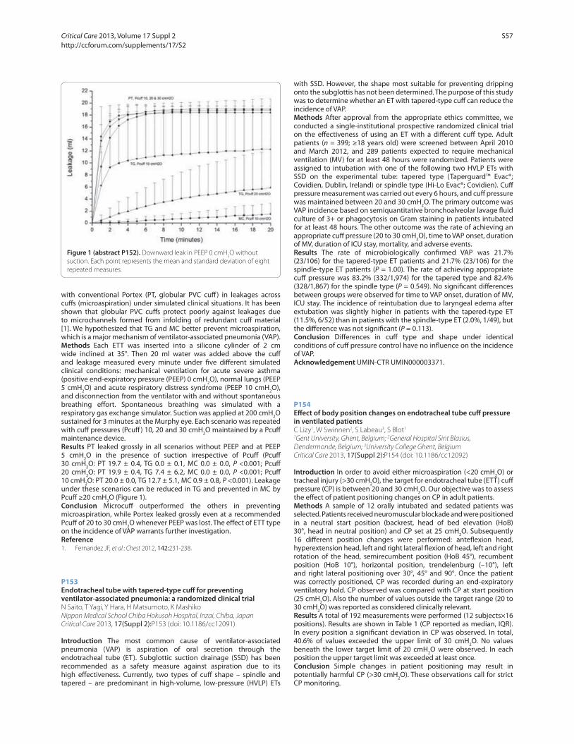
with conventional Portex (PT, globular PVC cuff ) in leakages across cuff s (microaspiration) under simulated clinical situations. It has been shown that globular PVC cuff s protect poorly against leakages due to microchannels formed from infolding of redundant cuff material [1]. We hypothesized that TG and MC better prevent microaspiration, which is a major mechanism of ventilator-associated pneumonia (VAP).Methods Each ETT was inserted into a silicone cylinder of 2 cm wide inclined at 35°. Then 20 ml water was added above the cuff and leakage measured every minute under fi ve diff erent simulated clinical conditions: mechanical ventilation for acute severe asthma (positive end-expiratory pressure (PEEP) 0 cmH2O), normal lungs (PEEP 5 cmH2O) and acute respiratory distress syndrome (PEEP 10 cmH2O), and disconnection from the ventilator with and without spontaneous breathing eff ort. Spontaneous breathing was simulated with a respiratory gas exchange simulator. Suction was applied at 200 cmH2O sustained for 3 minutes at the Murphy eye. Each scenario was repeated with cuff pressures (Pcuff ) 10, 20 and 30 cmH2O maintained by a Pcuff maintenance device.Results PT leaked grossly in all scenarios without PEEP and at PEEP 5 cmH2O in the presence of suction irrespective of Pcuff (Pcuff 30 cmH2O: PT 19.7 ± 0.4, TG 0.0 ± 0.1, MC 0.0 ± 0.0, P <0.001; Pcuff 20 cmH2O: PT 19.9 ± 0.4, TG 7.4 ± 6.2, MC 0.0 ± 0.0, P <0.001; Pcuff 10 cmH2O: PT 20.0 ± 0.0, TG 12.7 ± 5.1, MC 0.9 ± 0.8, P <0.001). Leakage under these scenarios can be reduced in TG and prevented in MC by Pcuff ≥20 cmH2O (Figure 1).Conclusion Microcuff outperformed the others in preventing microaspiration, while Portex leaked grossly even at a recommended Pcuff of 20 to 30 cmH2O whenever PEEP was lost. The eff ect of ETT type on the incidence of VAP warrants further investigation.Reference1. Fernandez JF, et al.: Chest 2012, 142:231-238.
P153Endotracheal tube with tapered-type cuff for preventing ventilator-associated pneumonia: a randomized clinical trialN Saito, T Yagi, Y Hara, H Matsumoto, K MashikoNippon Medical School Chiba Hokusoh Hospital, Inzai, Chiba, JapanCritical Care 2013, 17(Suppl 2):P153 (doi: 10.1186/cc12091)
Introduction The most common cause of ventilator-associated pneumonia (VAP) is aspiration of oral secretion through the endotracheal tube (ET). Subglottic suction drainage (SSD) has been recommended as a safety measure against aspiration due to its high eff ectiveness. Currently, two types of cuff shape – spindle and tapered – are predominant in high-volume, low-pressure (HVLP) ETs
with SSD. However, the shape most suitable for preventing dripping onto the subglottis has not been determined. The purpose of this study was to determine whether an ET with tapered-type cuff can reduce the incidence of VAP.Methods After approval from the appropriate ethics committee, we conducted a single-institutional prospective randomized clinical trial on the eff ectiveness of using an ET with a diff erent cuff type. Adult patients (n = 399; ≥18 years old) were screened between April 2010 and March 2012, and 289 patients expected to require mechanical ventilation (MV) for at least 48 hours were randomized. Patients were assigned to intubation with one of the following two HVLP ETs with SSD on the experimental tube: tapered type (Taperguard™ Evac®; Covidien, Dublin, Ireland) or spindle type (Hi-Lo Evac®; Covidien). Cuff pressure measurement was carried out every 6 hours, and cuff pressure was maintained between 20 and 30 cmH2O. The primary outcome was VAP incidence based on semiquantitative bronchoalveolar lavage fl uid culture of 3+ or phagocytosis on Gram staining in patients intubated for at least 48 hours. The other outcome was the rate of achieving an appropriate cuff pressure (20 to 30 cmH2O), time to VAP onset, duration of MV, duration of ICU stay, mortality, and adverse events.Results The rate of microbiologically confi rmed VAP was 21.7% (23/106) for the tapered-type ET patients and 21.7% (23/106) for the spindle-type ET patients (P = 1.00). The rate of achieving appropriate cuff pressure was 83.2% (332/1,974) for the tapered type and 82.4% (328/1,867) for the spindle type (P = 0.549). No signifi cant diff erences between groups were observed for time to VAP onset, duration of MV, ICU stay. The incidence of reintubation due to laryngeal edema after extubation was slightly higher in patients with the tapered-type ET (11.5%, 6/52) than in patients with the spindle-type ET (2.0%, 1/49), but the diff erence was not signifi cant (P = 0.113).Conclusion Diff erences in cuff type and shape under identical conditions of cuff pressure control have no infl uence on the incidence of VAP.Acknowledgement UMIN-CTR UMIN000003371.
P154Eff ect of body position changes on endotracheal tube cuff pressure in ventilated patientsC Lizy1, W Swinnen2, S Labeau3, S Blot1
1Gent University, Ghent, Belgium; 2General Hospital Sint Blasius, Dendermonde, Belgium; 3University College Ghent, BelgiumCritical Care 2013, 17(Suppl 2):P154 (doi: 10.1186/cc12092)
Introduction In order to avoid either microaspiration (<20 cmH2O) or tracheal injury (>30 cmH2O), the target for endotracheal tube (ETT) cuff pressure (CP) is between 20 and 30 cmH2O. Our objective was to assess the eff ect of patient positioning changes on CP in adult patients.Methods A sample of 12 orally intubated and sedated patients was selected. Patients received neuromuscular blockade and were positioned in a neutral start position (backrest, head of bed elevation (HoB) 30°, head in neutral position) and CP set at 25 cmH2O. Subsequently 16 diff erent position changes were performed: antefl exion head, hyperextension head, left and right lateral fl exion of head, left and right rotation of the head, semirecumbent position (HoB 45°), recumbent position (HoB 10°), horizontal position, trendelenburg (–10°), left and right lateral positioning over 30°, 45° and 90°. Once the patient was correctly positioned, CP was recorded during an end-expiratory ventilatory hold. CP observed was compared with CP at start position (25 cmH2O). Also the number of values outside the target range (20 to 30 cmH2O) was reported as considered clinically relevant.Results A total of 192 measurements were performed (12 subjects×16 positions). Results are shown in Table 1 (CP reported as median, IQR). In every position a signifi cant deviation in CP was observed. In total, 40.6% of values exceeded the upper limit of 30 cmH2O. No values beneath the lower target limit of 20 cmH2O were observed. In each position the upper target limit was exceeded at least once.Conclusion Simple changes in patient positioning may result in potentially harmful CP (>30 cmH2O). These observations call for strict CP monitoring.
Figure 1 (abstract P152). Downward leak in PEEP 0 cmH2O without suction. Each point represents the mean and standard deviation of eight repeated measures.
Critical Care 2013, Volume 17 Suppl 2 http://ccforum.com/supplements/17/S2
S57

P155Towards safer airway management in the critically ill: lessons from National Audit Project 4N Santana-Vaz, S Tallowin, H Lewis, D Park, R O’Brien, JM PatelHeart of England NHS Foundation Trust, Birmingham, UKCritical Care 2013, 17(Suppl 2):P155 (doi: 10.1186/cc12093)
Introduction National Audit Project 4 (NAP4) highlighted the need to improve airway management in ICUs and key recommendations were the continuous use of end-tidal carbon dioxide (ETCO2) monitoring, pre-intubation checklists and diffi cult airway trolleys [1]. This complete cycle audit aimed to quantify the current state of airway management on our ICU and the eff ectiveness of implementing the NAP4 recommendations.Methods Data collection was carried out prospectively for both phases and included documentation of intubation, use of ETCO2 and the incidence of serious adverse events (SAEs). The contents of the intubation boxes were compared against the Diffi cult Airway Society (DAS) guidelines [2]. The re-audit was carried out 6 months following the introduction of a pre-intubation checklist, a documentation sticker, a diffi cult airway trolley and standardization of the basic bedside airway boxes with a checklist of contents. A training program in airway management for all ICU staff was also introduced.Results The baseline characteristics of both groups were similar. The initial audit included 45 patients and the re-audit 58 patients. In the initial audit 19% of patients had accurate documentation of intubation with the use of ETCO2. The continuous use of ETCO2 was 80%. Bedside airway boxes did not contain a checklist of standardized equipment. A diffi cult airway trolley was not located on the ICU. Following introduction of the changes described, a signifi cant improvement in the use of ETCO2 to confi rm intubation (46% vs. 19%, P <0.01) and continuous use of ETCO2 monitoring (100% vs. 80% P ≤0.0004) was observed. The use of the pre-intubation checklist (7%) and stickers (11%) was poor. All bedside boxes had a checklist and the required equipment. A diffi cult airway trolley is located on the ICU with an equipment list and DAS algorithm attached. No SAEs were recorded in either phase.Conclusion Serious airway complications are rare. Safer airway manage ment is essential in the ICU to prevent morbidity and mortality.
This audit demonstrated that simple measures in conjunction with a structured training program can help to improve the safety of airway management in critically ill patients. Further work is required to improve compliance with the pre-intubation checklist alongside a continued education program.References1. The Royal College of Anaesthetists [www.rcoa.ac.uk/nap4]2. Diffi cult Airway Society Equipment List
[http://www.das.uk.com/equipmentlistjuly2005.htm]
P156Quantitative assessment of endotracheal tube (ETT) biofi lm by micro-CT scan: evaluation of the eff ectiveness of ETT cleaning devicesA Coppadoro1, G Bellani1, T Mauri1, R Borsa1, V Meroni1, A Barletta1, M Teggia Droghi1, A Lucchini2, R Marcolin2, N Patroniti1, S Bramati2, A Pesenti11University of Milan-Bicocca, Monza, Italy; 2San Gerardo Hospital, Monza, ItalyCritical Care 2013, 17(Suppl 2):P156 (doi: 10.1186/cc12094)
Introduction The goal of this study was to assess the amount of biofi lm within the endotracheal tube (ETT) by CT scan and to evaluate in a bench test a device aimed at ETT biofi lm removal. Biofi lm grows within the ETT soon after intubation, resulting in increased resistance to airfl ow. Biofi lm detachment from the ETT can result in bacterial colonization of the distal airways. The use of an ETT cleaning device could eff ectively reduce the patient’s airfl ow resistance burden. The quantifi cation and distribution of biofi lm within the ETT might help to evaluate ETT cleaning device eff ectiveness.Methods We collected ETTs of 11 critically ill patients after extubation. Micro-CT scan (SkyScan 1176; Bruker, Belgium) was performed using a resolution of 35 μm. Axial sections of the 20 cm above the cuff were reconstructed, and the volume of secretions was assessed by a density criterion. Microbiological cultures of the ETT lavage fl uid were then obtained. Patient’s demographics and clinical data were recorded. In a diff erent set of bench experiments, we injected 1 ml water-based polymer into new ETTs of diff erent sizes. We measured resistance to airfl ow before and after using an ETT cleaning device (Airway Medix Closed Suction System; Biovo Technologies, Tel Aviv, Israel). We also obtained resistance values of intact ETTs as controls.Results The studied ETTs remained in place for a median of 7 days (IQR range 4 to 15). The amount of secretions assessed by CT scan was 0.293 ± 0.290 ml (range 0.032 to 0.777 ml). Secretion volumes were not related to patient severity at admission (SAPS 2, P/F ratio) or days of intubation; an inverse correlation with patient’s age was present (P = 0.032, R2 = 0.46). Bacterial growth was present in 9/11 (82%) ETT fl uids cultures and Candida spp. showed an elevated prevalence (6/11, 55%). In the bench tests, the cleaning device reduced resistance to airfl ow (diff erence before and after cleaning 5.5 (95% CI = 8.9 to 1.6) cmH2O/l/second, P = 0.006). After cleaning, resistance resulted higher than intact ETTs, although with a clinically negligible diff erence (diff erence 0.3 (95% CI = 0.2 to 0.6 cmH2O/l/second), P = 0.032).Conclusion Micro-CT scan is a feasible and promising technique to assess secretions volume in ETTs after extubation. The use of an ETT cleaning device decreases resistance to airfl ow in bench tests; the eff ectiveness of such a device in the clinical setting could be properly assessed by post-extubation CT scan.
P157Early identifi cation of patients at risk of diffi cult intubation in the ICU: development and validation of the MACOCHA score in a multicenter cohort studyA De Jong1, N Molinari1, N Terzi2, N Mongardon3, B Jung1, S Jaber1
1Montpellier University Hospital, Montpellier, France; 2Caen University Hospital, Caen, France; 3Cochin University Hospital, Paris, FranceCritical Care 2013, 17(Suppl 2):P157 (doi: 10.1186/cc12095)
Introduction Diffi cult intubation (DI) in the ICU is a challenging issue, associated with severe life-threatening complications [1,2]. The objective was to develop and validate a simplifi ed score for identifying patients with DI in the ICU and to report related complications.
Table 1 (abstract P154). Endotracheal tube cuff pressure values following patient position changes
Cuff P for Δ from Measurements pressure start position >30 cmH2O (cmH2O)a (25 cmH2O) (n (%))
Antefl exion head 33.5 (27.5 to 42.0) 0.005 9 (75)
Hyperextension head 30.5 (27.5 to 42.0) 0.004 6 (50)
Lateral fl exion head, left 26.5 (25.3 to 28.8) 0.025 1 (8)
Lateral fl exion head, right 27.0 (25.3 to 29.8) 0.049 2 (17)
Rotation head, left 27.0 (26.3 to 33.8) 0.003 3 (25)
Rotation head, right 28.0 (26.3 to 38.8) 0.020 4 (33)
Semirecumbent, 45° HoB 28.0 (25.3 to 31.5) 0.009 2 (17)
Recumbent, 10° HoB 28.0 (25.3 to 31.5) 0.016 3 (25)
Horizontal back rest 28.5 (25.3 to 31.5) 0.011 3 (25)
Trendelenburg, 10° 29.0 (25.3 to 32.0) 0.008 5 (42)
Left lateral position, 30° 30.5 (25.3 to 32.0) 0.010 6 (50)
Left lateral position, 45° 30.5 (27.3 to 32.8) 0.005 6 (50)
Left lateral position, 90° 34.0 (27.0 to 36.8) 0.006 7 (58)
Right lateral position, 30° 31.0 (27.0 to 34.5) 0.003 6 (50)
Right lateral position, 45° 32.5 (28.3 to 35.8) 0.002 8 (50)
Right lateral position, 90° 31.0 (29.3 to 35.0) 0.003 7 (58)
Total (192 measurements) – – 78 (40.6)
HoB, head of bed elevation. aCuff pressure values reported as median (fi rst to third quartile).
Critical Care 2013, Volume 17 Suppl 2 http://ccforum.com/supplements/17/S2
S58

Methods Data collected in a prospective multicenter-study from 1,000 consecutive intubations from 42 ICUs were used to develop a simplifi ed score of DI, which was then validated externally in 400 consecutive intubation procedures from 18 other ICUs and internally by bootstrap on 1,000 iterations.Results In multivariate analysis, the main predictors of DI (incidence = 11.3%) were related to the patient (Mallampati score III or IV, obstructive sleep apnea syndrome, reduced mobility of cervical spine, limited mouth opening), to pathology (severe hypoxia, coma) and to the operator (non-anesthesiologist). From the β-parameter, a seven-item simplifi ed score (MACOCHA score; Table 1) was built, with an area under the curve (AUC) of 0.89 (95% CI = 0.85 to 0.94). In the validation cohort (prevalence of DI = 8%), the AUC was of 0.86 (95% CI = 0.76 to 0.96), with a sensitivity of 73%, a specifi city of 89%, a negative predictive value of 98% and a positive predictive value of 36%. After internal validation by bootstrap, the AUC was 0.89 (95% CI = 0.86 to 0.93). Severe life-threatening events (severe hypoxia, collapse, cardiac arrest or death) occurred in 38% of the 1,000 cases. Patients with DI (n = 113) had signifi cantly higher severe life-threatening complications than those who had a non-DI (51% vs. 36%, P <0.0001).
Table 1 (abstract P157). MACOCHA score calculation worksheet
Points
Factors related to patient Mallampati score III or IV 5 Obstructive apnea syndrome 2 Reduced mobility of cervical spine 1 Limited mouth opening <3 cm 1
Factors related to pathology Coma 1 Severe hypoxemia (<80%) 1
Factor related to operator Non-anaesthesiologist 1
Total 12
Conclusion DI in ICU is strongly associated with severe life-threatening complications. A simple score including seven clinical items discriminates diffi cult and non-DI in ICU.References1. Jaber et al.: Crit Care Med 2006, 34:2355-2361.2. Jaber et al.: Intensive Care Med 2010, 36:248-255.
P158Incidence of major complications related to endotracheal intubation in ICUs of nonacademic Brazilian public hospitalsAB Cavalcanti1, K Normilio-da-Silva1, JC Acarine Mouro1, F Moreira1, AA Kodama1, R Del-Manto2, O Berwanger1
1Hospital do Coração – HCor, São Paulo, Brazil; 2Hospital Militar de Área de São Paulo – HMASP, São Paulo, BrazilCritical Care 2013, 17(Suppl 2):P158 (doi: 10.1186/cc12096)
Introduction Our objective is to determine the incidence of major complications related to endotracheal intubations (death, cardiac arrest, severe hemodynamic instability or severe hypoxemia) per-formed in the ICUs of nonacademic Brazilian public hospitals.Methods This is a report of the baseline phase of a cluster randomized trial with two parallel arms. In this baseline phase, we collected data from sequential patients needing endotracheal intubation in 17 ICUs from Brazil. Patients needing endotracheal intubation after cardiac arrest were excluded. In a second ongoing phase, not reported here, we have randomized ICUs to a multifaceted intervention to prevent complications of endotracheal intubation. Primary outcome was defi ned as the occurrence of death, cardiac arrest, severe cardiovascular instability (systolic blood pressure <60 mmHg at least one time, <90 mmHg and persisting >30 minutes or need to initiate vasopressors) or severe hypoxemia (SpO2 <80%) within 1 hour after intubation.Results We evaluated 246 intubations. The mean age was 49 ± 21.9 years; the SAPS 3 admission score was 57.4 ± 15.4. The most
common indications of intubation were acute respiratory failure (61.0%) and coma (17.1%). The primary outcome occurred in 138 of 246 intubations (56.1%). Within the fi rst hour, the incidence of severe hypotension was 47.2%, of severe hypoxemia was 20.3%, of cardiac arrest was 5.7% and of death was 4.5%.Conclusion Incidence of major complications associated with endotracheal intubation is unacceptably high in Brazilian public ICUs. There is an urgent need for eff ective measures to increase safety of this common procedure in our environment.
P159Tracheal intubation for a diffi cult airway using Airway scope®, KingVision® and McGRATH®: a comparative manikin study of inexperienced personnelJ Itai1, Y Tanabe2, T Nishida1, T Inagawa1, Y Torikoshi1, Y Kida1, T Tamura1, K Ota1, T Otani1, T Sadamori1, K Une1, R Tsumura1, Y Iwasaki1, N Hirohashi1, K Tanigawa1
1Hiroshima University Hospital, Hiroshima-shi, Japan; 2Hiroshima City Hospital, Hiroshima-shi, JapanCritical Care 2013, 17(Suppl 2):P159 (doi: 10.1186/cc12097)
Introduction The Airway scope® (AWS), the KingVision® (KV) and the McGRATH® (MG) are new indirect video laryngoscopes designed to facilitate eff ective and safe tracheal intubation under various conditions. However, there are few comparative studies as for performance in tracheal intubation attempted by inexperienced personnel. The purpose of this study was to evaluate success rates, time to intubation with use of these devices by inexperienced personnel in a simulated manikin diffi cult airway.Methods Twenty-nine fi fth-year medical students with no previous experience in tracheal intubation participated in this study. We used an advanced patient simulator (SimMan®; Laerdal Medical, Stavanger, Norway) to simulate normal and diffi cult airway scenarios including cervical spine rigidity, swollen tongue, and pharyngeal edema. The sequences in selecting devices and scenarios were randomized. Success rate for tracheal intubation, and the time required for visualization of the glottis (T1), tracheal intubation (T2), and infl ation of the lungs (T3) were analyzed. Also, numbers of audible dental click during the intubation attempt were recorded. The three diff erent intubation devices were tested in four diff erent scenarios by 29 students.Results All three devices had very high success rates of tracheal intubation (AWS 100%; KV 100%; MG 99%). In the normal airway, T1, T2 and T3 were AWS: 4.4 ± 3.3 seconds, 7.5 ± 3.8 seconds, 11.1 ± 3.8 seconds; KV: 6.9 ± 6.9 seconds, 10.3 ± 7.9 seconds, 14.1 ± 8.3 seconds; MG: 4.6 ± 1.3 seconds, 11.4 ± 5.2 seconds, 16.1 ± 5.4 seconds, respectively (NS). In the three diffi cult airway scenarios, T1, T2 and T3 were AWS: 5.3 ± 4.7* seconds, 10.4 ± 6.9* seconds, 14.2 ± 6.9* seconds; KV: 10.9 ± 12.8 seconds, 21.5 ± 18.1 seconds, 25.4 ± 18.3 seconds; MG: 13.0 ± 15.3 seconds, 26.2 ± 24.6 seconds, 31.0 ± 24.3 seconds, respectively (*P <0.05 AWS vs. KV and MG). The number of audible dental click sounds with the MG was greater than with the AWS and KV (AWS 5%*; KV 8%*; MG 28%*; *P <0.05 AWS and KV vs. MG).Conclusion The AWS, KV and MG had very high success rates of tracheal intubation and are suitable as intubation devices for inexperienced personnel. In the diffi cult airway, however, the intubation time with AWS was signifi cantly shorter than with KV and MG. These fi ndings suggested that AWS may be most useful device particularly in diffi cult situations such as emergency settings. Further studies in a clinical setting are needed to confi rm these fi ndings.
P160Resistance of neonatal endotracheal tubes: a comparison of four commercially available typesJ Spaeth, D Steinmann, J Guttmann, S SchumannUniversity Medical Center Freiburg, GermanyCritical Care 2013, 17(Suppl 2):P160 (doi: 10.1186/cc12098)
Introduction In mechanically ventilated neonates the fl ow-dependent resistance of the endotracheal tube (ETT) causes a noticeable pressure diff erence between airway and tracheal pressure [1]. This may potentially lead to retardation of the passive driven expiration and dynamic lung infl ation consecutively but more importantly increases
Critical Care 2013, Volume 17 Suppl 2 http://ccforum.com/supplements/17/S2
S59

the work of breathing. The aim of this study was to compare the resistive pressure drop of four commercially available neonatal ETTs with internal diameter (ID) of 2.0 mm.Methods The pressure–fl ow relationship of neonatal ETTs (internal diameter 2.0 mm) of four diff erent manufacturers (Mallinckrodt, Hi-Contour, Covidien, Dublin, Ireland; Portex, Cole’s Neonatal Tube, Smith Medical, St Paul, MN, USA; Rüsch, Silko Clear, Telefl ex Medical, Kernen, Germany; and Vygon, Pediatric Endotracheal Tube, Ecouen, France) was determined in a physical model consisting of a tube connector, an anatomically curved ETT and an artifi cial trachea. The model was ventilated with a sinusoidal gas fl ow with an amplitude of ±100 ml/second and a ventilation rate ranging from 40 to 60 cycles/minute. The coeffi cients of an approximation equation considering ETT resistance were fi tted separately to the measured pressure–fl ow curves for inspiration and expiration.Results The pressure drop profi les of all ETTs were nonlinearly fl ow dependent. The expiratory pressure drop (Pexp) slightly exceeded the inspiratory one (Pinsp). The ETT of Portex Cole’s Neonatal Tube had a signifi cant lower pressure drop (Pinsp 8.8 ± 0.3 cmH2O vs. Pexp –11.2 ± 0.2 cmH2O at an air fl ow of 100 ml/second) compared with all other ETTs (Mallinckrodt: Pinsp 19.2 ± 0.4 cmH2O vs. Pexp –24.3 ± 0.7 cmH2O; Rüsch: Pinsp 28.3 ± 0.8 cmH2O vs. Pexp –33.0 ± 0.8 cmH2O; Vygon: Pinsp 19.2 ± 0.4 cmH2O vs. Pexp –22.6 ± 0.6 cmH2O, all P <0.05) for all fl ow and ventilation rates, respectively.Conclusion The ETT resistance highly contributes to the total airway resistance in neonatal ventilation. The fl ow-dependent pressure drop of shouldered Cole’s Neonatal Tube (Portex) was up to 70% less in inspiration (resp. 67% in expiration) compared with straight tubes with an internal diameter corresponding to the narrow part of Cole’s tube. We conclude that neonatal intubation with a Cole’s tube can clearly reduce the resistive load due to the endotracheal tube and thus potentially prevent additionally work of breathing.Reference1. Guttmann J, et al.: Crit Care Med 2000, 28:1018-1026.
P161NT-proBNP and cardiac cycle effi ciency changes during extubation process in critically ill patientsF Franchi, P Mongelli, M Cozzolino, B Galgani, C Ragozzino, C Bianchi, B BiagioliUniversity of Siena, ItalyCritical Care 2013, 17(Suppl 2):P161 (doi: 10.1186/cc12099)
Introduction Several factors can lead to weaning failure from mechanical ventilation (MV). Among these, cardiac dysfunction is one of the main causes. NT-proBNP has been proposed as a biomarker of cardiovascular function during weaning from MV. Unfortunately it does not provide for a continuous monitoring of cardiac function. Pulse wave analysis may serve as a continuous bedside monitoring tool of cardiovascular performance. Cardiac cycle effi ciency (CCE) is an indirect index of left ventricular performance obtained by the pulse contour method MostCare (Vygon, Padova, Italy). The aim of this study was to evaluate the correlation between NT-proBNP and CCE and the potential usefulness of such variables during the weaning process from MV.Methods Twenty-two long-term (>48 hours) mechanically ventilated patients capable of performing a weaning trial of spontaneous breathing (SBT) were enrolled in the study. Inclusion criteria were: age >18 years and equipment with a standard arterial catheter line. Exclusion criteria were: neuromuscular disease, tracheotomy, renal failure, and traumatic brain injury. During the weaning process, NT-proBNP plasma levels, CCE, and standard hemodynamic and ventilatory data were collected 30 minutes before extubation (T1), 2 hours (T2) and 12 hours later (T3). After removal of tracheal tube, patients with a history of heart failure received continuous positive airway pressure (CPAP group). Patients with normal cardiac function were maintained with spontaneous breathing (SB group).Results Sixty-six paired NT-proBNP and CCE values were obtained. Patients in the SB group and in the CPAP group were 10 and 12, respectively. In both groups there was a trend towards an increase in NT-proBNP values after extubation, an opposite trend was observed regarding CCE values (P <0.05). NT-proBNP levels showed an increase
after extubation (T2, T3) compared with T1; conversely, CCE showed an inverse trend. Overall, a negative correlation was found between NT-proBNP and CCE values (R = –0.81, P <0.001). Signifi cant inverse correlations were found between NT-proBNP and CCE at T1, T2, and T3 (R = –0.91, –0.75 and –0.73 respectively; P < 0.001). The overall correlation between NT-proBNP and CCE was –0.74 in the SB group and –0.86 in the CPAP group. Standard hemodynamic and ventilatory data did not show signifi cant changes during the study.Conclusion NT-proBNP correlated well with CCE. The latter seems to be an additional attractive index of cardiovascular state that, in association with NT-proBNP changes, may provide information about cardiac function on a beat-by-beat basis during weaning process from MV.
P162Comparison of outcomes between early and late tracheostomy for critically ill patientsK Suzuki1, S Kusunoki1, T Yamanoue1, K Tanigawa2
1Hiroshima Prefectural Hospital, Hiroshima, Japan; 2Hiroshima University Hospital, Hiroshima, JapanCritical Care 2013, 17(Suppl 2):P162 (doi: 10.1186/cc12100)
Introduction Tracheostomy is one of the more commonly performed procedures in critically ill patients requiring long-term mechanical ventilation. However, the optimal timing or method of performing tracheostomies in this population remains to be established. In the present study, we compared outcomes of early and late tracheostomy in critically adult patients with diff erent clinical conditions.Methods All patients needing tracheostomy in the Critical Care Medical Center of Hiroshima Prefectural Hospital from January 2009 to December 2011 were surveyed. Patients with tracheostomy who were not indicated for mechanical ventilation were excluded from the subjects. Early tracheostomy (ET) was defi ned as <10 days after tracheal intubation and late tracheostomy (LT) was defi ned as ≥10 days after intubation. We compared patient characteristics, type of tracheostomy procedure, length of weaning from ventilator and outcomes between the groups. Data are shown as the mean ± SD, with unpaired t test and Mann–Whitney U test used for statistical analyses. Statistical signifi cance was accepted at P <0.05.Results One hundred patients were surveyed. The ET and LT groups included 49 and 51 patients, respectively. Tracheostomy was performed using a percutaneous procedure in 48 patients (ET: 25, LT: 23) and a surgical procedure in 52 patients (ET: 24, LT: 28). Sixty-two patients (ET: 34, LT: 28) survived to discharge and 16 patients died in the ICU (ET: 7, LT: 9). Fifty-six patients (ET: 31, LT: 25) were weaned from ventilator support and tracheostomy cannula was removed in 20 patients (ET: 11, LT: 9). There were no signifi cant diff erences in type of tracheostomy procedure, period from tracheostomy until ICU and hospital discharge, rate of patients who could be weaned from ventilator and removed tracheostomy cannula, and ICU and hospital mortality between the groups. The length of mechanical ventilation and the time to removal of tracheostomy cannula were signifi cantly shorter in the ET group (5 ± 7 vs. 26 ± 41 and 29 ± 24 vs. 94 ± 83 days, respectively).Conclusion In this retrospective study, early tracheostomy reduced the length of weaning after tracheostomy and the time to removal of tracheostomy cannula, while there were no diff erences in the length of ICU stay and patient outcome. In critically ill adult patients who require mechanical ventilation, a tracheostomy performed at an earlier stage may shorten the duration of artifi cial ventilation. A further randomized clinical trial is essential to determine the eff ectiveness and safety of early tracheostomy.Reference1. Arabi Y, et al.: Crit Care 2004, 8:R347-R352.
P163Ultrasound scanning for percutaneous dilatational tracheostomy: a systematic reviewR Pugh, A SlaterGlan Clwyd Hospital, Bodelwyddan, UKCritical Care 2013, 17(Suppl 2):P163 (doi: 10.1186/cc12101)
Introduction Percutaneous dilatational tracheostomy (PDT) remains a frequently performed procedure in the ICU. However, there is great
Critical Care 2013, Volume 17 Suppl 2 http://ccforum.com/supplements/17/S2
S60

variability in the course of blood vessels in the pre-tracheal area. A 5% risk of clinically relevant bleeding was recently reported for patients undergoing PDT [1]. We conducted a systematic review of reports evaluating clinical outcomes following use of ultrasound scanning (US) for PDT.Methods Two investigators performed a search of the literature using the following databases: CENTRAL, Embase, MEDLINE and SCOPUS. The following eligibility criteria were used: population including adults >16 years managed in the ICU; use of ultrasound to guide decision-making pre-PDT or guide PDT performance; report of clinically relevant outcome measures. Nonrandomised controlled trials were classifi ed according to Cochrane Non-Randomised Study Methods Group criteria [2] and evaluated for risk of bias.Results An initial search identifi ed 2,043 reports, of which 10 studies met eligibility criteria: eight case series, one randomised controlled trial (RCT) and one prospective cohort study, incorporating 488 patients. Two studies specifi cally reported data on patients with obesity (n = 29 patients) and one study reported data for a group of patients with spinal cord fi xation (n = 6). US was used to guide decision to perform PDT or surgical tracheostomy in fi ve studies, with decision to perform surgical tracheostomy ranging from 0 to 27% of cases. US was used to guide insertion point in seven studies, and used real-time in four studies. Times to perform US-guided PDT were reported in four studies (ranging from 8 to 12 minutes). No studies compared time taken with or without US. Data on complications of procedure were reported in nine studies. Minor bleeding was reported for eight cases (1.6% overall). Prolonged bleeding was reported in two cases (0.4%). There were no episodes of catastrophic bleeding among 488 cases. High risk of bias was identifi ed in fi ve studies in terms of patient selection. An intervention protocol was not defi ned in three reports. No attempt was made at blinding any aspect of the 10 studies.Conclusion Use of US guidance could theoretically help minimise risk of haemorrhagic complications during PDT and perhaps reduce time taken to perform PDT. However, there is currently inadequate evidence from controlled cohort studies or RCTs to suggest that routine use for PDT in selected or unselected groups improves clinically relevant outcome measure.References1. Delaney et al.: Crit Care 2006, 10:R55.2. Higgins JPT, Green S (Eds): Cochrane Handbook for Systematic Reviews of
Interventions. Version 5.1.0. The Cochrane Collaboration; 2011. [www.cochrane-handbook.org]
P164Evaluation of a new device for emergency transcricoid ventilation in a manikin modelP Persona, P Diana, A Ballin, F Baratto, M Micaglio, C OriClinica di Anestesia e Medicina Intensiva, Padova, ItalyCritical Care 2013, 17(Suppl 2):P164 (doi: 10.1186/cc12102)
Introduction Failed airway situations are potentially catastrophic events and require a correct approach with appropriate tools. Recently, Ventrain has been presented as a manual device for emergency ventilation through a small-bore cannula, which can provide expiratory assistance by applying the Venturi eff ect.Methods We used the SimulARTI Human Patient Simulator to evaluate Ventrain. Initially, we studied the eff ectiveness and security in ventilating and oxygenating the patient. In a second phase, the Ventrain performance was compared with what is considered to be the present gold standard (Quicktrach II, Portex Mini-Trach II Seldinger Kit, Melker Emergency Cricothyrotomy Catheter Set). Seven anesthesiologists performed an emergency transcricoid ventilation with each device in the same setting.Results Ventrain provided an average tidal volume of 334 ml and an average minute volume of 2.4 l in the considered situation, with a modifi cation of PAO2 from 32 to 702 mmHg and of PACO2 from 54.5 to 38.8 mmHg. In the second phase, the time needed to obtain an eff ective oxygenation with Ventrain was found to be shorter than other devices (median diff erence; vs. Minitrach –60 seconds; vs. Melker –35 seconds; vs. Quicktrach –25 seconds) (Figure 1); the ability to remove CO2 resulted bigger (average diff erence: vs. Minitrach –11.9;
vs. Melker –0.3; vs. Quicktrach –5.9) (Figure 2) and moreover the users judged it more favorably.Conclusion In this manikin study, Ventrain seemed to be able to appropriately oxygenate and ventilate a patient in a CICV situation. When compared with the best available choices, it has shown not to be inferior.Reference1. Cook TM, Nolan JP, Magee PT, Cranshaw JH: Needle cricothyroidotomy.
Anaesthesia 2007, 62:289-291.
P165Device for measuring bronchodilator delivery and response in resource-limited settingsCW Carspecken, D TalmorHarvard Medical School and Beth Israel Deaconess Medical Center, Boston, MA, USACritical Care 2013, 17(Suppl 2):P165 (doi: 10.1186/cc12103)
Introduction Eff ective delivery of aerosolized bronchodilators for patients with asthma is crucial for adequate therapy in critical care and emergent settings. Often administered with pressure-metered dose inhalers (pMDIs), bronchodilator delivery depends on the correct patient technique during administration [1] and the ability to measure treatment response, which are diffi cult to monitor at the point of care and particularly so in resource-poor settings where standard in-hospital monitoring is unavailable [2].Methods A point-of-care device for airfl ow measurement during bronchodilator delivery was designed and tested for use in resource-limited settings. The handheld device was constructed from a clinical aerosol delivery tube with a bidirectional sensor for pressure diff erential detection about the aerosol element (Figure 1). The custom low-cost
Figure 1 (abstract P164). Oxygenation time to oxygen alveolar pressure >100 mmHg.
Figure 2 (abstract P164). Mean carbon dioxide alveolar pressure at the start and at the end of the test.
Critical Care 2013, Volume 17 Suppl 2 http://ccforum.com/supplements/17/S2
S61

(<$11) electronics were designed such that no power supply would be needed apart from a computer or mobile device. Collection of airfl ow signal and calibration was performed with a standard 3 l syringe with fl ow volume measurement on one adult subject.Results Calibration of the assembled device over range of 0.5 to 3 l/second enabled conversion of pressure diff erence to air fl ow with a mean measured tube resistance of 0.258 (kPa*second)/l. Robust signal responses to an adult subject’s continuous respiratory maneuvers on the tube itself were demonstrated. Subject performance of the pMDI technique with subsequent tidal volume breathing was recorded and analyzed (Figure 1).Conclusion Design and calibration of a novel low-cost monitoring device for bronchodilator delivery monitoring enabled detection and recording of characteristic fl ow volume respiratory patterns for point-of-care diagnostics in resource-limited settings. Future work will require clinical testing and automated detection of the correct pMDI patient technique.References1. Ari A, et al.: Respir Care 2012, 57:613-626.2. Riviello E, et al.: Crit Care Med 2011, 39:860.
P166Evidence-based guidelines and protocols for the management of adult patients with a tracheostomy: a systematic reviewJ Siddiqui1, PB Sherren2, MA Birchall31North West London Hospitals NHS Trust, London, UK; 2Barts and The London NHS Trust, London, UK; 3The Royal National Throat Nose and Ear Hospital, London, UKCritical Care 2013, 17(Suppl 2):P166 (doi: 10.1186/cc12104)
Introduction Protocol-based care of the tracheostomised patient is important, as adverse events confer a high rate of mortality. Little is known regarding the existence of formal evidence-based guidelines on tracheostomy care. The aim of this study was to perform a systematic review for evidence-based guidelines on adult tracheostomy care.Methods A systematic search of PubMed, MEDLINE, guideline clearinghouses, centres of evidence-based practice, and professional societies’ guidelines relating to care of adult patients with a tracheostomy was performed by two reviewers. In addition, a Google search of publicly available tracheostomy care guidelines was performed. Search terms: (tracheostom* OR tracheotom*) AND (protocol* OR guideline* OR standard* OR management OR consensus OR algorithm*). Filters: English language, human, from 1 January 1990 to date, adult patients. Guideline appraisal criteria: the quality of guidelines retrieved was assessed using the Appraisal of Guidelines Research and Evaluation II (AGREE II) instrument [1].Results The search results are summarised in Table 1. A total of 80 guidelines were identifi ed. Five were found to satisfy the AGREE II criteria and only three related to the entire spectrum of tracheostomy management. The majority was informal and was not published or evidence based.Conclusion Five evidence-based guidelines on adult tracheostomy management were identifi ed. This may represent a paucity of evidence on the subject, suggesting that further clinical trials on the topic are needed to contribute to the evidence base. This also highlights the need for international consensus on the topic, to reduce duplication of eff orts, standardise practice, and improve outcomes.Reference1. Appraisal of Guidelines for Research and Evaluation: Instrument. AGREE II
[http://www.agreetrust.org/]
Figure 1 (abstract P165).
Table 1 (abstract P166). Summary of results returned following systematic review
Total Results with Full texts Guidelines AGREE instrumentDatabase results limits retrieved found compliant
PubMed 4,685 1,596 33
MEDLINE 3,859 1,337 17 26 2
CINAHL 711 172 6
Medical societies/guideline websites/clearinghouses N/A N/A N/A 54 3
Google 17,600,000 497 N/A
Critical Care 2013, Volume 17 Suppl 2 http://ccforum.com/supplements/17/S2
S62

P167Survey of emergency tracheostomy management in the East of England regionN Lawrence, L Oakley, C Swanavelder, M PalmerWest Suff olk Hospital, Bury St Edmunds, UKCritical Care 2013, 17(Suppl 2):P167 (doi: 10.1186/cc12105)
Introduction The 4th National Audit Project of the Royal College of Anaesthetists [1] concluded that the majority of airway-related signifi cant complications in ICUs resulted from displaced or blocked tracheostomies and recommended together with the Intensive Care Society and the National Tracheostomy Safety Project that each ICU in the UK should have an emergency airway management plan and guidelines [2]. The aim of this survey was to establish whether such guidelines exist and are familiar to those working within the ICUs of the East of England (EoE), their ease of availability in an emergency and the degree of emergency tracheostomy training within the region.Methods Data collection was via a telephone survey of 11 ICUs in the EoE training region during July 2012 with one senior ICU nurse and one ICU trainee questioned per hospital. Questions related to the existence and accessibility of guidelines for tracheostomy emergencies, and to the respondent’s degree of emergency tracheostomy training and their perceived availability of formal training.Results All 11 ICUs questioned perform and manage tracheostomies. Of 22 respondents, 10 knew of guidelines covering all of the emergencies described above and their location. Four respondents thought that these guidelines were accessible in an emergency setting, one-half of which were on computer systems requiring a login and search function. With regards to emergency management, 19 respondents felt competent in a tracheostomy emergency; almost exclusively through experience and in-house teaching. No respondents were aware of any formal emergency tracheostomy management courses.Conclusion Despite national guidance within the UK this survey highlights that implementation and awareness of emergency tracheostomy guidelines in ICUs in the EoE region is poor, and when present they are not readily accessible in an emergency. Emergency training has largely been informal and the availability of formal training courses has not been recognised. In order to improve patient safety there is a need to ensure that emergency tracheostomy management including guidelines, equipment and formalised tracheostomy emergency training are adopted and embraced universally.References1. Cook TM, et al.: Results of the 4th National Audit Project of the Royal
College of Anaesthetists. Br J Anaesth 2011, 106:632-642.2. McGrath BA, et al.: Multidisciplinary guidelines for the management of
tracheostomy and laryngectomy airway emergencies. Anaesthesia 2012, 67:1025-1041.
P168Can we keep neck breathers safe? A survey on training adequacy of medical staff caring for tracheostomy/laryngectomy patientsC Tjen, G Rajendran, S HutchinsonNorfolk and Norwich University Hospital, Norwich, UKCritical Care 2013, 17(Suppl 2):P168 (doi: 10.1186/cc12106)
Introduction A fatal incident related to a blocked tracheostomy tube prompted a review in our Trust. To provide safe tracheostomy care, changes in staffi ng, education and operational policies were recommended. Training of potential fi rst responders to tracheostomy or laryngectomy emergencies remains outstanding. We aim to quantify the training defi cit. Tracheostomies are common in critical care but these patients require ongoing management of an artifi cial airway on discharge to the ward and even the community. In 2010 our critical care unit cared for 108 tracheostomy patients, of which 30 were transferred to the wards. The 4th National Audit Project highlighted complications including hypoxic brain injury and death [1] and the National Patient Safety Agency recognised a number of avoidable aspects [2]. Existing guidelines for management of these patients including emergencies are not widely known.Methods An anonymous online survey was sent to all trainees who may respond to a tracheostomy emergency in our organisation. Trainees in anaesthesia/critical care, general medicine, general surgery,
ENT, thoracics and A&E were approached. All completed forms were included.Results We achieved a response rate of 39% (65/168). Respondents comprised: 33% anaesthesia/critical care, 47% medicine and 14% surgery. Over one-half (36/65) had managed tracheostomy/laryngec-tomy emergencies, with 42% (15/36) of these incidents occurring on the wards and one in an outpatient clinic. Only 20% (13/65) had received any formal training on management of a blocked/misplaced tracheostomy tube and only 18% (12/65) were aware of any guidelines. One-third of responders lacked confi dence in management of these emergencies and 88% felt they would benefi t from formal training including simulation.Conclusion The population of patients with exteriorised tracheas is increasing and represents a high-risk group. Management of airway emergencies in these patients is not part of standard life-support courses. According to our trainees, these scenarios are relatively common and a signifi cant proportion of fi rst responders are poorly equipped to deal with them. Our Trust will be including specifi c training on the emergency management of neck breathers as part of in-house resuscitation training. We would contend that national resuscitation courses should consider doing the same.References1. Cook T, et al.: Br J Anaesth 2011, 106:632-642.2. McGrath B, et al.: Postgrad Med J 2010, 86:522-525.
P169Laryngeal mask as a safe and eff ective ventilatory device during Blue Dolphin tracheostomy in the ICUA De Nicola1, MJ Sucre1, G Donnarumma1, A Corcione2
1San Leonardo Hospital, Castellammare di Stabia, Italy; 2Monaldi Hospital, Naples, ItalyCritical Care 2013, 17(Suppl 2):P169 (doi: 10.1186/cc12107)
Introduction Usually percutaneous tracheostomy is accomplished via the tracheal tube. Some severe complications during percutaneous dilatational tracheostomy (PDT) may be related to poor visualization of tracheal structures. The alternative implies extubation and re-insertion of a laryngeal mask (LMA). An accidental extubation as well as an injuring of the vocal cords (because of the infl ated cuff during dislocation) appears impossible in this method. Subjectively, the bronchoscopic view obtained via a LMA seems to be better than that obtained with an endotracheal tube (ET) [1,2].Methods In this prospective observational study, the bedside PDT was performed using the Ciaglia Blue Dolphin method in 150 critically ill patients. The patient’s tracheal tube was exchanged for a LMA Fastrach™ before undertaking PDT. The insertion of the LMA, the quality of ventilation, the blood gas values, the view of the tracheal puncture site, and the view of the balloon dilatation were rated as follows: very good (1), good (2), barely acceptable (3), poor (4), and very poor (5).Results PDTs with LMA were successful in 99.3% of the patients (n = 149). The ratings were 1 or 2 in 96% of cases with regards to ventilation and to blood gas analysis, in 96.6% for identifi cation of relevant structures and tracheal puncture site, and in 93.3% for the view inside the trachea during PDT. A rating of 5 was assigned to one patient requiring tracheal reintubation for inadequate ventilation. There were no damages to the bronchoscope or reports of gastric aspiration.Conclusion The Blue Dolphin PDT using a LMA showed defi nite advantages regarding inspection of dilation process. This method improves visualization of the trachea and larynx during a video-assisted procedure and prevents the diffi culties associated with the use of an ET such as cuff puncture, tube transection by the needle, accidental extubation, and bronchoscope lesions. The LMA results as an eff ective and successful ventilatory device during PDT. This may be especially relevant in cases of diffi cult patient anatomy where improved structural visualization optimizes operating conditions. The intensivist performing PDT should be scrupulous when deciding which method to use. In our ICU the Blue Dolphin PDT with LMA has become the procedure of choice.References1. Linstedt U, et al.: Anesth Analg 2010, 110:1076-1082.2. De Nicola A, Sucre MJ: Abstract 829. Proceedings Book of 40th Congress of the
Society of Critical Care Medicine 2011: January 15–19 2011; San Diego, CA. Mount Prospect, IL: Society of Critical Care Medicine.
Critical Care 2013, Volume 17 Suppl 2 http://ccforum.com/supplements/17/S2
S63

P170Real-time ultrasound-guided balloon dilatational percutaneous tracheostomy is a safe procedure in critically ill patients: an evaluation studyA Taha, A Shafi e, M Mostafa, P Wallen, H Hon, R MarktannerSheikh Khalifa Medical City, Abu Dhabi, United Arab EmiratesCritical Care 2013, 17(Suppl 2):P170 (doi: 10.1186/cc12108)
Introduction Balloon dilatational percutaneous tracheostomy with radial outward dilation minimizes bleeding and injury to tracheal rings [1]. Using the bronchoscope during the puncture of the trachea holds a risk of inaccurate placement, of hypoventilation and of needle puncturing the bronchoscope. This study was conducted in order to evaluate the safety features and feasibility of combining real-time ultrasound-guided puncture of the trachea with visualization of the needle path during tracheal puncture [2]. We provide precise localization of tracheal cartilage and proper site puncture for the stoma by ultrasound, with subsequent performance of balloon dilatational percutaneous tracheostomy using the Blue dolphin technique.Methods Twenty-fi ve patients including 15 males and 10 females, mean age 61 years, with age range from 23 to 102 years, underwent bedside percutaneous tracheostomy combining real-time ultrasound and the Blue Dolphin technique.Results The median time for the procedure was 15 (12 to 20) minutes. Targeted placement for the tracheostoma between the second and third or the third and fourth tracheal ring was achieved in 100%. No signifi cant complications (for example, tracheal bleeding, puncture posterior tracheal wall, misplacement of the tracheal cannula) occurred. One fractured tracheal ring was identifi ed using bronchoscopy after the procedure. No conversion into a bronchoscopically guided or into a surgical open technique was necessary.Conclusion This study demonstrates the feasibility and safeness combining real-time ultrasound guidance and balloon dilatational percutaneous tracheostomy (Blue Dolphin technique) in critically ill patients. In our division, this technique has become the standard bedside tracheostomy procedure because it combines excellent patient safety features with avoidance of intraprocedural tube misplacement, hypoventilation and accidental bronchoscopy damage even in technically diffi cult cases [3].References1. Gromann TW, et al.: Anesth Analg 2009, 108:1862-1866.2. Venkatakrishna Rajajee et al.: Crit Care 2011, 15:R54.3. Guinot et al.: Crit Care 2012, 16:R40.
P171Right ventricular restriction in interventional lung assist for acute respiratory distress syndromeG Tavazzi1, M Bojan2, S Canestrini2, M White2, S Price2
1University of Pavia Foundation Policlinico San Matteo IRCCS, Pavia, Italy; 2Royal Brompton Hospital, London, UKCritical Care 2013, 17(Suppl 2):P171 (doi: 10.1186/cc12109)
Introduction Acute cor pulmonale (ACP) is associated with increased mortality in patients ventilated for acute respiratory distress syndrome (ARDS). Interventional lung assist (iLA) allows a lung-protective ventilatory strategy, whilst allowing CO2 removal, but requires adequate right ventricular (RV) function. RV restriction (including presystolic pulmonary A wave) [1] is not routinely assessed in ARDS.Methods A prospective analysis of retrospectively collected data in patients with echo during iLA was performed. Data included epidemiologic and ventilatory factors, LV/RV function, evidence of RV restriction and pulmonary hemodynamics. Data are shown as mean ± SD/median (interquartile range).Results Thirty-two patients (45 ± 17 years), 22 male (68%), SOFA score 11.15 ± 2.38 were included. Pulmonary hypertension (PHT) was 53%, and hospital mortality 43%. Mortality was not associated with age, days on iLA, length of ICU stay, inotropic support, nitric oxide or level of ventilatory support, but was associated with pressor requirement (P = 0.005), a worse PaO2:FiO2 ratio (9.4 (7.8 to 12.6) vs. 15.2 (10.7 to 23.9), P = 0.009) and higher pulmonary artery pressures (56.5 mmHg (50 to 60) vs. 44.5 (40.5 to 51.2), P = 0.02). No echo features of ACP were
found, with no signifi cant diff erence between RV systolic function, pulmonary acceleration time and pulmonary velocity time integral between survivors and nonsurvivors. The incidence of RV restriction was high (43%), and independent of PHT, RV systolic function and level of respiratory support, but correlated with CO2 levels (restrictive 7.1 kPa (7.4 to 8.0) vs. 6.1 (5.8 to 6.8), P = 0.03). See Figure 1.Conclusion Typical echo features of ACP were not seen in this study, possibly because of the protective ventilatory strategies allowed by use of iLA. The incidence of RV restriction may refl ect more subtle abnormalities of RV function. Further studies are required to elucidate RV pathophysiology in critically ill adult patients with ARDS.Reference1. Cullen S, et al.: Circulation 1995, 91:1782-1789.
P172Left ventricular electromechanical dyssynchrony and mortality in cardiothoracic intensive careG Tavazzi1, M Bojan2, A Duncan3, A Vazir3, S Price3
1University of Pavia Foundation Policlinico San Matteo IRCCS, Pavia, Italy; 2Necker-Enfants Malades University Hospital, Paris, France; 3Royal Brompton Hospital, London, UKCritical Care 2013, 17(Suppl 2):P172 (doi: 10.1186/cc12110)
Introduction Global left ventricular electromechanical dyssynchrony (GLVD) is uncoordinated LV contraction that reduces the extent of intrinsic energy transfer from the myocardium to the circulation leading to a reduction in peak LV pressure rise, prolonged total isovolumic time (t-IVT) and fall in stroke volume [1]. This potentially important parameter is not routinely assessed in critically ill cardiothoracic patients.Methods A prospective analysis of retrospectively collected data in cardiothoracic ICU patients who underwent echocardiography was performed. In addition to epidemiological factors, echo data included comprehensive assessment of LV/RV systolic and diastolic function including Doppler analysis of isovolumic contraction/relaxation, ejection time (ET) and fi lling time (FT). t-IVT was calculated as (60 – (total ET + total FT)) and the Tei Index as (ICT + IRT) / ET. t-IVT >14 second/minute and Tei index >0.48 were used to defi ne GLVD [2]. Data are shown as mean ± SD/median (interquartile range).Results A total of 103 patients (63.5 ± 18.4 years), 65 male (63%), APACHE II score (14.6 ± 7.4) were included. The prevalence of GLVD was high (24/103, 22%) and associated with signifi cantly increased mortality, 7.5% vs. 25% (P = 0.02). There was no diff erence in requirement for cardiorespiratory support between the two populations, but there were signifi cant diff erences (no GLVD vs. GLVD) in requirement for
Figure 1 (abstract P171).
Critical Care 2013, Volume 17 Suppl 2 http://ccforum.com/supplements/17/S2
S64

pacing (35% vs. 62%, P = 0.02), atrial fi brillation (20% vs. 41%, P = 0.03), QRS duration (92.0 (80.0 to 120.0) vs. 116.5 (95.0 to 154.0), P = 0.01) and QTc (460.0 (416.0 to 498.5) vs. 477.5 (451.2 to 541.0), P = 0.02). There was no signifi cant diff erence in ejection fraction (no GLVD 43.0 (35.0 to 49.5) vs. GLVD 39.6 (29.5 to 49.7), P = 0.43), mitral regurgitation (40.5% vs. 62.5%, P = 0.06), or any other measures of LV systolic or diastolic function between the two groups. There was good correlation between the two methods used to assess dyssynchrony (LV t-IVT:LV Tei index correlation coeffi cient = 0.80, P <0.001).Conclusion GLVD that limits cardiac output is common in the cardiothoracic ICU, and signifi cantly related to mortality. When diagnosed, the underlying cause should be sought and treatment instigated to minimize the t-iVT (pacing optimization/revascularization/inotrope titration/volaemia optimization).References1. Duncan A, et al.: J Am Coll Cardiol 2003, 41:121-128.2. Tei C, et al.: J Am Coll Cardiol 1996, 28:658-664.
P173Safety of ultrasound-guided central venous access in critically ill patients with uncorrected coagulopathyG Reusz1, C Langer1, G Egervari1, P Sarkany1, A Csomos2
1Markhot Ferenc Hospital, Eger, Hungary; 2Semmelweis University, Budapest, HungaryCritical Care 2013, 17(Suppl 2):P173 (doi: 10.1186/cc12111)
Introduction Correction of coagulopathy before central venous catheter (CVC) insertion is a common practice; however, when ultrasound guidance is used this is controversial as mechanical complications are rare. Studies in oncology patients suggest that CVC placement without prior correction of coagulopathy is safe but no studies are available for critically ill patients and guidelines do not give recommendations [1,2]. We do not routinely correct coagulopathy, even if severe, when ultrasound guidance is used and the purpose of this retrospective study was to evaluate the safety of this practice.Methods Data for all ultrasound-guided interventions, including complications, are prospectively collected in our department for audit purposes; in this study we involved only CVC insertions in the ICU between February 2011 and November 2012. Electronic medical and laboratory records and paper-based nursing charts were retrospectively studied for all interventions, specifi cally looking for blood results, coagulation abnormalities and intervention-related complications.Results In the study period, ultrasound guidance was employed for a total of 291 central line insertions in 220 ICU patients. Coagulopathy was detected in 127 cases at the time of CVC placement (43.6%). On the day of CVC insertion, coagulation abnormalities were corrected in 20 cases (15.7%); 33 out of 50 patients with severe coagulopathy (66.0%) and 74 out of 77 patients with coagulopathy of moderate severity (96.1%) had no correction at all. Correction was started only after CVC insertion for reasons unrelated to CVC placement in a further eight and two patients with severe and less severe coagulopathy (16.0% and 2.6%), respectively. No bleeding complications were observed.Conclusion In patients undergoing CVC insertion in our ICU, coagulopathy is common. We observed uncomplicated CVC placement in all 41 patients with severe uncorrected coagulopathy and in a further 76 patients with coagulopathy of moderate severity. When combined with other studies, our data suggest that ultrasound-guided CVC placement without routine correction of coagulation abnormalities may be safe in the ICU.References1. Cavanna L, et al.: World J Surg Oncol 2010, 8:91.2. Lamperti M, et al.: Intensive Care Med 2012; 38:1105-1117.
P174Cyanoacrylate glue prevents early bleeding of the exit site after CVC or PICC placementG Scoppettuolo, MG Annetta, C Marano, E Tanzarella, M PittirutiCatholic University, Rome, ItalyCritical Care 2013, 17(Suppl 2):P174 (doi: 10.1186/cc12112)
Introduction Early bleeding from the exit site after CVC or PICC placement is a very common event that causes diffi culties in the
patient’s care and logistical problems. In our experience, the rate of signifi cant local bleeding after placement of PICCs without reverse tapering may be as high as 40% at 1 hour and 15% at 24 hours, while the rate of bleeding after placement of a large-bore dialysis catheter is above 50% at 1 hour.Methods The aim of this pilot study was to verify the effi cacy of a cyanoacrylate glue in reducing the risk of early bleeding at the exit site after CVC or PICC placement. We studied a group of adult patients consecutively undergoing placement of polyurethane CVCs or PICCs without reverse tapering in a non-intensive ward of our hospital. All lines were inserted according to the same protocol, which included 2% chlorhexidine antisepsis, maximal sterile barriers, ultrasound guidance, EKG guidance and securement with sutureless device. Two minutes after placement of the glue, the exit site was covered with a temporary gauze dressing, which was replaced by transparent membrane at 24 hours. All patients were assessed at 1 hour and at 24 hours.Results In 65 consecutive patients (45 PICCs, 11 dialysis catheters and nine CVCs), there was no signifi cant local bleeding at 1 hour or at 24 hours after catheter placement. No local adverse reaction occurred. No damage to the polyurethane of the catheters was detected.Conclusion Glue is an inexpensive and highly eff ective tool for avoiding the risk of early bleeding of the exit site after catheter placement. We also suggest that in the next future the glue might prove to have benefi cial collateral eff ects on the risk of extraluminal contamination (by reducing the entrance of bacteria in the space between the catheter and the skin), as well as on the risk of dislocation (by increasing the stability of the catheter inside the skin breach).
P175Intracardiac ECG for confi rmation of correct positioning of central venous catheters is safe and cost-eff ectiveM Eriksson, R DörenbergSurgical Sciences, Uppsala, SwedenCritical Care 2013, 17(Suppl 2):P175 (doi: 10.1186/cc12113)
Introduction About 10 years ago the use of chest radiographs as the golden standard to ensure correct positioning of central venous catheters (CVC) was questioned. The frequent use of CVCs was also challenged. We decided to retrospectively evaluate our routines in a large surgical unit in a Swedish university hospital.Methods All X-rays were centrally registered. Chest X-ray performed in our unit is almost entirely used to confi rm CVC positioning. The Certofi x CVC set for the Seldinger technique in combination with Certodyn – Universaladapter (B Braun, Germany) is now used as the routine equipment and the right jugular vein is our standard approach.Results In 2002 the total number of X-rays performed in patients at our unit was 2,306, which corresponds to the approximate number of inserted CVCs at that time, since a confi rmatory X-ray was routine. X-rays were rarely performed on other indications in our unit. X-ray costs were at that time approximately €300,000 (~€130/each). The year after, 1,726 chest X-rays were performed, refl ecting both the use of intracardiac confi rmation of correct CVC position and also a reduced use of CVCs. This trend has continued over time. In 2011 approximately 600 CVCs were inserted at our unit. X-rays were performed in about 20% of these cases. The cost for a chest X-ray is today ~€200, meaning that X-ray costs were approximately €24,000. We have not experienced any medical problems when intracardiac ECG was used for positioning confi rmation. On the contrary, aspiration of venous blood without apparent p-waves in a patient with sinus rhythm may suggest improper placement of the CVC; for example, the right brachial vein.Conclusion If we had continued to use CVCs at the same frequency as we did 10 years ago, and used X-ray confi rmation in practically all cases, we would have paid approximately €460,000 annually. Reduced use of CVCs, in combination with intracardiac confi rmation of CVC positioning, has not only allowed us to reduce costs associated with CVC insertion by more than €400,000, corresponding to a reduction rate of more than 90%, but also decreased the patient’s exposure to X-ray irradiation.References1. Stas M, et al.: Eur J Surg Oncol 2001, 27:316-320.2. Joshi A, et al.: Indian Crit Care Med 2008, 12:10-14.
Critical Care 2013, Volume 17 Suppl 2 http://ccforum.com/supplements/17/S2
S65

P176Is automated brachial cuff measurement of arterial pressure less accurate in cases of arrhythmia?K Lakhal1, S Faiz2, M Martin1, AS Crouzet1, F Reminiac2, S Ehrmann2, R Cinotti1, X Capdevila3, K Asehnoune4, Y Blanloeil1, B Rozec1, T Boulain2
1Centre Hospitalier Universitaire Laennec, Nantes, France; 2Centre Hospitalier Universitaire Bretonneau, Tours, France; 3Centre Hospitalier Universitaire Lapeyronie, Montpellier, France; 4Centre Hospitalier Universitaire Hotel-Dieu, Nantes, FranceCritical Care 2013, 17(Suppl 2):P176 (doi: 10.1186/cc12114)
Introduction In cases of arrhythmia, the beat-to-beat variation of arterial pressure (AP) may impair the accuracy of automated cuff measurements. Indeed, this oscillometric device relies on the detection of arterial wall oscillations. Our aim was to determine, in ICU patients, whether brachial cuff measurements are really less reliable during arrhythmia than during regular rhythm.Methods Patients with arrhythmia and carrying an intra-arterial catheter were prospectively and consecutively included in this multi-center study. After each arrhythmic inclusion, a regular rhythm patient was included. A second inclusion was possible in case of change in the cardiac rhythm. Three pairs of invasive and brachial cuff (Philips® MP70 monitor) measurements of mean arterial pressure (MAP) were respectively averaged. Some patients underwent a second set of measurements, after a cardiovascular intervention (passive leg raising, volume expansion, initiation of/increase in catecholamine infusion) allowing the assessment of MAP changes.Results In the 111 analyzed inclusions (in 103 patients) there was only one failure in displaying a brachial cuff measurement of MAP. Arrhythmic patients (atrial fi brillation 88%, frequent extrasystoles 7%, fl utter 5%) were similar (P >0.3) to patients in regular rhythm for MAP (median 74 (IQR 67 to 80) vs. 75 (69 to 84) mmHg), SAPS II score, BMI, arm circumference, norepinephrine administration (36% vs. 35%), mechanical ventilation (80% vs. 81%), and site of the intra-arterial catheter (radial artery: 80% vs. 85%). Between arrhythmic and regular rhythm patients: the agreement (Bland–Altman analysis) between invasive and brachial cuff measurements of MAP was similar (mean bias –0.4 ± 7.2 (limits of agreement –14/14) mmHg vs. 3.0 ± 8.2 (–13/19) mmHg, respectively); the detection of hypotension (invasive MAP <65 mmHg) by the brachial cuff was of similar reliability (area under the ROC curve (AUC) = 0.91 (95% CI = 0.80 to 0.97) vs. AUC = 0.98 (0.89 to 1), P = 0.2); and the detection of a response (>10% increase in MAP) to therapy was of similar reliability (AUC = 1 (0.85 to 1) (n = 22) vs. AUC = 0.99 (0.78 to 1) (n = 17), P = 0.5).Conclusion These preliminary results suggest that arrhythmia does not impair the reliability of automated cuff measurements of MAP.
P177Cardiac output monitoring in brain-stem-dead potential organ donors: an audit of current UK practiceCJ Wright1, A Broderick2, G Mandersloot3
1Glasgow Royal Infi rmary, Glasgow, UK; 2National Health Service Blood and Transplant, London, UK; 3The Royal London Hospital, London, UKCritical Care 2013, 17(Suppl 2):P177 (doi: 10.1186/cc12115)
Introduction Signifi cant changes in haemodynamics occur after brain stem death (BSD) and there is evidence that yield of transplantable organs may be decreased in donors who remain preload responsive prior to donation [1], suggesting that optimisation of the cardiac output (CO) may be benefi cial in potential organ donors. We describe current UK practice with regard to CO monitoring in this group.Methods We reviewed a database of 287 brain-stem-dead potential organ donors collected by specialist nurses in organ donation (SN-OD) over a 6-month period (30 April 2011 to 31 October 2011) across multiple UK centres. The database contained data on donor management in the period from initial SN-OD review to immediately prior to transfer to the operating theatre. We analysed data on CO monitoring and vasopressor/inotrope use. Where information was missing/not recorded in the dataset, the treatment referred to was interpreted as not given/not done.
Results Fifty-three patients (18.5%) had evidence of CO monitoring. LiDCO was the most popular method (Figure 1). A total of 264 (94%) patients received treatment with vasopressors and/or inotropes. CO data were utilised in a variety of ways (Figure 2).Conclusion The majority of potential donors require vasopressors and/or inotropes post BSD, but it seems only a minority currently have their CO monitored. There is variation in how CO data are utilised to direct haemodynamic management. We welcome the development of standardised bundle-driven donor management.Reference1. Murugan R, et al.; HIDonOR Study Investigators: Preload responsiveness is
associated with increased interleukin-6 and lower organ yield from brain-dead donors. Crit Care Med 2009, 37:2387-2393.
P178Indocyanine green plasma disappearance rate for assessment of liver function: re-evaluation of normal ranges and impact of biometric dataW Huber, M Kranzmayr, C Schultheiss, W Reindl, A Krug, B Saugel, U Mayr, RM SchmidKlinikum rechts der Isar, Technical University of Munich, GermanyCritical Care 2013, 17(Suppl 2):P178 (doi: 10.1186/cc12116)
Introduction The indocyanine green plasma disappearance rate (ICG-PDR) is a dynamic liver function test that can be non-invasively measured by pulse densitometry. ICG-PDR is associated with mortality and other markers of outcome. Due to predominant use of ICG-PDR in the ICU setting, the normal range is based on scarce data available outside the ICU and given with 18 to 25%/minute.Methods To prospectively re-evaluate the normal range and to analyze the potential impact of biometric data on ICG-PDR, we measured ICG-PDR (i.v. injection of 0.25 mg/kg ICG; LiMON, Pulsion, Munich,
Figure 1 (abstract P177). CO device.
Figure 2 (abstract P177). Use of CO data.
Critical Care 2013, Volume 17 Suppl 2 http://ccforum.com/supplements/17/S2
S66

Germany) in 95 outpatients with ulcerative colitis (UC). Due to a prevalence of primary sclerosing cholangitis (PSC) of 5% in UC, patients were asked regarding previous diagnosis of PSC. Additionally, serum bilirubin, AST, ALT, INR, AP, γGT and cholinesterase were determined. Association of biometric data (age, gender, height, weight) and diagnosis of PSC on ICG-PDR were evaluated using Spearman corre-lation, ROC and multivariate analysis (statistics: IBM SPSS 20).Results A total of 95 patients (54 male, 41 female), age 43.2 ± 15.5 (21 to 76) years, weight 74 ± 16 kg, height 175 ± 10 cm, BMI 23.7 ± 4, bilirubin 0.48 ± 0.27 mg/dl, AST 26.8 ± 11.4 U/l, ALT 26.8 ± 14.9 U/l, previous diagnosis of PSC 8/95 (8.4%). ICG-PDR ranged from 13.8 to 44.0 with a mean of 28.2 ± 6.8 and a median of 27.0%/minute. In univariate analysis ICG-PDR was signifi cantly associated with age (r = –0.480; P <0.001), weight (r = –0.262; P = 0.011) and female gender (r = 0.221; P = 0.032), but not to height (P = 0.681). In multivariate analysis (R = 0.459) including the variables age, gender, height, weight and diagnosis of PSC, only age was independently (P <0.001) associated with ICG-PDR. With each year in age, ICG-PDR decreased by 0.206%/minute. In ROC analysis, ICG-PDR above the upper normal range (>25%/minute) was signifi cantly associated with young age (AUC 0.746; P <0.001).Conclusion ICG-PDR (normal) values should be corrected for age. With a decrease in ICG-PDR of 0.206% per year, the range between the upper and lower normal level (25 to 18%/minute) is passed through within about 35 years of life. These fi ndings are in accordance with functional loss of other organ functions (for example, cardiac output, glomerular fi ltration rate) with increasing age. Normal ranges of innovative markers of organ function mainly derived from ICU populations should be re-evaluated outside the ICU.
P179Prediction of 28-day mortality by indocyanine green disappearance rate, other markers of hepatic function and transpulmonary thermodilution parameters: a prospective study in 154 patientsW Huber, D Ertekin, T Langer, B Saugel, T Lahmer, M Messer, C Spinner, C Schultheiss, RM SchmidKlinikum rechts der Isar, Technical University of Munich, GermanyCritical Care 2013, 17(Suppl 2):P179 (doi: 10.1186/cc12117)
Introduction Hepatic dysfunction has been associated with outcome of ICU patients. However, most scoring systems including APACHE II only marginally refl ect acute liver dysfunction on admission. Indocyanine green (ICG) is eliminated by hepatobiliary excretion. Therefore, the ICG plasma disappearance rate (ICG-PDR) is used as a dynamic liver function test. ICG-PDR has been associated with mortality in several studies.Methods A prospective study to compare prediction of 28-day mortality by ICG-PDR, other markers of liver function and scoring systems (primary endpoint). In the subgroup of patients with trans-pulmonary thermodilution (TPTD) monitoring (PiCCO device; Pulsion, Munich, Germany), predictive capabilities of ICG-PDR were compared with cardiac index (CI), extravascular lung water index (EVLWI), global end-diastolic volume index (GEDVI) and pulmonary vascular permeability index (PVPI). ICG-PDR (i.v. bolus of 0.25 mg/kg ICG; LiMON device, Pulsion) and all other predictors were determined within 48 hours after admission. Statistics: IBM SPSS 20.Results A total of 154 patients (46 female, 108 male), age 59 ± 13 years, APACHE II score 16.0 ± 5.7, SOFA score 7.6 ± 4.2. Etiology: sepsis 14.4%, cirrhosis 28.8%, GI bleeding 8.9%, ARDS 17.8%, cardiogenic shock 4.1%, acute renal failure 3.4%, various 22.6%. The 28-day mortality was signifi cantly predicted by APACHE II (ROC-AUC: 0.762; P <0.001) and SOFA (AUC: 0.784; P <0.001). Among markers of hepatic function on admission, ICG-PDR provided the largest AUC (0.742; P <0.001), which was larger than for GOT (AUC: 0.646; P = 0.019), bilirubin (AUC: 0.641; P = 0.023) and INR (AUC: 0.614; P = 0.067). Among TPTD parameters, only PVPI signifi cantly predicted 28-day mortality (AUC: 0.643; P = 0.043), whereas CI, GEDVI and EVLWI were not predictive. Prediction of 28-day mortality by SOFA could not be improved by models including any hepatic parameter. By contrast, ICG-PDR was independently (P = 0.036) associated with mortality when included in a model (R = 0.58) with APACHE II. This model based on APACHE II and ICG-PDR provided the largest of all ROC-AUCs (AUC: 0.804; P <0.001). ICG-PDR itself was
independently associated with age (P = 0.025), but not with any other biometric parameter (gender, weight and height).Conclusion ICG-PDR on admission is an independent predictor of 28-day mortality. Predictive capabilities particularly of APACHE II can be improved by combination with ICG-PDR. Among TPTD-derived parameters, only PVPI provides signifi cant prediction of mortality.
P180Mixed and central venous oxygen saturation are not interchangeable in patients with cardiogenic shock after cardiac surgeryS Romagnoli, P Balsorano, F Landucci, A De GaudioAOUC Careggi, Florence, ItalyCritical Care 2013, 17(Suppl 2):P180 (doi: 10.1186/cc12118)
Introduction Mixed venous oxygen saturation (SVO2) represents a well-recognized parameter of oxygen delivery (DO2)–consumption (VO2) mismatch and its use has been advocated in critically ill patients in order to guide hemodynamic resuscitation [1] and oxygen delivery optimization. Nevertheless, the pulmonary artery catheter (PAC) is not readily available and its use is not devoid of risks. Furthermore, its use has been decreasing in recent years in surgical and cardiac surgical patients as the benefi t of guiding therapy with this device is unclear [2-4]. Central venous oxygen saturation (ScVO2) has been suggested as an alternative to SVO2 monitoring due to its feasibility in several settings. Unfortunately concerns arise from its capability to correlate with SVO2, the relationship being infl uenced by several factors, such as hemodynamic impairment and pathological process. Hemodynamic instability and shock often complicate cardiac surgery, and the SVO2–ScVO2 relationship has not been specifi cally investigated in this setting. The aim of this study is to compare SVO2 and ScVO2 values in patients with cardiogenic shock after cardiac surgery.Methods A prospective observational study was designed and conducted. Inclusion criteria were: patients who had underwent elective or urgent/emergent cardiac surgery, with cardiac index (CI) <2.5 l/minute/m2 estimated by means of a PAC, left ventricle ejection fraction (LVEF) <40%, lactate >2 mmol/l, age >18 years. A central venous catheter (CVC) and a PAC were inserted for each patient before surgery in the same right internal jugular vein in accordance with standard procedure. Proper position of the PAC was confi rmed with pressure tracings and chest X-ray. Mixed and central venous blood samples were collected from the distal ports of the PAC and CVC respectively 30 minutes after ICU admission, and every 6 hours for a total of three samples in a 24-hour period for each patient. All blood samples were analyzed by a co-oximeter (Radiometer ABL800 fl ex; Radiometer, Copenhagen, Denmark). Statistical analysis was performed by Stats Direct (Ver.2.5.8, Cheshire, UK) and GraphPad (Vers. Prism 4.0; San Diego, CA, USA). All data were tested for normal distribution with the Kolmogorov–Smirnov test. Statistical analysis was performed by linear regression analysis. The agreement between absolute values of ScvO2 and SvO2 were assessed by the mean bias and 95% limits of agreement (LOA) ((mean bias ± 1.96)×standard deviation) according to the method described by Bland and Altman [5].Results A total of 20 patients were enrolled. In 18 out of 20 cases all three blood samples were collected. In two patients only two blood samples were drawn as they exited the inclusion criteria. Linear regression analysis between the two variables resulted in an r2 of 0.708. Bland–Altman analysis (Figure 1) for the pooled measurements of SvO2 and ScvO2 showed a mean bias and LOA of 6.82% (SD of bias 5.3) and –3.71 to +17.3% respectively.Conclusion ScVO2 has been advocated as an attractive and simple indicator of DO2–VO2 mismatch [6]. Its role as a surrogate of the well-established SVO2 has been investigated in several settings, and it has been purposed in the hemodynamic resuscitation of critically ill septic patients [1]. Nevertheless, the SVO2–ScVO2 relationship can be infl uenced by several factors due to ScVO2 dependency from global blood fl ow redistribution that can occur during hemodynamic impairments. It has been shown previously that in healthy people ScVO2 values tend to underestimate SVO2 values, due to the higher oxygen content from inferior vena cava [7]. During circulatory shock, not homogeneous oxygen extraction and regional blood fl ow
Critical Care 2013, Volume 17 Suppl 2 http://ccforum.com/supplements/17/S2
S67

redistribution make SVO2 a more reliable parameter suggesting the global adequacy of cardiac output rather than ScVO2. In this study we aimed at evaluating SvO2–ScVO2 diff erences in patients with cardiogenic shock, as defi ned by hyperlactatemia, low CI, and LVEF <40%, after cardiac surgery. Our results highlighted a great variability for these two parameters, with a clinically unacceptable mean bias and LOA. As expected, ScVO2 values were consistently higher.References1. Rivers E, et al.: Early goal-directed therapy in the treatment of severe sepsis
and septic shock. N Engl J Med 2001, 345:1368-1377.2. Edwards JD: Oxygen transport in cardiogenic and septic shock. Crit Care
Med 1991, 19:658-663.3. Hadian M, et al.: Evidence based of the use of the pulmonary artery
catheter: impact data and complications. Crit Care 2006, 10(Suppl 3):S11-S18.
4. Connors AF, et al.: The eff ectiveness of right heart catheterization in the initial care of critically ill patients. JAMA 1996, 276:889-897.
5. Bland JM, Altman DG: Statistical methods for assessing agreement between two methods of clinical measurement. Lancet 1986, 1:307-310.
6. Walley KR: Use of central venous oxygen saturation to guide therapy. Am J Respir Crit Care Med 2011, 184:514-520.
7. Reinhart K, et al.: Comparison of central-venous to mixed-venous oxygen saturation during changes in oxygen supply/demand. Chest 1989, 95:1216-1221.
P181Pulmonary hemodynamic disorder in pediatric sepsis and their correction with L-arginine infusionM Georgiyants, V KorsunovKharkov Medical Academy Post-Graduate Education, Kharkov, UkraineCritical Care 2013, 17(Suppl 2):P181 (doi: 10.1186/cc12119)
Introduction The acute respiratory distress syndrome and pulmonary hypertension (PH) is one of the factors of septic mortality. Some data demonstrate arginine defi ciency as an important pathogenic factor of septic PH. We suppose that intravenous L-arginine infusion improves NO production and reduce PH.Methods We examined 46 patients with sepsis, severe sepsis and septic shock in accordance with San Antonio Criteria. Organ dysfunction severity was defi ned according to SOFA. The mean pulmonary artery pressure (MPAP) was estimated by Doppler method, cardiac output (CO), and stroke volume (SV) – by ultrasound M-mode. SpO2, mean arterial pressure (MAP), blood gas, and plasma NO level was evaluated. The patients of Group 1 (n = 21, age 22.1 ± 8.5 months) had respiratory and hemodynamic support, and antibiotics. Group 2 (n = 25, age 27.0 ± 11.2 months) had the same treatment with continuous i.v. infusion of 5 ml/kg body weight 4.2% L-arginine solution during 24 hours.Results The patients of Groups 1 and 2 did not have statistical diff erence of SOFA (4.1 ± 1.0 vs. 4.4 ± 0.7), need for ventilator support (57.0 ± 11.0% vs. 44.0 ± 10.0%), and dose of inotropes (P >0.05). The reduction of MPAP and increase of SaO2 was signifi cantly higher in
Group 2 compared with Group 1 (P <0.001). The increase in plasma NO level was signifi cantly higher in Group 2 (Table 1).Conclusion L-Arginine i.v. infusion 5 ml/kg during 24 hours increased NO blood level and decreased MPAP in pediatric sepsis, but did not deteriorate hemodynamic system values.
P182Utility of transesophageal echocardiography in the ICU: a preliminary US perspectiveA Kaynar, D Phillips, H Gomez, M Lischner, S Melhem, K Subramaniam, M PinskyUniversity of Pittsburgh, PA, USACritical Care 2013, 17(Suppl 2):P182 (doi: 10.1186/cc12120)
Introduction While TEE is providing a direct assessment of the cardiac function and volume status as a diagnostic tool, until recently it has been impractical to be continuously available for monitoring. A new disposable, monoplanar TEE probe (ImaCor) can remain in the patient for up to 72 hours, allowing repeated measures of ventricular function and volume status, parameters needed to monitor response to therapy.Methods We assessed the benefi t these TEE data provided in the assessment of fi ve domains: hypovolemia, right ventricular dysfunction, left ventricular dysfunction, sepsis, and valvular abnormality. Bedside practitioners listed their diagnoses before and after seeing primary TEE images perform by trained physicians. We used a 0 to 5 Likert scale to assess diff erential diagnosis before and after the TEE, comparing changes using a paired t test.Results All requests for TEE were to access hemodynamic instability. A total of 18 patients were screened and nine were eligible, in which 16 total TEE studies were performed. There were no complications with TEE and all patients tolerated the long-term placement of the probe well. Of the fi ve diagnostic domains studied, right ventricular failure was the most commonly underdiagnosed contributor to the hemodynamic instability among patients prior to TEE (P = 0.054) (Figures 1 and 2).
Figure 1 (abstract P180).
Table 1 (abstract P181). Hemodynamic parameters before and after treatment
Group 1 Group 1 Group 2 Group 2Value before after before after
MAP (mmHg) 66.9 ± 3.4 68.8 ± 3.0 72.8 ± 2.0 71.8 ± 1.3
MPAP (mmHg) 43.5 ± 2.6 44.3 ± 3.4 50.8 ± 3.0 28.8 ± 2.1
CO (l/minute/m2) 4.2 ± 0.2 5.0 ± 0.4 4.8 ± 0.2 4.8 ± 0.3
SpO2 (%) 93.7 ± 1.2 93.4 ± 1.5 94.6 ± 0.7 98.1 ± 0.3
Figure 1 (abstract P182). Ventricular function according to the Likert scale.
Critical Care 2013, Volume 17 Suppl 2 http://ccforum.com/supplements/17/S2
S68

Conclusion Our results suggest that having continuously available TEE for monitoring and management of hemodynamically unstable patients increases awareness of right ventricular dysfunction in the ICU.References1. Vieillard-Baron A, et al.: Intensive Care Med 2004, 30:1734-1739.2. Monnet X, et al.: Intensive Care Med 2005, 31:1195-1201.
P183Cardiopulmonary monitoring in Thai ICUs: results of ICU-RESOURCE I surveysK Chittawatanarat1, A Wattanatham2, D Sathaworn2, C Permpikul3, TSCCM Study Group4
1Chiang Mai University, Chiang Mai, Thailand; 2Pramongkudklao Hospital, Bangkok, Thailand; 3Siriraj Hospital, Mahidol University, Bangkok, Thailand; 4Thai Society of Critical Care Medicine, Bangkok, ThailandCritical Care 2013, 17(Suppl 2):P183 (doi: 10.1186/cc12121)
Introduction Although rapid progress in ICU monitoring with advanced equipments has been developed, there were limited data on ICU monitoring in Thailand. The objective of this study was to determine the current utilization of monitoring in Thai ICUs.Methods A self-administered questionnaire was developed by the TSCCM research subcommittee. Data verifi cation was processed by an online medical research tools program (OMERET).Results A total of 350 questionnaires were sent to ICUs throughout Thailand. In total, 256 questionnaires were confi rmed after being received at the end of June 2012. Of these, 140 fi lled forms (56.9%) were returned for fi nal analysis. More than 70% of the ICUs had basic hemodynamic monitoring. Less than 10% of general and regional hospitals could perform cardiac output monitoring by thermodilution technique compared with 60% of academic teaching hospitals. New and advanced hemodynamic monitoring techniques such as pulse pressure variation, systolic pressure variation, stroke volume variation, PiCCO, Vigileo-Flo Tract, Pleth variability index device and echocardiography were available only in ICUs of academic teaching hospitals except ultrasound-based techniques including transthoracic and transesophageal echocardiography and USCOM. For respiratory monitoring, all ICUs had a SpO2 monitoring device but only one-half of them had end-tidal CO2 monitoring. Nearly 80% of ventilator support in participating ICUs was capable of displaying graphic waveform monitoring. Only 43.6% of participating ICUs had a ventilator machine that could calculate lung mechanics data. Advanced respiratory monitoring such as EIT and esophageal pressure monitoring are available only in ICUs of academic teaching hospitals. There was no
ICU in Thailand that was capable of measuring extravascular lung water. None of the Thai ICUs used transcutaneous PCO2, near-infrared spectroscopy, gut mucosal tonometry and sublingual sidestream dark-fi eld for tissue perfusion monitoring. Only four ICUs had transcutaneous PO2. However, measuring the level of lactate as one of the tissue perfusion markers was routinely performed in about 50% of the ICUs.Conclusion There were variations in monitoring performance in Thai ICUs. These vary by type of hospital. Academic ICUs had a tendency for advance monitoring in overall aspects. Some advance monitoring used in developed countries is also unavailable in Thailand.
P184Establishing a critical care echocardiography laboratoryK Yastrebov1, A McLean2
1The St George Hospital, Sydney, Australia; 2Nepean Hospital, Sydney, AustraliaCritical Care 2013, 17(Suppl 2):P184 (doi: 10.1186/cc12122)
Introduction Echocardiography is increasingly utilized by inten sive care physicians in everyday practice. Standardization of echocardiographic studies and reporting, quality assurance and medicolegal requirements necessitate establishment of a dedicated system within the critical care setting. We describe the process of setting up a critical care echocardiography (CCE) laboratory based on our experience from three separate ICUs.Methods A retrospective review and analysis of the process involved in establishment of echocardiography laboratories within ICUs.Results Creating a CCE service involves a number of stages and takes several years to achieve. Major components include staffi ng, equipment, quality control, study archiving and networking capability. For staffi ng the objective is to identify and recruit staff with adequate training and expertise in CCE, providing 24/7 specialist cover in addition to supporting and training junior medical and nursing staff . There is further a need to acquire funding for high-quality ultrasound machines and related hardware as well as long-term DICOM-based archiving and reporting systems. This should be based on projections of annual volumes of echo studies and corresponding digital storage. Networking connectivity is highly desirable, including obligatory back-up solutions and site allocations. A business case incorporating all the above should precede any development as identifi able funding sources and administrative approval are essential. The implementation stage requires the presence of a project leader who can organize the trialing of scanners, archiving, reporting and research systems, ensure compatibility with existing hospital and cardiology networks, and who can assist in individualizing archiving and reporting software refl ecting institutional and ICU specifi cs. Coordination with the IT department is very important. Clear contractual vendor obligations for service, maintenance and future upgrades of hardware and software need to be specifi ed. Training and credentialing of staff is best achieved within a systematic framework that includes ongoing competency review, education and QA programs. Partnership with cardiology may benefi t both groups. Major pitfalls are associated with poor initial training, lack of expertise and leadership, and bad vendor contracts.Conclusion Establishment of a CCE laboratory requires careful planning, and allocation of adequate human and fi nancial resources. Many potential problems can be identifi ed and prevented in advance. Strong expert leadership plays an important role.
P185Contrast-enhanced ultrasonography in the ICU: promising tool or exciting toy?I Göcze, R Herzog, BM Graf, A Agha, HJ Schlitt, K Pfi ster, E Jung, T BeinUniversity Medical Centre Regensburg, GermanyCritical Care 2013, 17(Suppl 2):P185 (doi: 10.1186/cc12123)
Introduction Contrast-enhanced ultrasonography (CEUS) is a dynamic digital ultrasound-based imaging technique, which allows quantifi cation of the microvascularisation up to the capillary vessels. As a novel method for assessment of tissue perfusion it is ideally designed for use in the ICU. CEUS is cost-eff ective and safe and can be repeatedly performed at the bedside without radiation and nephrotoxicity.
Figure 2 (abstract P182). Volume status, valve, sepsis according to the Likert scale.
Critical Care 2013, Volume 17 Suppl 2 http://ccforum.com/supplements/17/S2
S69

Methods The frequency of CEUS use in the multidisciplinary surgical ICU was retrospectively evaluated for the period from 1 September 2011 to 1 September 2012. Furthermore, contributions of this novel method to the management of critically ill ICU patients as well as its accuracy were assessed.Results In total, 33 CEUS studies were performed in critically ill ICU patients. The most frequent indications included: assessment of the liver perfusion, assessment of the pancreas and kidney perfusion after pancreas and kidney transplantation, assessment of the renal perfusion in acute kidney injury (AKI), assessment of active bleeding and assessment of the bowel perfusion. In all studies, the correct diagnosis was achieved and the transport of critically ill patients to the radiology department for further diagnostic procedures as well as application of iodinated contrast agents was avoided. In 16 cases signifi cant new fi ndings were detected. Twelve of them were missed by conventional standard Doppler ultrasound prior to CEUS. In assessment of seven cases with AKI, impaired or delayed perfusion and microcirculation of the kidney was observed in six patients. In three patients urgent surgical intervention was performed because of CEUS results. In three cases active bleeding was excluded at the bedside due to absence of contrast agent extravasation into hematoma (thigh and perihepatic) or into abdominal cavity, without need for complementary CT imaging or angiography. In one case the regular perfusion of intestinal anastomosis was confi rmed with no need for surgical exploration. None of patients undergoing CEUS manifested any adverse reactions or developed any complications associated with the imaging technique.Conclusion Contrast-enhanced ultrasonography clearly improves visualization of the perfusion in various tissues. It is very likely to be superior to standard Doppler ultrasound, and is safe and well tolerated in critically ill patients. Promising indications for the use of CEUS in the ICU may be the assessment of kidney microcirculation and assessment of liver perfusion in liver transplant and liver trauma patients.
P186Hemodynamic disclosure of septic shock patients by intensive care ultrasoundP Theerawit, Y Sutherasan, T HongpanatRamathibodi Hospital, Mahidol University, Bangkok, ThailandCritical Care 2013, 17(Suppl 2):P186 (doi: 10.1186/cc12124)
Introduction Even though invasive hemodynamic devices are usually used for assessment of septic shock victims, they cannot evaluate the heart function. LV dysfunction as well as right heart syndrome are not uncommon in sepsis and critical patients. Intensive care ultrasound discloses these data and leads to appropriate treatment.Methods The study was a prospective cross-sectional study. The measurement was performed within 24 hours of ICU admission. We excluded patients with history of COPD and pulmonary hypertension from any diseases. Only good-quality images acquired from subjects were included for analysis. The primary objective was to disclose how the hemodynamic changed in septic patients by ICU-US.Results A total of 133 septic patients were measured by ICU-US. Good image quality was acquired in 115 cases (86.47%). The mean ages were 57.48 ± 17.87 years. The three major causes of sepsis were pneumonia, unknown source, abdominal infection and bacteremia. Heart failure at admission was found only in 1.79%. Previous history of hypertension, DM, and coronary artery disease was found in 12.17%, 11.30%, and 1.74% of patients. The mean LV ejection fraction (LVEF) was 54 ± 15.43%. The percentages of patients with LVEF <35%, 35 to 40%, 41 to 45% and >45% were 13.2%, 7.5%, 8.5%, and 70.8% respectively. Diastolic dysfunction defi ned by E/A ratio <1 was observed in 47.5% of patients. A total 44.6% of cases had cardiac output under 4 l/minute whereas CO over 6 l/minute was found in 18.1% of cases. The average mean pulmonary artery pressure was 34.75 ± 15.13 mmHg. The proportion of patients with meanPAP over 25 mmHg was 76.2%. RV to LV ratio >1 was found in 42.4% of septic patients.Conclusion Cardiac dysfunction, namely left ventricle and probably right ventricle, was not uncommon in septic shock patients. Without intensive care ultrasound, all crucial information was delayed until patient deterioration and initial treatment may be harmful. Thus cardiac ultrasound should be used initially to disclose hemodynamic features before routine resuscitation is initiated.
References1. Krishnagopalan S, Kumar A, Parrillo JE, Kumar A: Myocardial dysfunction in
the patient with sepsis. Curr Opin Crit Care 2002, 8:376-388.2. Parker MM, McCarthy KE, Ognibene FP, et al.: Right ventricular dysfunction
and dilatation, similar to left ventricular changes,characterize the cardiac depression of septic shock in humans. Chest 1999, 97:126-131.
P187Infl uence of positive end-expiratory pressure on four-chamber longitudinal strain analysis by speckle tracking echocardiographyF Franchi, A Faltoni, M Cameli, S Cecchini, M Lisi, M Contorni, S Mondillo, S ScollettaUniversity of Siena, ItalyCritical Care 2013, 17(Suppl 2):P187 (doi: 10.1186/cc12125)
Introduction Speckle-tracking echocardiography (STE) has emerged as an ultrasound technique for accurately evaluating myocardial function also in critically ill patients. By tracking the displacement of the speckles during the cardiac cycle, the strain rate can be measured offl ine after adequate image acquisition. The aim of the study was to evaluate the eff ects of the positive end-expiratory pressure (PEEP) on four-chamber longitudinal strain (LS) analysis in critically ill patients.Methods We enrolled 20 consecutive patients (mean age 64 ± 18) who needed mechanical ventilation and were admitted to the ICU due to heterogeneous causes. Inclusion criteria were: hypoxia requiring PEEP titration, invasive arterial pressure monitoring, age >18. Exclusion criteria were: myocardial dysfunction, cardiac arrhythmias and valvular pathologies. The same operator performed three standard echocardiography measurements (MyLab 70 Xvision; Esaote), each of them after having increased PEEP at 5, 10, and 15 cmH2O (T1, T2, T3, respectively). Blood gas analysis, respiratory, and hemodynamic parameters provided by a pulse contour method were also recorded. STE analysis was performed offl ine (XStrain™MyLab 70 Xvision; Esaote).Results Left peak atrial LS (LA-PALS) was signifi cantly reduced from T1 to T2, and from T2 to T3 (40.2 ± 12%, 35.9 ± 9%, 28.4 ± 8%, T1, T2, T3, respectively; P <0.05). Right peak atrial LS (RA-PALS) and right ventricular (RV)-LS showed a signifi cant reduction only at T3 (RA-PALS: 44.7 ± 48.5% at T1, 35.9 ± 11% at T3; RV-LS: –20.2 ± 2% at T1, –16.3 ± 1.1% at T3; P <0.05). Left ventricular (LV)-LS did not change signifi cantly during titration of PEEP. Cardiac chambers’ volumes and cardiac output (CO) showed a signifi cant reduction at higher levels of PEEP. Pulse pressure variation was signifi cantly aff ected by higher levels of PEEP (P <0.05).Conclusion In hypoxic patients with normal cardiac function, PEEP titration determined a reduction of LA-LS, RA-LS and RV-LS values. LV-LS values were not infl uenced by PEEP changes. The fall in CO, observed with higher values of PEEP, seemed to be related to the impairment of preload and not of myocardial contractility. Whenever interpreting data on cardiac function obtained with longitudinal strain analysis, attention of the clinician should be drawn to diff erent levels of PEEP. The higher the PEEP, the more the probability of misleading interpretation of STE data.
P188Evaluation of a new calibration index suggesting recalibration of the pulse contour cardiac index by transpulmonary thermodilution: a prospective studyW Huber, K Waldleitner, S Mair, B Saugel, RM SchmidKlinikum rechts der Isar, Technical University of Munich, GermanyCritical Care 2013, 17(Suppl 2):P188 (doi: 10.1186/cc12126)
Introduction After calibration by thermodilution (TD), the PiCCO device is able to assess cardiac index (CI) using pulse contour (PC) analysis. The manufacturer suggests recalibration by TD after 8 hours. Recently, we suggested a calibration index indicating a certain probability of a relevant bias and triggering the next calibration. With changes of CIpc compared with the previous CItd being a key predictor of the bias of CIpc compared with the following CItd, the manufacturer implemented a soft alarm indicating changes in CIpc compared with the last CItd (thresholds adjustable to 15%, 25% and 35%). The aim of
Critical Care 2013, Volume 17 Suppl 2 http://ccforum.com/supplements/17/S2
S70

the study was prospective evaluation of predictive capabilities of the 15% alarm regarding a bias of CIpc compared with CItd exceeding critical thresholds of 15%, 20% and 25%.Methods A prospective analysis of 329 routine TPTD measurements in 70 patients. The CI-trend alarm was set to 15%, and CIpc, trend alarm (yes/no), relative and absolute changes in CIpc were recorded immediately before TD providing exact measurement of CItd. Predictive capabilities of the 15% trend alarm regarding the bias were evaluated using Spearman correlation, chi-square test, ROC analysis and Wilcoxon test (IBM SPSS 20).Results A total of 70 patients (24 female, 46 male), age 62 ± 14 years, APACHE II score 19.5 ± 7.5, SOFA score 8.9 ± 4.4. At 937 ± 904 (50 to 5,795) minutes after the last CItd, CIpc provided a bias of –0.0566 ± 0.761 l/minute*m2 compared with the next CItd. Percentage error was 34.6%. Absolute and relative bias of CIpc compared with the new CItd did not correlate to time to last TD, but correlated (all P <0.001) to CIpc itself (r = 0.333; r = 0.385) and relative (r = 0.531; r = 0.537) and absolute (r = 0.533; r = 0.529) changes in CIpc compared with last CItd. The 15% CIpc trend alarm was indicated before 83/329 measurements (25.2%). The amount of bias exceeded 15% and 20% in 101 (30.7%) and 79 (24%) of TDs. In TDs with indicated trend alarm (≥15% deviation of CIpc to the last CItd), the amount of bias more frequently exceeded 15% (P = 0.019), 20% (P <0.001) and 25% (P <0.001). Time to last calibration ≥8 hours was not associated with bias exceeding 15% (P = 0.735), 20% (P = 0.888) or 25% (P = 281).Conclusion Bias of CIpc compared with the next CItd does not depend on the time since last CItd. Changes in CIpc itself compared with the last CItd are associated with the bias. A 15% change in CIpc trend alarm provided by the new PiCCO algorithm is signifi cantly associated with bias values exceeding 15%, 20% or 25% of CItd, whereas time to last calibration >8 hours is not associated with bias exceeding these critical thresholds.
P189Validation of less-invasive hemodynamic monitoring with Pulsiofl ex in critically ill patients: interim results of a multicentre studyK Van de Vijver1, C Pigozzi1, L Vervliet1, V Vanbiervliet1, V Brabers1, I Vos1, H Maes1, N Van Regenmortel1, I De laet1, K Schoonheydt1, H Dits1, J Belda2, Z Molnar3, M Malbrain1
1Ziekenhuis Netwerk Antwerpen, ZNA Stuivenberg, Antwerp, Belgium; 2Hospital Clinico Universitario, Valencia, Spain; 3University of Szeged, HungaryCritical Care 2013, 17(Suppl 2):P189 (doi: 10.1186/cc12127)
Introduction Thermodilution (TD) is considered a gold standard for measurement of cardiac index (CI) in critically ill patients. The aim of this study is to compare intermittent bolus TD CI with intermittent automatic calibration CI (AutoCI) and two continuous CIs obtained by pulse contour analysis with PiCCO2 (PiCCI) and Pulsiofl ex (PuCCI).Methods Interim results of an ongoing prospective multicentre study in 53 patients. Age 58.7 ± 15.4, SAPS II score 51.4 ± 14.7 and SOFA score 10 ± 3.2. All patients underwent PiCCO monitoring via a femoral line whilst a radial line was kept in place during four 8-hour time periods (in the fi rst two periods, the Pulsiofl ex was connected to a radial line; in the last two it was connected to a femoral line). In the fi rst and third periods, the Pulsiofl ex was calibrated with TDCI, for the second and fourth periods Pulsiofl ex was calibrated with AutoCI. Simultaneous PiCCI and PuCCI measurements were obtained every 2 hours while simultaneous TDCI and AutoCI were obtained every 8 hours. We also looked at the eff ects of 40 interventions.
Results In total, 940 CCI and 382 TDCI values were obtained: 940 paired PiCCI and PuCCI; 358 paired AutoCI-TDCI measurements. TDCI values ranged from 1.5 to 6.9 l/minute/m2 (mean 3.6 ± 1.1), AutoCI from 1.8 to 7.2 (3.6 ± 0.9), PiCCI from 1.0 to 7.1 (3.5 ± 1.1) and PuCCI from 1.3 to 7.6 (3.6 ± 1). Pearson’s correlation coeffi cient comparing mean PuCCI and PiCCI values per patient had an R2 of 0.79. Comparison between AutoCI and TDCI had an R2 of 0.51. Changes in AutoCI correlated well with changes in TDCI (R2 = 0.44, concordance coeffi cient = 95.7), as did changes in PuCCI versus changes in PiCCI (R2 = 0.99, CC = 93.4%). Changes in PiCCI and PuCCI induced by an intervention correlated well with each other (R2 = 0.86, CC = 100%). The percentage error (PE) obtained by Bland and Altman analysis and R2 for the diff erent comparisons are presented in Table 1.Conclusion The preliminary results indicate that in unstable critically ill patients, CI can be reliably monitored with Pulsiofl ex technology via a femoral line. Pulsiofl ex was also able to keep track of changes in CI.
P190Interim results of an ongoing study on the use of non-invasive hemodynamic monitoring with Nexfi n in critically ill patientsK Van de Vijver, V Brabers, C Pigozzi, L Vervliet, V Vanbiervliet, H Maes, I Vos, M Peetermans, N Van Regenmortel, I De laet, K Schoonheydt, H Dits, M MalbrainZiekenhuis Netwerk Antwerpen, ZNA Stuivenberg, Antwerp, BelgiumCritical Care 2013, 17(Suppl 2):P190 (doi: 10.1186/cc12128)
Introduction The Nexfi n monitor (BMEYE, the Netherlands) enables continuous non-invasive analysis of blood pressure (MAP) as well as cardiac output (CO) measurements. The aim of the present study was to validate the Nexfi n in a mixed population of medical ICU patients.Methods Interim results of a prospective ongoing study in 77 patients admitted to the medical ICU (46 patients mechanically ventilated, M/F ratio 1/1). Age 65.6 ± 15.9, BMI 25.6 ± 4.8, APACHE II score 22.9 ± 10.9, SAPS II 48 ± 20.1, SOFA score 7.5 ± 4.5. A modifi ed outreach score (SOS) was calculated on admission. For all patients, simultaneous recording of arterial pressure by radial line (n = 78), PiCCO (n = 44) or by NIBP with arm cuff (n = 47) was compared with the Nexfi n monitor. Statistical analysis was performed with Student’s t test, Pearson correlation and Bland–Altman analysis.Results A total of 103 measurements in 77 patients were performed. In seven patients measurement with Nexfi n was not possible. For CO (55 paired measurements), values were 6 ± 2.1 l/minute (range 2.6 to 12). Pearson’s correlation coeffi cient comparing Nexfi n-CO with reference CO showed a good correlation (R2 = 0.52). Bland–Altman analysis comparing both CO techniques revealed a mean bias ± 2SD (LA) of 0.3 ± 3.6 l/minute (58% error). The MAP was 84.2 ± 15.6 mmHg (53 to 131.5) and values obtained with the Nexfi n correlated well with the reference method with an R2 of 0.72. Bland–Altman analysis comparing both MAP techniques revealed a mean bias ± 2SD (LA) of –0.3 ± 18 mmHg (20.9% error). However, Nexfi n-MAP did not correlate well with NIBP (R2 = 0.36). Hemoglobin values obtained with Nexfi n Massimo technique did not correlate well with laboratory values (R2 = 0.26, 33% error). The 26 patients that died in the ICU had higher APACHE II (P = 0.017), SAPS II (P <0.0001), SOFA (P <0.0001) and SOS (P = 0.004) scores and signifi cantly lower MAP (P <0.0001), hemoglobin (P = 0.01) and lower dp/dtmax (P = 0.003), a marker for contractility. There were no outcome diff erences with regard to subgroup analysis in patients with either low or high CO or SVR.Conclusion In unstable critically ill patients, MAP (and CO) can be monitored with the Nexfi n. The exact patient population for this
Table 1 (abstract P189). Results of Bland and Altman analysis
Pulsiofl ex AutoCal PE (CCl) (%) n R2 PE (TD-Pi) (%) n R2 PE (TD-Pu) (%) n R2
All All 37.9 940 0.73 22.8 382 0.88 38.5 382 0.66
All Yes 43.4 510 0.50 20.4 210 0.88 42.3 210 0.47
All No 27.8 430 0.83 25.6 172 0.85 32.5 172 0.74
Fem All 30.6 464 0.73 20.2 192 0.88 33.0 192 0.66
Rad All 44.2 476 0.58 25.2 190 0.85 43.7 190 0.56
Critical Care 2013, Volume 17 Suppl 2 http://ccforum.com/supplements/17/S2
S71

technology has yet to be defi ned and more patients are probably needed for pattern recognition, although the results indicate that low MAP and dp/dtmax are associated with poor outcome. In the future, Nexfi n data could theoretically be incorporated in a new outreach score.
P191Cost-eff ectiveness analysis of stroke volume variation guided perioperative hemodynamic optimizationJ Benes, J Zatloukal, A Simanova, I Chytra, E KasalThe Medical Faculty and Hospital in Plzen – Charles University Prague, Plzen, Czech RepublicCritical Care 2013, 17(Suppl 2):P191 (doi: 10.1186/cc12129)
Introduction Perioperative goal-directed therapy (pGDT) can sub-stantially improve the outcome of high-risk surgical patients [1]. But the approach needs an initial investment and increases the staff workload. Economic factors might participate in the weak adherence to the pGDT concept. Some model studies support pGDT cost-eff ectiveness, but real economic data based on a recent clinical trial are lacking. We performed an economic analysis of hemodynamic optimization using the stroke volume variation trial [2] in order to elucidate this issue.Methods The hospital care invoices of all 120 patients included in the trial were retrospectively extracted. Due to the nature of the data we have adopted the healthcare payer’s perspective. We performed a comparison of induced costs between the Vigileo (n = 60) and Control (n = 60) groups and constructed a cost tree using the study group and complications occurrence as distributive parameters. The incremental cost-eff ectiveness ratio per complication avoided was calculated and, fi nally, diff erent reimbursing categories were assessed as potential cost drivers.Results A decreased rate (18 vs. 35 patients) and number of complications (34 vs. 78) were observed in the original trials Vigileo group. The mean cost of intervened patient was lower (€2,877 ± 2,336 vs. €3,371 ± 3,238; P = 0.38). According to the cost-tree analysis, patients with complications (n = 53; 44%) consumed signifi cantly more resources (€235,623; 63%). A gain of €634 per avoided complications confi rms that the lower complications rate was the most important cost driver. Both the clinical care for patients costs (€505 vs. 912; P = 0.04) and ward stay costs (€244 vs. 402; P = 0.03) were decreased by the intervention. On the contrary, the intervention increased anaesthesia costs (€880 vs. 688; P = 0.001).Conclusion Intraoperative fl uid optimization with the use of stroke volume variation and the Vigileo/FloTrac system showed not only a substantial improvement of morbidity, but was also associated with an economic benefi t. This observed benefi t highly exceed the increased monitoring costs in our trial.Acknowledgements The study was supported by the MSM0021620819 and the Charles University Research Fund (project number P36).References1. Hamilton MA, et al.: Systematic review and meta-analysis on the use of
preemptive hemodynamic intervention to improve postoperative outcomes in moderate and high-risk surgical patients. Anesth Analg 2011, 112:1392-1402.
2. Benes J, et al.: Intraoperative fl uid optimization using stroke volume variation in high risk surgical patients: results of prospective randomized study. Crit Care 2010, 14:R118.
P192EV1000/VolumeView: a new device for a more reliable measurement of extravascular lung water index in patients with lung resectionsA Donati, C Melia, V Monaldi, R Domizi, E Damiani, A Carsetti, C Scorcella, R Castagnani, P PelaiaUniversità Politecnica delle Marche, Ancona, ItalyCritical Care 2013, 17(Suppl 2):P192 (doi: 10.1186/cc12130)
Introduction Hemodynamic monitoring is important in high-risk surgical patients in order to detect and correct circulatory instability, thereby improving outcome [1]. The extravascular lung water index
(EVLWI) refl ects pulmonary edema [2]. The new EV1000/VolumeView (Edwards Lifesciences) can accurately measure EVLWI corrected for the actual volume of lung parenchyma (EVLWIc). The aim of our study is to prove a stronger correlation between EVLWIc and PaO2/FiO2 compared with EVLWI in patients undergoing pulmonary resection.Methods A prospective observational study. Seven patients with lung cancer undergoing pulmonary resection were monitored using the EV1000 plathform. EVLWI was assessed by thermodilution at the following time points: after intubation (t1); during single-lung ventilation (t2); after lung resection (t3); after ICU admission (t4); 12 hours (t5) and 18 hours after ICU admission (t6). EVLWIc values were also collected at t3 and t4. PaO2/FiO2 was measured at the same time points.Results No signifi cant correlation was found between EVLWI and PaO2/FiO2 (r = –0.3124, P >0.05), while a signifi cant correlation was seen between EVLWIc and PaO2/FiO2 (r = –0.528, P =0.009; Figure 1).Conclusion Despite the small sample size, this study shows that in patients undergoing pulmonary resection the EVLWIc is more strongly correlated to PaO2/FiO2 than EVLWI. Therefore, the EV1000 may be a valuable tool for more reliable hemodynamic monitoring in this subgroup of patients.References1. Pinsky MR: Clin Chest Med 2003, 24:549-560.2. Sakka SG, et al.: Chest 2002, 122:2080-2086.
P193Goal-directed resuscitation therapy in high-risk patients undergoing cardiac surgery (GRICS study): a randomized controlled trial – preliminary resultsE Osawa1, A Rhodes2, J Vincent3, J Almeida1, J Fukushima1, B Pileggi1, C Park1, L Camara1, J Auler Jr1, R Chan1, M Piccioni1, M Lima1, F Galas1, L Hajjar1
1Heart Institute, São Paulo, Brazil; 2St George’s Healthcare NHS Trust and St George’s University of London, UK; 3Universite Libre de Bruxelles, Brussels, BelgiumCritical Care 2013, 17(Suppl 2):P193 (doi: 10.1186/cc12131)
Introduction A goal-directed resuscitation therapy (GDT) through optimization of cardiac output reduces complications in noncardiac surgeries. We investigated whether the implementation of a GDT protocol in high-risk cardiac surgery with the use of LiDCO Rapid reduces postoperative complications as compared with the standard of care.Methods We performed a prospective and randomized study whereby consecutive patients fulfi lling one high-risk criteria (EuroSCORE >5, ejection fraction <50%, recent myocardial infarction, unstable angina
Figure 1 (abstract P192). Correlation between PaO2/FiO2 and EVLWIc.
Critical Care 2013, Volume 17 Suppl 2 http://ccforum.com/supplements/17/S2
S72

or extracardiac arteriopathy) were allocated to GDT or conventional hemodynamic therapy. We excluded patients with endocarditis, previous use of dobutamine, need for IABP, high dose of vasopressors and emergency surgery. The GDT protocol involved hemodynamic resuscitation aimed at a target of a cardiac index >3 l/minute/m2 through a three-step approach: fl uid therapy of 250 ml lactated Ringer’s solution, dobutamine infusion up to a dose of 20 μg/kg/minute, and red blood cell transfusion to reach a hematocrit level above 28%.Results Twenty patients from the GDT group were compared with 20 control patients. Both groups were comparable concerning baseline characteristics and severity scores, except for a higher prevalence of hypertension and heart failure in the GDT group. Intraoperative data showed no diff erence regarding length of extracorporeal circulation, fl uid balance, transfusion or inotropic requirement. Patients from the GDT group were given more fl uids within the fi rst 8 hours as compared with the conventional group (1,250 ml vs. 500 ml, P <0.001). GDT patients showed a median ICU stay of 3 days (95% CI: 3 to 4) compared with 5 days in control patients (95% CI: 4 to 7). Moreover, hospital stay was less prolonged in GDT patients (10 days vs. 14 days, P = 0.043). Inotropic dependence was lower in the GDT group (29 hours vs. 55 hours, P = 0.003) as well as the cumulative dobutamine dosage (8 vs. 19 μg/kg/day, P = 0.025). Also, GDT group presented a lower incidence of infections, tachyarrithmias and acute renal failure (RIFLE R) when compared with the control group.Conclusion A hemodynamic therapy tailored to an optimized cardiac output reduced the length of ICU stay, vasoactive drug requirement and postoperative complications.Acknowledgement Grants received from FAPESP 2011/19987-5.
P194Perioperative volume management for esophageal cancer surgeryM Karaman Ilic, G Madžarac, J Kogler, D Stančić Rokotov, N HodobaUniversity Hospital Centre Zagreb, CroatiaCritical Care 2013, 17(Suppl 2):P194 (doi: 10.1186/cc12132)
Introduction Pulmonary complications are the primary reason for extending a patient’s stay in the ICU. The aim of this study was to investigate whether perioperative volume management can infl uence the PaO2/FiO2 value and total length of stay in the ICU.Methods Sixteen patients were divided into two groups: one group was treated with a restrictive approach (≤8 ml/kg/hour), and the other with a liberal approach (> 8 ml/kg/hour). Patients were randomly allocated using sealed envelopes. During the thoracic part of the surgical procedure, all patients received one-lung ventilation (OLV).Results In the group treated with a restrictive volume approach, patients received fl uids at the rate of 7.0 ± 1.0 ml/kg/hour. PaO2/FiO2 was 288 ± 14 after intubation and 270 ± 22 before extubation. In the group treated with a liberal volume approach, fl uids were replaced at 11.0 ± 2.0 ml/kg/hour. PaO2/FiO2 was 259 ± 24 after intubation and 223 ± 43 before extubation. Surgery combined with OLV was found to signifi cantly aff ect the PaO2/FiO2 value (ANOVA, F1,14 = 15.85a, P = 0.001, partial η2 = 0.531). The average PaO2/FiO2 level was signifi cantly higher in the restrictive-replacement group than in the liberal-replacement group (ANOVA, F1,14 = 9.66, P = 0.008, partial η2 = 0.408). There was no interaction between the groups (ANOVA, F1,14 = 1.7a, P = 0.215, partial η2 =0.108). Mean length of stay in the ICU was similar between the restrictive-replacement group (5.2 ± 2.3 days) and the liberal-replacement group (6.3 ± 1.6 days) (ANOVA, F1,14 = 0.814a, P = 0.382, partial η2 = 0.055).Conclusion Results from this small sample indicate that esophageal carcinoma surgery by itself had a detrimental eff ect on the PaO2/FiO2 value, which restriction of perioperative volume did not signifi cantly aff ect. Volume restriction also did not aff ect length of stay in the ICU.References1. Nisanevich V, Felsenstein I, Almogy G, Weissman C, Einav S, Matot I: Eff ect of
intraoperative fl uid management on outcome after intraabdominal surgery. Anesthesiology 2005, 103:25-32.
2. Wei S, Tian J, Song X, Chen Y: Association of perioperative fl uid balance and adverse surgical outcomes in esophageal cancer and esophagogastric junction cancer. Ann Thorac Surg 2008, 86:266-272.
P195Treatment-related cardiac complications associated with goal-directed therapy in high-risk surgical patients: a meta-analysisN Arulkumaran, C Corredor, M Hamilton, M Grounds, J Ball, A Rhodes, M CecconiSt George’s Hospital, London, UKCritical Care 2013, 17(Suppl 2):P195 (doi: 10.1186/cc12133)
Introduction We hypothesized that goal-directed therapy (GDT) is not associated with an increased risk of cardiac complications in high-risk, noncardiac surgical patients. Patients with limited cardiopulmonary reserve are at risk of mortality and morbidity after major surgery [1]. Augmentation of the oxygen delivery index (DO2I) with a combination of intravenous fl uids and inotropes (GDT) has been shown to reduce the postoperative mortality and morbidity in high-risk patients [2]. However, concerns regarding cardiac complications associated with fl uid challenges and inotropes used to augment cardiac output may deter clinicians from instituting early GDT in the very patients who are more likely to benefi t.Methods Systematic search of MEDLINE, Embase and CENTRAL databases for randomized controlled trials of GDT in high-risk surgical patients. Studies including cardiac surgery, trauma, and pediatric surgery were excluded to minimize heterogeneity. We reviewed the rates of all cardiac complications, arrhythmias, acute myocardial ischemia, and acute pulmonary edema. Meta-analyses were performed and forest plots drawn using RevMan software. Data are presented as odd ratios (ORs) (95% CIs), and P values.Results We identifi ed 23 randomized controlled trials including 2,219 patients, who reported cardiac complications. GDT was associated with a reduction in total cardiovascular complications (OR = 0.55 (0.39 to 0.78), P = 0.0007), and with a signifi cantly reduced incidence of arrhythmias (OR = 0.59 (0.38 to 0.91), P = 0.02). GDT was not associated with an increase in acute pulmonary edema (OR = 0.68 (0.42 to 1.10), P = 0.11) or acute myocardial ischemia (OR = 0.70 (0.38 to 1.27), P = 0.23). Subgroup analysis of overall cardiovascular complications revealed that the benefi t is most pronounced in patients receiving fl uid and inotrope therapy (OR = 0.55 (0.34 to 0.89), P = 0.01) to achieve a supranormal oxygen delivery target (OR = 0.50 (0.32 to 0.79), P = 0.003), guided by the use of minimally invasive cardiac outmonitoring (OR = 0.50 (0.33 to 0.77), P = 0.002).Conclusion Perioperative, physiologically guided, GDT in high-risk surgical patients is not associated and actually reduces postoperative cardiovascular complications.References1. Pearse RM, et al.: Lancet 2012, 380:1059-1065.2. Hamilton MA, et al.: Anesth Analg 2011, 112:1392-1402.
P196Estimation of potential cost-savings related to the implementation of perioperative hemodynamic goal-directed therapyT Simon, G MarxRWTH University Hospital Aachen, GermanyCritical Care 2013, 17(Suppl 2):P196 (doi: 10.1186/cc12134)
Introduction Many studies have demonstrated the ability of peri-operative hemodynamic goal-directed therapy (pGDT) to decrease post operative morbidity in patients undergoing medium-to-high-risk surgery [1]. As a result, pGDT may be a cost-saving strategy. Our goal was to provide an estimation of potential cost-savings based on recent literature.Methods The largest and most recent meta-analysis [1] on pGDT was used to estimate what could be the reduction of postoperative morbidity if pGDT was to be adopted. Costs related to the treatment of patients developing at least one (1+) postoperative complication were obtained from two recent US [2] and Swiss [3] publications. Potential cost-savings related to the adoption of pGDT were calculated according to the actual morbidity rate, assuming 0% pGDT use so far, and 100% compliance rate after implementation.Results The 2011 meta-analysis [1] of 29 RCTs (4,805 patients) showed that pGDT is associated with a reduction in the rate of patients developing 1+ postoperative complications with odd ratios ranging
Critical Care 2013, Volume 17 Suppl 2 http://ccforum.com/supplements/17/S2
S73

between 0.35 and 0.55. Importantly, these odd ratios were not related to the morbidity rates. In the US publication [2], extra costs for treating patients with 1+ complication were $17,949. In the Swiss (CH) publication [3], they were $34,446. See Table 1.Conclusion Depending on the pre-implementation morbidity rate, the degree of pGDT-induced morbidity reduction and the country, cost-savings ranged between $808 and $13,434 per patient. This large variability suggests that local/hospital estimations are desirable before starting pGDT implementation. These tailored evaluations would also allow more precise cost-saving estimations by taking into account the actual and expected pGDT compliance rates.References1. Hamilton MA, et al.: A systematic review and meta-analysis on the use of
preemptive hemodynamic intervention to improve postoperative outcomes in moderate and high-risk surgical patients. Anesth Analg 2011, 112:1392-1402.
2. Boltz et al.: Synergistic implications of multiple postoperative outcomes. Am J Med Qual 2012, in press.
3. Vonlanthen R, et al.: The impact of complications on costs of major surgical procedures. Ann Surg 2011, 254:907-913.
P197Cardiac output estimation: Vigileo and Mostcare versus echocardiographyS Romagnoli, D Quattrone, AR De GaudioAzienda Ospedaliero Universitaria Careggi, Florence, ItalyCritical Care 2013, 17(Suppl 2):P197 (doi: 10.1186/cc12135)
Introduction In the present study we analyzed the reliability for cardiac output (CO) measurement of Vigileo (Edwards Lifescience, Irvine, CA, USA) and MostCare (pressure recording analytical method; Vygon-Vytech, Padova, Italy) in comparison with transthoracic Doppler echocardiography (as the reference method) in patients undergoing vascular surgery.Methods Both Vigileo and MostCare were connected to the FloTrac transducer (Edwards Lifescience) for CO calculation. The data from Vigileo and MostCare were registered (COVIG and COMC respectively)
and compared with those calculated with the echocardiographic standard formulation (stroke volume = cross-sectional area×velocity time integral; COECHO = SV×heart rate). In every patient CO was measured twice: at baseline (T1) and after volume loading (500 ml lactate Ringer solution) (T2). Agreements between COVIG, COMC, and COECHO were evaluated by means of simple linear regression (r2) and Bland–Altman analysis.Results Twenty patients were enrolled in the study. Values of r2, bias and limit of agreement at T1 and T2 are summarized in Table 1. CO values ranged from 3.9 and 8.6 l/minute (echo), from 3.4 to 9.9 (Vigileo) and from 4 to 8.3 (MostCare); the Pearson’s and Bland–Altman methods showed poor agreement between COECHO and COVIG, demonstrating a tendency to overestimation (see Figure 1). The percentage of error (PE) was 51.7% at T1 and 49.3% at T2. On the contrary, MostCare measures showed good agreement with echocardiography (see Table 1) with a PE of 22.4% at T1 and of 17% at T2.Conclusion Vigileo did not prove to be a substitute to the reference system; pre-loaded data, necessary for vascular impedance estimation, may be one of the main limitations that made Vigileo measurements less accurate than the MostCare ones. On the contrary, MostCare, an uncalibrated totally independent system, was shown to properly estimate the vascular impedance in these hemodynamically stable patients. Further comparisons in unstable conditions are needed to confi rm our observations.References1. Romano SM, et al.: Crit Care 2002, 30:1834-1841.2. Geisen M, et al.: Curr Opin Crit Care 2012, 18:377-384.
Table 1 (abstract P196)
Actual morbidity rate no pGDT (%) 10 20 30 40 50 60
Expected morbidity rate pGDT (%) 3.5 to 5.5 7 to 11 11 to 17 14 to 22 18 to 28 21 to 33
Expected cost reduction/US patient ($) 808 to 1,167 1,615 to 2,333 2,423 to 3,500 3,231 to 4,667 4,039 to 5,833 4,846 to 7,000
Expected cost reduction/CH patient ($) 1,550 to 2,239 3,100 to 4,478 4,478 to 6,545 6,200 to 8,956 7,578 to 11,023 9,300 to 13,434
Table 1 (abstract P197)
Pearson r2 Bias LoA
COECHO vs. COVIG (T1) 0.31 (CI 0.24 to 1.63) –0.49 –3.41 to 2.43
COECHO vs. COMC (T1) 0.71 (CI 0.56 to 1.08) –0.055 –1.19 to 1.08
COECHO vs. COVIG (T2) 0.27 (CI 0.16 to 1.53) –0.49 –3.24 to 2.34
COECHO vs. COMC (T2) 0.79 (CI 0.66 to 1.107) –0.023 –0.94 to 0.9
CI, confi dence interval; LoA, limits of agreement.
Figure 1 (abstract P197). Bland–Altman analysis (T1).
Critical Care 2013, Volume 17 Suppl 2 http://ccforum.com/supplements/17/S2
S74

P198Brachial pulse waveform characteristics predict development of organ failure in septic patientsS Kazune, I JagmaneHospital of Traumatology and Orthopaedics, Riga, LatviaCritical Care 2013, 17(Suppl 2):P198 (doi: 10.1186/cc12136)
Introduction Pulse waveform characteristics (pulse wave transit time and augmentation index) are measures of arterial stiff ness. Previous studies have found an association between severity of acute infl ammatory states and increased arterial stiff ness but it is not known whether non-invasive pulse waveform analysis could predict development of multiple organ failure in septic patients. The purpose of this study was to evaluate the photoplethysmographic brachial artery pulse wave transit time and augmentation index and their changes in response to induced forearm ischemia in septic ICU patients and correlate these indices to the development of subsequent end organ damage.Methods A prospective observational study in patients with sepsis within 24 hours of admission. Severity of sepsis was assessed with APACHE II score (median 18.5) and SOFA score (median 7.5). Three-minute signal recording was done concurrently from the brachial artery at the elbow and the radial artery at the wrist with an originally designed photoplethysmograph at rest and after 5 minutes of induced forearm ischemia. Recordings were analyzed to obtain the pulse wave transit time and augmentation index at rest and 60 seconds after induced ischemia. The SOFA score was recalculated at 48 hours post recording.Results We studied 14 consecutive general ICU patients. There was a negative linear relationship between the pulse wave transit time (median 22.6 ms) at rest and increase in SOFA score in 48 hours (P = 0.02, r = 0.96). The postischemic pulse wave transit time increased in all patients (median 25.7 ms) but no association was found between the proportion of increase and subsequent change in SOFA. Correlation between rest (median 7.6) and postischemic (median 7.2) augmentation index and 48-hour SOFA scores was not statistically signifi cant (r = 0.57, P = 0.46).Conclusion This study indicates that in early sepsis pulse waveform characteristics could predict the risk of developing end organ failure. The pulse wave transit time is more robust than the augmentation index and could be easier to use in patients with poor perfusion. Vascular reactivity indices do not seem to have predictive value in this context.Reference1. Vlachopoulos C, Dima I, Aznaouridis K, Vasiliadou C, Ioakeimidis N, Aggeli C,
Toutouza M, Stefanadis C: Acute systemic infl ammation increases arterial stiff ness and decreases wave refl ections in healthy individuals. Circulation 2005, 112:2193-2200.
P199Computerized decision support system improves consistency of haemodynamic assessment amongst ICU team membersAE Aneman, R RanganathaLiverpool Hospital, Liverpool, AustraliaCritical Care 2013, 17(Suppl 2):P199 (doi: 10.1186/cc12137)
Introduction This study evaluated the potential of the NAVIGATOR (Applied Physiology) haemodynamic monitor to improve consistency of cardiovascular assessments amongst ICU members with diff erent levels of expertise and experience.Methods Patients (n = 20) post cardiac surgery were monitored by NAVIGATOR to display heart effi ciency (Eh), mean systemic fi lling pressure (Pms) and vascular resistance (SVR) against targets for mean arterial pressure and cardiac output set by the clinical team. Four categories of staff participated: nine consultants (C), eight senior registrars (SR), nine registrars (R) and 11 nurses (N) (median ICU experience 15, 7, 2 and 10 years, respectively) and were asked to score Eh, Pms, and SVR ranging (discrete steps of one) from –5 (grossly subnormal) to 0 (normal) to 5 (grossly supranormal) fi rst without (BLIND) and then given (OPEN) access to the NAVIGATOR display. Recommendations for therapeutic interventions were noted.
Agreement (maximum two steps deviation for each assessment between staff ; number of patients), disagreement (median and interquartile range of steps) and therapeutic agreement (intervention/s to change Eh, Pms and SVR in similar direction/s, number of patients) were recorded and analysed for statistical diff erence BLIND versus OPEN (Fisher’s exact test, Mann–Whitney test, P <0.05).Results Eh was commonly overestimated, Pms commonly under-estimated with no clear trend for SVR. Agreement amongst all categories of staff increased and disagreement score decreased for Eh, Pms and SVR (Table 1) comparing BLIND versus OPEN assessments. Agreement for therapeutic interventions also increased signifi cantly from 4/20 (BLIND) to 18/20 patients (OPEN).
Table 1 (abstract P199)
BLIND OPEN
Eh 6/20, 2 (1 to 3) 17/20*, 1 (0 to 1)*
Pms 6/20, 2.5 (2 to 4) 17/20*, 2 (1 to 2)*
SVR 8/20, 2 (1 to 2) 16/20*, 1 (1 to 2)*
Patients (n/20) and score (median and IQR). *P <0.05.
Conclusion The assessment of heart function, intravascular fi lling and resistance state in postoperative cardiac patients was made signifi cantly more consistent amongst diff erent ICU staff members using the NAVIGATOR haemodynamic monitor. Such enhanced consistency could potentially make the haemodynamic management more eff ective with improved clinical outcomes.
P200Volume quantifi cation by contrast-enhanced ultrasound and thermodilution: an in vitro comparisonIH Herold1, G Russo2, HC Van Assen2, HH Korsten1, M Mischi21Catharina Ziekenhuis Eindhoven, the Netherlands; 2University of Technology, Eindhoven, the NetherlandsCritical Care 2013, 17(Suppl 2):P200 (doi: 10.1186/cc12138)
Introduction In clinical practice, blood volumes (BV) are typically measured by thermodilution. Recently, contrast-enhanced ultrasound (CEUS) has been proposed as an alternative minimally invasive approach for BV assessment [1]. This method measures BV using a single peripheral injection of a small bolus of ultrasound contrast agent (UCA) detected by an ultrasound scanner. By measuring the acoustic backscatter, two indicator dilution curves (IDCs) can be derived from two diff erent sites in the circulatory system. IDC analysis permits deriving the mean transit time (MTT) the bolus takes to travel between the injection site and two measurement sites. Assessment of the BV between these sites is obtained by multiplying the diff erence in MTT (ΔMTT) by the blood fl ow. In this study, we compare diff erent volumes in an in vitro set-up by CEUS with true set-up volumes and thermodilution acquired volumes.Methods The in vitro set-up consisted of a centrifugal pump, a network of tubes with variable volumes, an electromagnetic fl owmeter to measure and adjust the generated fl ow, heating devices to maintain constant temperature (37°C), two thermistors for thermodilution measurement, an ultrasound transducer and a pressure stabilizer. A small bolus of UCA diluted in cold saline (1 mg SonoVue® in 20 ml saline at 4°C) was injected into the system. The cold UCA passage through a fi rst and a second region of interest (ROI) was measured simultaneously with the ultrasound transducer and the thermistors. The measurements were performed at diff erent fl ows and volumes. BVs were estimated using the two diff erent approaches, namely CEUS and thermodilution. The IDCs were processed and fi tted separately with a dedicated model to estimate the ΔMTT of the cold UCA bolus between the two ROIs and the two thermistors. All the measurements were repeated three times.Results A linear relation between BVs estimated by the two techniques was observed with a correlation coeffi cient of 0.94. The bias of CEUS with respect to the true volumes was –40.1 ml; the bias of thermodilution was 84.3 ml. The most prominent diff erences between the two techniques were observed in case of high volume and low fl ow, possibly due to diff erent transport kinetics between UCAs and heat.
Critical Care 2013, Volume 17 Suppl 2 http://ccforum.com/supplements/17/S2
S75

Conclusion Given the good correlation between BVs estimated with CEUS and thermodilution, CEUS is not inferior to thermodilution with the advantage of being minimally invasive.Reference1. Mischi M, et al.: IEEE Trans UFFC 2004, 51:1137-1141.
P201Successful introduction of cardiac index, fl uid responsiveness and oxygen delivery data into the primary survey at a central London major trauma centre and impact on time to CT, fl uid resuscitation and disposalS Helyar, I De Abreu, S Holloway, P HopkinsKing’s Health Partners AHSC, London, UKCritical Care 2013, 17(Suppl 2):P201 (doi: 10.1186/cc12139)
Introduction The use of cardiac output monitoring has been shown to be benefi cial in the setting of perioperative medicine and critical illness [1,2]. More recently, its application in the setting of major trauma has been described [3]. Here, we describe our preliminary experience of embedding bioreactance fl ow monitoring within the major trauma primary survey of severely injured patients and the subsequent eff ect on patient management.Methods Institutional ethical approval was obtained. Intubated major trauma patients were sequentially enrolled. Exclusions included major thoracic burns and children. Bioreactance fl ow monitoring (NICOM; Cheetah) was applied at the same time as ECG leads and the calibration step performed during handover from the prehospital team. Time to availability of oxygen delivery data was recorded and trauma team members surveyed regarding for perceived benefi ts and concerns from this monitoring. The infl uence of fl ow monitoring on fl uid resuscitation, time to CT and defi nitive disposal (to OR/ICU) was measured and compared with a control population matched for injury severity score, age and sex.Results Cardiac index was available at mean 10.6 minutes (median 9 minutes; SD 3.9), fl uid responsiveness at mean 35.9 minutes (median 35; SD 11.3) and oxygen delivery calculation at mean 25.3 minutes (median 25; SD 7.7). Passive leg raise was not performed in 63% of patients due to concerns about pelvic or brain injury. Volume of fl uid infused (mean 738 vs. 925 ml; P = 0.124), time to CT (mean 57.4 vs. 68.8 minutes; P = 0.08), and time to defi nitive disposal (mean 124.9 vs. 146.1 minute; P = 0.069) were all reduced in the fl ow monitored group, although not signifi cantly diff erent when compared with a matched
control group (Mann–Whitney U rank sum). Eighty-four percent of trauma team members surveyed felt the fl ow monitoring data to be useful, and only 11% felt it may impair clinical management.Conclusion Cardiac index, fl uid responsiveness and oxygen delivery data can be obtained inform a primary survey. Rather than introducing delays, the use of fl ow monitoring was associated with a trend towards decreased time to imaging; less fl uid use pre-damage control point and reduced time to defi nitive disposal. Further research is required to confi rm benefi ts and mechanism.References1. Shoemaker WC, et al.: Chest 1988, 94:1176-1186.2. Boyd O, et al.: JAMA 1993, 270:2699-2707.3. Dunham M, et al.: J Trauma Acute Care Surg 2012, 73:479-485.
P202Fluid responsiveness in pressure support ventilation: role of asynchronyA Messina, D Colombo, G Cammarota, M De Lucia, F Della Corte, P NavalesiUniversità Maggiore della Carità A. Avogadro, Novara, ItalyCritical Care 2013, 17(Suppl 2):P202 (doi: 10.1186/cc12140)
Introduction Pulse pressure variation (PPV) is a dynamic indicator of fl uid responsiveness, which is known to have a low sensibility and specifi city in patients ventilated in pressure support (PS) [1]. We aim to investigate patient–ventilator asynchrony as a potential source of hemodynamic interference in PS.Methods We performed a prospective study including PS ventilated patients who met inclusion criteria for fl uid depletion [1]. Patients who showed an asynchrony index (AI) exceeding 10% were included in the asynchrony group (AG). The remaining patients were included in the synchrony group (SG) [2]. Beat-to-beat hemodynamic variables were recorded through PRAM (Mostcare; Vytech Health srl, Padova, Italy). PPV cutoff of 13% was used to identify fl uid responders/nonresponders. A fl uid challenge of 500 ml normal saline was given in 5 minutes. An increase of 15% of cardiac index after 10 minutes indicated fl uid responsiveness.Results So far, eights patients showed an AI >10% while 16 did not. Overall sensitivity was 28.6% versus 50% in SG; overall specifi city was 76.5% versus 91.7% in AG. Overall Cohen’s k was 33.3% versus 61.2% in AG (see Figure 1). However, because none of the responders in the AG group was detected by PPV, statistical analysis was not feasible within this subgroup.
Figure 1 (abstract P202).
Critical Care 2013, Volume 17 Suppl 2 http://ccforum.com/supplements/17/S2
S76

Conclusion The consistency of PPV in predicting fl uid responsiveness during PS seems to be more reliable in the patients with better patient–ventilator synchrony.References1. Heenen S, et al.: Crit Care 2006, 10:R102.2. Vitacca M, et al.: Chest 2004, 126:851-859.
P203Comparison of pulse pressure variation with radial arterial systolic, diastolic and pulse transit time interval variation in pediatric patients undergoing liver transplantationYJ Moon, IG Jun, WJ Shin, BH Sang, GS HwangAsan Medical Center, Seoul, South KoreaCritical Care 2013, 17(Suppl 2):P203 (doi: 10.1186/cc12141)
Introduction In pediatric patients, dynamic preload indices to predict fl uid responsiveness have confl icting results in comparison with adults. A recent study demonstrated that pulse pressure variation (PPV) ≥16% has provided an accurate method for predicting fl uid responsiveness in pediatric congenital heart surgery. We aimed to compare PPV and respiratory systolic, diastolic and pulse transit time interval variation (STV, DTV and PTTV, respectively) as predictors of fl uid responsiveness during pediatric liver transplantation.Methods A total of 61 data from 16 pediatric patients, median age 5.4 years (range 0.1 to 9 years), were retrospectively evaluated from electrically recorded radial arterial and central venous pressure (CVP) waveform. The time from the onset of systolic upstroke to the dicrotic notch was defi ned as the systolic time interval (STI), and the time from dicrotic notch to the beginning of systolic upstroke was defi ned as the diastolic time interval (DTI). The time from peak R wave on electrocardiography to the onset of systolic upstroke was defi ned as the pulse transit time (PTT) interval. STV was calculated by averaging of three consecutive respiratory cycles with the following: (STImaximum – STIminimum) / STImean. The same method was used for calculating DTV, PTTV and PPV. STV, DTV and PTTV were corrected by cardiac period. Averaged CVP was used as a static preload index. PPV threshold ≥16% was used to discriminate fl uid responsiveness. Receiver operating characteristic (ROC) curves and Pearson’s correlation analysis were used for the comparison.Results PPV showed correlations with STV, DTV and PTTV (r = 0.65, 0.57 and 0.60, respectively), but less with CVP (r = –0.30). Area under ROC curves (AUC) of STV, DTV, PTTV and CVP were 0.834, 0.872, 0.832 and 0.613, respectively. Cutoff values of STV, DTV, PTTV and CVP were 7.7% (sensitivity/specifi city, 0.80/0.83), 7.7% (sensitivity/specifi city, 0.70/0.88), 8.7% (sensitivity/specifi city 0.67/1.0) and 3.1% (sensitivity/specifi city 0.50/0.85), respectively.Conclusion This study shows that STV, DTV and PTTV can be used as a surrogate for PPV ≥16%, suggesting that this novel method can be used to predict hemodynamic response during pediatric surgery.Reference1. Renner J, et al.: Br J Anaesth 2012, 108:108-115.
P204Mini-fl uid challenge: how much fl uid and what parameter to use?B Geerts, RB De Wilde, JJ Maas, LP Aarts, JR JansenLeiden University Medical Centre, Leiden, the NetherlandsCritical Care 2013, 17(Suppl 2):P204 (doi: 10.1186/cc12142)
Introduction The mini-fl uid challenge is a widely used strategy to manage fl uid loading in the ICU and OR. Although it might be a rational strategy, data on the mini-fl uid challenge and its reliability are very limited. We investigated the value of changes in pulse contour cardiac output as a result of a mini-fl uid challenge of 50 and 100 ml to predict fl uid loading responsiveness.Methods We measured the eff ects after the administration of 50, 100 and 500 ml bolus colloid infusions on CO (Modelfl ow (COm) and LiDCO (COli)), CVP and MAP in 21 patients on mechanical ventilation after elective cardiothoracic surgery. From the data we analysed the smallest volume that was predictive for the eff ects of 500 ml on cardiac output.Results COli and COm increased after 50, 100 and 500 ml fl uid loading. Best results are observed for changes in COm after 100 ml fl uid loading (area under the ROC 0.86, 95% CI between 0.65 and 1.00). A change in Modelfl ow CO of at least 4.3% has a sensitivity of 67% and a specifi city of 100% after 100 ml fl uid loading. Sensitivity is 60% and specifi city 83% for a similar cutoff in CO measured with the LiDCO device after 100 ml fl uid loading. In our patient population, MAP and COli did not predict responsiveness with more accuracy than mathematical chance. See Figure 1.Conclusion Changes in pulse contour CO can be used in a mini-fl uid challenge to assess fl uid responsiveness in our postcardiac surgery patients.
P205Effi cacy of intraoperatory optimisation of fl uids guided with transoesophageal Doppler monitorisation: a multicentre randomised controlled trialS Maeso1, R Villalba2, J Ripollés3, S Asuero2, J Blasco1, J Calvo3
1Agencia Lain Entralgo, Madrid, Spain; 2Hospital Universitario Ramón y Cajal, Madrid, Spain; 3Hospital Infanta Leonor, Madrid, SpainCritical Care 2013, 17(Suppl 2):P205 (doi: 10.1186/cc1243)
Introduction The objective is to compare stay in surgery monitored with oesophageal Doppler with unmonitored.Methods A randomized trial. We present preliminary results obtained in the fi rst 55 cases. The surgeries were general and urological. ISRCTN93543537.Results There were no diff erences in any of the baseline variables. A total of 69.1% were men. The mean age was 65.80 years. There were 63.6% general surgery and 36.4% urologic surgery. There were 81.5% open surgeries and 18.5% laparoscopic. The results were favorable to the intervention group for most outcomes; these diff erences did not
Figure 1 (abstract P204). Cardiac function curve: a fl uid challenge of 50 ml in a (non)responder.
Critical Care 2013, Volume 17 Suppl 2 http://ccforum.com/supplements/17/S2
S77

reach statistical signifi cance (Table 1). We emphasize the diff erence in postoperative stay of 3.4 days. Only complications results were against the intervention.Conclusion The preliminary results obtained for the postsurgical length of stay, as for most of the outcomes, were favorable to monitoring by oesophageal Doppler.
P206Looking for the infl exion point of the Frank–Starling curveHD Aya, M Geisen, C Ebm, N Fletcher, M Grounds, A Rhodes, M CecconiSt George’s Healthcare NHS Trust, London, UKCritical Care 2013, 17(Suppl 2):P206 (doi: 10.1186/cc12144)
Introduction Fluid responsiveness is defi ned based on an arbitrary increase of cardiac output (CO) or stroke volume (SV) of 10 to 15%. We hypothesise that the variation of heart effi ciency (Eh) and the slope (S) defi ned by the relative increase of CO over the relative increase of mean fi lling pressure (Pmsa) can be used as alternative defi nitions of fl uid responsiveness.Methods Patients admitted to the ICU were monitored with a calibrated LiDCOplus (LiDCO, UK) and Navigator (Applied Physiology, Australia) to estimate Pmsa and Eh (Pmsa – central venous pressure/Pmsa). A 250 ml fl uid challenge was performed over 5 minutes. Categorical data were compared by Pearson chi-square test. Correlation was assessed by Kappa test. The infl exion point of S to defi ne responders was obtained by ROC curve analysis.Results A total of 104 fl uid challenges were observed in 40 patients. ROC curve analysis reveals an area under the curve of 0.93 (95% CI = 0.85 to 1, P <0.001). The best cutoff for the slope was 0.76 (sensitivity 0.92, specifi city 0.93). The proportions of responders identifi ed by the ΔEh (Table 1) and by the slope method (Table 2) are smaller compared with the relative increase of SV method. Signifi cant correlation was found between both methods and the ΔSV (ΔEh κ = 0.54, P <0.001; S κ = 0.55, P <0.001). See Figure 1.Conclusion Moderate agreement is observed between new and current defi nitions of fl uid responsiveness.Reference1. Michard F, et al.: Chest 2002, 121:2000.
P207Prediction of stroke volume response to fl uid bolus in 100 childrenR Saxena, A Durward, I Murdoch, S TibbyGuy’s & St Thomas’ NHS Trust, London, UKCritical Care 2013, 17(Suppl 2):P207 (doi: 10.1186/cc12145)
Introduction Fluid overload is associated with poor outcome in the critically ill. Thus, an accurate predictor of a positive haemodynamic response (increase in stroke volume) to fl uid challenge is vital.Methods We studied the predictive value (positive response defi ned as change in stroke volume >15% after 10 ml/kg fl uid bolus) of a range of haemodynamic variables: static (CVP, active circulating volume, central blood volume, total end diastolic volume), dynamic (systolic pressure variation, stroke volume variation) and contactility (dp/dt), in a group of 100 ventilated children (median weight 10 kg). Variables were measured using transpulmonary ultrasound dilution and PRAM (an arterial pulse contour method).Results We performed 168 paired measurements (pre-fl uid and post-fl uid challenge), with a SV response rate of 45%. Overall predictive values were poor, but slightly better for static versus dynamic variables (Table 1). When SV response was analysed as a continuous variable, the two predictive multivariable variables were change in TEDVI and baseline dp/dt (r2 = 0.30, both P <0.001).
Table 1 (abstract P207). ROC areas for haemodynamic variables
AUC 95% CI
CVP 0.50 0.41 0.59
ACVI 0.62 0.54 0.71
CBVI 0.59 0.50 0.68
TEDVI 0.65 0.56 0.73
SPV 0.59 0.50 0.69
SVV 0.48 0.39 0.58
dp/dt 0.59 0.50 0.68
Conclusion The predictive ability for typical static and dynamic haemodynamic variables, when taken in isolation, is poor. However, improved prediction is seen when baseline contractility is taken into account.
P208Microcirculatory changes during goal-directed or mean arterial pressure-guided fl uid therapy in abdominal surgerySP De Wolf, J Stens, RJ Van der Zwan, N Koning, C BoerVU University Medical Centre, Amsterdam, the NetherlandsCritical Care 2013, 17(Suppl 2):P208 (doi: 10.1186/cc12146)
Introduction This study compared the eff ect of pulse pressure variation (PPV) and cardiac index (CI)-guided fl uid therapy versus mean arterial
Figure 1 (abstract P206).
Table 1 (abstract P205). Analysis of the outcome variables
ODM ControlOutcome Units (n = 32) (n = 23) P value
Surgery time Minutes 206.20 225.59 0.532
Total stay Days 13.92 18.19 0.292
Postsurgery Days 10.93 14.36 0.280
Time to enteral Days 2.73 3.13 0.567
Time to deambulation Days 2.52 3.00 0.359
Complications % 34.4 21.7 0.377
Table 1 (abstract P206). Distribution of events according to ΔSV and ΔEh
Response by ΔSV ≥10% Response by ΔEh ≥0 P value
Nonresponder 62 (59.6%) 76 (73.1%) <0.001
Responder 42 (40.4%) 28 (26.9%) <0.001
Table 2 (abstract P206). Distribution of events according to ΔSV and the slope (S)
Response by ΔSV ≥10% Response by S ≥0.76 P value
Nonresponder 62 (59.6%) 75 (72.1%) <0.001
Responder 42 (40.4%) 29 (27.9%) <0.001
Critical Care 2013, Volume 17 Suppl 2 http://ccforum.com/supplements/17/S2
S78

pressure (MAP)-guided fl uid therapy on microcirculatory perfusion in patients undergoing abdominal surgery.Methods Patients undergoing elective abdominal surgery were randomized into a PPV/CI-guided group (n = 11) or a MAP-guided (n = 12) group. PPV, CI and MAP were measured using the non-invasive fi nger arterial blood pressure measurement device ccNexfi n (Edwards Lifesciences BMEYE, Amsterdam, the Netherlands). Tidal volumes were ≥8 ml/kg with PEEP ≥8 mmHg. In both groups, MAP of 70 mmHg was maintained. In the PPV/CI group, an intraoperative algorithm was used keeping the PPV under 12% and CI above 2.5 l/minute/m2 using fl uid therapy and dobutamine and noradrenaline infusion, respectively. Sublingual microvascular perfusion was measured after anesthesia induction, and every subsequent hour using sidestream dark-fi eld imaging (Microscan; Microvision Medical, Amsterdam, the Netherlands). The perfused small vessel density (PVD) values were offl ine quantifi ed.Results The fi rst hour during surgery, the PPV/CI-guided group tended to receive more fl uids than the MAP-guided group (1,014 ± 501ml vs. 629 ± 463 ml; P = 0.07). At this time point, the PVD was slightly lower in the PPV/CI-guided group (16.7 ± 3.1 mm/mm2) when compared with the MAP-guided group (17.9 ± 3.9 mm/mm2; P = 0.41). In both groups the PVD remained stable during the fi rst 2 hours of surgery. However, 2 hours after the start of surgery, the PVD in the PPV/CI group restored and tended to be higher than in the MAP-guided group (21.1 ± 1.9 vs. 18.1 ± 3.4 mm/mm2; P = 0.09). After 1 hour of surgery, the administered fl uid volume correlated inversely with PVD (r = –0.59, P = 0.011).Conclusion Goal-directed fl uid management resulted in a higher administered fl uid volume in the beginning of surgery, and this was associated with a slightly reduced microcirculatory perfusion when compared with MAP-guided fl uid management. Microcirculatory perfusion tended to improve as surgery progressed in the goal-directed fl uid therapy group. Our fi ndings suggest that goal-directed and MAP-guided fl uid management are associated with distinct patterns in fl uid resuscitation, which may be of consequence for microvascular perfusion.
P209Postoperative imaging of the intestinal microcirculationI Abdo1, R Hall1, D Henzler1, V Cerny2, CH Lehmann1
1Dalhousie University, Halifax, Canada; 2Charles University, Medical Faculty in Hradec Kralove, Czech RepublicCritical Care 2013, 17(Suppl 2):P209 (doi: 10.1186/cc12147)
Introduction Sidestream dark-fi eld (SDF) imaging was introduced recently to study the sublingual microcirculation in humans. Patients with ileostomies off er a unique access to the intestinal microcirculation. To date, no reference ranges for standard microcirculatory parameters of the gut are available. Therefore, the aim of our study was to establish a database for postoperative microcirculatory parameters for ileostomies.Methods For this observational prospective cohort study 77 patients were screened. In total, 165 SDF measurements could be obtained. All patients included had bowel surgery for chronic infl ammatory bowel disease or intestinal malignancies and received an ileostomy. Patients were excluded if they had signs of local infection or bleeding, or if they were admitted to the ICU for sepsis or perioperative complications. The SDF device was gently inserted into the stoma at a depth of 3 to 4 cm, and fi ve real-time images were recorded. All videos recorded were analyzed offl ine using AVA® software (Microvision Medical, Amsterdam, the Netherlands). The following parameters were quantifi ed: microvascular fl ow index (MFI), total vessel density (TVD), perfused vessel density (PVD), and proportion of perfused vessels (PPV). Patients were followed for 3 days post surgery with fi ve images captured every day.Results We were able to capture clear images of the small intestine microvasculature. Distinct villi were visible with a dense network of microvessels (diameter: 6 to 17 μm). Mean TVD (± 2SD) was 19.3 (± 1.0) mm/mm2, PVD: 18.5 (± 1.1) mm/mm2, PPV: 94.5 (± 5)% and MFI: 2.8 (± 0.1). Patients’ age, sex and comorbidity had no signifi cant impact on postoperative microvascular parameters. No signifi cant changes of the microvascular parameters were observed during the fi rst 3 postoperative days.
Conclusion SDF imaging is a feasible, non-invasive bedside method to study the postoperative intestinal microcirculation. The established reference ranges are useful for early detection of postoperative local complications and studies of microcirculatory changes induced by systemic pathologies; for example, in sepsis.
P210Monitoring microcirculatory blood fl ow with a new sublingual tonometer in a porcine model of haemorrhagic shockP Palagyi, J Kaszaki, Z MolnarUniversity of Szeged, HungaryCritical Care 2013, 17(Suppl 2):P210 (doi: 10.1186/cc12148)
Introduction Tissue capnometry has been used to assess organ perfusion but it is not generally available at the bedside [1,2]. Our aim was to test a new sublingual capillary tonometer in haemorrhagic shock.Methods Thirty-six mini-pigs were anaesthetised, ventilated and divided into sham operated and shock groups. Instrumentation included: intestinal and sublingual tonometry, haemodynamic monitoring (PiCCO; Pulsion, Germany) and orthogonal polarization spectroscopy (OPS). After baseline measurements (T0) haemorrhagic shock was induced and maintained by reducing mean arterial pressure (MAP) to ~40 mmHg for 60 minutes. Measurements were repeated every 30 minutes (T1 to T6). Fluid resuscitation started after T2 aiming to increase MAP to 75% of the baseline value. OPS imaging was performed at T0, T2 and T6. Data are presented as the median (interquartile range), for statistical analysis Friedmann ANOVA, Mann–Whitney and Spearman tests were used as appropriate.Results Bleeding resulted in a signifi cant decrease in MAP and cardiac index, and an increase in heart rate. Macrohaemodynamic changes were accompanied by signifi cant changes in red blood cell velocity (RBCV) and a signifi cant increase in the intestinal and sublingual mucosal-to-arterial carbon dioxide partial pressure diff erence (PCO2 gap): from 4 (2 to 11) to 30 (23 to 37) mmHg in the sublingual, and from 25 (17 to 31) to 50 (33 to 64) mmHg in the ileum. RBCV decreased from 1,075 (945 to 1,139) to 520 (449 to 621) μm/second in the sublingual area, and from 646 (596 to 712) to 419 (350 to 451) μm/second in the ileum. There was signifi cant correlation between RBCV and PCO2 gap in sublingual and intestinal regions alike: r = –0.58; r = –0.71, P <0.0001, respectively.Conclusion In this model of haemorrhagic shock, sublingual PCO2 gap showed good correlation with RBCV, suggesting that this new sublingual capillary tonometer may be an appropriate tool for monitoring microcirculation at the bedside.References1. Creteur et al.: Curr Opin Crit Care 2006, 12:272-277.2. Cammarata et al.: Shock 2009, 31:207-211.
P211Sublingual microcirculatory changes during transient intra-abdominal hypertension: a study in laparoscopic surgery patientsL Maddison, K Riigor, J Karjagin, J StarkopfUniversity of Tartu, Tartu University Hospital, Tartu, EstoniaCritical Care 2013, 17(Suppl 2):P211 (doi: 10.1186/cc12149)
Introduction Microvascular alterations play an important role in development of organ failure [1]. It is not known whether increased intra-abdominal pressure (IAP) is associated with microcirculatory perfusion derangements. Our hypothesis was that transiently increased IAP is related to microcirculatory alterations in laparoscopic cholecystectomy patients.Methods Sixteen patients (14 female, two male) who underwent laparoscopic cholecystectomy were studied. Sublingual orthogonal polarization spectral (OPS) imaging was used to detect microcirculatory function. OPS was done before surgery, at least 15 minutes after initiation of pneumoperitoneum and 1 hour after the end of pneumo-peritoneum. The microcirculation cutoff value for vessels was 20 μm. Data are presented as medians with interquartile ranges.
Critical Care 2013, Volume 17 Suppl 2 http://ccforum.com/supplements/17/S2
S79

Results Patient median age was 54 (39 to 63) years, ASA score 2 (2 to 3), BMI 29.7 (24.9 to 34.7), haemoglobin concentration 138 (133 to 142) g/l, and hematocrit 42 (39 to 43). IAP was held at 12.5 (12 to 13) mmHg, median duration of pneumoperitoneum was 45 (24 to 55) minutes. Median MAP was 86 (69 to 93), abdominal perfusion pressure (APP) 73 (57 to 81) mmHg during the pneumoperitoneum. Median fl uid administration during anesthesia was 1,050 (1,000 to 1,400) ml. Altogether 448 microcirculation videos were taken. Interobserver variability was 24%. The following microcirculatory parameter values describe before, during and after pneumoperitoneum periods. Total vascular density was 19.4 (17.0 to 21.1); 18.5 (17.0 to 20.9); 19.3 (16.9 to 20.9) n/mm2. Perfused vessels density was 13.3 (10.9 to 15.2); 13.8 (8.9 to 18.0); 13.1 (11.0 to 16.0) n/mm2. Proportion of perfused vessels (PPV) was 61 (50 to 69); 64 (45 to 76); 60 (54 to 67)%. Microvascular fl ow index was 2.4 (2.0 to 2.5); 2.5 (2.0 to 3.0); 2.3 (2.0 to 2.9) and heterogeneity index was 0.8 (0 to 0.9); 0.6 (0 to 1.0); 0.6 (0 to 0.8). No signifi cant diff erences in microcirculatory parameters were observed between time points. PPV was somewhat less than that of described in healthy volunteers (61% vs. 90%) [2].Conclusion Microcirculatory alterations are mild during transient increase of intra-abdominal pressure in laparoscopic surgery patients.References1. De Backer D, et al.: Crit Care Med 2006, 34:1918-1924.2. Vellinga NAR, et al.: Crit Care Res Pract 2012. doi:10.1155/2012/121752
P212Changes in microcirculatory perfusion during cardiac surgery are paralleled by alterations in glycocalyx integrityC Boer1, NJ Koning1, J Van Teeff elen2, AB Vonk1, H Vink2
1VU University Medical Center, Amsterdam, the Netherlands; 2Maastricht University, Maastricht, the NetherlandsCritical Care 2013, 17(Suppl 2):P212 (doi: 10.1186/cc12150)
Introduction Previous studies demonstrate that loss of glycocalyx integrity is associated with impaired microvascular function. We investigated whether glycocalyx dimensions are reduced in patients undergoing cardiac surgery with or without cardiopulmonary bypass (CPB), and are paralleled by loss of microcirculatory perfusion using in vivo microcirculation measurements.Methods Patients undergoing on-pump surgery with nonpulsatile (n = 11) or pulsatile (n = 13) CPB or off -pump surgery (n = 13) underwent sublingual sidestream dark-fi eld imaging at baseline, during coronary grafting and upon ICU admission to assess perfused microvascular vessel density. Glycocalyx integrity was evaluated using the GlycoCheck Measurement Software, and expressed as the perfused boundary region (PBR). An increase in PBR represents deeper penetration of erythrocytes into the glycocalyx, and is indicative for compromised glycocalyx thickness.Results The perfused vessel density remained stable during off -pump surgery, while the PBR decreased from 2.6 ± 0.1 μm (baseline) to 2.3 ± 0.1 μm (ICU). Nonpulsatile CPB was associated with loss of microcirculatory perfusion during bypass (15.1 ± 2.7 mm/mm2) and ICU admission (15.3 ± 2.6 mm/mm2) compared with baseline (19.8 ± 2.8 mm/mm2). Pulsatile CPB reduced the perfused vessel density from 20.9 ± 2.4 to 16.7 ± 2.6 mm/mm2, but this was restored towards baseline levels upon ICU admission (20.3 ± 2.3 mm/mm2; P = 0.02 between groups). The PBR increased upon initiation of CPB in both groups, but remained elevated in the nonpulsatile fl ow group only. In the pulsatile group, the PBR started at 2.4 ± 0.1 μm, increased to 2.7 ± 0.1 μm during bypass but restored already during CPB towards a PBR value of 2.3 ± 0.1 μm (ICU). The PBR and perfused vessel density showed a good correlation (r = –0.65; P = 0.002), demonstrating loss of perfused vascular density when the glycocalyx is damaged.Conclusion The glycocalyx is damaged after initiation of CPB, and restoration is impaired after exposure to nonpulsatile fl ow during the use of the heart–lung machine. A reduction in glycocalyx thickness, represented by an increased PBR, correlates with impairment of perfused microvascular vessel density. Our data support the use of intraoperative PBR monitoring as a novel clinical indicator of microcirculatory perfusion.
P213Microvascular response to cold stress in healthy humansA Pranskunas1, R Rasimaviciute1, J Paltanaviciute1, L Daniuseviciute2, V Pilvinis1, M Brazaitis2
1Lithuanian University of Health Sciences, Kaunas, Lithuania; 2Lithuanian Sports University, Kaunas, LithuaniaCritical Care 2013, 17(Suppl 2):P213 (doi: 10.1186/cc12151)
Introduction Cold exposure can be adapted for exercise or therapeutic purposes, but its impact on microcirculation in healthy humans has not been well defi ned. We hypothesize that whole body cold stress may impair microcirculation.Methods Seven volunteers were recruited for the water immersion procedure. During the cooling protocol the volunteers every 20 minutes of immersion were asked to step out from the bath and rest for 10 minutes in a room environment and then return to the water bath for the next 20 minutes of immersion. This head-out immersion procedure in bath water at 14°C continued until the rectal temperature was dropped to 35.5°C or the time of 180 minutes was terminated. Maximum cold water immersion time was 120 minutes. Before, at the end of whole body cooling and 1 hour after cooling was ended, systemic hemodynamics and direct in vivo observation of the sublingual microcirculation were obtained with sidestream dark-fi eld imaging. Assessment of microcirculatory parameters of convective oxygen transport (microvascular fl ow index (MFI), proportion of perfused vessels (PPV)), and diff usion distance (perfused vessel density (PVD) and total vessel density (TVD)) was done using a semiquantitative method.Results During cooling and 1 hour after cooling was ended, a signifi cant increase in cardiac output (P = 0.028 and P = 0.043) was observed, but there were no changes in heart rate or mean arterial pressure in comparison with baseline variables. There were no signifi cant changes in PPV, MFI, PVD and TVD of small vessels in comparison with baseline variables during all observational time.Conclusion Defined cold exposure had no effect on the micro-circulation.
P214Microcirculatory response to experimentally-induced whole body heat stress in healthy humansA Pranskunas1, E Milieskaite1, I Maraulaite1, L Daniuseviciute2, Z Pranskuniene1, V Pilvinis1, M Brazaitis2
1Lithuanian University of Health Sciences, Kaunas, Lithuania; 2Lithuanian Sports University, Kaunas, LithuaniaCritical Care 2013, 17(Suppl 2):P214 (doi: 10.1186/cc12152)
Introduction Vasodilation and increased skin blood fl ow (also sweating) are infl uential in heat dissipation during heat exposure and exercise. It is unclear how heat stress infl uences microcirculation. Side dark-fi eld imaging visualizes the blood fl ow at the capillary level and helps to assess perfusion heterogeneity. Clinical and experimental data show that the sublingual region is clinically relevant for detecting microcirculatory alterations and more represents central microcirculation than cutaneous perfusion. We hypothesize that whole body heat stress may increase capillary density.Methods Eight healthy men with no history of cold and/or heat injury were recruited to this study. Passive body heating was performed by continuous immersion up to the waist in the water bath at 44°C and continued until rectal temperature reached 39.5°C. Before, at the end of whole body heating and 1 hour after heating was ended, systemic hemodynamics and direct in vivo observation of the sublingual microcirculation were obtained with sidestream dark-fi eld imaging. Assessment of microcirculatory parameters of convective oxygen transport (microvascular fl ow index (MFI), proportion of perfused vessels (PPV)), and diff usion distance (perfused vessel density (PVD) and total vessel density (TVD)) was done using a semiquantitative method. Vessels were separated into large (mostly venules) and small (mostly capillaries) using a diameter cutoff value of 20 μm.Results Whole body heating resulted in signifi cantly increased heart rate (P = 0.012) and cardiac output (P = 0.046) in comparison with baseline variables. One hour after heating was ended, the heart rate
Critical Care 2013, Volume 17 Suppl 2 http://ccforum.com/supplements/17/S2
S80

remained increased (P = 0.012), but cardiac output returned to baseline values. During all observational time the mean arterial pressure remain unaltered. There was no signifi cant diff erence in MFI and PPV of small vessels at the end of heating and 1 hour after heating in comparison with baseline variables. One hour after heating we observed signifi cant increase in PVD (P = 0.046) and TVD (P = 0.028) of small vessels regardless of lack of diff erence at the end of heating (P = 0.753 and P = 0.075, respectively) in comparison with baseline variables.Conclusion Whole body heating induced time-dependent changes in capillary density.
P215Observational study of the eff ects of age, diabetes mellitus, cirrhosis and chronic kidney disease on sublingual microvascular fl owT Reynolds1, S Jhanji2, A Vivian-Smith1, RM Pearse1
1Royal London Hospital, London, UK; 2Royal Marsden Hospital, London, UKCritical Care 2013, 17(Suppl 2):P215 (doi: 10.1186/cc12153)
Introduction Sidestream dark-fi eld (SDF) imaging is an important new technology that has been used to demonstrate microcirculatory abnormalities in a variety of critical illnesses [1]. The microcirculation is also aff ected by age and chronic comorbidities. However, the eff ect of these conditions on SDF microcirculatory parameters has not been well described.Methods Sublingual SDF images were obtained from fi ve groups of 20 participants: healthy volunteers under 25 years, healthy volunteers over 55 years, and stable patients over 55 years with one of diabetes mellitus (DM), cirrhosis and stage 5 chronic kidney disease (CKD). Microcirculatory parameters [1] between the groups were then com-pared for signifi cance using ANOVA for parametric data and the Kruskal–Wallis test for nonparametric data. This was approved by the local ethics committee.Results All DM patients were type 2, with mean glycated haemoglobin (HbA1c) of 8.8% (SD 1.7%). Seventeen cirrhotic patients were Child–Pugh–Turcotte score A and one was score B. For CKD, the mean estimated glomerular fi ltration rate was 11.5 ml/minute (SD 2.9). Median microvascular fl ow index (MFI) was 2.85 (IQR 2.75 to 3.0) for participants aged <25, 2.81 (2.66 to 2.97) for those aged >55, 2.88 (2.75 to 3.0) for those with DM, 3.0 (2.83 to 3.0) for those with cirrhosis and 3.0 (2.78 to 3.0) for those with CKD (P for diff erence = 0.14). There were no signifi cant diff erences in the proportion of perfused vessels and perfused vessel density between the groups. See Figure 1.Conclusion Older age, diabetes, and chronic kidney and liver disease need not be considered confounding factors for comparison of SDF microcirculatory parameters in the critically ill.Reference1. De Backer D, et al.: How to evaluate the microcirculation: report of a round
table conference. Crit Care 2007, 11:R101.
P216Lactate clearance as a determinant of mortality in surgical patientsFF Amorim1, EB Moura2, AR Santana1, FB Soares1, LG Godoy1, TA Rodrigues1, LJ Almeida1, GM FIlho1, TA Silva1, AP Amorim3, JA Neto2, MO Maia2
1Escola Superior de Ciências da Saúde, Brasília, Brazil; 2Unidade de Terapia Intensiva Adulto do Hospital Santa Luzia, Brasília, Brazil; 3Liga Academica de Medicina Intensiva do Distrito Federal (LIGAMI), Brasilia, BrazilCritical Care 2013, 17(Suppl 2):P216 (doi: 10.1186/cc12154)
Introduction Serial measurements of lactate over time may be a better prognosticator than a single lactate concentration [1]. Early lactate-guided therapy also reduces ICU length of stay and ICU and hospital mortality [2]. This study aims to assess the prognostic value of the lactate clearance (LC) in the fi rst 24 hours in surgical patients.Methods In a prospective cohort during 1 year, we followed consecu-tively enrolled patients admitted immediately postoperative to the surgical ICU of Hospital Santa Luzia, Brasília, Brazil. Patients were assigned to two groups: LC >10% and LC ≤10%. The primary outcome measure was mortality at 7 and 28 days. The secondary outcome included hospital and ICU length of stay (LOS).Results A total of 417 patients were followed. In total, 50.4% were male and 83% underwent elective surgery. The mean age was 59 ± 16, APACHE II score 8 ± 5, SAPS 2 26 ± 11. The mortality at 7 days was 0.95% (n = 4) and the mortality at 28 days was 2.15% (n = 9), respectively. Hospital mortality was 4.79% (n = 20). Sixty-one percent (n = 255) of the patients had LC >10% versus 39% (n = 162) with LC ≤10%. Those who had LC ≤10% were older (62 ± 16 vs. 57 ± 17, P = 0.00) and had greater APACHE II score (9 ± 6 vs. 7 ± 4, P = 0.00) and SAPS 2 (28 ± 12 vs. 25 ± 10, P = 0.02). There was no diff erence in ICU LOS (5 ± 12 vs. 4 ± 9 days, P = 0.54) and hospital LOS (10 ± 15 vs. 9 ± 11 days, P = 0.48). Initial lactate levels were lower in the group with LC ≤10% (1.1 ± 0.9 vs. 1.9 ± 1.6, P = 0.00); however, mean lactate was higher in 24 hours (2.0 ± 1.8 vs. 1.0 ± 0.7, P = 0.00). All of the patients who died in the fi rst 7 days had LC ≤10% (2.46%, n = 4, P = 0.02); this group also had a higher mortality at 28 days (4.32%, n = 7 vs. 0.78%, n = 2; P = 0.03). The relative risk for mortality LC ≤10% in 7 and 28 days was 1.02 (95% CI: 1.00 to 1.05) and 5.07 (95% CI: 1.17 to 27.09), respectively. Signifi cant diff erence was observed in the Kaplan–Meier survival curves for 7 and 28 days (P = 0.01 and 0.02, respectively). The sensibility of LC ≤10% was 100% (95% CI: 51 to 100%) for 7-day mortality and 78% (95% CI: 45 to 94%) for 28-day mortality. The specifi city was 62% (95% CI: 57 to 66%) for 7-day mortality and 62% (95% CI: 57 to 66%) for 28-day mortality.Conclusion Despite initial lactate levels, lactate clearance ≤10% proved to be a good predictor of mortality in 7 and 28 days in surgical patients admitted in the postoperative period to the ICU.References1. Nguyen HB, et al.: Crit Care Med 2004, 32:1637-1642.2. Jansen TC, et al.: Am J Respir Crit Care Med 2010, 182:752-756.
P217Assessment of the mottling score as a mortality predictor in critically ill patientsE Bastos de Moura, F Ferreira Amorim, C Darwin Silveira, M Oliveira MaiaHospital Santa Luzia, Brasília, BrazilCritical Care 2013, 17(Suppl 2):P217 (doi: 10.1186/cc12155)
Introduction The use of peripheral perfusion objective parameters to anticipate successful resuscitation in septic shock has been recently investigated [1]. The mottling score, a perfusion parameter used for decades, has been proposed to correlate with septic shock survival [2], and was tested in this study as a clinical tool in predicting mortality.Methods A prospective observational study was conducted, with patients consecutively admitted to a tertiary hospital ICU in Brasília, Brazil. From July 2011 to May 2012, all patients diagnosed with septic shock were enrolled. Demographic data, diagnoses, shock origin and severity scores were recorded. After initial resuscitation, the score was registered in the fi rst 3 days by the same observer, considering the score on the lower limb without an arterial catheter, or the worst between the lower limbs, and the worst in the 3 days. Exclusion criteria were terminal illness with no intervention decision and incomplete
Figure 1 (abstract P215).
Critical Care 2013, Volume 17 Suppl 2 http://ccforum.com/supplements/17/S2
S81

data. The scores are pooled in Group 1 (scores 0 and 1), Group 2 (scores 2 and 3) and Group 3 (scores 4 and 5) to compare mortality. Statistical analysis was made using the chi-square test.Results One hundred and seventeen patients were analyzed; 20 were excluded (18 terminal illness, two with incomplete data). Ninety-seven patients were included; the mean age was 72.8 years, mean SAPS II score was 46.8 (SD ± 15.7), mean APACHE II score was 19.2 (SD ± 8.1); mean norepinephrine dose was 1.25 μg/kg/minute; mean length of stay in ICU was 19.2 days (1 to 176); mottling score distribution was: score 0: 41 patients; score 1: 33 patients; score 2: 14 patients; score 3: two patients; score 4: three patients; score 5: four patients. The sepsis origin was as follows: 65, pulmonary (67%); 18, abdominal (18.5%); nine, urinary (9.5%); two, osseous (2%); one, mediastinal; one, skin and soft tissue; and one, central nervous system (1% each). Comparing the mortality in Groups 1, 2 and 3, we found a signifi cant diff erence (P = 0.042), even greater when considering 28-day mortality (P = 0.004). The Kaplan–Meier survival method showed P = 0.000.Conclusion The mottling score was an objective reproducible system to bedside use and a good predictor of septic shock mortality.References1. Hernandez G, et al.: Evolution of peripheral vs metabolic perfusion
parameters during septic shock resuscitation. A clinical-physiologic study. J Crit Care 2012, 27:283-288.
2. Ait-Oufella H, et al.: Mottling score predicts survival in septic shock. Intensive Care Med 2011, 37:801-807.
P218Impact of diff erent haemodynamic resuscitation strategies on brain perfusion and tissue oedema markers in a model of severe haemorrhagic shockKK Ida, DA Otsuki, LU Castro, TR Sanches, MH Shimizu, LC Andrade, JO Auler-Jr, LM MalbouissonFaculdade de Medicina, Universidade de São Paulo, BrazilCritical Care 2013, 17(Suppl 2):P218 (doi: 10.1186/cc12156)
Introduction This study aimed to compare the cerebral eff ects of terlipressin (TERLI) with conventional prehospital fl uid resuscitation with lactated Ringer’s solution (LR) in a model of haemorrhagic shock (HS).Methods Pigs (20 to 30 kg) were randomized into one of the groups: Sham (n = 2), HS (n = 9), LR (3× volume bled; n = 9) or TERLI (2 mg bolus; n = 9). HS induced to target MAP of 40 mmHg was maintained for 30 minutes. Brain tissue oxygen pressure (PbtO2), intracranial pressure (ICP), cerebral perfusion pressure (CPP), haemodynamics and blood gas analyses were assessed prior to HS (baseline) up to 120 minutes after treatment. Tissue markers of brain oedema (aquaporin-4 (AQP4) and Na–K–Cl cotransporter-1 (NKCC1)), apoptosis (pre-apoptotic protein (Bax)) and oxidative stress (thiobarbituric acid reactive substances (TBARS)) were also measured.Results Sham animals had no signifi cant changes in the variables assessed. HS resulted in a signifi cant decrease in CPP (mean varied from 36 to 39 mmHg), PbtO2 (from 23.6 to 26.6 mmHg), ICP (from 1 to 2 mmHg) and haemodynamics (MAP from 38 to 40 mmHg; CI from 1.8 to 2.1 l/minute/m2), and a signifi cant increase in blood lactate (from 6.7 to 8.9 mmol/l) and cerebral AQP4 (mean ± SE; 167 ± 54% of sham), NKCC1 (237 ± 47% of sham), Bax (167 ± 44% of sham) and TBARS. Fluid resuscitation was followed by an increase in ICP (from 7 to 9 mmHg) and a decrease in CPP (from 41 to 52 mmHg), with an increased expression of cerebral AQP4 (210 ± 56% of sham), NKCC1 (163 ± 32% of sham) and Bax (137 ± 24% of sham). Only TERLI restored baseline values of CPP (from 54 to 61 mmHg) and did not change the cerebral expression of AQP4 (100 ± 6% of sham), NKCC1 (100 ± 1% of sham), Bax (102 ± 6% of sham) and TBARS. Both TERLI and LR recovered baseline levels of PbtO2 (TERLI from 30.0 to 34.2 mmHg; LR from 29.4 to 40.7 mmHg) and MAP (TERLI from 53 to 64 mmHg; LR: 48 to 61 mmHg). Blood lactate levels were not recovered in any group (TERLI from 5.7 to 8.1 mmol/l; LR from 4.5 to 7.7 mmol/l).Conclusion TERLI recovered cerebral perfusion and oxygenation with no signifi cant changes in ICP and cerebral markers of oedema, apoptosis and oxidative stress. LR did not recover CPP probably due to a signifi cant increase in ICP caused by brain oedema, which may have
contributed to the cerebral apoptosis. None of the treatments caused cerebral oxidative stress.References1. Cavus E, et al.: Resuscitation 2009, 80:567-572.2. Bayram B, et al.: Am J Emerg Med 2011, 30:1176-1182.
P219Accuracy of the shock index and various modifi ed shock indexes to predict early mortality in patients suff ering from gastrointestinal haemorrhageJ St-Cyr Bourque, J Cliche, J Chauny, R Daoust, J Paquet, É PietteHôpital du Sacré-Coeur de Montréal, CanadaCritical Care 2013, 17(Suppl 2):P219 (doi: 10.1186/cc12157)
Introduction The shock index (SI) is an easy-to-use clinical tool that rapidly identifi es patients at risk of haemodynamic decompensation. Previous studies focused primarily on patients suff ering from pneumonia, pulmonary embolism, ruptured ectopic pregnancy and traumatic haemorrhagic shock. Modifi ed SIs have also been studied. Could the SI be accurate in patients suff ering from gastrointestinal (GI) haemorrhage? The aim of this study was to compare the performance of the SI with various modifi ed SIs and conventional vital signs in predicting 30-day mortality in a population of patients with a GI haemorrhage.Methods A single-center post-hoc analysis was conducted of prospectively collected data from patients diagnosed with a GI haemorrhage episode in an academic emergency department (ED) from March 2008 to December 2011. Data were extracted from two databases used at our ED. The SI (pulse/systolic blood pressure) and nine modifi ed SIs were calculated from the available fi rst documented vital signs. ROC curves were used to determine sensitivity and specifi city of the diff erent SIs in predicting 30-day mortality.Results Of the 770 patients included in the analysis, 52 died within 30 days. The standard SI at a cutoff point of 0.7 had the highest predictability and sensitivity of 30-day mortality (area under the curve (AUC) = 0.7, sensitivity = 0.79, specifi city = 0.56). In comparison, one of the modifi ed SIs (pulse/diastolic blood pressure) had 0.65 sensitivity and 0.71 specifi city (AUC = 0.73). A heart rate >100 bpm predicted 30-day mortality with 0.40 sensitivity and 0.82 specifi city (AUC = 0.63).Conclusion To our knowledge, this is the fi rst study to examine the relationship between the SI and mortality in patients with a GI haemorrhage. It appears that the standard SI, when compared with various modifi ed SIs and conventional vital signs, had the highest combined predictability and sensitivity of 30-day mortality in a population of patients suff ering from GI haemorrhage. Further prospective studies are needed to confi rm these fi ndings.
P220Inhaled carbon monoxide or nebulized sodium nitrite protect against hemorrhagic shock-induced mitochondrial dysfunctionH Gomez, D Escobar, B Ataya, L Gordon, O Ogundele, M Pinsky, S Shiva, B ZuckerbraunUniversity of Pittsburgh, PA, USACritical Care 2013, 17(Suppl 2):P220 (doi: 10.1186/cc12158)
Introduction Shock induces mitochondrial damage, which can lead to tissue injury and infl ammation. Resuscitative adjuncts to limit mitochondrial injury may be eff ective to reduce tissue injury and protect against the sequelae of hemorrhagic shock (HS). Others and we have demonstrated the protective eff ects of inhaled carbon monoxide (CO) or nebulized sodium nitrite (NaNO2) in models of HS. Our aim was to test the hypothesis that CO and NaNO2 protect against hemorrhagic shock-induced tissue injury/infl ammation by limiting mitochondrial damage and preventing bioenergetic failure.Methods Twenty anesthetized female Yorkshire pigs were subjected to severe hemorrhage until unable to compensate or 90 minutes, and were then resuscitated with volume/pressors. Muscle and platelet samples were obtained at baseline (BL) and 2 hours after resuscitation (EndObs). Animals were randomized to: standard of care (HSR, n = 5); HSR+CO (CO; 250 ppm×30 minutes, n = 6); or HSR+NaNO2 (NaNO2;
Critical Care 2013, Volume 17 Suppl 2 http://ccforum.com/supplements/17/S2
S82
11 mg in PBS×30 minutes, n = 6), and sham (n = 3). CO or NaNO2 were initiated ~30 minutes before resuscitation. Primary endpoints were changes in muscle and platelet mitochondrial respiration between BL and EndObs, quantifi ed by muscle respiratory control ratio (RCR, traditional respirometry), and by the change in proton-leak respiration (PLR) and mitochondrial reserve capacity in platelets. Secondary endpoint was mortality at EndObs.Results Skeletal muscle RCR decreased in the HSR group (P = 0.04) but not in sham. Decrease in RCR was primarily due to decreased ADP-dependent respiration, without change in state 4 respiration. HSR also resulted in platelet mitochondrial dysfunction as demonstrated by increased PLR and decreased reserve capacity. This correlated with increased platelet activation (%CD62P+ by fl ow cytometry) in HSR. CO or NaNO2 treatment prevented these deleterious changes in both muscle and platelet mitochondrial respiration, as well as limited HSR-induced platelet activation. CO treatment also improved reserve capacity compared with baseline. Mortality was higher in HSR than in CO or NaNO2 (80 vs. 33 and 33%, respectively).Conclusion In severe HS, mitochondrial injury in platelets and muscle was limited by CO or NaNO2. Although not powered for a secondary endpoint, mortality was double in HSR versus adjunctive therapies. This suggests that CO and NaNO2 may protect mitochondrial function by maintaining ATP-coupled respiration and reserve capacity, and that this may confer a survival advantage. However, further investigations are required.
P221Is norepinephrine more eff ective than other vasopressors for septic shock? A systematic review and meta-analysisF Zhou1, Z Peng2, W Zhang3, J Bishop2, Q Song1
1Chinese People’s Liberation Army General Hospital, Beijing, China; 2University of Pittsburgh, PA, USA; 3University of Pittsburgh School of Medicine, PA, USACritical Care 2013, 17(Suppl 2):P221 (doi: 10.1186/cc12159)
Introduction Norepinephrine has been widely used in septic shock. However, its eff ect remains controversial. We conduct a systematic review and meta-analysis to compare the eff ect between norepinephrine and other vasopressors.Methods The PubMed, Embase, and Cochrane Library databases from database inception until October 2012 were searched. We selected randomized controlled trials in adults with septic shock and compared norepinephrine with other vasopressors. The quality of each study included was assessed with Jadad score. After assessing for heterogeneity of treatment eff ect across trials using the I2 statistic, we used a fi xed eff ect model (P ≥0.1) or random-eff ects model (P <0.1) and expressed results as the risk ratio (RR) for dichotomous outcomes or the standardized mean diff erence (SMD) for continuous data with 95% CI.Results Eighteen trials (n = 2,715) met inclusion criteria, which compared norepinephrine with fi ve diff erent vasopressors (dopamine, vasopressin, epinephrine, terlipressin and phenylephrine). The mean Jadad score was 3.11. Overall, there was no diff erence in mortality in the comparisons between norepinephrine and vasopressin, epinephrine, terlipressin and phenylephrine (P >0.05, respectively). However, norepinephrine had a trend in decreasing mortality compared with dopamine (RR, 0.84; 95% CI, 0.68 to 1.02; P = 0.08). There were a decreased heart rate (HR) (SMD, –2.10; 95% CI, –3.95 to –0.25; P = 0.03), cardiac index (SMD, –0.73; 95% CI, –1.14 to –0.03; P = 0.004) and an increased systemic vascular resistance index (SVRI) (SMD, 1.03; 95% CI, 0.61 to 1.45; P <0.0001) with the treatment of norepinephrine compared with dopamine.Conclusion There is not suffi cient evidence to prove that norepinephrine is superior to vasopressin, epinephrine, terlipressin and phenylephrine in terms of mortality. However, norepinephrine is associated with a decreased HR, cardiac index and an increased SVRI, and appears to have a greater eff ect on decreasing mortality compared with dopamine.References1. Jadad AR, et al.: Control Clin Trials 1996, 17:1-12.2. Higgins JP, et al.: BMJ 2003, 327:557-560.
P222Vasopressin Versus Norepinephrine for the Management of Shock After Cardiac Surgery (VaNCS study): a randomized controlled trialL Hajjar1, JL Vincent2, A Rhodes3, D Annane4, F Galas1, J Almeida1, S Zeferino1, L Camara1, V Santos1, J Pereira1, E Osawa1, E Maciel1, A Rodrigues1, J Jardim1, D Blini1, E Araujo1, F Bergamin5, R Kalil Filho1, J Auler Jr1
1Heart Institute, São Paulo, Brazil; 2Universite Libre de Bruxelles, Brussels, Belgium; 3University of London, UK; 4Faculte Medicine Paris, France; 5Cancer Institute, São Paulo, BrazilCritical Care 2013, 17(Suppl 2):P222 (doi: 10.1186/cc12160)
Introduction Vasoplegic syndrome is a common complication after cardiac surgery, with negative impact on patient outcomes and hospital costs. Pathogenesis of vasodilatory phenomenon after cardiac surgery remains a matter of controversy. Loss of vascular tone can be partly explained by the depletion of neurohypophyseal arginine vasopressin stores. Vasopressin is commonly used as an adjunct to catecholamines to support blood pressure in refractory septic shock, but its eff ect on vasoplegic shock is unknown. We hypothesized that the use of vasopressin would be more eff ective on treatment of shock after cardiac surgery than norepinephrine, decreasing the composite endpoint of mortality and severe morbidity.Methods In this prospective and randomized, double-blind trial, we assigned patients who had vasoplegic shock to receive either vasopressin (0.01 to 0.06 U/minute) or norepinephrine (0.01 to 1 μg/kg/minute) in addition to open-label vasopressors. All vasopressor infusions were titrated and tapered according to protocols to maintain a target blood pressure. The primary endpoint was major morbidity according to STS (30-day mortality, mechanical ventilation >48 hours, mediastinitis, surgical re-exploration, stroke, acute renal failure). Secondary outcomes were time on mechanical ventilation, ICU and hospital stay, new infection, the time to attainment of hemodynamic stability, occurrence of adverse events and safety.Results A total of 300 patients underwent randomization, were infused with the study drug (148 patients received vasopressin, and 152 norepinephrine), and were included in the analysis. Patients who received vasopressin had a lower rate of morbidity (23.5% vs. 34%, P = 0.001) as compared with the norepinephrine group. The 30-day mortality rate was 6.1% in the norepinephrine group and 4.6% in the vasopressin group (P = 0.570). There were no signifi cant diff erences in the overall rates of serious adverse events (7.4% and 6.6%, respectively; P = 0.772).Conclusion Vasopressin reduces major morbidity after cardiac surgery as compared with norepinephrine among patients with cardiac surgery with vasoplegic shock.Acknowledgement Clinical Trials number NCT01505231.
P223Intraaortic counterpulsation in a second-level institution: indications, management and outcomeR Gómez López, P Fernández Ugidos, P Vidal Cortés, M Lorenzo Lage, A Tizón Varela, E Rodríguez ÁlvarezComplexo Hospitalario Universitario de Ourense, SpainCritical Care 2013, 17(Suppl 2):P223 (doi: 10.1186/cc12161)
Introduction Counterpulsation is an important support for patients with cardiac diseases. The use of these devices has been limited typically to hospitals with a cardiac surgery service. The aim of this study is to describe the management of patients with an intraaortic balloon pump (IABP) in our second-level institution.Methods An observational study that includes all patients with IABP in our hospital from January 2010 to September 2012, followed to November 2012. Epidemiological and clinical variables as an indication of IABP, therapeutic management and outcome were collected. Because of the small size sample, statistical analysis was limited to descriptive parameters.Results Seventeen patients (76.5% men, age 66.6 ± 11.6 (43 to 85) years, APACHE score 21.5 ± 14.9 (3 to 46) points) were treated with IABP. The reason for ICU admission was STEMI (n = 6 patients, 35.3%), no-STEMI (n = 5, 29.4%), acute heart failure (n = 4, 23.5%) and out-of-hospital
Critical Care 2013, Volume 17 Suppl 2 http://ccforum.com/supplements/17/S2
S83

cardiac arrest (n = 2, 11.8%). The indications of IABP were refractory cardiogenic shock (n = 12, 70.6%), high-risk percutaneous coronary intervention (n = 2, 11.8%), refractory angina (n = 1, 5.9%), refractory pulmonary edema (n = 1, 5.9%) and electrical storm (n = 1, 5.9%). Five IABPs (27.4%) were inserted in the ICU and the rest in the catheterization laboratory. Six patients (35.3%) suff ered a cardiac arrest prior to hemodynamic stabilization. Three patients (17.6%, the electrical storm and the out-of-hospital cardiac arrest) died before coronary catheterization was performed and the other three were treated with mild therapeutic hypothermia. Thirteen patients (76.5%) needed invasive mechanical ventilation. In six patients (35.3%) invasive hemodynamic monitoring was performed (one pulmonary artery catheter, fi ve PICCO). Transthoracic echocardiography was performed in all patients and transesophageal in six (35.3%). Six of the patients (35.3%) were transferred to the reference centre for immediate coronary artery bypass grafting (CABG). No complications were reported during the transfer. During the ICU stay, femoral artery pseudoaneurysm was reported in one of the patients and inguinal hematoma after IABP withdrawal in two (no transfusion required). Four patients (27.4%) died because of refractory cardiogenic shock despite revascularization. The length of mechanical support was 1.67 ± 1.5 days (0 to 6).Conclusion In our hospital the IABP was mainly used in refractory cardiogenic shock because of myocardial ischemia, with an all-cause mortality of 41.2%. Low rates of complications were observed. Transfer of patients with IABP was performed safely.
P224Impact of left ventricular ejection fraction and elevated fi lling pressures on mortality in mechanically ventilated patients in severe sepsis or septic shockS Gillespie, J PulidoMayo Clinic, Rochester, MN, USACritical Care 2013, 17(Suppl 2):P224 (doi: 10.1186/cc12162)
Introduction Myocardial dysfunction in septic shock is common and the presentation is broad. There are confl icting data regarding the prognostic implications of low left ventricular (LV) ejection fraction and elevated E/e’ ratio on mortality in this patient population. We sought to assess the impact of LV dysfunction and elevated E/e’ ratio on 30-day mortality in mechanically ventilated patients with severe sepsis or septic shock.Methods Fifty-eight mechanically ventilated patients with severe sepsis or septic shock admitted from 1 August 2007 to 31 January 2009 were prospectively evaluated with transthoracic echocardiogram within 24 hours of admission. Left ventricular ejection fraction was assessed using the modifi ed Simpson method as recommended by the American Society of Echocardiography. Normal LV function was defi ned as LVEF 40%. Mitral infl ow pulsed wave Doppler of peak E waves and tissue Doppler imaging (TDI) of the septal mitral annulus peak velocities were measured, the E/e’ ratio was obtained. Elevated LV fi lling pressures was defi ned as E/e’ >15.Results All cause 30-day mortality was 50% (n = 29). Forty-six patients (79%) had normal LV function. Forty-four (76%) patients had normal LV fi lling pressures. Patients who survived had lower E/e’ ratio, (median E/e’ 10.6 ± 4 vs. 13.6 ± 7, P = 0.1) but this was not statistically signifi cant. However, when defi ned as E/e’ >15, the mortality was 71%. On the contrary, patients with low LVEF had a mortality of 41% and LVEF was no diff erent between survivors and nonsurvivors (55 ± 16 vs. 58 ± 14, P = 0.34).Conclusion Myocardial dysfunction is a well-known entity in patients with septic shock. The clinical spectrum of this entity is broad, including LV, RV and diastolic dysfunction. Although the E/e’ ratio has been a known prognostic indicator in other cardiac conditions, its role in these patients is less clear. This study demonstrated that when septal mitral annulus E/e’ was >15, it was a better marker for mortality than LVEF in mechanically ventilated patients with severe sepsis or septic shock. Larger studies incorporating diastolic evaluation and TDI should be performed to further clarify this fi nding.References1. J Cardiothorac Vasc Anesth 2011, 25:526-535.2. Crit Care Med 2009, 37:441-447.3. Mayo Clin Proc 2012, 87:620-628.
P225Eff ect of β-blockers and calcium channel blockers on shock index predictability in patients suff ering from urosepsisJ Cliche, J St-Cyr Bourque, R Daoust, J Chauny, J Paquet, E PietteSacré-Coeur Hospital, Montreal, CanadaCritical Care 2013, 17(Suppl 2):P225 (doi: 10.1186/cc12163)
Introduction In our busy emergency departments the identifi cation of patients at risk of rapid hemodynamic decompensation is crucial. The shock index (SI) (pulse/systolic blood pressure) is a non-invasive clinical sign associated with more complications if higher than 0.7. The eff ect of β-blockers (BB) and calcium channel blockers (CCB) on the SI has not yet been described. Most studies excluded these patients, owing to the medications’ eff ect on the cardiac pulse. Considering that BB and CCB are commonly prescribed, we studied their eff ect on the SI’s predictability on 30-day mortality in a urosepsis population.Methods This single-center post-hoc analysis of prospectively collected data was conducted in an academic Canadian emergency department (ED) between March 2008 and February 2011. Data were extracted from two institutional databases. We included patients with a fi nal diagnosis of urosepsis, sepsis, pyelonephritis and urinary tract infection. Selected patients also had a documented positive urine culture. The SI predictability on 30-day mortality was calculated for patients taking BB and/or CCB as well as patients taking none of these medications. Sensitivity and specifi city were determined using ROC curves. t tests were used to compare mean SI between both groups.Results Our urosepsis population contained 364 patients, of which 129 (35.4%) were either using a BB, a CCB or both before their admission. Mean age was 74.9 years and 48.1% of the patients were women. A total of 36 patients died (9.9%) in a 30-day period. The group taking either BB or CCB had a signifi cantly lower mean SI (0.76, 95% CI: 0.72 to 0.81 vs. 0.93, 95% CI: 0.89 to 0.98, P <0.0001). In our urosepsis population, a SI of 0.7 had a sensitivity of 0.76 and a specifi city of 0.27 (area under the curve (AUC): 0.596) for patients taking neither BB nor CCB. In the group taking either or both medications, a SI of 0.7 had a sensitivity of 0.57 and a specifi city of 0.42 (AUC: 0.578). In both groups, lowering the SI to 0.5 increased the sensitivity to more than 0.95 but lowered specifi city signifi cantly.Conclusion To our knowledge this is the only study analysing the eff ect of BB and CCB on the SI predictability of 30-day mortality. Our results indicate that SI cannot be used to accurately predict mortality with patients suff ering from urosepsis. In both our groups, SI performance was poor, as shown by the ROC curves. BB or CCB did not infl uence these results.
P226Levosimendan in critically ill adults: a utilisation reviewJ Aron1, S Harrison2, A Milne2, S Patel2, R Maharaj21Lewisham NHS Trust, London, UK; 2King’s College Hospital NHS Trust, London, UKCritical Care 2013, 17(Suppl 2):P226 (doi: 10.1186/cc12164)
Introduction Levosimendan improves haemodynamic performance and may have cardioprotective eff ects. Small trials have demonstrated effi cacy in well-defi ned populations [1]. Given the uncertain benefi ts we describe contemporaneous patient selection, utilisation of levosimedan, haemodynamic eff ects and outcomes.Methods The study was performed at a single centre’s three ICUs. Data from 38 consecutive admissions, from 2010 to 2012, treated with levosimendan were collected retrospectively. Demographics, illness severity, comorbidity, haemodynamic, metabolic, biochemical, resource utilisation, organ support and hospital outcomes were analysed.Results Our cohort had a mean age of 58 (95% CI = 53.4 to 64.4). Only 18% underwent cardiothoracic surgery: sepsis, pulmonary emboli and myocardial/pericardial diseases were also treated with levosimendan. Admission characteristics included mean PaO2/FiO2 ratio (PFR) of 186.7 mmHg (95% CI = 160 to 212) and mean cardiac index (CI) of 2.3 (95% CI 1.9 to 2.8). The median APACHE II score was 24 (interquartile range 18.5 to 30). Levosimendan was initiated at a variable point during ICU treatment (days 0 to 14) and was usually the third inotrope
Critical Care 2013, Volume 17 Suppl 2 http://ccforum.com/supplements/17/S2
S84

(range 0 to 5) commenced. Treatment with levosimendan (Table 1) resulted in improved CI and PFR with reductions in EVLW, BE, lactate and creatinine. The ICU length of stay was 15.4 days (95% CI = 13.2 to 17.6) and the hospital mortality was 54%. No signifi cant adverse eff ects were reported.Conclusion Levosimendan is usually used in patients with cardiogenic shock unresponsive to conventional inotropes or mechanical augmentation. The mortality of this group is high but represents patients with shock refractory to conventional treatment. Its use in sepsis, myocarditis and pulmonary embolism is not well established. In this group levosimendan appears to have a favourable eff ect on gas exchange, renal function and tissue perfusion. Limitations include retrospective analysis and missing data.Reference1. Landoni G, et al.: Eff ects of levosimendan on mortality and hospitilisation.
A meta-analysis of RCT. Crit Care Med 2012, 40:634-646.
P227Levosimendan versus dobutamine in Tako-tsubo cardiomyopathyFR Righetti, MP Parolini, GC CastellanoSt. Boniface Hospital Verona, ItalyCritical Care 2013, 17(Suppl 2):P227 (doi: 10.1186/cc12165)
Introduction Tako-tsubo cardiomyopathy is a clinical entity charac-terized by a dysfunction of the left ventricle, usually transient, which manifests itself with symptoms that can mimic an acute coronary syndrome. It is characterized by transient ballooning modifi cation of the left ventricular apex, probably due to neurogenic stimuli resulting in low cardiac output syndrome. The aim of this prospective randomized study was to evaluate, by serial transesophageal echocardiography (TEE), what is the best drug treatment between levosimendan and dobutamine to restore a satisfactory cardiac function in the case of low cardiac output.Methods Twelve adult patients, aged 18 years, were admitted to the ICU with Tako-tsubo cardiomyopathy at entrance. The patients were divided randomly into two groups: levosimendan (six patients) treated with levosimendan and standard treatment, and the control group (six patients) with dobutamine and standard treatment. In all patients, serial TEE was performed studying the systolic function, by ejection fraction of the left ventricle with Simpson’s method. The TEEs were performed at the entrance of the patient, after 12 and 24 hours of treatment. The results were expressed as mean with standard deviation. For the comparison between the two groups we used the Fisher test, considered signifi cant with P <0.05.Results Patients in the two groups were statistically comparable with respect to sex (P = 0.31) and age (P = 0.53). The causes of the syndrome of Tako-tsubo were: subarachnoid hemorrhage (six patients) after coronary artery bypass graft (four patients), and polytrauma (two patients). All patients had low cardiac output. In the levosimendan group the ejection fraction at entrance was 25 ± 6%, after 12 hours 36 ± 10%, and 47 ± 5% after 24 hours. In the control group the ejection fraction at entrance was 24 ± 7%, after 12 hours 28 ± 6% and after 24 hours 33 ± 4%. Comparing the two groups we reached statistical signifi cance, P = 0.026.Conclusion Comparing the two groups, we noticed that both started from a low cardiac output. However, in the group who used the drug therapy based on levosimendan we saw a return of systolic function of the left ventricle to near-normal levels within 24 hours, while in the
control group there remains a dysfunction in systolic function. We have shown the drug therapy based on levosimendan contributes to improving the systolic function of the left ventricle compared with treatment with dobutamine despite the initial cardiac stunning.Reference1. Landoni G, et al.: Eff ects of levosimendan on mortality and hospitalization.
A metanalysis of randomized controlled studies. Crit Care Med 2012, 40:634-646.
P228Combined eff ects of ivabradine with dobutamine or levosimendan in isolated perfused heartsD Konrad, R Vicenzi-Moser, M Vicenzi, U Aldenhoff , E Schwarzl, W TollerMedical University Graz, AustriaCritical Care 2013, 17(Suppl 2):P228 (doi: 10.1186/cc12166)
Introduction Dobutamine and levosimendan improve cardiac contractility in patients with heart failure, septic cardiomyopathy or cardiac surgery. Tachycardia in these cases is undesired because of aggravating ischemia. Ivabradine, a selective sinus node inhibitor, does not aff ect contractility. A combination of ivabradine with positive inotropic drugs might be favorable. Thus, we compared the cardiac eff ects of dobutamine or levosimendan alone, and combined with ivabradine using the Langendorff method of isolated perfused hearts.Methods Isolated guinea pig hearts (n = 37) were perfused with incremental doses of dobutamine (10 nM to 10 μM) or levosimendan (30 nM to 10 μM) either alone or combined with 3 μM ivabradine. Heart rate (HR), left ventricular pressures, contractility (+dLVP/dt) and relaxation (–dLVP/dt) were recorded. Data for each drug (dobutamine or levosimendan) were analyzed by two-way ANOVA for repeated measures including the two main eff ects of ivabradine and drug dose and their interaction. Data are reported as mean ± standard deviation.Results Ivabradine decreased the HR from 223 ± 18 to 196 ± 15 bpm (P <0.05). Contractility and relaxation remained unchanged. Ivabradine reduced the positive chronotropic eff ect at all doses of dobutamine (10 nM: 232 ± 37 vs. 187 ± 19, 100 nM: 265 ± 37 vs. 211 ± 40, 1 μM: 316 ± 35 vs. 250 ± 39, 10 μM: 320 ± 33 vs. 235 ± 40 bpm; P <0.05). It shifted the maximum positive inotropic action of dobutamine to lower dose ranges (100 nM: 2,924 ± 841 vs. 2,978 ± 955, 300 nM: 3,743 ± 925 vs. 4,795 ± 1,298, 1 μM: 4,138 ± 935 vs. 4,896 ± 1,861 mmHg/second; P <0.05). A comparable shift was seen for relaxation (100 nM: –2,178 ± 686 vs. –2,520 ± 742, 300 nM: –2,615 ± 726 vs. –3,150 ± 888, 1 μM: –2,903 ± 752 vs. –2,972 ± 967 mmHg/second; P <0.05). Levosimendan increased the HR only at high doses. With ivabradine, no positive chronotropic eff ect of levosimendan was observed (100 nM: 185 ± 30 vs. 162 ± 22, 1 μM: 208 ± 28 vs. 166 ± 29, 10 μM: 242 ± 27 vs. 168 ± 36 bpm; P <0.05). Ivabradine attenuated the positive inotropic eff ect of levosimendan (100 nM: 2,303 ± 303 vs. 1,737 ± 262, 1 μM: 2,977 ± 481 vs. 1,940 ± 449, 10 μM: 3,480 ± 941 vs. 2,189 ± 542 mmHg/second; P <0.05) but did not signifi cantly alter its lusitropic eff ect.Conclusion Addition of ivabradine to dobutamine attenuates its chronotropic actions without diminishing its inotropic eff ects. A combination of levosimendan with ivabradine does not seem to provide benefi t. Clinical studies are necessary to confi rm these experimental results.
P229Infl uence of temperature on the cardiac action of dobutamine and levosimendan in isolated perfused heartsU Aldenhoff , R Vicenzi-Moser, M Vicenzi, D Konrad, E Schwarzl, W TollerMedical University Graz, AustriaCritical Care 2013, 17(Suppl 2):P229 (doi: 10.1186/cc12167)
Introduction Inotropic agents (catecholamines or calcium-sensitizers) are frequently used in hypothermic as well as hyperthermic patient conditions. Divergent results from animal experiments raise doubt as to whether they act to the same extent at diff erent body temperatures. Thus, we studied the infl uence of clinically relevant temperatures on the hemodynamic eff ects of dobutamine and levosimendan using the Langendorff model of isolated perfused hearts.
Table 1 (abstract P226). Treatment outcomes
Variable Mean 95% CI
CI Δd5 – d0 +1.34 0.4 to 2.2
PFR Δd5 – d0 +284 223 to 345
BE Δd5 – d0 +2.9 0.85 to 5
Lac Δd5 – d0 –1 –2 to 0.2
d0, treatment commenced.
Critical Care 2013, Volume 17 Suppl 2 http://ccforum.com/supplements/17/S2
S85

Methods Isolated guinea pig hearts (n = 60) were perfused with incremental doses (10 nM to 10 μM) of dobutamine or levosimendan either at normothermic (37°C), hyperthermic (40°C) or hypothermic (34°C) perfusion conditions. Contractility (+dLVP/dt), relaxation (–dLVP/dt), left ventricular pressures and heart rate were recorded. Data with increasing drug dosage were calculated in percent from baseline for each temperature tested. Data for each drug were analysed by two-way ANOVA for repeated measures including the two main eff ects of temperature and drug dose and their interaction. Data are reported as mean ± standard deviation.Results The positive inotropic action of dobutamine was least at 37°C, more pronounced at 40°C and best at 34°C (37°C vs. 40°C vs. 34°C: 300 nM 280 ± 71 vs. 301 ± 94 vs. 345 ± 99%, 1 μM 310 ± 74 vs. 327 ± 92 vs. 389 ± 144%, 10 μM 297 ± 69 vs. 339 ± 96 vs. 359 ± 175%; P <0.05). Dobutamine’s positive lusitropic eff ect was not signifi cantly altered by temperature. The positive inotropic action of levosimendan was best at 37°C, in hyperthermia and hypothermia only the three highest doses of levosimendan increased contractility (37°C vs. 40°C vs. 34°C: 100 nM 121 ± 21 vs. 134 ± 15 vs. 87 ± 12%, 300 nM 135 ± 22 vs. 137 ± 17 vs. 94 ± 15%, 1 μM 153 ± 22 vs. 147 ± 21 vs. 108 ± 21%, 3 μM 172 ± 19 vs. 150 ± 24 vs. 115 ± 23%, 10 μM 173 ± 31 vs. 156 ± 28 vs. 117 ± 28%; P <0.05). The positive lusitropic eff ect of levosimendan at 37°C was almost absent in hypothermia and hyperthermia (P <0.05).Conclusion In isolated perfused hearts, dobutamine has its best positive inotropic eff ect in hypothermia whereas levosimendan increases contractility best at normothermic conditions. Clinical studies are necessary to confi rm these experimental results.
P230Confl icting results between V/Q SPECT and angioscan in pulmonary embolism: what to do?JM Martel, MN Nadeau, RD Daoust, JP Paquet, JC ChaunyHôpital du Sacré-Coeur de Montréal, CanadaCritical Care 2013, 17(Suppl 2):P230 (doi: 10.1186/cc12168)
Introduction The objective of this study is to determine the fi nal clinical diagnosis of patients who underwent the double investigation for pulmonary embolism with confl icting results. Pulmonary embolism is a prevalent pathology in the emergency department (ED). A certain proportion of patients undergo a double radiological investigation (V/Q SPECT and angioscan), which incurs higher costs and X-ray doses. However, no study to date has addressed this issue.Methods This retrospective study included patients who underwent a double investigation in the ED of Hôpital du Sacré-Coeur de Montréal from April 2008 to October 2012. Patients were selected from a computerized database of medical prescriptions (MediaMed Technologie). Data were then extracted from the patient’s fi les: patient characteristics, radiology report, diagnosis and treatment. Descriptive statistics were conducted.Results In all, 125 patients underwent the double investigation. Our sample had a mean age of 63.1 years (SD ±18.6) and was composed of 82 (65.6%) women. One hundred and fi fteen patients (92%) underwent the V/Q SPECT fi rst. The result of 66 (52.8%) SPECT was intermediate or indeterminate. The fi nal diagnosis was pulmonary embolism for 23 patients (18.4%). A signifi cant proportion of patients (19, 38.0%) had confl icting results with the two tests. In this subpopulation, four (21.1%) had a fi nal diagnosis of pulmonary embolism. In the 16 patients with a result of high probability V/Q SPECT and negative angioscan, one (6.3%) had a fi nal diagnosis of pulmonary embolism, but three (100%) with low probability SPECT and positive angioscan were given this fi nal diagnosis.Conclusion Most patients underwent the double investigation because of intermediate or indeterminate V/Q SPECT results. In case of confl icting results, clinicians based their decision on the angioscan. In future studies, it would be useful to identify contributing factors for this discordance.
P231Intraabdominal hypertension and abdominal compartment syndrome in patients admitted to a medical and surgical ICU at a third referral university hospitalN Chindavech1, S Yenarkart2, S Kongsayreepong2
1Siriraj Hospital, Mahidol University, Bangkok, Thailand; 2Division of Critical Care Medicine, Bangkok, ThailandCritical Care 2013, 17(Suppl 2):P231 (doi: 10.1186/cc12169)
Introduction Intraabdominal hypertension (IAH), especially abdominal compartment syndrome (ACS), can aff ect organ function leading to multiple organ failure. Appropriate and prompt management could improve survival. Less recognition of this problem in critically ill patients has been reported. The aim of this study was to study the prevalence, predictive factors and clinical outcome of IAH and ACS in a mixed population of critically ill patients by intermittent measuring bladder pressure during the ICU stay.Methods This prospective observational study was done in 130 adult patients (age >18 years) admitted to a medical and general surgical ICU at a third referral university hospital during June to November 2011. Variables about the patient’s profi le laboratory data and clinical outcome as ICU and hospital length of stay, and ICU, in-hospital and 28-day mortality were recorded.Results There were 33 (25.4%) medical and 93 (74.6%) surgical ICU patients in this study. Surgical patients had higher prevalence of IAH than medical patients (57.8% vs. 33.3%, P = 0.015). Medical patients were admitted with severe sepsis/septic shock, AKI, pneumonia and ARDS. Surgical patients were more acutely ill with high ASA (III to IV), severity score, underwent emergency abdominal surgery and received more transfusion but were no diff erent in type of fl uid replacement. Signifi cant risk factors of IAH were coagulopathy (OR = 2.09, 95% CI = 1.62 to 2.69), intraabdominal infection (OR = 1.87, 95% CI = 1.40 to 2.48), retroperitonium hematoma (OR = 1.82, 95% CI = 1.36 to 2.44), marked ascites (OR = 1.76, 95% CI = 1.32 to 2.36), acidosis (pH <7.2) (OR = 1.82, 95% CI = 1.37 to 2.43), severe sepsis/septic shock (OR = 1.63, 95% CI = 1.14 to 2.33), and massive transfusion (OR = 1.51, 95% CI = 1.10 to 2.08). Patients with IAH had more reopened surgeries and had higher ICU, hospital and 28-day mortality. Sixteen (12.3%) patients had ACS, 15 patients underwent emergency surgery, two patients had temporary abdominal closured and one-half of the patients had severe abdominal sepsis and massive transfusion. Fourteen patients died despite temporary abdominal closure. Delayed release abdominal tamponade were most causes of death.Conclusion The prevalence and morbidity/mortality of IAH and ACS were high in this institute. Early appropriate and prompt management, especially fl uid and releasing a tamponade eff ect, were important.
P232Red cell distribution width predicts cardiovascular complications after high-risk surgeryM Geisen, HD Aya, C Ebm, MA Hamilton, J Ball, M Grounds, A Rhodes, M CecconiSt George’s Healthcare NHS Trust, London, UKCritical Care 2013, 17(Suppl 2):P232 (doi: 10.1186/cc12170)
Introduction The red-cell distribution width (RDW) is associated with cardiovascular morbidity and mortality and is a predictor of ICU survival. It was the aim of this study to investigate the potential of RDW to predict postoperative cardiovascular complications (new onset of treated arrhythmias, myocardial ischaemia or initiation of vasopressor support) and its association with markers of tissue perfusion (serum lactate >2.5 mmol/l).Methods Analysis of prospectively recorded data for a register of patients admitted to an 18-bed ICU in a large teaching hospital after undergoing high-risk surgery. Haemodynamic and laboratory parameters during the fi rst 12 hours of ICU stay were recorded as well as demographic characteristics. Assessment for postoperative complications was performed using the postoperative morbidity survey and the Clavien-Dindo classifi cation. In addition, clinical outcome data (hospital mortality, length of ICU stay, length of hospital stay, readmission to the ICU) were recorded.
Critical Care 2013, Volume 17 Suppl 2 http://ccforum.com/supplements/17/S2
S86

Results A total of 119 patients were included. Seventy-six (63%) patients developed complications postoperatively. Thirty patients (25.2%) developed cardiovascular complications. These patients had a higher median RDW than patients without cardiovascular compli-cations (14.7 vs. 13.8%, P <0.05). RDW on ICU admission was associated with serum lactate concentration on ICU admission: receiver operating characteristic analysis showed an area under the curve of 0.68 (SE = 0.06, P = 0.005; see Figure 1). RDW >14.35% predicted hyperlactataemia with a sensitivity of 76.0% and a specifi city of 71.1%.Conclusion RDW is a potential parameter for perioperative risk stratifi cation. Further studies will have to elucidate the ability of acute elevations in RDW to refl ect impaired tissue perfusion.References1. Bazick HS, et al.: Crit Care Med 2011, 39:1913-1921.2. Hunziker H, et al.: Crit Care 2012, 16:R89.
P233Consequences of obesity in outcomes after cardiac surgery: analysis of the ARIAM registryEC Curiel-Balsera1, J Muñoz-Bono1, MJ Delgado-Amaya1, R Hinojosa-Pérez2, A Reina-Toral3, A Gordillo-Brenes4, R Rivera-Fernández1
1Hospital Regional Carlos Haya, Málaga, Spain; 2Virgen del Rocio Hospital, Seville, Spain; 3Virgen de las Nieves Hospital, Granada, Spain; 4Puerta del mar Hospital, Cádiz, SpainCritical Care 2013, 17(Suppl 2):P233 (doi: 10.1186/cc12171)
Introduction Obesity is a disease that aff ects a large part of the population and has been associated with worse outcomes after cardiac surgery. The aim of our study is to evaluate the consequences of obesity related to postoperative complications, hospital length of stay and mortality.Methods An observational, prospective, multicenter study of patients included in the ARIAM registry of adult cardiac surgery between March 2008 and March 2011. We analyzed clinical variables, the surgical procedure, postoperative complications and mortality, comparing the group of patients with body mass index (BMI) greater or less than 30 kg/m2.Results The study included 4,712 patients with a mean age of 64.03 (SD ±12.08) years, BMI 28.53 (SD ±4.7) and EuroSCORE 5.58 (SD ±2.91). In 1,940 patients (35.7%) BMI was >30 kg/m2. There were no diff erences
in the development of overall postoperative complications (33% in obese and 35.8% in nonobese, P = 0.07), although less appreciated were reoperation rate or stroke, as well as further development of postoperative renal failure. After adjusting for severity and length of cardio bypass time, obese patients had lower mortality without being statistically signifi cant, OR = 0.94 (0.79 to 1.04). There were no diff erences in ICU length of stay, but obese patients had greater ward length of stay, 9.04 (10.43) versus 1.18 (9.2) days, P = 0.01.Conclusion Obese patients undergoing cardiac surgery have mortality, rate of complications and length of stay similar to nonobese patients. Obese patients required less reoperation but developed more frequent postoperative renal failure.References1. Jin R, et al.: Circulation 2005, 111:3359-3365.2. Stamous SC: Ann Thorac Surg 2011, 91:42-48.
P234Myocardial injury in critically ill patients admitted with noncardiac diagnosesJ Lo1, K Lei2, I Webb2, J Coutts2, J Chambers2, A Griffi ths2, J Smith2, E Connell3, P Collinson3, J Peacock1, D Treacher2, M Ostermann2
1King’s College London, London, UK; 2Guys & St Thomas Foundation Hospital, London, UK; 3St George’s Hospital, London, UKCritical Care 2013, 17(Suppl 2):P234 (doi: 10.1186/cc12172)
Introduction In the critically ill, the incidence of raised cardiac troponin T (cTnT) levels is high. Although the mechanisms of myocardial injury are not well understood, raised cTnT levels are associated with increased mortality. The aim of our study was to determine the incidence, prevalence and outcome of silent myocardial injury as determined by raised cTnT levels and concomitant ECG changes in critically ill patients admitted for noncardiac reasons.Methods ECGs were taken and cTnT was measured daily during the fi rst week and on alternate days during the second week until discharge from the ICU or death. After completion of the study, all cTnT levels and ECGs were analysed independently and patients were classifi ed into four groups: defi nite MI (cTnT ≥15 ng/l and defi nite ECG changes of MI), possible MI (cTnT ≥15 ng/l and ischaemic changes on ECG), troponin rise alone (cTnT ≥15 ng/l with no ischaemic ECG changes), or normal. All medical notes were reviewed independently by two ICU clinicians.Results A total of 144 patients were included in the analysis (42% female; mean age 61.9 (SD 16.9); mean APACHE II score 19.4). In total, 121 patients (84%) had at least one cTnT level ≥15 ng/l during their stay in the ICU. Twenty patients (14%) fulfi lled criteria for a defi nite MI, of whom 65% were septic and 50% were on noradrenaline at the time (ICU and hospital mortality: 25% and 30%, respectively). Thirty-nine patients (27%) had a possible MI, of whom 69% were septic and on noradrenaline (ICU and hospital mortality: 31% and 41%, respectively). Sixty-two patients (43%) had a raised troponin without ECG, of whom 69% were septic and 50.7% were on noradrenaline (ICU and hospital mortality: 23% and 31%, respectively). Twenty-three patients had normal cTnT results and serial ECGs, of whom 61% had sepsis. ICU and hospital mortality was 4%. Only 25% of defi nite MIs and 18% of possible MIs were recognised by the clinical teams at the time.Conclusion Eighty-four per cent of critically ill patients had a raised cTnT level at some stage during their stay in the ICU. More than 40% of patients fulfi lled criteria for a possible or defi nite MI, of whom only 20% were recognised clinically. ICU and hospital outcome were signifi cantly worse in patients with a cTnT rise. The proportion of patients with sepsis was similar between the patients with a defi nite, possible or no MI.
P235Validation of the GRACE score risk in our populationL Sayagues1, J Sieira1, E Abbu2, J Chico3, C Pena3, J Gonzalez Juantey2
1HULA, Lugo, Spain; 2CHUS, Santiago de Compostela, Spain; 3CHUVI, Vigo, SpainCritical Care 2013, 17(Suppl 2):P235 (doi: 10.1186/cc12173)
Introduction The Global Registry of Acute Coronary Events (GRACE) risk score provides an estimation of the probability of death within
Figure 1 (abstract P232). Prediction of hyperlactataemia according to RDW.
Critical Care 2013, Volume 17 Suppl 2 http://ccforum.com/supplements/17/S2
S87

6 months of hospital discharge in patients with acute coronary syndrome (ACS). Our aim was to assess the validity of this risk score in our contemporary cohort of patients admitted to our third-level hospital.Methods The study involved 1,185 consecutive patients with ACS evaluated between February 2004 and February 2009. Their virtual status was determined 6 months after hospital discharge and the validity of the GRACE risk score was evaluated.Results In total, 450 (38.8%) patients were admitted for STEMI and 725 (61.2%) for NSTEMI. Percutaneous revascularisation was performed in 846 (71.5%). The median GRACE risk score was 121 (interquartile range 96/144). Mortality after discharge was 4.4%. The calibration of the GRACE score was Hosmer–Lemeshow P >0.2 and its discriminatory capacity was excellent. Area under the ROC curve was 0.86, 95% CI 0.807 to 0.916, in all patients. See Table 1.
Table 1 (abstract P235)
GRACE risk Total STEMI NSTEMI
Low 270 (22.7) 177 (38.3) 93 (12.8)
Medium 334 (28.2) 141 (30.7) 193 (26.7)
High 580 (49) 142 (30.9) 438 (60.5)
Conclusion The GRACE risk score for predicting death within 6 months of hospital discharge was validated and can be used in patients with ACS. It would be perfect in the future to include the GRACE risk score in the medical records of this type of patients. Also it would be very interesting to validate this in a multicentric study.
P236Outcomes of incomplete and complete revascularization in ACSL Sayagues1, J Sieira1, E Abbu Assi2, J Chico3, C Pena3, J Gonzalez Juanatey2
1HULA, Lugo, Spain; 2CHUS, Santiago de Compostela, Spain; 3CHUVI, Vigo, SpainCritical Care 2013, 17(Suppl 2):P236 (doi: 10.1186/cc12174)
Introduction This is a prospective study in which all ACS cases attending a level 3 hospital were collected consecutively from February 2004 to 2010, and were clinically followed-up until 2012.Methods Of a total 4,589 cases, only 2,515 were revascularized with ICP (1,742 complete and 907 incomplete; 160 failed). We split the cases into two main groups: STEMI and NSTEMI.Results We observed a 0.6 higher accumulative survival rate in patients with complete ICP over patients who underwent surgery, incomplete ICP or mixed treatment. Those with conservative measures solely have, obviously, worst prognosis. See Figures 1 and 2.
Conclusion In our study patients undergoing ICP have higher survival rates in comparison with cardiac surgery except those >65 years old and diabetic groups in which cardiac surgery has higher ratios than ICP. In the rest of the groups, no matter how many coronary arteries were aff ected, only those with complete ICP present higher survival rates. It would be important to repeat this study in a multicentric cohort.
P237Non-invasive valve implantation with TAVI versus conventional aortic valve replacementE Trujillo-García, C Joya-Montosa, MJ Delgado-Amaya, E Curiel-BalseraHospital Regional Carlos Haya, Málaga, SpainCritical Care 2013, 17(Suppl 2):P237 (doi: 10.1186/cc12175)
Introduction In patients with severe and inoperable aortic stenosis, some trials have proven that TAVI excels standard procedures and has also been proven less cost-eff ective. We aim at reviewing short-term results after 2 years since the implementation of this technique in our premises.Methods A study of a retrospective cohorts of patients who underwent isolated aortic valve replacement (AVR) by either conventional surgery or TAVI (CoreValve device) from June 2010 to December 2011 at the University Hospital Carlos de Haya (Málaga, Spain). Clinical epidemiologic, complication and short-term outcome variables were registered. Qualitative variables are expressed as percentages, while quantitative variables are expressed as means and SD. Fisher’s exact test and Mann–Whitney’s U-test were used where necessary (5% maximum error ratio).Results A total number of 27 TAVI and 154 isolated AVR procedures were completed. Intervention typology was chosen according to the recommendations of scientifi c societies, apart from patients’ fulfi llment of the anatomic criteria required for percutaneous implant. Mean age was 67 ± 11 years (54% males) in AVR and 80 ± 6 years (44% males) in TAVI (P <0.05). The additive EuroSCORE in AVR was 7 ± 2 and 9 ± 2 in TAVI (P <0.05). However, 55.6% of the percutaneous-valve patients presented previous coronary-tree alterations with stent implantation, while only 7% of AVR patients showed these alterations (P <0.001). ICU mortality in TAVI and AVR patients was 3.7% and 8.2%, respectively (P = NS). Regarding complications, 48.1% of TAVI patients showed altered heart rhythm and 33% required a permanent pacemaker. Electrical disorders were observed in 4% of AVR patients, while 1.9% of these patients required a permanent pacemaker (P <0.001 for both). Reoperation was necessary in 14.8 and 1.9% of TAVI and AVR patients, respectively (P <0.001).Conclusion Even with our limited experience, TAVI patients are observed to be older, to involve higher surgical risk, and to have undergone previous coronary-tree interventions. Although no
Figure 1 (abstract P236). FEVI and prognosis.
Figure 2 (abstract P236). Incomplete and complete revascularization.
Critical Care 2013, Volume 17 Suppl 2 http://ccforum.com/supplements/17/S2
S88

signifi cant diff erences were found regarding mortality, a higher rate of complications was observed in TAVI patients. With no short-term diff erences, a signifi cant rate of postsurgical complications, and a cost-effi ciency handicapped technique, analysis of long-term outcomes seems necessary to assess TAVI’s advantages over conventional AVR.
P238Time to recover from shock is determinant of a positive fl uid balance in septic shockS Lobo, AL CunhaFaculdade de Medicina de São José do Rio Preto, BrazilCritical Care 2013, 17(Suppl 2):P238 (doi: 10.1186/cc12176)
Introduction Excess fl uids may be harmful in critically ill patients. We aimed to evaluate the cumulative fl uid balance during 7 days in patients with septic shock after recovery from shock.Methods A prospective and observational study in septic shock patients. Patients with MAP >65 mmHg and lactate <2.0 mEq/l were included <12 hours after weaning from vasopressors. Daily fl uid balance was registered during 7 days after the enrollment. Patients were divided into two groups according to the full cohort’s median cumulative fl uid balance administered during the period of shock (use of vasopressors) calculated on study day 1: Group 1 ≤4.4 l (n = 20) and Group 2 >4.4 l (n = 20).Results Cumulative fl uid balance was 1.6 ± 1.8 l in Group 1 and 10.2 ± 4.1 l in Group 2 and 8.5 ± 5.3 l in Group 1 and 18.5 ± 7.9 l in Group 2 on study day 8 (P <0.001 for both). Time for recovery from shock was predictive of receiving larger volume of fl uids (OR: 1.38, 95% CI: 1.08 to 1.75, P = 0.009). After zeroing fl uid balance on study day 2, 7 days cumulative fl uid balance continues to increase in both groups (Figure 1). Patients in Group 2 had more prolonged length of stay in the ICU and in hospital than patients in Group 1.
Conclusion After recovery from septic shock we notice a huge accu-mulated fl uid balance. A more positive fl uid balance was associated with a more prolonged length of stay in the ICU and in the hospital.
P239Lactate clearance is a predictor of sustained bleeding in emergency room patients with moderate upper gastrointestinal bleedingT Wada1, A Hagiwara2, N Yahagi1, A Kimura2
1University of Tokyo, Japan; 2National Center for Global Health and Medicine, Tokyo, JapanCritical Care 2013, 17(Suppl 2):P239 (doi: 10.1186/cc12177)
Introduction There is no useful predictor of sustained upper gastro-intestinal bleeding (UGIB). Glasgow–Blatchford scoring (GBS) is based on simple clinical and laboratory variables and can predict whether a
UGIB patient needs an intervention or not. However, the intervention which the GBS mentions includes not only endoscopy but also blood transfusion. Therefore, we cannot determine whether a UGIB patient needs urgent endoscopy or just blood transfusion by GBS alone. We hypothesized that high lactate clearance (CLac) would decrease the likelihood of sustained UGIB.Methods This is a retrospective study. UGIB patients, who visited the emergency department (ED) of the National Center for Global Health and Medicine from April 2011 to March 2012 and received urgent endoscopy in the ED, were enrolled. We collected for each patient the GBS, the blood lactate value on arrival in the ED, the blood lactate value after bolus administration of 20 to 40 ml/kg Ringer’s acetate (initial fl uid therapy) and the report of urgent endoscopy. We classifi ed the severity of UGIB according to GBS. A score ≤12 was classifi ed as moderate, and a score ≥13 was classifi ed as severe. CLac was defi ned as the percentage decrease in blood lactate from the time of arrival in the ED to the time when an initial fl uid therapy was fi nished. CLac <50% was defi ned as low, and CLac ≥50% was defi ned as high. Whether a patient had sustained bleeding or not was determined based on the report of urgent endoscopy. The relationship between CLac and sustained bleeding was examined by Fisher’s exact test, and P <0.05 was considered statistically signifi cant.Results Seventy-nine patients were enrolled. Fifty-one patients were with moderate UGIB, and 28 patients were with severe UGIB. As indicated in Tables 1 and 2, there was a signifi cant relationship between CLac and sustained bleeding in moderate UGIB (P = 0.02). On the other hand, there was no signifi cant relationship between CLac and sustained bleeding in severe UGIB (P = 0.58).
Table 1 (abstract P239). Relationship between lactate clearance and sustained bleeding in moderate UGIB (n = 51)
Sustained bleeding Nonsustained bleeding (n = 19) (n = 32) P value
CLac <0.5 18 (95%) 21 (65%) 0.02
CLac ≥0.5 1 (5%) 11 (34%)
Table 2 (abstract P239). Relationship between lactate clearance and sustained bleeding in severe UGIB (n = 28)
Sustained bleeding Nonsustained bleeding (n = 6) bleeding (n = 22) P value
CLac <0.5 4 (67%) 18 (82%) 0.58
CLac ≥0.5 2 (33%) 4 (18%)
Conclusion If an initial fl uid therapy for moderate UGIB results in high CLac, the bleeding might already have stopped. For such a patient, we may save urgent endoscopy.
P240Muscular glucose assessed by microdialysis and blood glucose can predict mortality in septic shockA Massoudi, M Belhadj Amor, C Romdhani, A Ben Gabsia, I Labbene, M FerjaniMilitary Hospital of Tunis, TunisiaCritical Care 2013, 17(Suppl 2):P240 (doi: 10.1186/cc12178)
Introduction The aim of our study was to assess the muscular glucose by microdialysis and its association with mortality in septic shock patients.Methods We conducted a preliminary prospective study. We included septic shock patients hemodynamically optimized according to international recommendations. A microdialysis catheter was inserted in the femoral quadriceps. Interstitial fl uid samples were collected every 6 hours for 5 days. The determination of muscular glucose was performed by the CMA 600 analyzer (CMA/Microdialysis AB, Sweden). We also performed a dosage of concomitant blood glucose. The study population was divided into two groups according to hospital mortality. Statistic analysis: Mann–Whitney test and chi-squared test:
Figure 1 (abstract P238).
Critical Care 2013, Volume 17 Suppl 2 http://ccforum.com/supplements/17/S2
S89

comparisons between groups. Quantitative variables were expressed as mean ± standard deviation or median (interquartile range) as appropriate.Results We included 12 patients with septic shock. The mortality rate was 50%. Demographics were comparable between groups except for age (66 ± 9 vs. 41 ± 12, dead patients vs. survivors, respectively; P = 0.002). Pneumonia was the major cause of septic shock (10 patients). We analysed 167 blood samples and 166 muscular glucose samples. We found a positive association between muscular glucose, blood glucose and mortality. Tissue glucose was signifi cantly higher among dead patients compared with survivors at the 54th hour. Comparing all data, muscular glucose (P = 0.02) and blood glucose (P = 0.007) were signifi cantly higher in dead patients (Table 1).
Table 1 (abstract P240). Association between muscular glucose, blood glucose and mortality
Dead patients Survivors (n = 6) (n = 6) P value
Muscular glucose (mmol/l) 9.4 (5.66; 13.71) 7.87 (5.62; 10.41) 0.02
Blood glucose (mmol/l) 10.9 (8.3; 15.5) 8.6 (6.9; 11.4) 0.007
Conclusion Our data suggest that muscular glucose assessed by microdialysis and blood glucose are associated with mortality in septic shock patients. Therefore, muscular glucose may refl ect the metabolic alterations and microcirculatory dysfunction induced by septic shock.
P241Constipation in critically ill patients and its relationship to feeding and weaning from respiratory supportE Spodniewska, A GuhaRoyal Liverpool University Hospital, Liverpool, UKCritical Care 2013, 17(Suppl 2):P241 (doi: 10.1186/cc12179)
Introduction Constipation can be defi ned as a failure of the bowels to open for 3 consecutive days. It is a common problem in intensive care settings but not many studies have so far raised the subject.Methods The audit had the Trust Audit Committee’s approval. The existing protocol was used as the benchmark. Patients were studied prospectively to assess compliance with the local bowel protocol, incidence of constipation and relationship to weaning from respiratory support and feeding. All HDU and all mechanically ventilated ICU patients who stayed on the ward for more than 3 days were included, except for patients after bowel surgery and patients with encephalopathy.Results Among the 24 HDU and 21 ICU patients audited in the Royal Liverpool University Hospital, 67% and 57% respectively were constipated. Laxatives were prescribed when patients had not opened their bowels for 3 days in 25% HDU and 75% ICU cases. Taking into consideration that the median age, APACHE II score and length of stay for constipated and nonconstipated patients were similar, the relationship to feeding and respiratory support were assessed. Constipated patients required mechanical ventilation for an average of 6.8 days and nonconstipated for 4.3 days. Failure to feed was observed at least once in 58% constipated and 44% nonconstipated ICU patients and 19% constipated and 12.5% nonconstipated HDU patients.Conclusion There was a high prevalence of constipation among critical care patients with poor adherence to the bowel protocol, which requires improvements in staff awareness and new recommendations (drug chart amendment, and so forth). Duration of mechanical ventilation and failure to feed were greater in constipated than nonconstipated patients. This may indicate a common correlation and further studies are warranted. Due to the possible signifi cant impact of constipation on patients’ recovery, each critical care unit should introduce a bowel protocol or ensure compliance with the existing one before the evidence is clearly established.References1. Mostafa SM: Constipation and its implications in the critically ill patient. Br
J Anaesth 2003, 91:815-819.2. Palácio de Azevedo R: Intestinal constipation in intensive care unit. Rev Bras
Ter Intensiva 2009, 21:324-331.
P242Nasogastric tube dislodgement: a problem on our ICUB Morton, R Hall, T Ridgway, O Al-RawiLiverpool Heart & Chest, Liverpool, UKCritical Care 2013, 17(Suppl 2):P242 (doi: 10.1186/cc12180)
Introduction It was noted on our unit that dislodgement of nasogastric tubes occurred commonly. This can lead to an increased risk of aspiration, interruptions in nutritional support, skin breakdown and radiographic exposure [1]. It is recommended that the position of nasogastric tubes should be confi rmed by aspiration and pH testing, with radiographic confi rmation used only when this is not possible [2].Methods We performed a retrospective review of chest X-ray (CXR) requests for the 3-month period June to August 2012 using the trust radiology information system. The proportion of CXR requests for confi rmation of position and patient demographics were measured with an estimation of the fi nancial cost performed.Results There were 541 patients admitted to the critical care area in the study period. In total, 207 out of 2,340 (8.8%) CXRs performed were for confi rmation of position. Repeated X-rays were required in some patients (see Table 1); these patients were older and tended to have a longer length of stay. A mobile CXR costs £25 in our trust, if one CXR is accepted per patient with a nasogastric tube; there was an excess of 160 images with a cost of £4,000 in the 3-month period.
Table 1 (abstract P242)
Mean days CXR for Total NG Total from fi rst to MeanNG Patients CXR CXR Proportion last CXR age
1 47 47 321 0.15 8.45 67.55
2 16 32 181 0.18 15.94 69.63
3 7 21 87 0.24 17.86 70.00
4 10 40 149 0.27 15.90 69.20
5 3 15 52 0.29 23.33 66.00
6 3 18 61 0.30 40.00 76.67
8 2 16 52 0.31 39.50 80.00
9 2 18 46 0.39 29.50 83.00
Conclusion An excess of CXRs were performed for confi rmation of nasogastric tube in our patient population. The recommended methods for position confi rmation were reinforced amongst medical staff . The high number of repeated imaging for some patients indicates that dislodgement of tubes was also a problem. We propose that nasogastric tubes should be bridled after fi rst dislodgement or at tracheostomy insertion to minimise dislodgement in the future.References1. Lorente L, et al.: Accidental catheter removal in critically ill patients: a
prospective and observational study. Crit Care 2004, 8:R229-R233.2. National Patient Safety Agency: Reducing Harm Caused by the Misplacement of
Nasogastric Feeding Tubes. NPSA/2011/PSA002. NHS; 2011. [http://www.nrls.npsa.nhs.uk/alerts/?entryid45=129640]
P243Handling of dietary protein in critically ill patientsF Liebau1, O Rooyackers1, L Van Loon2, J Wernerman1
1Karolinska University Hospital Huddinge, Stockholm, Sweden; 2NUtrIm, Maastricht University, Maastricht, the NetherlandsCritical Care 2013, 17(Suppl 2):P243 (doi: 10.1186/cc12181)
Introduction Protein turnover measurements by stable isotope techniques can be applied to assess the nutritional/metabolic status in critically ill patients and their response to feeding. Because of uncertain gastrointestinal transport and uptake of nutrients, their contribution needs to be measured separately. We studied whole-body protein kinetics, with special emphasis on the contribution of dietary protein, in ICU patients and healthy controls.
Critical Care 2013, Volume 17 Suppl 2 http://ccforum.com/supplements/17/S2
S90

Methods Mechanically ventilated, not enterally fed ICU patients (n = 9) were recruited from an interdisciplinary ICU. Healthy, overnight-fasted volunteers (n = 6) served as reference. A primed constant i.v. infusion of 2H-labeled phenylalanine (Phe) and tyrosine was used to quantify whole-body protein metabolism. Patients remained on parenteral nutrition (PN) as clinically indicated; controls received PN starting 2.5 hours before starting enteral feeding. Intrinsically 13C-Phe-labeled casein was infused for 6 hours by nasogastric tube at 0.75 g protein/hour, together with maltodextrin at 2.73 g/hour. Protein breakdown, synthesis, net balance, and Phe splanchnic extraction were calculated before and at the end of the enteral feeding period, using equations for steady-state whole-body protein kinetics. Comparisons were made by Wilcoxon matched pairs and Mann–Whitney U tests; values are reported as mean ± SD.Results Protein net balance was lower in patients than in the reference group at baseline (–1.8 ± 1.7 vs. 0.6 ± 0.6 mg/kg BW/hour, P = 0.003), and after enteral feeding (–1.1 ± 1.5 vs. 0.6 ± 0.6 mg/kg BW/hour, P = 0.049). Recovery of labelled Phe from enteral feeding into the systemic circulation was higher in the reference group as compared with patients (20.3 + 11.2% vs. 7.0 + 4.8%, P = 0.005). Enteral feeding did not aff ect protein metabolism in the reference group. In patients, protein breakdown became slightly lower during enteral feeding (10.6 ± 3.3 vs. 11.2 ± 3.3 mg/kg BW/hour, P = 0.086) and protein net balance became slightly higher (–1.1 ± 1.5 vs. –1.8 ± 1.7 mg/kg BW/hour, P = 0.086).Conclusion Intrinsically isotope-labelled casein can be used to quantify dietary contribution to protein metabolism in critically ill patients. Hypocaloric enteral feeding marginally improved protein balance in these patients. The low recovery of enterally administered labelled amino acid underlines the need to quantify uptake from the gastrointestinal tract when protein turnover measurements are performed in critically ill patients on enteral nutrition.
P244Confi rmation of nasogastric tube placement in critical careM Moore, R ThomsonSt Georges Healthcare NHS Trust, London, UKCritical Care 2013, 17(Suppl 2):P244 (doi: 10.1186/cc12182)
Introduction Placement of nasogastric tubes (NGTs) is commonplace in critical care. Misplacement of NGTs is rare and considered a never event [1]. Strategies to avoid never events (by confi rming NGT position) include pH analysis of gastric secretions or chest X-ray confi rmation of tube position. For this reason the authors set out to establish the effi ciencies surrounding safe placement of NGTs in a 17-bed adult cardiothoracic critical care unit in a large teaching hospital.Methods This small-scale study of 25 NGT placements during a 5-week period collated data supplied by questionnaire by healthcare workers responsible for NGT placements.Results Analysis of Adverse Incident Reports identifi ed no never events of misplaced NGTs within the previous 10 years. This audit revealed that the commonest type of NGT was a radio-opaque tube with stylet (corfl o) (92% of placements), with occasional use of the electromagnetic placement system (cortrak) (8% of placements). Sizes 10 (40%) and 12 (56%) were most common. Tube placement was confi rmed by: X-ray (72%); pH of aspirates (35%); electromagnetic tube placement (one patient). The time taken from decision to place NGT to use varied (range 15 to 510 minutes). Little distinction was seen in the time taken to use and NGT confi rmed by aspirate alone (205 minutes) or by X-ray (220 minutes), although the shortest interval was seen in electromagnetic NGT placement (15 minutes). The cost of NGTs confi rmed by aspirate alone was low (approximately £10.00), higher with X-ray confi rmation/electromagnetic placement (approximately £45.00).Conclusion Despite the small dataset the results demonstrate a concerning delay in the application of enteral feeding and/or drug administration. Whilst reassuring in the steps taken to avoid never events, this study demonstrates that there may be delays in time-critical administration of enteral medicine or optimal nutritional practices. This study reveals a signifi cant problem with aspirating gastric contents for pH testing, necessitating a large number of X-ray position
confi rmations. Even if the frequency and volume of gastric aspiration were greater, there is a belief that pH testing may not be suffi ciently accurate (since many factors alter patients’ gastric pH). It is possible that new technologies such as electromagnetic NGT placement may allow faster/equally safe practices. Further study including cost/benefi t analysis will be needed to confi rm this.Reference1. National Patient Safety Agency: Patient Safety Alert (5): Reducing the Harm
Caused By Misplaced Nasogastric Feeding Tubes. London: NPSA; 2005. [http://www.nrls.npsa.nhs.uk/resources/?EntryId45=59794]
P245pH testing to confi rm nasogastric tube position on the ICU: are we wasting our time?P Temblett, S GeorgeMorriston ICU, Swansea, UKCritical Care 2013, 17(Suppl 2):P245 (doi: 10.1186/cc12183)
Introduction For such a simple procedure, the insertion and position checking of nasogastric (NG) tubes can be very problematic. The UK’s National Patient Safety Agency declared that ‘Placement of NG tubes together with confi rmation of correct placement can carry signifi cant risks’ and recommends that measuring the pH of NG aspirate should be the fi rst-line method of determining correct NG position (safe range ≤5.5) [1].Methods In order to assess the usefulness of pH testing of NG aspirates in critical care patients, a prospective survey of pH testing of NG tube aspirate was carried out. This was undertaken both in patients who had a newly placed NG tube and in patients who were having regular/routine checks of their existing NG tube.Results A total of 168 separate pH readings in 41 ICU patients receiving continuous enteral nutrition were recorded. In total, 137/168 patients were receiving proton pump inhibitors (PPIs). Eighteen readings were from newly placed NG tubes and 150 readings from old NG tubes. Fifty-three per cent of routine pH readings were falsely high; that is, pH 6 or above despite the NG tube being in the stomach (Figure 1). Twenty-eight per cent of newly placed NG tubes had falsely high pH readings (Figure 2).Conclusion In this population of ICU patients, routine/daily checks of NG pH aspirate appear to be limited. This is almost certainly due to the use of continuous NG feed together with PPIs. The usefulness of pH testing in newly placed NG tubes, however, appears more reliable.
Figure 2 (abstract P245). pH values – new NG tubes.
Figure 1 (abstract P245). pH values – routine checks.
Critical Care 2013, Volume 17 Suppl 2 http://ccforum.com/supplements/17/S2
S91

Reference1. National Patient Safety Agency: Reducing the Harm Caused by Misplaced
Nasogastric Feeding Tubes. NPSA/PSA001./PSA002. NPSA; 2005, 2011. [www.nrls.npsa.nhs.uk]
P246Eff ect of probiotic containing lactobacillus, bifi dobacterium and streptococcus thermophilus in critically ill patientsM Ebrahimi Mamaghani, S Sanaie, A Mahmoudpour, H HamishehkarTabriz University of Medical Sciences, Tabriz, IranCritical Care 2013, 17(Suppl 2):P246 (doi: 10.1186/cc12184)
Introduction Sepsis is the most common cause of death in ICUs [1]. Destruction of intestinal barrier function and increased translocation of bacteria to systemic blood fl ow can lead to sepsis [2]. Probiotics may have benefi cial eff ects in improvement of critically ill patients by modulating intestinal barrier and reduction of infl ammation [3]. The aim of this trial was to determine the eff ect of probiotic on infl ammatory biomarkers and mortality rate of sepsis in critically ill patients in the ICU.Methods This double-blind, randomized controlled trial was conducted on 40 critically ill patients admitted to the ICU. They were randomly assigned to receive placebo or probiotic for 7 days. The APACHE score, Sequential Organ Failure Assessment (SOFA) and systemic concentrations of IL-6, procalcitonin (PCT) and protein C were measured before initiation of the study and on days 4 and 7. Also, 28-day mortality was evaluated for each patient.Results IL-6 and PCT levels decreased and protein C levels increased signifi cantly in probiotic group over the treatment period (P <0.001). There was a signifi cant diff erence in IL-6, PCT and protein C levels of the 7th day between two groups (P = 0.001, 0.006 and <0.001, respectively). Compared with controls, probiotic was eff ective in improving APACHE and SOFA scores in 7 days (P <0.001). There was signifi cant diff erence between the probiotic and control group in the 28-day mortality rate (20% vs. 55% respectively, P = 0.048).Conclusion Probiotics reduce infl ammation and mortality rate in critically ill patients and might be considered as an adjunctive therapy to sepsis.References1. Angus DC, Linde-Zwirble WT, Lidicker J, Clermont G, Carcillo J, Pinsky MR:
Epidemiology of severe sepsis in the USA: analysis of incidence, outcome, and associated costs of care. Crit Care Med 2001, 29:1303-1310.
2. Hassoun HT, Kone BC, Mercer DW: Post-injury multiple organ failure: the role of gut. Shock 2001, 15:1-10.
3. Morrow LE: Probiotics in the intensive care unit. Curr Opin Crit Care 2009, 15:144-148.
P247Selenium levels in patients with diff erent sources of sepsisL French, P TemblettMorriston Hospital, Swansea, UKCritical Care 2013, 17(Suppl 2):P247 (doi: 10.1186/cc12185)
Introduction The aim of this study is to establish whether diff erent types of sepsis have an impact on selenium levels. Selenium is an essential trace element involved in antioxidant and immunological reactions. Selenium levels have been shown to be low in patients with systemic infl ammatory response syndrome and sepsis. Selenium replacement has been recommended in patients with sepsis [1,2]. Greater than 5 days of supplementation may also help to prevent the development of new infections on ICUs [3].Methods This is a prospective survey where selenium levels were collected from patients admitted with septic shock to a tertiary ICU, for 6 months from October 2010 to March 2011.Results Selenium levels were measured in 31 patients with septic shock. Abdominal and chest sepsis were the main sources of infection. Those with an abdominal source of sepsis had the lowest levels, as shown in Table 1. All septic shock patients who had selenium levels taken within the fi rst 10 days of admission had subnormal levels (<0.8 mg/dt), and after 10 days had levels within the normal range, as shown in Figure 1.
Table 1 (abstract P247). Mean selenium levels in diff erent sources of sepsis
Source Quantity Level (mg/dl)
Abdominal 12 0.43
Chest 12 0.71
Skin 3 0.55
CNS 2 0.53
GU 2 0.58
Conclusion All patients admitted with early septic shock had subnormal selenium levels. Patients with an abdominal source of septic shock had the lowest levels. This survey supports previous studies indicating early supplementation may be benefi cial in septic shock patients.References1. Berger MM: Nutr Clin Pract 2006, 21:438-449.2. Angstwurm MW, et al.: Crit Care Med 2007, 35:118-126.3. Andrews PJ, et al.: BMJ 2011, 342:d1542.
P248Is a combined i.v. and enteral glutamine regimen superior to a single i.v. glutamine regimen in severe thoracic trauma?D Pavelescu, I Grintescu, I Luca-Vasiliu, L MireaEmergency Hospital Floreasca, Bucharest, RomaniaCritical Care 2013, 17(Suppl 2):P248 (doi: 10.1186/cc12186)
Introduction Glutamine regulates many biological functions in preserving the cell, acts as a key respiratory fuel and nitrogen donor for rapidly dividing cells, and modulates the expression of many genes associated with metabolism, cell defences and repair, and cytokine production. In severe thoracic trauma, glutamine supplementation is essential because the body consumes more than it produces and glutamine eff ects become dependent on its route of delivery.Methods Fifty-two patients 19 to 78 years old with surgery for severe thoracic trauma were assessed in two groups: Group A received 0.3 to 0.5 g/kg/day i.v. glutamine + 20 g enteral glutamine for 7 days, supplementation to enteral nutrition; Group B receive only i.v. glutamine supplementation to enteral nutrition 0.3 to 0.5 g/kg/day for 7 days. Weaning time, the duration of p.o. ileus, incidence and time to resolution of VAP, glycemic level and the percentage decrease of CRP at 96 hours were assessed in both groups.
Figure 1 (abstract P247). Selenium levels before and after 10 days of admission.
Critical Care 2013, Volume 17 Suppl 2 http://ccforum.com/supplements/17/S2
S92

Results Weaning time and the duration of p.o. ileus were signifi cantly lower in Group A; although the incidence of VAP is similar in both groups, the time of VAP resolution is lower, the glycemic control is better in Group A. The percentage of CRP decrease is higher in Group A. See Figure 1.Conclusion Glutamine becomes an essential amino acid in severe thoracic trauma and when the patients are fed other than TPN (enteral, oral); although hard evidence is lacking, both administration routes may be effi cient as soon as possible.
P249Clinical eff ect of altered lipid emulsion composition containing fi sh oil in postoperative patients following abdominal surgery: a prospective, randomized, open-label, comparative, multicenter phase 3 clinical studyM Keum1, S Hong1, H Han2, D Yoon3, J Seo4, I Yun5
1Asan Medical Center, Seoul, South Korea; 2Seoul National University Bundang Hospital, Seongnam, South Korea; 3Gangnam Severance Hospital, Seoul, South Korea; 4Samsung Medical Center, Seoul, South Korea; 5Konkuk University Medical Center, Seoul, South KoreaCritical Care 2013, 17(Suppl 2):P249 (doi: 10.1186/cc12187)
Introduction Omega-3 fatty acids have been shown to decrease infl ammatory responses after trauma and surgery resulting in
Figure 1 (abstract P248). Results.
Critical Care 2013, Volume 17 Suppl 2 http://ccforum.com/supplements/17/S2
S93
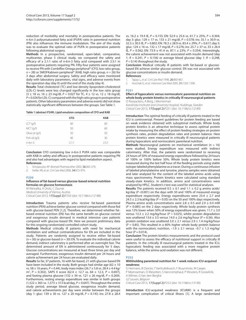
reduction of morbidity and mortality in postoperative patients. The n-6:n-3 polyunsaturated fatty acid (PUFA) ratio in parenteral nutrition (PN) also infl uences the immune modulation. The aim of this study was to evaluate the optimal ratio of PUFA in postoperative patients following abdominal surgery.Methods In a prospective, randomized, open-label, comparative, multicenter, phase 3 clinical study, we compared the safety and effi cacy of a 2.1:1 ratio of n-6:n-3 fatty acid compared with 2.5:1 in postoperative patients requiring PN. Fifty-four patients were assigned to receive PN with Combifl ex Omega peripheral® (CFO, low ratio group, n = 28) or SMOFKabiven peripheral® (KAB, high ratio group, n = 26) for 3 days after abdominal surgery. Safety and effi cacy were monitored daily with laboratory parameters, vital signs, and adverse events from the operation day (day 0) until the end of the study (day 4).Results Total cholesterol (TC) and low-density lipoprotein-cholesterol (LDL-C) levels were less changed signifi cantly in the low ratio group (3 ± 18 vs. 16 ± 23 mg/dl, P = 0.027 for TC, 4 ± 12 vs. 12 ± 18 mg/dl, P = 0.026 for LDL-C) compared with the high ratio group in postoperative patients. Other laboratory parameters and adverse events did not show statistically signifi cant diff erences between the groups. See Table 1.
Table 1 (abstract P249). Lipid emulsion composition of CFO and KAB
CFO KAB
LCT (g/l) 60 60
MCT (g/l) 50 60
Olive oil (g/l) 50 50
Fish oil (g/l) 40 30
n-6:n-3 2.1:1 2.5:1
Conclusion CFO containing low n-6:n-3 PUFA ratio was comparable with KAB in safety and effi cacy in postoperative patients requiring PN and also had advantages with regard to lipid metabolism aspect.References1. Simopoulos AP: Biomed Pharmacother 2002, 56:365-379.2. Heller AR, et al.: Crit Care Med 2006, 34:972-979.
P250Infl uence of fat-based versus glucose-based enteral nutrition formulas on glucose homeostasisM Wewalka, A Drolz, C ZaunerMedical University of Vienna, AustriaCritical Care 2013, 17(Suppl 2):P250 (doi: 10.1186/cc12188)
Introduction Trauma patients who receive fat-based parenteral nutrition (PEN) achieve better glucose control compared with those fed with glucose-based PEN [1,2]. Therefore, we determined whether fat-based enteral nutrition (EN) has the same benefi t on glucose control and exogenous insulin demand in medical intensive care patients compared with glucose-based EN. Here we present preliminary data for this ongoing randomized controlled cohort study.Methods Medical critically ill patients with need for mechanical ventilation and without contraindications for EN are included in the study. Patients are randomly assigned to receive either fat-based (n = 30) or glucose-based (n = 30) EN. To evaluate the individual calorie demand, indirect calorimetry is performed after an overnight fast. The determined amount of EN is administered continuously for 5 days. Glucose concentrations are measured at least three times per day and averaged. Furthermore, exogenous insulin demand per 24 hours and calorie achievement per 24 hours are evaluated daily.Results So far, 37 patients, 16 with fat-based, 21 with glucose-based EN have been included in the study. Both groups had similar age (62 ± 10 vs. 58 ± 16 years, P = 0.44), body mass index (26.7 ± 5.8 vs. 28.4 ± 4.4 kg/m2, P = 0.302), SAPS II score (62.4 ± 12.7 vs. 64 ± 12.3, P = 0.697), and fasting plasma glucose (132 ± 34 vs. 121 ± 26 mg/dl, P = 0.269). Furthermore, resting energy expenditure was similar in both groups (1,522 ± 365 vs. 1,573 ± 313 kcal/day, P = 0.647). Throughout the entire study period, average blood glucose, exogenous insulin demand, and calorie achievements per day were similar between the groups (day 1: gluc: 139 ± 30 vs. 127 ± 20 mg/dl, P = 0.143; ins: 27.8 ± 28.4
vs. 16.2 ± 19.4 IE, P = 0.155; EN: 52.9 ± 25.6 vs. 61.7 ± 25%, P = 0.304; day 3: gluc: 129 ± 17 vs. 133 ± 21 mg/dl, P = 0.578; ins: 35.1 ± 30.9 vs. 32.3 ± 30.5 IE, P = 0.802; EN: 78.3 ± 28.9 vs. 83.4 ± 29%, P = 0.617; day 5: gluc: 124 ± 16 vs. 132 ± 17 mg/dl, P = 0.276; ins: 25.7 ± 37 vs. 33 ± 26.4 IE, P = 0.562; EN: 73.9 ± 41.4 vs. 87.1 ± 23%, P = 0.354). Interestingly, the calorie achievement was not associated with insulin demand (day 1: R = 0.241, P = 0.156) or average blood glucose (day 1: R = 0.248, P = 0.14) throughout the study.Conclusion Medical critically ill patients with fat-based or glucose-based EN achieve similar glucose control. EN was not associated with glucose concentrations or insulin demand.References1. Tappy L, et al.: Crit Care Med 1998, 26:860-867.2. Huschak G, et al.: Intensive Care Med 2005, 31:1202-1208.
P251Eff ect of hypocaloric versus normocaloric parenteral nutrition on whole body protein kinetics in critically ill neurosurgical patientsO Rooyackers, A Berg, J WernermanKarolinska Institutet and University Hospital, Huddinge, SwedenCritical Care 2013, 17(Suppl 2):P251 (doi: 10.1186/cc12189)
Introduction The optimal feeding of critically ill patients treated in the ICU is controversial. Present guidelines for protein feeding are based on weak evidence obtained with suboptimal methods. Whole body protein kinetics is an attractive technique to assess optimal protein intake by measuring the eff ect of protein feeding strategies on protein synthesis rates, protein degradation rates and protein balance. Here protein kinetics were measured in critically ill neurosurgical patients during hypocaloric and normocaloric parenteral nutrition.Methods Neurosurgical patients on mechanical ventilation (n = 16) were studied. Energy expenditure was measured with indirect calorimetry. After that, the patients were randomized to receiving 24 hours of 50% of measured energy expenditure followed by 24 hours of 100% or 100% before 50%. Whole body protein kinetics were measured during the last half hour of the feeding periods using stable isotope-labeled phenylalanine as a tracer. During a continuous infusion of labeled phenylalanine and tyrosine, plasma samples were obtained and later analyzed for the content of the labeled amino acids using mass spectrometry. Protein kinetics were calculated using standard steady-state kinetics. In addition, amino acid concentrations were analyzed by HPLC. Student’s t test was used for statistical analyses.Results The patients received 0.5 ± 0.1 and 1.1 ± 0.2 g amino acids/kg/day (P <0.001) on the days with 50 and 100% of measured energy expenditure respectively. Energy expenditures were 23.4 ± 2.7 and 24.5 ± 2.3 kcal/kg/day (P = 0.05) on the 50 and 100% days respectively. Plasma amino acids concentrations were 2.8 ± 0.5 and 2.9 ± 0.4 mM (P = 0.085) on the 2 days respectively. Whole body protein synthesis was 12% lower when 50% of energy expenditure was given, 11.7 ± 3.0 versus 13.3 ± 2.2 mg/kg/hour (P = 0.025), whilst protein degradation was unaltered 13.6 ± 3.5 versus 14.0 ± 2.6 mg/kg/hour (P = 0.56). Also protein oxidation was unaltered 3.0 ± 2.1 versus 2.9 ± 1.4 mg/kg/hour (P = 0.85). This resulted in a 60% higher whole body protein balance with the normocaloric nutrition, –1.9 ± 2.1 versus –0.7 ± 1.3 mg/kg/hour (P = 0.014).Conclusion The protein kinetics measurements and the protocol used were useful to assess the effi cacy of nutritional support in critically ill patients. In the critically ill neurosurgical patients treated in the ICU, hypocaloric feeding was associated with a more negative protein balance, while the amino acid oxidation was not diff erent.
P252Withholding parenteral nutrition for 1 week reduces ICU-acquired weaknessG Hermans, B Clerckx, T Vanhullebusch, F Bruyninckx, M Casaer, P Meersseman, D Mesotten, S Vancromphaut, P Wouters, R Gosselink, A Wilmer, G Van den BergheUZ Leuven, BelgiumCritical Care 2013, 17(Suppl 2):P252 (doi: 10.1186/cc12190)
Introduction ICU-acquired weakness (ICUAW) is a frequent and important complication of critical illness [1]. A large randomized
Critical Care 2013, Volume 17 Suppl 2 http://ccforum.com/supplements/17/S2
S94

controlled trial (EPaNIC: clinicaltrials.gov: NCT00512122) [2] showed that withholding parenteral nutrition during the fi rst week of ICU stay whereby tolerating substantial caloric defi cit (late PN) accelerated recovery and shortened weaning time as compared with early parenteral substitution for defi cient enteral feeding (early PN). We examined the impact of late PN, as compared with early PN, on incidence and recovery of ICUAW.Methods A preplanned subanalysis of adult patients included in the EPaNIC trial. The study was performed between October 2008 and November 2010 and included those patients who required intensive care for ≥8 days as well as a computer-generated, admission category-matched, random sample of short-stay ICU patients, the latter to correct for possible bias evoked by earlier ICU discharge in one of the two study groups. Assessors blinded for treatment allocation evaluated muscle strength clinically three times weekly from awakening onward and performed nerve conduction studies and electromyography (NCS and EMG) weekly. The primary outcome was the incidence of ICUAW, diagnosed clinically by the Medical Research Council (MRC) sum score (<48/60) [3] at fi rst evaluation. Secondary outcomes included ICUAW at worst and last MRC evaluation, recovery from ICUAW and incidence of abnormal fi ndings on NCS and EMG. All analyses were performed on the total dataset and on a for-baseline characteristics propensity score-matched sample to correct for possible imbalances between the groups.Results Clinical ICUAW evaluation was performed in 600 patients (matched n = 558), electrophysiological testing in 730 (matched n = 684). Late PN reduced the incidence of ICUAW at fi rst evaluation from 43.1% to 34.4%, P = 0.03 (matched: early PN 41.6%, late PN 33.3%, P = 0.04). Signifi cantly fewer patients in the late PN group developed weakness at any time during ICU stay (late PN 37.0%, early PN 46.4%, P = 0.02; matched: late PN 36.2%, early PN 45.2%, P = 0.03). ICUAW may have recovered faster with late PN than with early PN (P = 0.05, matched P = 0.06). Other outcomes were not diff erent.Conclusion As compared with early PN, late PN reduced the incidence of ICUAW and may have accelerated recovery thereof.References1. Stevens et al.: Int Care Med 2007, 33:1876-1891.2. Casaer et al.: N Engl J Med 2011, 365:506-516.3. De Jonghe et al.:, JAMA 2002, 288:2859-2867.
P253Impact of early parenteral nutrition on muscle and adipose tissue compartments during critical illnessL Langouche1, MP Casaer2, W Coudyzer2, D Vanbeckevoort2, B De Dobbelaer2, FG Güiza1, PJ Wouters2, D Mesotten2, G Van den Berghe2
1KU Leuven, Belgium; 2University Hospitals Leuven – KU Leuven, BelgiumCritical Care 2013, 17(Suppl 2):P253 (doi: 10.1186/cc12191)
Introduction The goal of enhanced nutrition in critically ill patients is to improve outcome by reducing lean tissue wasting. However, such eff ect has not been proven. This study aimed to assess the eff ect of early administration of parenteral nutrition (PN) on muscle volume and composition by repeated quantitative computer tomography (qCT).Methods We performed a preplanned substudy of a randomized controlled trial (EPaNIC) that compared early initiation of PN when enteral nutrition was insuffi cient (early PN) with tolerating a pronounced nutritional defi cit for 1 week in the ICU (late PN) [1]. Late PN prevented infections and accelerated recovery. We studied 15 EPaNIC study neurosurgical patients requiring prescheduled repeated follow-up CT scans and six healthy volunteers matched for age, gender and BMI. Repeated abdominal and femoral qCT images were obtained in a standardized manner on median ICU day 2 (IQR 2 to 3) and day 9 (8 to 10). Intramuscular, subcutaneous and visceral fat compartments were delineated manually. Muscle and adipose tissue volume and composition were quantifi ed using standard Hounsfi eld Unit ranges.Results Critical illness evoked substantial loss of femoral muscle volume in 1 week, irrespective of the nutritional regimen. Early PN reduced the quality of the muscle tissue, as refl ected by the attenuation, revealing increased intramuscular water/lipid content. Early PN also increased the volume of adipose tissue islets within the femoral muscle compartment. These changes in skeletal muscle integrity correlated with caloric
intake. In the abdominal muscle compartments, changes were similar, albeit smaller. Femoral and abdominal subcutaneous adipose tissue compartments were unaff ected by disease and nutritional strategy.Conclusion Early PN did not prevent the pronounced wasting of skeletal muscle observed over the fi rst week of critical illness. Moreover, early PN increased the amount of adipose tissue within the muscle compartments.Reference1. Casaer MP, et al.: N Engl J Med 2011, 365:506-517.
P254Withholding parenteral nutrition during the fi rst week of critical illness increases plasma bilirubin but lowers the incidence of cholestasis and gallbladder sludgeYM Vanwijngaerden1, L Langouche1, M Gielen1, Y Debaveye1, M Casaer1, C Liddle2, S Coulter2, R Brunner1, P Wouters1, A Wilmer1, G Van den Berghe1, D Mesotten1
1University Hospitals KU Leuven, Belgium; 2University of Sydney, AustraliaCritical Care 2013, 17(Suppl 2):P254 (doi: 10.1186/cc12192)
Introduction Cholestatic liver dysfunction (CLD) during critical illness, defi ned by hyperbilirubinemia, often occurs and is associated with poor outcome. Parenteral nutrition (PN) is assumed to aggravate CLD. However, hyperbilirubinemia more frequently occurred when the start of PN was delayed until day 8 in the ICU (late PN) [1]. Late PN accelerated recovery as compared with early initiation of PN.Methods This was a preplanned subanalysis of a large randomized controlled trial on early versus late initiation of PN (n = 4,640) [1]. Plasma total bilirubin was quantifi ed in all patients daily while in the ICU. Liver enzymes ALT, AST, GGT and ALP were quantifi ed twice weekly in all patients while in the ICU. In a random predefi ned subset of patients, circulating bile salts were also quantifi ed with MS-HPLC at baseline and on day 3, day 5 and the last day in the ICU (n = 280). Gallbladder sludge was evaluated by ultrasound on ICU day 5 by blinded assessors (n = 776).Results From day 1 after randomization until the end of the 7-day intervention window, plasma bilirubin was higher in the late PN than in the early PN group (all P <0.001). In the late PN group, as soon as PN was started on day 8, plasma bilirubin also fell and the two groups became comparable. Maximum levels of GGT, ALP and ALT during the ICU stay were higher in the early PN group (all P <0.01). Compared with baseline, the circulating glycine and taurine conjugated primary bile salts were elevated on day 3, day 5 and last day of the ICU stay (P <0.01 for all). However, there was no diff erence between the two groups. More patients in the early PN than in the late PN group had gallbladder sludge on day 5 (45% vs. 37%; P = 0.04).Conclusion Tolerating substantial caloric defi cit by withholding PN until day 8 of critical illness increased circulating levels of bilirubin but reduced the occurrence of gallbladder sludge and lowered GGT, ALP and ALT levels. These results suggest that hyperbilirubinemia during critical illness dies not necessarily refl ect cholestasis and instead may be an adaptive response. Additional analyses on a propensity score-matched patient population are ongoing.Reference1. Casaer et al.: N Engl J Med 2011, 365:506-516.
P255Impact of early parenteral nutrition on catabolismJ Gunst1, I Vanhorebeek1, MP Casaer1, G Hermans1, PJ Wouters1, J Dubois2, K Claes1, M Schetz1, G Van den Berghe1
1KU Leuven, Belgium; 2Jessa Hospital, Hasselt, BelgiumCritical Care 2013, 17(Suppl 2):P255 (doi: 10.1186/cc12193)
Introduction Prolonged critically ill patients enter a state of hypercatabolism and muscle weakness, which has been associated with increased morbidity and mortality. Early, full feeding of ICU patients has been advocated to counteract catabolism. However, a large, multicenter study found that early parenteral nutrition (PN) had no signifi cant impact on mortality and even increased dependency on intensive care with, among others, a signifi cant prolongation of
Critical Care 2013, Volume 17 Suppl 2 http://ccforum.com/supplements/17/S2
S95

the duration of renal replacement therapy (RRT) [1]. The impact of the intervention on early markers of catabolism has not been investigated.Methods We studied the impact of early versus late PN on daily markers of catabolism in the ICU in the total study population and in propensity score-matched subgroups of long-stay patients. In addition, we calculated the net incorporation rate of the extra amino acids supplied by early PN.Results Plasma urea, the urea/creatinine ratio and nitrogen excretion increased over time in the ICU. Early PN further increased these markers of catabolism, from the fi rst day of amino acid infusion onward, and only marginally improved the nitrogen balance. Also in the group that received PN only after the fi rst week in the ICU, ureagenesis was increased by infusing amino acids. Over the fi rst 2 weeks, approximately two-thirds of the extra amino acids supplied by early PN were net wasted in urea. The above fi ndings were confi rmed in propensity score-matched subgroups of long-stay patients. The higher urea levels with early PN, rather than the kidney function as such, may have driven the observed longer duration of RRT, as supported by multiple regression analysis.Conclusion The extra amino acids supplied by early PN appeared ineffi cient to reverse the negative nitrogen balance, not because of insuffi cient amino acid delivery, but rather because of insuffi cient incorporation with, instead, increased degradation into urea. The substantial catabolism of the extra amino acids, leading to pronounced urea generation, may have prolonged the duration of RRT in the early PN group.Reference1. Casaer et al.: N Engl J Med 2011, 365:506-517.
P256Impact of early versus late parenteral nutrition on morphological and molecular markers of atrophy and autophagy in skeletal muscle of critically ill patientsI Vanhorebeek, MP Casaer, F Güiza, S Derde, I Derese, PJ Wouters, Y Debaveye, J Gunst, G Hermans, G Van den BergheKU Leuven, BelgiumCritical Care 2013, 17(Suppl 2):P256 (doi: 10.1186/cc12194)
Introduction Muscle weakness of critical illness is associated with prolonged dependency on ventilatory support and delayed rehabilitation. Muscle wasting related to poor nutrition has long been considered a major determinant, whereas the importance of myofi ber integrity only recently emerged [1-4]. We hypothesized that nutrient restriction early during illness aggravates atrophy while preserving myofi ber integrity by activating the crucial cellular quality control pathway autophagy. The latter could be important to preserve muscle function.Methods Critically ill patients (n = 122) were randomized to early (early-PN) or late (late-PN) initiation of parenteral nutrition to complete failing enteral nutrition, while maintaining normoglycemia (80 to 110 mg/dl) with insulin, in the EPaNIC study [5]. Vastus lateralis biopsies were harvested after 1 week and compared with matched controls (n = 20).Results As compared with controls, muscle from critically ill patients showed reduced myofi ber density, a shift to smaller (especially type I) myofi bers, lower myosin and actin mRNA, upregulated mRNA of the ubiquitin ligases muscle-ring-fi nger-1 and atrogin-1, a small increase in the autophagosome formation marker LC3-II/LC3-I, and increases in the autophagic substrates ubiquitin and p62 (all P ≤0.006). Late-PN, resulting in a larger caloric defi cit than early-PN, had no substantial impact on atrophy markers. In contrast, late-PN increased LC3-II/LC3-I (P = 0.02), which coincided with less accumulation of ubiquitinated proteins/aggregates (P = 0.05). Fewer patients on late-PN developed muscle weakness as compared with early-PN (42% vs. 68%, P = 0.05). In multivariable analysis, a lower LC3-II/LC3-I ratio (P = 0.05) and higher myofi ber density (P = 0.04) were independently associated with muscle weakness.Conclusion Early-PN did not counteract muscle atrophy whereas it suppressed autophagy and aggravated weakness. Statistically, muscle weakness was not explained by atrophy or wasting but rather by impaired autophagy and preservation of muscle density. Thus, tolerating nutrient restriction early during critical illness may preserve myofi ber integrity by activating autophagy.
References1. Debaveye, Van den Berghe: Annu Rev Nutr 2006, 26:513-538.2. Masiero et al.: Cell Metab 2009, 10:507-515.3. Vanhorebeek et al.: J Clin Endocrinol Metab 2011, 96:E633-E645.4. Derde et al.: Endocrinology 2012, 153:2267-2276.5. Casaer et al.: N Engl J Med 2011, 365:506-517.
P257Impact of closing an emergency department on a neighbouring teaching hospital: the concentrate eff ectJ Millar, R Wilson, P O’Connor, R McLaughlinRoyal Victoria Hospital, Belfast, UKCritical Care 2013, 17(Suppl 2):P257 (doi: 10.1186/cc12195)
Introduction Closure of an acute hospitals emergency department (ED) has important ramifi cations for those centres expected to take up the resultant workload. The continued reconfi guration of emergency care is likely to produce an increasing number of these scenarios. Little evidence is available to support planning of such initiatives and thus the implications are diffi cult to anticipate. This study aims to demonstrate one hospital’s experience of the rationalisation of emergency care and its eff ect on workload.Methods This retrospective study was conducted in a large teaching hospital. Activity data were analysed for a 12-month period following the closure of a neighbouring ED. The results were subsequently compared against the year prior to closure. Attendance, triage data, admission rates and waiting times were compared across the two periods, as were workload data for all grades of physician. The chi-squared test was used to examine diff erences between groups.Results In the period studied, the gross attendance fi gure increased by 20,480 (33.72%), whilst the admission rate rose from 22 to 27%. Following closure of the neighbouring ED, the proportion of high-acuity patients attending our institution increased dramatically, with the proportion of category one and two patients (Manchester Triage Scale) increasing by 8.33% (P = 0.076) and 18.80% (P <0.001), respectively. Likewise, ambulance arrivals increased out of proportion to the total increase in attendances (P = 0.016). Admissions from the ED to the ICU increased by 63.04%. Consultants workloads now include 50% more category 1 and 2 patients (P = 0.001).Conclusion Reconfi guration of emergency care can have dramatic implications for existing services; these may not always be anticipated. Rationalisation of ED’s may result in a concentration of high-acuity patients accompanied by a downturn in the numbers of patients whose presentations are amenable to care delivered in other settings. This abrupt change in case mix requires a re-examination of existing workforces and their seniority.
P258Overcrowding estimation in the emergency department: is the simplest score the best?V Winkin, V Pinckaers, V D’Orio, A GhuysenCHU Liège, BelgiumCritical Care 2013, 17(Suppl 2):P258 (doi: 10.1186/cc12196)
Introduction Emergency department (ED) overcrowding is a major international problem with a negative impact on both patient care and providers. Among validated methods of measurement, emergency physicians have to choose between simple and complex scores [1,2]. The aim of the present study was to compare the complex National Emergency Department Overcrowding Scale (NEDOCS) with the simple occupancy rate (OR) determination. We further evaluated the correlation between these scores and a qualitative assessment of crowding.Methods The study was conducted in two academic hospitals and one county hospital in Liège, Chênée and Verviers; each with an ED census of over 40,000 patient visits per year. Samplings occurred over a 2-week period in January 2011, with fi ve sampling times each day.Results ED staff considered overcrowding as a major concern in the three EDs. Median OR ranged from 68 to 100, while the NEDOCS ranged from 64.5 to 76.3. We found a signifi cant correlation between
Critical Care 2013, Volume 17 Suppl 2 http://ccforum.com/supplements/17/S2
S96

the OR and the NEDOCS (Pearson R = 0.973, 0.974 and 0.972), as well as between the OR, the NEDOCS and the subjective evaluation (P = 0.001).Conclusion Crowding evaluation in the ED requires validated, easy-to-use, scores. Our study indicates that the simple OR is as accurate as the complex NEDOCS, allowing continuous crowding assessment.References1. Hwang U, McCarthy ML, Bernstein SL, et al.: Measures of crowding in the
emergency department: a systematic review. Acad Emerg Med 2011, 18:527-538.
2. Hoot NR, Zhou C, Jones I, Aronsky D: Measuring and forecasting emergency department crowding in real time. Ann Emerg Med 2007, 49:747-755.
P259GP-led walk-in centre in the UK: another way for urgent healthcare provisionM Arain, J Nicholl, M CampbellThe University of Sheffi eld, UKCritical Care 2013, 17(Suppl 2):P259 (doi: 10.1186/cc12197)
Introduction It is evident that accident and emergency departments are overloaded with patients, which results in delays in healthcare provision [1]. A large proportion of patients consist of patients with minor illness that can be seen by a healthcare provider in a primary care setting. The aim of the study was to determine the characteristics of patients using GP walk-in services, patients’ satisfaction and the eff ect on emergency department (ED) services.Methods The survey was conducted in Sheffi eld and Rotherham walk-in centres over 3 weeks during September and October 2011. A self-reported, validated questionnaire was used to conduct survey on the patients presenting at these centres. We estimated that a sample size of around 400 patients from each centre was required to achieve statistically robust results. A post-visit, short questionnaire was also sent to those who agreed for the second questionnaire and provided contact details. ED data were also obtained from April 2008 to March 2010, 1 year before and 1 year after the opening of the GP walk-in centre. Data were entered and analysed in PASW Statistics 18. Ethical approval of the study was obtained from the NHS ethical review committee.Results A total of 1,030 patients participated in the survey (Rotherham 501; Sheffi eld 529). The mean age of the participants was 32.1 years at Sheffi eld and 30.88 years at Rotherham. A higher proportion of users were female, around 59% at both centres. Most of the patients rated high for convenience of the centre opening hours and location (above 85%, apart from the location of Sheffi eld centre, which was rated high by around 72% of the research participants). Overall 93% patients were satisfi ed with the service at Rotherham centre and around 86% at the Sheffi eld centre. Based on the estimation of the monthly counts of patients attending ED and the GP walk-in centre, around 14% monthly reduction in minor attendances at ED was expected. However, ED routine data did not show any signifi cant reduction in minor attendances as a result of the opening of the GP walk-in centre.Conclusion These walk-in centres have been shown to increase accessibility to healthcare service through longer opening hours and walk-in facility. Although the eff ect on the reduction of patients’ load at the ED is not visible as these centres cover a fraction of the population, the centre has a potential to divert patients from the ED.Reference1. Gerard K, Lattimer V: Preferences of patients for emergency services
available during usual GP surgery hours: a discrete choice experiment. Fam Pract 2005, 22:28-36.
P260Establishing a new emergency department: eff ects on patient fl owV Rautava1, T Valpas1, M Nurmikari2, A Palomäki11Kanta-Häme Central Hospital, Hämeenlinna, Finland; 2City of Hämeenlinna, FinlandCritical Care 2013, 17(Suppl 2):P260 (doi: 10.1186/cc12198)
Introduction Overcrowding in emergency departments (EDs) is a widely known problem. It causes problems and delays in the ED and
has a negative impact on patient safety [1]. The aim of this study was to analyse whether a reform of emergency care can reduce patient fl ow into the ED.Methods A substantial reform of emergency care took place in the province of Kanta-Häme in Southern Finland. Three separate out-of-hours services in primary healthcare (PHC) and one ED in the hospital were combined into one large ED in April 2007. Basic principles of the new ED were: the ED is only for those patients who are seriously ill or injured, and need immediate care; PHC (healthcare centres) take care of acute ordinary illnesses and nonserious injuries during offi ce hours. To achieve these principles a regional fi ve-scale triage system was planned and implemented. The information plan was established. Citizens were systematically informed about the principles of the new ED by mail, articles in the newspapers and interviews in the radio and television. The ED’s Internet pages were planned and established. The number of patient visits (Hämeenlinna region) was analyzed 2 years before and after establishing the new ED.Results During the 2-year period before the establishment of the new ED the mean number of GP patient visits was 1,845 ± 43/month. During the 2-year period after the reform the number was diminished to 1,364 ± 21/month. This change was not associated with the increase of the patient visits taken care of by specialists and hospital residents. See Figure 1.Conclusion An extensive reform of the emergency services can notably reduce patient fl ow into the ED.Reference1. Boyle A, et al.: Emerg Med Int 2012:838610. [Epub ahead of print]
P261Prehospital EKG evaluation in Rio de Janeiro ambulancesRV Vasconcellos, FE Erthal, RV VargasInstituto Nacional de Cardiologia, Rio de Janeiro, BrazilCritical Care 2013, 17(Suppl 2):P261 (doi: 10.1186/cc12199)
Introduction Rio de Janeiro’s Fire Squad is responsible for EMS in the city. During 2010 we implemented 10 ambulances with EKG transmission capability in our city. Our intention was to access the prevalence of acute myocardial infarction in the prehospital setting.Methods We used the Aerotel HeartView EKG system to acquire patient examination and a blackberry phone to transmit and receive the PDF EKG trace. The PDF comes with the cardiologist’s interpretation from a remote hospital, the HCOR São Paulo.Results We realized 503 EKG examinations in total. Of these, 248 (49%) had as the chief complaint chest pain, 101 (20%) shortness of breath, 47 (9%) syncope, 36 (7%) palpitation; other complaints were 15%. We detected 32 examinations (6.36%) with ST elevation MI and
Figure 1 (abstract P260). GP patient visits before and after the reform of emergency services.
Critical Care 2013, Volume 17 Suppl 2 http://ccforum.com/supplements/17/S2
S97

Figure 1 (abstract P261). Acute inferior MI with complete heart block.
Figure 2 (abstract P261). Paroxysmal supraventricular tachycardia.
Critical Care 2013, Volume 17 Suppl 2 http://ccforum.com/supplements/17/S2
S98

44 examinations (8.75%) with ST depression. Atrial fi brillation was detected in 43 examinations (8.5%). See Figures 1 and 2.Conclusion This experience gave us an idea of the prevalence for acute ST elevation MI in the prehospital setting, so that we can better develop our prehospital thrombolysis protocol and focus our training for cardiology care.Reference1. Ioannidis JP, Salem D, Chew PW, Lau J: Accuracy and clinical eff ect of out-of-
hospital electrocardiography in the diagnosis of acute cardiac ischemia: a meta-analysis. Ann Emerg Med 2001, 37:461-470.
P262Technique of ultrasound-guided peripheral venous access in the emergency roomMG Annetta, G Scoppettuolo, M Biancone, F Toni, M PittirutiCatholic University, Rome, ItalyCritical Care 2013, 17(Suppl 2):P262 (doi: 10.1186/cc12200)
Introduction In emergency situations, patients may need a fast and reliable peripheral venous access, which sometimes may be diffi cult to obtain, because of poor visualization of the superfi cial veins due to edema, obesity, hypovolemia or local abnormalities. In such cases, insertion of a central line is potentially time consuming and possibly associated with complications. Furthermore, central lines inserted in emergency are known to be at high risk of infection, so guidelines recommend that they should removed within 24 to 48 hours. In this setting, ultrasound-guided placement of a peripheral venous access might be more rapid, safer and more cost-eff ective than a central line.Methods We have reviewed retrospectively our experience with the emergency use of 18 G or 20 G polyurethane catheters, 8 to 10 cm long, inserted by direct Seldinger technique.Results In 1 year, 76 long peripheral catheters were inserted in emergency conditions, using ultrasound guidance. The success rate was 100%; most lines lasted >1 week and were used for diff erent purposes, including contrast medium injection.Conclusion The direct Seldinger technique allows a rapid and safe placement of the catheter in a vein of the arm or of the forearm, even when the vessel cannot be palpated or seen, as long as it can be visualized by ultrasound and it is not deeper than 2 cm. The long life of this type of peripheral line (up to 2 weeks) is guaranteed by the material (polyurethane being more biocompatible than Tefl on) and by the length of the catheter (which reduces the risk of dislodgment). Also, these catheters are particularly cost-eff ective if compared with a central line or with a midline catheter, since a complete kit including catheter, 20 G needle and 20 cm guidewire costs between €15 and €20.
P263FIB the fractured femurH Shahzad, M Majeed, D Yeo, V Gupta, U SalankeUniversity Hospital, Birmingham, UKCritical Care 2013, 17(Suppl 2):P263 (doi: 10.1186/cc12201)
Introduction The lumbar plexus block provides excellent analgesia after hip and knee surgery. One approach to the three major nerves (femoral, lateral cutaneous and obturator) of the lubar plexus is the fascia iliaca compartment block, fi rst described by Dalens and colleagues. Using this blind approach, complete nerve block in the distribution of the lumbar plexus may be achieved only in 38% of cases. Ultrasound-guided regional techniques off er a number of advantages including real-time needle guidance and direct observation of local anesthetic spread within tissue planes.Methods We hypothesized that real-time UFIB could be successfully performed in the ED and would provide an excellent adjunct or alternative to repeat doses of IVMS for pain control in patients with HFx. The study was conducted at University Hospital, Birmingham, where we see about 90,000 patients every year. All patients with confi rmed femoral (neck/shaft) fractures and pain score >7 were included in the study. Patients with local wounds, or suspected signifi cant pelvic injury were excluded. A combination of 15 ml lignocaine 1% and 15 ml bupvivcaine 0.25% was used. VAS (0 to 10), pain on movement of the
leg and patient satisfaction were used to assess the outcome. The assessments were made before the FIB, at 15 minutes, 30 minutes and 45 minutes.Results A convenience sample of 19 patients was enrolled in this study. The compartment block was placed by three mergence ultrasound trainers. The mean age was 58.5 years. There were 13 females and six males. The mean pain score was 9.5 at time 0. The mean pain score had improved to 5.5 at 15 minutes, 4.5 at 30 minutes and 3 at 45 minutes, respectively. The patient satisfaction was scored 4.5 on a scale of 1 to 5 where 5 was the most satisfi ed. No patient required any further analgesia up to 90 minutes and no issues were raised about the pain or discomfort upon patient transfer. We have no documented complications or side eff ects.Conclusion FIB is now widely used by non-anaesthetic trainees to combat pain in preoperative care due to its safe landmarks. Our results have shown that it can be used safely and eff ectively for pain management in hip fracture by emergency physicians, who are trained in the technique, on the shop fl oor.
P264Abdominal pain in adolescent females: a single-centre audit and review of managementF Gallagher, H Jahn, F DaviesLeicester Royal Infi rmary, Leicester, UKCritical Care 2013, 17(Suppl 2):P264 (doi: 10.1186/cc12202)
Introduction Abdominal pain in adolescent females has undergone recent changes with regards to its management under various specialities. The authors report a single-centre audit looking at the correct investigation and management of 12-year-old to 16-year-old girls with abdominal pain in the emergency department setting.Methods A single-centre audit and retrospective analysis of patients took place using case notes and computerised records. Documentation was analysed using statistical analysis and minimum standards were set and reviewed.Results After exclusion criteria 62 females between the ages of 12 and 16 presented to the paediatric emergency department in Leicester with abdominal pain as the predominant admission symptom during a 12-month period. Documentation of the gynaecological history was poor (menstrual history 47%, sexual history 14%, contraception 8%), as was the performance of basic investigations (urine dipsticks 65%, pregnancy test 42%). Documentation was analysed with regard to discharge diagnosis. Ultrasound investigation was performed on seven of the patients but only once admitted to various specialities. No ultrasound was undertaken upon admission.Conclusion Improvement in documentation of minimum standards for these patients is needed. A multidisciplinary care pathway could improve outcome. Consideration should be given to whether early ultrasound investigation is appropriate and there is a further need for investigation as to whether this would improve longer term outcomes.
P265Multidisciplinary approach to improving documentation of visual acuity in patients presenting with ocular traumaL Low, M JohnstonNHS Tayside, Dundee, UKCritical Care 2013, 17(Suppl 2):P265 (doi: 10.1186/cc12203)
Introduction This study aimed to implement a multidisciplinary quality improvement project in Perth Royal Infi rmary A&E department to improve documentation of visual acuity (VA) in patients presenting with ocular injury.Methods The improvement project involves a three-pronged multi-disciplinary approach: ensure that equipment required for VA testing (Snellen chart and pinhole mask) was readily available; encourage VA testing at fi rst point of contact with A&E staff , both nursing/medical staff ; and refresher online course on how to test for VA quickly and accurately, in the 6/X format. We compared the pre-intervention (2 September to 2 October 2012) and post-intervention (11 October to 19 November 2012) rates of VA documentation using the chi-square test.
Critical Care 2013, Volume 17 Suppl 2 http://ccforum.com/supplements/17/S2
S99
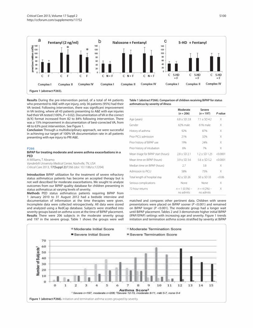
Results During the pre-intervention period, of a total of 44 patients who presented to A&E with eye injury, only 36 patients (95%) had their VA tested. Following intervention, there was signifi cant improvement in VA testing, where all 43 patients presenting to A&E with eye injuries had their VA tested (100%, P = 0.02). Documentation of VA in the correct (6/X) format increased from 82 to 84% following intervention. There was a 15% improvement in documentation of best-corrected VA, from 48 to 63% post intervention. See Figure 1.Conclusion Through a multidisciplinary approach, we were successful in achieving our target of 100% VA documentation rate in all patients presenting with eye injury to PRI A&E.
P266BiPAP for treating moderate and severe asthma exacerbations in a PEDA Williams, T AbramoVanderbilt University Medical Center, Nashville, TN, USACritical Care 2013, 17(Suppl 2):P266 (doi: 10.1186/cc12204)
Introduction BiPAP utilization for the treatment of severe refractory status asthmaticus patients has become an accepted therapy but is not well described for moderate exacerbations. We sought to analyze outcomes from our BiPAP quality database for children presenting in status asthmaticus at varying levels of severity.Methods PED status asthmaticus patients requiring BiPAP from 1 January 2010 to 31 August 2012 had a bedside interview and documentation of information at the time therapies were given. Incomplete data were collected retrospectively. All data were stored and analyzed using a RedCap database. Subjects were stratifi ed into severity groups based on asthma score at the time of BiPAP placement.Results There were 206 subjects in the moderate severity group and 197 in the severe group. Table 1 shows the groups were well
matched and compares other pertinent data. Children with severe presentations were placed on BiPAP sooner (P <0.001) and remained on BiPAP longer (P <0.001). The moderate group had a longer wait until BiPAP placement. Tables 2 and 3 demonstrate higher initial BiPAP (IPAP/EPAP) settings with increasing age and severity. Figure 1 trends initiation and termination asthma scores stratifi ed by severity at BiPAP
Figure 1 (abstract P265).
Table 1 (abstract P266). Comparison of children receiving BiPAP for status asthmaticus by severity of illness
Moderate Severe (n = 206) (n = 197) P value
Age (years) 6.8 ± SD 2.8 7.1 ± SD 4.2 X
Gender 62% male 61% male X
History of asthma 92% 87% X
Prior PICU admission 21% 32% X
Prior history of BiPAP use 19% 24% X
Prior history of intubation 6% 7% X
Mean triage for BiPAP start (hours) 2.8 ± SD 2.1 1.2 ± SD 1.25 <0.0001
Mean time on BiPAP (hours) 3.9 ± SD 3.6 5.6 ± SD 5.2 <0.0001
Median time on BiPAP (hours) 2.7 3.8 X
Admission to PICU 58% 75% X
Total length of hospital stay 42 ± SD 26 50 ± SD 33 <0.06
Serious complications None None X
72-hour returns n = 1 (0.5%) – n = 4 (2%) – X no admits no admits
Figure 1 (abstract P266). Initiation and termination asthma scores grouped by severity.
Critical Care 2013, Volume 17 Suppl 2 http://ccforum.com/supplements/17/S2
S100

placement. More of the severe group was admitted to the PICU and had overall longer hospitalizations (P <0.06). None experienced severe complications.Conclusion BiPAP is a benefi cial therapy for children presenting to the PED with severe asthma exacerbations. It may have utility for less severe asthma exacerbations.
P267Report of an outbreak of toxicity from a novel drug of abuse in the UK: Eric-3C Morden, S Haig, C KellyGreat Western Hospital, Swindon, UKCritical Care 2013, 17(Suppl 2):P267 (doi: 10.1186/cc12205)
Introduction We present a case series of toxicity due to a novel sub-stance in the UK: Eric-3. Novel drugs of abuse are becoming more common throughout the world, and they represent particular diffi culties in their acute management. A recent report from the European Monitoring Centre for Drugs and Drug Addiction and Europol has reported 49 new psychoactive substances reported via its early warning system.Methods This was a retrospective case-note review over a 6-month period. Patients were included if their presentation was due to recent ingestion of Eric-3. Physiological data, symptoms, outcome and destination of the patient from the emergency department were collected. Postmortem toxicological analysis was obtained for one of the two patients who died.Results Forty-one attendances were identifi ed from 18 patients. Two patients died and fi ve were admitted to the ICU. Heart rate and temperature on arrival tended to be above normal (mean heart rate was 112 bpm, with an SD of 18; mean temperature was 37.45°C with an SD of 0.95). In total, 63.4% of attendances included agitation and 34.1% choreiform movements. α-Methyltryptamine and 3-/4-fl uoroephedrine were found in the blood of one of the patients who died.Conclusion In this outbreak in the UK, Eric-3 gave symptoms similar to other stimulants known as legal highs, including death. It may have been a novel substance, 3-/4-fl uoroephedrine. This underlines the need for prospective data collection and early national and international information sharing.
References1. EMCDDA: Europol Annual Report on the Implementation of Council Decision
2005/387/JHA. Lisbon: EMCDDA/Europol; 2011.2. Brandt SD, et al.: Drug Test Anal 2010, 2:377-382.3. Sikk K, et al.: Acta Neurol Scand 2007, 115:385-389.4. Stepens A, et al.: N Engl J Med 2008, 358:1009-1017.5. Murphree HB, et al.: Clin Pharmacol Ther 1961, 2:722-726.
P268Abstract withdrawn
P269Thallium group poisoning incident in Japan 2011Y Namba, R Suzuki, J Sasaki, M Takayasu, K Watanabe, D Kenji, M Hayashi, Y Kitamura, M Kawamo, H Masaki, E Kyuuno, M Hayashi, M Yamaguchi, A MaedaShowa University Fujigaoka Hospital, Yokohama, JapanCritical Care 2013, 17(Suppl 2):P269 (doi: 10.1186/cc12207)
Introduction Thallium is an odorless, tasteless, heavy metal that has been often used for intentional poisonings. In severe patients, thallium poisoning produces neuromuscular symptoms such as extreme pain and muscle weakness.Methods Five case reports.Results All patients worked at a pharmaceutical factory. They joined a tea party held at their workplace at the end of April 2011. The fi ve patients drank tea from a teapot someone had put thallium in. A few days later, they complained of femoral numbness and pain caused by pressure. About a week later, three of fi ve patients had profound hair loss. Three weeks after the party, they came to our ER. We thought that their symptoms might be caused by some chemicals. We searched the keywords: ‘lower extremity pain’, ‘hair loss’ and ‘poison’ in the Internet. As a result, thallium, mercury, lead, and so forth, were suspicious metals. In those metals, thallium was most likely because it was used in their factory. We immediately examined the blood concentration of several metals and ordered iron(III)hexacyanoferrate(II) that is known as the antidote for thallium poisoning. Only thallium was positive in the blood metal concentration test. Three patients consented to oral administration of an antidote. Two patients rejected administration because their symptoms were mild and getting better. All symptoms of all patients gradually disappeared by August. We also followed up the course of blood concentration of thallium. The concentration in three patients who took the antidote was reduced more rapidly than the two patients who did not take it.Conclusion All patients recovered without any sequelae. Three patients’ hair started to grow 3 months from ingestion of thallium, and after half a year their hair was restored to their former state. We had diffi culty ordering iron(III)hexacyanoferrate(II) because this is also known as an antidote for cesium. On 11 March 2011 a megathrust earthquake and tsunami hit Japan and the giant tsunami gave rise to an accident at a nuclear power generation plant. Because the rumor of radioactive substances including cesium might be spread was the talk in the city near the nuclear power plant, the authorities put the antidote under heavy supervision. We could also collect the data for the course of thallium concentration. Thallium concentration of the patients who had an antidote was reduced more rapidly but these patients had a loose stool, thought to be a side eff ect of this antidote.Reference1. Centers for Disease Control and Prevention: Thallium poisoning from eating
contaminated cake – Iraq, 2008. MMWR Morb Mortal Wkly Rep 2008, 57:1015-1018.
P270Heart rate variability in children with tricyclic antidepressant intoxicationEC Dinleyici, Z Kilic, S Sahin, R Tutuncu-Toker, M Eren, ZA Yargic, P Kosger, B UcarEskisehir Osmangazi University Faculty of Medicine, Eskisehir, TurkeyCritical Care 2013, 17(Suppl 2):P270 (doi: 10.1186/cc12208)
Introduction Tricyclic antidepressant (TCA) intoxication is among the most common causes of poisonings in our country. The TCA group of
Table 2 (abstract P266). Mean settings by age: moderate group
Age (years) IPAP EPAP
<3 (n = 22) 12.1 ± SD 2.1 6.4 ± SD 1.3
3 to 4 (n = 48) 12.1 ± SD 1.6 6.4 ± SD 1.2
5 to 6 (n = 51) 13.0 ± SD 2.3 6.6 ± SD 1.3
7 to 8 (n = 24) 13.9 ± SD 2.1 7.2 ± SD 1.2
9 to 10 (n = 28) 13.9 ± SD3.2 7.4 ± SD 2.0
11 to 12 (n = 25) 13.8 ± SD 1.8 7.0 ± SD 1.3
13 to 14 (n = 10) 13.8 ± SD 3.2 7.4 ± SD 1.4
15 to 17 (n = 8) 17.0 ± SD 2.4 9.5 ± SD 0.9
Table 3 (abstract P266). Mean settings by age: severe group
Age (years) IPAP EPAP
<3 (n = 21) 12.7 ± SD 2.0 6.8 ± SD 1.3
3 to 4 (n = 50) 13.3 ± SD 2.8 6.8 ± SD 1.4
5 to 6 (n = 26) 13.9 ± SD 2.2 7.3 ± SD 1.3
7 to 8 (n = 29) 15.0 ± SD 2.4 7.7 ± SD 1.5
9 to 10 (n = 26) 15.7 ± SD 3.5 8.0 ± SD 1.8
11 to 12 (n = 22) 14.5 ± SD 3.2 7.6 ± SD 1.6
13 to 14 (n = 9) 15.3 ± SD 3.2 8.0 ± SD 1.7
15 to 17 (n = 14) 17.5 ± SD 3.0 9.5 ± SD 1.8
Critical Care 2013, Volume 17 Suppl 2 http://ccforum.com/supplements/17/S2
S101

drugs aff ects the central nervous and cardiovascular systems, resulting in severe arrhythmia and death. Heart rate variability (HRV) analysis is a non-invasive assessment method that allows evaluation of the cardiac autonomic (sympathetic and parasympathetic) activity. The aim of this study was to evaluate HRV in children requiring ICU stay due to TCA poisoning.Methods Twenty children with isolated TCA poisoning aged between 3 and 16 years who were hospitalized in the pediatric ICU, between March 2009 and July 2010, and 20 healthy children as a control group were enrolled. Clinical and electrocardiographic (ECG) fi ndings were noted in the TCA poisoning group. In both groups, 24-hour time domain HRV analysis (SDNN, SDANN, SDNNi, RMSDD, NN50, and pNN50) was performed. We also recorded frequency domain analysis results at the fi rst 5 minutes and the last 5 minutes of the 24-hour record (VLF, nLF, nHF, LF/HF ratio).Results The average level of TCA in the study group was 1,116 ± 635 and TCA levels were positively correlated with the duration of QRS interval (P <0.01). In time-domain nonspectral evaluation, SDNN (P <0.001), SDNN (P <0.05), RMSDD (P <0.01), and pNN50 (P <0.01) were found signifi cantly lower in the TCA intoxication group compared with the control group, while NN50 (P <0.01) was signifi cantly higher in value. The spectral analysis (frequency domain) of data recorded at fi rst 5 minutes after intensive care admission showed that the values of the nLF (P <0.05) and LF/HF ratio (P = 0.001) were signifi cantly higher in the TCA intoxication group than the controls, while nHF (P = 0.001) values were signifi cantly lower. The frequency domain spectral analysis of data recorded at the last 5 minutes showed a lower nHF (P = 0.001) in the TCA intoxication group than the controls, and the LF/HF ratio was signifi cantly higher (P <0.05) in the intoxication group. SDNN (P <0.001), RMSDD (P <0.01), SDNNi (P <0.01), and pNN50 (P <0.01) levels were higher in patients with positive ECG fi ndings than those without positive ECG fi ndings. The LF/HF ratio was higher in seven children with seizures (P <0.001).Conclusion Existing fi ndings give us an idea about HRV’s value to determine arrhythmia and predict convulsion risk in TCA poisonings. HRV can be used as a non-invasive method in determining the treatment and prognosis of TCA poisoning.
P271Diagnosing anisakiasis in the emergency departmentT Takabayashi, S Ishimatsu, N Otani, T Mochizuki, R MiyamichiSt. Luke’s International Hospital, Tokyo, JapanCritical Care 2013, 17(Suppl 2):P271 (doi: 10.1186/cc12209)
Introduction Humans can be incidentally parasitized by third-stage Anisakis larvae after ingestion of raw or undercooked seafood. Although the clinical symptom of anisakiasis is abdominal pain, the clinical fi nding is nonspecifi c and may be misdiagnosed as appendicitis, ileus, and so forth. Every year, >40,000 patients visit our ED and our hospital near the Tsukiji market (famous seafood market in Tokyo). We often observed cases of anisakiasis in patients who visited the hospital with abdominal pain as the chief complaint. Thus, we researched to determine factors useful to diagnose anisakiasis.Methods We retrospectively reviewed data of 83 patients (58 men, 25 women) diagnosed with anisakiasis in our ED (22 July 2003 to 22 July 2012) and examined the usefulness of clinical history, blood test, diagnostic imaging, and so forth, for anisakiasis diagnosis. Diagnosis was made after (A) endoscopically proven Anisakis polypide and (B) a hematologically positive Anisakis antibody (IgG, IgA) and CT diagnostic imaging.Results Of the 83 patients, 39 had gastric anisakiasis and 44 had small intestinal anisakiasis. All gastric and small intestinal anisakiasis patients were diagnosed by methods A and B, respectively. A blood test was unable to show the specifi c infl ammatory reaction. A history of raw or undercooked seafood ingestion was noted in 95.2% (79/83) of the patients. This was observed in 100% (39/39) of the gastric anisakiasis patients and 91% (40/44) of the small intestinal anisakiasis patients. With regard to the development of symptoms, symptoms for gastric anisakiasis developed within 48 hours and reached a peak in less than 6 hours, whereas the symptoms for small intestinal anisakiasis reached a peak in 48 hours and persisted for a maximum of 5 days. Diagnostic CT imaging revealed that all the patients with gastric anisakiasis
demonstrated edematous wall thickening of gastric mucosa, and ascites was observed in 44.4% (12/27) of these patients. The patients with small intestinal anisakiasis demonstrated limited edematous wall thickening of the intestinal tract, and ascites was observed in 90.9% (40/44) of these patients. Furthermore, phlegmon of mesentery fat was observed in 72.7% (32/44) of the small intestinal anisakiasis patients.Conclusion When the cause of abdominal pain cannot be determined by initial assessment, anisakiasis should be considered, especially if the patient has a history of raw or undercooked seafood ingestion. In the ED, certain methods of diagnosis are evaluation of the time to develop symptoms and CT imaging, and a history of raw or undercooked seafood ingestion should be considered in the diagnosis.References1. Valle et al.: J Med Case Rep 2012, 6:114.2. David Hwang et al.: Chonnam Med J 2012, 48:73-75.
P272Implementation of a sepsis protocol in a community hospital using a Telemedicine ProgramM Steinman1, CA Abreu Filho1, A Andrade2, R Cal1, N Akamine1, J Teixeira2, E Silva1, A Kanamura1, M Cenderoglo1, C Lottenberg1
1Hospital Israelita Albert Einstein, São Paulo, Brazil; 2Hospital Municipal Dr. Moysés Deutsch, São Paulo, BrazilCritical Care 2013, 17(Suppl 2):P272 (doi: 10.1186/cc12210)
Introduction Sepsis has high incidence around the world, approximately 400,000 new cases occur annually in Brazil. In developing countries it is still an important cause of death due to poor adherence to best practice medicine protocols, mainly in community hospitals. The Brazilian mortality rate of septic shock is around 60%. The aim of this study is to describe the fi rst Brazilian initiative of implementation of a Telemedicine (TM) Project for therapeutic support of septic patients in a community hospital in São Paulo, Brazil.Methods Since May 2012, a TM Program has been implemented at two hospitals in São Paulo – Hospital Municipal Dr. Moysés Deutsch (HMMD), a public, secondary hospital, and Hospital Israelita Albert Einstein (HIAE), a tertiary private philanthropic entity – due to a partnership with Brazilian Health Ministry. A TM Central Command was located at HIAE with Endpoint 97 MXP Cisco® Solution and a mobile Intern MXP ISDN/IP Cisco® for the remote hospital (HMMD) via dedicated GB/sec connection. Imaging examinations were evaluated using PACS technology. At HMMD, the Sepsis Protocol, based on the Surviving Sepsis Campaign, was started for every recruited patient admitted to the emergency department (ED) or the ICU, and assessed by the Central Command through TM with an experienced consultant.Results Over a 6-month period, 33 patients with diagnosis of sepsis (including severe sepsis and septic shock) admitted to HMMD were evaluated by skilled doctors of HIAE via TM, and the Surviving Sepsis Campaign protocol was instituted. In total, 87.8% of the consultations originated from the ICU and 12.2% from the ED; 16 patients were male (48.4%), mean age was 44.2 years old. TM improved diagnosis in 12.1% and infl uenced clinical management in 87.9% of the cases; in 11 patients (33.3%) TM consultation led to antibiotic change. Hospital mortality rate was 33.3%.Conclusion The implementation of the TM Project at a community hospital has a major impact in the eICU and eED Sepsis Management Program, and improved compliance with recommended care bundles.Reference1. Beale R, et al.: Promoting Global Research Excellence in Severe Sepsis
(PROGRESS): lessons from an international sepsis registry. Infection 2009, 37:222-232.
P273Initial Brazilian experience of Telestroke for thrombolysis in a community hospitalCA Abreu Filho1, A Caluza1, M Steinman1, G Silva1, R Cal1, N Akamine1, J Teixeira2, A Andrade2, E Silva1, A Kanamura1, M Cenderoglo1, C Lottenberg1
1Hospital Israelita Albert Einstein, São Paulo, Brazil; 2Hospital Municipal Dr. Moysés Deutsch, São Paulo, BrazilCritical Care 2013, 17(Suppl 2):P273 (doi: 10.1186/cc12211)
Critical Care 2013, Volume 17 Suppl 2 http://ccforum.com/supplements/17/S2
S102

Introduction Stroke is the leading cause of death in Brazil, the number of stroke patients receiving thrombolysis therapy in the developing world is extremely low. Prehospital delay, fi nancial constraints, and lack of infrastructure are the main barriers for thrombolysis therapy in developing countries. In this way, telemedicine (TM) allows knowledge transfer of medical expertise to remote community hospitals, where there is a shortage of specialized doctors. The aim of this study is to describe the fi rst Brazilian initiative of a Telestroke Program for therapeutic support and monitoring cases of acute stroke, and venous thrombolysis of eligible cases in a community hospital in São Paulo, Brazil.Methods Since May 2012 a TM Program has been implemented at two hospitals in São Paulo – Hospital Municipal Dr. Moysés Deutsch (HMMD), a public, secondary hospital, distant about 60 minutes from the nearest tertiary center, and Hospital Israelita Albert Einstein (HIAE), a tertiary private philanthropic entity – due to a partnership with Brazilian Health Ministry. Patients admitted to the community hospital’s emergency department with acute ischemic stroke as possible diagnosis are submitted to brain imaging examinations, and considered for remote consultation. A TM Central Command was located at HIAE with Endpoint 97 MXP Cisco® Solution and a mobile Intern MXP ISDN/IP Cisco® for the remote hospital (HMMD). Imaging examinations were evaluated using PACS technology. At HIAE the TM Center has skilled doctors, including neurologists, available for consultation 24 hours, 7 days a week. They discussed the neurological cases via TM, and selected stroke patients for intravenous thrombolysis when timely (3 hours symptom onset).Results HMMD receives an average of 30 cases of stroke monthly, and thrombolysis did not occur before the implementation of the TM Project, because of the lack of neurologists available to conduce the cases. After implementation of the TM Program, six cases of ischemic stroke were thrombolyzed with alteplase; only one case (16%) progressed to death from septic shock, and one case (16%) presented symptomatic intracranial hemorrhage.Conclusion Thrombolysis in ischemic stroke reduces 30% the risk of disability and 18% the mortality rate. This procedure has been only feasible to be done in the community setting because of the implementation of the TM Project, which permits the presence of a real time consultation with a specialized neurological team from a tertiary center.
P274Analysis of emergency calls achieved in a French emergency dispatching centre: what resources for which patientsO Tilak, J Cuny, N Assez, P Goldstein, E WielLille University Hospital Center, Lille Cedex, FranceCritical Care 2013, 17(Suppl 2):P274 (doi: 10.1186/cc12212)
Introduction Following patients referred by the French emergency dispatching centre (SAMU 15) in diff erent hospital services (emergency or ICU) is not always easy. The purpose of this study was to analyze each call arriving in the emergency dispatching centre, defi ne resources for transportation, the host hospital department, and analyze the short-term outcome of these patients.Methods We conducted a prospective, observational study, by collecting all data recorded at the emergency dispatching centre in a continuous 24-hour period. Each call creating a medical fi le was included. We identifi ed the type of call, various administrative information, the purpose of call, resources triggered, medical advice, hospital department admission, and clinical evolution.Results A total of 877 patients were included; 67.80% calling from home, 15.63% calling from a public place. Firemen ambulances were sent in 38.89%, private ambulance in 24.65% and an emergency medical ambulance (SMUR) in 11.23%. Simple medical advice without transportation was given in 13.19% of cases. In total, 22.18% of patients were entrusted to the family; 2.89% of patients refused transportation; 69.94% of triggered fi remen ambulances were done by a centre 18 call, without any medical regulation by an emergency physician. A total of 68.59% patients were referred to the emergency department, 1.2% in ICU, and 1.8% in cardiac ICU. In the emergency room, 50.78% of patients received a simple medical consultation with biological
analyses, and then returned home. In total, 25.50% of patients were hospitalized in a medical or surgical department, and 12.42% in the short-term hospitalised unit of the emergency department (stay duration <24 hours). Some 5.10% of patients worsened and were oriented in the ICU. A total 3.77% of patients in a cardiac ICU. In total, 73.84% of patients had stay duration less than 6 hours in the ED, 24.45% <24 hours. Forty percent of patients supported by fi remen and 54% supported by private ambulance left the hospital after a single medical consultation.Conclusion Nearly 70% of patients calling the French emergency medical dispatching centre are sent to hospital. Those transportations are supported for two-thirds of cases by a private ambulance or fi remen ambulance. One out of two patients only receive a simple medical consultation in the ED, and go back home. This may concur to the defi ciency of using general medicine in town. They prefer using emergency services for free. Only one patient out of four was hospitalized more than 24 hours.
P275Reform of emergency services: immediate eff ects on cardiac care unit and ICU patient intakeAO Alaspää, T Valpas, V Rautava, A PalomäkiKanta-Häme Central Hospital, Hämeenlinna, FinlandCritical Care 2013, 17(Suppl 2):P275 (doi: 10.1186/cc12213)
Introduction Early onset eff ective care in the emergency department (ED) has been reported to have a great infl uence on the intensive care patients’ morbidity and mortality [1]. Little is known about the infl uence of the reorganisation of the ED on patient intake to the ICUs. The aim of this study was to analyse monthly intake of patients from the ED to the cardiac care unit (CCU) and ICU before and after the reform of emergency services.Methods In Kanta-Häme Central Hospital, a new ED started on 1 April 2007. Four older emergency rooms were combined into one bigger emergency department and an observation ward was introduced with continuous follow-up of vital signs. This study is a retrospective analysis of the patient intake to the CCU and ICU 1 year before and after the reorganisation. Using as data the Finnish Intensive Care Quality Consortium (Intensium, Finland) database and the cardiac database of the hospital, patient transfer from ED to the ICU and CCU was collected and analysed. Monthly pre/post comparisons were carried out statistically by a nonparametric Wilcoxon signed-rank test.
Figure 1 (abstract P275). Combined patient fl ow to the ICU and CCU before and after the reformation of the ED.
Critical Care 2013, Volume 17 Suppl 2 http://ccforum.com/supplements/17/S2
S103

Results The total decrease in monthly patient infl ow from ED to the ICU and CCU was 30.1% (P = 0.003); that is, from the mean of 47.7 ± 8.2 to 33.3 ± 8.3 patients (Figure 1). The 7.1% decrease in patients taken into the ICU (12.9 ± 4.1 to 12.0 ± 4.6 patients) was not statistically signifi cant. However, the intake from the ED to the ICU decreased by 38.6% (from 34.8 ± 6.5 to 21.3 ± 5.1 patients) (P = 0.002).Conclusion According to our results the reform of the ED may have a signifi cant role in the total infl ow of patients to the CCU and ICU.Reference1. Rivers E, et al.: N Engl J Med 2001, 345:1368-1377.
P276Overtriage and undertriage in a prehospital system over 7 yearsL Carenzo, F Barra, A Messina, D Colombo, T Fontana, F Della CorteUniversitè del Piemonte Orientale A. Avogadro, Novara, ItalyCritical Care 2013, 17(Suppl 2):P276 (doi: 10.1186/cc12214)
Introduction The Novara 118 emergency medical system (EMS) dispatch center manages medical emergency calls coming from a region that spreads out over 1,400 km2 and includes 88 towns and a population of 385,000 people; inhabitant density is 275 inhabitants/km2.Methods Data collection from 1 January 2005 to 31 December 2011 was obtained (EMS software SaveOnLine® Suite 4; REGOLA s.r.l., Turin, Italy). Triage used was the Medical Priority Dispatch System and severity was defi ned by color codes.Results During the study period the Novara 118 dispatch center managed a total of 122,384 EMS interventions. Median (interquartile) overestimation (regardless of severity) was 10.0% (2.4 to 14.1%) while median underestimation was 1.5% (0.8 to 2.5%). See Tables 1 and 2.
Table 1 (abstract P276). Overtriage (%)
Overtriage
2005 2006 2007 2008 2009 2010 2011
Other 3.03 2.47 5.30 4.95 4.06 4.58 4.17
Cardiac 13.23 15.30 27.06 23.72 19.28 19.17 20.14
Drunk 0.00 0.00 0.00 0.00 0.00 0.00 0.00
Poisoning 10.19 18.92 17.91 8.47 8.85 15.20 11.81
Oncological 0.00 2.78 1.05 1.03 3.03 2.47 0.00
Neurologic 9.97 10.05 15.59 15.18 14.57 13.32 13.32
Not identifi ed 13.03 13.15 11.84 11.07 10.33 11.79 7.23
Mental 1.30 0.41 2.09 2.34 2.30 1.27 2.35
Respiratory 10.67 10.89 22.09 18.63 16.29 18.23 14.12
Trauma 12.97 12.68 10.42 8.59 6.78 7.74 7.56
Table 2 (abstract P276). Undertriage (%)
Undertriage
2005 2006 2007 2008 2009 2010 2011
Other 1.01 0.77 2.87 1.66 1.44 1.48 1.31
Cardiac 2.16 1.44 1.64 1.60 2.15 1.60 1.14
Drunk 0.00 1.89 0.00 0.00 0.00 0.00 0.00
Poisoning 8.33 0.00 0.00 1.69 0.00 1.60 0.00
Oncological 95.74 0.56 0.00 1.03 3.03 2.47 0.00
Neurologic 2.42 2.32 2.96 3.67 3.05 1.40 2.46
Not identifi ed 1.72 2.13 1.87 2.20 2.04 2.74 3.50
Mental 1.74 0.83 1.05 1.17 0.00 0.42 0.39
Respiratory 2.30 4.16 3.76 3.75 2.57 2.72 2.46
Trauma 1.07 0.92 1.50 1.41 1.36 1.31 1.46
Conclusion Overall there were no statistical diff erences between the observation years either for overtriage or undertriage. When observed individually, trauma showed the only signifi cant overtriage reduction over time; there were no individual modifi cations in undertriage over time.Reference1. Neely KW, Eldurkar JA, Drake ME: Acad Emerg Med 2000, 7:174-180.
P277Lorca’s hospital evacuation and medical attention by the emergency departmentL Escobar Alvaro1, J Jimenez Gonzalez1, A Pelaez Ballesta1, A Corbatón Anchuelo2
1Hospital Rafael Mendez, Lorca, Spain; 2Hospital Clínico San Carlos, Madrid, SpainCritical Care 2013, 17(Suppl 2):P277 (doi: 10.1186/cc12215)
Introduction On 11 May 2011 two moderate magnitude earthquakes hit the city of Lorca, southeast Spain. They caused 11 deaths, including two pregnant women and their babies, more than 350 injured, and moderate or severe damage to 80% of the building in the city, including Lorca’s public hospital that had to be evacuated.Methods A descriptive study of the fi les of Lorca’s hospital and the clinical records of patients that attended our service in the 20 hours following the second earthquake on 11 May.Results A total of 225 patients were relocated. Simultaneously 224 patients were treated by the emergency service, until the evacuation was completed and after then, from 10:00 pm to 3:00 pm the next day, 47 more in a fi eld hospital placed just outside the hospital building. See Figures 1 and 2.
Figure 1 (abstract P277). Hospital evacuation.
Figure 2 (abstract P277). Emergency service taking care of patients during evacuation.
Critical Care 2013, Volume 17 Suppl 2 http://ccforum.com/supplements/17/S2
S104

Conclusion In less than 3 hours 225 patients were evacuated and 224 were given attention by the emergency service of Lorca’s hospital, with the support of personnel from other services of the hospital. The emergency service of the hospital continues to be operative in the building until evacuation is completed and in a fi eld hospital later.
P278War injuries and civilian accidents in Afghan childrenR Kedzierewicz1, P Ramiara2, M Puidupin3, S Mérat4
1CMA des Alpes, Barby, France; 2HIA Sainte Anne, Toulon, France; 3HIA Desgenettes, Lyon, France; 4HIA Begin, Saint Mandé, FranceCritical Care 2013, 17(Suppl 2):P278 (doi: 10.1186/cc12216)
Introduction We intended to compare paediatric traumatic injuries due to war and others related to civilian accidents (CA).Methods We conducted an observational epidemiologic retrospective study on paediatric cases (<18 years old) seen at the emergency room of Kabul International Airport NATO role 3 medical treatment facility between 2009 and 2012.Results During 3 years, 341 children were admitted to the emergency room (ER) with a mean age of 10 years (9.49 to 10.51). Eighty-eight per cent of children (301) were traumatized, 39% of them (118) due to war injuries (WI) and 61% (183) due to CA. Forty-three per cent of WI (gunshot wound 34%, explosion 66%) but only 19% of injuries due to CA (falls 24%, transport accidents 56%, burns 9%, penetrating injuries 9%, unknown cause 2%) involved polytraumas defi ned as a New Injury Severity Score (NISS) >15 (P <0.001). Patients experiencing a traumatic injury related to war had lower Paediatric Trauma Scores (PTS) than those injured in a CA (PTS = 6.4 (5.7 to 7.0) vs. PTS = 9.0 (8.7 to 9.4) respectively, P <0.00001), higher ISS (ISS = 16.7 (13.8 to 19.6) vs. ISS = 8.9 (7.5 to 10.2) respectively, P <0.00001), NISS (P <0.0001), mortality rate predicted by the Trauma Injury Severity Score (TRISS, P <0.02) or A Severity Characterization of Traumas (ASCOT, P <0.001). The result is longer overall hospitalization of patients having WI (P < 0.001) and a higher number of surgeries (P <0.02). After the ER, 54% of patients with WI were hospitalized in the ICU (86% of them after surgery) but only 26% of patients involved in a CA (71% after surgery). As many patients with WI as involved in a CA (40%) were admitted to the ward (89% of patients with WI after surgery but only 63% of patients with injuries due to a CA). Thirty-three per cent of patients involved in a CA returned home and one was transferred, whereas only three patients with WI returned home after being in the ER, three patients were transferred and one died in the operating room. Observed paediatric mortality in our medical treatment facility was 2.9% (10 children out of 341): three children died of WI, three due to a CA and one of septic shock due to a medical cause.Conclusion War injuries are more prone to cause polytrauma than CA. According to the PTS, ISS, NISS, TRISS and ASCOT, children experiencing WI have higher severity scores and predicted mortality rate than others, stay longer in the hospital and have more surgeries.
P279Intensive care and disaster medicine: the role of a compendiumY Haraguchi1, Y Tomoyasu1, H Nishi1, M Hoshino1, T Tsubata2, M Sakai1, E Hoshino1
1Disaster Medicine Compendium Team, Japan, Tokyo, Japan; 2Keiyo Hospital, Tokyo, JapanCritical Care 2013, 17(Suppl 2):P279 (doi: 10.1186/cc12217)
Introduction There were several catastrophes in the last decades. The make-up of systematic measuring and life-saving medical systems, including intensive care, is thought to be an urgent and essential issue. Our eff orts for establishing a disaster medicine and education system are presented.Methods Mega-disasters or catastrophes are researched basically on actual medical experience; that is, the 9/11 attack in 2001, Hurricane Katrina in 2005, Indian Ocean earthquake with tsunami in 2004, the Chernobyl incident in 1986 as well as the Higashinihon earthquakes with tsunami, which was followed by the severest degree of nuclear disaster in 2011.
Results Our research indicated that disaster medicine should be established systematically or it is necessary to compile a compendium of disaster medicine from a broad perspective or from a bird’s-eye and long-term view. The Japanese version was tentatively completed with 22 volumes as of the fi nancial year 2005, of which nearly three-quarters are written in Japanese. Although this worked partly during the above-shown catastrophe in Japan 2011, several problems are left to be solved; that is, the insuffi cient operation system of the Japan DMAT or Disaster Medical Assistant Team that seemed to have caused a large number of preventable deaths.Conclusion The large number of casualties during a major disaster is a global problem, even in the developed countries. When the role of the intensivist is reviewed, many roles were verifi ed to be important; that is, as a leader of a medical team or triage offi cer as well as a professional in the fi eld of specifi c intensive care. However, there are many problems to be solved in the fi elds of disaster medicine. In order to solve the diversifi cation or the various medical problems, it is necessary to compile or systematize a disaster medicine of the world version. The concept of the compendium and our process of trial are shown in relation to intensive care.
P280Factors involved in ICU mortality following medical retrieval: identifying diff erences between ICU survivors and nonsurvivorsM Kennedy1, P Visser1, L Harriss2
1Ambulance Victoria, Melbourne, Australia; 2Monash University, Melbourne, AustraliaCritical Care 2013, 17(Suppl 2):P280 (doi: 10.1186/cc12218)
Introduction The study aimed to determine factors related to ICU mortality in critically ill patients transferred by Adult Retrieval Victoria (ARV) medical staff . Patients who died in the ICU after interhospital transfer were compared with those who survived.Methods This is a retrospective cohort study of ARV cases between 1 January 2009 and 30 June 2010. Retrieval data were linked with data from the ANZICS CORE APD (Australia and New Zealand Intensive Care Society Centre for Outcome and Resource Evaluation Adult Patient Database). Victoria Data Linkage (VDL) performed linkage of the data. Data included demographic and clinical data obtained during transfer and data related to outcome measures in the ICU.Results Of the 601 cases transferred by ARV during the study period, 549 were eligible for linkage to 25,543 ANZICS APD case records for the same period. VDL matched 460 of these cases (83.8%). Logistic regression analysis revealed the variables associated with mortality were advanced age (OR = 1.02, 95% CI = 1.00 to 1.04, P = 0.02), cardiac principal referral problems (OR = 1.84, 95% CI = 1.02 to 3.32, P = 0.04), lower mean arterial blood pressure (OR = 0.97, 95% CI = 0.95 to 0.99, P = 0.005) and tachycardia (OR = 1.02, 95% CI = 1.00 to 1.03, P = 0.008) on arrival at the destination hospital.Conclusion Advanced age, lower mean arterial blood pressure and tachycardia towards the completion of transfer were associated with increased ICU mortality in this population. Clinicians should be aware of the additional risk for cardiac patients.References1. Hill AD, Vingilis E, Martin CM, Hartford K, Speechley KN: Interhospital transfer
of critically ill patients: demographic and outcomes comparison with nontransferred intensive care unit patients. J Crit Care 2007, 22:290-295.
2. Flabouris A, Hart GK, George C: Outcomes of patients admitted to tertiary intensive care units after interhospital transfer: comparison with patients admitted from emergency departments. Crit Care Resusc 2008, 10:97-105.
3. Duke GJ, Green JV: Outcome of critically ill patients undergoing interhospital transfer. Med J Aust 2001, 174:122-125.
P281Algorithm for the resuscitation of traumatic cardiac arrest patients in a physician-staff ed helicopter emergency medical servicePB Sherren, C Reid, K Habig, B BurnsGreater Sydney Area HEMS, Sydney, AustraliaCritical Care 2013, 17(Suppl 2):P281 (doi: 10.1186/cc12219)
Introduction Survival rates following traumatic cardiac arrest (TCA) are known to be poor but resuscitation is not universally futile [1].
Critical Care 2013, Volume 17 Suppl 2 http://ccforum.com/supplements/17/S2
S105

There are distinct diff erences in the pathophysiology between medical cardiac arrests and TCA. Traumatic pathologies associated with an improved chance of successful resuscitation include hypoxia, tension pneumothorax and cardiac tamponade [1]. The authors believe a separate algorithm is required for the management of out-of-hospital TCA attended to by a highly trained physician and paramedic team.Methods A suggested algorithm for TCA was developed based on the Greater Sydney Area Helicopter Emergency Medical Service’s standard operating procedures and current available evidence.Results An algorithm for the general management of TCA can be seen in Figure 1. In TCA, priority should be given to catastrophic haemorrhage control (tourniquets, direct pressure, haemostatic agents, pelvic and long bone splintage) and volume resuscitation. Simultaneous oxygena tion optimisation should occur with proactive exclusion of tension pneumothoraces with bilateral open thoracostomies. Cardiac ultrasound (US) should be used to help exclude cardiac tamponade and assist in prognostication. The US presence of true cardiac standstill versus low pressure state/pseudo-PEA, and an ETCO2 <1.3 kPa carries a grave prognosis in TCA. Given the high incidence of hypovolaemia, hypoxia and obstructive shock prior to TCA, the role of adrenaline and chest compressions are limited. Figure 2 shows a suggested algorithm for the management of penetrating TCA requiring prehospital thoracotomy.Conclusion The suggested algorithm is designed for a highly trained physician-led prehospital team and aims to maximise the number of neurologically intact survivors in out-of-hospital TCA.Reference1. Lockey D, et al.: Ann Emerg Med 2006, 48:240-244.
P282Are physicians required during HEMS winch rescue missions?PB Sherren, C Hayes-Bradley, C Reid, B Burns, K HabigGreater Sydney Area HEMS, Sydney, AustraliaCritical Care 2013, 17(Suppl 2):P282 (doi: 10.1186/cc12220)
Introduction A winch-capable helicopter emergency medical service (HEMS) off ers several advantages over standard rescue operations. Little is known about the benefi t of physician winching in addition to a highly trained paramedic. We analysed the mission profi les and interventions performed during rescues involving the winching of a physician in the Greater Sydney Area HEMS (GSA-HEMS).Methods All winch missions involving a physician from August 2009 to January 2012 were identifi ed from the prospectively completed GSA-HEMS electronic database. A structured case-sheet review for a predetermined list of demographic data and physician-only interventions (POI) was conducted.Results We identified 130 missions involving the winching of a physician, of which 120 case sheets were available for analysis. The majority of patients were traumatically injured (90%) and male (85%) with a median age of 37 years. Seven patients were pronounced life extinct on the scene. A total of 63 POI were performed on 48 patients. Administration of advanced analgesia was the most common POI making up 68.3% of interventions. Patients with abnormal RTSc2 scores were more likely to receive a POI when compared with those with normal RTSc2 (P = 0.03). The performance of POI had no effect on median scene times (45 vs. 43 minutes; P = 0.51). See Tables 1 and 2.Conclusion Our high POI rate of 40% coupled with long rescue times and the occasional severe injuries supports the argument for winching doctors. Not doing so would deny a signifi cant proportion of patients time-critical interventions, advanced analgesia and procedural sedation.
Figure 1 (abstract P281). Traumatic cardiac arrest algorithm.
Critical Care 2013, Volume 17 Suppl 2 http://ccforum.com/supplements/17/S2
S106

P283Improving maintenance of critical care land and aeromedical transfer equipmentD Ashton-Cleary, N BoydRoyal Cornwall Hospital, Truro, UKCritical Care 2013, 17(Suppl 2):P283 (doi: 10.1186/cc12221)
Introduction The aim was to assess the content and state of repair of equipment carried for transfer of critical care patients to other hospitals. By chance, several items of date-expired stock were identifi ed in the
transfer kit whilst moving a patient to a tertiary centre. This raised the possibility of a more extensive problem with the equipment bags. Due to the geographical location of our district general hospital we undertake around 70 transfers of critical care patients to other hospitals per year (16% by air) and it is clearly important that our equipment is well maintained for these journeys.
Figure 2 (abstract P281). Thoracotomy algorithm.
Table 1 (abstract P282). Demographic data, timings and Coded Revised Trauma Score (RTSc2)
Total (n = 120)
Male 102 (85)
Median (IQR) age (years) 37 (26 to 53)
Received a physician-only intervention 48 (40)
Pronounced life extinct on arrival 7 (5.8)
RTSc2
7.8408 104 (86.7)
7.0001 to 7.8407 2 (1.7)
6.0001 to 7.0000 4 (3.3)
5.0001 to 6.0000 4 (3.3)
4.0001 to 5.0000 0 (0)
<4.0001 6 (5)
Table 2 (abstract P282). Interventions performed
Physician-only intervention Number of interventions (n = 63)
Analgesia/procedural sedation
Intravenous ketamine 42 (66.7)
Intravenous fentanyl 1 (1.6)
Fascia iliaca compartment block 1 (1.6)
Airway management
Rapid sequence induction and intubation 4 (6.3)
Surgical airway 1 (1.6)
Circulatory support
Adult intraosseous access 1 (1.6)
Blood transfusion 2 (3.2)
Orthopaedic manipulation of joint/limb 6 (9.5)
Thoracostomy 1 (1.6)
Diagnostic ultrasound 1 (1.6)
Hypertonic saline administration 3 (4.8)
Data presented as n (%).
Critical Care 2013, Volume 17 Suppl 2 http://ccforum.com/supplements/17/S2
S107

Methods We maintain two identical sets of equipment (syringes, fl uid, airway management items, and so forth) and drug bags to take on transfers; one equipment and one drug bag taken on each trip. The contents of all four bags were checked and itemised. By careful consideration of the aims of the bags (to provide emergency equipment and drugs for managing one patient during an en-route emergency) a new inventory was devised. Excess items were removed to lighten the bags and improve accessibility to the essential items. Expired stock was removed. A daily checking procedure and tamper-proof seals on the bags were instigated and the bags were reassessed 12 months later.Results A total of 13.9% of drug items and 29.2% of equipment items had expired or would do so within 30 days of the initial assessment. The combined weight of one equipment and one drug bag was reduced from 14 to 9 kg (36% reduction) by introducing the new inventory. At reassessment in November 2012, only 10 items of equipment (3.2%) were expired or near to expiry and there were no expired drug items (4.1% near to expiry). In total, 0.3 kg (26 small items) of extraneous equipment had been added through over-restocking and was removed.Conclusion These bags are designed for a clinician to manage a patient when an emergency arises during transfer of a critical care patient. By the introduction of simple measures, the risks posed by expired items or cluttered equipment bags have almost been eradicated. Signifi cant weight savings have been made; this off ers improved ergonomics for staff and is also an important consideration for aeromedical operations. Our department was surprised to discover the extent of decline of our equipment and it may be that other departments would fi nd themselves in a similar position. The anaesthetic registrars who routinely escort the transfer patients have a vested interest to maintain this equipment and this has secured their buy-in to the new checking procedure with clear results.
P284Safety standards for intrahospital transfer of critical care patientsD Ashton-ClearyRoyal Cornwall Hospital, Truro, UKCritical Care 2013, 17(Suppl 2):P284 (doi: 10.1186/cc12222)
Introduction The aim was to assess care of patients during intrahospital transfer. The UK Royal College of Anaesthetists has defi ned auditable standards for the care of patients and the training of escorting medical and nursing staff in this context [1].Methods Patients in a 27-bed combined general and neurosurgical critical care unit were studied in January 2011 and May 2012. Patients undergoing radiology department imaging or intervention were identifi ed from the electronic imaging library. Records of these transfers were sought in the critical care electronic notes and the standards of documentation graded on a fi ve-point scale (very good, good, average, minimal, absent). Documentation of the grade and training of escorting staff was also sought. Between the two study periods, a transfer safety checklist was introduced.Results A total of 20.9% of 143 patients underwent one or more transfers in January 2011 (40 transfers). In May 2012, 26.4% of 151 patients underwent 57 transfers. In the fi rst period, documentation was graded as minimal (limited to a statement that the patient had left the critical care unit) or absent in 77.5% of transfers. In the 62.5% of patients transferred whilst on invasive ventilation, 88.0% had no documentation by the doctor and in 84.0% it was not known which doctor had escorted the patient. There was only slight improvement in the second period (71.9% minimal or absent documentation, 80.0% no documentation by the doctor, 72.0% not known which doctor escorted). In the documentation available, six severe complications were noted during the second period (including episodes of severe bradycardia, hypotension and pupil dilatation).Conclusion On average our unit conducts nearly two critical care transfers each day. Severe complications seem to complicate at least 10% of these, stressing the risk, need for good care and ongoing training. The intervention made in this audit had little impact on the standard of documentation. However, it has raised the issue within the consciousness of the staff . It is important to identify interventions that have failed to reach a gold standard to provide the impetus to seek other solutions. As a result of this study, the author has devised new
hospital protocols and specifi c training courses to improve standards of transfer medicine locally. The study also identifi ed our portable head CT scanner to have the potential to reduce transfers by 52% and so this has been strongly promoted.Reference1. Morgan G: Critical care services. In Raising the Standard: A Compendium of
Audit Recipes. 2nd edition. London: Royal College of Anaesthetists; 2006.
P285Prehospital hyperoxemia does not infl uence the functional neurological outcome in polytraumatized patients with traumatic head injuryV Vujanovic Popovic1, T Pelcl1, M Spindler1, Z Klemenc Ketis2, M Strnad1
1ZD dr. Adolfa Drolca Maribor, Slovenia; 2University of Maribor, SloveniaCritical Care 2013, 17(Suppl 2):P285 (doi: 10.1186/cc12223)
Introduction The association between hyperoxemia and neurological outcome in trauma patients is not clear. We examined the association between prehospital hyperoxemia and neurological outcome in polytraumatized patients.Methods This was a retrospective study of polytraumatized patients with traumatic head injury who were endotracheal intubated and ventilated with supplemental oxygen (100%) in the prehospital emergency setting. Arterial partial oxygen pressure (PaO2) was measured after arrival at the hospital trauma center. We included patients with initial PaO2 above 160 mmHg (hyperoxemia group). The severity of the trauma was determined upon the admission to the hospital by the Injury Severity Scale (ISS) and the outcome was assessed at the discharge from the hospital using the Glasgow Coma Scale (GCS), Glasgow Outcome Scale (GOS) and Cerebral Performance Categories scale (CPC). Mann–Whitney’s test was used for data analysis.Results Sixty patients were involved in the study. Forty-eight (80%) of them were men and 86.7% sustained blunt trauma. Hyperoxemia was present in 41.6% of patients. Initial average ISS was 38, in patients with normoxemia 32.5 and in patients with hyperoxemia 35.4. Discharge GCS, GOS and CPC in the hyperoxemia group compared with the normoxemia group were 9.86 versus 9.33 (P = 0.503), 2.52 versus 2.24 (P = 0.613) and 3.10 versus 3.19 (P = 0.936) with the duration of hospitalization of 26.64 days versus 27.72 days (P = 0.984).Conclusion Prehospital hyperoxemia did not infl uence the functional neurological outcome. One of the reasons for this fi nding could be the short arrival time to the trauma center where repeated analyses of arterial blood gases were performed. Therefore, correction of fraction of inspired oxygen according to the arterial blood gas analysis shortens the time of hyperoxemia, thus reducing neuronal brain damage.References1. Beynon et al.: Brain tissue oxygen monitoring and hyperoxic treatment in
patients with traumatic brain injury. J Neurotrauma 2012, 29:2109-2123.2. Brenner et al.: Association between early hyperoxia and worse outcomes
after traumatic brain injury. Arch Surg 2012, 16:1-5.3. Davis et al.: Both hypoxemia and extreme hyperoxemia may be
detrimental in patients with severe traumatic brain injury. J Neurotrauma 2009, 26:2217-2223.
P286Innate immune response-mediated late increase in SuPAR in multi-trauma patientsK Timmermans, M Kox, M Vaneker, P PickkersRadboud University Nijmegen Medical Center, Nijmegen, the NetherlandsCritical Care 2013, 17(Suppl 2):P286 (doi: 10.1186/cc12224)
Introduction The soluble form of urokinase-type plasminogen activator (suPAR) has been identifi ed as a marker for immune activation and is demonstrated to accurately predict outcome in patients with sepsis or infectious diseases. In multi-trauma patients a considerable immunological response also occurs that is related to multiple organ failure and patient outcome. We investigated the kinetics of suPAR, correlation with the immune response and outcome in multi-trauma patients.Methods Blood was obtained from adult multi-trauma patients (n = 63) on arrival at the emergency room (ER) of the Radboud University
Critical Care 2013, Volume 17 Suppl 2 http://ccforum.com/supplements/17/S2
S108

Nijmegen Medical Centre and days 1, 3, 5, 7, 10 and 14 following trauma. Plasma concentrations of TNFα, IL-6, IL-10, IFNγ, IL-8 and MCP-1 were determined by Luminex, and SuPAR concentrations using ELISA. Clinical data were collected from electronic patient fi les. Concentrations, areas under the curve (AUC) and regression coeffi cients were statistically analyzed. Spearman correlation coeffi cients were calculated and diff erences between survival/nonsurvival groups were analyzed using unpaired Student t tests.Results SuPAR values at admission to the ER were higher in nonsurvivors compared with survivors (n = 16, mean ± SEM 4.1 ± 0.6 ng/ml vs. n = 40, 3.0 ± 0.2 ng/ml, P = 0.03). SuPAR levels increased in time. An increase of suPAR did not predict or precede death, however. SuPAR AUC from ER to day 5 tended to correlate with injury severity score (r = 0.5, P = 0.07). Plasma cytokines in the ER did not correlate with suPAR measured at the same time (for example, TNFα: r = 0.2, P = 0.37, IL-10: r = –0.02, P = 0.91), while cytokine concentrations at the ER did correlate with suPAR levels at days 3 (TNFα: r = 0.6, P <0.01, IL-10: r = 0.5, P = 0.02) and 5 (TNFα: r = 0.7, P <0.01).Conclusion Plasma concentrations of SuPAR measured at admission to the ER are associated with overall survival of multi-trauma patients. Furthermore, suPAR concentrations increased during hospital admission, with most pronounced increases found in patients that suff ered more serious injury and related to the innate immune response determined in the ER. These results indicate that suPAR is an innate immune response-induced late mediator in multi-trauma patients.
P287Single-dose estrogen infusion can amplify brain levels of Sonic hedgehog, a signal protein for neuro stem cells and repair following the indirect brain injury resulting after severe torso burnsPE Pepe1, JG Wigginton1, JW Gatson1, J Simpkins2, D Maass1, K AbdelFattah1, AH Idris1, V Warren1, JP Minei11University of Texas Southwestern Medical Center, Dallas, TX, USA; 2University of North Texas, Fort Worth, TX, USACritical Care 2013, 17(Suppl 2):P287 (doi: 10.1186/cc12225)
Introduction Severe burn patients are often noted to have subsequent neurocognitive problems. Experimentally, we have found striking, prolonged elevations of infl ammatory markers in the brain (for example, IL-6) even when the injury occurs in a remote anatomic location. This neuroinfl ammatory response can also be signifi cantly blunted by a single post-burn dose of estrogen. Sonic hedgehog (SHH), an important signaling protein found in the brain, controls and directs diff erentiation of neural stem cells, infl uencing brain regeneration and repair by generating new neurons throughout life. As estrogens not only blunt infl ammation but also exert an infl uence on a variety of stem cells, we hypothesized that 17β-estradiol (E2) might aff ect levels of SHH in the post-burn rat brain.
Methods Male rats (n = 44) were assigned randomly into three groups: controls/no burn (n = 4); burn/placebo (n = 20); and burn/E2 (n = 20). Burned rats received a 40% 3° TBSA dorsal burn, fl uid resuscitation and one dose of E2 or placebo (0.5 mg/kg intraperitoneally) 15 minutes post burn. Eight animals from each of the two burn groups (burn/placebo and burn/E2) were sacrifi ced at 24 hours and at 7 days, respectively (sham group at 7 days only), with four each of the two burn groups sacrifi ced at 45 days. Brain tissue samples were analyzed by ELISA for SHH.Results Mean levels of SHH levels were signifi cantly elevated within 24 hours as much as 45 days post injury in burned animals receiving the 17β-estradiol (>1,200 pcg/mg) as compared with the placebo-treated burned animals (<700 pg/mg) and controls (<300 pcg/mg). See Figure 1.Conclusion Early, single-dose estrogen administration following severe burn injury signifi cantly elevated levels of SHH in brain tissue. This fi nding may represent an extremely novel and important pathway for both neuroprotection and neuroregeneration in burn patients.
P288Feasibility and experience of using exception from informed consent in a pilot study of immediate estrogen infusion for hypotensive trauma patientsJG Wigginton1, PE Pepe1, V Warren1, K AbdelFattah1, JW Gaston1, J Simpkins2, JP Minei1, D Maass1, AH Idris1
1University of Texas Southwestern Medical Center, Dallas, TX, USA; 2University of North Texas, Fort Worth, TX, USACritical Care 2013, 17(Suppl 2):P288 (doi: 10.1186/cc12226)
Introduction Many proposed resuscitative therapies for cardiac arrest and trauma will require the earliest possible intervention and would occur under volatile circumstances, making true informed consent for clinical trials unfeasible. The purpose here was to report our experience using exception to informed consent during the inaugural pilot study of infusing estrogen for acute injury, the so-called RESCUE Shock study.Methods Fifty patients were enrolled in RESCUE Shock in which estrogen or placebo was infused as soon as possible in the emergency department for trauma patients with a low systolic blood pressure (<90 mmHg) at two level I trauma centers. They were all treated with a single-dose estrogen or placebo infusion within 2 hours using exception from informed consent following US federal guidelines.Results Investigator-initiated exception from informed consent studies is feasible, with our FDA IND approval obtained in 31 days, IRB 1 approval in 25 days, and IRB 2 approval in 24 days. Community consultation/notifi cation was successfully accomplished with no one opting out and 47/50 enrolled patients or their legal representatives were notifi ed of participation (one died unidentifi ed, two died with no known contact). The average number of days to verbal notifi cation of patients or advocates was 6.55 days (range 0 to 51 days) as the study team began notifi cation only after the patient or family was able to reasonably understand information about the study. No one decided against continued follow-up. Overall, patients and their families were very enthusiastic about participation and the data safety monitoring board had no safety concerns after reviewing all study data.Conclusion Although delayed notice of participation occurs for many justifi able reasons, the use of exception from informed consent for novel, time-sensitive resuscitation studies is not only crucial, but can be feasible, and well accepted by patients, their advocates and communities at large.
P289Early administration of a single dose of parenteral estrogen decreases infl ammation in the heart for 45 days after severe burnsPE Pepe, JG Wigginton, JW Gatson, AH IdrisUniversity of Texas Southwestern Medical Center, Dallas, TX, USACritical Care 2013, 17(Suppl 2):P289 (doi: 10.1186/cc12227)
Introduction Patients with severe burn injury experience a rapid elevation in multiple circulating proinfl ammatory cytokines, with the levels correlating with both injury severity and outcome. In animal
Figure 1 (abstract P287).
Critical Care 2013, Volume 17 Suppl 2 http://ccforum.com/supplements/17/S2
S109

models, accumulations of these cytokines have been observed in remote organs, including the heart, brain and lungs. However, data are lacking regarding the long-term levels of cytokines in the heart following severe burn injury and also how infusion of parenteral estrogen, a powerful anti-infl ammatory agent, would aff ect these levels. Using a rat model, we studied the eff ects of a full-thickness third-degree burn on cardiac levels of IL-6 and TNFα over 45 days with and without 17β-estradiol infusion.Methods A total of 168 male rats were assigned randomly to one of three groups: (1) Sham burn (no actual injury) group (n = 8); (2) Placebo (no treatment) burn group (n = 80); and (3) E2 (estrogen treatment) burn group (n = 80). Groups 2 and 3 had 40% TBSA third-degree dorsal burns, early fl uid resuscitation and, 15 minutes post burn, 0.5 mg/kg intraperitoneal estrogen (Group 3) or placebo (Group 2). From each group of 80, eight animals were sequentially sacrifi ced (and cardiac tissue was sampled for IL-6, TNFα, IL-1β) at one of 10 respective time points: 0, 0.5, 1, 2, 4, 6, 8, 18, 24 hours and day 7 post burn (at day 7 only for the eight shams). The markers were measured by ELISA method.Results In the burned rats, 17β-estradiol signifi cantly decreased the cardiac levels of TNFα at all time points through 45 days post burn, with the sham animal levels (30 pg/mg) more comparable with the estradiol group (70 pg/mg), and signifi cantly less than the placebo animals (332 pg/mg). Similarly, IL-6 levels in the sham animals (70 pg/mg) were comparable with the estradiol group (86.5 pg/mg), and signifi cantly less than the placebo group (730 pg/mg), even at 45 days post burn.Conclusion Following severe burn injury in an animal model, an early single dose of estrogen can decrease the prolonged let alone the early onset of cardiac infl ammation. Based on these data, clinical studies of estrogen infusions should be seriously entertained as estrogen may not only be an inexpensive, simple adjunctive therapy in burn management, it may also obviate the need for many subsequent interventions altogether and even diminish mortality.
P290Accuracy of FAST scan in blunt abdominal trauma in a major London trauma centreS Fleming1, R Bird2, K Ratnasingham3, S Sarker3, M Walsh2, B Patel31Royal National Orthopaedic Hospital Stanmore, London, UK; 2Royal London Hospital, London, UK; 3Barts Cancer Institute, London, UKCritical Care 2013, 17(Suppl 2):P290 (doi: 10.1186/cc12228)
Introduction Blunt abdominal trauma (BAT) is a leading cause of morbidity and mortality. Rapid diagnosis and treatment with the Advanced Trauma Life Support guidelines are vital, leading to the development of focused assessment with sonography in trauma (FAST).Methods A retrospective study carried out from January 2007 to 2008 on all patients who presented with BAT and underwent FAST scan. All patients subsequently had a CT scan within 2 hours of admission or a laparotomy within 2 days. The presence of intraperitoneal free fl uid was interpreted as positive.Results One hundred patients with BAT presented; 71 had complete data. The accuracy of FAST in BAT was 59.2%; in these, 31 (43.7%) were confi rmed by CT and 11 (15%) by laparotomy. There were 29 (40.8%) inaccurate FAST scans, all confi rmed by CT. FAST had a specifi city of 94.7% (95% CI: 0.75 to 0.99) and sensitivity of 46.2% (95% CI: 0.33 to 0.60). A positive predictive value of 0.96 (0.81 to 0.99) and negative predictive value of 0.39 (0.26 to 0.54). Fisher’s exact test shows positive FAST is signifi cantly associated with intra-abdominal pathology (P = 0.001). Cohen’s chance corrected agreement was 0.3. Twenty-one out of 28 who underwent laparotomies had positive FAST results, indicating accuracy of 75% (95% CI: 57 to 87%).Conclusion Patients with false negative scans requiring therapeutic laparotomy is concerning. In unstable patients, FAST may help in triaging and identifying those requiring laparotomy. Negative FAST scans do not exclude abdominal injury. Further randomised control trials are recommended if the role of FAST is to be better understood.Reference1. Smith J: Focused assessment with sonography in trauma (FAST): should its
role be reconsidered? Postgrad Med J 2010, 86:285-291.
P291Level of lipopolysaccharide-binding protein and microbial landscape with account of severity of sepsis syndromes in polytraumaIM Ustyantseva, OI Khokhlova, OV Petukhova, YA ZhevlakovaFederal Scientifi c Clinical Center of Miners’ Health Protection, Leninsk-Kuznetsky, RussiaCritical Care 2013, 17(Suppl 2):P291 (doi: 10.1186/cc12229)
Introduction The aim was estimation of the clinical and predictive signifi cance of the level of lipopolysaccharide-binding protein (LBP) in blood serum and examination of microbial landscape with account of severity of sepsis syndromes in polytrauma.Methods Clinical examination was performed, which included 99 patients with polytrauma according to sepsis syndrome: SIRS (n = 18), local infection (n = 36), sepsis (n = 27), severe sepsis (n = 12), septic shock (n = 6) to the criteria of АССР/SССМ. The microorganism identifi cation was performed using iEMS Reader MF (Labsystems) with the La Chema multimicrotests. The content of LBP in blood serum was assessed with IMMULITE ONE (USA) with the reagents DPC (USA). Statistical analysis of the data was performed with Statistica 6.0. The numerical characteristics of variables are presented as Me (LQ to UQ). The analysis of diff erences was carried with the Kruskal–Wallis test for multiple comparison of independent groups, with Friedman one-way analysis of variance. The diff erences were statistically signifi cant with P <0.05.Results In 81% of the critically ill patients with polytrauma the post-traumatic period was accompanied with development of infectious complications, Gram-negative (K. pneumoniae, Acinetobacter spp., E. coli) and Gram-positive (S. Epidermidis, S. aureus). Sepsis was diagnosed on 8 to 10 days in 45% of the patients. The signifi cant increase of LPS-BP was found in the fi rst 3 days of the follow-up, compared with the control values (6.7 times higher in SIRS group (χ2(n = 18, df = 3) = 52.8666, P <0.001); 9.9 times higher in the group with local infection (χ2(n = 36, df = 3) = 91.6629, P <0.001); 15.2 times higher in the sepsis group; 20.5 times higher in the severe sepsis group; 47.3 times higher in the septic shock group (χ2(n = 6, df = 3) = 11.0339, P = 0.0115)), whereas the fi rst positive results of the microbiological examination were obtained only on 5 to 7 days. The diagnostic sensitivity of threshold concentration of LBP in blood serum (335 mkg/ml) was 84%, diagnostic specifi city was 88% (ROC curve: 0.88).Conclusion The high incidence of the diagnostic levels of LBP in blood serum in patients with sepsis in the early term, before microbiological confi rmation of infection, allows one to use this parameter as an early marker of development of purulent septic complications conditioned by Gram-negative microfl ora.
P292Risk factors for the development of complications following blunt chest trauma: a new risk stratifi cation toolC Battle1, H Hutchings2, S Lovett2, P Evans2
1Morriston Hospital, ABMU Health Board, Swansea, UK; 2Swansea University, Swansea, UKCritical Care 2013, 17(Suppl 2):P292 (doi: 10.1186/cc12230)
Introduction The aim of the study was to investigate the risk factors for the development of complications following blunt chest trauma and to develop a risk stratifi cation tool to assist in the management of this patient group. The diffi culties in the management of this patient group in the emergency department (ED) due to the development of late complications are well recognised in the literature [1].Methods Between 2009 and 2011 a total of 276 patients were admitted to hospital from the ED of a regional trauma centre in Wales, with the primary diagnosis of blunt chest trauma. Patients with immediate life-threatening injuries were excluded. Data were collected retrospectively and included risk factors (age, number of rib fractures, comorbidity, pre-injury anticoagulant use, smoking status, oxygen saturations and respiratory rate on initial assessment in the ED), and outcomes (mortality, any pulmonary morbidity, length of stay of 7 days or more and need for ICU admission). Development of complications was defi ned as the occurrence of one or more of the outcomes investigated. Multivariable logistic regression using fractional polynomials was used to identify
Critical Care 2013, Volume 17 Suppl 2 http://ccforum.com/supplements/17/S2
S110

risk factors and develop a risk stratifi cation tool. The signifi cant risk factors in the model were selected using backward elimination with the Akaike Information Criterion (AIC) at a signifi cance level of 0.05. The c index and the Hosmer–Lemeshow (H–L) test were calculated to assess discrimination and calibration of the risk stratifi cation tool respectively.Results A total of 161 patients out of the 276 admitted developed complications following blunt chest trauma. Implementation of backward elimination using AIC values resulted in a fi nal model based on the signifi cant risk factors; age, oxygen saturations, number of rib fractures, presence of chronic lung disease and pre-injury anticoagulant use (all P <0.05). The c index for the tool was 0.80 and the H–L score was 9.22 (P = 0.32), indicating good predictive capabilities of the tool.Conclusion The results of this study highlight the risk factors for the development of complications following blunt chest trauma. A risk stratifi cation tool has also been developed that could assist in the prediction of poor outcomes in this patient group. The next stage is to complete a prospective validation study.Reference1. Ahmad MA, et al.: Assessment of severity of chest trauma. Injury 2010,
41:981-983.
P293Guidelines for chest drain insertion do not protect relevant anatomical structuresJ Bowness1, P Kilgour1, S Whiten1, I Parkin1, J Mooney2, P Driscoll11St Andrews University, St Andrews, Fife, UK; 2University of Salford, UKCritical Care 2013, 17(Suppl 2):P293 (doi: 10.1186/cc12231)
Introduction We have reported the risk of chest drain insertion inferior to the diaphragm when using current international guidelines [1]. Another complication is damage to signifi cant peripheral nerves, such as the long thoracic nerve causing winging of the scapula [2]. We assessed these risks using: the European Trauma Course method, a patient’s handbreadth below their axilla just anterior to the midaxillary line; the British Thoracic Society safe triangle [3]; and the Advanced Trauma Life Support (ATLS) course guidance [4].Methods We used the above guidelines to place markers (representing chest drains) in the thoracic wall of 16 cadavers bilaterally (32 sides), 1 cm anterior to the midaxillary line. Subsequent dissection identifi ed the course and termination of the long thoracic nerve, the site of lateral cutaneous branches of intercostal nerves, and their relation to the markers.Results The long thoracic nerve was found in the fi fth intercostal space in 16 of 32 cases, always in or posterior to the midaxillary line. Contrary to the description in Grays’ Anatomy (40th edition) it terminated before the inferior border of serratus anterior. Most commonly it was found to end by branching in the fourth (right) or fi fth (left) intercostal space (range third to sixth). Lateral cutaneous branches of intercostal nerves were found in the fi fth intercostal space in 25 of 32 cases. Contrary to the description in Last’s Anatomy (12th edition) they always passed anterior to the midaxillary line (and marker).Conclusion Placement 1 cm anterior to the midaxillary line minimises risk to the long thoracic nerve and lateral cutaneous branches of intercostal nerves. We therefore conclude that not all areas of the British Thoracic Society safe triangle are indeed safe, and anteroposterior placement should follow the European Trauma Course and ATLS guidelines: just anterior to the midaxillary line (for example, 1 cm).References1. Bowness J, et al.: It’s higher than you think: chest drains and the 5th ICS.
Clin Anatomy, in press.2. Kesienne EB, et al.: Tube thoracostomy: complications and its management.
Pulm Med 2012:256878.3. Laws D, et al.: BTS guidelines for the insertion of a chest drain. Thorax 2003,
58:ii53-ii59.4. American College of Surgeons Committee on Trauma: Advanced Trauma Life
Support for Doctors, Course Manual. Chicago: American College of Surgeons; 2008:108.
P294Prediction score of indications for whole body computed tomography in blunt trauma patientsH Oda, R Sasaki, A Hagiwara, A KimuraNational Center For Global Health and Medicine, Tokyo, JapanCritical Care 2013, 17(Suppl 2):P294 (doi: 10.1186/cc12232)
Introduction Whole body computed tomography (WBCT) appears to be useful for the early detection of clinically occult injury, although its indications have been controversial. The purpose of this study was to develop a clinical prediction score to clarify the indications for blunt trauma patients with multiple injuries (MI) who require WBCT.Methods We conducted a retrospective study of 173 patients with blunt trauma who underwent WBCT at our emergency center between June 2011 and July 2012. We chose the presence or absence of MI (Injury Severity Score ≥15) in need of surgical intervention as the outcome variable. We used bivariate analyses to identify variables potentially predicting the presentation of MI. The predictor variables were confi rmed by multivariate logistic regression analyses. We assigned a score based on the corresponding coeffi cients.Results Among the 173 patients enrolled, 53 were in the MI group. Four predictors were found to be independently signifi cant by the logistic analysis: (1) body surface wound ≥3 regions, (2) positive focused assessment with sonography for trauma, (3) white blood cell count ≥11,000/μl, and (4) D-dimer ≥8 μg/ml. Score 1 was assigned to predictor (1), score 2 was assigned to predictors (2), (3) and (4). A prediction score was calculated for each patient by adding these scores. The area under the receiver operating characteristic curve was 0.89. No patients with a score of 1 or less had MI (Figures 1 and 2).Conclusion In patients with a score of 1 or 0, the presence of MI is less likely. These patients may not require WBCT, and selective CT scans of body parts based on clinical presentation should be considered.
Figure 1 (abstract P294). Prediction versus MI probability (validation group).
Figure 2 (abstract P294). Clinical scorings. Multivariate, logistic regression analyses.
Critical Care 2013, Volume 17 Suppl 2 http://ccforum.com/supplements/17/S2
S111

References1. Huber-Wgner S, et al.: Lancet 2009, 373:1455-1461.2. Kimura A, et al.: Acad Emerg Med 2012, 19:734-735.
P295Prehospital blood transfusion: 5-year experience of an Australian helicopter emergency medical servicePB Sherren, B BurnsGreater Sydney Area HEMS, Sydney, AustraliaCritical Care 2013, 17(Suppl 2):P295 (doi: 10.1186/cc12233)
Introduction There is an emerging body of evidence suggesting that early packed red blood cell transfusion accompanied by fresh frozen plasma, while limiting crystalloids, confers a survival benefi t in major trauma [1]. Prehospital blood transfusion has been infrequently described, and concerns over expense, transfusion reactions, risk of disease transmission, short shelf half-life and diffi cult storage have limited the interest of prehospital providers.Methods All Greater Sydney Area HEMS (GSA-HEMS) prehospital missions involving a blood transfusion over a 66-month period were identifi ed and reviewed. The prospectively completed GSA-HEMS electronic database was utilised to identify patients and extract data.Results We identifi ed 158 missions involving a prehospital blood transfusion, of which 147 patient datasets were complete. The majority of patients had a blunt mechanism of injury (93.9%) and were male (69.3%) with a median (IQR) age of 34.5 (22 to 52) years (Table 1). The majority of patients were haemodynamically unstable, with a median (IQR) heart rate and systolic blood pressure of 115 (90 to 130) and 80 (65 to 105) mmHg, respectively. Twenty-two patients (15.0%) were pronounced life extinct on the scene. A total of 382 units of packed red blood cells were transfused, with a median of 3 units (range 1 to 6). No early transfusion reactions were noted. A variety of prehospital interventions accompanied the transfusions, ranging from rapid sequence intubation through to thoracotomies (Table 2).Conclusion Despite the controversies over the role of fl uids in the prehospital environment, the carriage and use of blood is both feasible and safe in a physician-led helicopter emergency medical service.Reference1. Davenport R, et al.: J Trauma 2011, 70:90-95.
P296Severe hyperkalemia is prevalent and can be detected by quantitative electrocardiographic fi ndings and medication history of calcium channel blocker among the patients with symptomatic or extreme bradycardiaSB ChonKangwon National University Hospital, Chuncheon, South KoreaCritical Care 2013, 17(Suppl 2):P296 (doi: 10.1186/cc12234)
Introduction Severe hyperkalemia (serum potassium ≥6.0 mmol/l with electrocardiography (ECG) change) should be detected and treated immediately [1,2]. Among symptomatic or extreme bradycardia patients, we sought its prevalence and prediction rule by history, vital sign, and ECG [3,4].Methods A retrospective cross-sectional study was performed on patients with symptomatic (heart rate (HR) ≤50/minute with dyspnea, chest pain, altered mentality, dizziness/syncope/presyncope, general weakness, oliguria, or shock) or extreme (HR ≤40/minute) bradycardia at an ED of an academic hospital from June 2008 to March 2012. Risk factors of severe hyperkalemia were chosen by multiple logistic regression analysis among history (gender, age, comorbidity, and current medication), vital sign, and ECG (maximal precordial T wave amplitude, PR and QRS intervals). Scoring index was derived by summing up of simplifi ed regression coeffi cients of independent risk factors.Results A total of 169 cases were enrolled. Mean age was 71.2 years (SD, 12.5 years). Females numbered 87 (51.5%). Thirty-six cases (21.3%) had severe hyperkalemia. Four variables were independent risk factors of severe hyperkalemia (simplifi ed scores in parentheses): medication of calcium channel blocker (CCB: 2); maximal precordial T ≥8.5 mV (2); PR (atrial fi brillation or junctional bradycardia: 1); and HR ≤40/minute (1). (Nagelkerke R2 = 0.503, AUC = 0.849 (95% CI 0.786 to 0.899).) Sensitivity and specifi city reached 0.75 and 0.83 when total score was ≥3. For score ≥4, positive likelihood ratio reached 5.54 (sensitivity 0.50, specifi city 0.91).Conclusion Severe hyperkalemia is prevalent among symptomatic or extreme bradycardia patients and could be detected immediately by a scoring index composed of quantitative ECG parameters and history of medication of CCB.References1. Nyirenda MJ, et al.: Hyperkalaemia. BMJ 2009, 339:b4114.2. Alfonzo AV, et al.: Potassium disorders – clinical spectrum and emergency
Table 1 (abstract P295). Demographics of patients receiving a prehospital blood transfusion
Total (n = 147)
Male (%) 102 (69.3)
Age (years), median (IQR) 34.5 (22 to 52)
Mechanisms of injury (%)
Motor vehicle collision 87 (59.1)
Motor bike collision 20 (13.6)
Pedestrian versus car 9 (6.1)
Gunshot wound/stabbing 9 (6.1)
Fall from a height 5 (3.4)
Recreational 6 (4.1)
Other 11 (7.5)
Number of patients trapped on arrival (%) 45 (30.6)
Scene time (minutes), mean (SD) 49.9 (27.8)
Time from tasking to arrival at hospital (minutes), mean (SD) 126.5 (51.3)
Heart rate, median (IQR) 115 (90 to 130)
Systolic blood pressure (mmHg), median (IQR) 80 (65 to 105)
RTSo2, median (IQR) 5.967 (4.083 to 6.904)
Total number of PRBC units transfused 382
Total number of PRBC units wasted 66
Volume of crystalloid (ml), median (IQR) 500 (0 to 1,500)
Pronounced life extinct on scene 22 (15.0)
Table 2 (abstract P295). Interventions performed
Intervention Total (n = 147)
Rapid sequence intubation 96 (65.3)
Cold endotracheal intubation 15 (10.2)
Surgical airway 1 (0.7)
Thoracostomy (open or tube) 59 (40.1)
Thoracotomy 3 (2.0)
Pelvic binder or fracture splintage 89 (60.5)
Intraosseous insertion sites 22 (15.0)
Humerus 19
Tibia 10
Femur 1
Tourniquet application 15 (10.2)
Hypertonic saline administration 16 (10.9)
E-FAST performed 27 (18.4)
Negative results 9
Positive results (fi ndings) 18
Abdominal free fl uid 14
Pneumothorax 2
Haemothorax 1
Thoracic ultrasound only; all negative 2 (1.3)
Cardiac ultrasound only; no cardiac motion 1 (0.7)
Critical Care 2013, Volume 17 Suppl 2 http://ccforum.com/supplements/17/S2
S112

management. Resuscitation 2006, 70:10-25.3. Neumar RW, et al.: Part 8: adult advanced cardiovascular life support: 2010
American Heart Association Guidelines for Cardiopulmonary Resuscitation and Emergency Cardiovascular Care. Circulation 2010, 122:S729-S767.
4. Severi S, et al.: Heart rate response to hemodialysis-induced changes in potassium and calcium levels. J Nephrol 2001, 14:488-496.
P297Prehospital risk stratifi cation following out-of-hospital cardiac arrestY Goto1, T Maeda1, Y Goto2
1Kanazawa University Hospital, Kanazawa, Japan; 2Yawata Medical Center, Komatsu, JapanCritical Care 2013, 17(Suppl 2):P297 (doi: 10.1186/cc12235)
Introduction Multivariate analyses have identifi ed factors that have enabled the development of sophisticated equations and scoring models with the ability to predict outcomes following out-of-hospital cardiac arrest (OHCA). However, implementation of such outcome prediction models in research and clinical practice has been slow. The more crucial aspect of these predictions is the lack of prehospital risk stratifi cation for OHCA patients. Prehospital risk stratifi cation for patients after OHCA may help clinicians guide in-hospital strategies, particularly in the emergency department. The purpose of this study was to develop a simple and generally applicable prehospital risk-stratifi cation scheme for patients after OHCA.Methods We analyzed data for 390,226 adult patients (age ≥18 years) after nontraumatic OHCA, from a prospectively recorded nationwide Utstein-style Japanese database for 2005 to 2009. The endpoint was 1-month survival with favorable neurological outcome (Cerebral Performance Category (CPC) 1 to 2).Results Multivariate logistic regression analysis indicated the following three prehospital variables as signifi cant high-ranking factors for predicting favorable 1-month outcomes: shockable initial rhythm (odds ratio (OR), 5.87; 95% CI, 5.23 to 6.60), witnessed arrest (OR, 3.05; 95% CI, 2.83 to 3.28), and age (≥18 to <71 years; OR, 3.24; 95% CI, 2.97 to 3.53). Using recursive partitioning analysis for development cohort data (2005 to 2008, n = 307,896), we stratifi ed prehospital risk: if OHCA with shockable initial rhythm was witnessed, the probability of CPC 1 to 2 was 20.0% (age, ≥18 to <71 years; grade 1) or 10.3% (age, ≥71 years; grade 2); if OHCA with shockable initial rhythm was unwitnessed, the probability was 6.8% (age, <81 years; grade 3) or 1.8% (age, ≥81 years; grade 4a); if OHCA with unshakable initial rhythm was witnessed, the probability was 1.4% (grade 4b); and if OHCA with unshakable initial rhythm was unwitnessed, the probability was 0.3% (grade 5). The c-statistics for risk stratifi cation of the development and validation cohorts (2009 external data, n = 82,330) were 0.853 (95% CI, 0.846 to 0.859) and 0.875 (95% CI, 0.865 to 0.885), respectively. The odds ratios for CPC 1 to 2 at 1 month between patients at very high and very low were 79.5 (95% CI, 72.5 to 87.4) and 122.1 (95% CI, 102.0 to 147.3), respectively, in these cohorts.Conclusion Prehospital risk stratifi cation (grade 1 to 5) of patients after OHCA using three prehospital factors (shockable initial rhythm, witnessed arrest, and age) accurately classifi es the severity of OHCA and may help clinicians guide in-hospital strategies.
P298Multidisciplinary Medical Emergency Team System increases survival in cancer patients undergoing cardiopulmonary resuscitationL Hajjar1, F Galas2, S Vieira2, J Almeida2, E Osawa2, C Park2, J Fukushima2, E Angelo2, A Pinheiro2, J Auler Jr2
1Heart Institute, São Paulo, Brazil; 2Cancer Institute of University of São Paulo, BrazilCritical Care 2013, 17(Suppl 2):P298 (doi: 10.1186/cc12236)
Introduction In the last years, ICU admission has been high in cancer patients due to increased survival related to advances in treatment. Patients with cancer reportedly have poor outcomes from cardiopulmonary resuscitation (CPR). The goal of this study was to evaluate the eff ectiveness of a multidisciplinary Medical Emergency Team System in the CPR outcomes in cancer patients.
Methods We performed a prospective study between January 2011 and December 2012 at a university cancer reference hospital in Brazil. Consecutive patients undergoing cardiac arrest and receiving CPR were identifi ed, and a database was used to register baseline characteristics of patients, data for CPR and 30-day survival. During the fi rst 12 months of the study (phase 1), patients received CPR according to the institution’s protocol guided by diff erent physicians. During the last 11 months of the study (phase 2), a multidisciplinary Medical Emergency Team System consisting of a specialized team of ICU nurses, physiotherapists and physicians using an electronic calling system was responsible for the CPR.Results A total of 182 patients experienced in-hospital cardiac arrest and received CPR. Most patients were in hospital due to medical admission (82%), had solid tumors (85%), and had localized disease (60%). Pulseless electric activity was the predominant arrest rhythm (54%) and 45% of patients were resuscitated on the ICU. Overall 30-day survival was 9.3%. In the phase 1 of the study, 30-day survival was 3.7%, and in phase 2, survival was 14% (P = 0.017). There were no diff erences in the two phases of the study regarding baseline characteristics of patients, rhythm and place of CPR.Conclusion Overall survival from CPR in cancer patients compares favorably with survival rates in noncancer patients. A multidisciplinary Medical Emergency Team System increases survival in cancer patients undergoing cardiopulmonary resuscitation.Reference1. Rerisfeld GM, et al.: Survival in cancer patients undergoing in-hospital
cardiopulmonary resuscitation: a meta-analysis. Resuscitation 2006, 71:152-160.
P299Focused cardiac ultrasound in resuscitationE Fish, L Fuchs, G Gulati, D Talmor, A Oren-GrinbergBeth Israel Deaconess Medical Center, Boston, MA, USACritical Care 2013, 17(Suppl 2):P299 (doi: 10.1186/cc12237)
Introduction Focused cardiac ultrasound is recognized as a vital tool in critical care medicine. Few studies, however, have examined the utility of this modality in resuscitation. While integration of ultrasound can supplement resuscitation by potentially establishing the etiology of cardiac arrest, it must be done safely to avoid interruption of compressions. The aim of this study was to examine the integration of focused cardiac ultrasound into resuscitations in our hospital.Methods We performed a retrospective observational study of patients undergoing resuscitation at Beth Israel Deaconess Medical Center, Boston, USA, from 2009 to 2012. Inclusion criteria were age >17 years
Figure 1 (abstract P299). Ultrasound fi ndings.
Critical Care 2013, Volume 17 Suppl 2 http://ccforum.com/supplements/17/S2
S113

and performance of focused cardiac ultrasound during resuscitation. Recorded variables included admission data, code demographics and diagnosis, therapy outside standard ALS protocol, time to return of spontaneous circulation (ROSC), and outcomes data.Results Of 33 eligible patients, 12 were excluded due to incomplete cardiac ultrasound reports; 21 patients were enrolled. Cohort demographics included: 57.1% male, average age 64.3 years, average BMI 28.5 kg/m2, average Charlson score 5.5. Resuscitations took place on the wards (52.4%), ICU (42.9%), or operating room (4.8%). Most patients had an initial unshockable rhythm (71.5%). The most common ultrasound fi nding was cardiac standstill (47.6%) (Figure 1). The most common intervention as a result of the ultrasound was initiation of a pressor infusion (33.3%), of which 71.4% were ionotropes. Additional therapies included blood transfusion (4.8%), heparin (9.5%), tPA (4.8%), cardiac catheterization (4.8%), and surgery (9.5%). ROSC was achieved in 37.5% of patients; average time to ROSC was 13 minutes. A total 33.3% of patients who underwent ALS were alive at hospital discharge and 28.6% at 1 year.Conclusion Focused cardiac ultrasound is a feasible adjunct to ALS resuscitation and may assist in the early identifi cation of reversible causes of cardiac arrest. Care must be taken to ensure no interruptions to cardiac compressions are made by performance during pulse checks. Further studies are needed to examine the outcomes associated with its integration into resuscitations.References1. Deakin CD, et al.: Resuscitation 2010, 81:1305-1352.2. Robson R. Resuscitation 2010, 81:1453-1454.3. Breitkreutz R. et al.: Crit Care Med 2007, 35:S150-S161.
P300Acute intrathoracic gastric herniation as a rare cause of cardiac arrestDW Hoelen, AL Van Duijn, CL Meuwese, JP Ruurda, MA SikmaUMC Utrecht, the NetherlandsCritical Care 2013, 17(Suppl 2):P300 (doi: 10.1186/cc12238)
Introduction In this case report, we describe a patient who presented with a cardiac arrest as a result of an obstructive shock, which progressed into cardiac arrest, caused by an acute para-esophageal gastric herniation.Methods Our patient, with a medical history of a laparoscopic repair of a symptomatic diaphragmatic hernia 6 months prior, presented herself at the emergency department with pain in the upper abdomen and nausea. The physical examination, laboratory tests and X-ray of the thorax were normal and she was sent home. Twenty-four hours later paramedics were summoned to our patient because of increased complaints. On arrival of the paramedics she had a normal electrocardiogram (ECG) and during the transfer from her bed to the stretcher she collapsed due to pulseless electric activity (PEA), for which cardiopulmonary resuscitation was started. Sinus rhythm and output was regained after several minutes and the patient was transported to the hospital. At arrival in the hospital, the X-ray of the thorax showed an intrathoracic stomach and a signifi cant mediastinal shift to the right.Results After emergency laparotomy, which concerned correcting the gastric herniation and resection of an ischemic part of stomach, the patient remained hemodynamically stable. Cardiac ischemia was ruled out based on ECG, laboratory fi ndings, cardiac ultrasound and cardiac computed tomography. The ultrasound in the emergency department did show a distended right ventricle and normal left function, which disappeared later (after repositioning the stomach), which is evidence for the mediastinal shift as a cause for the PEA.Conclusion We are the fi rst to describe a patient requiring cardiopulmonary resuscitation for progressive obstructive shock, due to an intrathoracic stomach. Especially after a laparoscopic repair of a diaphragmatic hernia, this is a rare cause for shock and cardiac arrest, which requires a diff erent medical approach.
P301Implementation of dispatcher-assisted cardiopulmonary instructions using the ALERT protocol: preliminary results in BelgiumA Ghuysen1, S Stipulante1, M El FAssi1, A Donneau2, V D’Orio1, R Tubes1
1CHU – Ulg Liège, Belgium; 2Liège University, Liège, BelgiumCritical Care 2013, 17(Suppl 2):P301 (doi: 10.1186/cc12239)
Introduction Early bystander cardiopulmonary resuscitation (CPR) is a key factor in improving survival from out-of-hospital cardiac arrest (OOH-CA). The ALERT algorithm, a simple and eff ective compression-only telephone CPR protocol, has the potential to help bystanders initiate CPR. This study evaluates the eff ectiveness of the implementation of this protocol in the Liege dispatching centre.Methods We designed a before-and-after study based on a 3-month retrospective assessment of the adult victims of OOH-CA in 2009, before the implementation of the ALERT protocol in the Liege dispatching centre, and the prospective evaluation of the same 3-month period in 2011, immediately after the implementation of this protocol. Data were extracted from ambulance, paramedical and medical intervention teams fi les, as well as the audio recordings of the dispatching centre.Results There were 233 OOH-CAs detected in the fi rst period and 235 in the second. Victims were predominantly male (59%, both periods), aged 66 and 64 years, respectively. Callers were family members in 52% in 2009 and 64% in 2011. In 2009, only 9.9% victims benefi ted from bystander CPR, while there were 22.5% in 2011 (P < 0.0002). Reasons for protocol underuse were: assistance not off ered (42.3%), caller remote from the victim (20.6%) or emotionally distressed (15.5%). Mean no-fl ow time decreased from 253 seconds in 2009 to 168 seconds in 2011 (NS). Ten victims were admitted in ROSC to hospital in 2009 and 13 in 2011 (P = 0.09).Conclusion Using the ALERT protocol in the Liege dispatching centre signifi cantly improved the numbers of patients in whom bystander CPR was attempted. Dispatchers must embrace this new opportunity to help callers and be encouraged to accept the responsibility of initiating such assistance.Reference1. Ghuysen A, Collas D, Stipulante S, Donneau AF, Hartstein G, Hosmans T,
Vantroyen B, D’Orio V: Dispatcher-assisted telephone cardiopulmonary resuscitation using a French-language compression-only protocol in volunteers with or without prior life support training: a randomized trial. Resuscitation 2011, 82:57-63.
P302Prehospital epinephrine administration and survival among patients with unshockable initial rhythm after out-of-hospital cardiac arrestY Goto1, T Maeda1, Y Goto2
1Kanazawa University Hospital, Kanazawa, Japan; 2Yawata Medical Center, Komatsu, JapanCritical Care 2013, 17(Suppl 2):P302 (doi: 10.1186/cc12240)
Introduction Epinephrine has been a cornerstone of cardiac resuscitation and advanced cardiac life support since the 1960s. However, there is little evidence from clinical trials that epinephrine administration after out-of-hospital cardiac arrest (OHCA) improves long-term survival. There would be subsets of patients for whom epinephrine administration is in fact benefi cial. Our objective was to determine whether prehospital epinephrine administration would improve survival at 1 month in OHCA patients with unshockable initial rhythm.Methods We analyzed data for 383,045 adult OHCA patients with unshockable initial rhythm, from a prospectively recorded nationwide Utstein-style Japanese database for 2007 to 2010. We divided these patients into two cohorts: prehospital epinephrine administration cohort (n = 30,237) and non-epinephrine administration cohort (n = 352,808). The endpoints were 1-month survival after OHCA, prehospital return of spontaneous circulations (ROSCs), and 1-month survival with favorable neurological outcome (Cerebral Performance Category (CPC) scale, categories 1 to 2) at 1 month.Results The rate of 1-month survival was 3.72% for the epinephrine administration cohort and 2.49% for the non-epinephrine adminis-tration cohort, 17.9% versus 3.0% for prehospital ROSC, and 0.57% versus 0.77% for CPC 1 to 2 (all P <0.0001). Positive associations were observed between epinephrine administration and 1-month survival (adjusted odds ratio (aOR), 1.18; 95% CI, 1.11 to 1.27), and prehospital ROSC (aOR, 5.50; 95% CI, 5.29 to 5.72; all P <0.0001). Negative association was observed between epinephrine administration and CPC 1 to 2 (aOR, 0.56; 95% CI, 0.48 to 0.66; P <0.0001). Multivariate logistic analysis revealed that age (<66 years; aOR, 4.31; 95% CI, 2.47 to 8.01), total dose
Critical Care 2013, Volume 17 Suppl 2 http://ccforum.com/supplements/17/S2
S114

of epinephrine (1 mg; aOR, 3.65; 95% CI, 2.61 to 5.18), call-response time (<5 minutes; aOR, 3.58; 95% CI, 1.98 to 6.69), witnessed arrest (aOR, 2.17; 95% CI, 1.51 to 3.16), and pulseless electrical activity as an initial rhythm (aOR, 2.02; 95% CI, 1.46 to 2.80) were signifi cantly associated with CPC 1 to 2 at 1 month in the epinephrine administration cohort.Conclusion In OHCA patients with unshockable initial rhythm, prehospital epinephrine administration signifi cantly increased the rate of survival at 1 month after cardiac arrest. The best single predictor for favorable neurological outcomes at 1 month following prehospital epinephrine administration after cardiac arrest was age (<66 years) followed by total dose of epinephrine (1 mg) and then by call-response time (<5 minutes).
P303Impact of termination of resuscitation for out-of-hospital cardiopulmonary arrest in JapanT Fukuda, N Ohashi, M Gunshin, T Matsubara, S Nakajima, Y Kitsuta, N YahagiGraduate School of Medicine, The University of Tokyo, JapanCritical Care 2013, 17(Suppl 2):P303 (doi: 10.1186/cc12241)
Introduction What can one anticipate from the introduction of termination of resuscitation (TOR) for patients suff ering out-of-hospital cardiopulmonary arrest (OHCA) in Japan? Irrespective of whether patients have made a living will requesting that medics do not attempt resuscitation, eff orts are made to resuscitate over 90% of OHCA patients in Japan [1,2]; the number of people resuscitated exceed 120,000 every year. The 2010 American Heart Association (AHA) Guidelines for cardiopulmonary resuscitation (CPR) and emergency cardiovascular care (ECC) defi ned the criteria for TOR; this initiative may help reduce the number of unnecessary hospital transports by 40 to 60% and hold down medical costs [3].Methods This was a single-center retrospective cohort study of patients who suff ered OHCA and were transported to our hospital between April 2009 and March 2011. We investigated the patients’ characteristics, whether they met the TOR criteria, and their outcome at the time of hospital discharge.Results A total of 195 patients (mean age, 69 years), 67% of whom were male, were transported to our hospital after suff ering OHCA. Cardiopulmonary arrest was witnessed in 52 cases (27%). The 2010 AHA Guidelines for CPR and ECC regarding the criteria for TOR were applied in 126 cases (65%), of whom 113 (90%) were dead on arrival, and 13 were successfully resuscitated and admitted. The outcomes for these 13 patients were as follows: 10 died in the hospital, two patients were discharged with a Glasgow Pittsburgh Cerebral Performance Category (CPC) score of 1, and one patient was transferred to another hospital with a CPC score of 3.Conclusion In our study, 65% of the patients who were transported to the hospital after OHCA met the criteria for TOR. Outcomes for patients who met the TOR criteria were signifi cantly worse than those who did not meet the criteria (2.4% vs. 14.5%, P <0.005). In Japan, eff orts are made to resuscitate almost all individuals who suff er OHCA, but 75% of those patients die within a day. In light of the fact that even the medical cost for each of these patients who die within a day amounts to US$1,500 [4], the introduction of TOR will have a particularly strong impact in Japan.References1. Berdowski J, et al.: Resuscitation 2010, 81:1479-1487.2. Fukuda T: J Jpn Assoc Acute Med 2010, 23:101-108.3. Morrison LJ, et al.: N Engl J Med 2006, 355:478-487.4. Fukuda T: J Jpn Soc Intensive Care Med 2012, 19:275.
P304Survival from cardiopulmonary arrest after regular working hours in a tertiary-care hospital: retrospective studyA Mir, A Hussein, A Al Enezi, M Almaani, M Shah, A Shah, B DaoudKing Fahd Medical City, Riyadh, Saudi ArabiaCritical Care 2013, 17(Suppl 2):P304 (doi: 10.1186/cc12242)
Introduction Detection and treatment of cardiopulmonary arrest and their antecedents may be less eff ective at night and weekend than weekdays because of hospital staffi ng and response factors [1]. Early
detection and resuscitation of cardiopulmonary arrest are crucial for better clinical outcome. We conducted our study to evaluate event survival of in-hospital cardiopulmonary arrest after regular working hours in nonmonitored areas of a tertiary-care center.Methods A retrospective chart review of all adult patients who developed in-hospital cardiopulmonary arrest between January 2010 and December 2011. Working hours are defi ned as 07:00 to 17:00 Saturday to Wednesday. Event survival is defi ned as return of spontaneous circulation (ROSC) for more than 20 minutes. Adult patients 18 years and above who suff ered from in-hospital cardiopulmonary arrest were included. Patients were excluded if they had cardiopulmonary arrest in the emergency department, had implantable cardiovascular device, arrested in monitored areas or had pre-existing DNR orders. Data analysis was accomplished using SAS, version 9.3 (SAS institute, Inc., Cary, NC, USA).Results A total of 430 cardiopulmonary arrest events occurred in-hospital between January 2010 and December 2011. A total of 326 patients were excluded because 252 occurred in the ICU, 70 in the coronary care unit and four in the stork unit. In total, 104 patients were enrolled; 50.9% where female and 49.1% male. Median age was 61 years. A total 41.3% of the arrests were due to respiratory arrest and 58.7% due to cardiac arrest. Out of 104 patients who developed cardiopulmonary arrest, 47 (45.19%) occurred during regular working hours, and 57 (54.81%) occurred after regular working hours (P = 0.0081). The event survival was 87.23% during regular working hours compared with 47.37% for the patients who developed cardiopulmonary arrest after regular working hours (P <0.0001). In total, 53.19% of those who developed cardiopulmonary arrest during regular working hours were discharged alive from the ICU compared with 26.32% of those who developed cardiopulmonary arrest after regular working hours.Conclusion Event survival and survival to discharge were signifi cantly higher in patients who developed cardiopulmonary arrest during regular working hours.Reference1. Peberdy MA,Ornato JP, Larkin L, et al.: Survival from in-hospital cardiac arrest
during nights and weekends. JAMA 2008, 299:785-792.
P305Cognitive performance and capacity to return home following out-of-hospital cardiac arrestJ Petrie, C Lockie, S Brett, R Stümpfl eImperial College Healthcare NHS Trust, London, UKCritical Care 2013, 17(Suppl 2):P305 (doi: 10.1186/cc12243)
Introduction Before the introduction of primary percutaneous coronary intervention (PCI) and therapeutic hypothermia (TH) to out-of-hospital cardiac arrest (OOHCA) management, survival to hospital discharge with intact neurological function was poor [1,2]. We aimed to quantify the survival and degree of neurological impairment in OOHCA patients admitted to our ICU since the adoption of post-OOHCA bundles.Methods Sixty-nine consecutive OOHCA patients admitted to the ICU at Hammersmith Hospital from 1 January 2011 to 30 June 2012 were identifi ed and reviewed from hospital databases. Cognitive status was scored using Cerebral Performance Category (CPC); 1 to 2 normal–mild and 3 to 4 moderate–severe neurological impairment. Scores were determined from ICU summaries, occupational and physiotherapy reports. Hospital discharge outcomes were determined from hospital databases.Results TH was initiated in 93% (64/69) of OOHCA patients and 87% (40/46) with ischaemic cardiac aetiology underwent PCI. ICU survival was 58% (40/69); 65% (26/40) scoring CPC 1 to 2 and 35% (14/40) CPC 3 to 4 at ICU discharge. Two patients with CPC 2 improved to CPC 1 during their hospital stay. All patients with CPC 1 to 2 survived to hospital discharge; two required general rehabilitation before returning home. Only 43% (6/14) of CPC 3 to 4 patients survived to hospital discharge; none returned home. Two went into hospice care, one was repatriated to another hospital and three went to neuro-rehab. No CPC 3 to 4 patients improved CPC scores after ICU discharge. Overall hospital survival was 46%. See Table 1.Conclusion OOHCA patients admitted to our ICU had a 46% chance of surviving to hospital discharge. Most patients left hospital with good neurological status (CPC 1 to 2); moderate–severe neurological
Critical Care 2013, Volume 17 Suppl 2 http://ccforum.com/supplements/17/S2
S115

disability (CPC 3 to 4) was seen in 19%, greater than previously reported [1]. A higher proportion (35%) of patients discharged from the ICU had moderate–severe neurological disability; most subsequently died in hospital (62%). These fi gures may represent better ICU outcomes subsequent to adoption of OOHCA bundles but suggest further work is required in neuro-disabled survivors.References1. Cobbe SM, et al.: BMJ 1999, 312:1633-1637.2. Holzer M, et al.: N Engl J Med 2002, 346:549-556.
P306Start value of cerebral saturation in prehospital cardiac arrest patients: does it mean something?C Genbrugge, I Meex, S Scheyltjens, J Dens, C De DeyneZOL, Genk, BelgiumCritical Care 2013, 17(Suppl 2):P306 (doi: 10.1186/cc12244)
Introduction During cardiopulmonary resuscitation (CPR) monitoring possibilities are limited. Parnia and colleagues investigated the feasibility and role of near-infrared spectroscopy (NIRS) during CPR in cardiac arrest patients (CA) [1]. NIRS could have a role in predicting return of spontaneous circulation (ROSC). Recently, the Equanox® with four wavelengths sensor was validated to provide absolute data on regional cerebral saturation [2]. We measured cerebral oxygenation (rSO2) during CPR with NIRS technology and analyzed the diff erences between initial cerebral saturations in patients achieving ROSC compared with patients without ROSC.Methods With IRB approval, rSO2 was measured with NIRS during resuscitation in 18 out-of hospital CA patients. The Equanox® Advance (NONIN), a NIRS monitoring device that measures absolute rSO2 values, was applied on the right side of the patient’s forehead when the medical emergency team arrived in a resuscitation setting. Placement of the probe did not interfere with the advanced life support algorithm. The sensor remained on the patient’s forehead during resuscitation, and if ROSC was reached the probe was removed on arrival at the emergency department. If ROSC was not achieved, the probe was removed prehospital. ROSC was defi ned as ROSC during more than 20 minutes. The Mann–Whitney test was utilized for comparison of survivor and nonsurvivor data. Student’s t test was performed to compare the initial rSO2.Results Of the 18 patients, nine patients had ROSC (survivors). The initial rhythm was the same in both groups, six patients in each group had asystole as initial rhythm. In the group of survivors were six female patients, in the nonsurvivors were two female patients. The mean age in ROSC and no-ROSC groups is respectively 75.8 years (SD ±12.8) and 69.4 years (SD ±22.9, P = 0.48). The mean rSO2 at arrival of the emergency medical team was 31.56% (SD ±29.4) and 12.78% (SD ±12.7) respectively in the ROSC group and no-ROSC group (P = 0.1). The mean time between collapse and start of CPR (basic life support of bystanders) was 6.9 minutes (SD ±8.2) in the no-ROSC group and 8.2 minutes (SD ±7.08, P = 0.69) in the ROSC group.Conclusion Initial rSO2 values in out-of hospital CA patients with ROSC showed a tendency towards higher values compared with nonsurvivors, but no signifi cant diff erence could be demonstrated, probably related to the small number of patients included in this preliminary report.References1. Parnia et al.: Resuscitation 2012, 83:982-985.2. MacLeod et al.: J Cardiothorac Vasc Anesth 2012, 26:1007-1014.
P307Intensive care admission after cardiac arrest: cardiac versus noncardiac causes and consequences for treatmentT Hargreaves1, H Kingston1, M Crews1, M Mogk2, I Welters3
1Royal Liverpool University Hospital, Liverpool, UK; 2Moredata GmbH, Giessen, Germany; 3Liverpool University, Liverpool, UKCritical Care 2013, 17(Suppl 2):P307 (doi: 10.1186/cc12245)
Introduction In-hospital (IHCA) and out-of-hospital cardiac arrest (OHCA) are associated with high mortality [1]. Studies suggest that up to 68% of OHCA is due to acute coronary syndrome, with 38% requiring percutaneous coronary intervention (PCI) [1]. However, revascularisation may not always be available or address the underlying pathology. This study aimed to establish the prevalence of diff erent aetiologies for IHCA and OHCA, and the use of emergency treatment for these patients.Methods A retrospective case-note review of all patients admitted between 2008 and 2011 to the ICU of an inner-city university hospital after OHCA or IHCA. Biometric data, presenting cardiac rhythm, presumed cause of arrest, management and outcomes were recorded. The Kruskal–Wallis test was used for numerical data analysis and chi-square test for categorical data.Results Data were analysed for 64 patients – 44 (69%) following OHCA and 20 (31%) after IHCA. The median APACHE score for OHCA was 17 and for IHCA was 23.5 (P = 0.001). Hospital survival rate was 10% (n = 2) for IHCA and 38.6% (n = 17) for OHCA (P <0.02). A total of 34.1% (n = 15) OHCA were due to myocardial infarction (MI) compared with 10% (n = 2) of IHCA (P <0.05). The most prevalent aetiologies were MI (n = 17), hypoxia (n = 10), cardiac other (n = 5), sepsis (n = 4), arrhythmia (n = 3) and PE (n = 3). In two IHCA patients more than one likely cause of arrest was reported and in 19 cases no cause was identifi ed. The presenting rhythm was ventricular fi brillation (VF) in 45.3% (n = 29), pulseless electrical activity in 32.8% (n = 21) and asystole in 20.3% (n = 13). A total of 9.4% (n = 6) were thrombolysed and one (1.6%) patient was referred for emergency PCI.Conclusion As previously reported [2], IHCA was associated with a worse prognosis than OHCA. The OHCA survival rate was better than reported elsewhere [3]. The percentage of IHCA attributed to MI was low. Only one OHCA patient was referred for emergency PCI. Routine coronary angiography with ad hoc PCI in VF OHCA has been associated with increased survival [4]. Greater availability of PCI post OHCA could further improve mortality in patients with a primary cardiac pathology. Further investigation should include management of noncardiogenic cardiac arrest.References1. Stub et al.: Am J Cardiol 2011, 107:522-527.2. Girotra et al.: N Engl J Med 2012, 367:1912-1920.3. Herlitz et al.: Heart 2003, 89:25-30.4. Cronier et al.: Crit Care 2011, 15:122.
P308Comparison of 1-year neurological outcome between intra-hospital and extra-hospital cardiac arrest survivors submitted to mild therapeutic hypothermia in a community-based setting in BrazilCA Abreu Filho1, A Andrade2, A Neto2, S Santos2, M Bracco2, E Silva1, A Baruzzi1, J Teixeira2
1Hospital Israelita Albert Einstein, São Paulo, Brazil; 2Hospital Municipal Dr. Moysés Deutsch, São Paulo, BrazilCritical Care 2013, 17(Suppl 2):P308 (doi: 10.1186/cc12246)
Introduction Mild therapeutic hypothermia (MTH) is the most powerful therapy to improve survival and neurologic outcome after out-of-hospital cardiac arrest. Such benefi t may also occur for unconscious patients after in-hospital cardiac arrest. The aim is to compare 1-year evolution of neurological outcomes of patients treated with MTH after in-hospital versus out-of-hospital cardiac arrest.Methods A prospective study of patients treated with MTH after cardiac arrest in a community hospital in São Paulo, Brazil. After return of spontaneous circulation, unconscious survivors received MTH using topical ice and cold saline infusions in order to achieve a 32 to 34°C goal temperature, within 6 hours of cardiac arrest, and maintained
Table 1 (abstract P305). Discharge and neurological status of OOHCA patients
Discharge Hospital discharge destination
Neuro-Score ICU Hospital Home rehab Repatriation Hospice
CPC 1 20 22 19 0 3 0
CPC 2 6 4 4 0 0 0
CPC 3 4 3 0 3 0 0
CPC 4 10 3 0 0 1 2
Total 40 32 23 3 4 2
Critical Care 2013, Volume 17 Suppl 2 http://ccforum.com/supplements/17/S2
S116

for 24 hours. Esophageal temperature was monitored; continuous intravenous sedation-analgesia was maintained for 48 hours after initiation of MTH. The Glasgow Outcome Scale (GOS) was used to analyze the neurological outcomes after hospital discharge.Results From January 2009 to April 2012, 84 patients submitted to MTH were divided into two groups: Group 1, 54 patients presented out-of-hospital cardiac arrest; and Group 2, 30 patients had intra-hospital cardiac arrest. Both groups were similar regarding to gender; Group 2 tended to be older (mean age 44.3 years vs. 33.5 years, P = 0.07), and had more frequently asystole as the cardiac arrest rhythm (45% vs. 15 %, P = 0.10). Group 2 had shorter duration of resuscitation (12.3 minutes vs. 33.7 minutes, P = 0.03), longer time to hypothermia initiation (309.3 minutes vs. 212.8 minutes, P = 0.04), longer hospital stay after cardiac arrest (50.7 days vs. 32.4 days, P = 0.01) and worse neurological outcome, characterized by the presence of GOS ≤3 at 30 days (68.4% vs. 35.1%, P = 0.03). Hospital mortality was 5.5% in Group 1 and 13.3% in Group 2 (P = 0.21). The 1-year survival rate was 85.1% in Group 1 and 83.3% in Group 2 (P = 0.43); after 1 year follow-up, GOS ≤3 was present in 30.4% of Group 2 patients and in 11.1% of Group 1 patients (P = 0.04).Conclusion Midterm neurological outcome of MTH after in hospital cardiac arrest seems to be not as good as after out-of-hospital cardiac arrest. Delay in hypothermia initiation, older age and associated comorbidities could explain the worse evolution of this group of patients.
P309Primary percutaneous coronary angioplasty and therapeutic hypothermia in out-of-hospital cardiac arrestR Hunt, M Holl, A Bailey, P MacnaughtonDerriford Hospital, Plymouth, UKCritical Care 2013, 17(Suppl 2):P309 (doi: 10.1186/cc12247)
Introduction The benefi t of primary percutaneous intervention (PCI) in the management of out-of-hospital cardiac arrest (OHCA) is not clear cut [1]. It has historically been used in patients with ST elevation on post-resuscitation electrocardiogram (ECG) although this is a poor predictor of acute coronary occlusion after cardiac arrest [2]. This study investigates the benefi t of PCI regardless of post-resuscitation ECG. Benefi t is widely claimed for therapeutic hypothermia, so cooling parameters were included.Methods We analysed all 41 consecutive adults admitted post OHCA to a university hospital ICU between January 2010 and December 2011. Patients received PCI regardless of ECG changes. A Cox proportional hazards model was used to determine the relationship between PCI, cooling and survival to discharge. Routinely collected data such as demographics and details of resuscitation (OHCA Utstein data) were also included.Results Survival to hospital discharge was 41% with 29% of survivors discharged to a neurological rehabilitation centre. Multivariate analysis using a Cox proportional hazards model showed PCI to be an independent predictive factor of survival, unrelated to ECG (hazards ratio, 0.0583; 95% CI, 0.0076 to 0.4485). Cooling had no signifi cant impact on patient survival. See Figure 1.Conclusion In this small retrospective study primary PCI appears to be an independent predictor of survival after OHCA. This is consistent
with other studies suggesting benefi t for primary PCI regardless of the post-resuscitation ECG [3]. Cooling was not found to improve survival to discharge but further analysis is required to determine impact on neurological function.References1. Kern KB: Optimal treatment of patients surviving out-of-hospital cardiac
arrest. JACC Cardiovasc Interv 2012, 5:597-605.2. Spaulding CM, et al.: Immediate coronary angiography in survivors of out-
of-hospital cardiac arrest. N Engl J Med 1997, 336:1629-1633.3. Dumas F, et al.: Immediate percutaneous coronary intervention is
associated with better survival after out-of-hospital cardiac arrest. Circ Cardiovasc Interv 2010, 3:200-207.
P310Infrared pupillometry for outcome prediction after cardiac arrest and therapeutic hypothermiaT Suys, N Sala, AO Rossetti, M OddoLausanne University Hospital, Lausanne, SwitzerlandCritical Care 2013, 17(Suppl 2):P310 (doi: 10.1186/cc12248)
Introduction Sedation and therapeutic hypothermia (TH) modify neurological examination and alter prognostic prediction of coma after cardiac arrest (CA). Additional tools, such as EEG and evoked potentials, improve prediction of outcome in this setting, but are not widely available and require signifi cant implementation.Methods Using a new device for infrared pupillometry, we examined the value of quantitative pupillary light reactivity (PLR) to predict outcome in comatose post-CA patients treated with TH. Twenty-four comatose CA patients treated with TH (33°C, 24 hours) were prospectively studied. The percentage variation in PLR was measured during TH (12 hours from CA), using the NeuroLight Algiscan® (IDMED, Marseille, France). For each patient, three consecutive measures were performed and the best value was retained for analysis. The relationship of PLR
Figure 1 (abstract P309). Comparison of hazards ratios post Cox analysis.
Table 1 (abstract P310). False-positive and false-negative rates for outcome (% of patients)
Outcome
Good Bad (CPC 1 to 2) (CPC 3 to 5) (%) (%) P value FNR (%) FPR (%)
PLR
≤10% 0 100 <0.0001 0 0
>10% 100 0
BRS
Absent 0 33 0.02 0 28
Present 39 28
MR
Absent 11 50 0.02 11 11
Present 28 11
Critical Care 2013, Volume 17 Suppl 2 http://ccforum.com/supplements/17/S2
S117

with survival and neurological outcome (CPC scores) at 3 months was analyzed, and the predictive value of PLR was compared with that of standard clinical examination (motor response and brainstem refl exes) performed at 48 hours from CA.Results Quantitative PLR was strongly associated with survival (median left-eye PLR 14% (11 to 16%) variation in survivors vs. 5.5% (4 to 8.5%) in nonsurvivors, P < 0.0001) and 3-month neurological outcome (14% (11 to 21%) in patients with CPC 1 to 2 vs. 5.5% (4 to 8.5%) in those with CPC 3 to 5, P <0.0001). Comparable fi ndings were obtained using right-eye PLR. A PLR >10% was 100% predictive of patient prognosis, with false-positive and false-negative rates of 0% for outcome. Clinical examination was signifi cantly associated with outcome; however, motor response (MR) and brainstem refl exes (BRS) yielded higher false-positive and false-negative rates than PLR (Table 1).Conclusion Quantitative PLR appears highly accurate and superior to standard neurological examination to predict outcome in patients with post-CA coma. Further study is warranted to confi rm these promising fi ndings.Acknowledgements Supported by Grants from the Swiss National Science Foundation (FN 320030_138191) and the European Critical Care Research Network (ECCRN).
P311Eff ect of moderate hyperventilation and induced hypertension on cerebral tissue oxygen saturation in comatose post-cardiac arrest patients treated with hypothermiaT Suys, N Sala, M OddoLausanne University Hospital, Lausanne, SwitzerlandCritical Care 2013, 17(Suppl 2):P311 (doi: 10.1186/cc12249)
Introduction Maintenance of adequate brain perfusion is an essential component of post-resuscitation care. Mean arterial pressure (MAP) and PaCO2 are important determinants of brain perfusion; however, no precise guidelines exist for optimal MAP and PaCO2 targets in comatose post-cardiac arrest (CA) patients.Methods Using NIRS, we examined changes in non-invasive cerebral tissue oxygen saturation (SctO2) following moderate hyperventilation (HV) and induced hypertension (IH) in comatose CA patients treated with therapeutic hypothermia (TH). A prospective pilot study including comatose patients successfully resuscitated from out-of-hospital CA treated with TH (33°C for 24 hours, using cold saline and surface cooling device), monitored for continuous SctO2 with the Foresight NIRS system (CAS Medical Systems, Branford, CT, USA). Moderate hyperventilation was induced for approximately 30 minutes by decreasing PaCO2 from ~40 to ~30 mmHg, at stable MAP. After PaCO2 normalization, MAP was increased from ~70 to ~90 mmHg by intravenous infusion of norepinephrine, at stable PaCO2. Eff ects of MV and IH on SctO2 were analyzed with a paired t test.Results Ten patients (mean age, 69.5; mean time to ROSC, 19 minutes) were studied during the stable TH maintenance phase. Results are summarized in Figure 1. MV was associated with a signifi cant reduction of SctO2 from baseline (75% (73 to 76) to 69% (67.5 to 71.5), P <0.001).
No signifi cant changes in SctO2 were found after IH (74 (72 to 76) vs. 75 (73 to 75.5), P = 0.24).Conclusion Moderate HV was associated with signifi cant reduction in cerebral saturation, whilst IH may be detrimental after CA and TH, whilst increasing MAP to supranormal levels with vasopressors does not improve cerebral oxygenation. These data stress the importance of strict control of PaCO2 following CA and TH to avoid secondary cerebral ischemic insults.Acknowledgements MO is supported by Grants from the Swiss National Science Foundation (FN 320030_138191) and The European Critical Care Research Network (ECCRN).
P312Eff ects of viscosity on cerebral blood fl ow after cardiac arrestL Bisschops, G Pop, S Teerenstra, J Van der Hoeven, C HoedemaekersRadboud University Nijmegen Medical Centre, Nijmegen, the NetherlandsCritical Care 2013, 17(Suppl 2):P312 (doi: 10.1186/cc12250)
Introduction After cardiac arrest, microcirculatory reperfusion dis orders develop despite adequate cerebral perfusion pressure. Increased blood viscosity strongly hampers the microcirculation, resulting in plugging of the capillary bed, arteriovenous shunting and diminished tissue perfusion. The aim of the present study was to assess blood viscosity in relation to cerebral blood fl ow in patients after cardiac arrest.Methods We performed an observational study in 10 comatose patients after cardiac arrest. Patients were treated with hypothermia for 24 hours. Blood viscosity was measured ex vivo using a Contraves LS300 viscometer. Mean fl ow velocity in the middle cerebral artery (MFVMCA) was measured by transcranial Doppler (TCD) at the same time points.
Figure 1 (abstract P311). Changes in SctO2 after moderate HV and IH tests (n = 10 patients).
Figure 1 (abstract P312).
Critical Care 2013, Volume 17 Suppl 2 http://ccforum.com/supplements/17/S2
S118

Results The median viscosity on admission was 9.12 (8.19 to 11.19) mPa.second, and remained stable at 3 and 6 hours after admission. From 6 hours after admission, viscosity decreased signifi cantly to 3.66 (3.12 to 4.04) mPa.second (P <0.001). Median MFVMCA was low (27.0 (23.8 to 30.5) cm/second) on admission, and signifi cantly increased to 63.0 (51.0 to 80.0) cm/second at 72 hours (P <0.001). There was a signifi cant association between viscosity and the MFVMCA (P = 0.0019). See Figure 1.Conclusion Viscosity decreases in the fi rst 3 days after cardiac arrest and is strongly associated with an increase in cerebral blood fl ow. Since viscosity is a major determinant of cerebral blood fl ow, repeated measurements may guide therapy to help restore cerebral oxygenation after cardiac arrest.
P313Therapeutic mild hypothermia after cardiac arrest in shockable and nonshockable rhythms: does it improve both survival and neurological outcome?A GuptaFortis Escorts Heart Institute, New Delhi, IndiaCritical Care 2013, 17(Suppl 2):P313 (doi: 10.1186/cc12251)
Introduction Although therapeutic mild hypothermia (TMH) after resuscitation from cardiac arrest (CA) has been postulated and studied to be associated with good outcome of the patients, there is no dearth of data that does not favour TMH. Our aim was to fi nd out whether TMH is associated with good outcome after CA in shockable rhythm (SR) compared with nonshockable rhythm (NR), in terms of survival as well as neurological outcome.Methods We reviewed medical records of all CA patients (in-hospital or out-of-hospital arrest) in whom cardiopulmonary resuscitation (CPR) was performed at our hospital from 1 February 2011 to 31 January 2012 (12 months). The following information was collected: fi rst documented rhythm, whether TMH done or not, and two outcome measures including survival to hospital discharge and neurological outcome at the time of hospital discharge. A measure of good neurological outcome was Cerebral Performance Category score 1 or 2 (CPC, fi ve-point scale; 1 = good cerebral performance to 5 = brain death). Then we quantifi ed the association of TMH with SR as well as NR for both the parameters of outcome – that is, survival to hospital discharge and good neurological outcome – by logistic regression analysis.Results We had 297 CA patients (168 SR, 129 NR) in whom CPR was done. Return of spontaneous circulation was achieved in 90 patients. TMH was induced in 57 patients (33 SR, 24 NR). Survival to hospital discharge was observed in 27 patients (18/33 (54.5%) SR, 9/24 (37.5%) NR), out of which 18 patients (10/33 (30%) SR, 8/24 (33%) NR) had good neurological outcome. On analysis, TMH was found to be associated with increased odds of survival to hospital discharge (although statistically not signifi cant) in SR patients compared with NR patients (odds ratio (OR) = 2.00; 95% CI = 0.68 to 5.85; P = 0.2837), but it was not associated with any better neurological outcome in terms of CPC score in patients presenting with SR rather than NR (OR = 0.87; 95% CI = 0.28 to 2.68; P = 1.0000). Rather, the odds for good neurological outcome were more in favour of NR (pulseless electrical activity/asystole).Conclusion Although TMH might be associated with better survival chances in patients presenting with SR, neurological outcome was no better (rather worse) in this group of patients when compared with patients with NR as the fi rst documented rhythm.
P314Agreement between temperature measurements during cooling and warming through the esophagusEB Kulstad1, DM Courtney2, P Shanley3, A Metzger3, T Matsuura3, J Rees4, K Lurie3, S McKnite5
1Advocate Christ Medical Center, Oak Lawn, IL, USA; 2Northwestern University, Chicago, IL, USA; 3University of Minnesota Medical Center, Minneapolis, MN, USA; 4Advanced Circulatory Systems, Minneapolis, MN, USA; 5Minneapolis Medical Research Foundation, Minneapolis, MN, USACritical Care 2013, 17(Suppl 2):P314 (doi: 10.1186/cc12252)
Introduction Measurement of temperature during treatment with therapeutic hypothermia can yield variable values depending on
where temperature is measured. Measurements of temperature can be made rectally, intravascularly, vaginally, or from the bladder, but agreement between these sites is often uncertain. We measured temperature at multiple sites during experimental induction and reversal of hypothermia in swine with a novel esophageal cooling device, hypothesizing that agreement between sites would fall within standard acceptance criteria (average diff erence ± 1.96 standard deviation of the diff erence) in Bland–Altman analyses.Methods Five female Yorkshire swine (mean weight 65 kg) were anesthetized and cooled for 24 hours, then gradually rewarmed, with a novel esophageal heat transfer device powered by an external chiller (Gaymar MediTherm III). Swine temperature was measured intra-vascularly, rectally, and either vaginally or in the bladder. Comparisons between temperature readings were then made via Bland–Altman plots.Results Agreement between the diff erent sources of temperature measurement was generally high, with less than 10% outliers beyond ±1.96 SD. The best agreement was seen between intravascular tempera ture measurement and bladder temperature measurement (SD = 0.14°C), with vaginal measurements showing less agreement (SD = 0.23°C), and rectal measurements showing the least (SD = 0.31°C) with intravascular.Conclusion Bladder temperature measurements demonstrated the best agreement with intravascular temperature measurements during cooling with an esophageal heat transfer device; however, reasonable agreement was demonstrated with rectal and vaginal temperature measurements, suggesting that these sites are also acceptable for use.Reference1. Arrich J, Holzer M, Herkner H, Mullner M: Hypothermia for neuroprotection
in adults after cardiopulmonary resuscitation. Cochrane Database Syst Rev 2009, 4:CD004128.
P315Association between oxygenation and 6-month mortality during post-cardiac arrest careM Skrifvars1, J Vaahersalo1, M Reinikainen2, S Bendel3, J Kurola3, M Tiainen1, R Raj1, V Pettilä1, T Varpula1, FinnResusci Study Group1
1Helsinki University Hospital, HUS, Finland; 2North Karelia Central Hosital, Joensuu, Finland; 3Kuopio University Hospital, Kuopio, FinlandCritical Care 2013, 17(Suppl 2):P315 (doi: 10.1186/cc12253)
Introduction Optimal oxygenation level during post-cardiac arrest (PCA) care is currently undefi ned, and studies have suggested harm from hyperoxia exposure [1]. We aimed to assess the optimal oxygenation level and possible associations of time-weighted exposure to hyperoxia on outcome in patients during PCA care.Methods We conducted a prospective observational cohort study in 21 ICUs in Finland between 2009 and 2010. The Utstein Guidelines were used for collecting resuscitation and PCA care data, such as initial rhythm and delay to return of spontaneous circulation (ROSC). Measured arterial blood oxygen values during the fi rst 24 hours from admission to the ICU were divided into the following predefi ned oxygenation categories: low (<10 kPa), normal (10 to 19 kPa), intermediate (20 to 29 kPa), and high (>30 kPa). Exposure to hyperoxia was defi ned as paO2 levels higher than 40 kPa [1]. Time spent in diff erent oxygenation categories and the highest, lowest and median oxygen values during the fi rst 24 hours were calculated and included in separate multivariate regression models along with age, delay to ROSC, initial rhythm and the use of therapeutic hypothermia for the prediction of 6-month mortality.Results A total of 489 patients were included. The average number of paO2 measurements during the fi rst 24 hours was eight per patient. A total of 6% of patients experienced paO2 values higher than 40 kPa at any time during the fi rst 24 hours. Average times spent in each time oxygenation category during the fi rst 24 hours were as follows: low 14%, normal 69%, intermediate 14%, and high 2% of the time. Survivors spent less time in the low band (P = 0.029) and more time in the intermediate band (P = 0.029) compared with nonsurvivors. The median paO2 during the fi rst 24 hours was higher in survivors than in nonsurvivors (15 kPa vs. 14 kPa, P = 0.016) but there was no diff erence in lowest (11 kPa vs. 10 kPa, P = 0.162) or the highest paO2 values
Critical Care 2013, Volume 17 Suppl 2 http://ccforum.com/supplements/17/S2
S119
(22 kPa vs. 20 kPa, P = 0.054). In separate multivariate models neither time spent in the low or the intermediate categories, or the median, highest or lowest paO2 was found to correlate with mortality.Conclusion In this multicentre observational study we were unable to defi ne an optimal oxygenation level during PCA care, but hypoxia seemed to be more harmful than hyperoxia. Exposure to hyperoxia was less common than in previous trials, and we were unable to confi rm previous fi ndings indicating an association with mortality.Reference1. Kilgannon JH, et al.: JAMA 2010, 303:2165-2171.
P316Neuron-specifi c enolase and bispectral index/suppression ratio for prognostication after cardiac arrestM Van Laer1, K Deschilder2, P Lormans2, J Gillet1, W Stockman2
1UZ Leuven, Belgium; 2HHRM, Roeselare, BelgiumCritical Care 2013, 17(Suppl 2):P316 (doi: 10.1186/cc12254)
Introduction Neuron-specifi c enolase (NSE) values >33 μg/ml [1] and low bispectral index (BIS) [2] values correlate with bad outcome after cardiac arrest (CA).Methods In this nonblinded prospective study, we observed all CA patients from February 2011 until September 2012 surviving at least 24 hours. NSE was measured between 24 and 72 hours after CA. From October 2011 onward, we recorded BIS and suppression ratio (SR) values as soon as possible after arrival in the ICU. Patients treated with therapeutic hypothermia (TH) (33°C for 24 hours) received cisatracurium. Cerebral Performance Category (CPC) [3] 1 and 2 were considered good outcome, CPC 3 to 5 bad outcome and were recorded after 3 months. Statistical analysis was performed using SPSS statistics 19.Results NSE >45 occurred in 24/68 patients (35.3%) and invariably correlates with bad outcome. The positive predictive value (PPV) NSE >45 for bad outcome is 100%. No patient in this group ever had a GCS ≥12. NSE >33 and <45 occurred in 16/68 patients (23.5%). Thirteen out of 16 patients (81.2%) had bad outcome. However, 7/16 patients (43.8%) woke up at some time (GCS ≥12). NSE <33 occurred in 28/68 patients (41.2%), 17/28 patients (60.7%) had good outcome and 23/28 patients (88.4%) had GCS ≥12 at some time. The PPV NSE <33 for good outcome is 60.7%. The BIS and SR were measured in only 28 patients. Initial BIS ≤10 occurred in 13/28 patients (46.4%) and correlates with bad outcome in 12/13 patients (92.3%). BIS >30 occurred in nine patients, 6/9 (66.7%) had good outcome. Initial SR ≥75 occurred in 11/28 patients (39.3%) and invariably correlates with bad outcome. NSE >25 and SR >60 occurred in 15/28 patients (53.6%) and invariably correlates with bad outcome.Conclusion NSE >45 uniformly correlates with bad outcome after CA. However, we urge caution for the use of intermediate values (33 to 45). In preliminary data, we report that SR >75 might correlate with bad outcome and that combining NSE and SR might improve the predictive value. Also, low NSE and good initial BIS values correlate with preserved cerebral potential and should encourage the clinician.References1. Zandbergen et al.: Neurology 2006, 66:62-68.2. Leary et al.: Resuscitation 2010, 81:1133-1137.3. Ajam et al.: Scand J Trauma Resusc Emerg Med 2011, 19:38.
P317Motor scores, therapeutic hypothermia and neurological outcome after cardiac arrestM DavidsonPlymouth Hospitals NHS Trust, Plymouth, UKCritical Care 2013, 17(Suppl 2):P317 (doi: 10.1186/cc12255)
Introduction Accurate prediction of neurological outcome after cardiac arrest is desirable to prevent inappropriate withdrawal of life-sustaining therapy in patients who could have a good neurological outcome, and to limit active treatment in patients whose ultimate neurological outcomes are poor. Established guidelines to predict neurological outcome after cardiac arrest were developed before the
widespread use of therapeutic hypothermia. The American Association of Neurology guidelines [1] currently recommend that absent or extensor motor scores on day 3 post arrest are reliable indicators or poor neurological outcome with a false positive rate of 0 to 3%.Methods A review of existing literature was undertaken to examine whether the utility of motor scores to predict poor neurological outcome is infl uenced by the use of therapeutic hypothermia.Results Six studies were identifi ed [2-7] that investigated the use of motor scores on day 3 post cardiac arrest in patients who had received therapeutic hypothermia. False positive rates (defi ned as 1 – specifi city) for predicting poor neurological outcome were calculable in fi ve of the six studies [2-6] and were 14%, 24%, 11%, 25% and 12% respectively. In all studies the FPR for motor scores of extension or worse were signifi cantly higher than the 0% (0 to 3% 95% CIs) in the AAN guidelines.Conclusion Motor scores at day 3 post cardiac arrest of extension or worse do not reliably predict poor neurological outcome when therapeutic hypothermia has been used. Clinical neurological fi ndings may not be valid predictors of poor neurological outcome after therapeutic hypothermia.References1. Wijdicks et al.: Neurology 2006, 67:203-210.2. Al Thenayan et al.: Neurology 2008, 71:1535-1537.3. Rossetti et al.: Ann Neurol 2010, 67:301-307.4. Bisschops et al.: Resuscitation 2011, 82:696-701.5. Cronberg et al.: Neurology 2011, 77:623-630.6. Samaniego et al.: Neurocrit Care 2011, 15:113-119.7. Fugate et al.: Ann Neurol 2010, 68:907-914.
P318Diff erences of calcium binding protein immunoreactivities in the young hippocampal CA1 region from the adult following transient ischemic damageCW Park, HY Lee, JH Cho, MH WonKangwon National University, Chuncheonsi, South KoreaCritical Care 2013, 17(Suppl 2):P318 (doi: 10.1186/cc12256)
Introduction It has been reported that the young are much more resistant to transient cerebral ischemia than the adult.Methods In the present study, we compared the chronological changes of calcium binding proteins (CBPs) (calbindin 28k (CB-D 28k), calretinin (CR) and parvalbumin (PV)) immunoreactivities and levels in the hippocampal CA1 region of the young gerbil with those in the adult following 5 minutes of transient cerebral ischemia induced by the occlusion of both the common carotid arteries.Results In the present study, we examined that about 90% of CA1 pyramidal cells in the adult gerbil hippocampus died at 4 days post ischemia; however, in the young hippocampus, about 56% of them died at 7 days post ischemia. We compared immunoreactivities and levels of CBPs, such as CB-D 28k, CR and PV. The immunoreactivities and protein levels of all the CBPs in the young sham were higher than those in the adult sham. In the adult, the immunoreactivities and protein levels of all the CBPs were markedly decreased at 4 days post ischemia; however, in the young, they were apparently maintained. At 7 days post ischemia, they were decreased in the young; however, they were much higher than those in the adult.Conclusion In brief, the immunoreactivities and levels of CBPs were not decreased in the ischemic CA1 region of the young 4 days after transient cerebral ischemia. This fi nding indicates that the longer maintenance of CBPs may contribute to a less and more delayed neuronal death/damage in the young.
P319Early hypothermia improves survival and reduces the rise of serum biomarkers after traumatic brain injury in swineM Kumar, AD Goldberg, M Kashiouris, L Keenan, A RabinsteinMayo Clinic, Rochester, MN, USACritical Care 2013, 17(Suppl 2):P319 (doi: 10.1186/cc12257)
Introduction Poor outcomes in clinical trials on the use of therapeutic hypothermia following traumatic brain injury (TBI) may be due to the
Critical Care 2013, Volume 17 Suppl 2 http://ccforum.com/supplements/17/S2
S120

delay in reaching target temperature [1]. We hypothesize that early and rapid induction of hypothermia will mitigate neuronal injury and improve survival in a swine model of TBI.Methods Twenty domestic cross-bred pigs (34 to 35 kg) were subjected to a 5 ATM (100 ms) lateral fl uid percussion TBI. The brain temperature and ICP were measured using Camino®. Serum biomarkers for neuronal injury – S-100β, neuron-specifi c enolase, glial fi brillary acid protein (GFAP), and neurofi laments heavy chain – were measured daily using enzyme-linked immunosorbent assay. Twelve of the injured animals were rapidly cooled to 32°C within 90 minutes of the injury using a transpulmonary hypothermia technique [2]. Hypothermia was maintained for 48 hours. Eight injured control animals were maintained at 37°C. In both groups, anesthesia (isofl urane 1%) was discontinued and the animals were weaned off the ventilator after 48 hours. Five days post injury, the surviving animals were euthanized and necropsied. The data were analyzed using a log-rank (Mantel–Cox) test, and ANOVA.Results Ten of the 12 hypothermia and four of the eight normothermia animals survived to the end of the 5-day study (χ2 = 2.597, df = 1, P = 0.1071). Although the probability of type I error between survival curves was 11%, the study was clinically signifi cant and showed a clear trend toward improved survival with hypothermia. The intracranial pressures were signifi cantly (P <0.05) lower in the hypothermia group. Both interventions – that is, general anesthesia and hypothermia – mitigated the rise of serum biomarkers following TBI. However, the suppression of biomarkers was sustained during the recovery period only in the hypothermia group. With the exception of the GFAP levels, the curves of all biomarkers were signifi cantly diff erent between the groups.Conclusion Our preliminary fi ndings show early initiation, rapid induction, and prolonged maintenance (48 hours) of cerebral hypothermia to lower intracranial pressure, blunt the rise in serum biomarkers, and improve survival following TBI.References1. Clifton GL, et al.: Lancet Neurol 2011, 10:131-139.2. Kumar M., et al.: Anesth Analg 2012, 114:S-160.
P320Epidemiology of moderate and severe traumatic brain injury in Cairo University Hospital in 2010T Montaser1, A Hassan2
1Shobra General Hospital, Cairo, Egypt; 2King Khaled University Hospital, Riyadh, Saudi ArabiaCritical Care 2013, 17(Suppl 2):P320 (doi: 10.1186/cc12258)
Introduction Traumatic brain injury (TBI) is a contributing factor to approximately one-third of all injury-related deaths in the USA annually. Updated statistical records for TBI in Egypt are lacking. The current research is aiming to estimate the prevalence of TBI in Egypt in order to develop a comprehensive TBI prevention program.Methods A 1-year period (one calendar month every quarter of 2010) descriptive epidemiological study of moderate and severe TBI cases admitted to the emergency department, Cairo main university hospital. The data collection sheet included personal data (age, sex and residency), incident-related data (cause, nature and time of injury) and both clinical and radiological fi ndings.
Results Table 1 shows the magnitude of the problem, highlighting the leading causes of TBI in Egypt in 2010. Male sex was predominantly aff ected, 79% of cases. Moderate and severe injuries account for 17.2% of all TBI presented cases.Conclusion TBI is a serious public health problem in Egypt. Further data interpretation over wider periods of time should be conducted for better understanding of TBI prevalence is highly recommended to develop eff ective injury prevention programs.
P321Alcohol intoxication impedes the recognition of traumatic brain injury in the prehospital setting and may worsen 6-month outcomeR Raj, J Siironen, R Kivisaari, M Kuisma, M SkrifvarsHelsinki University Central Hospital, Helsinki, FinlandCritical Care 2013, 17(Suppl 2):P321 (doi: 10.1186/cc12259)
Introduction Transport directly to a neurosurgical trauma center has shown to reduce mortality in patients with traumatic brain injury (TBI). We hypothesized that alcohol intoxication may impede prehospital recognition of TBI, resulting in transportation to a non-neurosurgical hospital.Methods A retrospective analysis of TBI patients admitted to a designated neurosurgical trauma center’s ICU in 2009/10 and primarily treated by the Emergency Medical Service (EMS). Patients were divided into two categories based either direct or indirect trauma transfer by the EMS. Directly transferred patients are directly transported to the neurosurgical trauma center from the injury scene while indirectly transported patients are initially transported to another non-neurosurgical hospital before re-transfer to the trauma center. Data from patient journals and EMS forms were extracted. The blood alcohol level (BAL) was measured by the EMS using an alcohol breath-test. Logistic regression modeling was used to identify variables present at scene associated with transport destination.Results Totally 470 patients met the inclusion criteria; 60% were transported directly and 40% indirectly. In the direct group 15% of patients had a positive BAL, compared with 26% for those indirectly transported. In the logistic regression model, factors associated with direct transport were: BAL ≥2.3‰ (OR: 0.06, CI: 0.01 to 0.36), male gender (OR: 0.35; CI: 0.16 to 0.76), GCS 13 to 15 (OR: 0.28; CI 0.10 to 0.74), high-energy trauma (OR: 9.42; CI 2.15 to 41.20), major extracranial injury (OR: 7.92; CI 2.57 to 24.41), EMS physician telephone consultation (OR: 6.02; CI 2.51 to 14.11) or presence on scene (OR: 8.63; CI 3.50 to 21.26) and incident at a public place outside (OR: 3.05; CI: 1.34 to 6.4) and inside (OR: 2.92; CI: 1.07 to 8.01). Median time delay to trauma center admission was 1:07 hours (IQR: 0.52 to 1:28) for directly transported patients and 4:06 (IQR: 2.54 to 5:43) for those indirectly transported (P <0.001). There was a clear trend towards poorer neurological outcome for patients with delayed trauma center admission in univariate analysis (P = 0.001).Conclusion Heavily alcohol intoxicated TBI patients are commonly initially transported to a non-neurosurgical trauma center and this may worsen 6-month neurological outcome.References1. Hartl R, et al.: J Trauma 2006, 60:1250-1256.2. Shahin H, et al.: J Trauma 2010, 69:1176-1181.
Table 1 (abstract P320). Causes of TBI in diff erent age groups in Egypt
NO = 844/ Abuse/ Violence/ Struck by/ Other causes/ Age (years) 4 months FFH MVC neglect assaults against not known
1 to 5 54 (6%) 20 (37%) 10 (19%) 8 (15%) – 8 (15%) 8 (14%)
6 to 18 91 (11%) 28 (31%) 20 (22%) 5 (6%) 6 (5 %) 22 (25%) 10 (11%)
19 to 29 150 (18%) 49 (33%) 45 (30%) – 14 (9%) 18 (12%) 24 (16%)
30 to 55 381 (45%) 114 (30%) 134 (35%) – 19 (5%) 50 (13%) 64 (17%)
Above 55 168 (20%) 47 (28%) 41 (24%) 11 (7%) 5 (3%) 25 (15%) 39 (23%)
Critical Care 2013, Volume 17 Suppl 2 http://ccforum.com/supplements/17/S2
S121

P322Predictive ability of the IMPACT-TBI prognostic model is improved by adding markers of coagulationR Raj, J Siironen, R Kivisaari, L Handolin, J Hernesniemi, M SkrifvarsHelsinki University Central Hospital, Helsinki, FinlandCritical Care 2013, 17(Suppl 2):P322 (doi: 10.1186/cc12260)
Introduction One of the most used prognostic models for traumatic brain injury is the IMPACT-TBI model, which predicts 6-month mortality and unfavorable outcome. Our aim was to study whether adding markers of coagulation improves the model’s predictive power when accounting for extracranial injury.Methods Patients with a TBI admitted to a designated trauma center in 2009/10 were screened retrospectively and included according to the IMPACT study criteria. The predictive outcome was calculated for included patients using the full IMPACT-TBI model. To assess coagulopathy and extracranial injury we used the prothrombin time percentage (PT), platelet count (109), and injury severity score (ISS). PT, platelets and ISS were added to the IMPACT model, creating new models. The predictive power of the new models was tested by comparing their area under the receiver operating characteristic curve (AUC) with the original model.Results Totally 342 were included. Six-month mortality was 32% and unfavorable outcome 57%. The IMPACT model’s predictive power for mortality and neurological outcome; AUC: 0.85, CI: 0.81 to 0.89; and AUC: 0.81, CI: 0.76 to 0.86. Nonsurvivors and patients with an unfavorable outcome had lower platelets (159; 165 vs. 191; 198), PT (76; 77 vs. 86; 90) and higher ISS (50; 44% ISS >25 vs. 34; 32% ISS >25) compared with survivors and favorable outcome (P <0.05). Addition of PT but not ISS nor platelets resulted in a signifi cant improvement of the IMPACT model’s predictive power for 6-month mortality; AUC: 0.87, CI: 0.81 to 0.89, P = 0.017 (Figure 1). In multivariate analysis PT remained an independent signifi cant predictor of outcome when adjusting for IMPACT prognosis and ISS (OR: 0.985, CI: 0.975 to 0.996).Conclusion Addition of PT to the IMPACT model improves the predictive power for 6-month mortality but not neurological outcome when adjusting for ISS.References1. Steyerberg EW, et al.: PLoS Med 2008, 5:e165.2. Marmarou A, et al.: J Neurotrauma 2007, 24:239-250.
P323Nonischemic endogenous lactate production in humans with severe traumatic brain injuryN Sala1, T Suys1, JB Zerlauth1, J Bloch1, P Magistretti2, R Meuli1, M Oddo1
1Lausanne University Hospital, Lausanne, Switzerland; 2EPFL, Lausanne, SwitzerlandCritical Care 2013, 17(Suppl 2):P323 (doi: 10.1186/cc12261)
Introduction Evidence suggest that endogenous lactate, produced by aerobic glycolysis, is an important substrate for neurons, particularly in
conditions of increased energy demand. This study aimed to examine brain lactate metabolism in patients with severe traumatic brain injury (STBI).Methods A prospective cohort of STBI patients monitored with cerebral microdialysis (CMD) and brain tissue oxygen (PbtO2) was studied. Brain lactate metabolism was assessed by quantifi cation of elevated CMD lactate samples (>4 mmol/l). These were matched to pyruvate and PbtO2, and dichotomized as hyperglycolytic (CMD pyruvate >119 μmol/l) versus nonhyperglycolytic or as hypoxic (PbtO2 <20 mmHg) versus nonhypoxic. Data were expressed as percentages per patient. Global brain perfusion (categorized as oligemic, normal or hyperemic) was assessed with CT perfusion (CTP).Results Twenty-four patients (total 1,782 CMD samples) were studied. Samples with elevated CMD lactate were frequently observed (41 ± 8% SEM of individual samples). Brain lactate elevations were predominantly hyperglycolytic (73 ± 8.2%), whilst only 14 ± 6.3% of them were hypoxic. Trends over time of both lactate patterns are shown in Figure 1. On CTP (n = 17; average 48 hours from TBI) hyperglycolytic lactate was always associated with normal or hyperemic CTP, whilst hypoxic lactate was associated with oligemic CTP (Table 1).
Table 1 (abstract P323). Lactate elevations and brain perfusion
Oligemia (%) Normal (%) Hyperemia (%)
HG lactate 0 58 42
HX lactate 100 0 0
HG, hyperglycolytic; HX, hypoxic.
Conclusion Our fi ndings suggest predominant nonischemic lactate release after TBI and identify, for the fi rst time, an association between cerebral hyperglycolytic lactate production and normal to supranormal brain perfusion. Our data support the concept that lactate may be used as energy substrate by the injured human brain.Acknowledgement Supported by the Swiss National Science Foundation.
P324Japan Coma Scale used in the prehospital setting can predict clinical outcome in severe pediatric traumaT Yagi, N Saito, Y Hara, H Hisashi Matumoto, K MashikoNippon Medical School Chiba Hokusoh Hospital, Inzai/Chiba, JapanCritical Care 2013, 17(Suppl 2):P324 (doi: 10.1186/cc12262)
Introduction In the prehospital setting, it is diffi cult to use the Glasgow Coma Scale (GCS) to evaluate the consciousness state using in pediatric patients with severe trauma. The Japan Coma Scale (JCS) is a consciousness scale used widely in Japan and, with its four grades, is simpler and quicker to use than the GCS. This study examined whether
Figure 1 (abstract P322).
Figure 1 (abstract P323).
Critical Care 2013, Volume 17 Suppl 2 http://ccforum.com/supplements/17/S2
S122

the JCS can predict clinical traumatic brain injury (TBI) and outcome in pediatric patients aged 3 to 15 years in the prehospital setting.Methods This retrospective study analyzed data from the Japan Trauma Data Bank. Registered pediatric patients aged 3 to 15 years with severe trauma (maximum Abbreviated Injury Scale score ≥3 or Injury Severity Score ≥9) were divided into four groups according to JCS score in the prehospital setting (Grade 0: alert, Grade 1: possible eye-opening, not lucid, Grade 2: possible eye-opening upon stimulation, Grade 3: no eye-opening and coma). TBI was defi ned as maximum head AIS ≥3.Results A total of 1,562 patients were included (Grade 0: 673, Grade 1: 410, Grade 2: 230, Grade 3: 249). Victims of blunt trauma accounted for 98.1%. Median age was 9 (interquartile range: 7 to 12) years, median ISS was 16 (9 to 21). There was strong agreement between the JCS in prehospital setting and GCS scores on arrival at hospital (r = –0.745, P <0.001). Multivariate analysis adjusted for age and ISS revealed that the JCS was independently associated with TBI (odds ratio (OR): 2.5; 95% CI: 2.1 to 2.8, P <0.001) and hospital mortality (OR: 3.8; 95% CI 2.4 to 6.0, P <0.001). See Table 1.
Table 1 (abstract P324)
TBI, n (%) Mortality (%)
JCS grade 0 213 (31.6) 0.1
JCS grade 1 225 (62.2) 0.7
JCS grade 2 184 (80.0) 2.6
JCS grade 3 230 (92.4) 15.7
Conclusion There was strong association between JCS score and clinical outcome in pediatric patients with severe trauma. The results support the use of the JCS in the prehospital transport destination criteria for children.
P325Eff ect of positive end-expiratory pressure on ultrasound measurement of optical sheath nerve diameter: preliminary studyT Aslanidis, E Anastasiou, E Geka, E Efthimiou, A Myrou, A Kontos, D Papadopoulos, E Boultoukas, M Giannakou-PeftoulidouA.H.E.P.A. University Hospital, Thessaloniki, GreeceCritical Care 2013, 17(Suppl 2):P325 (doi: 10.1186/cc12263)
Introduction Non-invasive ocular ultrasonography has recently been proposed to detect elevated intracranial pressure (ICP). On the other hand, the eff ect of positive end-expiratory pressure (PEEP) on ICP is well documented. The aim of the present ongoing study is to record the eff ect of changes of airway pressures on optical sheath nerve diameter (OSND).
Methods In this prospective observational study, measurements of OSND were carried out in 21 patients of a polyvalent 10-bed adult ICU before and 2 minutes after changes of PEEP from a baseline of 5 with 1 cmH2O increments, to a maximum of 12. Two measurements were performed with a 7.5 MHz probe (MicroMaxx; Fujifi lm Sonosite Inc., USA) in each eye before and after PEEP changes. Demographic data (APACHE II score, age, sex, diagnosis) Ppeak and Pm were also recorded. Exclusion criteria were intracranial pathology, ocular neuritis and trauma. Normality with P–P plots and the Kolmogorov–Smirnov test were calculated followed by bivariate and single linear regression analysis (IBM SPPS v17, signifi cance P <0.05).Results Seventy-two measurements were included for further analysis. Mean ± SD values were: PEEP (7.9 ± 2.3), Ppeak (24.35 ± 5), Pm (12.03 ± 3.16), OSNDright (4.03 ± 0.79), OSNDleft (4.1 ± 0.79), ΔOSND (0.24 ± 0.2), while values for age and APACHE II score were 62 ± 4 years and 14.4 ± 1.4 respectively. PEEP and OSND seem to have a moderate relation (tendency equation: OSND = 2.454 + 0.581×PEEP) compared with the weaker eff ect of Ppeak and Pm (Table 1).
Table 1 (abstract P325). Correlations (r2) between airway pressures and OSND
OSNDright OSNDleft
PEEP 0.581* 0.571*
Ppeak 0.467* 0.448*
Pm 0.491** 0.467**
*P <0.01, **P <0.05.
Conclusion Our study identifi ed a moderate relation between PEEP and OSND and a weaker one between Ppeak, Pm and OSND. Thus, in selected cases OSND could serve as a bedside marker of eff ect of airway pressure to ICP. Yet, larger studies are needed to come to a safer conclusion.Reference1. Geeraerts T, et al.: Int Care Med 2008, 34:2062-2067.
P326National survey of current protocols and management of the traumatic brain injury patients in UK ICUsB Lewinsohn, S Panchatsharam, S Wijayatilake, A Billini, G Delacedra, R Jain, J Khan, R Shetty, A LewinsohnQueens Hospital, Romford, UKCritical Care 2013, 17(Suppl 2):P326 (doi: 10.1186/cc12264)
Introduction Following primary neurological insult, initial manage ment of traumatic brain-injured (TBI) patients has a clearly defi ned pathway [1]. However, after arrival at tertiary centers, further manage ment is not
Figure 1 (abstract P326).
Critical Care 2013, Volume 17 Suppl 2 http://ccforum.com/supplements/17/S2
S123

standardized. Intracranial hypertension (ICH), systemic hypotension, hypoxia, hyperpyrexia and hypocapnia have all been shown to independently increase mortality [2]. Despite numerous studies, there is currently no level 1 evidence to support any specifi c management [3]. Our objective was to provide an overview of the current clinical management protocols in the UK.Methods Thirty-one ICUs managing patients with severe TBI were identifi ed from the RAIN (Risk Adjustment In Neurocritical care) study, and a telephone survey was conducted.Results A total 97% of units used a cerebral perfusion pressure protocol for the initial management, with 83% targeting pressures of 60 to 70 mmHg and 17% aimed for >70 mmHg. Ninety-one percent of units monitored CO2 routinely with 61% targeting CO2 of 4.5 to 5 kPa (Figure 1). Regarding osmotherapy, mannitol was still the preferred agent, with 48% of units using it as fi rst line; 32% used hypertonic saline, while 20% of units used either depending on clinicians’ preference. Sixteen percent questioned were currently enrolled on the Eurotherm hypothermia trial, while 16% never used hypothermia and one unit used prophylactic hypothermia routinely. The remaining 65% of units used hypothermia only to manage refractory ICH.Conclusion There is no clear consensus on the initial targets used. The surviving sepsis campaign showed that protocol-led care can reduce mortality [4]. Perhaps it is time for a similar approach to be adopted, with specialists coming to together to review the evidence and formulate guidelines that can then be tested.References1. NICE: Head Injury; Triage, Assessment, Investigation and Early Management of
Head Injury. London: National Collaborating Centre for Acute Care; September 2007.
2. Wijayatilake et al.: Updates in the management of intracranial pressure in traumatic brain injury. Curr Opin Anaesthesiol 2012, 25:540-547.
3. Guidelines for the Management of Severe Traumatic Brain Injury. 3rd edition. New York: Brain Trauma Foundation; 2007.
4. Barochia et al.: Bundled care for septic shock. Crit Care Med 2010, 38:668-678.
P327Low-frequency autoregulation index for calculation of optimal cerebral perfusion pressure in severe traumatic brain injuryF Guiza1, G Meyfroidt1, M Schuhmann2, G Van den Berghe1, I Piper3, B Depreitere1
1University Hospitals Leuven, Belgium; 2University Hospital Tübingen, Germany; 3Southern General Hospital, Glasgow, UKCritical Care 2013, 17(Suppl 2):P327 (doi: 10.1186/cc12265)
Introduction Traumatic brain injury (TBI) is a major cause of permanent disability and death in young patients. Controversy exists regarding the optimal cerebral perfusion pressure (CPP) required in TBI management. A tool for monitoring autoregulation and determining an optimal CPP is the pressure reactivity index (PRx), defi ned as a moving correlation coeffi cient between the mean arterial blood pressure (MAP) and intracranial pressure (ICP) at a frequency of at least 60 Hz. This requirement of high frequency has constrained its use to a few academic centers. An association was shown between outcome and continuous optimal CPP based on 4 hours of PRx [1]. We present a novel low-frequency autoregulation index (LAx), based on correlations between ICP and MAP at a standard minute-by-minute time resolution.Methods A total of 182 patients from the Brain-IT [2] multicentre European database had registered outcome and ICP and MAP for the fi rst 48 ICU hours. Twenty-one TBI patients admitted to the university hospitals of Leuven, Belgium and Tubingen, Germany were continuously monitored using ICM+ software (Cambridge Enterprise) allowing for continuous PRx calculation. Autoregulation indices versus CPP plots for PRx and LAx were computed to determine optimal CPP every minute during the fi rst 48 ICU hours [1].Results On the Brain-IT database, LAx resulted in an optimal CPP for 90% of the fi rst 48 hours. Table 1 shows recommendations with respect to outcome. In the Leuven–Tübingen database, PRx and LAx resulted in 44% and 92% recommendations respectively. The average diff erence between methods was 5.26 mmHg.Conclusion The diff erences in optimal CPPs derived from PRx and LAx were not clinically signifi cant. LAx allowed for recommendations to be
computed for longer periods. Signifi cantly better outcome (Table 1) was observed in patients for whom optimal CPP derived from LAx was maintained.References1. Aries M, et al.: Crit Care Med 2012, 40:2456-2463.2. Piper I, et al.: Acta Neurochirurgica 2003, 145:615-628.
P328Cerebral rSO2 monitoring in pediatric altered mental status patientsT Abramo, I KaneVanderbilt School of Medicine, Nashville, TN, USACritical Care 2013, 17(Suppl 2):P328 (doi: 10.1186/cc12266)
Introduction Pediatric patients with altered mental status (AMS) present with poor histories resulting in delayed testing and potential poor outcomes. Non-invasive detection for altered cerebral physiology related to TBI would improve resuscitation and outcome. Cerebral rSO2 (rcSO2) studies demonstrate its utility in certain neurological emergencies.Methods A retrospective analysis of rcSO2 utility in AMS. rcSO2 data were collected every 30 seconds for AMS patients who had a head CT. Patients with a negative head CT were compared with those with an abnormal head CT. ROC analysis was performed to fi nd the AUC for each summary statistic and performance characteristics. Subgroup analysis was done to determine whether rcSO2 predicted injury and location.Results rcSO2 readings across 5, 15, 20, and 30 minutes were stable (Figure 1). rcSO2 readings with one or both sides <50% or a wide diff erence between L and R cerebrum was predictive of an abnormal CT scan. A mean diff erence of 4.2 was 82% sensitive for detecting a CT lesion with 62% specifi city, 88% PPV, and 52% NPV; a mean diff erence of 12.2 was 100% specifi c for an abnormal head CT. Lower mean rcSO2 readings localized to the CT pathology side, and higher rcSO2 readings trend toward the EDH group.Conclusion Cerebral rcSO2 monitoring can non-invasively detect altered cerebral physiology and pathology related to TBI as the cause for pediatric altered mental status. The utility of rcSO2 monitoring has shown its potential for localizing and characterizing intracranial lesions among these altered children. Further studies utilizing rcSO2 monitoring as an adjunct tool in pediatric altered mental status evaluation and management are ongoing.
P329Intravenous paracetamol for fever control in acute brain-injured patients: cerebral and hemodynamic eff ectsE Picetti1, I Rossi1, P Ceccarelli1, S Risolo1, P Schiavi2, V Donelli1, A Crocamo1, M Antonini1, M Caspani11I Servizio Anestesia e Rianimazione, Azienda Ospedaliero-Universitaria di Parma, Italy; 2Neurochirurgia e Neurotraumatologia, Azienda Ospedaliero-Universitaria di Parma, ItalyCritical Care 2013, 17(Suppl 2):P329 (doi: 10.1186/cc12267)
Introduction Fever is a dangerous secondary insult for the injured brain [1]. We investigated the cerebral and hemodynamic eff ects of intravenous (i.v.) paracetamol administration for the control of fever in neurointensive care unit (NICU) patients.Methods The i.v. paracetamol (1 g in 15 minutes) was administered to NICU patients with a body temperature (Temp.) >37.5°C. Its eff ects on mean arterial pressure (MAP), heart rate (HR), intracranial pressure (ICP), cerebral perfusion pressure (CPP), jugular venous oxygen saturation (SjVO2) and Temp. were recorded at the start of paracetamol infusion (T0) and after 30 (T30), 60 (T60) and 120 (T120) minutes. Interventions for the maintenance of CPP >60 mmHg or ICP <20 mmHg were recorded.
Table 1 (abstract P327)
Nonsurvivors Survivors P value
Time within CPPopt-% 18.3 (15.5; 24.4) 24.8 (19.7; 28.7) 0.0004
|CPP-CPPopt| (mmHg) 7.05 (4.98; 9.73) 5.37 (4.49; 6.82) 0.0024
Critical Care 2013, Volume 17 Suppl 2 http://ccforum.com/supplements/17/S2
S124

Results Fifteen NICU patients (nine subarachnoid hemorrhage, fi ve traumatic brain injury, mean age 54.9 ± 16.8, seven (50%) males, median GCS 7) were prospectively studied. We analyzed the administration of one dose of paracetamol for each patient (total 14 cases). After infusion of paracetamol we found a decrease of Temp. (from 37.8 ± 0.3 to 37.4 ± 0.4°C, P <0.001), MAP (from 94.7 ± 9.9 to 86.1 ± 6.7 mmHg, P = 0.008), CPP (from 79.6 ± 13.1 to 70.8 ± 7.6 mmHg, P = 0.011) and HR (from 71.5 ± 14.9 to 63.8 ± 16.3 bpm, P <0.001) with respect to the starting value (ANOVA for repeated measures), whereas ICP and SjVO2 remained unchanged (Figure 1). In fi ve cases norepinephrine infusion was started for CPP <60 mmHg. In another two cases, for the same
reason, the norepinephrine dosage was augmented. The proportion of patients who had infusion of norepinephrine increased from 42.8% at T0 to 78.6% at T120 (P = 0.02, chi-square for trends).Conclusion Use of i.v. paracetamol is eff ective in the maintenance of normothermia in acute brain-injured patients. However, adverse hemodynamic eff ects, which could represent a secondary insult for the injured brain, must be rapidly recognized and treated.Reference1. Greer DM, Funk SE, Reaven NL, Ouzounelli M, Uman GC: Impact of fever on
outcome in patients with stroke and neurologic injury. Stroke 2008, 39:3029-3035.
Figure 1 (abstract P328).
Figure 1 (abstract P329). *P <0.05 versus T0.
Critical Care 2013, Volume 17 Suppl 2 http://ccforum.com/supplements/17/S2
S125

P330Retrospective analysis of the hemodynamic eff ects of induction of barbiturate coma in patients with refractory elevated intracranial pressureS D’Hollander1, M Vander Laenen1, JB Gillet2, C De Deyne1, R Heylen1, W Boer1, F Jans1
1ZOL Genk, Belgium; 2UZ Leuven, BelgiumCritical Care 2013, 17(Suppl 2):P330 (doi: 10.1186/cc12268)
Introduction Standard clinical practice in patients with elevated intracranial pressure (ICP) is to keep cerebral perfusion pressure (CPP) at (or above) 60 mmHg: we performed this retrospective study to analyze whether induction of barbiturate coma impedes compliance to this target CPP value. It has been repetitively shown that, although proven as an effi cient therapy for refractory elevated ICP, barbiturate coma may cause a decrease in mean arterial pressure (MAP) and CPP [1].Methods All patients that had received sodium thiopental in our ICU during 2011 were identifi ed and their medical and nursing records were retrospectively analyzed. The eff ect of administration of sodium thiopental (loading dose 3 to 5 mg/kg, followed by a continuous infusion adjusted to obtain an EEG burst suppression ratio between 40 and 80%) on MAP, ICP and CPP was evaluated. Values are reported as mean ± SD. The changes over time in ICP, MAP and CPP are reported as means with their 95% CI.Results In 2011, a total of 20 patients were treated in our ICU with barbiturate coma for refractory elevated ICP. In fi ve patients, systemic hypothermia was simultaneously induced. These patients were excluded from further analysis. Six patients died during their stay in the ICU and nine patients could be discharged to the neurosurgical ward. The mean peak ICP value before induction of barbiturate coma was 26 ± 3 mmHg, and the ICP value 6 hours later was 20 ± 6 mmHg. The MAP at those same time points was 91 ± 12 mmHg and 81 ± 11 mmHg, respectively. The CPP, calculated from both previous values, was 66 ± 13 mmHg and 61 ± 11 mmHg, respectively. At both time points, the dose of noradrenaline was comparable (10.1 ± 9.6 μg/minute and 11.5 ± 13.2 μg/minute, respectively). Analysis of the change in ICP, MAP and CPP over the 6-hour period showed a signifi cant decrease in ICP of –5.8 mmHg (–9.3; –2.3) (mean, 95% CI). The MAP signifi cantly decreased by –10.4 mmHg (–19.1; –1.7). The decrease in CPP was not signifi cant (–4.6 mmHg (–14.2; 5.0)).Conclusion This retrospective study indicates that inducing a thiopental coma in patients with refractory elevated ICP eff ectively reduces ICP. Although the concomitant reduction in MAP was signifi cant, the resulting decrease in CPP was small and nonsignifi cant. Moreover, the target CPP of 60 mmHg was maintained after induction of barbiturate coma.Reference1. Haddad et al.: Scand J Trauma Resusc Emerg Med 2012, 20:12-27.
P331Hospital readmissions following traumatic brain injuryA Boutin, K Francisque, L Moore, F Lauzier, X Neveu, A TurgeonUniversité Laval, Québec, CanadaCritical Care 2013, 17(Suppl 2):P331 (doi: 10.1186/cc12269)
Introduction Evaluating resource utilization is paramount in critically ill patients with traumatic brain injury (TBI), but little is known on readmissions after hospital discharge. We evaluated rates and determinants of unplanned readmission following TBI.Methods We conducted a multicenter retrospective cohort study from April 1998 to March 2009. Data were obtained from a Canadian provincial trauma system, based on mandatory contribution from 59 trauma centres, and a hospital discharge database. Patients aged ≥16 years with TBI (ICD-9 or ICD-10 codes of 850-854 and S06, respectively) were included. Patients who died during the index hospitalization, who lived outside the province, who could not be linked with the hospital discharge database were excluded. We collected baseline and trauma characteristics, hospital admissions in the 12 months preceding index
admission, and readmissions in the 12 following months. Primary outcome was unplanned readmission 30 days, 3 months and 6 months post discharge. We evaluated sociodemographic and clinical factors associated with readmissions using a logistic regression model.Results Among 18,342 adult patients with TBI identifi ed in the registry, 14,777 patients were included among which 2,363 had severe, 1,106 moderate and 11,308 mild traumatic brain injury. Most patients were young (mean age: 52 ± 23 years) and had no comorbidity (73.6%). Overall, 1,032 patients (7.0%) were readmitted within 30 days, 12.7% within 3 months and 17.6% within 6 months. At 30 days post discharge, 311 (30.1%) were readmitted for a complication. The median length of stay was 8 days (Q1 to Q3: 3 to 20). More than 10% of patients aged ≥75 years with ≥1 comorbidity or with ≥1 admission prior to index hospitalisation were readmitted. The severity of the TBI was not an independent predictor of readmission. Age, highest AIS, number of comorbidities, number of admissions prior to index hospitalization, level of index trauma center and discharge destination were associated with readmissions on multivariate analysis.Conclusion Readmissions in the months following TBI are frequent, but were not found to be associated with the TBI severity. Further studies evaluating reasons for readmission are warranted in order to develop strategies to prevent such events.
P332Risk factors of pituitary disorders following traumatic brain injuryF Lauzier1, O Lachance1, I Cote1, B Senay1, P Archambault1, F Lamontagne2, A Boutin1, M Shemilt1, L Moore1, F Bernard3, C Gagnon1, D Cook4, AF Turgeon1
1Universite Laval, Quebec, Canada; 2Universite de Sherbrooke, Canada; 3Universite de Montreal, Canada; 4McMaster University, Hamilton, CanadaCritical Care 2013, 17(Suppl 2):P332 (doi: 10.1186/cc12270)
Introduction Pituitary disorders following traumatic brain injury (TBI) are frequent, but their determinants are poorly understood. We performed a systematic review to assess the risk factors of TBI-associated pituitary disorders.Methods We searched MEDLINE, Embase, Scopus, The Cochrane Library, BIOSIS, and Trip Database, and references of narrative reviews for cohort, cross-sectional and case–control studies enrolling at least fi ve adults with TBI in whom ≥1 pituitary axis was tested and one potential predictor reported. Two independent investigators selected citations, extracted data and assessed the risk of bias. We pooled the data from all studies assessing a specifi c predictor, regardless of the pituitary axis being evaluated. When more than one pituitary axis was assessed, we used the data related to hypopituitarism or the data from the most defective axis. When a pituitary axis was evaluated several times, we used assessment farthest from the injury. A meta-analysis was performed using random eff ect models and I2 was used to evaluate heterogeneity. Studies were considered at low risk of bias if the authors defi ned inclusion/exclusion criteria, did not use voluntary sampling, and tested > 90% of patients with proper detailed diagnostic criteria.Results Among 13,559 citations, we included 26 studies (1,708 patients). Increased age was associated with pituitary disorders (weighted mean diff erence = 3.2, 95% CI = 0.3 to 6.1, 19 studies, 1,057 patients, I2 = 59%). This fi nding was no longer signifi cant when only considering studies with low risk of bias. TBI severity was associated with an increased risk of developing pituitary disorders (RR = 1.49, 1.24 to 1.77, I2 = 17% for moderate/severe vs. mild TBI; RR = 1.78, 1.09 to 2.91, I2 = 80% for severe vs. mild/moderate TBI), while sex was not (RR for male = 1.05, 0.98 to 1.13, 15 studies, 870 patients, I2 = 0%). Among CT scan fi ndings, only skull fractures were associated with pituitary disorders (RR = 1.75, 1.13 to 2.70, six studies, 345 patients, I2 = 47%). An insuffi cient number of studies with low risk of bias assessing the association between GCS, CT scan fi ndings and pituitary disorders was retrieved to perform meta-analysis.Conclusion Age, TBI severity and skull fractures are associated with an increased risk of pituitary disorders. Further studies are necessary to identify additional factors that will help developing targeted screening strategies.
Critical Care 2013, Volume 17 Suppl 2 http://ccforum.com/supplements/17/S2
S126

P333Vasopressin reduces the cumulative epinephrine dose in hypothermic swine with traumatic brain injuryAD Goldberg, MG Kashiouris, L Keenan, A Rabinstein, M KumarMayo Clinic, Rochester, MN, USACritical Care 2013, 17(Suppl 2):P333 (doi: 10.1186/cc12271)
Introduction Prevention of secondary neurologic injuries is paramount for improved neurologic outcomes after traumatic brain injury (TBI). Evidence suggests that although therapeutic hypothermia (TH) lowers intracranial pressure and attenuates secondary cerebral insults after TBI [1], it also induces hypotension. Brief episodes of mild hypotension in brain-injured patients can trigger secondary injuries, which have been associated with increased mortality in patients with TBI [2]. Vasopressin mitigates hypotension in septic shock and improves coronary perfusion in hypothermic cardiac arrest models [3]. We hypothesized that a low-dose vasopressin infusion may reduce the cumulative epinephrine dose in hypothermic, brain-injured swine.Methods Six domestic cross-bred pigs were subjected to epinephrine infusion after general anesthesia, standardized TBI and transpulmonary hypothermia (32°C for 48 hours). All animals received the same care, aiming for a mean arterial pressure >60 mmHg. At hour 24, animals received additional vasopressin infusion at 0.04 units/minute. We measured the cumulative epinephrine dose for each animal pre and post vasopressin infusion (Figure 1) and performed a two-sample Wilcoxon rank-sum test, comparing the median cumulative epinephrine doses in the two groups.
Results The median cumulative epinephrine dose in the animals that received the vasopressin infusion was 715 mg with a 25th to 75th interquartile range (IQR) of 320 to 930 mg. The median cumulative epinephrine dose in the control group was 2,044 mg (IQR 1,640 to 2,344 mg). This was statistically signifi cant (P = 0.003), based on the Wilcoxon rank-sum test.Conclusion A low-dose infusion of vasopressin can signifi cantly reduce vasopressor requirements and improves hemodynamics in hypothermic, brain-injured swine. This hemodynamic stability may improve neurological outcomes.References1. Polderman KH, et al.: Intensive Care Med 2002, 28:1563-1573.2. Fearnside MR, et al.: Br J Neurosurg 1993, 7:267-279.3. Voelckel WG, et al.: Anesth Analg 2000, 91:627-634.
P334Long-term functional outcome in adults with severe TBI: a meta-analysisM Asselin1, Y Lachance1, G Lalonde1, A Boutin1, M Shemilt1, L Moore1, F Lauzier1, R Zarychanski2, P Archambault1, F Lamontagne1, D Fergusson3, A Turgeon1
1Université Laval, Québec, Canada; 2University of Manitoba, Winnipeg, Canada; 3University of Ottawa, CanadaCritical Care 2013, 17(Suppl 2):P334 (doi: 10.1186/cc12272)
Introduction Severe traumatic brain injury (TBI) is a major cause of death and of severe neurologic sequelae. Long-term functional outcome of TBI and its best timing of assessment are not well understood, and may be evaluated too prematurely in clinical studies because of resources required to do so without too much missing data. Hence, we conducted a systematic review of studies in severe TBI patients to evaluate the long-term functional outcome. We hypothesized that functional impact measured by the Glasgow Outcome Scale (GOS), or the extended version (GOSe), may plateau after several months in patients with severe TBI.Methods We performed a systematic review of randomized controlled trials and cohort studies (prospective and retrospective) in patients with severe TBI. We searched MEDLINE, Embase, Cochrane Central, BIOSIS, CINAHL and Trip Database from their inception to December 2011. References of included studies were searched for additional studies. Two reviewers independently determined study eligibility and collected data. The primary outcome measure was the proportion of unfavourable functional outcome (GOS 1 to 3 or GOSe 1 to 4) at 6 to 12 months, 12 to 18 months, 18 to 24 months and more than 24 months after severe TBI. We calculated Freeman Tukey-type arcsine square-root transformations and pooled data using random-eff ect models. Heterogeneity was assessed with the I2 test and sensitivity analyses were based on a priori hypotheses.Results In total, 4,432 studies were assessed for eligibility; 209 studies (n = 31,540) were included. In the 188 studies using the GOS, a poor functional outcome was observed in 56.6% (95% CI = 54.0 to 59.1%, I2 = 91%), 51.9% (95% CI = 38.0 to 59.0%, I2 = 84%), 57.0% (95% CI = 48.2 to 55.5%, I2 = 93%) and 56.9% (95% CI = 48.2 to 65.1%, I2 = 93%) of patients at 6 to 12 months, 12 to 18 months, 18 to 24 months and beyond 24 months, respectively. In the 18 studies using GOSe, a poor functional outcome was observed in 62.9% of patients at 6 to 12 months (95% CI = 55.9 to 69.2%, I2 = 90%) and 54.6% at 12 to 18 months (95% CI = 43.2 to 65.8%, I2 = 90%). Heterogeneity was present in most analyses and was not entirely explained by the planned sensitivity analyses.Conclusion Considering that the incidence of patients with an unfavourable outcome remained constant at diff erent assessments, a follow-up of severe TBI patients longer than 12 months does not provide incremental information. Functional outcomes measured longer than 12 months after the injury may not be warranted in clinical studies.
P335Monitoring of severe traumatic brain injury patients in UK ICUs: a national surveyS Panchatsharam, B Lewinsohn, G De La Cerda, D WijayatilakeQueen’s Hospital, Romford, UKCritical Care 2013, 17(Suppl 2):P335 (doi: 10.1186/cc12273)
Introduction Prevention of secondary brain injury is the cornerstone in the management of patients with severe traumatic brain injury (TBI) and raised intracranial pressure (ICP). Although a variety of monitoring methods are available, due to lack of strong evidence their use varies considerably [1]. The objective of this survey was to provide an overview of the current practice in monitoring of patients with severe TBI in all neuro-ICUs across the UK.Methods The ICUs managing adult patients with severe TBI were identifi ed from the RAIN (Risk Adjustment In Neurocritical care) study sites, and the Society of British Neurosurgeons. Thirty-one centers were identifi ed, and a telephonic survey was conducted by the investigators. Data were collected using a single-page questionnaire containing 18 questions.Results All 31 (100%) units used ICP monitoring and parenchymal ICP bolt was the most widely used method (77%). Thirty (96.7%) units used a cerebral perfusion pressure protocol to guide therapy. Twenty-eight (90.3%) units used continuous capnography. Twelve (38.7%) units used transcranial Doppler. Eight (25.8%) units used partial pressure of oxygen in brain tissue. Four (12.9%) units used microdialysis. Only one unit used jugular bulb oximetry. None of the units used near-infrared spectrometry or optic nerve sheath diameter. Eight units (25.8%) used a cerebral function monitor and seven (22.5%) units used the bispectral index for guiding depth of sedation.
Figure 1 (abstract P333). Eff ect of vasopressin on cumulative amount of epinephrine administered.
Critical Care 2013, Volume 17 Suppl 2 http://ccforum.com/supplements/17/S2
S127

Conclusion This survey shows that there is no clear consensus on what types of monitoring should be used to guide management of patients with TBI. A CPP protocol, based on measurement of ICP, is the most widely used method, although a recent randomized control trial did not show any benefi t in outcome [2]. Other invasive monitoring methods, although they may help in individualized care, are still not yet popular to due lack of strong evidence. Larger multicentre portfolio studies are needed to establish their benefi ts.References1. Wijayatilake et al.: Updates in the management of intracranial pressure in
traumatic brain injury. Curr Opin Anaesthesiol 2012, 25:540-547.2. Chesnut et al.: A trial of intracranial-pressure monitoring in traumatic brain
injury. N Engl J Med 2012, 367:2471-2481.
P336Survival analysis after head injury: is a normal INR reassuring?K Leclerc-GagneHôpital Sacré-Coeur de Montréal, CanadaCritical Care 2013, 17(Suppl 2):P336 (doi: 10.1186/cc12274)
Introduction Intracranial bleeding after head injury is an important issue in the emergency department (ED), and elevated INR increases this risk [1]. A retrospective study showed an association between diff erent levels of normal INRs (<1.6) and elevated risk of intracranial bleeding, with a signifi cant elevation of this risk over an INR threshold of 1.3 [2]. The objective of the study was to evaluate clinical impact of an INR within the normal range in patients with head trauma. We compared mortality between patients with INR <1.3 and those with INR ≥1.3 to <1.6.Methods A post hoc analysis of prospective data collected from 3,356 patients seen in a tertiary-care ED from March 2008 to February 2011. We included patients aged 16 years old and over with an INR <1.6 and a head CT performed within 24 hours of the trauma. We followed these patients until December 2012, performed a chi-square test between mortality of the two groups and calculated the hazard ratio (HR) from survival analysis using Cox regression.Results Patient mean age was 55.1 years (SD ±23), 65% were men and mean follow-up duration was 3.1 years (SD ±0.8). A total of 115 patients (15.9%) died during follow-up: 16 (36.4%) in the group with INR ≥1.3 and 99 (14.6%) in the group with INR <1.3 (P <0.001). Results showed a
signifi cantly higher risk of death in the group with INR ≥1.3: HR = 2.99 (95% CI = 1.8 to 5.1); P <0.001 (Figure 1).Conclusion In patients with head injury and normal INR (<1.6), there is an association between an INR ≥1.3 and higher risk of death. Therefore, it would be useful to request an INR in patients presenting with a head injury when bleeding is suspected, even in the absence of anticoagulant.References1. Lingsma HF, et al.: Early prognosis in traumatic brain injury: from
prophecies to predictions. Lancet Neurol 2010, 9:543-554.2. Chauny JM, et al.: Non Elevated INR and Post Traumatic Intracranial Bleeding.
Day of Research in Neurotraumatology. Montreal: Hôpital du Sacré-Cœur; 22 June 2012.
P337One-year extended Glasgow Outcome Scale and hospital mortality predictors in patients with severe traumatic brain injury in BrazilR Turon1, FR Ferreira1, D Prado1, P Kurtz2, M Damasceno1
1Hospital de Clínicas de Niteroi, Brazil; 2Clinica São Vicente, Rio de Janeiro, BrazilCritical Care 2013, 17(Suppl 2):P337 (doi: 10.1186/cc12275)
Introduction Traumatic brain injury (TBI) is a leading cause of death and disability worldwide. Because mortality seriously underestimates the impact of TBI, an outcome tool including vegetative state and severe disability should be used. Our purpose was to study the long-term outcome of a small cohort of severe TBI patients in Brazil.Methods This was a retrospective analysis of a prospectively collected database. We included 34 consecutive adult patients admitted with severe TBI to a tertiary private hospital in Rio de Janeiro, Brazil between 2009 and 2011. We analyzed data on demographics, admission, clinical scores, imaging and complications, as well as hospital mortality and 1-year extended Glasgow Outcome Scale (eGOS) of all patients.Results We analyzed 33 patients with severe TBI. Mean age was 36 (± 20) years and 79% were male. All patients were mechanically ventilated and 70% underwent ICP monitoring. Overall hospital mortality was 36% and 60% had unfavorable outcome (eGOS <4) after 1 year. In univariate analysis, higher APACHE II scores, the presence of midriasis and intracranial hypertension were more frequent in nonsurvivors compared with survivors. Furthermore, older age, higher APACHE II score and worse GCS on admission were associated with unfavorable outcome after 1 year, as measured by eGOS <4.Conclusion Our descriptive results suggest a signifi cant burden of severe TBI in a small cohort of young patients. Moreover, severity of the primary disease, age and pupil refl exes, as well as intracranial hypertension seemed to be associated with worse neurological outcome.
P338Early respiratory deterioration predicts poor outcome after aneurysmal subarachnoid hemorrhageE Van Lummel, L Vergouw, M Van der JagtErasmus MC, Rotterdam, the NetherlandsCritical Care 2013, 17(Suppl 2):P338 (doi: 10.1186/cc12276)
Introduction Pulmonary complications are frequently occurring medical complications after aneurysmal subarachnoid hemorrhage (aSAH) [1]. Early respiratory deterioration (ERD) may be associated with delayed cerebral ischemia (DCI) or outcome and would then be a potential target for therapeutic interventions. We investigated whether respiratory deterioration within the fi rst 72 hours after admission predicted DCI or poor outcome.Methods We conducted a retrospective study in 137 consecutively admitted patients with aSAH, admitted between October 2007 and October 2011 to the ICU of a university hospital. ERD was defi ned as increased need for ventilatory support the second or third day after admission (Table 1). Elective intubation for a surgical procedure was not included as ERD. Inclusion criteria were availability of detailed information on respiratory status and level of support, admission within 48 hours after hemorrhage and age ≥18 years. Multivariable
Figure 1 (abstract P336). Survival analysis of patients with INR <1.3 and with INR ≥1.3.
Critical Care 2013, Volume 17 Suppl 2 http://ccforum.com/supplements/17/S2
S128

survival analysis was used to investigate associations of DCI, death and Glasgow Outcome Scale (GOS) with ERD adjusted for condition on admission, Hijdra score, treatment of ruptured aneurysm and pulmonary comorbidity. GOS was assessed at 3 to 6 months after the bleed. DCI was defi ned as described recently [2].Results Mean age of the patients was 55.9 (± 12.7) and 63.5% was female. A total 46.7% of the patients developed DCI. Mortality was 25.6%. Forty percent of the patients were classifi ed as having ERD. ERD was not associated with DCI (adjusted HR = 1.48; 95% CI = 0.88 to 2.49; P = 0.14). ERD showed a trend towards an association with mortality (adjusted HR = 2.22; 95% CI = 0.96 to 5.14; P = 0.06; additionally adjusted for age, and rebleed). A clear association was found between absence of ERD and functional outcome with ordinal logistic regression analysis (0.98 point increase in GOS score at 3 to 6 months; 95% CI = 0.14 to 1.82; P = 0.02; additionally adjusted for age and rebleed).Conclusion ERD within 72 hours after admission is associated with increased risk of poor functional outcome after aSAH, but not DCI. Further investigations are required to assess whether prevention of ERD may improve outcome.References1. Friedman JA, et al.: Neurosurgery 2003, 52:1025-1031.2. Vergouwen MD, et al.: Stroke 2010, 41:2391-2395.
P339Evaluation of proximal–distal velocity gradient in spastic middle cerebral artery after aneurismal subarachnoid hemorrhageM Gotti1, F Stretti1, S Piff eri1, V Conte1, M Zara1, N Stocchetti21Fondazione IRCCS Ca’ Granda-Ospedale Maggiore Policlinico, Milan, Italy; 2Fondazione IRCCS Ca’ Granda-Ospedale Maggiore Policlinico, Milan University, Milan, ItalyCritical Care 2013, 17(Suppl 2):P339 (doi: 10.1186/cc12277)
Introduction Delayed cerebral ischemia (DCI) worsens neurological outcome in patients with aneurysmal subarachnoid hemorrhage (aSAH). DCI pathogenesis is multifactorial and not completely understood, but vasospasm plays a central role. Transcranial color-coded duplex sonography (TCCDS) is a non-invasive bedside tool to explore cerebral vessels, but specifi city is still suboptimal. The aim of this study was to evaluate the proximal–distal gradient of mean cerebral blood fl ow velocity (CBF-V) in middle cerebral artery (MCA) as a possible indicator of critical vasospasm.Methods Consecutive aSAH patients (WFNS 1 to 5, age 18 to 80 years) admitted to NeuroICU between November 2011 and September 2012 were included in this study. TCCDS was used to assess CBF-V in MCA of both sides: we defi ned TCCDS vasospasm as CBF-V >120 cm/second. Magnetic resonance angiography (MRA, 3D TOF HR) was performed to evaluate vasospasm at early (<3 days) and delayed (7 to 10 days) time points. When patients underwent MRA, in the same clinical conditions, we recorded CBF-V with TCCDS at two levels: at the origin (proximal CBF-V) and at the end (distal CBF-V) of the MCA. Then we calculated the absolute value of proximal–distal CBF-V gradient for MCA of both sides. We defi ned clinical vasospasm as the appearance of a new neurological defi cit confi rmed by imaging. The relationships between the absolute value of TCCDS CBF-V gradient and (1) TCCDS vasospasm, (2) MRA vasospasm and (3) clinical vasospasm were explored.
Results We included 26 consecutive aSAH patients (WFNS 1 to 5, age 57 ± 12 years). The absolute value of MCA CBF-V gradient was higher in: (1) vessels aff ected by TCCDS vasospasm when compared with vessels not aff ected (63 cm/second, IR 23 to 85 and 15 cm/second IR 7 to 23 respectively, P <0.001); (2) vessels aff ected by MRA vasospasm when compared with vessels not aff ected (43 cm/second, IR 18 to 78 and 13 cm/second IR 6 to 22, P <0.001); and (3) in patients who developed clinical vasospasm than in patients who did not (56 cm/second IR 11 to 124 and 16 cm/second IR 8 to 30 respectively, P <0.01). Considering only the subset of MCAs aff ected by MRA vasospasm, the CBF-V gradient was higher when clinical vasospasm was also present (75 cm/second IR 56 to 124 and 23 cm/second IR 17 to 30 respectively, P <0.05).Conclusion Proximal–distal CBF-V gradient of MCA might be a reliable indicator of critical vasospasm but further studies are warranted to defi ne threshold values and specifi city in aSAH patients.
P340Microparticles in aneurismal subarachnoid hemorrhage: role in acute and delayed cerebral ischemiaV Conte1, A Artoni1, S Magnoni1, M Leoni1, L Capuano1, V Civelli1, N Stocchetti21Fondazione IRCCS Ca’ Granda-Ospedale Maggiore Policlinico, Milan, Italy; 2Fondazione IRCCS Ca’ Granda-Ospedale Maggiore Policlinico, Milan University, Milan, ItalyCritical Care 2013, 17(Suppl 2):P340 (doi: 10.1186/cc12278)
Introduction Microthrombosis has been demonstrated in early and delayed cerebral ischemia after aneurysmal subarachnoid hemorrhage (aSAH). Markers of coagulation activation as microparticles (MPs) are an established risk factor for thrombosis. Our hypothesis was that levels of microparticles might correlate with aSAH severity, early cerebral ischemia (ECI) and delayed cerebral ischemia (DCI).Methods Consecutive aSAH patients (age 18 to 80) admitted to our department between November 2011 and September 2012 were enrolled in the study. Total MPs (anxV+), platelet MPs (PLT-MPs, anx+/CD41+), tissue factor MPs (TF-MPs, anx+/CD142+), and endothelial MPs (E-MPs, anx+/CD144+) were assessed in venous blood samples. Measurement of MPs levels were obtained at early (<3 days) and delayed phase (7 to 10 days). Multiparameter MRI (T2; FLAIR; T1; AngioRM 3D TOF HR; DWI) was performed to evaluate ischemic damage and vasospasm at the same time points. Qualitative evaluation of severity of ischemic damage was performed on DWI sequences. Levels of MPs were evaluated comparing diff erent groups in terms of SAH severity (World Federation of Neurological Surgeons score or WFNS), ECI severity and occurrence of delayed complications (vasospasm and DCI).Results Twenty-fi ve patients (age 57 ± 12 years, WFNS 1 to 5) were included in the analysis. Overall increased levels of MPs were observed at early phase after aSAH when compared with controls. Contrarily to our hypothesis, in the early phase no signifi cant diff erences of MP level were observed in WFNS 4 to 5 (n = 9) when compared with WFNS 1 to 2 (n = 16) patients. Interestingly, patients with most severe ECI and DCI showed the highest values of MPs, but this trend did not reach statistical signifi cance, probably because of the small number of patients included in the study.Conclusion Microparticles and microthrombosis may increase the severity of early and delayed ischemic damage after aSAH.
P341Intracranial pressure after subarachnoid hemorrhageA Lombardo1, T Zoerle1, ER Zanier2, L Longhi1, N Stocchetti11Fondazione IRCCS Ca’ Granda-Ospedale Maggiore Policlinico, University of Milan, Italy; 2Mario Negri Institute, Milan, ItalyCritical Care 2013, 17(Suppl 2):P341 (doi: 10.1186/cc12279)
Introduction Elevated intracranial pressure (ICP) may have deleterious eff ects on cerebral metabolism and mortality after aneurysmal subarachnoid hemorrhage (SAH) [1,2], but its relevance has not yet been well explored. Aims of this study are to track ICP changes after SAH, to identify clinical factors associated with it and to explore the relationship between ICP and outcome.
Table 1 (abstract P338). Categories of respiratory support
Level of respiratory supportCategory (compared with fi rst admission day)
1 Up to 5 l O2
2 >5 l O2 by nasal canulla or Venturi mask
3 Nonrebreathing mask or non-invasive positive-pressure ventilation
4 Invasive mechanical ventilation
When mechanically Decreased PaO2/FiO2 ratioterminated
Critical Care 2013, Volume 17 Suppl 2 http://ccforum.com/supplements/17/S2
S129

Methods A total of 116 consecutive SAH patients with ICP monitoring were enrolled. Episodes of ICP >20 mmHg for at least 5 minutes and the mean ICP value for every 12-hour interval were analyzed. The highest mean ICP collected in every patient was identifi ed. ICP values were analyzed in relation to clinical and CT fi ndings; 6-month outcome and ICU mortality were also introduced in multivariable logistic models.Results Eighty-one percent of patients had at least one episode of elevated ICP and 36% had a highest mean ICP >20 mmHg. The number of patients with highest mean ICP >20 mmHg or with episodes of HICP was maximum at day 3 after SAH and decreased only after day 7. Neurological status, aneurysmal rebleeding, amount of blood on CT and CT ischemic lesion occurred within 72 hours from SAH were signifi cantly related to highest mean ICP >20 mmHg in a multivariable model. Patients with highest mean ICP >20 mmHg showed signifi cantly higher mortality in ICU. However, ICP is not an independent predictor of 6 months unfavorable outcome.Conclusion Elevated intracranial pressure is a common complication in the fi rst week after SAH. It is associated with early brain injury severity and ICU mortality.References1. Heuer GG, et al.: Relationship between intracranial pressure and other
clinical variables in patients with aneurismal subarachnoid hemorrhage. J Neurosurg 2004, 101:408-416.
2. Nagel A, et al.: Relevance of intracranial hypertension for cerebral metabolism in aneurismal subarachnoid hemorrhage. Clinical article. J Neurosurg 2009, 111:94-101.
P342Prevalence of pituitary disorders associated with aneurysmal subarachnoid hemorrhageF Lauzier, I Cote, A Bureau, M Shemilt, A Boutin, L Moore, C Gagnon, AF TurgeonUniversite Laval, Quebec, CanadaCritical Care 2013, 17(Suppl 2):P342 (doi: 10.1186/cc12280)
Introduction Pituitary disorders are an often-neglected consequence of aneurysmal subarachnoid hemorrhage (aSAH). We systematically reviewed their prevalence, aiming particularly at studies with low risk of bias.Methods We searched Embase, MEDLINE, The Cochrane Library, Trip Database, references of included studies and narrative reviews. We included cohort studies, cross-sectional studies and RCTs published in any language that tested the integrity of ≥1 pituitary axis in adults
with aSAH. Studies including more than 50% of non-aneurismal SAH were excluded. Studies were considered at low risk of bias if the authors defi ned inclusion/exclusion criteria, avoided voluntary sampling, and tested >90% of included patients with proper detailed diagnostic criteria. Studies testing all pituitary axes were considered as evaluating hypopituitarism, which was defi ned as the dysfunction of ≥1 axis. We used a Freeman Tukey-type arcsine square-root transformation and pooled prevalences using the DerSimonian–Laird random-eff ect method. We determined the degree of heterogeneity with I2 values.Results Among 12,363 citations, we included 28 studies (1,628 patients). Patients were mostly female (64%) aged 50.5 ± 5.4. Sixteen studies reported the severity of aSAH, 14 reported the procedure for securing the aneurysm and 13 reported the location of aneurysm. Overall, hypopituitarism was observed in 53.4% of patients at short-term (<3 months), 36.5% at mid-term (3 to 12 months) and 34.2% at long-term (>12 months) (Table 1). There was an insuffi cient number of studies with low risk of bias to perform sensitivity analyses according to study quality.Conclusion The exact prevalence of pituitary disorders following aSAH remains uncertain, mainly due to high heterogeneity and the small number of studies with low risk of bias. However, the prevalence seems to decrease during the recovery phase. The prevalence, risk factors and clinical signifi cance of pituitary disorders in aSAH will require further rigorous evaluation.
P343Correlation between the timing of endovascular coiling or microsurgical clipping and long-term outcomes of patients after aneurysmal subarachnoid haemorrhageN Ojukwu, R Jain, S Wijayatilake, A Bellini, R Shetty, J Khan, G De la CerdaQueen’s Hospital, London, UKCritical Care 2013, 17(Suppl 2):P343 (doi: 10.1186/cc12281)
Introduction Aneurysmal subarachnoid haemorrhage (aSAH) is associated with a high morbidity and mortality. Although UK Anaesthesia Guidelines advocate early coiling or clipping of the aneurysm within the fi rst 72 hours of admission for all grades of aSAH, the optimal timing of treatment and whether this is linked with better neurological long-term outcome are a subject of debate [1]. We aimed to investigate whether the timing of the occlusion of the aneurysm translates into better outcome.Methods A retrospective analysis of prospective collected data in a tertiary neuroscience centre from January to September 2012. All
Table 1 (abstract P342)
Hypopituitarism GH ACTH TSH Gonadal ADH
Short term
All studies 53.4% (20.5 to 84.7) 30.4% (11.5 to 53.2) 13.2% (4.4 to 25.1) 49.2% (0.1 to 17.7) 26.1% (8.8 to 48.0) 6.3% (2.7 to 11.0)
n (patients) 5 (235) 8 (346) 13 (537) 7 (327) 8 (351) 3 (481)
I2 96.0% 93.5% 90.2% 89.1% 93.0% 61.1%
Studies of low risk of bias (patients) 2(56) – – 2(56) 1(26) –
Mid-term
All studies 36.5% (13.6 to 62.8) 7.3% (0.6 to 18.3) 8.9% (1.3 to 21.0) 8.8% (2.4 to 18.0) 3.9% (0.0 to 11.9) 3.7% (0.6 to 13.5)
n (patients) 4 (109) 6 (158) 7 (208) 5 (176) 6 (178) 2(56)
I2 85.7% 70.9% 79.4% 59.4% 66.8% 29.0%
Studies with low risk of bias (patients) 1(40) – – 1(40) – –
Long-term
All studies 34.2% (18.7 to 51.4) 17.1% (9.4 to 26.4) 11.2% (4.8 to 19.4) 2.3% (0.4 to 5.3) 3.2% (0.7 to 6.8) 0% (0 to 1.5)
n (patients) 12 (486) 13 (531) 13 (531) 12 (486) 13(351) 3 (142)
I2 92.5% 82.9% 82.9% 49.0% 62.9% 0%
Studies with low risk of bias (patients) 2(40) 2(40) 2(40) 2(40) 1(10) –
Critical Care 2013, Volume 17 Suppl 2 http://ccforum.com/supplements/17/S2
S130

patients were managed according to the local Guidelines for the management of aSAH. Outcome was assessed at 3 months using the extended Glasgow Outcome Scale (GOSE) defi ning good recovery as a GOSE ≥7 and poor outcome as GOSE ≤6.Results A total of 28 patients were included within the study period. Three patients were not expected to survive the fi rst 24 hours and were not included in the study. Seventeen patients were classifi ed as good grade aSAH (WFNS I to III) and eight as poor grade (IV to V). Twenty-two patients underwent successful coiling while the other three required clipping due to unsuccessful coiling. We did not fi nd any correlation between the timing of coiling/clipping and the 3-month GOSE (Figure 1). A total 44% of the patients had a poor 3-month GOSE while 56% had a good long-term functional outcome. The overall mortality rate was 21%.Conclusion Overall mortality in patients with aSAH is low when aneurysm is treated early post rupture of aneurysm. We did not fi nd any correlation between the timing of occlusion of aneurysm and the 3-month functional outcome.Reference1. van der Schaaf I, et al.: Stroke 2006, 37:572-573.
P344Time course of neuropsychological functioning after aneurysmal subarachnoid hemorrhageB Zarino1, G Bertani1, V Conte1, S Magnoni1, A Di Cristofori1, N Stocchetti21Fondazione IRCCS Ca’ Granda-Ospedale Maggiore Policlinico, Milan, Italy; 2Fondazione IRCCS Ca’ Granda-Ospedale Maggiore Policlinico, Milan University, Milan, ItalyCritical Care 2013, 17(Suppl 2):P344 (doi: 10.1186/cc12282)
Introduction Early and delayed cognitive dysfunctions are an under-studied issue after aneurysmal subarachnoid hemorrhage (aSAH), irrespective of neurological outcome. The aim of this study was to describe early and delayed changes of cognitive functions, activities of everyday life, and quality of life in aSAH patients.Methods Consecutive aSAH patients admitted to our ICU between November 2011 and September 2012 were prospectively studied. Patients underwent neuropsychological evaluation at early (<3 days, 10 days) and delayed time points (1 month, 3 months). Patients were tested for language, verbal fl uency, short-term and long-term memory, attention, executive functions, praxis, and neglect. Impairments in activities of everyday life were assessed using the Activities of Daily Living scale and the Instrumental Activities of Daily Living scale. The SF-36 was used to assess the quality of life at 3 months. Since complications of aneurysm treatment in addition to aSAH severity may signifi cantly aff ect cognitive status, patients were evaluated according to the World Federation of Neurological Surgeons score after treatment (WFNSpt).
Results All WFNSpt 1 to 2 patients completed neuropsychological tests at each time point. WFNSpt 3 and WFNS 4 patients were testable in 80% and 50% of the cases respectively at early time points. WFNS 5 patients were not testable at any time point. In all testable patients, cognitive functions were severely impaired at early time points. At 3 months in WFNS 1 to 3 a good recovery of language defi cits while only a partial recovery of attention, memory and executive functions were observed; at the same time point 70% of WFNS 4 patients became testable, but they had a worse recovery of all cognitive functions. At 1 month after SAH less than 30% of patients return to work, at 3 months approximately 50%. Despite a good recovery of everyday life activities at 3 months, for all patients quality of life was lower than a normal population.Conclusion Cognitive dysfunction has diff erent time courses after aSAH: signifi cant defi cits in diff erent cognitive domains, worse quality of life and diffi culties in return to work persist in more than 50% of patients at 3 months following SAH.
P345Neuroprotective eff ect of a new synthetic aspirin–decursinol adduct in a rat model of ischemic strokeYH Lee, CW Park, JH Cho, MH WonKangwon National University, Chuncheonsi, South KoreaCritical Care 2013, 17(Suppl 2):P345 (doi: 10.1186/cc12283)
Introduction Stroke is a major cause of death. This study investigated the preventative eff ect of a new synthetic drug on brain function in experimentally induced ischemic stroke.Methods Male Sprague–Dawley rats were administered aspirin (ASA), decursinol (DA) or ASA-DA before and after ischemic insults. Brain and neuronal damage were examined by TTC staining, PEP-CT, NeuN immunohistochemistry and F-J B histofl uorescence. Gliosis was also observed by GFAP and iba-1 immunohistochemistry.Results Pretreatment with 20 mg/kg, but not 10 mg/kg, of ASA-DA protected against ischemic neuronal death and damage, and its neuroprotective eff ect was much more pronounced than that of ASA or DA alone. In addition, treatment with 20 mg/kg ASA-DA reduced the ischemia-induced activation of astrocytes and microglia.Conclusion Our fi ndings indicate that ASA-DA, a new synthetic drug, prevents against transient focal cerebral ischemia, which provides a resource for the development of its clinical application for stroke.
P346Prevalence of acute critical neurological disease in children: a global epidemiological assessment (PANGEA)E Fink1, R Tasker2, J Beca3, MJ Bell1, RS Clark1, J Hutchison4, M Vavilala5, RS Watson1, L Weissfeld6, PM Kochanek1, DC Angus7, PANGEA Investigators1
1Children’s Hospital of Pittsburgh, PA, USA; 2Children’s Hospital Boston, MA, USA; 3Starship Children’s Hospital, Auckland, New Zealand; 4The Hospital for Sick Children, Toronto, Canada; 5Seattle Children’s Hospital, Seattle, WA, USA; 6University of Pittsburgh, PA, USA; 7University of Pittsburgh Medical Center, Pittsburgh, PA, USACritical Care 2013, 17(Suppl 2):P346 (doi: 10.1186/cc12284)
Introduction Acute neurological injury is a leading cause of morbidity and mortality in children. Global prevalence and regional disparities of etiology, interventions, and outcomes are unknown. The aim of this point-prevalence study was to measure the burden of pediatric neurological injury and to describe variations in interventions and outcomes in ICUs.Methods One hundred and three ICUs on six continents enrolled subjects on 4 specifi c days in a 1-year period. Included subjects were between ages 7 days and 17 years who were diagnosed with acute traumatic brain injury, stroke, cardiac arrest, central nervous system infection or infl ammation, status epilepticus, spinal cord lesion, hydrocephalus, or brain mass. Sites completed a secure web-based case report form that included subject and hospital demographics, details about the neurological disease, interventions, length of stay, and Pediatric Cerebral Performance Category (PCPC) score (good outcome = PCPC 1 to 3) and mortality at hospital discharge.
Figure 1 (abstract P343). Time of coiling/clipping versus GOSE boxplot.
Critical Care 2013, Volume 17 Suppl 2 http://ccforum.com/supplements/17/S2
S131

Results Of 3,113 subjects screened, 1,009 (32%) met enrollment criteria. The mean number of subjects enrolled per site for each study day was 2.4. Most sites were dedicated pediatric ICUs with a mean number of 22 ICU beds (range 3 to 72). ICUs had resources to invasively monitor intracranial pressure (93%), continuous electroencephalography (14%), invasive and non-invasive brain tissue oxygenation (14% and 57%), and somatosensory evoked potentials (39%). There were on average 11 ICU faculty and six fellows per site, and nearly one-half reported a neurocritical care ICU team. Subjects were 58% male and 52% white, and 60% had normal pre-admission PCPC scores (85%). Status epilepticus and cardiac arrest (both 21%) had the highest prevalence. Sixty-one per cent of subjects were mechanically ventilated during ICU admission. ICU length of stay was a mean 29 days (median 43 days) and hospital LOS was a mean 43 days (median 61 days). Survival at hospital discharge was 87% with 58% of subjects discharged home and 17% to inpatient rehabilitation.Conclusion Acute neurological disease is a signifi cant pediatric health issue. These data suggest a vital need for increased research and healthcare resources to assist in the challenge of improving outcomes for these children.
P347Survival analysis of patients taking dabigatran who consulted an emergency department for acute bleeding: a fi rst alarmOA Arlet, EN Notebaert, JP Paquet, RD Daoust, MV Vincent, JC ChaunyHôpital du Sacré-Coeur de Montréal, Université de Montréal, CanadaCritical Care 2013, 17(Suppl 2):P347 (doi: 10.1186/cc12285)
Introduction The newly approved oral anticoagulant dabigatran has no eff ective antidote. We therefore suspected an overall increase in mortality in patients presenting to the emergency department (ED) with a bleeding complication on dabigatran compared with warfarin or aspirin.Methods We conducted a post hoc analysis on a database of all patients admitted to a tertiary-care ED with any kind of bleeding or suspicion of one from March 2011 to August 2012 who were taking dabigatran, warfarin, or aspirin. The primary endpoint was long-term survival. Patients were censored at death or at the end of the study period (7 December 2012). We performed a Cox proportional hazard model, controlled for age, to calculate the hazard ratio (HR) for dabigatran versus warfarin and one for warfarin versus aspirin. Statistical signifi cance was set at α = 0.05 and results are presented with 95% CI.Results In total, 943 patients met the inclusion criteria with a mean follow-up period of 1 year. The mean age was 74.3 years and 50.4% were men. A total of 108 deaths (11.5%) were recorded within the follow-up period; eight (25%) for dabigatran compared with 44 (12.6%) for warfarin and 56 (9.9%) for aspirin. The mortality risk for patients on dabigatran was signifi cantly higher than for patients on warfarin: HR = 2.1 (95% CI: 1.0 to 4.5), P = 0.05 after controlling for age. Aspirin had a lower (but not statistically signifi cant) mortality risk compared with warfarin; HR = 0.75 (95% CI: 0.50 to 1.14), P = 0.18 after controlling for age.Conclusion The results showed higher overall mortality in patients who presented to the ED with a bleeding complication and were taking dabigatran compared with warfarin or aspirin. Physicians should be aware of the potential higher mortality with dabigatran over warfarin when treating a bleeding patient. However, this was a single-centre retrospective analysis with a small number of patients taking dabigatran (n = 32), and further studies are needed to corroborate the results.
P348Bedside anti-Xa measurement for therapeutic assessment of a prophylactic anticoagulation regimenB Dewulf1, I Herck2, F De Somer2, K Francois2, J Decruyenaere2
1AZ Sint-Jan, Brugge, Belgium; 2University Hospital, Gent, BelgiumCritical Care 2013, 17(Suppl 2):P348 (doi: 10.1186/cc12286)
Introduction Dose adjustments of low molecular weight heparin (LMWH) based on daily anti-Xa measurement by chromogenic
assay remain controversial in daily clinical practice. One of the major obstacles is the cost of such a test. An aff ordable and reliable bedside test could change practice to an individual tailored dosing of LMWH. The aim of our study was to evaluate whether a prophylactic dose regimen of 40 mg enoxaparine in cardiac surgical patients increases the anti-Xa activity to the level necessary for effi cient prevention of a thromboembolic event [1]. Secondarily we tested whether there was a reliable correlation between a bedside anti-Xa measurement compared with a two-stage chromogenic assay at the laboratory [2].Methods This was an open, single-centre, prospective, nonrandomized clinical trial at a university hospital. All patients that needed prophylactic dosing of enoxaparine after cardiac surgery were duly informed and after giving written consent we included 44 patients with a mean Euroscore of 1.66. The demographic specifi cations, medical and surgical history of all patients were collected. Anti-Xa activity was measured at three diff erent points in time. We determined baseline, peak and trough anti-Xa activity: preoperatively, and respectively 4 hours after the third dose of enoxaparine and 30 minutes before the fourth dose. Each measurement was done with both techniques, the two-stage chromogenic assay at the laboratory (Biophen®) and the bedside assay (Hemochron® Jr).Results Our dose regimen of enoxaparine achieved in one-half of the included patients a suffi cient anti-Xa activity for prevention of thromboembolic events. One-half of the patients with insuffi cient anti-Xa activity had a body mass index over 30 kg/m2. Comparison of the bedside assay with the two-stage chromogenic assay by means of the Pearson’s correlation coeffi cient showed correlation of the two tests if no variables were taken into account. In the Bland–Altman analysis we could not confi rm this correlation.Conclusion The bedside anti-Xa activity assay with a Hemochron device tends to show some correlation with the two-stage chromogenic assay, but insuffi cient to be used as an alternative, in this small but uniform patient population. Use of a standard dosing protocol for enoxaparine administration is prone for underdosage in post-cardiac surgery patients and may increase postoperative morbidity.References1. Ribic C, et al.: J Crit Care 2009, 24:197-205.2. Gehrie E, et al.: Am J Hematol 2012, 87:194-196.
P349Eff ective dosage of enoxaparin for intensive care patientsS Robinson, A Zincuk, U Larsen, P ToftOdense University Hospital, Odense, DenmarkCritical Care 2013, 17(Suppl 2):P349 (doi: 10.1186/cc12287)
Introduction We hypothesized that higher doses of enoxaparin would improve thromboprophylaxis without increasing the risk of bleeding. Critically ill patients are predisposed to venous thromboembolism, leading to increased risk of adverse outcome [1]. Peak anti-factor Xa (anti-Xa) levels of 0.1 to 0.4 IU/ml, 4 hours post administration of enoxaparin, refl ect adequate thromboprophylaxis for medico-surgical patients.Methods The sample population consisted of 78 patients, randomized to receive subcutaneous (s.c.) enoxaparin: 40 mg ×1 (control group), versus 30 mg ×2, 40 mg ×2 or 1 mg/kg ×1 (test groups) for a period of 3 days. Anti-Xa activity was measured at baseline, and at 4, 12, 16 and 24 hours post administration on each day. Patients did not diff er signifi cantly between groups.Results On day 1 of administration, doses of 40 mg ×1 and 40 mg ×2 yielded similar mean peak anti-Xa of 0.20 IU/ml and 0.17 IU/ml respectively, while a dose of 30 mg ×2 resulted in subtherapeutic levels of anti-Xa (0.08 IU/ml). Patients receiving 1 mg/kg enoxaparin achieved near-steady-state levels from day 1 with mean peak anti-Xa levels of 0.34 IU/ml. Steady-state anti-Xa was achieved for all doses of enoxaparin at day 3. At steady state, mean peak anti-Xa levels of 0.13 IU/ml and 0.15 IU/ml were achieved with doses of 40 mg ×1 and 30 mg ×2 respectively. This increased signifi cantly to 0.33 IU/ml and 0.40 IU/ml for doses of 40 mg ×2 and 1 mg/kg enoxaparin respectively (P = 0.0000) (Figure 1). A dose of 1 mg/kg enoxaparin yielded therapeutic anti-Xa levels for over 80% of the study period. There were no adverse eff ects.
Critical Care 2013, Volume 17 Suppl 2 http://ccforum.com/supplements/17/S2
S132

Conclusion Current standard s.c. doses of 40 mg ×1 enoxaparin (Europe) or 30 mg ×2 (North America) yield levels of anti-Xa thought to be subtherapeutic for critically ill patients. A weight-based dose yielded the best anti-Xa levels, allowed the establishment of near-steady-state levels from the fi rst day of enoxaparin administration, and may thus be more appropriate, convenient, and eff ective. A new study using 1 mg/kg enoxaparin s.c. with clinical endpoints has recently been approved by the Danish Ministry of Health.Reference1. Cook D, et al.: N Engl J Med 2011, 364:1305-1314.
P350Abstract withdrawn
P351Anti-Xa activities following 5,000 IU and 7,500 IU of s.c. dalteparin in critically ill patients and 5,000 IU in medical patients: a prospective randomized studyGH Heinz1, U Priglinger2, I Pabinger3
1University Clinic of Internal Medicine II, Vienna, Austria; 2University Clinic of Emergency Medicine, Vienna, Austria; 3University Clinic of Internal Medicine I, Vienna, AustriaCritical Care 2013, 17(Suppl 2):P351 (doi: 10.1186/cc12289)
Introduction Unfractionated heparin is preferred over LMWH in ICU patients but LMWH is used more frequently in many European ICUs. Thromboprophylaxis with standard doses of nadroparin and enoxaparin has been shown to result in signifi cantly lower anti-Xa in ICU patients when compared with medical patients [1,2].Methods ICU patients (SAPS 44 ± 16, MV, n = 44; pressors n = 32) received 7,500 IU (Group 1, n = 25) or 5,000 IU dalteparin s.c. (Group 2, n = 29). Twenty-nine medical patients receiving 5,000 IU dalteparin served as controls (Group 3).Results Group 2 had signifi cantly lower areas under the Xa curve (AUC) compared with Groups 1 and 3 (Table 1). Diff erences were not signifi cant between Groups 1 and 3. Peak anti-Xa activities (Cmax-anti-Xa)
were delayed (tmax-anti-Xa) in Group 2 compared with Groups 1 and 3 (Table 1).Conclusion In ICU patients a s.c. dose of 5,000 IU dalteparin results in signifi cantly lower Xa activities when compared with normal ward patients. A s.c. dose of 7,500 IU dalteparin in ICU patients resulted in kinetics and peak anti-Xa activities comparable with medical patients receiving 5,000 IU dalteparin.References1. Dörffl er-Melly J, deJonge E, dePont AC, et al.: Bioavailability of subcutaneous
low-molecular-weight heparin to patients on vasopressors. Lancet 2002, 359:849-850.
2. Priglinger U, Delle Karth G, Geppert A, et al.: Prophylactic anticoagulation with enoxaparin: is the subcoutaneous route appropriate in the critically ill? Crit Care Med 2003, 31:1405-1409.
P352Randomized double-blind placebo-controlled PK/PD study on the eff ects of a single intravenous dose of the anti-hepcidin Spiegelmer NOX-H94 on serum iron during experimental human endotoxemiaL Van Eijk1, DW Swinkels1, A John1, F Schwoebel2, F Fliegert2, L Summo2, S Vauleon2, JM Laarakkers1, K Riecke2, P Pickkers1
1Radboud University Nijmegen Medical Centre, Nijmegen, the Netherlands; 2NOXXON Pharma AG, Berlin, GermanyCritical Care 2013, 17(Suppl 2):P352 (doi: 10.1186/cc12290)
Introduction Anemia is very frequently encountered on the ICU. Increased hepcidin production is one of the cornerstones of the pathophysiology of anemia of infl ammation. The fi rst-in-class hepcidin antagonist NOX-H94, a PEGylated anti-hepcidin L-RNA oligonucleotide, is in development for targeted treatment of anemia of infl ammation. We investigated whether NOX-H94 prevents the infl ammation-induced serum iron decrease during experimental human endotoxemia.Methods A randomized, double-blind, placebo-controlled trial in 24 healthy young men. At T = 0 hours, 2 ng/kg E. coli endotoxin was administered intravenously (i.v.), followed by 1.2 mg/kg NOX-H94 or placebo i.v. at T = 0.5 hours. Blood was drawn serially after endotoxin administration for measurements of infl ammatory parameters,
Figure 1 (abstract P349). Peak anti-Xa over 3 days with varying doses of enoxaparin.
Table 1 (abstract P351). Pharmacokinetics
Group 1 (n = 25) Group 2 (n = 29) Group 3 (n = 29) ICU, 7,500 IU ICU, 5,000 IU Normal ward, 5,000 IU P overall
AUC-anti-Xa0–12 hours (U/l*hour) 2.51 (1.15 to 4.61) 1.27 (1.15 to 4.4) 2.58 (1.45 to 4.87) <0.001*
AUC-anti-Xa12_00 (U/l*hour) 1.37 (0.58 to 13) 1.47 (0.65 to 6.3) 0.89 (0.35 to 3.88) 0.003**
Cmax-anti-Xa (U/l) 0.29 (0.10 to 0.52) 0.14 (0.1 to 0.43) 0.33 (0.14 to 0.65) <0.001***
tmax-anti-Xa (hours) 3 (3 to 12) 4.5 (1 to 12) 3 (3 to 6) 0.017§
Cmax, peak anti-Xa level; tmax, time of anti-Xa peak. Medians and range. *Group 1 versus 2, P = 0.001; Group 1 versus 3, P = 0.51. **Group 1 versus 2, P = 0.96; Group 1 versus 3, P = 0.045; Group 2 versus 3, P = 0.13. ***Group 1 versus 2, P = 0.001; Group 1 versus 3, P = 0.29. §Group 1 versus 3, P = 0.09; Group 1 versus 2, P = 0.8; Group 2 versus 3, P = 0.018.
Critical Care 2013, Volume 17 Suppl 2 http://ccforum.com/supplements/17/S2
S133

cytokines, NOX-H94 pharmacokinetics, total hepcidin-25, and iron parameters. The diff erence of serum iron change from baseline at T = 9 hours was defi ned as the primary endpoint.Results Endotoxin administration led to fl u-like symptoms. Infl am-matory parameters (CRP, body temperature, leucocytes, and plasma levels of TNFα, IL-6, IL-10, and IL-1RA) peaked markedly and similarly in both treatment groups. NOX-H94 was well tolerated. Plasma concen-trations peaked at 0.7 ± 0.4 hours after the start of administration, after which they declined according to a two-compartment model, with a T1/2 of 22.5 ± 4.28 hours. In the placebo group, serum iron increased from 19.0 ± 7.6 μg/l at baseline to a peak at T = 3 hours, returned close to baseline at T = 6 hours and decreased under the baseline concentration at T = 9 hours, reaching its lowest point at T = 12 hours. In the NOX-H94 group, serum iron concentrations rose until T = 6 hours and then slowly declined until T = 24 hours. From 6 to 12 hours post LPS, the serum iron concentrations in NOX-H94-treated subjects were signifi cantly higher than in placebo-treated subjects (P <0.0001, ANCOVA).Conclusion Experimental human endotoxemia induces a robust infl am matory response and a subsequent decrease in serum iron. Treatment with NOX-H94 had no eff ect on innate immunity, but eff ectively prevented the infl ammation-induced drop in serum iron concentrations. These fi ndings demonstrate the clinical potential of the anti-hepcidin drug NOX-H94 for further development to treat patients with anemia of infl ammation.
P353Haemoglobin concentration on admission to intensive care infl uences hospital mortality rates and length of stay: a retrospective studyM Adams, P Dean, P Doherty, S Noble, A MackayVictoria Infi rmary, Glasgow, UKCritical Care 2013, 17(Suppl 2):P353 (doi: 10.1186/cc12291)
Introduction The association between haemoglobin concentrations and mortality has been studied in patients with various comorbidities [1,2]. This study aims to determine the association between haemo-globin levels on admission to intensive care and patient length of stay and mortality.Methods A retrospective collection of data from patient admissions to a single fi ve-bed ICU over a 15-year period identifi ed 5,826 patients between April 1994 and November 2012. Patients were split into groups according to haemoglobin concentration on admission. The data were analysed to determine whether there was any relationship between haemoglobin concentration at ICU admission and any of our outcome measures (unit and hospital mortality, unit and hospital length of stay).Results Patients with haemoglobin concentrations ≤10 g/dl and >10.1 g/dl were used in mortality comparisons. Patients with a haemo globin concentration ≤10 g/dl had an increase in ICU mortality compared with those with haemoglobin levels >10 g/dl (OR = 1.36, 95% CI = 1.30 to 1.71, P <0.0001). A similar diff erence was seen with hospital mortality (≤10 g/dl 37.7% vs. >10 g/dl 27.3%, P <0.0001). Unit length of stay was signifi cantly longer in patients with admission Hb ≤10 g/dl (5.34 days) compared with an admission Hb >10 g/dl (4.08 days), P <0.0001. Hospital length of stay was also signifi cantly longer in patients with Hb ≤10 g/dl versus Hb >10 g/dl (21.6 days vs. 15.5 days, P <0.0001). There was seen to be an inverse correlation between haemoglobin concentration and patient age (r = –0.174; P <0.0001).Conclusion Haemoglobin concentrations ≤10 g/dl on admission to the ICU are associated with an increase in ICU mortality, hospital mortality, unit length of stay and hospital length of stay when compared with patients admitted with haemoglobin concentrations >10 g/dl.References1. Zhao Y, et al.: Crit Care 2007, 11:P234.2. Sekhon MS, et al.: Crit Care 2012, 16:R128.
P354Infl uence of epidural anesthesia on the hemocoagulation disorders and quantity of septic complications in patients with acute necrotizing pancreatitisO Tarabrin, S Shcherbakov, A Tkachenko, O Kushnir, I GrychushenkoOdessa National Medical University, Odessa, UkraineCritical Care 2013, 17(Suppl 2):P354 (doi: 10.1186/cc12292)
Introduction According to many authors, acute necrotizing pancreatitis (ANP) still remains one of the diffi cult problems of abdominal surgery. The complexity of the pathogenesis of the disease, features of the pancreas pathomorphology, abdominal hypertension, and high mortality (30 to 70%) necessitate a search for new ways to treat this disease.Methods The study was conducted in 44 patients with ANP, who were divided into two groups according to type of analgesia: epidural or opioids. Patients from the fi rst group (n = 23) had epidural analgesia by ropivacaine 6 to 14 mg/hour during 7 to 10 days, and from the second group (n = 21) opioid analgesia by trimeperidine 20 mg three times a day during the same period. We monitored the level of septic and thrombohemorrhagic complications by clinical and instrumental data, during the month after treatment starting. The hemostatic system was evaluated using indicators of hemoviscoelastography (Mednord-01M analyzer).Results It was found that all patients with ANP initially have hypercoagulation and fi brinolysis inhibition. Levels of hemostatic disorders correlate with the level of septic complications, treatment in the ICU, and mortality. In the fi rst group we noted a deep vein thrombosis, two pneumonia, seven pseudopancreatic cysts and abscesses, two deaths and time of stay in the ICU as 15.4 days. In the second group: three cases of deep vein thrombosis, four pneumonia, 10 pseudopancreatic cysts and abscesses, two episodes of gastroduodenal bleeding, fi ve deaths and time of stay in the ICU as 27.8 days.Conclusion Using epidural anesthesia in patients with ANP reduced the number of septic complications on 36.6%, and reduced the mortality rate from 23.8% (second group) to 8.7% (fi rst group). We think that violations of blood coagulation and microcirculation are the basis for ischemia, necrosis in tissues and septic complications. Epidural analgesia is an eff ective method to decrease the level of septic and thrombohemorrhagic complications and mortality in ANP patients.
P355Hemoviscoelastography: a new method of hemostasis monitoringO Tarabrin, S Galich, D Gavrychenko, G Mazurenko, A SuhanovOdessa National Medical University, Odessa, UkraineCritical Care 2013, 17(Suppl 2):P355 (doi: 10.1186/cc12293)
Introduction Deep vein thrombosis of lower extremities and pulmonary embolism occupy an important place in the structure of postoperative morbidity and mortality.Methods After ethics approval and informed consent, we studied the functional state of hemostasis in a group of 57 healthy volunteers, who were not receiving drugs aff ecting coagulation, and 43 patients with postphlebothrombotic syndrome (PPTS). In the PPTS patients we conducted baseline studies of coagulation state and daily monitoring of dynamic changes in the functional state of hemostasis, a comparative evaluation of performance low-frequency piezoelectric vibration hemoviscoelastography (LPVH) and platelet aggregation test (PAT), standard coagulation tests (SCT), and thromboelastogram (TEG).Results We found that LPVH correlated with SCT, PAT and TEG. However, our proposed method is more voluminous: indexes ICC (the intensity of the contact phase of coagulation), t1 (the time for the contact phase of coagulation), and AO (initial rate of aggregation of blood) are consistent with PAT; indexes ICD (the intensity of coagulation drive), CTA (a constant thrombin activity) and CP (the clot intensity of the polymerization) are consistent with SCT and TEG. In addition, the advantage of this method is to determine the intensity of fi brinolysis – with the indicator IRIS (the intensity of the retraction and clot lysis).Conclusion LPVH allows one to make a total assessment of all parts of hemostasis: from initial viscosity and platelet aggregation to coagulation and lysis of clots, as well as their interaction. These fi gures are objective and informative, as evidenced by close correlation with the performance of traditional coagulation methods.
P356Thromboelastographic monitoring of fondaparinux in surgical patientsB Martinez1, R Giacomello1, R Paniccia2
1University Hospital of Udine, Italy; 2University of Florence, ItalyCritical Care 2013, 17(Suppl 2):P356 (doi: 10.1186/cc12294)
Introduction Fondaparinux is currently the fi rst therapeutic choice in several clinical settings such as deep venous thrombosis (DVT)
Critical Care 2013, Volume 17 Suppl 2 http://ccforum.com/supplements/17/S2
S134

prophylaxis in orthopedics or in cases of acute coronary syndromes. The main drawback of FOND is that routine monitoring is not currently available. This could be a problem during the management of critical and surgical patients, especially in cases of old patients and renal failure. The aim of this study is to evaluate the ability of thromboelastography (TEG) to determine the level of anticoagulation due to FOND in a surgical population.Methods We prospectively analyzed all patients to whom elective major orthopedic surgery was consecutively performed in a 2-month period. All the patients received FOND 2.5 mg in the postoperative period according to ACCP 2012 Guidelines. Native and heparinase (hep) TEG (Haemoscope Corporation, Niles, IL, USA) tests activated with kaolin were performed using whole blood citrated samples at four times: T0, before FOND administration; T1, 2 hours after administration; T2, 17 hours after administration (half-life); T3, 24 hours after administration. The following native and hep TEG parameters were analyzed: reaction time (R), α angle, maximum amplitude (MA) and coagulation index (CI). These parameters were compared with levels of anti-Xa. Unvariate analysis and Spearman’s test were applied to our data.Results Eighteen patients were analyzed. Ten patients met the inclusion criteria. The mean R value increased from T1 to T3. The mean R parameter was in the normal range at any phase of the study and there was no signifi cant diff erences between the R mean value at the diff erent phases. The lowest value of R was at T1, which coincides with plasmatic peak concentration of FOND. This value did not correlate with anti-Xa mean value at T1, which showed the highest value at that time. There was signifi cant diff erence between the mean native and hep R value only at T2 (P <0.05), native and hep α angle at T3, MA and MA hep at T2 (P <0.01) and CI and CI hep at T3 (P <0.02). Only the parameter MA had signifi cant variation over time (P <0.02).Conclusion R represents the time necessary to thrombin formation and in the presence of FOND we hypothesized a prolonged R time. In our population, TEG performed with citrated kaolin-activated whole blood was not able to detect prophylatic doses of FOND in every phase. On the contrary, levels of anti-Xa were able to reveal the exact pharmacokinetics of the drug. Further studies including a large number of patients are necessary.References1. Falck-Ytter Y, et al.: Chest 2012, 141:2.2. FUTURA/OASIS Trial Group, Steg PG, Jolly SS, Mehta SR, et al.: Low-dose vs
standard dose unfractioned heparin for percutaneous coronary intervention in acute coronary syndromes treated with fondaparinux: the FUTURA/OASIS randomized trial. JAMA 2010, 304:1339-1349.
P357Urokinase-induced fi brinolysis in thromboelastography (UKIF-TEG) to assess fi brinolysis in critically ill patientsL Zacchetti, M Panigada, M Cressoni, T Marchesi, L GattinoniFondazione IRCCS Ca’ Granda Ospedale Policlinico Milano, Milan, ItalyCritical Care 2013, 17(Suppl 2):P357 (doi: 10.1186/cc12295)
Introduction Coagulopathy, particularly a trend toward hyper-coagula bility and hypofi brinolysis, is common in critically ill patients and correlates with worse outcome. Available laboratory coagulation tests to assess fi brinolysis are expensive and time demanding. We investigated whether a modifi ed thromboelastography with the plasminogen activator urokinase (UKIF-TEG) [1] may be able to evaluate fi brinolysis in a population of critically ill patients.Methods UKIF-TEG was performed as follows: fi rst urokinase was added to citrate blood to give fi nal concentrations of 160 UI/ml, then thromboelastography (TEG) analysis was started after kaolin activation and recalcifi cation with calcium chloride. Basal TEG (no addition of urokinase) was also performed. Fibrinolysis was determined by the loss of clot strength after the maximal amplitude (MA), and recorded as Ly30 (percentage lysis at 30 minutes after MA) and as Ly60 (percentage lysis at 60 minutes after MA).Results UKIF-TEG was performed on 17 healthy volunteers and 18 critically ill patients. Ly60 was predicted by Ly30 according to an exponential function, so we used Ly30 as an indicator of clot lysis. Basal TEG showed increased coagulability and a trend toward less fi brinolysis in critically ill patients compared with healthy volunteers (reaction time
8.7 ± 3.4 minutes vs. 12.2 ± 1.9 minutes, P <0.001; α-angle 59.7 ± 9.4 vs. 47.2 ± 11.8, P <0.01). This reduction of fi brinolysis was more evident at a urokinase concentration of 160 UI/ml (Figure 1).Conclusion UKIF-TEG could be a feasible point-of-care method to evaluate fi brinolysis in critically ill patients.Reference1. Gallimore MJ, et al.: J Thromb Haemost 2005, 3:2506-2513.
P358Bleeding complications after central line insertions and the relevance of pre-procedure coagulation tests and blood component therapyTK Kander, US SchöttLund University, Lund, SwedenCritical Care 2013, 17(Suppl 2):P358 (doi: 10.1186/cc12296)
Introduction Bleeding complications in association with insertion of central venous catheters are reported in 0.5 to 1.6% of cases, are rarely fatal, but sometimes require intervention. The aim of this study was to map pre-procedural variables at the insertion of a central venous catheter and to investigate whether any independent variable could be identifi ed as an independent risk factor for bleeding complications associated with insertion of central venous catheters.Methods In this retrospective study, we investigated 1,737 consecutive insertions of central venous catheters on 1,444 patients in a large university hospital during 2009 to 2010. Pre-procedural coagulation status, blood component use, type of catheter, insertion site, and complications during insertion were recorded and compared with bleeding complications documented in electronic charts.Results No serious bleeding complications were recorded in connection with the insertions of central venous catheters. Sixteen out of 1,769 (0.9%) insertions caused grade 2 bleeding, being defi ned as a bleeding requiring prolonged compression at the insertion site. Insertion of a large-bore central dialysis catheter was found to be an independent risk factor for bleeding complications. Neither conventional coagulation tests nor accidental arterial puncture or the number of needle passes could predict bleeding complications in this study.Conclusion This retrospective study shows that serious bleeding complications in association with central line insertion are uncommon and that insertion of dialysis catheter is an independent risk factor for mild bleeding complications. Coagulopathy, measured with conventional coagulation tests, is not an independent risk factor for bleeding complication at insertion of central venous catheters.
P359Effi cacy of tranexamic acid in decreasing blood loss during cesarean sectionV Zaporozhan, O Tarabrin, D Gavrychenko, G Mazurenko, O Saleh, I LyoshenkoOdessa National Medical University, Odessa, UkraineCritical Care 2013, 17(Suppl 2):P359 (doi: 10.1186/cc12297)
Introduction Despite signifi cant progress in obstetric care, the problem of bleeding during labor remains unsolved. Annually in the world, 125,000 women die from obstetric hemorrhage.
Figure 1 (abstract P357).
Critical Care 2013, Volume 17 Suppl 2 http://ccforum.com/supplements/17/S2
S135

Methods We performed a randomized, double-blind study in 37 patients who underwent cesarean section. Patients were divided into two groups: the fi rst group (n = 19) received preoperative (30 minutes before operation) tranexamic acid 10 mg/kg; the second group (n = 18) received preoperative placebo. The condition of hemostasis was monitored by haemoviscoelastography.Results All patients included in the study before surgery had moderate hypercoagulation and normal fi brinolysis: increasing the intensity of clot formation (ICF) to 11.4% compared with normal rates; the intensity of the retraction and clot lysis (IRCL) was 16.45 ± 1.40 in both groups. At the start of the operation in patients (Group 1), ICF decreased by 9.7% (P <0.05), and IRCL decreased by 27.6% (P <0.05) compared with preoperatively. In Group 2, there was ICF decrease by 8.8% (P <0.05), and IRCL increase by 11.4% (P <0.05) compared with preoperatively. At the end of the operation, the condition of hemostasis in both groups came almost to the same value – moderate hypocoagulation, depressed fi brinolysis. In both groups there were no thrombotic complications. Intraoperative blood loss in the fi rst group was 300 ± 40.5 and in the second was 500 ± 60.6.Conclusion Using of tranexamic acid before surgery signifi cantly reduces intraoperative blood loss by 60%, without thrombotic complications.
P360In vitro correction of hypothermic and dilutive crystalloid and colloid rotational thromboelastography-monitored coagulopathy with fi brinogen and factor XIIID Winstedt, O Thomas, US SchöttLund University, Lund, SwedenCritical Care 2013, 17(Suppl 2):P360 (doi: 10.1186/cc12298)
Introduction Rotational thromboelastography (ROTEM) can detect dilutive and hypothermic eff ects on coagulation and evaluate corrective treatments. The aim of this in vitro study was to study whether fi brinogen concentrate alone or combined with factor XIII could reverse colloid-induced and crystalloid-induced coagulopathies in the presence and absence of hypothermia.Methods Citrated venous blood from 10 healthy volunteers was diluted by 33% using 130/0.42 hydroxyethyl starch or Ringer’s acetate. ROTEM was used to evaluate the eff ect of addition of either fi brinogen concentrate corresponding to 4 g/70 kg, or this fi brinogen dose combined with factor XIII equivalent to 20 IU/kg. Blood was analyzed at 33 or 37°C with ROTEM ExTEM and FibTEM reagents.Results A signifi cant dilutive response was shown in both groups: hypocoagulation was greater in the starch group. Hypothermia lengthened the following: ExTEM clotting time (CT), clot formation time and α angle; FibTEM maximal clot formation (MCF). Irrespective of temperature, fi brinogen overcorrected Ringer’s acetate’s eff ects on all ROTEM parameters and partially reversed starch’s eff ects on ExTEM CT and FibTEM MCF. FibTEM demonstrated that factor XIII provided an additional procoagulative eff ect in the Ringer’s acetate group at both temperatures but not the starch group. The only ExTEM parameter to be improved by addition of factor XIII was MCF at 33°C.Conclusion ROTEM shows that fi brinogen concentrate can reverse dilutive coagulation defects induced by colloid and crystalloid at both 33 and 37°C. Some additional reversal was provided by factor XIII: higher doses of both fi brinogen and factor XIII may counteract starch’s eff ects on clot structure.
P361Dose–response of fi brinogen and factor XIII concentrate for correcting albumin-induced coagulopathyUS Schött, DW Winstedt, JH HannaLund University, Lund, SwedenCritical Care 2013, 17(Suppl 2):P361 (doi: 10.1186/cc12299)
Introduction Natural colloid albumin induces a lesser degree of dilutional coagulopathy than synthetic colloids. Fibrinogen concentrate has emerged as a promising strategy to treat coagulopathy, and factor XIII (FXIII) works synergistically with fi brinogen to correct coagulopathy following haemodilution with crystalloids. Objectives were to examine the ability of fi brinogen and FXIII concentrates to reverse albumin-induced dilutional coagulopathy.
Methods High and low concentrations of both fi brinogen and FXIII were used to reverse coagulopathy induced by 1:1 dilution in vitro with 5% albumin of blood samples from healthy volunteers, monitored by rotational thromboelastometry (ROTEM).Results Haemodilution with albumin signifi cantly attenuated EXTEM maximum clot fi rmness (MCF), α angle (AA), clotting time (CT) and clot formation time (CFT), and FIBTEM MCF (P <0.001). Following haemodilution, both doses of fi brinogen signifi cantly corrected all ROTEM parameters (P ≤0.02), except the lower dose did not correct AA. Compared with the lower dose, the higher dose of fi brinogen signifi cantly improved FIBTEM MCF and EXTEM MCF, AA and CFT (P <0.001). The lower dose of FXIII did not signifi cantly correct any of the ROTEM parameters, and the high dose only improved EXTEM CT (P = 0.004). All combinations of high/low concentrations of fi brinogen/FXIII signifi cantly improved all ROTEM parameters examined (P ≤0.001). Fibrinogen concentration generally had a greater eff ect on each parameter than did FXIII concentration; the best correction of ROTEM parameters was achieved with high-dose fi brinogen concentrate and either low-dose or high-dose FXIII.Conclusion Fibrinogen concentrate successfully corrected initiation, propagation and clot fi rmness defi cits induced by haemodilution with albumin, and FXIII synergistically improved fi brin-based clot strength.Reference1. Anesth Analg 2008, 106:1360-1365.
P362Intraoperative cell savage for life-threatening haemorrhagic caesarean sectionAS Ducloy1, C Barre-Drouard2, E Jaillette2, S Depret-Mosser2, B Wibaut2, B Vallet2, S Susen2
1Hôpital Jeanne de Flandre, Lille, France; 2Centre hospitalier regional universitaire Lille, FranceCritical Care 2013, 17(Suppl 2):P362 (doi: 10.1186/cc12300)
Introduction Postpartum hemorrhage (PPH) remains the leading cause of maternal mortality. After IOCS introduction in our north of France tertiary-care obstetric unit, the consecutive cohort of severe PPH was collected prospectively and compared regarding the use of IOCS or not.Methods A case–control prospective open-label study comparing the management and outcome of a consecutive cohort of 324 severe PPHs in elective and emergency hemorrhagic caesarean sections (CS) over 3 years.Results IOCS was used in 70 severe PPHs and 254 severe PPH controls were managed without IOCS. Placenta accreta can be selected as the best indication for RBC restitution. In the 1,500 to 3,000 ml PPH, allogeneic transfusion was decreased in the IOCS group: 17.6 versus 56.3% (P = 0.006); PRBC: 0 (0 to 3) versus 3 (0 to 6) (P = 0.045). IOCS spared 87 blood bank PRBC (17,374 ml); that is, 24.2% of the total transfusion need. No amniotic fl uid embolism has been observed in the group with IOCS whereas one case appeared in the control group without IOCS.Conclusion Regarding the literature [1-4] and our study, IOCS could be used safely in PPH during CS. A leukocyte fi lter for retransfusion has been recommended and Rhesus isoimmunization must be precluded and monitored by repeated fetal RBC testing.References1. Rainaldi et al.: Br J Anaesth 1998, 80:195-198.2. Bernstein et al.: Anesth Analg 1997, 85:831-833.3. Catling et al.: Int J Obstet Anesth 1999, 8:79-84.4. Waters et al.: Anesthesiology 2000, 92:1531-1536.
P363Does protamine infusion reduce postoperative blood loss and heparin rebound in cardiac surgery patients? A retrospective analysis of 240 patients on the cardiac ICUE Helme, H Meeran, N Al-SubaieSt George’s Hospital, London, UKCritical Care 2013, 17(Suppl 2):P363 (doi: 10.1186/cc12301)
Introduction Cardiac surgery is associated with signifi cant blood loss. Teoh and colleagues demonstrated a reduction in postoperative
Critical Care 2013, Volume 17 Suppl 2 http://ccforum.com/supplements/17/S2
S136

bleeding with the use of a protamine infusion and an abolishment of heparin rebound [1]. The aim of this study was to see whether the use of postoperative protamine infusions in our cardiac ITU was associated with a reduction in heparin rebound and blood loss.Methods Data from 240 cardiac surgery patients were retrospectively analysed. Of these, 157 had routine management with a bolus of protamine to correct the activated clotting time and then expectant management of subsequent bleeding, and 47 had the same but also a protamine infusion of 10 to 80 mg/hour for between 3 and 8 hours postoperatively. Blood loss was measured at 1, 6, 12 and 24 hours. In all, excessive bleeding was investigated using thromboelastography (TEG). Rebound heparinisation was determined by a ratio of R-times (heparinase/plain) <0.8. The Mann–Whitney U test and the chi-squared test were used to assess statistical signifi cance.Results There was no signifi cant diff erence in blood loss between the two groups. Blood loss at 1 hour in the infusion and non-infusion group was 145 and 88 ml, respectively (P = 0.06); at 6 hours: 450 and 392 ml (P = 0.5); at 12 hours: 620 and 595 ml (P = 0.62); and at 24 hours: 971 and 872 ml (P = 0.12). There was also no signifi cant diff erence in those getting heparin rebound with 40% in the infusion group and 47% in the non-infusion group (P = 0.54).Conclusion Unlike Teoh and colleagues [1], we did not fi nd an advantage in using protamine infusions. That there were still cases of heparin rebound in the infusion group suggests that the infusion was not as eff ective as expected and/or the dose was inadequate. However, previous studies assessed heparin rebound using isolated clotting parameters [1,2]. Here, we used TEG. As TEG measures the thrombodynamic properties of whole blood coagulation, perhaps it is a more reliable indicator of heparin activity? As a retrospective study, there are limitations; namely, the nonstandardised management of the patients and the potential bias in the anaesthetists’ selection of patients for an infusion. This group may be inherently higher risk for bleeding. However, heparin rebound is common and protamine is a simple, relatively safe and low-cost intervention compared with transfusion and so further study is needed.References1. Teoh K, et al.: J Thorac Cardiovasc Surg 2004, 128:211-219.2. Teoh K, et al.: Circulation 1993, 88:420-425.
P364Transfusion Requirements in Surgical Oncology Patients (TRISOP): a randomized, controlled trialJ Almeida1, F Galas1, E Osawa1, J Fukushima1, S Moulin1, C Park1, E Almeida1, S Vieira1, J Vincent2, A Rhodes3, M Balzan1, J Inacio1, H Palomba1, R Nakamura1, F Bergamin1, A Sandrini1, U Ribeiro Jr1, J Auler Jr1, L Hajjar1
1Instituto do Cancer do Estado de São Paulo, Brazil; 2Erasme Hospital, Université libre de Bruxelles, Brussels, Belgium; 3St George’s Healthcare NHS Trust and St George’s University of London, UKCritical Care 2013, 17(Suppl 2):P364 (doi: 10.1186/cc12302)
Introduction The purpose of this study was to evaluate whether a restrictive strategy of red blood cell (RBC) transfusion was superior to a liberal one for reducing mortality and severe clinical complications among patients undergoing major cancer surgery.Methods The trial was designed as a phase III, randomized, controlled, parallel-group, superiority trial. The inclusion criteria were adult patients with cancer who were undergoing major abdominal surgery requiring postoperative care in an ICU. The patients were randomly allocated to treatment with either a liberal RBC transfusion strategy (transfusion when hemoglobin levels decreased below 9 g/dl) or a restrictive RBC transfusion strategy (transfusion when hemoglobin levels decreased below 7 g/dl). The primary outcome was a composite endpoint of death or severe complications. The patients were monitored for 30 days.Results A total of 1,521 patients were screened for eligibility and 248 met the inclusion criteria. After exclusions for medical reasons or a lack of consent, 198 patients were included in fi nal analysis, with 101 allocated to the restrictive group and 97 to the liberal group. The
primary composite endpoint – all-cause mortality, cardiovascular complications, acute respiratory distress syndrome, acute kidney injury requiring renal replacement therapy, septic shock or reoperation at 30 days – occurred in 19.6% of the patients in the liberal strategy group and in 35.6% in the restrictive group (P = 0.012). The liberal strategy group had a signifi cantly lower 30-day mortality rate as compared with the restrictive group (8.2% (95% CI, 4.2 to 15.4%) vs. 22.8% (95% CI, 15.7 to 31.9%), respectively, P = 0.005). The occurrence of cardiovascular complications was lower in the liberal group than in the restrictive group (5.2% (95% CI, 2.2 to 11.5%) vs. 13.9% (95% CI, 8.4 to 21.9%), respectively, P = 0.038). The restrictive strategy group had a higher 60-day mortality rate as compared with the liberal group (23.8% (95% CI, 16.5 to 32.9%) vs. 11.3% (95% CI, 6.5 to 9.2%), respectively, P = 0.022).Conclusion The liberal RBC transfusion strategy with a hemoglobin trigger of 9 g/dl was associated with fewer major postoperative complications in patients undergoing major cancer surgery compared with the restrictive strategy.Acknowledgement ClinicalTrials.gov NCT 01560494.
P365Red blood cell transfusion is a predictor of mortality and morbidity in children undergoing cardiac surgeryC Colognesi, R Maia, L Hajjar, F Bergamin, J Fukushima, E Osawa, J Almeida, L Camara, S Zeferino, V Guimaraes, F GalasHeart Institute, São Paulo, BrazilCritical Care 2013, 17(Suppl 2):P365 (doi: 10.1186/cc12303)
Introduction Red blood cell (RBC) transfusion is associated with morbidity and mortality in critically ill patients. Congenital cardiac surgeries are associated with high rates of bleeding and consequently with high rates of allogeneic transfusion. We aimed to evaluate the association of transfusion with worse outcomes in children undergoing cardiac surgery.Methods We performed a prospective cohort study of 205 patients undergoing cardiac surgery for congenital heart disease. We recorded baseline characteristics, RACHS-1 score, intraoperative data, transfusion requirement and severe postoperative complications as need for reoperation, acute kidney injury, arrhythmia, severe sepsis, septic shock, bleeding, stroke, and death during 30 days. We performed univariate analysis using baseline, intraoperative and postoperative variables. Selected variables (P <0.10) were included in a forward stepwise multiple logistic regression model to identify predictive factors of a combined endpoint including 30-day mortality and severe complications.Results One hundred and thirty-six patients (66.3%) were exposed to RBC transfusion. In the intraoperative room, 63.4% of patients received at least one RBC unit, and in the ICU, 11.2% of children were transfused. From all patients, 66 (32.1%) presented the combined endpoint. Patients with complications had higher RACHS-1 score, were younger (69 months (0 to 137) vs. 73 months (37 to 138), P <0.001), had a lower weight (13 kg (3 to 23) vs. 20 kg (12 to 36), P <0.001), a longer time of surgery (475 minutes (410 to 540) vs. 353 (275 to 433), P <0.001), a longer duration of cardiopulmonary bypass (205 minutes (175 to 235) vs. 106 minutes (73 to 123), P = 0.003), a lower SVO2 at the end of surgery (59% (IQR 39 to 80) vs. 78% (71 to 83), P <0.001), a higher arterial lactate at the end of surgery (6.9 mmol/l (4.3 to 9.2) vs. 2.7 mmol/l (73 to 123), P = 0.003), a lower intraoperative hematocrit (26.2 ± 5.6% vs. 29.5 ± 6% (P <0.001)) and a lower hematocrit at the end of surgery (33.4 ± 6.7% vs. 36.9 ± 6.9% (P <0.001)) as compared with patients without complications. Patients with complications were more exposed to RBC transfusion in the intraoperative room (75% vs. 57%, P = 0.011) and in the ICU (21% vs. 6.4%, P = 0.002). In an adjusted model of logistic regression, RBC transfusion is an independent risk factor of combined endpoint (OR 4.25 (95% CI, 1.359 to 13.328), P = 0.013).Conclusion Blood transfusion after pediatric cardiac surgery is a risk factor for worse outcome including 30-day mortality. Avoiding blood transfusion must be a goal of best postoperative care.
Critical Care 2013, Volume 17 Suppl 2 http://ccforum.com/supplements/17/S2
S137

P366Red blood cell transfusion results in worse outcomes in patients with left ventricular dysfunction undergoing on-pump cardiac surgeryR Miadda, C Colognesi, L Hajjar, M Sundin, L Camara, S Zeferino, F Bergamin, A Leme, V Guimaraes, F GalasHeart Institute, São Paulo, BrazilCritical Care 2013, 17(Suppl 2):P366 (doi: 10.1186/cc12304)
Introduction Cardiac surgeries are associated with high rates of bleeding and consequently with high rates of allogeneic transfusion. Patients with left ventricular dysfunction usually present a higher incidence of complications after cardiac surgery, including low output syndrome and organ dysfunction. It is a matter of controversy whether this subgroup of patients need higher hemoglobin levels to optimize oxygen delivery. We aimed to evaluate the association of transfusion with worse outcomes in patients with left ventricular dysfunction.Methods A prospective cohort study was conducted in 215 patients undergoing cardiac surgery, presenting ejection fraction <35%. We recorded baseline characteristics, intraoperative data, transfusion requirement and severe postoperative complications as need for reoperation, acute kidney injury, arrhythmia, severe sepsis, septic shock, bleeding, stroke, and death during 30 days. We performed univariate analysis using baseline, intraoperative and postoperative variables associated with severe postoperative complications. Selected variables (P <0.10) were included in a forward stepwise multiple logistic regression model to identify predictive factors of a combined endpoint including 30-day mortality and severe complications.Results Two hundred and fi fteen patients were included in the study. The mean ejection fraction was 31%. From all patients, 129 (54.4%) presented the combined endpoint. The incidence of RBC transfusion was higher in the group with complications compared with the group without complications both intraoperative and postoperative, P <0.001. Patients with complications were older, had lower initial hemoglobin levels and had higher EuroSCORE. At multivariate analysis, age (OR = 1.07, 95% CI = 1.013 to 1.137, P = 0.016) and perioperative RBC transfusion (OR = 7.78, 95% CI = 2.13 to 28.41, P = 0.002) were independent predictors of severe postoperative complications after cardiac surgery.Conclusion In patients with left ventricular dysfunction, red blood transfusion after cardiac surgery is a risk factor for worse outcome including 30-day mortality.
P367Defi ning the need for a massive transfusion in severe blunt traumatic patientsT Umemura, H Ishikura, Y Nakamura, K Hoshino, T Nishida, T KamitaniFukuoka University, Fukuoka, JapanCritical Care 2013, 17(Suppl 2):P367 (doi: 10.1186/cc12305)
Introduction We do not have enough criteria to make a judgment of the need for a massive transfusion (MT) in severe blunt traumatic patients. As a scoring system to predict the need for a MT, we usually use the Assessment Blood Consumption score (ABCs) and/or the Trauma-Associated Severe Hemorrhage score (TASHs). However, for these scoring systems, the procedure is slightly complicated. The aim of this study was to establish a predictor of a MT using coagulation or fi brinolysis markers.Methods A retrospective analysis of MT was conducted in patients with severe blunt traumatic injury, which was defi ned as Injury Severity Score (ISS) of 16 or more admitted to the ICU between 1 June 2009 and 31 December 2010. Blood samples were collected from patients immediately after arriving at our level I trauma center. We defi ned the patients who received more than 10 unit packed red blood cells (PRBCs) within the fi rst 24 hours as a MT group and who received less than 9 units PRBCs as a non-MT group. After the demographic data, number of units of PRBCs and the need for massive transfusions were recorded and analyzed in each groups, we compared data between two groups.Results There were 114 patients who met the inclusion criteria. Fifty patients received blood transfusions (43.9%; 50/114). There were 27
patients in the MT group (23.7%; 27/114) and 87 in the non-MT group. The MT group was signifi cantly higher in the ratio of females (P <0.001), ISS (P <0.01), PT-INR (P <0.001), APTT (P <0.05), ABCs (P <0.001) and TASHs (P <0.001) than in the on-MT group. On the other hand, the MT group was signifi cantly lower in Ps (P <0.05) and fi brinogen level (P <0.001) than the non-MT group. In the receiver operating characteristics (ROC) analysis, the area under the curve (AUC) to distinguish a MT was the highest for TASHs (0.831, P <0.001), followed by fi brinogen (0.758, P <0.001), and ABCs (0.732, P <0.001). Fibrinogen was only a predictor of a MT without a scoring system such as ABCs and TASHs, and the optimal cutoff value was 205 mg/dl.Conclusion We found that the level of fi brinogen was the most valuable predictor of a MT in the coagulation or fi brinolysis markers. It is certain that the level of fi brinogen at admission was not as useful as the TASHs about predicting a MT in this study. Whereas the scoring systems require the assessment of several factors, the measurement of fi brinogen is simple, easy and quick. We strongly suggest that the level of fi brinogen will be a useful predictor of a MT at in severe blunt traumatic patients.References1. Timothy C, et al.: J Trauma 2009, 66:346-352.2. Yücel N, et al.: J Trauma 2006, 60:1228-1236.3. Sørensen B, et al.: Semin Thromb Hemost 2012, 38:268-273.
P368Association between red blood cell transfusions and clinical outcome in critically ill childrenP Demaret1, M Tucci2, T Ducruet2, H Trottier2, J Lacroix2
1CHC – Clinique de l’Espérance, Montegnée, Belgium; 2CHU Sainte-Justine, Montreal, CanadaCritical Care 2013, 17(Suppl 2):P368 (doi: 10.1186/cc12306)
Introduction Red blood cell (RBC) transfusions are frequent in critically ill children. Their benefi ts are clear in several situations. However, issues surrounding their safety have emerged in the past decades. It is important to identify the potential complications associated with RBC transfusions, in order to evaluate their risk–benefi t ratio better.Methods A single-center prospective observational study of all children admitted to the pediatric intensive care unit (PICU) over a 1-year period. The variables possibly related to RBC transfusions were identifi ed before the study was initiated, and their presence was assessed daily for each child. In transfused cases (TCs), a variable was considered as a possible outcome related to the transfusion only if it was observed after the fi rst transfusion.Results During the study period, 913 admissions were documented, 843 of which were included in the study. Among them, 144 (17%) were transfused. When comparing TCs with nontransfused cases (NTCs), the odds ratio (OR) of new or progressive multiple organ dysfunction syndrome (NPMODS) was 2.39 (95% CI = 1.58 to 3.62, P <0.001). This association remained statistically signifi cant in the multivariate analysis for children with admission PRISM score ≤5 (OR = 2.41, 95% CI = 1.08 to 5.37, P = 0.032). TCs were ventilated longer than NTCs (14.1 ± 32.6 days vs. 4.3 ± 9.6 days, P <0.001). This diff erence was still signifi cant after adjustment using a Cox model. Moreover, we observed an adjusted dose–eff ect relationship between RBC transfusions and length of mechanical ventilation. The PICU length of stay was signifi cantly increased for TCs (12.4 ± 26.2 days vs. 4.9 ± 10.2 days, P <0.001), even after multivariate adjustment (hazard ratio of PICU discharge for TCs: 0.61, 95% CI = 0.5 to 0.74, P <0.001). We also observed an adjusted dose–eff ect relationship between RBC transfusions and PICU length of stay. The paired analysis for comparison of pre-transfusion and post-transfusion values showed that the arterial partial pressure in oxygen was signifi cantly reduced after the fi rst transfusion (mean diff erence 42.8 mmHg, 95% CI = 27.2 to 58.3, P <0.001). The paired analysis also showed an increased proportion of renal replacement therapy, while the proportions of sepsis, severe sepsis and septic shock did not diff er.Conclusion RBC transfusions were associated with prolonged mechanical ventilation and prolonged PICU stay. The risk of NPMODS was increased in some transfused children. Moreover, our study questions the ability of stored RBCs to improve oxygenation in critically ill children. These results should help to improve transfusion practice in the PICU.
Critical Care 2013, Volume 17 Suppl 2 http://ccforum.com/supplements/17/S2
S138

P369Microcirculation and blood transfusions during sepsis: leukoreduced (LR) versus non-LR red blood cellsA Donati1, E Damiani1, R Domizi1, C Scorcella1, A Carsetti1, S Tondi1, R Castagnani1, N Mininno1, P Pelaia1, C Ince2
1Università Politecnica delle Marche, Ancona, Italy; 2Academic Medical Center, Amsterdam, the NetherlandsCritical Care 2013, 17(Suppl 2):P369 (doi: 10.1186/cc12307)
Introduction Microcirculatory alterations during sepsis impair tissue oxygenation, which may be further worsened by anemia. Blood transfusions proved not to restore O2 delivery during sepsis [1]. The impact of storage lesions and/or leukocyte-derived mediators in red blood cell (RBC) units has not yet been clarifi ed [2]. We compared the eff ects of leukoreduced (LR) versus nonLR packed RBCs on microcirculation and tissue oxygenation during sepsis.Methods A prospective randomized study. Twenty patients with either sepsis, severe sepsis or septic shock requiring RBC transfusion randomly received nonLR (Group 1, n = 10) or LR (Group 2, n = 10) fresh RBCs (<10 days old). Before and 1 hour after transfusion, microvascular
density and fl ow were assessed with sidestream dark-fi eld imaging sublingually. Thenar tissue O2 saturation (StO2) was measured using near-infrared spectroscopy and a vascular occlusion test was performed.Results The De Backer score (P = 0.02), total vessel density (P = 0.08), perfused vessel density (P = 0.04), proportion of perfused vessels (P = 0.01), and microvascular fl ow index (P = 0.04, Figure 1) increased only in Group 2. The StO2 upslope (Figure 2) during reperfusion increased in both groups (P <0.05). In Group 1 the baseline StO2 and StO2 downslope during ischemia increased, probably refl ecting a lower O2 consumption.Conclusion Unlike nonLR RBCs, the transfusion of fresh LR RBCs seems to improve microvascular perfusion and might help to restore tissue oxygenation during sepsis.References1. Marik PE, et al.: JAMA 1993, 269:3024-3029.2. Sparrow RL: Blood Transfus 2010, 8(Suppl 3):s26-s30.
P370Massive blood transfusion for obstetric haemorrhageS Simmons1, WE Pollock1, L Phillips2, S McDonald3
1Mercy Hospital for Women, Heidelburg, Australia; 2Monash University, Prahran, Australia; 3La Trobe University, Bundoora, AustraliaCritical Care 2013, 17(Suppl 2):P370 (doi: 10.1186/cc12308)
Introduction Obstetric haemorrhage remains a leading cause of maternal mortality and severe morbidity. Cardiovascular and haemostatic physiology alters in pregnancy and massive transfusion protocols have been implemented for obstetric haemorrhage based on limited evidence. The objective of this study was to examine the pattern and rate of blood products used in massive transfusion for obstetric haemorrhage in a tertiary obstetric hospital.Methods Massive transfusion was defi ned as 5 or more units of red blood cells within 4 hours in accordance with the Australian Massive Transfusion Registry defi nition. Following ethics approval, all cases fi lling this criterion were identifi ed in the hospital’s birthing and blood bank systems. Data were extracted from the medical histories and analysed using SPSS. P <0.05 was considered statistically signifi cant.Results Twenty-eight women in three years (2009 to 2011) underwent a massive transfusion for obstetric haemorrhage, with nine receiving more than 10 units of RBCs in 24 hours. Eleven (39%) were admitted to the ICU and 11 underwent a hysterectomy, of which six were admitted to the ICU. The median estimated blood loss was 4,335 ml (IQR 3,000 to 5,000). Median blood product delivery was RBC 8 units (IQR 6 to 13); FFP 4 units (IQR 4 to 8); platelets 4 units (IQR 0 to 8) and cryoprecipitate 3 units (IQR 0 to 10). One-half of the women received the fi rst four units of RBCs in less than 34 minutes. Other blood products were started a median of 49 minutes, 44 minutes and 75 minutes after the RBC transfusion commenced, respectively. Eight women had a fi brinogen level <0.8 g/l on the initial coagulation test during the haemorrhage. The remaining 20 women had a median fi brinogen level of 3.7 g/l (IQR 3.15 to 4.9). There was no diff erence in the transfusion of RBCs (P = 0.20), FFPs (P = 0.96) and platelets (P = 0.48) in women who showed an initial low fi brinogen and those who did not, although there was a diff erence in the number of units of cryoprecipitate (P <0.05). The median lowest Hb during the haemorrhage was 66 g/l (IQR 51 to 80) and median discharge Hb was 103 g/l (IQR 96 to 113). No blood product reaction was noted and there was one death.Conclusion Massive transfusion for obstetric haemorrhage involved rapid blood product administration with no consistent pattern in the ratio of products administered.
P371Blood transfusion practices in the ICU of a level 1 trauma centre and tertiary cardiac unitY Mustafa, B PouchetUniversity Hospitals Birmingham NHS Foundation Trust, Birmingham, UKCritical Care 2013, 17(Suppl 2):P371 (doi: 10.1186/cc12309)
Introduction Blood transfusions are associated with longer ICU and hospital inpatient durations, and an increase in mortality [1]. This study was undertaken to investigate whether the practice of packed red cell
Figure 2 (abstract P369). Blood transfusion and microvascular reactivity.
Figure 1 (abstract P369). Blood transfusion and microvascular fl ow.
Critical Care 2013, Volume 17 Suppl 2 http://ccforum.com/supplements/17/S2
S139

(PRC) transfusions in the ICU was in accordance with the best clinical evidence. A number of studies, most notably the TRICC study [2], have shown that indications for ICU blood transfusions are a haemoglobin (Hb) level of <7 g/dl or evidence of acute haemorrhage [3]. These criteria were therefore employed.Methods This study prospectively examined episodes of PRC unit transfusions over a 2-month period in the ICU of a large level 1 trauma centre and a tertiary cardiac unit. The number of PRC units transfused in each episode was recorded by nurses, along with the proposed indication and concurrent Hb level. The data were analysed to assess the number of transfusions administered contrary to the guidelines, along with the average Hb level at which a PRC unit was transfused and the average number of units administered per episode.Results A total 175 units of PRC were transfused in the ICU, over 105 episodes during the 2-month period (excluding immediately postoperative transfusions). Ninety-four units (53.7%) administered in 64 transfusion episodes (61.0%) occurred contrary to the guidelines. In 89.3% of these cases the recorded reason for transfusion was an apparently low Hb level. The median (IQR (range)) Hb level at which patients were transfused: within guidelines was 6.9 g/dl (6.6 to 7.7 (4.8 to 13.9)); within guidelines, excluding cases of acute blood loss, was 6.6 g/dl (6.5 to 6.8 (5.5 to 6.9)); and outside the guidelines was 7.7 g/dl (7.4 to 8.2 (7.0 to 9.7)). One unit of PRC was transfused in 57 episodes (54.3%), 2 units of PRC were transfused in 36 episodes (34.3%), and 3 to 6 units were transfused in 12 episodes (11.4%), with two-thirds of the latter due to acute haemorrhage.Conclusion Our results indicate a liberal transfusion threshold currently exists in the ICU. Patients are frequently receiving excessive PRC transfusions for Hb levels above the recommended concentration. In the 2-month study period, these were associated with a cost of approximately £12,220. We recommend increased staff awareness of the guidelines to reduce the number of unnecessary transfusions. This would decrease exposure of ICU patients to unnecessary risks of blood transfusion, reduce cost of treatment and help to preserve a valuable resource.References1. Corwin HL, et al.: Crit Care Med 2004, 32:39-52.2. Hebert PC: Br J Anaesth 1998, 81(Suppl 1):25-33.3. Napolitano et al.: Crit Care Med 2009, 37:3124-3157.
P372C1 inhibitor attenuates pulmonary infl ammation in an in vivo model of transfusion-related acute lung injuryM Müller1, PR Tuinman1, G Jongsma1, AM Tuip-de Boer1, L Boon2, SS Zeerleder1, NP Juff ermans1
1Academic Medical Center, Amsterdam, the Netherlands; 2Bioceros, Utrecht, the NetherlandsCritical Care 2013, 17(Suppl 2):P372 (doi: 10.1186/cc12310)
Introduction Transfusion-related acute lung injury (TRALI) has a high incidence in critically ill and surgical patients and contributes to adverse outcome, while specifi c therapy is absent. Recently it was demonstrated that complement activation plays a pivotal role in TRALI. We aimed to determine whether a C1 inhibitor is benefi cial in a two-hit mouse model of antibody-mediated TRALI.Methods BALB/c mice were primed with lipopolysaccharide (LPS, from E. coli 0111:B4) that was administered intraperitoneally in a dose of 0.1 mg/kg, after which TRALI was induced by injecting MHC-I antibody against H2Kd (IgG2a,k) at a dose of 2 mg/kg. Mice infused with PBS or LPS served as controls. Concomitantly, mice infused with the MHC-I antibody were treated with C1 inhibitor (Cetor®; Sanquin, Amsterdam, the Netherlands) in a dose of 400 IU/kg intravenously. After infusion, mice were mechanically ventilated with a lung-protective pressure-controlled mode for 2 hours and then sacrifi ced, after which a bronchoalveolar lavage (BAL) was done. Statistics were analyzed by one-way ANOVA, values expressed as mean and standard deviation.Results Injection of LPS and MHC-I antibodies resulted in TRALI, indicated by increased levels of protein in the BAL fl uid, wet/dry ratios and levels of KC, MIP-2 and IL-6. C1 inhibitor Cetor® signifi cantly reduced total protein in BAL fl uid from 792 (169) to 421 (230) μg/ml (P <0.001) and tended to reduce the wet/dry ratio from 5.3 ± 0.3 to
4.9 ± 0.5 (P = 0.09). Cetor® also reduced BALF levels of MIP-2 from 214 (54) to 127 (22) pg/ml (P <0.01). KC and IL-6 levels were not aff ected.Conclusion In a model of antibody-mediated TRALI, C1 inhibitor attenuated pulmonary infl ammation. C1 inhibition may be a potential benefi cial intervention in TRALI.
P373Incidence, risk factors and outcome of TRALI after cardiac surgeryA Dijkhuizen, R De Bruin, S ArbousLeiden University Medical Center, Leiden, the NetherlandsCritical Care 2013, 17(Suppl 2):P373 (doi: 10.1186/cc12311)
Introduction Transfusion-related acute lung injury (TRALI) is a syndrome that presents as a sudden onset of respiratory distress 6 hours after transfusion of blood products. The diagnosis is based on clinical and radiographic fi ndings. Particularly at risk for TRALI are cardiac surgery patients. However, specifi c patient risk factors and data on outcome are largely unknown. The aim of this study was to investigate incidence, risk factors and outcome of TRALI in cardiac surgical patients on cardiopulmonary bypass.Methods All thoracic surgery patients from a university hospital in the Netherlands of 18 years and older admitted to the ICU from January 2009 until December 2011 were screened. Included patients were observed during surgery and the fi rst 24 hours on the ICU for the onset of possible TRALI. The Canadian Consensus Conference TRALI defi nition was used. Two independent physicians blinded to the predictor variables scored the chest radiographs for the onset of bilateral interstitial abnormalities on 2K monitors. When interpretation diff ered, chest radiographs were reviewed by a third physician to achieve consensus. The European System for Cardiac Operative Risk Evaluation (EURO score) and the American Association of Anesthesiology (ASA) were scored before surgery. By calculating the Acute Physiology and Chronic Health Evaluation (APACHE) II and IV scores the severity of illness was determined on arrival in the ICU.Results In total, 1,787 cardiac surgical patients were included. A total of 19 (1.1%) patients developed TRALI within 24 hours following surgery. Patients developing TRALI were older compared with patients not developing TRALI, mean age respectively 71 and 65 years (P = 0.035). Furthermore, patients developing TRALI had higher APACHE II, APACHE IV, EURO and ASA score (P = 0.000, P = 0.000, P = 0.000 and P = 0.37). Compared with patients not developing TRALI, patients developing TRALI had a longer cardiopulmonary bypass time. Of the 19 patients developing TRALI, four patients (21.1%) died during ICU stay, in total six patients (31.6%) died during hospital stay. Patients developing TRALI were ventilated longer and had a longer ICU and hospital stay.Conclusion Older age, higher APACHE II, IV, EURO and ASA scores and a longer time on cardiopulmonary bypass and ventilation time are risk factors for the development of TRALI. The ICU and hospital stay are longer, and the prognosis is bad for patients developing TRALI.Reference1. Vlaar APJ, et al.: Crit Care Med 2010, 38:771-778.
P374Albumin-induced coagulopathy is less severe and more eff ectively reversed with fi brinogen concentrate than is synthetic colloid-induced coagulopathyDW Winstedt, JH Hanna, US SchöttLund University, Lund, SwedenCritical Care 2013, 17(Suppl 2):P374 (doi: 10.1186/cc12312)
Introduction Volume resuscitation is essential to restore normovolemia during hemorrhagic shock, burns and sepsis. However, synthetic colloids cause dilutional coagulopathy. The aims were to determine whether the natural colloid albumin induces a lesser degree of coagulopathy compared with synthetic colloids, and the comparative eff ectiveness of fi brinogen concentrate to reverse coagulopathy following dilution with these solutions.Methods Rotational thromboelastometry-based tests were used to examine coagulation parameters in samples from 10 healthy volunteers, in undiluted blood and samples diluted 1:1 with saline,
Critical Care 2013, Volume 17 Suppl 2 http://ccforum.com/supplements/17/S2
S140

Ringer’s acetate, hydroxyethyl starch (HES) 130/0.4, buff ered HES 130/0.4, 3% dextran 60, 6% dextran 70 or 5% albumin. Samples were analyzed before and after addition of 2 mg/ml fi brinogen concentrate.Results EXTEM maximum clot fi rmness (MCF), clot formation time (CFT) and α angle (AA) decreased signifi cantly upon dilution with all colloid solutions (P <0.001), although a signifi cantly greater coagulopathic eff ect was seen for samples diluted with synthetic colloid solutions versus albumin (P ≤0.001). A signifi cant reduction in the platelet component of clot strength (EXTEM MCF – FIBTEM MCF) was seen for samples diluted with synthetic colloids (P <0.001) but not albumin (P = 0.10). Following addition of fi brinogen, FIBTEM MCF, EXTEM MCF and EXTEM AA were signifi cantly higher, and EXTEM CFT was signifi cantly shorter in samples diluted with albumin versus those treated with HES or dextran (P ≤0.001).Conclusion Hemodilution using albumin induced a lesser degree of coagulopathy compared with the synthetic colloids HES and dextran. In addition, albumin-induced coagulopathy was more eff ectively reversed following addition of fi brinogen concentrate compared with coagulopathy induced by synthetic colloids.
P375Network meta-analysis of clinical trials of fl uid treatments in sepsis demonstrates improved survival with albumin compared with crystalloid and hydroxyethyl starchM Bansal1, A Farrugia2, G Martin3
1Plasma Protein Therapeutics Association, Annapolis, MD, USA; 2University of Western Australia, Crawley, Australia; 3Emory University School of Medicine, Atlanta, GA, USACritical Care 2013, 17(Suppl 2):P375 (doi: 10.1186/cc12313)
Introduction Fluid resuscitation is widely practiced in treating sepsis. Comparative assessment of the diff erent fl uid modalities is hampered by a paucity of direct trials. We present a network meta-analysis for assessing the relative eff ectiveness of two fl uid treatments in sepsis when they have not been compared directly in a randomized trial but have each been compared with a common treatment.Methods A systematic review of trials sepsis yielded 13 trials for assessment in network meta-analysis. The indirect comparison between albumin, HES and crystalloid was conducted using Bayesian methods for binomial likelihood, fi xed-eff ects network meta-analysis with a Monte Carlo Gibbs sampling method. Studies in septic patients with crystalloid as a reference treatment compared with any formulation of the colloid treatments albumin or HES were included, as were direct head-to-head trials between the two colloids.Results Odds ratios between the diff erent treatments were obtained (Figure 1). Ranking the interventions [1] demonstrated that albumin ranked highest in lowering mortality at a 96.4% probability compared with 3.6% and 0.01% for crystalloid and HES, respectively.Conclusion Albumin as a fl uid therapy in sepsis is associated with the lowest mortality of the three modalities studied.
Reference1. Salanti G, Ades AE, Ioannidis JP: Graphical methods and numerical
summaries for presenting results from multiple-treatment meta-analysis: an overview and tutorial. J Clin Epidemiol 2011, 64:163-171.
P376Assessing the economic impact of catheter-related bloodstream infections when switching from open to closed system delivery of human albumin solutions in ICUsF Axelsen1, P Riss2
1Baxter Healthcare, Zurich, Switzerland; 2Medical University of Vienna, AustriaCritical Care 2013, 17(Suppl 2):P376 (doi: 10.1186/cc12314)
Introduction Hospital-acquired infections create a signifi cant burden to the healthcare system. To understand the economic impact, a model was developed to assess the cost of diff erences in catheter-related bloodstream infections (BSI) in ICUs based on either closed or open albumin systems.Methods A model was developed with the aim of showing the economic consequences of diff erences between intravenous (i.v.) infusions of albumin via open or closed infusion containers. The model took a healthcare perspective in the UK. The impact of central venous catheter-related BSI was evaluated. The model took diff erences in BSI rates and the associated cost consequences for the hospital into account. The model bases on the evidence that closed system delivery helps to reduce the risk of external contamination, which decreases the BSI rate and reduces overall mortality [1-3]. We assessed the total costs using public list prices and literature. The model assumes that infection rates for using open and closed albumin delivery systems are similar to any other closed or open nonalbumin i.v. fl uid delivery system used in the ICU. The model looks at reduction in central-line-associated BSI and the potential cost saving of this reduction.Results The economic model shows the potential cost saving by switching from open to closed albumin administration systems. It is measured in occurrence of BSI in the ICU, estimated occurrence of BSI in the albumin population and incidence of overall mortality in the ICU and cost of BSI per albumin patient. Finally, the model describes the potential cost savings by switching to a closed system. Using closed albumin delivery systems may overall provide a cost saving for the hospital and healthcare sector. Assuming 800 patients treated in an ICU per year with all in all 4,800 ml albumin, the total annual cost saving would be £24,000 switching from open to closed albumin infusions.Conclusion Switching from an open to a closed albumin infusion system may result in a reduction in the risk of external contamination, resulting in less infection and may provide a cost saving for the healthcare system.References1. Maki et al.: Infect Control Hosp Epidemiol 2011, 32:50-58.2. Tacconelli et al.: J Hosp Infection 2009, 72:97-103.3. Franzetti et al.: Epidemiol Infect 2009, 137:1041-1048.
P377Comparing the hemodynamic eff ects of six diff erent colloidsN Kteniadakis, V Grosomanidis, E Oloktsidou, B Fyntanidou, C Nouris, K Kotzampasi, D VasilakosAristotle Medical School, Thessaloniki, GreeceCritical Care 2013, 17(Suppl 2):P377 (doi: 10.1186/cc12315)
Introduction Despite the fact that colloids are widely used, doubt still remains as to which colloid is the best and what is the ideal volume of administration. The aim of our study was to compare the hemodynamic eff ects of six diff erent colloids.Methods A total of 180 patients were enrolled in our study undergoing general surgery procedures, who were assigned to six groups (A, B, C, D, E, F). After fasting fl uid replacement and induction of anesthesia, 500 ml Gelofusine, Haemaccel, Voluven, HES 6%, HES 10% and Hemohes were administered to each group respectively. An oesophageal Doppler monitor probe was inserted into the patients for measuring stroke volume (SV) and cardiac output (CO). Standard monitoring included ECG, IBP, ETCO2 and SpO2, and heart rate (HR). Systemic arterial pressure
Figure 1 (abstract P375). Forest plot of results of Bayesian fi xed-eff ect network meta-analysis of mortality.
Critical Care 2013, Volume 17 Suppl 2 http://ccforum.com/supplements/17/S2
S141

(SAP), SV and CO were recorded directly before the administration of any colloid (T0) and every 5 minutes for the next 1 hour (T1 to T12). Kolmogorov–Smirnov was used to test normal distribution of data and ANOVA was used for the statistical analysis. P <0.05 was considered statistically signifi cant.Results Demographic data and ASA classifi cation did not diff er statistically signifi cant among the six groups of the study. CO, SV, HR and SAP did not show any statistically signifi cant evolution compared with their baseline value during the study period. Moreover, there were no statistically signifi cant diff erences among the six study groups with regard to any of the recorded parameters.Conclusion According to our results, volume replacement with the six colloids tested in our study did not result in any hemodynamic response. Within comparison of these six colloids did not reveal any statistically signifi cant diff erence in any of the parameters recorded according to our protocol.
P378Strong ion diff erence and arterial bicarbonate concentration as cornerstones of the impact of fl uid therapy on acid–base balanceD Ottolina, M Ferrari, L Zazzeron, E Scotti, M Stanziano, C Rovati, C Marenghi, L Gattinoni, P CaironiFondazione IRCCS Ca’ Granda – Ospedale Maggiore Policlinico Università degli Studi di Milano, Milan, ItalyCritical Care 2013, 17(Suppl 2):P378 (doi: 10.1186/cc12316)
Introduction The biochemical characteristics of infused fl uids may be important in regulating acid–base balance, by modifying plasmatic volume and strong ion diff erence. In vitro and animal studies [1,2] have shown that volume and strong ion diff erence of infused fl uids (SIDin) as well as the arterial baseline bicarbonate concentration (HCO3-a) infl uence acid–base variations. Our aim was to verify these changes in critically ill patients after surgery.Methods An electronic-dedicated database was created to retro-spectively collect volume, type of fl uids infused and plasmatic acid–base balance variations in postoperative ICU patients from admission to 9:00 am of the day after. SIDin was calculated as the average SID of all fl uids infused during the whole study period (crystalloids, colloids and blood products). Arterial base excess variation (ΔBEa) was computed as the diff erence between values at 9:00 am on the day after and those at entry. We report data from all patients admitted in 2006 and 2007 (650 patients).Results Nine patients not receiving intravenous infusions were excluded. The remaining population was divided into three groups according to SIDin distribution (Group 1, 18 ± 12; Group 2, 47 ± 6; Group 3, 55 ± 0 mEq/l). We observed a progressive increment in ΔBEa between the groups (1.1 ± 2.0 vs. 2.8 ± 2.9 vs. 3.0 ± 2.8 mmol/l, P <0.001). We further subdivided each group by the median value of baseline HCO3-a (24.3 (22.3 to 26.1) mmol/l) and we analyzed the ΔBEa: we observed a greater increase in patients with lower baseline HCO3-a (Group 1, 1.8 ± 2.9 vs. 0.2 ± 2.6, mmol/l, P <0.001; Group 2, 4.0 ± 2.7 vs. 1.5 ± 2.6, mmol/l, P <0.001; Group 3, 4.4 ± 2.8 vs. 1.7 ± 2.0 mmol/l, P <0.001), as compared with those with higher baseline levels. When the study population was divided into quartiles of the diff erence between SIDin and HCO3-a, ΔBEa appeared to increase with the rise of such diff erence (P <0.001).Conclusion SIDin aff ects the acid–base status per se and in relation-ship with HCO3-a. We verifi ed this hypothesis in critically ill patients, highlighting the importance of the diff erence between SIDin and HCO3-a, which better describes and predicts the acid–base modifi cations to fl uid therapy.References1. Carlesso E, et al.: The rule regulating pH changes during crystalloid
infusion. Intensive Care Med 2011, 37:461-468.2. Langer T, et al.: In vivo conditioning of acid–base equilibrium by crystalloid
solutions: an experimental study on pigs. Intensive Care Med 2012, 38:686-693.
P379Microcirculatory response to fl uid challenge: should we prefer balanced colloids to rebalance tissue perfusion?A Donati, E Damiani, R Domizi, C Scorcella, A Carsetti, S Tondi, R Castagnani, N Mininno, V Monaldi, P PelaiaUniversità Politecnica delle Marche, Ancona, ItalyCritical Care 2013, 17(Suppl 2):P379 (doi: 10.1186/cc12317)
Introduction Fluid resuscitation should improve tissue oxygenation in hypovolemia, besides restoring macrohemodynamic stability [1]. We evaluated the microvascular response to fl uid challenge with diff erent colloid solutions and its relation to macrohemodynamics.Methods An observational study of patients receiving a fl uid challenge (500 ml colloids in 30 minutes) according to the attending physician’s decision. Before and after the infusion, sublingual microcirculation was evaluated with sidestream dark-fi eld imaging (Microscan; Microvision Medical, Amsterdam, the Netherlands). Microvascular fl ow and density were assessed for small vessels [2]. The cardiac index (CI), intrathoracic blood volume index (ITBVI) and extravascular lung water index (ELWI) were measured in seven patients with PiCCO2 (Pulsion Medical System, Munich, Germany).Results Ten patients (two sepsis, four trauma, three intracranial bleeding, one post surgery) received either saline-based hydroxyethyl starch (HES) 130/0.4 (Amidolite®; B.BraunSpA; n = 5) or balanced HES 130/0.42 (Tetraspan®; B.BraunSpA; n = 5). The CI (P = 0.02) and ITBVI (P = 0.07) tended to increase, the EVLWI did not change. Microvascular fl ow and density improved in the whole sample. No correlation was found between macro-circulatory and micro-circulatory parameters. Balanced HES led to a greater increase in capillary density than NaCl HES (Figure 1).Conclusion Balanced HES may be more effi cacious than saline-based HES in recruiting the microcirculation, thereby improving tissue O2 delivery.References1. Futier et al.: Crit Care 2011, 15:R214.2. De Backer et al.: Crit Care 2007, 11:R101.
P380Procedural sedation in the emergency departmentMS Shah1, FS Shah2, KP Pope3, AS Abbas4
1Luton and Dunstable Hospital, London, UK; 2Birmingham Hospitals NHS Trust, Birmingham, UK; 3Chesterfi eld NHS Trust, Chesterfi eld, UK; 4Colchester Hospital, Colchester, UKCritical Care 2013, 17(Suppl 2):P380 (doi: 10.1186/cc12318)
Introduction Are safety guidelines being followed when administering procedural sedation in the emergency department? Between November 2004 and November 2008, the NPSA received 498 alerts of patients being given the wrong dose of midazolam for procedural sedation [1]. In the fi rst 5 years of midazolam use there were 86 deaths, most related to procedural sedation [2].Methods We searched through the controlled drugs book in resuscitation over a 2-month period and found a list of patients who had received midazolam or fentanyl. From this, we could make a search for the relevant A and E notes for these patients. From these notes, we looked for (see shorthand in Table 1): verbal consent documentation (consent), past medical history recorded (pmhx), safe initial dose of midazolam (midaz), pre-procedure monitoring (pre), post-procedure monitoring (post), and monitoring for 1 hour before discharge (1hr). Following introduction of a reminder in the controlled drugs book/sedation room and staff education, the case notes were analysed over another 2-month period (24 sets of notes) to assess practise against safety guidelines.Results See Table 1 (key for shorthand in Methods).Conclusion The re-audit notices within the procedural sedation room and controlled drug book front cover served as a reminder of good practise. The visibility of this reminder (within the CD book) helped ensure better adherence to the audit standard. This reminder will now be kept within the CD book.References1. [http://www.npsa.nhs.uk/corporate/news/
reducing-risk-of-midazolam-overdose-in-adults]
Critical Care 2013, Volume 17 Suppl 2 http://ccforum.com/supplements/17/S2
S142
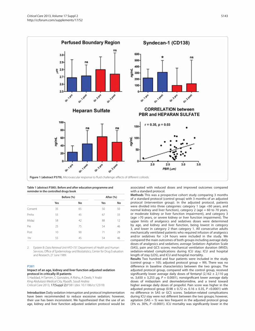
2. Epstein B: Data Retrieval Unit HFD-737. Department of Health and Human Services, Offi ce of Epidemiology and Biostatistics, Center for Drug Evaluation and Research; 27 June 1989.
P381Impact of an age, kidney and liver function adjusted sedation protocol in critically ill patientsS Haddad, H Tamim, C Gonzales, A Rishu, A Deeb, Y ArabiKing Abdulaziz Medical City, Riyadh, Saudi ArabiaCritical Care 2013, 17(Suppl 2):P381 (doi: 10.1186/cc12319)
Introduction Daily sedation interruption and protocol implementation have been recommended to reduce excessive sedation; however, their use has been inconsistent. We hypothesized that the use of an age, kidney and liver function adjusted sedation protocol would be
associated with reduced doses and improved outcomes compared with a standard protocol.Methods This was a prospective cohort study comparing 3 months of a standard protocol (control group) with 3 months of an adjusted protocol (intervention group). In the adjusted protocol, patients were divided into three categories: category 1 (age <60 years, and normal kidney and liver function), category 2 (age = 60 to 70 years, or moderate kidney or liver function impairment), and category 3 (age >70 years, or severe kidney or liver function impairment). The upper limits of analgesics and sedatives doses were determined by age, and kidney and liver function, being lowest in category 3, and lower in category 2 than category 1. All consecutive adults mechanically ventilated patients who required infusion of analgesics and/or sedatives for >24 hours were included in the study. We compared the main outcomes of both groups including average daily doses of analgesics and sedatives; average Sedation–Agitation Scale (SAS), pain and GCS scores; mechanical ventilation duration (MVD); sedation-related complications during ICU stay; ICU and hospital length of stay (LOS), and ICU and hospital mortality.Results Two hundred and four patients were included in the study (control group = 105; adjusted protocol group = 99). There was no diff erence in baseline characteristics between the two groups. The adjusted protocol group, compared with the control group, received signifi cantly lower average daily doses of fentanyl (2,162 ± 2,110 μg vs. 3,650 ± 3,253 μg, P = 0.0001), nonsignifi cant lower average daily doses of midazolam and dexmedetomidine, and a trend toward higher average daily doses of propofol. Pain score was higher in the adjusted protocol group (0.98 ± 0.72 vs. 0.16 ± 0.35, P <0.0001) with no diff erence in SAS or GCS scores. Sedation-related complications during ICU stay were not diff erent between the two groups; however, agitation (SAS = 5) was less frequent in the adjusted protocol group (3% vs. 30%, P <0.0001). ICU mortality was signifi cantly lower in the
Figure 1 (abstract P379). Microvascular response to fl uid challenge: eff ects of diff erent colloids.
Table 1 (abstract P380). Before and after education programme and reminder in the controlled drugs book
Before (%) After (%)
Yes No Yes No
Consent 35 65 50 50
Pmhx 55 45 67 33
Midaz 58 42 88 12
Pre 25 75 54 46
Post 10 90 71 29
1hr 55 45 54 46
Critical Care 2013, Volume 17 Suppl 2 http://ccforum.com/supplements/17/S2
S143

adjusted protocol group (18% vs. 36%, P = 0.004) with no signifi cant diff erences in MVD, ICU and hospital LOS, and hospital mortality.Conclusion The use of an age, kidney and liver function adjusted sedation protocol is associated with lower doses of analgesics and sedatives, lower risk of agitation and lower ICU mortality.
P382Risk and benefi t of protocol sedation and daily interruption of sedation for mechanically ventilated patients: a systematic reviewK Hosokawa1, M Egi2, M Nishimura3
1Kyoto Prefectural Yosanoumi Hospital, Kyoto, Japan; 2Okayama University Hospital, Okayama, Japan; 3Tokushima University Hospital, Tokushima, JapanCritical Care 2013, 17(Suppl 2):P382 (doi: 10.1186/cc12320)
Introduction Protocol-directed sedation (PS) and daily interruption of sedation (DIS) are two major strategies to optimize sedation depth for mechanically ventilated patients. To clarify the clinical risk and benefi ts of PS and DIS, we performed a systematic review of randomized controlled trials (RCTs) in previous literature.Methods We searched the MEDLINE database from January 1990 to October 2012 for studies on PS and DIS in critically ill patients. English-language manuscripts were included when they assessed the impact of PS or DIS on outcomes in critically ill patients requiring mechanical ventilation for >48 hours. PS is defi ned as sedation practices implemented by ICU staff using a sedation scale and a written protocol. DIS is defi ned as strategies with cessation of the continuous sedatives.Results We retrieved 902 citations. Among them, nine RCTs fulfi lled inclusion criteria. PS was compared with the standard practice in one RCT, DIS with the standard practice in four, PS with DIS in two, and PS with PS plus DIS (PS/DIS) in two. PS or DIS reduced the duration of mechanical ventilation by 28.9% and ICU stay by 27.8% compared with the standard practice. Neither the duration of mechanical ventilation nor ICU stay diff ered among DIS, PS and PS/DIS. Mortality did not diff er among the three strategies. DIS increased the daily dose of sedatives and the workload of nurses in one recent RCT.Conclusion PS or DIS decreased the duration of mechanical ventilation and ICU stay. PS seems to be superior to DIS based on the doses of sedatives and the workload of nurses.
P383Chemical and physical compatibility of continuous intravenous drug infusion combinations used in paediatric intensive careC Cole1, A Fox1, M Van Der Merwe2, L Dickson2, K Ball2, A Bevan1, A Hocking1, J Pappachan1
1University Hospitals Southampton, UK; 2University of Portsmouth, UKCritical Care 2013, 17(Suppl 2):P383 (doi: 10.1186/cc12321)
Introduction The aim of this research was to provide clinically relevant evidence for Y-site compatibility of drug infusion combinations used in the PICU. Pharmacists and clinicians regularly have to interpret limited published data, particularly when more than two drugs are Y-sited. The risk of potential incompatibility must be balanced against that of additional line insertion.Methods A full 28-factorial design (total 256 combinations) was used to investigate chemical and physical compatibility of fi ve drugs (clonidine, morphine, ketamine, midazolam and furosemide). The drugs were studied at their highest commonly infused concentrations and exposed to three variations in environmental conditions (diluent: sodium chloride 0.9% or glucose 10%; temperature 25 or 37°C; and normal room lighting or blue light phototherapy). Chemical stability was assessed using HPLC; >10% reduction in concentration indicated incompatibility. Physical incompatibility was confi rmed by precipitation, pH or colour change.Results Environmental conditions had no eff ect on the drug mixtures. The precipitation observed in incompatible combinations was due to either a change in pH, or with ketamine the presence of benzethonium chloride. Of 31 possible drug combinations, 12 were incompatible. A further three combinations were incompatible at extreme pH, or were of concern and so should be avoided. The incompatible formulations
all contained furosemide. All combinations of the sedative agents studied were chemically and physically compatible.Conclusion This work provides evidence for Y-site compatibility of morphine, midazolam, clonidine and ketamine in any combination, which will potentially reduce the need for extra intravenous lines. Furosemide is incompatible with any of these sedative drugs and must be infused via a separate line. These results will aid clinical decision-making and help satisfy the requirements of recent UK Department of Health legislation relating to the mixing of medicines.Reference1. Mixing of Medicines Prior to Administration in Clinical Practice: Medical
and Non-medical Prescribing [http://www.dh.gov.uk/en/Publicationsandstatistics/Publications/PublicationsPolicyAndGuidance/DH_116361]
P384Abstract withdrawn
P385Benzodiazepines induce hyperglycemia in rats by aff ecting peripheral disposal of glucoseVS Salice, FV Valenza, MP Pizzocri, LV Valenti, GC Chevallard, MU Umbrello, SG Gatti, SF Fargion, GI Iapichno, LG GattinoniUniversità degli Studi Milano, Milan, ItalyCritical Care 2013, 17(Suppl 2):P385 (doi: 10.1186/cc12323)
Introduction In light of the interest in the relationship between glycemia control in critically ill subjects and outcome, we set up a study to investigate whether benzodiazepine, commonly used in anesthesia and ICUs, interferes with glucose metabolism and to explore the mechanism.Methods A total of 40 sedated and paralyzed Sprague–Dawley rats (301 ± 55 g) were investigated in four consecutive studies. (1) To investigate the eff ects of diazepam on blood glucose, 15 rats were randomly assigned to intraperitoneal anesthesia with tiopenthal 80 mg/kg (DZP0), tiopenthal 40 mg/kg + diazepam 5 mg/kg (DPZ5) or tiopenthal 40 mg/kg + diazepam 15 mg/kg (DZP15). Blood levels of glucose (GEM premier 3000; IL) were measured at time intervals over 2 hours. (2) Ten animals randomized to DZP0 or DZP5 underwent an intravenous glucose tolerance test with glucose bolus (0.5 g/kg). Acute insulin response, the mean value of blood insulin (Insulin ELISA kit; Millipore) from 2 to 10 minutes after glucose bolus, was measured as index of insulin secretion. (3) A hyperinsulinemic euglycemic clamp obtained by a continuous intravenous infusion of insulin (130 mUI/kg/minute) was run in 10 animals randomized to DZP0 or DZP5 and the glucose infusion rate (GIR, mg/kg/minute) was assessed [1]. (4) The eff ect of midazolam on blood glucose was tested in fi ve additional animals (M5: tiophental 40 mg/kg + midazolam 5 mg/kg). Data are presented as mean ± SEM. Statistical analysis (ANOVA, t test) was conducted with Sigma Stat 3.1 (Systat Software).Results (1) Diazepam was associated with higher levels of blood glucose in a dose–response fashion: DZP0 128 ± 7 mg/dl, DZP5 166 ± 7.3, DZP15 197 ± 7 (P <0.05). (2) The acute insulin response to intravenous glucose tolerance test was similar in DZP0 and DZP5 (DZP0 3.97 ± 0.42 ng/ml, DZP5 3.68 ± 0.44, P = 0.68), while blood glucose levels were diff erent (DZP0 192 ± 5 mg/dl, DZP5 217 ± 5, P <0.05). (3) During hyperinsulinemic euglycemic clamp, blood glucose levels were similar (109 ± 3 mg/dl, P = 0.2), but the DZP5 group showed a trend through lower values of GIR (DZP0 30.8 ± 2 mg/kg/minute, DZP5 24.7 ± 2, P = 0.08). (4) Infusion of midazolam was associated with higher blood glucose levels (DZP0 128 ± 5 mg/dl, M5 151 ± 6, P <0.05).Conclusion Both diazepam and midazolam signifi cantly alter plasma glucose levels in rats. Peripheral disposal of glucose rather than altered pancreas secretion of insulin explains the benzodiazepine-associated hyperglycemia.Reference1. DeFronzo: Am J Physiol 1979, 273:E214-E223.
Critical Care 2013, Volume 17 Suppl 2 http://ccforum.com/supplements/17/S2
S144
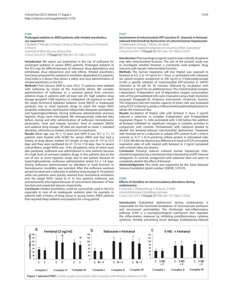
P386Prolonged sedation in ARDS patients with inhaled anesthetics: our experienceS Redaelli, P Mangili, V Ormas, S Sosio, L Peluso, F Ponzoni, N Patroniti, A PesentiUniversity of Milan-Bicocca, Monza, ItalyCritical Care 2013, 17(Suppl 2):P386 (doi: 10.1186/cc12324)
Introduction We report our experience in the use of isofl urane for prolonged sedation in severe ARDS patients. Prolonged sedation in the ICU may be diffi cult because of tolerance, drug dependence and withdrawal, drug interactions and side eff ects. Inhaled anesthetics have been proposed for sedation in ventilator-dependent ICU patients. AnaConDa is a device that allows a safety and easy administration of inhaled anesthetics in the ICU.Methods From January 2009 to June 2012, 15 patients were sedated with isofl urane by means of the AnaConDa device. We consider administration of isofl urane as a washout period from common sedative drugs in patients with (at least one of ): high sedative drug dosage (propofol ≥300 mg/hour or midazolam ≥8 mg/hour) to reach the target Richmond Agitation Sedation Score (RASS) or inadequate paralysis; two or more hypnotic drugs to reach the target RASS (propofol, midazolam, hydroxyzine, haloperidol, diazepam, quetiapine); and hypertriglyceridemia. During isofl urane administration previous hypnotic drugs were interrupted. We retrospectively collected data before, during and after administration of isofl urane: hemodynamic parameters, renal and hepatic function, level of sedation (RASS) and sedative drug dosage. All data are reported as mean ± standard deviation, otherwise as median (minimum to maximum).Results Mean age was 43 ± 12 years and SAPS II was 35.7 ± 11; 13 patients were treated with ECMO for severe ARDS and four had a history of drug abuse; median ICU length of stay was 41 (15 to 127) days and they were ventilated for 41 (12 to 114) days. Due to severe critical illness, target RASS was –4 for all patients, most of which were also paralysed. Isofl urane was administered in nine patients because of a high level of common sedative drugs, in fi ve patients due to the use of two or more hypnotic drugs and in one patient because of hypertrigliceridemia. Isofl urane administration lasted 5.6 ± 1.8 days. During isofl urane administration no alteration in renal function or hemodynamic instability was recorded. After the isofl urane washout period we observed a reduction in sedative drug dosage in 10 patients while two patients were quickly weaned from mechanical ventilation and the target RASS raised to 0. In two patients isofl urane was precautionarily interrupted because of concomitant alteration of liver function and suspected seizures respectively.Conclusion Inhaled anesthetics could be successfully used in the ICU especially in case of an inadequate sedation plan; for example, in patients with a history of drug abuse or young severe ARDS patients that required deep sedation and paralysis for a long period.
P387Involvement of mitochondrial ATP-sensitive K+ channels in fentanyl-induced mitochondrial dysfunction of cultured human hepatocytesS Djafarzadeh, M Vuda, J Takala, SM JakobBern University Hospital (Inselspital) and University of Bern, SwitzerlandCritical Care 2013, 17(Suppl 2):P387 (doi: 10.1186/cc12325)
Introduction Pharmacological agents used to treat critically ill patients may alter mitochondrial function. The aim of the present study was to investigate whether fentanyl, a commonly used analgesic drug, interacts with hepatic mitochondrial function.Methods The human hepatoma cell line HepG2 was exposed to fentanyl at 0.5, 2 or 10 ng/ml for 1 hour, or pretreated with naloxone (an opioid receptor antagonist) at 200 ng/ml or 5-hydroxydecanoate (5-HD; a specifi c inhibitor of mitochondrial ATP-sensitive K+ (KATP) channels) at 50 μM for 30 minutes, followed by incubation with fentanyl at 2 ng/ml for an additional hour. The mitochondrial complex I-dependent, II-dependent and IV-dependent oxygen consumption rates of the permeabilized cells were measured using a high-resolution oxygraph (Oxygraph-2k; Oroboros Instruments, Innsbruck, Austria). The respiratory electron transfer capacity of intact cells was evaluated using FCCP (carbonyl cyanide p-trifl uoromethoxyphenylhydrazone) to obtain the maximum fl ux.Results Incubation of HepG2 cells with fentanyl (1 hour, 2 ng/ml) induced a reduction in complex II-dependent and IV-dependent respiration (Figure 1). Cells pretreated with 5-HD before the addition of fentanyl exhibited no signifi cant changes in complex activities in comparison with controls. Pretreatment with naloxone tended to abolish the fentanyl-induced mitochondrial dysfunction. Treatment with fentanyl led to a reduction in cellular ATP content (0.24 ± 0.06 in controls vs. 0.17 ± 0.14 μmol/mg cellular protein in stimulated cells; P = 0.02). We did not observe any diff erence in basal or FCCP-uncoupled respiration rates of cells treated with fentanyl at 2 ng/ml compared with controls (data not shown).Conclusion Fentanyl reduces cultured human hepatocyte mito-chondrial respiration by a mechanism that is blocked by a KATP channel antagonist. In contrast, antagonism with naloxone does not seem to completely abolish the eff ect of fentanyl.Acknowledgement This study was supported by the Swiss National Science Foundation (grant number 32003B_127619).
P388Eff ects of clonidine on microcirculatory alterations during endotoxemiaK Schmidt, C Philipsenburg, A Zivkovic, S HoferUniversitätsklinikum Heidelberg, GermanyCritical Care 2013, 17(Suppl 2):P388 (doi: 10.1186/cc12326)
Introduction Endothelial dysfunction during endotoxemia is responsible for the functional breakdown of microvascular perfusion and microvessel permeability. The cholinergic anti-infl ammatory pathway (CAP) is a neurophysiological mechanism that regulates the infl ammatory response by inhibiting proinfl ammatory cytokine synthesis, thereby preventing tissue damage. Endotoxemia-induced
Figure 1 (abstract P387). Cellular oxygen consumption after incubation with fentanyl, naloxone or 5-HD.
Critical Care 2013, Volume 17 Suppl 2 http://ccforum.com/supplements/17/S2
S145

microcirculatory dysfunction can be reduced by cholinergic CAP activation. Clonidine improves survival in experimental sepsis [1] by reducing the sympathetic tone, resulting in the parasympathetic-mediated CAP activation. The aim of this study was to determine the eff ects of clonidine on microcirculatory alterations during endotoxemia.Methods Using fl uorescent intravital microscopy, we determined the venular wall shear rate, macromolecular effl ux and leukocyte adhesion in mesenteric postcapillary venules of male Wistar rats. Endotoxemia was induced over 120 minutes by intravenous infusion of lipopolysaccharide (LPS). Control groups received an equivalent volume of saline. Clonidine 10 μg/kg was applied as i.v. bolus in treatment groups. Animals received either (i) saline alone, (ii) clonidine 10 minutes prior to saline administration, (iii) clonidine 45 minutes prior to LPS administration, (iv) clonidine 10 minutes prior to LPS administration, (v) clonidine 30 minutes after LPS administration or (vi) LPS alone.Results All LPS groups (iii to vi) showed a signifi cantly reduced venular wall shear rate compared with the saline group after 120 minutes. There were no signifi cant diff erences between the numbers of adhering leukocytes in the clonidine/LPS groups (iii, iv, v) and the LPS group after 120 minutes. Macromolecular effl ux signifi cantly increased in all groups over the time period of 120 minutes. After 120 minutes there was no diff erence between the LPS group and the clonidine 10 minutes prior to LPS administration group (iv) whereas all other groups (i, ii, iii, v) showed a signifi cantly reduced macromolecular effl ux compared with the LPS group.Conclusion Clonidine has no positive eff ect on microhemodynamic alterations and leukocyte–endothelial interaction during endotoxemia. The reduction of capillary leakage in clonidine-treated groups depends on the time interval relative to the initiation of endotoxemia. Endothelial permeability and leukocyte activation are regulated by diff erent pathways when stimulated by clonidine during endotoxemia. We conclude that clonidine might have an important time-dependent anti-infl ammatory and protective eff ect on endothelial activation during infl ammation.Reference1. Hofer S, et al.: Crit Care 2009, 13:R11.
P389Conjunctival administration of S+ketamine in rabbitsL Hess1, J Malek2, A Kurzova2, Z Simunkova1, M Votava2
1Institute for Clinical and Experimental Medicine, Prague, Czech Republic; 23rd Medical Faculty of Charles University, Prague, Czech RepublicCritical Care 2013, 17(Suppl 2):P389 (doi: 10.1186/cc12327)
Introduction Delivering analgesics via conjunctival application could provide rapid and convenient pain relief in disaster medicine. There are sporadic reports from the USA concerning inhalation administration of aerosol with various drugs producing a wide variety of eff ects from anxiolysis, sedation, and loss of aggressiveness to immobilisation. We attempted to determine in an animal experiment whether conjunctival administration of S+ketamine could produce signifi cant eff ect without side eff ects.Methods After ethic committee approval, 10 rabbits were administered conjunctival S+ketamine 2.5 mg/kg. Measured parameters were SpO2, blood pressure (BP) and heart rate (HR) before administration and in 1-minute intervals and immobilisation time (loss of righting refl ex). The measurements were performed for 20 minutes. Conjunctival irritation was measured 1, 10 and 20 minutes after administration according to modifi ed technical standard EN IS O 10993-10. ANOVA test was used for statistical analysis of hemodynamic parameters.Results The immobilisation time was 207 ± 60 seconds. There were no changes in cardiorespiratory parameters (initial, 10 and 20 minutes after administration: HR 255.5 ± 24.7, 265.8 ± 26.0 and 267.8 ± 20.4, systolic BP 114.3 ± 8.6, 108.2 ± 10.7 and 113.9 ± 11.9 mmHg, diastolic BP 66.8 ± 11.6, 68.4 ± 10.6 and 69.7 ± 9.3 mmHg and SpO2 99.2 ± 1.0, 98.2 ± 0.6 and 98.7 ± 0.5). These results are in contrast to conjunctival administration of sufentanil 5 μg/kg that caused a signifi cant decrease of SpO2 and HR [1]. We can speculate that the reason for stability of cardiorespiratory parameters was due to the sympathoadrenergic
eff ect of ketamine or due to the method of administration. There were no signs of conjunctival irritation in any animal (S+ketamine is a preservative-free solution).Conclusion Conjunctival S+ketamine 2.5 mg/kg in rabbits produced rapid onset without changes in cardiorespiratory parameters and without signs of irritation of the eye. The results of our project warrant further research to increase the variety of drugs and methods of their administration for anxiolysis, sedation and analgesia in disaster medicine.Acknowledgement The study was supported by scientifi c grant IGA NT 11284.Reference1. Malek J, et al.: Conjunctival administration of sufentanil in rabbits. Eur J Pain
2011, Suppl 5:212.
P390Use of propofol for procedural sedation reduces length of stay in the emergency departmentJ Millar, F Adamson, P O’Connor, R Wilson, E Ferrie, R McLaughlinRoyal Victoria Hospital, Belfast, UKCritical Care 2013, 17(Suppl 2):P390 (doi: 10.1186/cc12328)
Introduction Procedural sedation is used in the emergency department (ED) to facilitate short but painful interventions. Many patients are suitable for discharge after completion. Ideally, the agent used to achieve sedation should not have a prolonged eff ect, allowing safe discharge in the shortest time frame. We hypothesised that propofol, with its short onset and off set, may reduce length of stay (LOS) in comparison with traditional benzodiazepines.Methods Data from a prospective registry were analysed for the period 1 August 2011 to 31 January 2012. Patients who underwent procedural sedation and who were discharged from the ED were identifi ed. Individuals were grouped as having received propofol, midazolam or a combination of the two. All were discharged when fully alert and able to eat and drink. Demographic details and the type of procedure undertaken were extracted. ANOVA was performed to identify diff erences in the length of stay between groups, in addition to descriptive analysis.Results During the study period 75 patients underwent procedural sedation and were discharged from the ED. The median age was 40 years and 57% were male. The commonest procedure performed was shoulder reduction (52%). In the propofol group (n = 20) the mean LOS was 100 minutes compared with 165 minutes in those receiving midazolam (n = 40) and 141 minutes in those receiving a combination (n = 15), P = 0.004. There was no diff erence in adverse events between groups. See Figure 1.Conclusion Propofol is increasingly used in EDs for procedural sedation due to its short duration of action. This study suggests that a shorter duration of action and faster recovery may result in a reduced LOS in the ED.
Figure 1 (abstract P390). Length of stay (minutes) in the ED.
Critical Care 2013, Volume 17 Suppl 2 http://ccforum.com/supplements/17/S2
S146

P391Pre-PRIS? Prospective monitoring for early markers of propofol infusion syndromeM Stovell, S Smith, M Udberg, W Loh, P NairWalton Centre Neurology & Neurosurgery, Liverpool, UKCritical Care 2013, 17(Suppl 2):P391 (doi: 10.1186/cc12329)
Introduction The use of propofol for sedation in intensive care has been associated with the propofol infusion syndrome (PRIS) characterised by cardiac dysfunction, metabolic acidosis, renal failure, rhabdomyolysis and hyperlipidaemia. We prospectively monitor biochemical markers that we believe demonstrate early signs of this dangerous, often fatal syndrome. When this Pre-PRIS state is identifi ed, propofol is withdrawn whilst the syndrome is still reversible.Methods We prospectively audited our monitoring of these markers over a 4-month period in propofol-sedated patients: propofol infusion rate, creatine kinase (CK), triglycerides (TG), creatinine, lactate, pH and base defi cit. We defi ned the criteria for Pre-PRIS as requiring a CK ≥320 mmol/l that had doubled from its base level and a rise in TG ≥1.7 IU/l; both that followed a trend with propofol dose.Results Data were collected from 54 adults; 50 received an infusion for ≥24 hours. We noted a signifi cant CK rise in 11 (22%) of our patients that could be attributed to propofol alone. They all had raised TG (≥1.7 IU/l); six (12%) were markedly raised (≥3.4 IU/l). No patients became signifi cantly acidotic, developed cardiac arrhythmias or renal failure as a result of propofol. In the 11 patients who met our criteria, highest daily propofol dose varied from 1.5 to 4 mg/kg/hour. Pre-PRIS developed from between 2 and 6 days of infusion. The mean propofol dose in patients displaying Pre-PRIS was 2.2 mg/kg/hour whilst in those not meeting our criteria it was 2.0 mg/kg/hour. No patients developed sequelae of full PRIS. See Table 1.
Table 1 (abstract P391)
Total Trauma Nontrauma
≥24 hours infusion 50 34 16
CK + TG ≥3.4 6 6 0
CK + TG ≥1.7 5 3 2
Conclusion We propose that a paired rise in CK and TG that can be attributed to propofol alone represents a Pre-PRIS state that is at risk of developing into full PRIS. We noted this in 22% of our patients, all on modest doses of propofol. It is unclear what proportion of patients will develop the full syndrome as it is not ethically possible to continue propofol in this situation. We advocate daily monitoring of CK and TG to identify Pre-PRIS so that propofol can be reduced or substituted to avoid the morbidity and mortality of the full syndrome.
P392Safety profi le of 1,008 cases of propofol sedation in the emergency departmentS Bradburn, G Lloyd, B NewsteadRoyal Devon and Exeter Hospital, Exeter, UKCritical Care 2013, 17(Suppl 2):P392 (doi: 10.1186/cc12330)
Introduction Until recently there were no guidelines for the reporting of adverse events (AEs) during procedural sedation [1,2]. A consensus document released in 2012 by the world SIVA International Sedation Task Force proposed a benchmark for defi ning AEs [3]. We analysed 1,008 cases of procedural sedation in the emergency department.Methods The study is based on 1,008 patients who received procedural sedation with propofol in the emergency department between December 2006 and March 2012. Patients were selected and sedated to a strict protocol by ED consultant staff . We applied the AE tool by performing a search through patient records, discussion with consultants performing the sedation and consensus opinion.Results From 1,008 cases we identifi ed 11 sentinel (six of hypotension, fi ve cases of hypoxia), 34 moderate, 25 minor and three minimal risk adverse events.
Conclusion The study shows a 1% adverse event rate. This supports use of propofol sedation by emergency physicians but within the limits of a strict governance framework. Our safety analysis using the World SIVA adverse events tool provides a reference point for further studies.References1. Green SM, Yealy DM: Procedural sedation goes Utstein: the Quebec
guidelines. Ann Emerg Med 2009, 53:436-438.2. Green SM, Krauss B: Barriers to propofol use in emergency medicine. Ann
Emerg Med 2008, 52:392-398.3. Mason KP, Green SM, Piacevoli Q; International Sedation Task Force: Adverse
event reporting tool to standardize the reporting and tracking of adverse events during procedural sedation: a consensus document from the World SIVA International Sedation Task Force. Br J Anaesth 2012, 108:13-20.
P393Use of physical restraint in Dutch ICUs: prevalence and motivesRJ Raijmakers1, RL Vroegop1, H Tekatli1, M Van den Boogaard2, AW Van der Kooi1, AJ Slooter1
1University Medical Center Utrecht, the Netherlands; 2Radboud University Nijmegen Medical Centre, Nijmegen, the NetherlandsCritical Care 2013, 17(Suppl 2):P393 (doi: 10.1186/cc12331)
Introduction Physical restraints are used to facilitate essential care and prevent secondary injuries. However, physical restraint may be regarded as humiliating. It may lead to local injury and increase the risk of delirium and post-traumatic stress syndrome. Research on physical restraint is scarce. The aim of this study is to investigate the scope of physical restraint use.Methods Twenty-one ICUs ranging from local hospitals to academic centres were each visited twice and 327 patients were included. We recorded characteristics of restrained patients, motives and awareness of nurses and physicians.Results Physical restraint was applied in 74 (23%) patients, ranging from 0 to 54% in diff erent hospitals. Frequent motives for restraint use were ‘possible threat to airway’ (36%) and ‘pulling lines/probes’ (31%). Restrained subjects more often had a positive CAM-ICU (34% vs. 16%, P <0.001), could less frequently verbally communicate (14% vs. 49%, P <0.001), and received more often antipsychotics (49% vs. 28%, P <0.001), or benzodiazepines (55% vs. 36%, P = 0.003). The use of physical restraint was registered in the patient’s fi les in 48% of cases. Of the 310 interviewed nurses, 258 (83%) were familiar with a physical restraint protocol and 89 (29%) used it in any situation. Thirty percent of the 60 interviewed physicians were aware of the physical restraint status of their patients.Conclusion Physical restraint is frequently used in Dutch ICUs, but the frequency diff ers strongly between diff erent ICUs. Attending physicians are often not aware of physical restraint use.
P394Factors infl uencing the use of physical restraints in Canadian ICUsB Sneyers1, E Luk2, M Perreault3, D Williamson3, L Rose2, S Mehta4, L Burry4
1Université Catholique de Louvain, Brussels, Belgium; 2Lawrence S. Bloomberg Faculty of Nursing, University of Toronto, Ontario, Canada; 3Université de Montréal, Canada; 4Mount Sinai Hospital, University of Toronto, Ontario, CanadaCritical Care 2013, 17(Suppl 2):P394 (doi: 10.1186/cc12332)
Introduction Physical restraint (PR) use in critically ill patients has been associated with delirium, unplanned extubation, prolonged ICU length of stay, and post-traumatic stress disorder. Our objectives were to defi ne prevalence of PR use, and to examine patient, treatment, or institutional factors associated with their use in Canadian ICUs.Methods We conducted an analysis of a database of sedative, analgesic, antipsychotic and neuromuscular blocker prescribing practices and PR use in 711 mechanically ventilated (MV) adults in 51 Canadian ICUs (3,619 patient-days). Data were collected during a 2-week period (2008 to 2009). Logistic and Poisson regression analysis were used to identify factors associated with PR use and number of days of use respectively.Results PR was used on 1 or more days in 52.6% (374/711) of patients. For patients restrained, the mean number of days restrained was
Critical Care 2013, Volume 17 Suppl 2 http://ccforum.com/supplements/17/S2
S147

4.1 ± 4.0 and the mean number of restraint applications per patient was 1.2 ± 0.5. Treatment characteristics associated with PR use were: daily benzodiazepine dose (OR = 1.01, 95% CI: 1.00 to 1.01), daily opiate dose (OR = 1.00, 95% CI: 1.00 to 1.01), daily sedation interruption (OR = 1.84, 95% CI: 1.24 to 2.72), use of antipsychotics (OR = 3.42, 95% CI: 1.98 to 5.91) and agitation (Sedation–Agitation Scale (SAS) >4, OR = 2.55, 95% CI: 1.20 to 5.44). Additionally, number of days of PR use was increased according to the following: daily sedation interruption (IRR = 4.52, 95% CI: 2.09 to 9.75), use of antipsychotics (IRR = 18.65, 95% CI: 7.95 to 43.77), the presence of a heavily sedated score (SAS <3, IRR = 2.57, 95% CI: 1.06 to 6.24) or the presence of an adverse event, such as self-extubation (IRR = 10.46, 95% CI: 2.55 to 42.92). Hospital characteristics associated with PR use included the proportion of MV patients in the ICU. University hospitals were associated with reduced PR use, compared with community hospitals (OR = 0.272, 95% CI: 0.15 to 0.50). Nonmodifi able patient characteristics (age, sex, APACHE score, patient category, prior substance abuse, prior psychotropic medication, pre-existing psychiatric condition or dementia) were not associated with PR use.Conclusion We identifi ed multiple factors associated with PR use and number of days of PR use. Particularly among those related to treatment, strategies such as daily sedation interruption or use of antipsychotics are associated with PR use and increasing the number of days of use.
P395Current practices in ICU delirium management: a prospective multicenter study in the NetherlandsZ Trogrlic, E Ista, A Slooter, J Bakker, M Van der Jagt, on behalf of iDECePTIvE Study GroupErasmus MC University Medical Center, Rotterdam, the NetherlandsCritical Care 2013, 17(Suppl 2):P395 (doi: 10.1186/cc12333)
Introduction As part of a multicenter prospective study on barriers and facilitators for implementation of protocolled care for delirious critically ill patients, we aimed to describe current practices in delirium management in a representative cohort of ICU patients.Methods We included consecutively admitted patients during a 4-month period in a prospective multicenter study in six ICUs (one academic, teaching and nonteaching hospitals) in the southwest of the Netherlands. We assessed: percentage of patients screened for delirium with validated screening tools; pharmacological treatment with haloperidol or other antipsychotic drugs; psychohygiene (use of hearing aids and or glasses, preventing sleep disturbances); and access to early mobilization and physiotherapy. Delirium was pragmatically defi ned as administration of haloperidol and/or delirium reported by physicians or ICU nurses in patient records, as assessed by a designated research nurse. Diff erences between centers were tested with non-parametric tests.Results We assessed 1,576 patients, corresponding with 8,150 ICU treatment days with a median length of stay of 3 days (IQR 2 to 5). The mean age of the patients was 62 years (SD = 16) and 58% were male. Delirium occurred in 23% (356/1,576) of patients with a median duration of 3 days (IQR 2 to 7) and ranged from 11 to 40% for each ICU. Delirium assessment with the CAM-ICU at any point during ICU stay was performed in 38% of all patients. Screening with CAM-ICU was applied in three ICUs, in 29 to 96% of the patients in these centers. Of 3,564 documented screening days with the CAM-ICU, it was positive in 1,459 (41%); in only 120 (8%) of these CAM-ICU-positive days there was a documented action or treatment started for delirium. However, patients still received haloperidol on 52% (n = 766) of all CAM-ICU-positive days. Patients received benzodiazepines in 49% (n = 1,141) of patient sedation days. Delirium preventive interventions were physiotherapy (19% of 8,150 ICU days), mobilization (10%), glasses use (2.6%) and hearing aid use (0.3%). Presence of hearing or visual impairment at admission was not documented in 65% of patients.Conclusion Daily screening for ICU delirium with a validated screening instrument is applied in less than one-half of the time in critically ill patients and management of delirium is often not guided by this screening. Haloperidol was used as the fi rst-choice medication. Measures aimed at delirium prevention (psychohygiene and early
mobilization) were carried only in a small minority or were not documented. To implement protocolled delirium care in the region at study, a multifaceted tailored implementation program is needed.
P396Intraoperative dexamethasone and delirium after cardiac surgery: a randomized clinical trialAC Sauër, AJ Slooter, DS Veldhuijzen, MM Van Eijk, D Van DijkUniversity Medical Center Utrecht, the NetherlandsCritical Care 2013, 17(Suppl 2):P396 (doi: 10.1186/cc12334)
Introduction The objective of this study is to investigate the eff ect of intraoperative administration of dexamethasone versus placebo on the incidence of delirium in the fi rst four postoperative days after cardiac surgery.Methods Within the context of the large multicenter Dexamethasone for Cardiac Surgery (DECS) trial [1] for which patients were randomized to 1 mg/kg dexamethasone or placebo at induction of anesthesia, a monocenter substudy was conducted. The primary outcome of this study was the incidence of delirium in the fi rst four postoperative days. Secondary outcomes were duration of delirium, use of restrictive measures and sedative, antipsychotic and analgesic requirements. Delirium was assessed daily by trained research personnel, using the Richmond Agitation Sedation Scale and the Confusion Assessment Method. Medical, nursing and medication charts were evaluated for signs of delirium and use of prespecifi ed medication. Analysis was by intention to treat.Results Of 768 eligible patients, complete data on delirium could be collected in 738 patients. The incidence of delirium was 14.2% in the dexamethasone group and 14.9% in the placebo group (odds ratio = 0.95, 95% CI = 0.63 to 1.43). No signifi cant diff erence was found on the duration of delirium between the intervention (median = 2 days, interquartile range 1 to 3 days) and placebo (median = 2 days, interquartile range 1 to 2 days) group (P = 0.45). The use of restrictive measures and administration of sedatives, haloperidol, benzodiazepine and opiates were comparable between both groups.Conclusion Intraoperative injection of dexamethasone seems not to aff ect the incidence or duration of delirium in the fi rst 4 days after cardiac surgery, suggesting this regimen is safe to use in the operative setting with respect to psychiatric adverse events.Reference1. Dieleman JM, et al.: JAMA 2012, 308:1761-1767.
P397Current perspectives, beliefs and practices concerning delirium in critically ill patients: a multicenter survey among Dutch healthcare professionalsZ Trogrlic, M Van der Jagt, P Van der Voort, J Bakker, E Ista, on behalf of iDECePTIvE Study GroupErasmus MC University Medical Center, Rotterdam, the NetherlandsCritical Care 2013, 17(Suppl 2):P397 (doi: 10.1186/cc12335)
Introduction The beliefs, knowledge and practices regarding ICU delirium among ICU professionals may vary. This may interfere with the implementation of the Dutch ICU Delirium guideline. We aimed to get insight into potential barriers and facilitators for delirium guideline implementation that may help to fi nd an eff ective implementation strategy.Methods An online survey was sent to healthcare professionals from the six participating ICUs. Respondents included ICU physicians, nurses and delirium experts (psychiatrists, neurologists, geriatricians, nurse experts). The survey consisted of statements on beliefs, knowledge and practices towards ICU delirium. Agreement with statements by more than 75% of respondents were regarded as facilitating items and agreement lower than 50% as barriers for implementing protocolled care.Results Of the 565 surveys distributed, 360 were completed (63.7%). The majority of respondents were ICU nurses (79%). Delirium was considered a major problem (83%) that requires adequate treatment (99%) and is underdiagnosed (81%). Respondents considered that
Critical Care 2013, Volume 17 Suppl 2 http://ccforum.com/supplements/17/S2
S148

routine screening of delirium can improve prognosis (95%). However, only a minority (20%) answered that delirium is preventable. Only 39% of the respondents had received any training about delirium in the previous 3 years and 77% of them found training useful. The mean delirium knowledge score was 6.6 out of 10 (SD = 1.54). When all groups were mutually compared, nurses scored lower than delirium experts (ANOVA, P = 0.013). The respondents (58%; n = 210) from three ICUs indicated that CAM-ICU assessment was department policy. However, 50% (n = 106) of these respondents felt unfamiliar with CAM-ICU and only 47% (n = 99) of them indicated that a positive CAM-ICU was used for treatment decisions. Haloperidol was the fi rst-choice pharmacological treatment. Only 21% of all respondents knew that a national ICU delirium guideline existed, but in-depth knowledge was generally low.Conclusion Our survey showed that healthcare professionals considered delirium an important but underdiagnosed form of organ failure. In contrast, screening tools for delirium are scarcely used, knowledge can be improved and protocolled treatment based on positive screening is often lacking. These results suggest that the focus of implementation of ICU delirium management should not be on motivational aspects, but on knowledge improvements, training in screening tools and implementation of treatment and prevention protocols.
P398International validation of the delirium prediction model for ICU patients (PREDELIRIC): a multicenter observati onal studyM van den Boogaard1, L Schoonhoven2, E Maseda3, C Plowright4, C Jones5, A Luetz6, PV Sackey7, P Jorens8, LM Aitken9, F van Haren10, JG van der Hoeven1, P Pickkers1
1Radboud University Nijmegen Medical Centre, Nijmegen, the Netherlands; 2Radboud University Nijmegen Medical Centre, Scientifi c Institute for Quality of Healthcare, Nijmegen, the Netherlands; 3Hospital Universitario La Paz, Madrid, Spain; 4Medway Maritime Hospital, Kent, UK; 5Whiston Hospital, Prescot, UK; 6Charité – Universitaetsmedizin Berlin, Germany; 7Karolinska University Hospital, Stockholm, Sweden; 8University of Antwerpen, Belgium; 9Princess Alexandra Hospital, Woolloongabba, Australia; 10Canberra Hospital, Canberra, AustraliaCritical Care 2013, 17(Suppl 2):P398 (doi: 10.1186/cc12336)
Introduction Delirium is a serious and frequent disorder in ICU patients. The aim of this study was to internationally validate the existing Prediction of Delirium ICU (PREDELIRIC) model to predict delirium in ICU patients.Methods A prospective multicenter cohort study was performed in eight ICUs in six countries (Table 1). The 10 predictors (age, APACHE II score, urgent and admission category, infection, coma, sedation, morphine use, urea level, metabolic acidosis) were collected within 24 hours after ICU admission. CAM-ICU was used to identify ICU delirium.Results A total of 2,852 adult ICU patients were screened and 1,824 (64%) were included. Main exclusion reasons were length of stay <1 day (19.1%) and sustained coma (4.1%). Mean ± CAM-ICU compliance
was 82 ± 16% and inter-rater reliability 0.87 ± 0.17. Overall delirium incidence was 19.9% (range 27.2%). Despite signifi cant diff erences between centres on all 10 predictors, the overall area under the receiver operating characteristic curve (AUROC) of the eight centers was 0.77 (95% CI: 0.74 to 0.79).Conclusion The overall predictive value of the PREDELIRIC model was good, indicating that the prediction model can now be used internationally.
P399Delirium in ICU patients is associated with increased temperature variabilityAW Van der Kooi, TH Kappen, RJ Raijmakers, IJ Zaal, AJ SlooterUniversity Medical Center Utrecht, the NetherlandsCritical Care 2013, 17(Suppl 2):P399 (doi: 10.1186/cc12337)
Introduction Delirium is an acute disturbance of consciousness and cognition. It is a common disorder in the ICU and associated with impaired long-term outcome [1,2]. Despite its frequency and impact, delirium is poorly recognized by ICU physicians and nurses using delirium screening tools [3]. A completely new approach to detect delirium is to use monitoring of physiological alterations. Temperature variability, a measure for temperature regulation, could be an interesting parameter for monitoring of ICU delirium, but this has never been investigated before. The aim of this study was to investigate whether temperature variability is aff ected during ICU delirium.Methods We included patients in whom days with delirium could be compared with days without delirium, based on the Confusion Assessment Method for the ICU and inspection of medical records. Patients with conditions aff ecting thermal regulation, including infectious diseases, and those receiving therapies aff ecting body temperature were excluded. Twenty-four ICU patients were included after screening 334 delirious ICU patients. Daily temperature variability was determined by computing the mean absolute second derivative of the temperature signal. Per patient, temperature variability during delirious days was compared with nondelirium days using a Wilcoxon signed-rank test. With a linear mixed model, diff erences between delirium and nondelirium days with regard to temperature variability were analysed adjusted for daily mean Richmond Agitation and Sedation Scale scores, daily maximum Sequential Organ Failure Assessment score, and within-patient correlation.Results Temperature variability was increased during delirium days compared with days without delirium (mean diff erence = –0.007, 95% CI = –0.004; –0.011, P <0.001). Adjusting for confounders did not alter our fi ndings (adjusted mean diff erence = –0.005, 95% CI = –0.008; –0.002, P <0.001).Conclusion Temperature variability is increased during delirium in ICU patients, which refl ects the encephalopathy that underlies delirium. Opportunities for delirium monitoring using temperature variability should be further explored. Particularly, in combination with electroencephalography it could provide the input for an objective tool to monitor delirium.References1. Pisani MA, et al.: Am J Respir Crit Care Med 2009, 180:1092-1097.2. Ely EW, et al.: JAMA 2004, 291:1753-1762.3. van Eijk MM, et al.: Am J Respir Crit Care Med 2011, 184:340-344.
P400Long-term outcome of delirium in critically ill patientsA Wolters, D Van Dijk, O Cremer, D De Lange, A SlooterUMC Utrecht, the NetherlandsCritical Care 2013, 17(Suppl 2):P400 (doi: 10.1186/cc12338)
Introduction In ICU patients, little research has been performed on the relationship between delirium and long-term outcome, including health-related quality of life (HRQoL), cognitive functioning and mortality. In addition, results seem to be inconsistent. Furthermore, in studies that reported increased mortality in delirious patients, no proper adjustments were made for severity of illness during ICU admission. This study was conducted to investigate the association
Table 1 (abstract P398). PREDELIRIC AUROC of diff erent centers
Delirium (%) AUROC 95% CI
Belgium (%) 86 (15.2) 0.79 0.74 to 0.84
Germany (%) 60 (26.9) 0.85 0.80 to 0.91
Spain (%) 23 (18.0) 0.88 0.81 to 0.94
Sweden (%) 30 (39.0) 0.71 0.60 to 0.83
Australia Brisbane (%) 42 (12.8) 0.81 0.75 to 0.88
Australia Canberra (%) 23 (11.8) 0.80 0.72 to 0.89
UK_Prescot (%) 73 (30.8) 0.65 0.57 to 0.73
UK_Kent (%) 26 (36.6) 0.76 0.65 to 0.88
PREDELIRIC overall 363 (19.9) 0.77 0.74 to 0.79
Critical Care 2013, Volume 17 Suppl 2 http://ccforum.com/supplements/17/S2
S149

between ICU delirium and long-term HRQoL, cognitive functioning and mortality. The hypothesis was that delirious patients have worse long-term outcome in comparison with nondelirious patients.Methods A prospective observational cohort study was conducted. A median of 12 months after ICU discharge, questionnaires were sent to all survivors. HRQoL and cognitive functioning were measured with the EuroQol-6D. Age, gender and severity of illness were considered relevant covariates. Severity of illness was estimated using the APACHE IV score and the maximal SOFA score during admission. HRQoL was investigated with linear regression analysis, cognitive functioning using logistic regression and mortality with Cox regression analysis.Results The patient population consisted of 690 patients admitted to the ICU, subdivided into delirious (n = 257) and nondelirious patients (n = 433). During follow-up, 181 (26%) patients died. The response rate of the questionnaire was 70.6%. After adjusting for the predefi ned covariates, delirium was signifi cantly associated with a lower HRQoL (adjusted β: –0.137; 95% CI = –0.140 to –0.005) and more mild and severe cognitive impairment (adjusted odds ratio: respectively: 2.3; 95% CI = 1.3 to 4.2 and 5.8; 95% CI = 1.3 to 15.2). No signifi cant association between delirium and long-term mortality was found (adjusted hazard ratio: 1.0; 95% CI = 0.7 to1.4).Conclusion Delirium during ICU admission was associated with lower HRQoL and worse cognitive functioning, 1 year after discharge. Furthermore, delirium on the ICU was not associated with long-term mortality after adjusting for relevant covariates, including severity of illness during ICU admission.
P401Gastrointestinal symptoms in the ICU: a comparison of primary and secondary acute gastrointestinal injuryA Reintam Blaser, J StarkopfUniversity of Tartu, Tartu University Hospital, Tartu, EstoniaCritical Care 2013, 17(Suppl 2):P401 (doi: 10.1186/cc12339)
Introduction We aimed to clarify the diff erences between primary and secondary acute GI injury.Methods A total of 2,690 consecutive adult patients were retro-spectively studied during their fi rst week in the ICU. Pathology in the GI system or laparotomy defi ned the primary GI insult. If GI symptoms developed without primary GI insult it was considered secondary GI injury. Absent bowel sounds (BS), vomiting/regurgitation, diarrhoea, bowel distension, GI bleeding, and high gastric residuals (GRV >1,000 ml/24 hours) were recorded daily.Results In total, 2,690 patients (60.4% male), median age 59 years (range 16 to 92), were studied. Eighty-four per cent of them were ventilated, 72% received vasopressor/inotrope. Median (IQR) APACHE II score was 14 (9 to 21) and SOFA on the fi rst day was 6 (3 to 10). A total 35.5% had primary GI pathology. During the fi rst week 34% of patients had absent BS, 38% vomiting/regurgitation, 9% diarrhoea, 7% bowel distension, 6% high GRV and 5% GI bleeding. All symptoms except diarrhoea occurred more often (<0.001) in patients with primary GI insult. Eighty-fi ve per cent of patients with primary GI insult versus 46% without developed at least one GI symptom. The incidence of GI symptoms was signifi cantly higher in nonsurvivors. ICU mortality was lower in patients with primary than secondary GI injury (43.6% vs. 61.2%, P = 0.046). Nonsurvivors without primary GI insult developed GI symptoms later (Figure 1).Conclusion Primary and secondary acute GI injury have diff erent incidence, dynamics and outcome.
P402Eff ects of proton pump inhibitor versus H2-receptor antagonist stress ulcer prophylaxis on ventilator-associated pneumonia: a pilot studyJF Fogas, KK Kiss, FG Gyura, ZT Tóbiás, ZM MolnárUniversity of Szeged, HungaryCritical Care 2013, 17(Suppl 2):P402 (doi: 10.1186/cc12340)
Introduction Ventilator-associated pneumonia (VAP) is the most frequent ICU-acquired infection among patients receiving mechanical
ventilation with relative risk of 9 to 27% and with mortality of 25 to 50% [1,2]. One of the promoting factors of VAP is the increased pH of the gastric acid, which occurs when H2-receptor antagonists (H2RA) or proton pump inhibitors (PPI) are used for stress ulcer prophylaxis. The aim of this study was to investigate eff ects of PPI versus H2RA on the occurrence of VAP and, as second endpoint, the occurrence of gastrointestinal bleeding (GIB).Methods After ethics committee approval mechanically ventilated (>48 hours) adults were recruited in a prospective randomized controlled, double-blind clinical trial and randomised into PPI or H2RA groups. Patients with admission diagnosis of pneumonia or other acute infl ammatory pulmonary disease, severe sepsis/septic shock, and/or regular PPI/H2RA users were excluded. The diagnostic criteria for VAP were: leukocytosis, increased PCT level, fever, purulent tracheal secretion, positive microbiological result of tracheal sample, and new/increased infi ltrate on chest X-ray. Data are presented as median (interquartile range, IQR). For statistical analysis Mann–Whitney U test and chi-square tests were used.Results Out of 94 patients recruited 14 were excluded due to early weaning/death (n = 9) or protocol violation (n = 6). The remaining 79 patients were analysed (PPI, n = 38; H2RA, n = 41). There was no signifi cant diff erence (P >0.05) between the groups regarding demographics: age 67 (56 to 77) versus 72 (58 to 79) years; male/female: 23/15 versus 25/16; length of mechanical ventilation: 5 (3 to 9) versus 5 (2 to 8) days; APACHE II score: 28 (22 to 32) versus 26 (21 to 36) (PPI vs. H2RA, respectively). There was no signifi cant diff erence in the number of cases with VAP in the PPI versus H2RA groups: 9 (24%) versus 10 (24%). None of the patients developed GI bleeding during their stay on the ICU.Conclusion The results of this pilot study suggest that there may be no diff erence in the incidence of VAP and GI bleeding if stress ulcer prophylaxis is performed by H2RA or PPI. As the latter is more expensive, its use as fi rst choice in critical care should be questioned.
Figure 1 (abstract P401). Daily dynamics of the number of GI symptoms. P <0.001 between survivors and nonsurvivors in all time points.
Critical Care 2013, Volume 17 Suppl 2 http://ccforum.com/supplements/17/S2
S150

However, the completion of the study on the planned 198 patients is required to come to the fi nal conclusions.References1. Efrati S, et al.: J Clin Monit Comput 2010, 24:161-168.2. Torres A, et al.: Intensive Care Med 2009, 35:9-29.
P403Early diagnosis of liver failure in septic patients using the maximal liver function capacity test (LiMAx test): comparison with conventional methodsM Kaff arnik, M Stockmann, J LockCharite Berlin Campus Virchow, Berlin, GermanyCritical Care 2013, 17(Suppl 2):P403 (doi: 10.1186/cc12341)
Introduction Patients with bacterial sepsis often suff er from multiorgan failure. No reliable parameter exists for the exact estimation of the liver function. The indocyaningreen test (ICG test) showed a correlation between liver function and mortality rate. The new maximal liver function capacity test (LiMAx test) is a real-time method to investigate the liver function.Methods Thirty septic patients were prospectively included in the study. The LiMAx test was performed on days 0, 2, 5 and 10 after sepsis onset and was compared with ICG test and liver-specifi c laboratory parameters. Primary endpoint was the mortality rate after 90 days. Secondary endpoint was the comparison with the ICG test.Results The LiMAx test showed low results initially with increasing values on days 5 and 10. The 90-day mortality rate in patients with a low LiMAx test on day 2 (<100 μg/kg/hour) was signifi cantly higher than in patients with a LiMAx test >100 μg/kg/hour. The LiMAx test was comparable with the ICG test. Patients with indication for renal replacement therapy during hospital treatment showed signifi cantly lower LiMAx values than patients without dialysis. See Figure 1.Conclusion With the LiMAx test we can detect a liver dysfunction in septic patients early on days 0 to 2. The LiMAx test is equal with the ICG test and a result of <100 μg/kg/hour on day 2 suggests a low probability of survival in septic patients.References1. Stockmann et al.: New liver function test for prediction of postoperative
outcome in liver surgery. HPB 2010, 12:139.2. Lock et al.: Early diagnosis of primary nonfunction and indication for
reoperation after liver transplantation. Liver Transpl 2010, 16:172.
P404Impact of liver resection on portal venous pressure and renal functionP Biesenbach, T Grünberger, B Payer, A Ferlitsch, W Druml, E Fleischmann, F Luf, J MühlbacherMedical University of Vienna, AustriaCritical Care 2013, 17(Suppl 2):P404 (doi: 10.1186/cc12342)
Introduction Liver dysfunction – in correlation to the severity of functional impairment – but also any increase in portal pressure per se (hepatorenal refl ex) can induce alterations in renal function and ultimately result in hepatorenal syndrome (HRS). In this prospective investigation we determined the impact of liver resection on portal venous pressure by measuring the hepatic venous pressure gradient (HVPG), on concentrations of vasoactive peptides and on renal function.Methods Twenty patients (mean age 66.3 years) undergoing elective liver resection surgery because of malignant tumor were assessed and grouped according to resection size: (Group 1) hemihepatectomy, n = 13 versus (Group 2) segmentectomy, n = 7. HPVG was measured before and after resection by canulation of a hepatic vein under fl uoroscopic guidance, liver function was assessed by the indocyanine green plasma disappearance rate (ICG-PDR).Results HVPG increased in Group 1 from 3.7 to 5.4 mmHg (P <0.05) and decreased in Group 2 (4.8 to 4.3 mmHg, P = NS) (Table 1). Liver function as assessed by ICG-PDR decreased in Group 1 by day 1 (18.5 to 15.3, P <0.05) and remained stable in Group 2 (25.5 to 26.8, P = NS). Renin, aldosterone, ADH, adrenaline, noradrenaline and dopamine increased signifi cantly (P <0.05) in Group 1 during operation. Group 2 showed a signifi cant rise only in ADH and dopamine. Acute kidney injury occurred in fi ve of 13 patients in Group 1 (P <0.05).
Table 1 (abstract P404)
Group 1 Group 2
Renin 36.4 7.9
PostOP 189.7* 30.5
Day 1 258 31.3
ADH 3.49 2.59
PostOP 16.11* 10.8*
Day 1 10.8 2.39
Creatine 0.8 0.83
Day 1 1.25* 0.91
*P <0.05 compared with preOP value.
Conclusion Depending on resection size liver resection acutely increases portal venous pressure and induces neurohumoral activation resulting in compromised renal function and increased risk of developing AKI.
P405Value of the preoperative Model for End-stage Liver Disease score in predicting postoperative outcome in living donor liver transplantation recipientsY Nassar, K Omar, A Battah, A MwafyCairo University, Giza, EgyptCritical Care 2013, 17(Suppl 2):P405 (doi: 10.1186/cc12343)
Introduction Partial liver grafting predisposes recipients to a unique set of potential technical and anatomic complications that are not prevalent in whole deceased donor grafts. We aimed to assess the preoperative Model for End-stage Liver Disease (MELD) score, postoperative 30-day clinical and laboratory follow-up, and detection of the value of MELD score in predicting 30-day postoperative outcome of LDLT recipients.Methods A prospective multicenter registry involving 142 patients who underwent LDLT from October 2004 to December 2010. Preoperative
Figure 1 (abstract P403). Kaplan–Meier survival curve of patients with high and low LiMAx results.
Critical Care 2013, Volume 17 Suppl 2 http://ccforum.com/supplements/17/S2
S151

MELD score was calculated: (0.378×log (bilirubin) (mg/dl) + 1.120×log (INR) + 0.957×log (creatinine) (mg/dl) + 0.643)×10 then rounded to the nearest integer. Postoperative daily recording of all the clinical and laboratory data was done for a period of 30 days.Results The survival rate recorded in our study was 86.62%. The most frequent complications were renal complications (86.6%), pulmonary complications (73.9%), neurological complications (14%), cardiovascular complications (12.6%), infectious complications (13.3%), intra-abdominal infections (10.5%) and immunosuppressant toxicity (7.7%). MELD score proved to be a good predictor of perioperative outcome, patients with MELD score (>12) was associated with less favorable outcome, accuracy was (80.3%).Conclusion MELD score >12 was associated with less favorable outcome with an accuracy of 80.3%.
P406Does just fungal evidence increase the mortality of critically ill patients with liver cirrhosis? A retrospective analysisT Lahmer, U Mayr, M Messer, B Saugel, C Schultheiss, R Schmid, W HuberUniversity Hospital Munich, GermanyCritical Care 2013, 17(Suppl 2):P406 (doi: 10.1186/cc12344)
Introduction Patients with liver cirrhosis (LC) are susceptible to a variety of complications. Especially, infection disorders can reduce their life expectancy markedly. The aim of this retrospective analysis was to detect the incidence and prognosis in patients with LC and fungal evidence (FE).Methods A 3-year retrospective analysis of ICU patients with LC. Patients with LC with/without FE were compared with a matched control group based on comparable age and APACHE II score. Specimens were collected based on clinical and/or laboratory (infection signs) reasons and were analyzed by microbiological standard methods.Results A total of 101 patients with LC were enrolled in this analysis. Basic data for both groups: Child–Pugh Score: 4*A, 18*B, 79*C; reason for ICU admission: 28*hepatorenal syndrome, 18*SBP, 20*GI bleeding, 19*sepsis, 16*pneumonia; etiology of LC: 83*alcoholic, 8*NASH, 8*viral (hepatitis B/C), 2*PSC. Fifty-fi ve (54%) patients with LC had FE (mean age: 57.6 ± 14 years, sex M/F: 40/15, APACHE II score 27 ± 2). Compared with the 46 patients with LC but without FE (mean age: 59.2 ± 12 years, sex M/F: 35/11, APACHE II score 23 ± 4), these patients have a signifi cantly higher APACHE II score (27 ± 2 vs. 23 ± 4; P <0.001) and signifi cantly higher mortality (43/55 (78%) vs. 15/46 (33%); P <0.001). Multivariate analysis showed that FE is associated with a higher mortality (P <0.001) but is not associated with age, sex or CHILD score. Seventeen (31%) of the 55 patients in the control group had FE (screening patients n = 147, mean age 65.8 ± 12 years, sex M/F: 35/20, APACHE II score 26.5 ± 3, reason for ICU admission: 19*pneumonia, 16*sepsis, 9*intracerebral haemorrhage, 7*acute abdomen, 4*lymphatic disorder). The mortality rate was signifi cantly lower (27/55 (49%) vs. 43/55 (78%); P = 0.003) as compared with patients with LC and FE. The main localisation of fungal was in both groups the lung (46 vs. 14), followed by urine (18 vs. 2), blood culture (6 vs. 1) and ascites (4 vs. 0). Predominantly candida species were detected (especially C. albicans/glabrata), fi ve patients in the LC group had aspergillus fumigatus. In LC patients, FE could be detected in many cases in several compartments.Conclusion ICU patients with LC have a high incidence of FE. The mortality rate is therefore signifi cantly higher as compared with patients with LC without FE and also as compared with a control group without LC. Therefore early antimycotic treatment should be considered.
P407Predictors of persistent organ failure in ICU patients with severe acute pancreatitisI Aleksandrova, M Ilynskiy, S Rei, V Kiselev, G Berdnikov, L MarchenkovaHospital Research Institute for Emergency Medicine N.V. Sklifosovsky, Moscow, RussiaCritical Care 2013, 17(Suppl 2):P407 (doi: 10.1186/cc12345)
Introduction Severe acute pancreatitis (SAP) requiring admission to an ICU is associated with high mortality (hospital mortality reached 42%)
and long lengths of stay [1]. Survival among patients with predicted SAP at admission has been shown to correlate with the duration of organ failure (OF) [2]. The systemic determinant of severity in a new classifi cation of acute pancreatitis (AP) is also based on identifi cation of patients with transient or persistent OF [3].Methods The aim of the study was to retrospectively determine the predictors of early persistent OF in ICU patients with SAP. The analysis involved 152 patients. The median time interval between the onset of AP and admission was 24 (9; 48) hours. The patients were divided into two groups: the fi rst group (n = 46) had transient OF and the second group (n = 106) had persistent OF. The ability of the APACHE II score, total SOFA score and number of organ/system failure to discriminate transient from persistent OF was explored with receiver operating characteristic (ROC) curves.Results Hospital mortality was signifi cantly higher in the second group as compared with the fi rst group (45% vs. 7%, P = 0.000); while infectious complications were 39% versus 11% (P = 0.001) and median lengths of ICU stay were 8 (5; 18) days for the second group and 6 (4; 7) days for the fi rst group (P = 0.001). Optimum cutoff levels (by ROC curve analysis) were APACHE II score ≥12 (sensitivity 0.790; 1 – specifi city 0.119), total SOFA score ≥4 (sensitivity 0.829; 1 – specifi city 0.190), and failure ≥2 organs/systems (sensitivity 0.819; 1 – specifi city 0.214). See Table 1.
Table 1 (abstract P407)
Variable AUC Standard error 95% CI
APACHE II score 0.934 0.023 0.889 to 0.980
SOFA score 0.908 0.026 0.857 to 0.960
Number OF 0.901 0.026 0.849 to 0.952
AUC, areas under the curves.
Conclusion Persistent OF in ICU patients with SAP is associated with the highest risk of hospital mortality. Early identifi cation of patients with a high likelihood of persistent OF (APACHE II score ≥12, total SOFA score ≥4, failure ≥2 organs/systems) is an important goal in determining optimal management of ICU patients with SAP.References1. Harrison DA, et al.: Crit Care 2007, 11(Suppl 1):S1.2. Johnson CD, et al.: Gut 2004, 53:1340-1344.3. Dellinger EP, et al.: Ann Surg 2012. [Epub ahead of print]
P408Increased plasma levels of thrombopoietin in patients with severe acute pancreatitisL Pigozzi1, O Bosco2, B Vizio2, M Loiacono1, M Lucchiari1, G Mengozzi1, C Moiraghi1, G Montrucchio2, E Lupia1
1Città della Salute e della Scienza Hospital, Torino, Italy; 2University of Torino, ItalyCritical Care 2013, 17(Suppl 2):P408 (doi: 10.1186/cc12346)
Introduction The aim of this study was to evaluate the accuracy of thrombopoietin (TPO) plasma levels as a biomarker of clinical severity in patients with acute pancreatitis (AP). TPO is a humoral growth factor that stimulates megakaryocyte proliferation and diff erentiation [1]. Furthermore, it favors platelet aggregation and polymorphonuclear leukocyte activation [2]. Elevated plasmatic concentrations of TPO have been shown in patients with critical diseases, including ACS, burn injury and sepsis [2]. In particular, clinical severity is the major determinant of elevated TPO levels in patients with sepsis [3]. AP is a relatively common disease whose diagnosis and treatment are often diffi cult, especially in the clinical setting of the emergency department (ED). About 20% of patients with AP develop a severe form of the disease. In order to early identify those patients aff ected by severe AP, several biomarkers have been studied. No data regarding TPO plasma levels in patients with AP are currently available.Methods We enrolled patients with AP at the moment of the fi rst clinical evaluation in the ED. AP patients were classifi ed as having mild or severe forms of AP on the basis of the APACHE II score (≥8). TPO concentrations were determined by ELISA.
Critical Care 2013, Volume 17 Suppl 2 http://ccforum.com/supplements/17/S2
S152

Results We studied 41 patients with AP (17 severe and 24 mild pancreatitis). No diff erences for gender and age were detected between patients with mild and severe disease. TPO plasma levels were signifi cantly higher in patients with severe AP (99.33 ± 23.68 pg/ml) than in those with mild AP (50.81 ± 6.73 pg/ml) (Figure 1). The ROC curve led us to calculate a cutoff value of 51.55 pg/ml. This value identifi ed correctly 12 out of the 17 patients aff ected by severe AP.Conclusion TPO may be proposed as biomarker of clinical severity in patients with AP at the time of fi rst evaluation in the ED. Further studies, involving larger numbers of patients, are needed in order to validate these preliminary data.References1. Kaushansky K: J Thromb Haemost 2003, 1:1587-1592.2. Lupia E, et al.: Mediators Infl amm 2012, in press.3. Zakynthinos SG, et al.: Crit Care Med 2004, 32:1004-1010.
P409Severe renal dysfunction measured by pRIFLE in children is an independent factor associated with death and not predicted by PIM2J Piva1, P Lago2, G Pedro3, F Cabral31UFRGS, Porto Alegre, Brazil; 2Hospital de Clinicas de Porto Alegre, Brazil; 3PUCRS, Porto Alegre, BrazilCritical Care 2013, 17(Suppl 2):P409 (doi: 10.1186/cc12347)
Introduction The objective was to evaluate the eff ect of the renal dysfunction measured by pRIFLE on clinical outcome in children admitted to the pediatric intensive care unit (PICU).Methods A retrospective cohort involving all children admitted between August 2009 and July 2010 to a Brazilian PICU located in a university-affi liated hospital. Patients were classifi ed according to the pRIFLE at admission as well the maximum pRIFLE during the PICU stay. The endpoints were: length of stay (LOS) in the PICU, LOS on mechanical ventilation, mortality rate quoted by predicted mortality of the Pediatric Index of Mortality (PIM2).Results In total, 375 children were included in the study. A total 169 (45%) of them presented renal dysfunction according to the pRIFLE. Renal injury and renal failure were associated with higher LOS in the PICU and length of mechanical ventilation. The observed and predicted mortality by PIM2 did not show diff erences. However, the more severe renal presentation (Injury and Failure) was associated with higher mortality rate and was not predicted by PIM2 (8% vs. 11%, P <0.01).Conclusion Renal dysfunction measured by pRIFLE is a very common fi nding (45%) among pediatric patients admitted to the PICU. The more severe renal aff ection is associated with higher mortality, length of MV and length of PICU stay.References1. Picca S, Picardo S: Acute kidney injury in an infant after cardiopulmonary
bypass. Semin Nephrol 2008, 28:470-476.2. Hassinger AB, Becker CL: Predictive power of serum cistatin C. Pediatr Crit
Care Med 2012, 13:435-440.
P410Thorough evaluation for the new acute kidney injury criteria by Kidney Disease Improving Global OutcomesJ Izawa, S Uchino, T Fujii, T Arii, T Fukushima, S KawanoJikei University School of Medicine, Tokyo, JapanCritical Care 2013, 17(Suppl 2):P410 (doi: 10.1186/cc12348)
Introduction Two previous classifi cations of acute kidney injury (AKI) that are known as RIFLE criteria and AKIN criteria have shown that AKI is associated with increased morbidity and mortality. Diff erences in predicting ability for prognosis, however, have been reported. In 2012, Kidney Disease Improving Global Outcomes (KDIGO) created the new AKI criteria, combining RIFLE and AKIN criteria. However, such a combination might cause inconsistency among each defi nition in the criteria. We have investigated all of the defi nitions in the new KDIGO criteria in detail.Methods This is a retrospective historical cohort study including adult patients admitted to the ICU (Jikei University, Tokyo, Japan) between January 2010 and October 2011. Patients undergoing chronic dialysis were excluded. KDIGO criteria were applied to all patients to diagnose AKI. Hospital mortality of patients with AKI diagnosed by the 10 defi nitions in the criteria was compared.Results A total of 2,399 patients were evaluated. AKI occurred in 26.6% with standard defi nition of KDIGO; 18.0% with creatinine criteria alone; 15.5% with urine output alone. By multivariable analysis, each AKI stage was associated with hospital mortality: 5.6%, odds ratio 2.95, for Stage 1; 10.1%, odds ratio 5.52, for Stage 2; 30.2%, odds ratio 21.30, for Stage 3. Crude hospital mortality stratifi ed by the 10 defi nitions showed increasing trends with stage progression. Mortality of the three defi nitions in Stage 1 was from 4.2% to 8.8%. Stage 2 had two defi nitions and their mortality was 12.1% and 12.9%. Stage 3 had fi ve defi nitions and their mortality was from 20.5% to 55.6%.Conclusion AKI defi ned by the new KDIGO criteria was associated with increased hospital mortality. In addition, defi nitions in the KDIGO criteria seem to be appropriate because of clear relations between mortality and stage progression.
P411Urinary neutrophil gelatinase-associated lipocalin as an early predictor of acute kidney injury in cardiac surgery patientsT García Rodríguez San MiguelHospital Santa Creu i Sant Pau, Barcelona, SpainCritical Care 2013, 17(Suppl 2):P411 (doi: 10.1186/cc12349)
Introduction To evaluate whether urinary neutrophil gelatinase-associated lipocalin (uNGAL) detects acute kidney injury (AKI) earlier than the estimated glomerular fi ltration rate (eGFR) in cardiac surgery patients.Methods Two-hundred and seventy-four adult patients undergoing cardiac surgery were consecutively included from February to December 2011. Exclusion criteria were absence of diuresis due to end-stage renal disease or chronic renal failure and a previous cardiac catheterism with i.v. contrast use the week before surgery. Four serial blood and urine samples immediately before (PRE) and after (POST) surgery, and 1 day (1d) and 2 days (2d) after surgery were obtained. uNGAL was measured in an Architect 6200 (Abbott Diagnostics). AKIN criteria were used to diagnose AKI. The study was approved by the local ethics committee and all patients gave informed consent. Delta uNGAL was defi ned as the diff erence between the PRE and the POSTs concentrations.Results One-hundred and eighty-one patients (66.1%) were men; mean age was 68.2 ± 12.2 years. Valve replacement was performed in 123, coronary artery bypass graft (CABG) in 81, valve surgery + CABG in 48, cardiac transplant in fi ve, aorta aneurism surgery in nine, and other procedures in eight patients. ICU and hospital stays were 6.7 ± 8.1 and 15.7 ± 13.9 days, respectively. Renal replacement therapy (RRT) was required in 16 patients (5.8%) within 48 hours of ICU stay and in 28 patients (10.2%) within 4 weeks. Mortality at 28 days was 2.9%. Eighty-six patients (31.4%) were diagnosed with AKI within 48 hours of surgery. Area under the ROC curve of POST uNGAL for AKI diagnosis was 0.72 (0.66 to 0.79) (P <0.0001) at an optimal cutoff value of 180 μg/l,
Figure 1 (abstract P408). Plasma TPO levels in patients with AP.
Critical Care 2013, Volume 17 Suppl 2 http://ccforum.com/supplements/17/S2
S153

with 78.7% specifi city, 64% sensitivity and 74.1% accuracy. uNGAL advanced diagnosis of AKI in 44 patients (51.2%), whereas diagnosis was achieved at the same time as AKI criteria in 11 patients; AKI criteria outperformed uNGAL in only 36% of cases. Accordingly, uNGAL was useful to diagnose postoperative AKI in 63.9% of cases. Median delta uNGAL was 12.5 (from –1.9 to 71.1) and 154.5 (from 16.6 to 484.5) μg/l in non-AKI and AKI patients, respectively (P <0.0001) and its area under the ROC curve for AKI prediction was 0.70 (0.63 to 0.77) (P <0.0001).Conclusion Compared with AKIN criteria, a urinary NGAL concentration >180 μg/l anticipates AKI diagnosis in more than 50% of cardiac surgery patients in the fi rst 24 to 48 hours after intervention.
P412Atrial natriuretic peptide attenuates metabolic acidosis and infl ammation of the kidney, lung and heart in a rat model of renal ischemia–reperfusion injuryM Khin Hnin Si, C Mitaka, M Tulafu, S Abe, S IkedaTokyo Medical and Dental University Graduate School, Tokyo, JapanCritical Care 2013, 17(Suppl 2):P412 (doi: 10.1186/cc12350)
Introduction Renal ischemia–reperfusion injury (IRI) is a common cause of acute kidney injury and occurs in various clinical conditions including shock and cardiovascular surgery. Renal IRI releases proinfl ammatory cytokines within the kidney. Atrial natriuretic peptide (ANP) has natriuretic, diuretic and anti-infl ammatory eff ects [1] and plays an important role of regulating blood pressure and volume homeostasis. The hypothesis was that renal IRI induces infl ammation not only in the kidney but also in remote organs such as the lung and heart and ANP attenuates renal injury and infl ammation in the kidney, lung and heart.Methods Male Sprague–Dawley rats were anesthetized with pento-barbital. Tracheostomy was performed and rats were ventilated at VT 10 ml/kg with 5 cmH2O PEEP. The right carotid artery was catheterized for blood sampling and continuous blood pressure measurements. The right femoral vein was catheterized for infusion of saline or ANP. Rats were divided into three groups; IRI group (n = 10), left renal pedicle was clamped for 30 minutes; IRI+ANP group (n = 10), left renal pedicle was clamped for 30 minutes, ANP (0.2 μg/kg/minute, for 3 hours 25 minutes) was started 5 minutes after clamp; and Sham group (n = 6), the sham-operated rats. Hemodynamics, arterial blood gas, and plasma lactate levels were measured at baseline and at 1 hour, 2 hours and 3 hours after declamp. The mRNA expression of IL-6 in the kidney, lung, and heart were
measured. The kidney, lung and heart were immunostained to examine the localization of IL-6 and NF-κB and assigned an expression score. The wet/dry ratio of the lung was also measured.Results Renal IRI induced metabolic acidosis, pulmonary edema, mRNA expression of IL-6 in the kidney, lung and heart. Renal IRI increased immunohistochemical localization of IL-6 in the proximal convoluted tubule of the left kidney and NF-κB in the bronchial epithelial cells of the lung. ANP attenuated metabolic acidosis, pulmonary edema and expression of IL-6 mRNA in the kidney, heart, and lung. ANP decreased immunohistochemical localization of IL-6 in the left kidney and NF-κB in the lung.Conclusion These fi ndings suggested that infl ammation within the kidney after renal IRI was extended into the lung and heart. ANP attenuated metabolic acidosis and infl ammation in the kidney, lung and heart in a rat model of renal IRI. ANP may attenuate organ cross-talk between the kidney, lung and heart.Reference1. Vollmar AM: The role of atrial natriuretic peptide in the immune system.
Peptide 2005, 26:1086-1094.
P413Perioperative sodium bicarbonate to prevent acute kidney injury after cardiac surgery: a multicenter double-blind randomized controlled trialA Haase-Fielitz1, M Haase1, M Plass2, P Murray3, M Bailey4, R Bellomo5, S Bagshaw6
1Otto von-Guericke University, Magdeburg, Germany; 2German Heart Center, Berlin, Germany; 3University College, Dublin, Ireland; 4ANZIC, Melbourne, Australia; 5Austin Hospital, Melbourne, Australia; 6University of Alberta, CanadaCritical Care 2013, 17(Suppl 2):P413 (doi: 10.1186/cc12351)
Introduction Evidence suggests a nephroprotective eff ect of urinary alkalinization in patients at risk of acute kidney injury (AKI).Methods In a multicenter, double-blind, RCT we enrolled 350 adult cardiac surgery patients. At induction of anesthesia, patients received either 24 hours of intravenous infusion of sodium bicarbonate (5.1 mmol/kg) or sodium chloride (5.1 mmol/kg). The primary endpoint was the proportion of patients developing AKI.Results Sodium bicarbonate increased urinary pH (from 6.0 to 7.5, P <0.001). More patients in the bicarbonate group (83/174 (47.7%)) developed AKI compared with control (64/176 (36.4%), OR = 1.60 (95% CI, 1.04 to 2.45); unadjusted P = 0.032). A greater postoperative
Figure 1 (abstract P413).
Critical Care 2013, Volume 17 Suppl 2 http://ccforum.com/supplements/17/S2
S154

increase in urinary NGAL in patients receiving bicarbonate infusion was observed compared with control (P = 0.011). The incidence of postoperative RRT was similar but hospital mortality was increased in patients treated with bicarbonate compared with chloride (11/174 (6.3%) vs. 3/176 (1.7%), OR 3.89 (1.07 to 14.2), P = 0.031). See Figure 1.Conclusion On this basis of our fi ndings we do not recommend the use of perioperative infusions of sodium bicarbonate to reduce the incidence or severity of AKI in this patient group.
P414Tubular damage biomarkers linked to infl ammation or iron metabolism predict acute kidney injuryA Haase-Fielitz1, S Westphal1, R Bellomo2, P Devarajan3, M Westerman4, P Mertens1, M Haase1
1Otto von-Guericke University, Magdeburg, Germany; 2Austin Hospital, Melbourne, Australia; 3Cincinnati Children’s Hospital, Cincinnati, OH, USA; 4Intrinsic LifeSciences, La Jolla, CA, USACritical Care 2013, 17(Suppl 2):P414 (doi: 10.1186/cc12352)
Introduction Here, we compare the value of acute tubular damage markers measured early in the course after cardiac surgery for prediction of postoperative AKI compared with conventional markers used in clinical routine for risk assessment of acute renal function loss.Methods One hundred adult patients undergoing cardiac surgery in the control arm of a RCT (NCT00672334) were analyzed. We quantifi ed the following biomarkers in urine: NGAL, hepcidin, midkine, IL-6, α1-microglobulin; in plasma: NGAL, hepcidin, haptoglobin, CK, CKMB, CRP, leukocytes, lactate, urea and creatinine.Results Preoperatively, no biomarker predicted AKI. At ICU arrival, four urinary tubular damage markers showed good discriminatory ability (AUC ≥0.80) for subsequent development or absence of AKI (Figure 1). An excellent predictive value was found for uNGAL/uhepcidin ratio (AUC 0.90, Figure 1). This ratio combines an AKI prediction marker (NGAL) and a marker of protection from AKI (hepcidin), potentiating their individual discriminatory values. Contrarily, at ICU admission, none of the plasma biomarkers was a good early AKI predictor with AUC-ROC ≥0.80.Conclusion Several urinary markers of acute tubular damage predict AKI after cardiac surgery and the biologically plausible combination of NGAL and hepcidin provides excellent AKI prediction.
P415Renal response and acid–base balance alterations during furosemide administrationL Zazzeron, D Ottolina, E Scotti, M Ferrari, M Stanziano, C Rovati, F Zadek, C Marenghi, L Gattinoni, P CaironiFondazione IRCCS Ca’ Granda – Ospedale Maggiore Policlinico, Milan, ItalyCritical Care 2013, 17(Suppl 2):P415 (doi: 10.1186/cc12353)
Introduction Furosemide is one of the most employed diuretics in the ICU for its ability to induce negative water balance. However, one common side eff ect is metabolic alkalosis [1]. We aimed to describe the time course of urinary excretion and changes in plasmatic acid–base balance in response to the administration of furosemide.Methods We connected the urinary catheter of 39 ICU patients to a quasi-continuous urine analyzer (Kidney INstant monitorinG®), allowing measurement of pH (pHU), sodium, chloride, potassium and ammonium concentrations (Na+U, Cl–U, K+U, NH4+U) every 10 minutes. The study period lasted 3 hours after a single intravenous bolus of furosemide (time 0). In 13 patients receiving two or more administrations over a longer period (46 (26 to 49) hours), according to clinical needs, we reviewed data on fl uid therapy, hemodynamics and acid–base balance from the beginning to the end of the observation.Results Ten minutes after furosemide administration, Na+U and Cl–U rose from 65 ± 6 to 140 ± 5 and from 109 ± 6 to 150 ± 5 mEq/l respectively, while K+U fell from 60 ± 5 to 39 ± 4 mEq/l (P <0.001 for all electrolytes vs. time 0) with a consequent increase in urinary anion gap (AGU = Na+U + Cl–U – K+U). Urinary output increased from 10 (5 to 19) to 53 (29 to 71) ml/10 minutes (P <0.05). After the fi rst hour Cl–U remained higher than Na+U, which progressively decreased, leading to a reduction in AGU and pHU over time. In parallel, a progressive increment in NH4+U was observed. In patients receiving more than one administration we observed an increase in arterial base excess (1.8 ± 0.8 vs. 5.0 ± 0.6 mmol/l, P <0.001) and plasmatic strong ion diff erence (SIDpl) (31 (30 to 33) vs. 35 (34 to 36) mEq/l, P = 0.01) during the study period. These changes were due to a decrease in plasmatic Cl– concentration (109.0 ± 1.1 vs. 106.6 ± 0.9 mEq/l, P = 0.009). Plasmatic sodium and potassium concentrations did not change. In these patients, considering the total amount of administered fl uids and urine, a negative water and chloride balance was observed (–460 ± 403 ml and –48 ± 48 mEq, respectively).Conclusion Furosemide acts immediately after administration, causing a rise in urinary output, Na+U and Cl–U concentrations. Loop-diuretic-induced metabolic alkalosis may be due to an increased urinary chloride loss and the associated increase in SIDpl.Reference1. Wilcox CS: Metabolic and adverse eff ects of diuretics. Semin Nephrol 1999,
19:557-568.
P416Red cell distribution width is not a predictor of mortality in acute kidney injuryP Purvis1, J Kinsella1, R Docking2
1University of Glasgow, UK; 2Glasgow Royal Infi rmary, Glasgow, UKCritical Care 2013, 17(Suppl 2):P416 (doi: 10.1186/cc12354)
Introduction Given the signifi cant morbidity and mortality associated with acute kidney injury (AKI), there is a need to fi nd factors to help aid decision-making regarding levels of therapeutic support. As a prognostic biomarker, the red cell distribution width (RDW) has attracted interest in the setting of critical care when added to existing scoring systems [1]. By examining RDW in a previously studied AKI cohort, we aimed to evaluate the utility of this routine blood test.Methods A cohort of 209 mixed critical care patients who received renal replacement therapy for AKI had their demographic and biochemical data retrieved from electronic databases. Outcomes were gathered for ICU and hospital mortality. Incomplete datasets were discarded, leading to 153 complete sets. RDW data were taken from the fi rst sample after admission to the ICU, as were all other biochemical values apart from pre-RRT creatinine and potassium. Overall cohort characteristics were gathered, and two groups were created: those with a RDW value within normal range (≤14.5%) and those with a greater than normal value
Figure 1 (abstract P414).
Critical Care 2013, Volume 17 Suppl 2 http://ccforum.com/supplements/17/S2
S155

(>14.5%). We then further subgrouped RDW to assess the correlation between rising levels and ICU mortality.Results A total 77.1% of our cohort had a RDW greater than the normal laboratory range at time of ICU admission. Key baseline characteristics (age, APACHE II score, length of stay, ICU mortality) did not diff er signifi cantly between patients with normal and abnormal RDW. When subgroup analysis was performed, no statistically signifi cant correlation between rising RDW and ICU mortality was found (Spearman correlation = 0.426, P = 0.233).Conclusion In this cohort of critically ill patients with AKI, RDW was not found to be a predictor of mortality. Our results contradict those of recent studies [1,2]. However, both groups of RDW patients in our study suff ered a higher ICU mortality than in other studies. To further explain these fi ndings, we intend to perform multivariate logistic regression analysis and assess the eff ect of social deprivation on RDW.References1. Hunziker et al.: Red cell distribution width improves the simplifi ed acute
physiology score for risk prediction in unselected critically ill patients. Crit Care 2012, 16:R89.
2. Oh et al.: Red blood cell distribution width is an independent predictor of mortality in acute kidney injury patients treated with continuous renal replacement therapy. Nephrol Dial Transplant 2012, 27:589-594.
P417Value of Tc-99m DMSA renal scan in assessing the prognosis and outcome of acute renal failure due to sepsisS Fawzi, K Mashour, A Amin, H GamalCairo University, Cairo – Manial, EgyptCritical Care 2013, 17(Suppl 2):P417 (doi: 10.1186/cc12355)
Introduction Acute renal failure (ARF) is a common complication in patients admitted to the ICU. Sepsis is also a well-known risk factor for the development of ARF. The combination of ARF and severe sepsis was reported to carry a mortality up to 70% whereas the mortality of ARF alone is 40 to 45%. The aim of the study is to evaluate the role of renal perfusion scanning in detecting the prognosis and outcome of patients with acute renal failure due to sepsis.Methods Forty patients with acute renal failure due to sepsis, aged between 15 and 74 years, were enrolled in the study. They were monitored for their ICU prognosis and outcome after doing renal perfusion scanning. All patients were subjected to routine ICU and laboratory investigations including APACHE II and SOFA score.Results Thirty patients had normal renal scan and 10 patients had abnormal renal scan. The mortality percentage was higher among abnormal renal scan cases (three out of 10, 30%) compared with cases with normal renal scan (seven out of 30, 23.3%) with nonsignifi cant P value: 0.6. The median length of stay/day in ICU was longer among nonsurvivors than survivors 15.5 ± 10, 11.5 ± 8, P value: 0.058 (approaching signifi cance). APACHE II score was higher in nonsurvivors than survivors 23.9± 3.2, 19.6± 4.2, P value: 0.0001. The percentage of mortality among cases that needed mechanical ventilation was higher (nine out of 16, 56.3%) compared with mortality cases that did not need mechanical ventilation (one out of 24, 4.2% with P value: 0.0001).Conclusion ARF may exert an independent adverse eff ect on outcome in septic and septic shock patients. It is also a risk factor for mortality. Tc-99m DMSA scanning is useful for detecting renal dysfunction and help to predict the outcome and prognosis.Reference1. Ympa YP, et al.: Am J Med 2005, 118:827-832.
P418Estimated glomerular fi ltration rate based on hospital discharge creatinine may signifi cantly overestimate renal function and underestimate chronic kidney disease in survivors of critical illnessI Kolic, J Purdell-Lewis, CJ Kirwan, JR ProwleRoyal London Hospital, Barts Health NHS Trust, London, UKCritical Care 2013, 17(Suppl 2):P418 (doi: 10.1186/cc12356)
Introduction Acute kidney injury (AKI) complicates over 50% of ICU admissions. Episodes of AKI are a major risk factor for development or progression of chronic kidney disease (CKD); however, methods of estimated glomerular fi ltration rate (eGFR) may be poorly calibrated
to survivors of critical illness who may have reduced muscle mass. We hypothesized that eGFR may underestimate rates and severity of CKD in ICU survivors.Methods A retrospective observational study of renal function in all patients admitted to a London teaching hospital ICU for ≥5 days and surviving to hospital discharge in 2011. We excluded cases with current or new diagnosis of end-stage renal disease or renal transplant. We assessed AKI in ICU by KDIGO 1 criteria and hospital discharge eGFR by the CKD-EPI equation. For comparison we assumed a normal GFR in a healthy individual as 120 ml/minute/1.73 m2 at age 20 decreasing by 0.8 per year over age 20.Results We identifi ed 282 patients, 180 of whom had AKI. Median age was 50 and 68% were male. Median hospital discharge serum creatinine was 57 μmol/l (range 16 to 654), median eGFR was signifi cantly higher than predicted normal GFR for age at 115 versus predicted 95 (P <0.001, median diff erence 16). In patients who had not had AKI discharge the eGFR was 119 versus normal predicted 98 (P <0.001, median diff erence 19), suggesting that eGFR could be overestimating true GFR in our population by at least a factor of 1.23 (Figure 1). Applying this correction factor to eGFRs of patients who had recovered from AKI resulted in 44% more diagnoses of CKD (eGFR <60) at hospital discharge (36 vs. 25).Conclusion eGFR may overestimate renal function in survivors of critical illness confounding identifi cation of CKD in this at-risk population. Prospective studies with measurement of actual GFR are required to assess the burden of CKD in survivors of critical illness.
P419Varying models of intra-abdominal hypertension and their eff ect on renal function in a porcine modelR Wise1, R Rodseth1, I De Laet2, L Luis3, L Correa-Martin4, M Garcia4, G Castellanos5, M Malbrain2
1University of Kwazulu Natal, Durban, South Africa; 2ZNA Stuivenberg, Antwerp, Belgium; 3Neuron NPh, S.A., Granada, Spain; 4Jesús Usón Minimally Invasive Surgery Centre, Cacères, Spain; 5‘Virgen de la Arrixaca’ University Hospital, Murcia, SpainCritical Care 2013, 17(Suppl 2):P419 (doi: 10.1186/cc12357)
Introduction Intra-abdominal hypertension (IAH) is an independent predictor of renal impairment and mortality [1]. Organ dysfunction caused by the pressure eff ect of IAH is well understood, but how this is modifi ed in the presence of bowel obstruction is unclear. The aim of this study was to determine how diff erent IAH models cause renal dysfunction in a pig model.Methods Twenty-four pigs were divided into three groups; a control group (n = 5), a pneumoperitoneum (Pn) (n = 10), and an intestinal occlusion (Oc) model (n = 10). IAP was maintained for 3 hours at 20 mmHg during which time creatinine, urea, urine output, potassium, and glomerular fi ltration pressure (GFP) were measured. Statistical analysis was performed using repeated-measures ANOVA.Results Over the fi rst 3 hours there was a statistically signifi cant diff erence between the control group and both IAH models for
Figure 1 (abstract P418). eGFR at hospital discharge versus expected normal GFR for age in 280 ICU survivors.
Critical Care 2013, Volume 17 Suppl 2 http://ccforum.com/supplements/17/S2
S156

potassium (P = 0.002), urea (P = 0.045), creatinine (P = 0.012), GFP (P <0.001), and urine output (P = 0.003). Over the full 5-hour period there was a statistically signifi cant diff erence in the potassium measurement between the Pn and the Oc model (P = 0.01). The other parameters did not show a signifi cant diff erence: urea (P = 0.171), creatinine (P = 0.074), GFP (P = 0.141), and urine output (P = 0.242).Conclusion As expected the IAH models resulted in signifi cantly worse renal function after 3 hours. This early renal dysfunction may be as a result of an early infl ammatory process that has been associated with the pathophysiology of acute kidney injury. Potassium was signifi cantly elevated in the Pn group as compared with the Oc group. Early changes in potassium levels with IAH may be a marker of early renal dysfunction and the usefulness of other renal biomarkers, such as NGAL, prompts further investigation.Reference1. Malbrain ML, et al.: Incidence and prognosis of intraabdominal
hypertension in a mixed population of critically ill patients: a multiple-center epidemiological study. Crit Care Med 2005, 33:315-322.
P420Renal resistive index can predict response to furosemide in septic patients with oliguriaI Gornik, A Godan, V GasparovicUniversity Hospital Centre Zagreb, CroatiaCritical Care 2013, 17(Suppl 2):P420 (doi: 10.1186/cc12358)
Introduction Oliguria is common in septic patients and is frequently therapeutically addressed with loop diuretics; that is, furosemide. Diuretic treatment in shock and hypovolemia is not rational, but can be tried in oliguric patients with normovolemia or hypervolemia and without hypotension. In such patients it still does not always increase dieresis and can also be harmful. The resistive index is a measure of pulsatile blood fl ow that refl ects the resistance to blood fl ow caused by the microvascular bed distal to the site of measurement. It can refl ect functional status of the tissue distal to the point of measurement. We investigated whether measuring the renal resistive index (RI) could be helpful in determining which patients will respond to furosemide treatment.Methods We included medical ICU patients with sepsis and oliguria (urine output <1 ml/kg/hour) who were prescribed i.v. furose mide. Patients with known chronic renal failure, hypovolemia (CVP <10 mmHg) or severe hypotension (MAP <80 mmHg) were excluded. Resistive index (1 − (end diastolic velocity / maximum systolic velocity)×100) was measured in at least three segmental arteries of both kidneys, the average of all measurements was reported as the result. Repeated assessments were viewed as independent if separated by more than 24 hours. Furosemide was given intravenously in the dose of 40 mg after RI measurement. Positive response to furosemide was defi ned as doubling of hourly dieresis or achieving urine output >1.5 ml/kg/hour after drug administration.Results We included 47 patients with a total of 59 measurements. In 28 cases patients had positive response to furosemide. Median RI in responders was 0.67 (range 0.55 to 0.78) and in nonresponders 0.79 (range 0.58 to 0.81); P = 0.027. Construction of receiver operating characteristic curve showed 83% sensitivity and 81% specifi city for the cutoff RI 0.73. No other measured patient characteristic was found to be predictive of response to diuretic treatment.Conclusion Our results show that the RI could be used to guide diuretic treatment in nonhypovolemic, nonhypotensive septic patients. Further studies are needed to confi rm those preliminary results.
P421Urinary chitinase 3-like 1, a novel biomarker for acute kidney injury in adult cardiac intensive care patients: a pilot studyJ De Loor1, E Hoste2, I Herck2, K Francois2, L De Crop2, C Clauwaert2, K Demeyere1, E Meyer1
1Faculteit diergeneeskunde, Merelbeke, Belgium; 2UZ Gent, BelgiumCritical Care 2013, 17(Suppl 2):P421 (doi: 10.1186/cc12359)
Introduction As a proof of concept, the potential added value of chitinase 3-like 1 (CHI3L1) as a more early and specifi c diagnostic parameter for acute kidney injury (AKI) was investigated in adult ICU patients that underwent elective cardiac surgery.
Methods A prospective single-center cohort study started in May 2012 in an academic cardiac surgery ICU. Blood and urine were collected preoperatively (baseline), at ICU admission and at several time points until 48 hours after ICU admission. Patients of age <18 years were excluded, as well as patients with AKI based on the KDIGO sCr and urine output criteria, patients with chronic kidney disease stage 5 and/or with recent renal transplantation (≤3 months before cardiac surgery). AKI was defi ned on KDIGO sCr criteria only. Baseline sCr was assessed as the sCr concentration measured before start of cardiac surgery. Urinary CHI3L1 was normalized to urinary creatinine. For both serum and urine CHI3L1, we measured changes from baseline. Data are expressed as median (interquartile range), and number (%).Results Data for 17 patients (nine males, eight females) were generated for interim analysis. Five patients (29.4%), of which four were females, developed AKI KDIGO class 1. AKI patients were older compared with non-AKI patients (82.0 years (77.5 to 83.5) vs. 66.5 (60.3 to 75.8); P = 0.002). The baseline sCr did not diff er signifi cantly between the AKI (1.12 mg/dl (0.80 to 1.15)) and non-AKI (0.90 mg/dl (0.86 to 1.01); P = 0.383) group. Baseline serum CHI3L1 was highly variable in both AKI and non-AKI patients. Moreover, no trend towards a higher serum CHI3L1 level in AKI versus non-AKI patients was observed either at baseline or during the study follow-up. In contrast, the increase in the urinary CHI3L1 to urinary creatinine ratio from baseline was higher in the AKI versus non-AKI group at time points 4 hours and 6 hours after admission to the ICU (2.00 ng/mg Cr (1.38 to 3.03) vs. 0.26 (0.05 to 0.88); P = 0.002, resp. 2.47 (1.09 to 7.26) vs. 0.23 (–0.18 to 0.74); P = 0.006) and, more importantly, occurred before the diagnosis of AKI by sCr (>16 hours after ICU admission).Conclusion This interim analysis of an ongoing pilot study, with 300 ICU patients in three cohorts (general ICU, pediatric and adult cardiac surgery ICU), shows promising results for urinary CHI3L1 (ng/mg Cr) as an early diagnostic biomarker for AKI in adult ICU patients that undergo elective cardiac surgery.
P422Use, type and duration of cardiopulmonary bypass are related to acute kidney injury occurrence and urinary NGAL concentrationsT García Rodríguez San MiguelHospital Santa Creu i Sant Pau, Barcelona, SpainCritical Care 2013, 17(Suppl 2):P422 (doi: 10.1186/cc12360)
Introduction To analyze whether variables related to cardiopulmonary bypass (CPB) infl uence acute kidney injury (AKI) occurrence and urinary neutrophil gelatinase-associated lipocalin (uNGAL) in cardiac surgery patients.Methods A total of 274 adult cardiac surgery patients were consecu-tively included from February to December 2011. Exclusion criteria were absence of diuresis due to end-stage renal disease or chronic renal failure and cardiac catheterism with i.v. contrast in the week before surgery. CPB, when performed, was used as standard CPB (SCPB) or MiniCPB. We obtained four serial blood and urine samples, immediately before (PRE) and after (POST) surgery, and 1 day (1d) and 2 days (2d) after surgery. uNGAL was measured by Architect 6200 (Abbott Diagnostics). AKIN criteria were used to diagnose AKI. The study was approved by the local ethics committee and all patients gave informed consent.Results One hundred and eighty-one patients (66.1%) were men; mean age was 68.2 ± 12.2 years. ICU and hospital stays were 6.7 ± 8.1 and 15.7 ± 13.9 days, respectively. Twenty-eight-day mortality was 2.9%. Eighty-six patients (31.4%) were diagnosed with AKI within 48 hours after surgery. In total, 219 patients required CPB (195 SCPB, 24 MiniCPB) and 55 did not (no-CPB). Seven no-CPB patients (12.7%) developed AKI and their median uNGAL POST was 330 (42.6 to 489.9) μg/l compared with 13.6 (6.9 to 38.3) μg/l in the 48 patients without AKI (P <0.0001). Of the 195 patients undergoing SCPB, 76 (38.9%) developed AKI and 119 did not; POST uNGAL was 204 (34.8 to 575.7) μg/l and 44.5 (13.2 to 175.8) μg/l (P <0.0001), respectively. In the 24 patients under MiniCPB, POST uNGAL was 113 (58.8 to 211.8) μg/l in those (three patients, 12.5%) developing AKI and 19.1 (9.2 to 41.8) μg/l in those without AKI (P = 0.01). Aortic clamp time (r = 0.31, P <0.0001) and cardiopulmonary bypass time (r = 0.30, P <0.0001) correlated with POST uNGAL concentrations.
Critical Care 2013, Volume 17 Suppl 2 http://ccforum.com/supplements/17/S2
S157

Incidence of AKI in patients without CPB or with MiniCPB was identical and signifi cantly lower (P <0.0001) than in SCPB patients.Conclusion The incidence of AKI was higher in patients undergoing SCPB. Aortic clamp and cardiopulmonary bypass time correlated with the POST uNGAL concentration. The higher values found in patients without AKI undergoing SCPB suggest that a subclinical AKI, only detectable by uNGAL, could exist in this group.
P423Symmetrical dimethylarginine is a more sensitive biomarker of renal dysfunction than creatinineJJ Dixon1, K Lane1, RN Dalton2, IA MacPhee1, BJ Philips1
1Acute Kidney Injury Research Group, St George’s Hospital and University of London, UK; 2King’s College, University of London, UKCritical Care 2013, 17(Suppl 2):P423 (doi: 10.1186/cc12361)
Introduction Current defi nitions of acute kidney injury (AKI) are based upon changes in serum creatinine (SCr) concentration and urine output: both have limitations in patients with AKI. Diagnosis may be delayed if using these criteria alone. We have previously validated a method of measuring the glomerular fi ltration rate (GFR) intended for use in patients with AKI (a continuous infusion of very low dose iohexol; CIVLDI). In this study we compare the performance of symmetrical dimethylarginine (SDMA) against SCr and CIVILDI. SDMA is the structural isomer of the endogenous nitric oxide inhibitor asymmetrical dimethylarginine. SDMA increases in parallel with SCr. Despite this, no formula exists to estimate GFR from SDMA concentration. Tubular secretion of SCr may be as high as 40%. SDMA may be a more sensitive biomarker of renal dysfunction than SCr; however, the tubular reabsorption of SDMA is unknown. The aims were to compare the performance of SDMA against SCr and accurately measured GFR, and to derive a formula to estimate GFR from SDMA concentration.Methods Seventeen volunteers had GFR measured twice via measuring the clearance of a 5 ml i.v. bolus of iohexol, or CIVLDI. Serum and urine iohexol, Cr and SDMA were measured by high-performance liquid chromatography/tandem mass spectrometry (HPLC-ms/ms) at 10 time points. Fractional excretion of SDMA and SCr were calculated using iohexol as the denominator. SDMA was plotted against measured GFR, and estimated GFR equations were derived from linear, quadratic and third-order polynomial plots.Results Mean GFR measured by single injection was 78.7 ± 28.5 ml/minute/1.73 m2, and 78.9 ± 28.6 ml/minute/1.73 m2 when measured by CIVLDI (P = 0.82). Mean SDMA concentration was similar on both occasions (641 ± 38 vs. 623 ± 22 nmol/l; P = 0.68). Tubular reabsorption of SDMA was lower in subjects with GFR <60 ml/minute/1.73 m2 (12 ± 8% vs. 18 ± 8% in subjects with GFR >60 ml/minute/1.73 m2; P = 0.0002), and tubular secretion of SCR was higher in subjects with GFR <60 ml/minute/1.73 m2 (29% vs. 23%; P <0.0001). The third-order polynomial equation (r = 0.93) estimated GFR better than quadratic (r = 0.92) and linear (r = 0.87) equations. Bland–Altman comparison revealed no bias when the third-order equation was used (precision: ±20 ml/minute/1.73 m2).Conclusion SDMA appears to be an accurate and precise estimate of GFR and a more sensitive biomarker of renal dysfunction than SCr. We predict SDMA will perform better than SCr as a biomarker of AKI. This forms the basis of a future study.
P424Prognostic value of neutrophil gelatinase-associated lipocalin and transpulmonary thermodilution-derived parameters within 48 hours after admissionW Huber, J Rauch, B Saugel, S Mair, M Messer, T Lahmer, C Schultheiss, P Luppa, RM SchmidKlinikum Rechts der Isar, Technical University of Munich, II. Medizinische Klinik, Munich, GermanyCritical Care 2013, 17(Suppl 2):P424 (doi: 10.1186/cc12362)
Introduction Outcome of ICU patients is predicted by multifactorial scores such as APACHE II and SOFA. Furthermore, markers of single organ failure such as the extravascular lung water index (EVLWI) and several biomarkers of acute kidney injury (AKI) have been associated
with mortality. Regarding a delayed increase in creatinine, other biomarkers of AKI including neutrophil gelatinase-associated lipocalin (NGAL) have been suggested. It was the aim of our study to compare the predictive capabilities of NGAL and transpulmonary thermodilution (TPTD)-derived parameters within 48 hours after ICU admission.Methods Urinary NGAL, serum creatinine and BUN as well as TPTD-derived parameters were measured 0 hours, 12 hours, 24 hours and 48 hours after ICU admission. Primary endpoint: prediction of ICU mortality. Secondary endpoint: requirement of renal replacement therapy (RRT). Statistics: ROC-AUC. IBM SPSS 20.Results There were 91 patients (34 female, 57 male), 65 ± 14 years, APACHE II score 20.6 ± 8.3, SOFA score 9.3 ± 4.2. Etiology: 10 ARDS, 36 sepsis, 22 cirrhosis, six pancreatitis, eight cardiogenic shock, two CNS-aff ections, seven various. Mortality was best predicted by SOFA (ROC-AUC: 0.747; P <0.001) and APACHE II (AUC: 0.705; P <0.001). Among admission parameters of renal function only NGAL signifi cantly predicted mortality (AUC: 0.647; P = 0.023), whereas creatinine (P = 0.290) and BUN (P = 0.067) were not predictive. ROC-AUCs for NGAL further increased after 12 hours (0.659), 24 hours (0.691; P = 0.01) and 48 hours (0.728; P = 0.004). NGAL on admission also predicted requirement of RRT during the ICU stay (AUC: 0.678; P = 0.015), which was also predicted by creatinine (AUC 0.688; P = 0.010) and BUN (AUC: 0.649; P = 0.043). Among baseline TPTD parameters, only the pulmonary vascular permeability index (PVPI) signifi cantly predicted mortality (AUC: 0.700; P = 0.007). The EVLWI (AUC 0.628; P = 0.083) slightly failed signifi cance. Cardiac index, global end-diastolic volume index and heart rate were not predictive. Furthermore, PVPI was the only TPTD-derived parameter to predict requirement of RRT (AUC: 0.693; P = 0.009).Conclusion Urinary NGAL on admission signifi cantly predicts mortality, whereas creatinine and BUN were not predictive. The predictive capabilities of NGAL further increased after 12 hours, 24 hours and 48 hours, with the ROC-AUC of NGAL 48 hours exceeding the AUC of the APACHE II score. NGAL on admission also predicts requirement of RRT. Among TPTD-derived parameters, PVPI on admission is signifi cantly associated with mortality and requirement of RRT.
P425Assessment of neutrophil gelatinase-associated lipocalin as a biomarker of acute kidney injury in acute heart failure patientsH Michalopoulou, H Michalopoulou, P Stamatis, J Xenogiannis, D StamatisMetaxa Hospital, Athens, GreeceCritical Care 2013, 17(Suppl 2):P425 (doi: 10.1186/cc12363)
Introduction Growing evidence hints that bidirectional interaction between heart failure and kidney disease and renal insuffi ciency is a strong predictor of mortality as well as causally linked to the progression of heart failure. Neutrophil gelatinase-associated lipocalin (NGAL) is an early predictor of acute kidney injury (AKI). We evaluated the impact of NGAL on morbidity and mortality in patients with acute heart failure.Methods Seventy-six patients presenting with symptoms consistent with acute heart failure (median age 72 years, 56% male) were enrolled. Plasma NGAL levels were measured by an ELISA at admission and compared with the glomerular fi ltration rate (eGFR) and B-natriuretic peptide (BNP) levels. The primary outcome was AKI development defi ned by RIFLE criteria (fall in GFR >25% or creatinine rise ≥50% from baseline, or a fall in urine output <0.5 ml/kg/hour) and secondary outcomes were duration of hospital stay and in-hospital mortality.Results AKI developed in 11 patients (14.4%). The subgroup that during hospitalization developed renal dysfunction had increased NGAL values with respect to patients with preserved renal function (146 ± 42 vs. 84 ± 24 ng/ml, P <0.05). NGAL signifi cantly associated with admission eGFR (r = 0.72, P <0.01) and BNP levels (r = 0.80, P <0.01). For prediction of AKI, NGAL >140 ng/ml has been related to renal insuffi ciency with sensitivity of 83% and specifi city of 74% with area under the receiver operator curve of 0.70 (0.58 to 0.82). In the follow-up setting, NGAL was associated with recurrent hospital admission for heart failure (HR: 3.1, CI: 1.5 to 6.2) and cardiac death (HR: 2.2, CI: 1.8 to 4.5).Conclusion NGAL is emerging as a promising biomarker of AKI in the setting of acute heart failure and elevated NGAL levels indicate a poor prognosis in this population regarding morbidity and mortality.
Critical Care 2013, Volume 17 Suppl 2 http://ccforum.com/supplements/17/S2
S158

P426Neutrophil gelatinase-associated lipocalin predicts postoperative fl uid overload after cardiac surgeryM Haase1, P Devarajan2, P Michael3, R Bellomo4, A Haase-Fielitz1
1Otto von-Guericke University, Magdeburg, Germany; 2Cincinnati Children’s Hospital Medical Center, Cincinnati, OH, USA; 3German Heart Center, Berlin, Germany; 4Austin Hospital, Melbourne, AustraliaCritical Care 2013, 17(Suppl 2):P426 (doi: 10.1186/cc12364)
Introduction Neutrophil gelatinase-associated lipocalin (NGAL), measured early after cardiac surgery, has been demonstrated to predict postoperative acute kidney injury (AKI). Fluid overload potentially masks a subsequent acute renal function loss through dilution of serum creatinine and maintenance of urine output just above AKI-defi ning criteria.Methods We investigated the early postoperative value of NGAL versus that of simultaneously measured serum creatinine to predict subsequent fl uid overload. We studied 100 adult cardiac surgery patients in the control arm of a RCT (NCT00672334). Severe postoperative fl uid overload was defi ned as positive fl uid balance >10% of preoperative body weight within 48 hours after surgery.Results Severe postoperative fl uid overload was present in 5% of patients with a mean positive fl uid balance of 15.8 ± 9.5 l. At ICU admission, urine NGAL predicted severe fl uid overload (AUC-ROC 0.82 (95% CI = 0.70 to 0.94)) (Figure 1) and mortality (AUC 0.88 (0.78 to 0.97)). Serum creatinine measured at the same time did not predict severe fl uid overload (AUC 0.52 (0.26 to 0.79)) or mortality (AUC 0.61 (0.16 to 0.99)).Conclusion Early NGAL-guided adjustments to fl uid management may reduce organ edema after cardiac surgery. Findings should be validated in larger cohorts.
P427Survivors of acute kidney injury requiring renal replacement therapy rarely receive follow-up: identifi cation of an unmet needCJ Kirwan, R Taylor, JR ProwleThe Royal London Hospital, Barts Health NHS Trust, London, UKCritical Care 2013, 17(Suppl 2):P427 (doi: 10.1186/cc12365)
Introduction Acute kidney injury (AKI) occurs in more than 50% of ICU admissions, requiring renal replacement therapy (RRT) in around 10% of cases. There is now increasing evidence that AKI is a risk factor for the development and progression of chronic kidney disease (CKD); however, when AKI occurs as a complication of critical illness appropriate follow-up may be neglected. Accordingly, we reviewed the follow-up of renal function in all patients who received RRT on our ICU and survived to hospital discharge.Methods A retrospective audit of patients who received RRT in a central London adult critical care unit during 2011.Results Of 921 patients admitted, 203 received RRT with 109 surviving to hospital discharge. We excluded 52 patients who had end-stage renal disease, renal transplant or known glomerular disease. Of the remaining 57 AKI patients, median age was 60 (range: 18 to 77) and 37 (65%) were male. Median discharge creatinine was 74.5 μmol/l (27 to 662). Forty-two (74%) were off ered follow-up, but in only six cases (11%) was this to nephrology services. Twenty-eight attended follow-up (fi ve to nephrology) at a median time of 6 weeks; however, creatinine was measured at in only 14 and in six of these it had risen (by median 16.5 μmol/l). In addition, 14 patients had creatinine measured 3 to 6 months post discharge and in eight it had risen (by median 31.5 μmol/l).Conclusion Follow-up of patients who received RRT for AKI in the ICU was poor and they were rarely referred to nephrologists. Where renal function was measured after discharge, there was evidence of progressive renal dysfunction; however, renal function was often not assessed. We propose an algorithm for clinicians to guide follow-up. See Figure 1.
P428Aetiology and outcomes for dialysis-dependent acute kidney injury patients on the ICUM Hameed1, P Carmichael21Salford Royal NHS Foundation Trust, Salford, UK; 2The Royal Wolverhampton Hopsitals NHS Trust, Wolverhampton, UKCritical Care 2013, 17(Suppl 2):P428 (doi: 10.1186/cc12366)
Introduction AKI is a common occurrence in sick hospitalized patients, in particular those admitted to intensive care. Published data suggest that 4 to 5% of all critically ill patients develop severe AKI and require initiation of renal replacement therapy (RRT) [1,2]. Such patients have high mortality rates often exceeding 60% [2]. We aimed to review the outcomes of patients admitted to the ICU and required renal replacement therapy for AKI. We examined whether aetiology of AKI, comorbidity burden, hospital length of stay and treatment in ICU had any signifi cant association with survival in the study cohort.Methods During 2009, 56 patients were identifi ed to have received RRT with AKI who were admitted to the ICU at the Royal Wolverhampton Hospitals NHS Trust. Computerised and paper-based case records were examined for these patients to collect the data. AKIN classifi cation was used to classify the severity of AKI.
Figure 1 (abstract P426). NGAL at ICU admission predicts fl uid overload of >10% of body weight.
Figure 1 (abstract P427). A suggested follow-up pathway for AKI survivors.
Critical Care 2013, Volume 17 Suppl 2 http://ccforum.com/supplements/17/S2
S159

Results Median age at admission was 66 years (27 to 85) with 29 males and 27 females. Thirty-one (55.4%) patients had sepsis and 20 (35.7%) patients had ATN as the main cause of AKI. Thirty-two patients (57%) had three organ failures at the time of commencement of RRT. Forty-six patients (82.1%) received haemofi ltration only. Thirty-two (57%) patients died, with more than 80% of these occurring in the ITU. There was no signifi cant diff erence in survival when compared with duration of haemofi ltration, length of stay, number of organs failed and number of comorbidities. However, signifi cantly more patients that died had AKI due to sepsis (P = 0.003) or if they received mechanical ventilation (P = 0.48) or inotropes (0.04). Of the 27 patients who survived until discharge from hospital, 18 (66.7%) had normal renal function, eight (29.6%) had AKIN stage I and only one patient required maintenance haemodialysis.Conclusion Individuals who develop dialysis-dependent AKI in the ICU setting in general terms either die or recover. Sepsis is the most common association with death. The need for mechanical ventilation and inotropic therapy are both associated with increased incidence of death.References1. Metnitz PG, et al.: Eff ect of acute renal failure requiring renal replacement
therapy on outcome in critically ill patients. Crit Care Med 2002, 30:2051-2058.
2. Uchino S, et al.: Acute renal failure in critically ill patients: a multinational, multicenter study. JAMA 2005, 294:813-818.
P429Effi ciency of substitutive renal therapy in the complex intensive care of multiple organ failure in patients with polytraumaS Kravtsov, A Shatalin, V Agadzhanyan, D SkopintsevFederal State Budgetary Medical Prophylactic Institution ‘Scientifi c Clinical Center of the Miners’ Health Protection’, Leninsk-Kuznetsky, RussiaCritical Care 2013, 17(Suppl 2):P429 (doi: 10.1186/cc12367)
Introduction This study was to evaluate the effi ciency of the early start of intermittent substitutive renal therapy in patients with polytrauma complicated by multiple organ failure syndrome.Methods Forty-two patients with polytrauma complicated by multiple organ failure syndrome were included in the study. The age of the patients was from 20 to 60 years (38.3 ± 1.6 years average). All patients were divided into two equal groups. In the control group (CG) the criteria for the start of the substitutive renal therapy were: hyperkalemia ≥6 mmol/l, plasma creatinine ≥280 μmol/l, diuresis ≤20 ml/hour. In the investigation group (IG) there were subtests to carry out the substitutive renal therapy, allowing one to start it in the earlier period of the multiple organ failure progression. These are increase of Na+ >150 mmol/l, osmolarity >300 mOsm/l, elevation of the plasma toxicity according to the average molecule concentration ≥1.0, diuresis decrease ≤40 ml/hour. These were examined: lethality, quantity of the substitutive renal therapy procedures, mechanical lung ventilation duration (MLV), intensive care and hospital duration. The substitutive renal therapy was carried out by AK-200-Ultra apparatus (Gambro, Sweden). The statistical analysis was realized using Statistica 6.1 and the Mann–Whitney U test.Results The average quantity of the substitutive renal therapy procedures in the CG was 13.4 ± 0.7, in the IG it was 8 ± 0.6 (P <0.05). The recuperation of the renal excretory functions was on 19 ± 1 day in 12 patients of the CG, and on 11 ± 1.3 day in the IG, from the moment of substitutive renal therapy start (P <0.05). Lethality in the CG was 43% (nine patients), and in the IG it was 29% (six patients, P <0.05). The duration of the MLV in the CG and IG was 21 ± 1.2 days and 16 ± 1.2, respectively (P <0.05). In the IG the duration of the ICU was lower by 23%, hospitality duration was lower by 17% (P <0.05).Conclusion The effi ciency of the substitutive renal therapy depends directly on the hydroelectrolytic and metabolic changes and toxicosis degrees in the polytrauma complicated by multiple organ failure syndrome. The early start of the dialysis methods treatment allows one to achieve the earlier recuperation of the renal functions and to decrease the lethality level by 14%.
P430Can treatment with the molecular adsorbent recirculation system be the solution for type-1 hepatorenal syndrome?L LavayssiereUniversity Hospital, Toulouse, FranceCritical Care 2013, 17(Suppl 2):P430 (doi: 10.1186/cc12368)
Introduction The aim of this retrospective study was to determine whether the molecular adsorbent recirculation system (MARS) can improve renal function in HSR1 patients.Methods Thirty-two patients with chronic liver disease and HRS1 were treated by MARS sessions that were performed every other day. The endpoint was renal function improvement by 28 days after diagnosis of HRS1 that was defi ned as a serum creatinine level <133 μmol/l. Partial renal recovery was defi ned as a 10% decrease in baseline serum creatinine level.Results The mean number of MARS sessions required by each patient was 3.5 ± 1.5. The median time between admission and the start of MARS therapy was 3 (0 to 15) days. Of the total patients, 13 (40%) had improved renal function. Among these, nine (28%) had complete renal recovery. Among the patients that survived, only 40% (6/15) had improved renal function. When MARS was started, 53% of patients had failed renal failure according to RIFLE criteria. Seven patients received a liver transplant after diagnosis of HRS. Of these, four had complete or partial recovery after transplantation (57%) versus nine of the 25 patients who did not undergo liver transplantation (36%), P = NS. The 28-day survival rate was 47%.Conclusion MARS therapy improved renal function in only very few patients with HRS1. Even if liver transplantation is the best option, MARS could be used as a bridge until transplantation. Some authors argue that prolonged duration of dialysis (>1 month) before transplantation may result in chronic renal failure probably because there are two etiologies of AKI in end-stage liver disease: acute tubular necrosis (ATN) and real functional renal failure. ATN before liver transplantation has been identifi ed as a risk factor for patients’ mortality and chronic renal failure at 1 and 5 years post liver transplantation. Further prospective controlled studies including large number of patients are required.References1. Nadim MK, Genyk YS, Tokin C, et al.: Impact of etiology of acute kidney
injury on outcomes following liver transplantation: acute tubular necrosis versus hepatorenal syndrome. Liver Transpl 2012, 18:539-548.
2. Cholongitas E, Senzolo M, Patch D, Shaw S, O’Beirne J, Burroughs AK: Cirrhotics admitted to intensive care unit: the impact of acute renal failure on mortality. Eur J Gastroenterol Hepatol 2009, 21:744-750.
P431Eff ect of continuous high-volume hemofi ltration on patients with acute respiratory distress syndromeA Kuzovlev1, E Tishkov2, O Bukaev2
1V.A. Negovsky Scientifi c Research Insitute of General Reanimatology RAMS, Moscow, Russia; 2A.I. Evdokimov Moscow State University of Medicine and Dentistry, Moscow, RussiaCritical Care 2013, 17(Suppl 2):P431 (doi: 10.1186/cc12369)
Introduction The aim of this study was to investigate the eff ects of intermittent venovenous high-volume hemofi ltration (IHVHF) on extravascular lung water, respiratory function and cytokine removal in patients with acute respiratory distress syndrome (ARDS) associated with postoperative multiple organ dysfunction syndrome in severe abdominal infection patients.Methods Thirty patients with severe abdominal infection and ARDS were randomized into the IHVHF group (n = 15) and control group (n = 15). All patients were mechanically ventilated. The IHVHF technique with a rate of 100 to 120 ml/kg/hour during 6 to 8 hours was used. The PiCCOplus system was used to monitor cardiac output (CO), extravascular lung water index (EVLWI) and intrathoracic blood volume index (ITBVI). Arterial partial pressure of oxygen (PaO2), arterial partial pressure of carbon dioxide (PaCO2), oxygenation index (PaO2/FiO2) and dynamic lung compliance (Cdyn), oxygen delivery and oxygen consumption were measured. Serum concentrations of TNFα, IL-6, IL-8
Critical Care 2013, Volume 17 Suppl 2 http://ccforum.com/supplements/17/S2
S160

and lactate were measured by ELISA method. The APACHE II scores before and after IHVHF therapy were detected.Results All indexes in the control group did not show any signifi cant improvement before and after treatment (P <0.05). There was a signifi cant decrease of serum concentrations of TNFα, IL-6, IL-8 and lactate, increase of CO and decrease of EVLWI 24 hours after IHVHF (P <0.05). The indexes of oxygen delivery, consumption, PaO2, and Cdyn improved signifi cantly, PaO2/FiO2 increased markedly compared with those before IHVHF and the control group. Signifi cant diff erences were detected in all of the hemodynamics and oxygen indexes (P <0.05).Conclusion IHVHF presents with a signifi cant benefi cial eff ect on respiratory function in patients with ARDS as a result of removal of cytokines, decrease in EVLWI, improvement of hemodynamic parameters, and correction of disturbances in oxygen balance. IHVHF adjuvant treatment for ARDS reduces pulmonary edema, improves PaO2/FiO2 and Cdyn and mechanical ventilation parameters. IHVHF may be a promising treatment for ARDS in postoperative multiple organ dysfunction syndrome in severe abdominal infection patients.Reference1. Honoré P, et al.: Anestesiol Reanimatol 2008, 6:4-11.
P432Dialyzer clearance of myoglobin with middle molecule fi lter and low blood fl ow CVVHD in patients with rhabdomyolysis-associated acute kidney injuryI Leppanen1, T Ahonen1, J Tenhunen2
1Tampere University Hospital, Tampere, Finland; 2Uppsala University Hospital, Uppsala, SwedenCritical Care 2013, 17(Suppl 2):P432 (doi: 10.1186/cc12370)
Introduction Rhabdomyolysis-associated acute kidney injury can be treated with various modes of renal replacement therapy (RRT) [1,2]. We have used continuous venoveno hemodialysis (CVVHD) with middle molecule fi lter and low extracorporeal blood fl ow (100 to 180 ml/minute) to avoid albumin loss and hemodynamic instability.Methods We treated nine patients suff ering from rhabdomyolysis and acute kidney injury with various causes. We used a CVVHD mode with low (100 to 180 ml/minute) blood fl ow and a middle molecule fi lter (Ultrafl ux EMiC2; 1.8 m2 surface area, polysulfone membrane, cutoff at 40 kDa, blood fi lling volume 130 ml, blood fl ow range 100 to 350 ml/minute, maximum dialysate fl ow 1,000 ml/minute). Myoglobin and albumin concentrations were measured from prefi lter and postfi lter samples and dialyzer clearances were calculated (CLdial = blood fl ow×Cpre – Cpost/Cpre + UF×Cpre/Cpost). Measurements were taken at 0, 15 minutes, 30 minutes, 4 hours, 12 hours, 24 hours, 36 hours and 48 hours from the start of the CRRT or until CRRT was no longer needed.Results The baseline and prefi lter plasma albumin concentrations varied from 8 to 29 g/l. Postfi lter and post-treatment albumin concentrations remained comparable. Prefi lter concentrations of myoglobin (17.8 kDa) varied from 96,109 to 747 μg/l and the dialyzer clearance of myoglobin from 50.6 to 0 ml/minute. The mean dialyzer clearance was 23.1 ml/minute and the median clearance 22.2 ml/minute. Maximal changes between corresponding prefi lter and postfi lter samples were: absolute concentration change 27,091 μg/l, percentage 34%, dialyzer clearance 50.6 ml/minute. Clearances were achieved with low extracorporeal blood fl ow between 100 and 180 ml/minute, most commonly 120 ml/minute. Ultrafi ltration (UF) was used only at fi ve of the 56 time points calculated, because of hemodynamic instability. All patients required either vasopressor or vasopressor and inotrope support. The highest dialyzer clearance of myoglobin (50.6 ml/minute) was measured at the prefi lter myoglobin of 29,266 μg/l, blood fl ow 180 ml/ml and no UF.Conclusion In a rhabdomyolysis-associated kidney injury a middle molecule fi lter and a low blood fl ow CVVHD off er a safe and eff ective treatment choice for patients requiring vasopressor or vasopressor and inotrope for hemodynamic support.References1. Albert C, et al.: Crit Care Resusc 2012, 14:159-162.2. Sorrentino SA, et al.: Crit Care Med 2011, 39:184-186.
P433Daily fl uid balance predicts hospital mortality in critically ill patients receiving continuous renal replacement therapyM Kashiouris, A Akhoundi, S Chaudhary, V Velagapudi, A Goldberg, K KashaniMayo Clinic, Rochester, MN, USACritical Care 2013, 17(Suppl 2):P433 (doi: 10.1186/cc12371)
Introduction It has been suggested that fl uid balance is a biomarker in critically ill patients [1]. There is a paucity of randomized trials examining the eff ect of daily fl uid balance on outcomes in patients on continuous renal replacement therapy (CRRT). The RENAL trial did not fi nd mortality diff erence with higher CRRT dose [2], but did not investigate the eff ect of daily fl uid balance on patient outcomes. A post hoc analysis suggested survival benefi t in patients with negative fl uid balance [3]. In this study, we hypothesize that daily fl uid balance is an independent predictor of mortality in critically ill patients.Methods We conducted a retrospective cohort study in eight ICUs of a tertiary academic center. We constructed a robust clustered linear regression model of daily fl uid balance and all-cause hospital mortality among 595 critically ill patients receiving CRRT. We adjusted the model for the Charlson comorbidity score, the daily SOFA scores in the fi rst week after initiation of CRRT as well the type of ICU.Results After adjusting for the type of ICU and the daily severity of illness, patients who died had on average 779 ml higher daily fl uid balance compared with patients who survived (P <0.001, 95% CI = 385 to 1,173 ml, Figure 1). Severity of illness predicted daily fl uid accumulation; each additional point of the SOFA score predicted an additional 57 ml of extra daily fl uid (P = 0.002).
Conclusion Among critically ill patients who are receiving CRRT, daily fl uid balance is an independent predictor of mortality, even after adjusting for the daily severity of illness and comorbidities.References1. Bagshaw SM, et al.: Crit Care 2008, 12:169.2. Bellomo R, et al.: N Engl J Med 2009, 361:1627.3. Bellomo R, et al.: Crit Care Med 2012, 40:1753-1760.
P434Fluid balance and renal outcomes in patients requiring renal replacement therapy in the ICUJA Silversides, R Kuint, R Pinto, R Wald, M Hladunewich, NK AdhikariUniversity of Toronto, CanadaCritical Care 2013, 17(Suppl 2):P434 (doi: 10.1186/cc12372)
Introduction Fluid overload is associated with mortality in critically ill patients with acute kidney injury (AKI) [1-3]. We explored this relationship in patients with AKI who received renal replacement therapy (RRT) in the ICU to investigate the relationship between fl uid
Figure 1 (abstract P433). Daily fl uid balance and 95% CIs among survivors and nonsurvivors.
Critical Care 2013, Volume 17 Suppl 2 http://ccforum.com/supplements/17/S2
S161

balance and intradialytic hypotension with mortality and recovery of renal function.Methods We conducted a retrospective cohort study among patients aged ≥16 years who had RRT initiated and continued for ≥2 days in a level 2 or 3 ICU at two academic centres, and had fl uid balance data available. Patients with end-stage kidney disease, within 1 year of a renal transplant or who had RRT initiated to treat a toxic ingestion were excluded. We used multivariable logistic regression to determine the relationship between mean daily fl uid balance over the fi rst 7 days following RRT initiation and the outcomes of mortality and RRT dependence in survivors.Results A total of 522 patients were included (319 male, mean age 64 years); 264 (50.6%) died in hospital. Survivors and those who died were similar with respect to age, weight and incidence of heart failure, liver cirrhosis and primary renal diagnoses. Independent risk factors for hospital mortality were higher Sequential Organ Failure Assessment score at RRT initiation (odds ratio (OR) = 1.18, 95% confi dence interval = 1.11 to 1.25), higher Charlson comorbidity index (OR = 1.22, 1.09 to 1.37), lower baseline creatinine (OR = 0.984 per 10 μmol/l, 0.970 to 0.998), lower minimum mean arterial pressure (MAP) on day 1 (OR = 0.81 per 10 mmHg, 0.66 to 0.95) and more positive fl uid balance for the fi rst 7 days (OR 1.03 per 100 ml, 1.02 to 1.05). Of 172 hospital survivors, 57 (33.1%) were RRT dependent at discharge. Although univariable analysis suggested lower SOFA scores and higher baseline serum creatinine levels in those who remained RRT dependent, no factor was independently associated with RRT dependence at discharge in a multivariate model.Conclusion In this cohort of patients with AKI requiring RRT, a more positive fl uid balance over 1 week and lower initial minimum MAP were associated with mortality. Among survivors, a less positive fl uid balance was not associated with increased risk of RRT dependence at discharge, suggesting that conservative fl uid management does not signifi cantly attenuate renal recovery.References1. Bouchard J, et al.: Kidney Int 2009, 76:422-427.2. Grams ME, et al.: Clin J Am Soc Nephrol 2011, 6:966-973.3. Payen D, et al.: Crit Care 2008, 12:R74.
P435Pharmacokinetics and pharmacodynamics of esomeprazole and ranitidine during continuous venovenous hemodiafi ltrationV Fuhrmann1, P Schenk2, W Jaeger3, M Miksits3, R Kitzberger4, U Holzinger1, C Madl51Medical University Vienna, Austria; 2Landesklinikum Thermenregion Hochegg, Grimmenstein, Austria; 3University Vienna, Austria; 4Kaiser Franz Josef Krankenhaus, Vienna, Austria; 5Krankenanstalt Rudolfstiftung Wien, Vienna, AustriaCritical Care 2013, 17(Suppl 2):P435 (doi: 10.1186/cc12373)
Introduction Esomeprazole, a proton pump inhibitor, and ranitidine, a histamine-2 receptor antagonist are frequently used in intensive care patients requiring stress ulcer prophylaxis. Continuous venovenous hemodiafi ltration (CVVHDF) is an important extracorporeal renal replacement therapy in critically ill patients suff ering from multiple organ failure. This study investigates the pharmacokinetics and pharmacodynamics of esomeprazole and ranitidine in anuric critically ill patients undergoing CVVHDF.Methods Esomeprazole 40 mg was administered intravenously once daily in nine intensive care patients and ranitidine 50 mg was administered intravenously three times daily in nine intensive care patients with acute renal failure undergoing CVVHDF who required stress ulcer prophylaxis. The concentration of esomeprazole and ranitidine in serum and ultradiafi ltrate was determined by high-performance liquid chromatography. The intragastric pH was obtained via intragastric pH-metry.Results The mean peak prefi lter concentration, AUC0–24, volume of distribution, half-life, total clearance and hemodiafi ltration clearance of esomeprazole were 6.9 μmol/l, 30.6 μmol.hour/l, 53.3 l, 2.0 hours, 1.4 l/hour and 0.2 l/hour, respectively. In the ranitidine group the mean peak prefi lter concentration, AUC0–8, volume of distribution, half-life, total clearance and hemodiafi ltration clearance were 5.5 μmol/l, 12.0 μmol.hour/l, 91.4 l, 14.4 hours, 5.8 l and 1.3 l, respectively. Median time of
intragastric pH >5 and median intragastric pH decreased signifi cantly in the ranitidine group over time (P <0.05) but not in the esomeprazole group.Conclusion Although pharmacokinetics of esomeprazole and raniti-dine are only little infl uenced by CVVHDF, we found a signifi cant decrease of intragastric pH in patients treated with ranitidine. Esome-prazole may be superior for long-term stress ulcer prophylaxis in critically ill patients undergoing CVVHDF.
P436Pharmacokinetics of gentamicin and vancomycin during continuous venovenous hemofi ltration in critically ill septic patients with acute kidney injuryN Petejova1, J Duricova1, A Martinek1, J Zahalkova2
1University Hospital, Ostrava, Czech Republic; 2Hospital, Sternberk, Czech RepublicCritical Care 2013, 17(Suppl 2):P436 (doi: 10.1186/cc12374)
Introduction Acute kidney injury (AKI) is a common complication of critical illness and sepsis [1]. Dosing of antibacterial agents in septic patients is complicated by altered pharmacokinetics due to both acute renal failure and critical illness [2]. Current dosing regimens for administration of gentamicin and vancomycin to septic patients with AKI on continuous venovenous hemofi ltration (CVVH) at a fi ltration rate of 45 ml/kg/hour are missing.Methods Seventeen septic patients with AKI treated with vancomycin and seven patients with gentamicin on CVVH were included. In the vancomycin group, patients received the fi rst dose of 1.0 g intravenously followed by 1.0 g/12 hours if not adjusted. In the gentamicin group, patients received a loading dose of 240 mg followed by a maintenance dose every 24 hours. The vancomycin maintenance dose was optimized to achieve AUC0–24/MIC ≥400 (Cmin >10 mg/l), gentamicin target was Cmax/MIC of 8 to 10. Maintenance doses were adjusted according to drug level simulation using a pharmacokinetic programme.Results The median vancomycin total clearance (Cltot) was 0.89 and 0.55 ml/minute/kg on the fi rst and second day of the study. CRRT clearance accounted for about 50 to 60% of vancomycin Cltot found in a population with normal renal function (0.97 ml/minute/kg). Vancomycin serum concentrations after the fi rst dose were below the required target of 10 mg/l as early as 6 hours in 10 patients. AUC0–24/MIC ≥400 ratio was achieved in 67% of patients on the fi rst day. The median gentamicin Cltot was 0.68 and 0.79 ml/minute/kg on the fi rst and second day of the study. CRRT clearance accounted for about 50% of gentamicin Cltot found in a population without renal impairment (0.73 ml/minute/kg). The target Cmax/MIC ratio was achieved in 78% of patients after the fi rst dose.Conclusion CVVH at a fi ltration rate of 45 ml/kg/hour leads to high removal of both antibiotics. Due to rapid change in patient’s clinical status it was impossible to predict a fi xed dosage regimen. We recommend administration of unreduced loading dose and: blood sampling as early as 6 hours after fi rst vancomycin dose; blood sampling 30 to 60 minutes after gentamicin administration and before the next dose; and the maintenance dose should be based on drug-level monitoring.References1. Uchino S, et al.: JAMA 2005, 294:813-818.2. Roberts JA, et al.: Crit Care Med 2009, 37:840-851.
P437Eff ects of two diff erent regional citrate anticoagulation CVVH protocols on acid–base status and phosphate supplementationS Morabito1, V Pistolesi1, V Pistolesi1, L Tritapepe2, L Tritapepe2, E Vitaliano3, E Vitaliano3, L Zeppilli1, L Zeppilli1, F Polistena1, F Polistena1, E Fiaccadori4, E Fiaccadori4, A Pierucci1, A Pierucci11Umberto I, Policlinico di Roma, ‘Sapienza’ University, Rome, Italy; 2Umberto I, Policlinico di Roma, Rome, Italy; 3Pertini Hospital, Rome, Italy; 4University of Parma Medical School, Parma, ItalyCritical Care 2013, 17(Suppl 2):P437 (doi: 10.1186/cc12375)
Introduction Regional citrate anticoagulation (RCA) is increasingly used in high bleeding risk patients undergoing CRRT. Regardless of anticoagulation protocol, hypophosphatemia occurs frequently in
Critical Care 2013, Volume 17 Suppl 2 http://ccforum.com/supplements/17/S2
S162

CRRT. The aim was to evaluate the eff ects on electrolyte and acid–base status of a new RCA-CVVH protocol using an 18 mmol/l citrate solution combined with a phosphate-containing replacement fl uid, compared with a previously adopted RCA-CVVH protocol combining a 12 mmol/l citrate solution with a conventional replacement fl uid.Methods Until September 2011, RCA-CVVH was routinely performed in our centre with a 12 mmol/l citrate solution and a postdilution replacement fl uid with bicarbonate (HCO3
– 32, Ca2+ 1.75, Mg2+ 0.5, K+ 2 mmol/l) (protocol A). In cases of metabolic acidosis, not related to inappropriate citrate metabolism and persisting after optimization of RCA-CVVH parameter setting, bicarbonate infusion was scheduled. Starting from September 2011, in order to optimize buff er balance and to reduce the need for phosphate supplementation, a new RCA-CVVH protocol has been designed using an 18 mmol/l citrate solution combined with a recently introduced phosphate-containing replacement fl uid with bicarbonate (HCO3
– 30, phosphate 1.2, Ca2+ 1.25, Mg2+ 0.6, K+ 4 mmol/l) (protocol B).Results In 30 cardiac surgery patients with AKI, acid–base status and electrolytes have been evaluated comparing protocol A (20 patients, running time 5,283 hours) versus protocol B (10 patients, 1,170 hours) (median (IQR)): pH 7.40 (7.36 to 7.44) versus 7.43 (7.41 to 7.47) (P <0.0001), bicarbonate 22.1 (20.9 to 23.5) versus 24.4 (23.2 to 25.6) mmol/l (P <0.0001), base excess –3.1 (–4.6 to –1.15) versus 0 (–1.5 to 1.1) (P <0.0001), systemic Ca2+ 1.16 (1.1 to 1.23) versus 1.14 (1.08 to 1.19) mmol/l (P <0.0001), phosphate 0.7 (0.5 to 1) versus 1.1 (0.9 to 1.4) mmol/l (P <0.0001). Protocol A required bicarbonate infusion in 90% of patients (6 ± 6.4 mmol/hour) and sodium phosphate supplementation in all cases (4.1 ± 2.4 g/day). A lower amount of phosphate supplementation (0.9 ± 2 g/day) was needed in 30% of patients undergoing protocol B while bicarbonate infusion was never required. Filter life was comparable (51.8 ± 36.5 vs. 46.8 ± 30.3 hours, P = NS).Conclusion Protocol B provided a buff er balance more positive than protocol A and allowed one to adequately control acid–base status without additional bicarbonate infusion and in the absence of alkalosis, despite the use of a standard bicarbonate concentration replacement solution. Furthermore, the combination of a phosphate-containing replacement fl uid appeared eff ective to prevent hypophosphatemia.
P438Abstract withdrawn
P439Muscle wasting in the ICU can be reliably monitored using ultrasoundH Jørgensen1, B Nedergaard2, T Gilsaa1
1Lillebaelt Hospital, Kolding, Denmark; 2Odense University Hospital, Svendborg, DenmarkCritical Care 2013, 17(Suppl 2):P439 (doi: 10.1186/cc12377)
Introduction The aim of this study was to establish the intraobserver and interobserver variation of ultrasonographic measurements of the rectus femoris muscle cross-section area (RF-CSA). Muscle wasting is frequent in the ICU, aff ecting more than one-half of the patients with severe sepsis [1]. Muscle mass reduces rapidly, and 15 to 20% is lost within the fi rst week [1]. To monitor muscle mass, ultrasound has the benefi ts of being both readily available in the ICU and non-invasive. Ultrasonographic measurement of RF-CSA has an almost perfect correlation with MRI (mean interclass correlation (ICC) = 0.999) [2] and RF-CSA is linearly related to maximum voluntary contraction strength in both healthy subjects and COPD patients (r = 0.78) [3].Methods The study had two purposes: to determine the intraobserver variation for RF-CSA by one observer scanning 15 healthy adult volunteers three times each at 2-day intervals; and to determine the interobserver variation for RF-CSA by two observers each scanning 15 adult ICU patients on the same day. Patients were in a supine position, legs in passive extension. The transducer was placed perpendicular to the long axis of the right thigh over the RF, two-thirds of the
distance from the anterior superior iliac spine to the superior patellar border [1]. RF-CSA was calculated by planimetry. At each scan, three measurements were made. For intraobserver variation, the 3×3 scans were analyzed using the interclass correlation coeffi cient. For interobserver variation, the three measurements from each observer were averaged and compared using Bland–Altman statistics.Results Intraobserver variation: 15 healthy adults, age 39.6 ± 2.4 years, weight 66.8 ± 2.3 kg, sex three male/12 female. ICC: 0.996 (95% CI: 0.990 to 0.998). Interobserver variation: 15 ICU patients, age: 77 ± 8.3 years, weight: 71.3 ± 9.1 kg, sex nine male/six female. Bland–Altman: bias: –0.07 cm2, 95% limits of agreement –0.188 to 0.048 cm2.Conclusion Ultrasonographic measurement of RF-CSA is easily learned and quickly performed. It has a very low intraobserver and interobserver variation and can be recommended as a reliable method for monitoring muscle wasting in the ICU.References1. Parry SM, et al.: BMJ Open 2012, e001891. doi:10.1136/bmjopen2. Reeves ND, et al.: Eur J Appl Physiol 2004, 91:116-118.3. Seymour JM, et al.: Thorax 2009, 64:418-423.
P440Critical illness induces nutrient-independent adipogenesis and accumulation of alternatively activated tissue macrophagesM Marques, S Vander Perre, A Aertgeerts, S Derde, F Guiza, M Casaer, G Hermans, G Van den Berghe, L LangoucheKU Leuven, BelgiumCritical Care 2013, 17(Suppl 2):P440 (doi: 10.1186/cc12378)
Introduction In artifi cially fed critically ill patients, adipose tissue reveals an increased number of small adipocytes and accumulation of M2-type macrophages [1]. We hypothesized that nutrient-independent factors of critical illness explain these fi ndings, and also that M2-macrophage accumulation during critical illness may not be limited to adipose tissue.Methods We performed a randomized investigation in a septic mouse model of critical illness and a study of ICU patient biopsies. In the critically ill mouse, we compared the eff ect of parenteral nutrition (n = 13) with fasting (n = 11) on body composition, adipocyte cell size, and macrophage accumulation in adipose tissue, liver and lung. Fed healthy control mice (n = 11) were studied for comparison. In vivo adipose tissue was harvested after 1 week of illness from human patients (n = 40) who participated in a RCT on early parenteral nutrition versus tolerating nutrient restriction [2], adipose tissue morphology was characterized and compared with healthy controls (n = 13).Results Irrespective of nutritional intake, critically ill mice lost body weight, total fat and fat-free mass. Part of the fat loss was explained by reduced ectopic fat accumulation. Adipocyte cell number and the adipogenic markers peroxisome proliferator-activated receptor γ and CCAT/enhancer binding-protein β increased with illness, again irrespective of nutritional intake. Macrophage accumulation with predominant M2-phenotype was observed in adipose tissue, liver and lungs of critically ill mice, further accentuated by fasting in visceral tissues. Macrophage M2-markers correlated with chemoattractant factor expression in all studied tissues. In human subcutaneous adipose tissue biopsies of critically ill patients, increased adipogenic markers and M2 macrophage accumulation were present irrespective of nutritional intake.Conclusion Adipogenesis and accumulation of M2-macrophages are hallmarks of critical illness, irrespective of nutritional management in humans and mice. Critical illness evokes macrophage polarization to the M2-state not only in adipose tissue but also in liver and lungs, which is further accentuated by fasting.References1. Langouche L, et al.: Am J Respir Crit Care Med 2010, 182:507-516.2. Casaer MP, et al.: N Engl J Med 2011, 365:506-517.
P441Abstract withdrawn
Critical Care 2013, Volume 17 Suppl 2 http://ccforum.com/supplements/17/S2
S163

P442Hypocalcemia during magnesium therapy for obstetric ICU admissionsPD Levin, A Szalat, M Vizana, CL Sprung, R Zaguri, Y HavivHadassah Hebrew University Medical Center, Jerusalem, IsraelCritical Care 2013, 17(Suppl 2):P442 (doi: 10.1186/cc12380)
Introduction Intravenous magnesium sulfate is commonly used in obstetric patients with pre-eclampsia. Following a case of acute symptomatic hypocalcemia we retrospectively examined a cohort of patients to investigate the frequency of hypocalcemia.Methods Obstetric patients were identifi ed from the ICU admissions database and divided into two groups – those treated with magnesium (for suspected pre-eclampsia) and those admitted for other obstetric indications (postpartum hemorrhage, infection, etc.). The baseline calcium values were compared, as well as the lowest and discharge values. Albumin and magnesium values were also compared. All comparisons used Student’s t test.Results Data were collected on 88 parturients admitted over 2 years including 40 (45%) who received magnesium and 48 (55%) who did not. Magnesium-treated women were younger (age: 31 ± 7 vs. 36 ± 5 years, P = 0.02). The baseline calcium concentrations were similar for the two groups (2.2 ± 0.2 vs. 2.2 ± 0.1 mmol/l, P = 0.85). Patients receiving magnesium had signifi cantly higher magnesium concentrations (2.1 ± 0.4 vs. 0.7 ± 0.2 mmol/l, P <0.001), and signifi cantly lower calcium concentrations during therapy (1.6 ± 0.3 vs. 1.9 ± 0.3 mmol/l, P <0.001). At discharge, the calcium levels were closer (magnesium treated 1.9 ± 0.2 vs. untreated 2.1 ± 0.1 mmol/l, P = 0.02). The albumin concentrations did not diff er between the two groups (magnesium treated 27 ± 13 vs. nontreated 33 ± 23 g/l, P = 0.134). Normal values: calcium 2.15 to 2.55 mmol/l, magnesium 0.7 to 0.95 mmol/l, albumin 35 to 50 g/l.Conclusion Magnesium therapy was associated with hypocalcemia. Potential causative mechanisms include a renal excretion interaction and magnesium-induced suppression of parathyroid hormone secretion. Physicians should be aware of the potential for symptomatic hypocalcemia during magnesium therapy.
P443Hyponatremia and intensive care outcomeP Dean, R Docking, D Govenden, A Davidson, A MackayVictoria Infi rmary, Glasgow, UKCritical Care 2013, 17(Suppl 2):P443 (doi: 10.1186/cc12381)
Introduction Disorders of sodium (Na+) and water homeostasis are common in hospitalised patients. Hyponatremia in particular has been associated with worse hospital outcome and length of stay [1]. We aimed to defi ne the incidence of hyponatremia (serum Na+ ≤134 mmol/l) in our intensive care population and to determine whether it was associated with ICU outcome or length of stay.Methods Demographics, APACHE II score, outcome data and admission sodium were retrieved from the Ward Watcher system in the Victoria Infi rmary ICU for 2,440 consecutive admissions from January 2005 to present. We divided patients into three groups depending on serum Na+ (≤134 mmol/l, 135 to 144 mmol/l, ≥145 mmol/l) and compared APACHE II score, length of stay and ICU outcome between patients with a low versus a normal serum Na+. Data were analysed using the chi-squared test, Student’s t test and the Mann–Whitney test where appropriate.Results Of the 2,440 patients studied, 1,993 had APACHE II data and serum Na+ recorded and so were included for analysis. In total, 453 patients (22.7%) had a serum Na+ ≤134 mmol/l and 1,388 patients (67.1%) had a serum Na+ of 135 to 144 mmol/l. Patients with a low Na+ had a higher mortality (OR = 1.48, 95% CI = 1.16 to 1.90, P <0.001), a higher APACHE II score (22 vs. 19, P <0.001) and higher mean age (60 years vs. 58 years, P <0.001) than patients with a normal serum Na+. Mean length of stay of patients with low serum Na+ was also longer (5.1 days vs. 4.6 days) although this was not statistically signifi cant (P = 0.09).Conclusion In summary, hyponatremia is a useful index of severity of illness in our ICU population. Whether this is a direct adverse eff ect
of low serum sodium levels, or if hyponatremia is simply a marker for ‘sicker’ patients, is not known.Reference1. Asadollahi et al.: Hyponatraemia as a risk factor for hospital mortality. QJM
2006, 99:877-880.
P444Strong ion gap can be accurately estimated with a simple bedside equationL Busse, L Chawla, R Panchamia, D Choi, E Nobakht, E Brasha-Mitchell, M Seneff George Washington University Medical Center, Washington, DC, USACritical Care 2013, 17(Suppl 2):P444 (doi: 10.1186/cc12382)
Introduction The anion gap (AG) is used routinely in the assessment of metabolic acidosis, but can be misleading in patients with hypoalbuminemia and other disorders commonly encountered in intensive care. This approach to acid–base analysis relies on assessment of pH, pCO2, sodium, bicarbonate and chloride, and can lead to underestimation or overestimation of the true electrochemical status of a patient, as it does not include important ions such as lactate, calcium, magnesium, and albumin. The strong ion gap (SIG) is an alternative to the AG and is based upon Stewart’s physical chemistry approach. However, the SIG is cumbersome to calculate. As such, a number of shortcut equations have been developed in an eff ort to approximate the SIG. We sought to compare three such equations, the Kellum corrected anion gap (KellAGc), the Moviat equation, and EZSIG, in an eff ort to evaluate precision and accuracy [1-3].Methods We conducted a retrospective chart review of consecutive patients admitted to the ICU of George Washington University Medical Center from September 2010 to March 2011. Of the 1,516 patients screened, 200 met inclusion criteria, which included availability of all laboratory components to calculate the SIG, obtained within 1 hour of each other. Demographic data and serum values for pH, pCO2, albumin, lactate, sodium, potassium, chloride, bicarbonate, magnesium, phosphate, and calcium were collected. The AG, SIG, KellAGc, EZSIG, and Moviat equations were subsequently calculated and compared using Pearson correlation and Bland–Altman analysis.Results The mean SIG was 3.25 ± 3.5. Mean values for KellAGc, Moviat, and EZSIG were 4.5 ± 5.0, 1.77 ± 2.2, and 3.6 ± 3.7, respectively. Pearson correlation coeffi cients for KellAGc, Moviat, and EZSIG when compared with the SIG were r = 0.77, P = 0.0001; r = 0.88, P = 0.001; and r = 0.89, P = 0.001, respectively. In Bland–Altman analysis, the mean bias for the test equations versus the SIG were: KellAGc (1.25), Moviat (–1.48), and EZSIG (0.40).Conclusion While all three equations correlated highly with the SIG, the EZSIG and Moviat outperformed the KellAGc in Pearson and Bland–Altman analysis. The EZSIG had a smaller bias than the Moviat equation and a slightly better correlation (0.89 vs. 0.88). In the assessment of critically ill patients, EZSIG is a candidate scanning equation for the measurement of the SIG when all SIG components are not available.References1. Kellum JA: Crit Care 2005, 9:500-507.2. Moviat M, et al: Crit Care 2003, 7:R41-R45.3. Busse L, et al.: Chest J 2011, 140(4_MeetingAbstracts):1012A-1012A.
P445Relative adrenal insuffi ciency in burnsA Mokline, L Gharsallah, I Rahmani, H Oueslati, B Gasri, S Tlaili, R Hammouda, A Ksontini, A Ghanem, AA MessadiBurn and Trauma Center, Tunis, TunisiaCritical Care 2013, 17(Suppl 2):P445 (doi: 10.1186/cc12383)
Introduction Relative adrenal insuffi ciency (RAI) is an uncommon disorder among burn patients, which can often go unrecognized. However, there are no or almost no data about the incidence of this disease in burns. The goal of the current study is to evaluate the incidence of RAI in burn patients during the acute phase.Methods A prospective study, approved by our Institutional Ethics Committee, was conducted in a 20-bed adult burn ICU at a
Critical Care 2013, Volume 17 Suppl 2 http://ccforum.com/supplements/17/S2
S164

university-affi liated teaching hospital in Tunis. Patients admitted within the fi rst 24 hours post burn with greater than 10% total body surface area (TBSA) burned were enrolled in this study from 1 January 2009 to 30 June 2010. Exclusion criteria were pregnancy, history of adrenal insuffi ciency, or steroid therapy within 6 months prior to burns. A short corticotrophin test (250 μg) was performed, and cortisol levels were measured at baseline (CS T0) and 60 minutes post test. Adrenal insuffi ciency was defi ned by a response ≤9 μg/dl. Relative adrenal insuffi ciency was further defi ned by a baseline cortisol >20 μg/dl.Results Patients were assigned into two groups: G1 (RAI, n = 7) and G2 (absence AI, n = 11). Comparative study of the two groups shows the results presented in Table 1.
Table 1 (abstract P445). Characteristics of the two groups
G1 G2 P value
Age 38 ± 13 34 ± 12 NS
TBSA 57.8 ± 30 26 ± 22 0.019
UBS 158 ± 100 70 ± 99 NS
ABSI 8 ± 3 6 ± 3 NS
CS T0 37 ± 11 19 ± 14 0.01
D CS 2.2 ± 1.8 19 ± 6 <0.001
Shock 6 (86%) 4 (36%) 0.04
Conclusion RAI is common in severely burned patients during the acute phase, and is associated with shock. Further prospective controlled studies will be necessary to establish risk factors of RAI in severely burned patients and its impact on their prognosis.
P446Albumin-adjusted calcium concentration should not be used to identify hypocalcaemia in critical illnessT Steele1, R Kolamunnage-Dona1, C Downey2, C Toh1, I Welters1
1University of Liverpool, UK; 2Royal Liverpool University Hospital, Liverpool, UKCritical Care 2013, 17(Suppl 2):P446 (doi: 10.1186/cc12384)
Introduction Hypocalcaemia is common in critical illness and accurate assessment is crucial. Small studies have shown that albumin-adjusted calcium (adjCa) does not accurately predict the ionised calcium (iCa) concentration in critically ill patients, yet adjCa continues to be widely used [1]. We investigated the reliability of using adjCa to identify hypocalcaemia in a large, diverse population requiring intensive care.Methods In a retrospective study of patients admitted to the ICUs of a tertiary care hospital between January 2008 and 2012, iCa and pH were extracted from routine blood gas results and total calcium, albumin and phosphate from routine biochemistry results. adjCa was calculated using a formula derived from and validated on the local population [2]. Sensitivity, specifi city, positive and negative predictive values (PPV and NPV) and area under the curve (AUC) of adjCa for predicting hypocalcaemia (iCa <1.1 mmol/l) were calculated.Results Data from 1,038 patients were included. Admission iCa was available for 976 (94%) patients. A total of 539 (55%) were hypocalcaemic (iCa <1.1 mmol/l). adjCa was available for 1,031 (99%), with 602 (58%) classifi ed as hypocalcaemic (adjCa <2.2 mmol/l). adjCa <2.2 mmol/l had sensitivity of 78% and specifi city of 63% for detecting iCa <1.1 mmol/l. The best previously published formula had an AUC of 0.81 (95% CI = 0.78 to 0.83), compared with an AUC of 0.78 (0.75 to 0.81) for the locally validated formula [1]. It was hypothesized that albumin, pH or phosphate derangements could aff ect the proportion of albumin-bound calcium and the performance of adjustment formulae. Albumin concentration was not correlated with the diff erence between adjCa and iCa. Weak but statistically signifi cant correlations were found for pH (r = 0.164; P <0.001) and phosphate (r = 0.135; P <0.001). The sensitivity and specifi city of adjCa <2.2 mmol/l for detecting hypocalcaemia did not improve when looking at patients with normal pH (sensitivity 60.4%; specifi city 69%) or phosphate (32.2%; 78.9%).Conclusion adjCa is a relatively poor predictor of iCa in critically ill patients. This cannot be explained by abnormalities of albumin,
phosphate or pH. With direct iCa measurements now readily available on ward-based analysers, adjCa should not be used to determine calcium status in critical illness.References1. Slomp J, et al.: Albumin-adjusted calcium is not suitable for diagnosis of
hyper- and hypocalcemia in the critically ill. Crit Care Med 2003, 31:1389-1393.
2. James M, et al.: Derivation and internal validation of an equation for albumin-adjusted calcium. BMC Clin Pathol 2008, 8:12.
P447Vitamin D status and overall and sepsis mortality in critically ill patientsP Zajic, C Schnedl, T Valentin, A Grisold, A Holl, S Farzi, S Fruhwald, H Dobnig, TR Pieber, K AmreinMedical University of Graz, AustriaCritical Care 2013, 17(Suppl 2):P447 (doi: 10.1186/cc12385)
Introduction Vitamin D status may aff ect hospital mortality and infectious complications in critical illness.Methods A total of 655 mixed critically ill patients with available 25(OH)D levels hospitalized between 2008 and 2010 were included. Cox regression analysis adjusted for SAPS 2, age and gender was performed. 25(OH)D levels were categorized by month-specifi c tertiles (high, intermediate, low) to refl ect seasonal variation. Primary endpoint was the correlation between 25(OH)D and hospital mortality. Secondary endpoints were sepsis mortality and blood culture positivity.Results Hospital mortality was signifi cantly higher in patients in the low (HR = 1.98) and the mid-range tertile (HR = 1.88) compared with the highest tertile. Vitamin D levels were signifi cantly lower in patients with lethal sepsis compared with other causes of death (12.3 ± 5.0 vs. 18.2 ± 11.2, P = 0.02). Blood culture positivity rates did not diff er between the groups (23.0% vs. 26.8% vs. 17.3%, P = 0.361). See Table 1.
Table 1 (abstract P447)
Low Intermediate High Tertile (n = 216) (n = 219) (n = 220) P value
25(OH)D (ng/ml) 10.4 (± 3.6) 17.6 (± 5.1) 30.7 (± 11.4) <0.001
SAPS 2 32 (± 15) 28 (± 16) 28 (± 16) 0.007
Hospital mortality 58 (26.9%) 48 (21.2%) 29 (13.2%) 0.002
Lethal sepsis 11 (5.1%) 9 (4.1%) 0 (0.0%) 0.005
Conclusion Low 25(OH)D status is predictive of all-cause and sepsis mortality in the critically ill. Interventional studies are needed to investigate the eff ect of vitamin D on mortality and sepsis incidence and outcomes.
P448Etiology of low testosterone levels in male patients with severe sepsis requiring mechanical ventilationA Bech, H Van Leeuwen, H De BoerRijnstate Hospital, Arnhem, the NetherlandsCritical Care 2013, 17(Suppl 2):P448 (doi: 10.1186/cc12386)
Introduction Low testosterone levels are frequently found in critically ill male patients. The etiology and clinical signifi cance is still poorly understood. In the present study we have investigated the kinetics and pathophysiology of altered gonadal hormone synthesis in male patients with severe sepsis and respiratory failure.Methods All male patients with severe sepsis and respiratory failure who were admitted to the ICU of a large teaching hospital in the Netherlands between September 2011 and June 2012 were included. Steroid hormone levels were measured on days 1, 3 and 7.Results In total, 18 patients were included. The mean age was 69 ± 2 years, mean weight 76 ± 2 kg, APACHE II score 23 ± 2 and most patients suff ered from pneumosepsis. On the fi rst day of intubation, total and free testosterone levels were extremely low in most patients and remained low during the fi rst week (Figure 1). 17β-Estradiol levels were elevated on day 1 and decreased during the fi rst week. LH and
Critical Care 2013, Volume 17 Suppl 2 http://ccforum.com/supplements/17/S2
S165

FSH levels were inappropriately low. All lipoprotein fractions and their apo-proteins were reduced as well as 17-OH-progesterone, DHEA and DHEAS. In contrast, androstenedione (adione) levels were elevated. This suggests preferential and stimulated synthesis of androstenedione (Figure 2). The high 17β-estradiol levels indicate that androstenedione is shunted into the estrogen pathway, a process that requires high aromatase activity. The high estradiol/total testosterone ratio supports this conclusion.Conclusion Hyperestrogenic hypotestosteronemia is a frequent fi nding in the acute phase of severe sepsis in male patients with respiratory failure. It is suggested to be caused by decreased androgen production and shunting of androgen to estrogen synthesis as a result of increased aromatase activity. The clinical relevance of gonadal hormone substitution needs further study.
P449Effi cacy of calcium replacement in hypocalcaemiaH Chatha, K SimWhiston Hospital, Liverpool, UKCritical Care 2013, 17(Suppl 2):P449 (doi: 10.1186/cc12387)
Introduction The aim of the study is to examine the effi cacy of calcium replacement in patients with hypocalcaemia in the ICU. Hypocalcaemia aff ects between 15 and 50% of admissions to the ICU [1].Methods Adjusted calcium results <2.0 mmol/l were followed up from admission for 5 days or until discharge or death. On each day
the adjusted calcium result and the calcium replacement were documented. Adequate replacement is defi ned as 40 ml of 10% calcium gluconate if the adjusted calcium is <2.0 mmol/l and 20 ml of 10% calcium gluconate if the adjusted calcium result is between 2.0 and 2.15 mmol/l.Results Eighty-eight adjusted calcium results were <2.0 mmol/l. In 74 results adequate replacement was given and the average increase in calcium was 0.2 mmol/l. This led to an increase in the adjusted calcium to >2.15 mmol/l in 36% of results and improvement to >2.0 mmol/l in 89% of results. In 14 results no replacement was given and the average increase in calcium was 0.07 mmol/l, which equated to 57% of calcium results remaining <2.0 mmol/l. Unpaired t test showed a P value of 0.0006. A total of 150 adjusted calcium results were between 2.0 and 2.15 mmol/l. In 111 results adequate replacement was given and the average increase was 0.006 mmol/l. The adjusted calcium fell to <2.0 mmol/l in 26% of results and normalised to >2.15 mmol/l in 23%. Where the adjusted calcium fell despite replacement, 51% of results were below 2.05 mmol/l. In 39 results no replacement was given, leading to an average fall in calcium of 0.03 mmol/l. Thirty-three percent of calcium results fell to <2.0 mmol/l and 20% of results normalised. Unpaired t test showed a P value of 0.07.Conclusion Calcium replacement with 40 ml of 10% calcium gluconate is eff ective when the adjusted calcium is <2.0 mmol/l. Replacement with 20 ml of 10% calcium gluconate when the adjusted calcium is between 2.0 and 2.15 mmol/l is not much more eff ective than no replacement. This suggests that either replacement is not needed or there is under-replacement in mild hypocalcaemia. Adjusted calcium may be giving falsely low calcium results in mild hypocalcaemia. The measurement of ionised calcium is a possible solution [2,3].References1. Zaloga GP: Crit Care Med 1992, 20:251-262.2. Slomp J, et al.: Crit Care Med 2003, 31:1389-1393.3. Byrnes MC, et al.: Am J Surg 2005, 189:310-314.
P450Endogenous melatonin in critically ill patientsVS Salice, IG Galluccio, BS Salihovic, IP Piva, FM Marazzo, CV Villa, MT Taverna, MU Umbrello, GM Mistraletti, GI IapichinoUniversità degli Studi Milano, Milan, ItalyCritical Care 2013, 17(Suppl 2):P450 (doi: 10.1186/cc12388)
Introduction Melatonin could have a meaningful role in critically ill patients, because of its immunomodulatory, antioxidant and sleep regulation properties; it is reduced in critical illness. The purpose of
Figure 1 (abstract P448).
Figure 2 (abstract P448).
Critical Care 2013, Volume 17 Suppl 2 http://ccforum.com/supplements/17/S2
S166

this study is to describe the endogenous blood melatonin values in ICU patients and their correlation with clinical parameters.Methods Seventy-three high-risk critically ill patients mechanically ventilated for >48 hours were enrolled. Blood samples for melatonin assay were collected between the 3rd and the 8th day of the ICU stay. Melatonin was determined by radioimmunoassay and ELISA. The peak and the area under the curve (AUC) calculated for each patient were correlated with the clinical parameters using the regression for quantiles test.Results Endogenous melatonin was found lower in critically ill patients compared with healthy subjects (Figure 1), although it showed a great individual variability and it generally maintained a night-time increase. In the univariate analysis the peak was found related to: blood creatinine (P = 0.034); patients in coma (P = 0.024); hospital mortality (P = 0.016). The AUC was found related to: SAPS II (P = 0.047); creatinine (P <0.001); AST (P <0.001); ALT (P <0.001); hospital mortality (P <0.022). Peak and AUC were found higher in nonsurvivor patients.Conclusion In accord with previous studies, the endogenous blood melatonin was found reduced in ICU patients. The higher melatonin peak in renal failure may be due to an increased distribution volume; greater AUC in patients with liver failure could be due to a less effi cient removal of the hormone from the systemic circulation. The fi nding of increased peak and AUC in nonsurvivor patients could be due to a hormonal response increased by the body stress reaction, potentially similar to cortisol [1], or to a higher production of a physiological antioxidant [2] with a decreased ability to use it.References1. Venkatesh B, et al.: Best Practice Res Clin End Met 2011, 25:719-733.2. Flaring UB, et al.: Intensive Care Med 2005, 31:1072-1078.
P451Succinate ameliorates mitochondrial oxygen consumption of metformin-intoxicated human plateletsA Protti1, M Monti2, A Lecchi1, A Artoni1, N Greppi1, L Gattinoni11Fondazione IRCCS Ca’ Granda Ospedale Maggiore Policlinico, Milan, Italy; 2Università degli Studi di Milano, Milan, ItalyCritical Care 2013, 17(Suppl 2):P451 (doi: 10.1186/cc12389)
Introduction Metformin intoxication inhibits mitochondrial complex I and oxygen consumption (VO2). Succinate bypasses complex I by donating electrons to complex II. The aim of this study was to clarify whether succinate ameliorates mitochondrial VO2 of metformin-intoxicated human platelets.
Methods Platelet-rich-plasma was incubated for 72 hours with metformin at a fi nal concentration of 0 mg/l (control), 1.66 mg/l (therapeutic dose) or 166 mg/l (toxic dose). Platelet VO2 was then measured with a Clark-type electrode, in the presence of glutamate plus malate (complex I electron donors) (fi nal concentration: 20 mmol/l for both) or succinate (complex II electron donor) (30 mmol/l), before and after adding cyanide (40 mmol/l). Mitochondrial (cyanide-sensitive) and extra-mitochondrial (cyanide-insensitive) VO2 were corrected for platelet count.Results The main results, from four preliminary experiments, are shown in Figure 1. In the presence of glutamate plus malate, only platelets incubated with a high dose of metformin had a mitochondrial VO2 signifi cantly lower than controls. In the presence of succinate, mitochondrial VO2 of controls did not change signifi cantly whereas that of platelets incubated with metformin did. The eff ect of succinate tended to become larger as the dose of metformin was increased from 0 up to 166 mg/l (0.3 ± 0.2 vs. 0.6 ± 0.3 vs. 1.0 ± 0.3 nmol/minute*106 cells) (P = 0.068). Even so, mitochondrial VO2 of platelets incubated with the highest dose of metformin did not return to the levels of controls. Extra-mitochondrial VO2 was always the same.
Figure 1 (abstract P450).
Figure 1 (abstract P451). Mitochondrial oxygen use of human platelets incubated with metformin.
Critical Care 2013, Volume 17 Suppl 2 http://ccforum.com/supplements/17/S2
S167
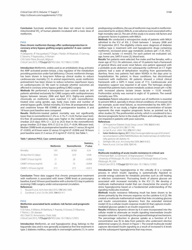
Conclusion Succinate ameliorates (but does not return to normal) mitochondrial VO2 of human platelets incubated with a toxic dose of metformin.
P452Does chronic metformin therapy off er cardiomyoprotection in coronary artery bypass grafting surgery patients? A case–control studyA Delaporte, JP Hacquebard, P Origer, I Pastijn, M Maatouk, O Germay, E Stevens, P Kapessidou, A RomanCHU Saint-Pierre, Brussels, BelgiumCritical Care 2013, 17(Suppl 2):P452 (doi: 10.1186/cc12390)
Introduction Metformin, widely used as an antidiabetic drug, activates the AMP activated protein kinase, a key regulator of the metabolism providing protection under fuel defi ciency. Chronic metformin therapy has been shown in long-term follow-up clinical studies to reduce cardiovascular mortality [1]. In animal experiments, acute metformin pretreatment has been shown to reduce ischemia–reperfusion injury on cardiomyocytes [2]. We want to evaluate whether outcomes are aff ected in coronary artery bypass grafting (CABG) surgery.Methods We performed a retrospective case–control study on 341 patients admitted exclusively for CABG surgery with cardiopulmonary bypass between January 2006 and April 2012. Eighty-one patients chronically on metformin were matched 1:3 with nonmetformin-treated ones using gender, age, body mass index and number of arterial bypass grafts. Global mortality, ICU-free 28 postoperative days and creatinine kinase MB (CKMB) at postoperative baseline, 8 and 18 hours post baseline were compared.Results Metformin-treated patient mortality was not signifi cantly lower than in nonmetformin (1.2% vs. 3.1%, P = 0.24, Fischer exact test). ICU-free 28 postoperative days were higher in the metformin group (median 23.4 days; 95% CI = 22.5 to 24.3) than in the nonmetformin group (median 22.9 days; 95% CI 22.3 to 23.5, P <0.04, Wilcoxon test). Median CKMB levels at postoperative baseline were 21 versus 26 ng/ml (P <0.003), at 8 hours were 22 versus 53 ng/ml (P <0.004) and 18 hours post baseline were 21.5 versus 27.5 ng/ml (P <0.012). See Table 1.
Table 1 (abstract P452). Case–control analysis
Statistics, Metformin Nonmetformin P value
Mortality 1/81 8/260 0.24
28 days free ICU (days) 23.4 22.9 <0.04
CKMB T 0 hours (ng/ml) 21 26 <0.003
CKMB T+8 hours (ng/ml) 22 53 <0.004
CKMB T+18 hours (ng/ml) 21.5 27.5 <0.012
Conclusion These data suggest that chronic preoperative treatment with metformin is associated with lower CKMB levels at postsurgery baseline, 8 and 18 hours post baseline with a shorter ICU stay in patients receiving CABG surgery under extracorporeal circulation.References1. Roussel R, et al.: Arch Intern Med 2010, 170:1892-1899.2. Gundewar S, et al.: Circ Res 2009, 104:403-411.
P453Metformin-associated lactic acidosis: risk factors and prognostic factorsV Di Falco, A Milano, M Battilana, F Araosta, A Grosso, D Albanese, F Petrini, L Di LiberatoASL Chieti SS. Annunziata, Chieti, ItalyCritical Care 2013, 17(Suppl 2):P453 (doi: 10.1186/cc12391)
Introduction Metformin, an oral hypoglycemic drug, belongs to the biguanide class and is now generally accepted as fi rst-line treatment in type 2 diabetes mellitus, especially in overweight patients [1]. In some
predisposing conditions, the use of metformin may result in metformin-associated lactic acidosis (MALA), a rare adverse event associated with a high mortality rate [2]. The aim of this study is to assess risk factors and prognostic factors in patients with MALA.Methods We conducted a retrospective study of patients with MALA admitted to the ICU of ASL 2 Chieti between 1 January 2008 and 30 September 2012. The eligibility criteria were: diagnosis of diabetes mellitus type 2, treatment with oral hypoglycemic drugs containing metformin, increased anion gap metabolic acidosis (pH <7.35, HCO3
– <22 mmol/l, lactate >5 mmol/l). For each patient we evaluated: sex, age, home care, SAPS II score [3], blood tests.Results Ten patients were selected, fi ve males and fi ve females, with a mean age of 72.2. On admission, nine of 10 patients had a framework of general illness and acute renal failure; one patient appeared with a probable acute abdomen not subsequently confi rmed on CT. Eight patients had a state of dehydration resulting from gastroenteritis, diarrhea, fever; two patients had taken NSAIDs in the days prior to hospitalization. No patient, in these conditions, has discontinued treatment with metformin. All patients showed a critical clinical framework with a SAPS II mean score of 77.5. Cardiovascular and respiratory support was required in all cases. The hemogas analysis showed that patients had a severe metabolic acidosis (mean pH = 6.96) with increased plasma lactate (mean lactate = 15.93 mmol/l). Prothrombin activity was normal in eight of 10 patients. The overall mortality was 70%.Conclusion Patient education on correct use of metformin is essential to prevent MALA, specially in those clinical conditions of increased risk (for example, acute renal failure), as recommended by the AIFA 2011 guidelines [4]. In our study, a higher plasma concentration of lactate represents the main negative prognostic factor, as pointed out by other studies [5]. The prothrombin activity, which is considered to be a decisive prognostic factor in the study of Peters and colleagues [6], was not impaired in patients with poor outcome.References1. UK Prospective Diabetes Study Group: Lancet 1998, 352:854-865.2. Lalau JD: Drug Saf 2010, 33:727-740.3. Le Gall JR, et al.: JAMA 1993, 270:2957-2963.4. Italian Medicines Agency 2011 [http://www.agenziafarmaco.gov.it/it/content/
raccomandazioni-sull%E2%80%99utilizzo-dei-medicinali-base-di-metformina-nella-gestione-del-diabete-m]
5. Dell’Aglio DM, et al.: Ann Emerg Med 2009, 54:818-823.6. Peters N, et al.: Crit Care 2008, 12:R149.
P454Multiscale modeling of acute insulin resistance in critical careA Pritchard-Bell1, G Clermont2, B Yegneswaran2, R Parker1
1University of Pittsburgh, PA, USA; 2University of Pittsburgh Medical Center, Pittsburgh, PA, USACritical Care 2013, 17(Suppl 2):P454 (doi: 10.1186/cc12392)
Introduction Stress hyperglycemia in the critically ill is a complex process in which insulin signaling is systematically hijacked to provide energy substrate for metabolic priorities such as cell healing or infection containment. Fluctuating levels of plasma glucose are associated with increased mortality in the ICU [1]. We develop a multiscale mathematical model that can characterize the severity of stress hyperglycemia based on a fundamental understanding of the signaling molecules involved.Methods Insulin resistance following insult has been shown to be driven primarily by the immune response via the cytokine IL-6 [2]. We created a multiscale mathematical model that links circulating glucose and insulin concentration dynamics from the extended minimal model [3] to a cellular insulin response model [4] that captures insulin-mediated glucose uptake in an insulin-responsive cell.Results Inhibitory dynamics driven by IL-6 were incorporated into the cellular model to attenuate an insulin signaling intermediate (insulin receptor substrate 1) according to the proposed biological mechanisms. The percentage reduction in glucose uptake as a function of IL-6 concentration was fi t to data from patients who underwent elective abdominal surgery [2], shown in Figure 1. The overall multiscale model captures decreased insulin signaling as a result of increased IL-6 levels and the subsequent hyperglycemia that may ensue.
Critical Care 2013, Volume 17 Suppl 2 http://ccforum.com/supplements/17/S2
S168

Conclusion A multiscale model has been developed to describe the inhibitory eff ects of IL-6 on insulin-mediated glucose uptake. Cellular inhibitory dynamics were shown to capture reduced insulin sensitivity on the macroscale, which could then be used to characterize insulin sensitivity and to provide insulin treatment advice to reduce glucose variability.References1. Van den Berghe G: N Engl J Med 2001, 345:1359-1367.2. Thorell A: Clin Nutr 1996, 15:75-79.3. Roy A: Diabetes Technol Ther 2006, 8:617-626.4. Sedaghat A: Am J Physiol Endocrinol Metab 2002, 283:E1084-E1101.
P455Impact of a real-time electronic persistent hyperglycemia alert on glucose control in the ICUK Colpaert, S Oeyen, T van den Driessche, C Danneels, J DecruyenaereGhent University Hospital, Ghent, BelgiumCritical Care 2013, 17(Suppl 2):P455 (doi: 10.1186/cc12393)
Introduction Hyperglycemia is frequently encountered in critically ill patients, and associated with adverse outcome. Improvement of glucose protocol adherence may be accomplished using electronic alerts. We confi gured a non-intrusive real-time electronic alert, called a GLYC sniff er, as part of our Intensive Care Information System (ICIS) that continuously evaluates the occurrence of persistent hyperglycemia and hypoglycemia. The GLYC sniff er is confi gured in such a way to give an alert: if two consecutive glucose values are >150 mg/dl, with a minimum interval of 60 minutes; or if a glucose value is <80 mg/dl. We wanted to evaluate whether the GLYC sniff er would improve the glucose control.Methods A single-center, prospective intervention study during a 6-month period in our 22-bed surgical ICU. Two study phases were compared: a 3-month pre-alert phase with no alerting to the nurses, and a 3-month intervention phase where the GLYC sniff er was alerting through the Clinical Notifi cation System of our ICIS.Results A total of 652 diff erent patients having a total of 699 admission episodes was recorded during the study period. There were no signifi cant diff erences between the two study groups regarding baseline demographic data, fi rst glucose value upon admission. A total of 2,335 GLYC sniff er alerts were recorded during the whole study period: 84.3% persistent hyperglycemia alerts (1,021 in the pre-alert group, 948 in the alert group), and 15.7% hypoglycemia alerts (139 vs. 227). A signifi cantly lower percentage of glucose values in the alert group were hyperglycemic (19.5% vs. 26.5%, P <0.001). The proportion of persistent hyperglycemic values (i.e. consecutive glucose values exceeding the limit of 150 mg/dl) was signifi cantly lower in the alert
group (9.9% vs. 15.4%, P <0.001). The patients in the alert group spend signifi cantly more time within the set target glucose interval of 80 to 150 mg/dl (82.3% vs. 75.0%, P = 0.009). A signifi cantly lower proportion of patients experienced a new-onset hypoglycemic event (<80 mg/dl) in the alert group (19.3% vs. 26.2%, P = 0.04).Conclusion A real-time electronic persistent glycemia sniff er resulted in a signifi cantly higher proportion of normoglycemia, without increasing the variability. Furthermore, hypoglycemic events occurred less frequently, and were resolved more timely. Smart alerting is able to improve quality of care, while diminishing the problem of alert fatigue.
P456Individualized targeted glucose control to avoid hypoglycemiaSP Gawel1, G Clermont2, T Ho1, BM Newman3, B Yegneswaran2, RS Parker1
1University of Pittsburgh, PA, USA; 2University of Pittsburgh Medical Center, Pittsburgh, PA, USA; 3Iowa State University, Ames, IA, USACritical Care 2013, 17(Suppl 2):P456 (doi: 10.1186/cc12394)
Introduction Hyperglycemia and hypoglycemia have been linked to worse outcomes in critically ill patients. While there is controversy as to the optimal tightness of glucose control in critically ill patients, there is agreement that an upper limit to safe glucose levels exists and that avoiding hypoglycemic episodes should be prioritized. Our algorithm can assist clinicians in maintaining blood glucose ([Gbl]) within a desired target range while avoiding hypoglycemia.Methods Our model predictive control (MPC) algorithm uses insulin and glucose as control inputs and a linearized model of glucose–insulin–fatty acid interactions. To allow the controller model to learn from data, a moving horizon estimation (MHE) technique tailored the tissue sensitivity to insulin to individual responses. Patient data ([Gbl] measurements, insulin and nutritional infusion rates) were from the HIDENIC database at the University of Pittsburgh Medical Center. [Gbl] measurements, typically hourly, were interpolated to impute a measurement every 5 minutes. The model captured patient [Gbl] via nonlinear least squares by adjusting insulin sensitivity (SI) and endogenous glucose production (EGP0). The resulting virtual patient (VP) is used to evaluate the performance of the MPC–MHE algorithm.Results MPC controller performance on one VP is shown in Figure 1. Across a population of 10 VPs, the average [Gbl] under MPC is 6.31 mmol/l, the average minimum is 4.62 mmol/l, the population individual minimum is 3.49 mmol/l and the average absolute average residual error is 0.83 mmol/l from a 5.6 mmol/l target. With standard intervention, the 10 VPs have an average [Gbl] of 9.32 mmol/l, an average minimum [Gbl] of 3.77 mmol/l, and a population minimum [Gbl] of 2.78 mmol/l. Algorithm performance deteriorates signifi cantly if the imputed sampling time exceeds 30 minutes, underlining the importance of dynamic variations in insulin sensitivity in this population.Conclusion The MPC–MHE algorithm achieves targeted glucose control in response to changing patient dynamics and multiple measured disturbances for a pilot population of 10 VPs. Furthermore, the MHE scheme updates patient parameters in real time in response to changing patient dynamics.
P457Hyperglycemia in intensive care identifi es patients at high risk of developing diabetes: preliminary data of a multicentre studyE Lukic, I Gornik, M Grgic-Medic, D Milicic, V Vegar, V Ivancan, M Peric, V Gasparovic, I Pavlic RenarUniversity Hospital Centre Zagreb, CroatiaCritical Care 2013, 17(Suppl 2):P457 (doi: 10.1186/cc12395)
Introduction A recent study showed that hyperglycaemia (blood glucose ≥7.8 mmol/l) in nondiabetic patients hospitalised in a medical ICU is associated with increased risk of diabetes [1]. We investigated a large mixed ICU population to confi rm these results.Methods This study retrospectively included patients with negative history of diabetes admitted to ICUs during the year 2007. We excluded patients receiving steroids, with newly diagnosed diabetes and those with end-stage disease. Patients were followed-up 5 years after index
Figure 1 (abstract P454). Reduced glucose uptake driven by increased plasma IL-6 levels.
Critical Care 2013, Volume 17 Suppl 2 http://ccforum.com/supplements/17/S2
S169

admission. Diagnosis of diabetes within 6 months from the index admission was presumed as revealing DM at inclusion, which excluded the patient. Patients who were taking glucocorticoids during the follow-up period were excluded. The rest of the patients, if consented, were assessed for their diabetic status with a single HbA1c measurement.Results A total of 2,696 patients were included in the study: 1,441 medical and 1,255 surgical, hyperglycaemia (≥7.8 mmol/l) was registered in 1,419 (52%). During the 5 years from index admission 409 died and 38 were taking glucocorticoids, and 10 refused participation. Diabetes was diagnosed in 189 patients during the 6 months after index admission. The remaining cohort of 2,320 patients had 1,169 patients who were hyperglycaemic during hospitalisation. Diabetes was already diagnosed in 132 patients, of whom 112 were hyperglycaemic in the ICU; prediabetes was already diagnosed in 121 patients, of whom 83 had ICU hyperglycaemia. Since 125 patients (55 hyperglycaemic) refused HbA1c testing, it was performed on 1,852 patients and revealed diabetes in 136 patients (107 had hyperglycaemia) and prediabetes in 103 patients (75 were hyperglycaemic in ICU). Overall, patients who had hyperglycaemia had 19.8% cumulative 5-year incidence of diabetes and 14.3% of prediabetes. Nonhyperglycaemic ICU patients had cumulative incidences of 4.5% and 6.6%, respectively. Relative risk for developing diabetes during 5 years after ICU admission is 4.2 (95% CI = 3.2 to 5.9) for patients who did have hyperglycaemia.Conclusion The results of this study confi rm in a large mixed medical/surgical ICU population that hyperglycaemia occurring in severe illness or the perioperative period is associated with increased risk of developing diabetes. Suggesting lifestyle changes to reduce the risk and regular follow-up should be implemented at least for these patients.Reference1. Gornik I, et al.: A prospective observational study of the relationship of
critical illness associated hyperglycaemia in medical ICU patients and subsequent development of type 2 diabetes. Crit Care 14:R130.
P458Association between average glucose levels and hospital mortality among critically ill patientsB Yegneswaran1, R Parket2, S Gawel2, A Pritchard-Bell2, T Ho2, G Clermont2
1UPMC Hamot, Erie, PA, USA; 2University of Pittsburgh, PA, USACritical Care 2013, 17(Suppl 2):P458 (doi: 10.1186/cc12396)
Introduction The objective of the study was to determine the association between average glucose level (AVG) and hospital outcomes in critically ill patients.Methods We performed a chart review of the MIMICII and HIDENIC databases, prospective cohorts of over 32,000 and 46,000 patients admitted to the ICUs at Beth Israel Deaconess Medical Center between 2001 and 2007 and the ICUs at University of Pittsburgh Medical Center between 1991 and 2008, respectively. All admissions of patients ≥18 years without the diagnosis of DKA and NKHS were included. Diabetics were identifi ed from ICD-9 documentation. Propensity score for death (pDead) was computed from either SAP1 (MIMICII) or APACHE III (HIDENIC) to assess the risk of death. Hypoglycemia was defi ned as AVG ≤60 mg/dl. AVG was computed as the area under the glucose curve throughout ICU admission. Mortality was examined within bins (each bin is categorized by a 10 mg/dl increase in AVG) and was compared between adjacent categories using a chi-square test. The same method was repeated among diabetics, nondiabetics, patients with lower (pDead greater than median) and higher (pDead lower than
Figure 1 (abstract P456).
Figure 1 (abstract P458).
Critical Care 2013, Volume 17 Suppl 2 http://ccforum.com/supplements/17/S2
S170

median) risk of death. Statistical signifi cance was noted following a Bonferroni correction.Results MIMICII database: 21,209 admissions met inclusion criteria. Mean age: 62.5 ± 16.4 years; female: 9,070 (42.8%); diabetes: 5,496 (25.9%); mean admission SAP1 score: 13.8 ± 5.2; mean LOS: 9 days (IQR 6.15); hospital mortality: 10.3%. HIDENIC database: 38,872 admissions met inclusion criteria. Mean age: 59.4 ± 17.3 years; female: 16,675 (42.9%); diabetes: 11,326 (29.1%); mean admission APACHE III score: 80.2 ± 14; mean LOS: 11 days (IQR 7.22); hospital mortality: 14.3%. In both databases, association between average glucose and hospital mortality followed a bathtub shape with mortality nadir between 80 and 130 mg/dl. No mortality diff erence was observed between adjacent AVG glucose bins in the bottom of the bathtub (Figure 1). This relationship was preserved in nondiabetics, and for patients with higher risk of death.Conclusion Low and high average glucose are associated with mortality.
P459Continuous glucose monitoring in critically ill adults: comparison of two diff erent calibration protocolsL Leelarathna1, S English2, H Thabit1, K Caldwell1, J Allen1, K Kumareswaran1, M Wilinska1, M Nodale1, J Mangat1, M Evans1, R Burnstein2, R Hovorka1
1University of Cambridge, UK; 2Addenbrookes Hospital, Cambridge, UKCritical Care 2013, 17(Suppl 2):P459 (doi: 10.1186/cc12397)
Introduction We evaluated the clinical and numerical accuracy of the Freestyle Navigator continuous glucose monitoring (CGM) system, in critically ill adults using two diff erent methods of calibration. Studies looking at intensive glucose control have yielded confl icting results with increased rates of iatrogenic hypoglycemia. Availability of accurate real-time glucose information may improve safety and effi cacy of glucose control in the critical care unit.Methods In a randomized prospective trial, paired CGM and reference glucose (hourly arterial blood gas (ABG)) were collected from 24 adults with critical illness (age 60 ± 14 years, BMI 29.6 ± 9.3 kg/m2, APACHE score range 6 to 19) and hyperglycemia (glucose ≥10 mmol/l or treated with intravenous insulin), over 48 hours. In 12 subjects, CGM was force-calibrated at variable 1 to 6 hourly intervals using ABG glucose (FC arm). In the other 12 subjects, the sensor was calibrated according to the manufacturer’s instructions (1, 2, 10, 24 hours after insertion) using arterial blood and built-in glucometer (MC arm).Results Two groups had similar characteristics at baseline. A total of 1,060 CGM/ABG pairs were analyzed and reference glucose ranged from 4.3 to 18.8 mmol/l. Median (IQR) absolute relative deviation was lower in the FC arm (7.0% (3.5, 13.0) vs. 12.8% (6.3, 21.8), FC vs. MC, P <0.001). Similarly, the percentage of points in the Clarke error grid zone A points meeting ISO criteria were higher with FC (87.8% vs. 70.2%). Sensor bias (median (IQR)) was signifi cantly lower in the FC arm (–0.1 (–0.7, 0.4) mmol/l vs. –1.1 (–2.3, –0.1) mmol/l, P <0.001). The median (IQR) interval between calibrations in FC arm was 169 (122, 213) minutes.Conclusion CGM accuracy in the MC arm was comparable with accuracy in subjects with type 1 diabetes. Further signifi cant improvements to CGM accuracy in critical care are possible by increasing the frequency of calibrations. Such accurate CGM may provide valuable information to guide insulin therapy in critically ill subjects.References1. Weinstein RL, et al.: Diabetes Care 2007, 30:1125-1130.2. Kovatchev B, et al.: Diabetes Care 2008, 31:1160-1164.
P460Root cause analysis of hypoglycemic events in critically ill patientsA McDonald, J KrinsleyStamford Hospital, Stamford, CT, USACritical Care 2013, 17(Suppl 2):P460 (doi: 10.1186/cc12398)
Introduction Hypoglycemia (H) is a complication of intensive insulin (IN) therapy of critically ill patients and is independently associated
with increased risk of mortality (M). Heightened attention to identifi ed risk factors (RF) and causes of hypoglycemic events (HE) may lead to improvement in glycemic control. A limited literature describes RF for H but a detailed root cause analysis (RCA) of individual HE has not been published.Methods This is a retrospective analysis of prospectively collected data including 835 patients admitted consecutively between 1 February 2012 and 31 October 2012 to the 16-bed medical–surgical ICU of a university-affi liated teaching hospital. The blood glucose (BG, mg/dl) target was 90 to 120 and the monitoring frequency was every 3 hours, or every 1 hour when the patient received i.v. IN infusion. HE data were collected by the bedside nurse, using a cutoff of BG <60, and information was abstracted from the unit’s database. RF included: shock (SH), renal insuffi ciency (RI), hepatic failure (HF), mechanical ventilation (MV), and diabetes mellitus (DM). Attributable causes included: IN treatment, improper monitoring frequency (MF), interruption of feeding (FE), and spontaneous (SP). We measured rates of severe (<40) and mild (40 to 69) H, and compared them as well as percentage of values in the optimum range (70 to 139) and hyperglycemic range (>139) with data from the preceding 3 months (PRE).Results Sixty-three (7.5%) patients sustained a total of 79 HE. They were older, more likely to be diabetic, and had higher APACHE IV predicted (37.3% vs. 15.4%, P <0.0001) and observed (25.4% vs. 8.6%, P <0.0001) M than those without H. RF: SH 24.1%; RI 24.1%; HF 8.9%; MV 46.8%; DM 36.5%. Attributable causes: IN 58.2%; MF 8.9%; FE 6.3%; SP 36.7%. BG <40, 40 to 69 and >139 decreased 78.6%, 39.0% and 9.3% (P = 0.0006, P <0.0001, P <0.0001). BG 70 to 139 increased 5.5% (P <0.0001). In total, 4.2% and 22.4% of PRE patients sustained severe and mild H. In total, 0.7% and 14.1% of RCA patients sustained severe and mild H (P = 0.0029, P = 0.0003).Conclusion IN treatment was associated with barely more than one-half of HE in this population of intensively monitored patients. Implementation of an initiative mandating RCA of every HE, including identifi cation of RF and attributable causes, was associated with a signifi cant improvement in glycemic control.
P461Continuous arterial and venous glucose monitoring by quenched chemical fl uorescence in ICU patients after cardiac surgeryS Bird1, L Macken1, O Flower1, F Bass1, N Hammond1, S Webb1, N Kennedy1, A Baker1, E Yarad1, C Chau2, M Librande2, P Strasma2, S Finfer1
1Royal North Shore Hospital, Sydney, Australia; 2Glumetrics, Irvine, CA, USACritical Care 2013, 17(Suppl 2):P461 (doi: 10.1186/cc12399)
Introduction Continuous glucose monitoring (CGM) in ICUs has the potential to improve patient safety outcomes. The GluCath Intravascular CGM System uses a novel quenched chemical fl uorescence sensing mechanism to measure glucose concentration in venous or arterial blood (BG). This is the fi rst report of this system deployed for 48 hours in both arteries and veins of ICU patients.Methods This ongoing clinical study evaluates up to two sensors per subject in 20 patients undergoing cardiac surgery. An arterial sensor is deployed via a standard 20 G radial artery catheter inserted for routine care and an optional venous sensor is deployed percutaneously in an upper arm vein. Data are presented from the fi rst fi ve patients. Outcome measures are qualitative (ease of use, workfl ow fi t) and quantitative (accuracy vs. reference analyzer). Sensors were inserted shortly after ICU admission, with ultrasound guidance for venous sensor insertion. In vivo calibration was performed at 1 and 2 hours, then each morning. Glucose values were recorded every 10 seconds by the system. Hourly arterial reference samples were analyzed via Radiometer ABL 800 Flex Blood Gas Analyzer (BGA).Results Arterial sensors were successfully deployed in all fi ve patients and did not interfere with clinical care, blood pressure monitoring or sampling. One arterial catheter failed resulting in sensor removal at 36 hours. The venous sensor was deployed in three patients, but removed from two patients due to thrombosis identifi ed during surveillance ultrasound examinations. A total of 202 reference BG samples ranging from 5.3 to 11.3 mmol/l were collected. Precision between arterial and venous sensors (n = 2) was 9.3% CV. Arterial sensor accuracy compared with BGA was 5.5% MARD. One hundred
Critical Care 2013, Volume 17 Suppl 2 http://ccforum.com/supplements/17/S2
S171
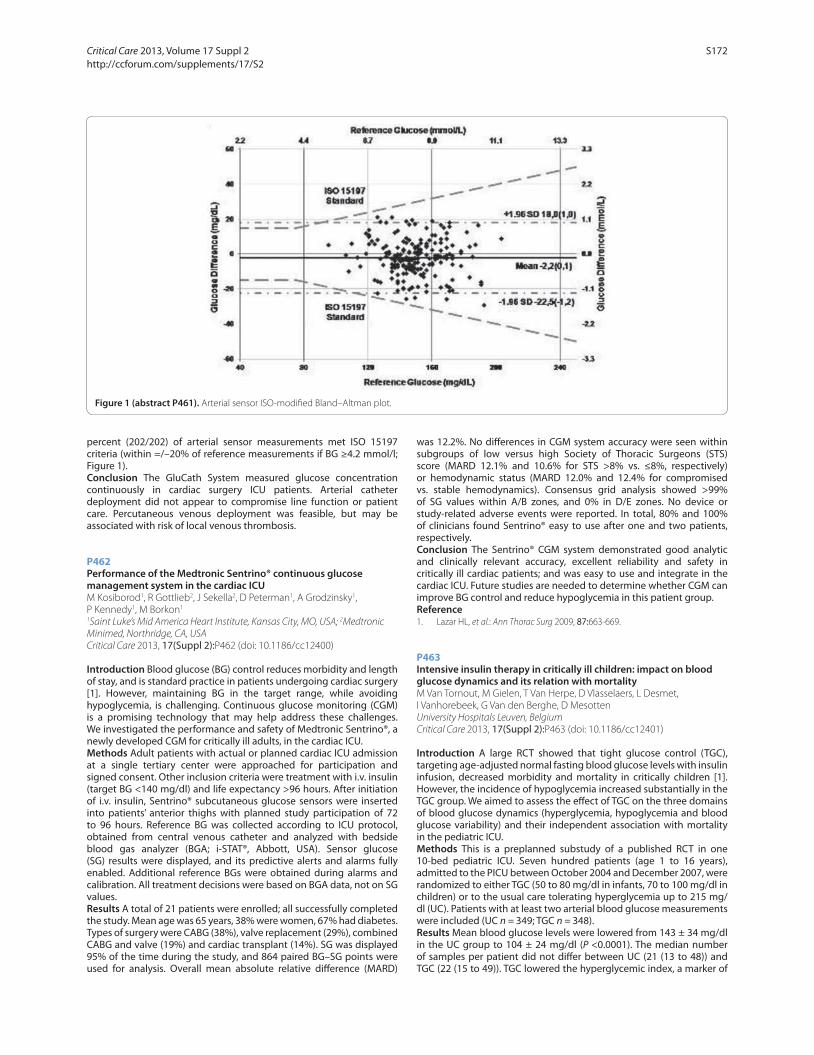
percent (202/202) of arterial sensor measurements met ISO 15197 criteria (within =/–20% of reference measurements if BG ≥4.2 mmol/l; Figure 1).Conclusion The GluCath System measured glucose concentration continuously in cardiac surgery ICU patients. Arterial catheter deployment did not appear to compromise line function or patient care. Percutaneous venous deployment was feasible, but may be associated with risk of local venous thrombosis.
P462Performance of the Medtronic Sentrino® continuous glucose management system in the cardiac ICUM Kosiborod1, R Gottlieb2, J Sekella2, D Peterman1, A Grodzinsky1, P Kennedy1, M Borkon1
1Saint Luke’s Mid America Heart Institute, Kansas City, MO, USA; 2Medtronic Minimed, Northridge, CA, USACritical Care 2013, 17(Suppl 2):P462 (doi: 10.1186/cc12400)
Introduction Blood glucose (BG) control reduces morbidity and length of stay, and is standard practice in patients undergoing cardiac surgery [1]. However, maintaining BG in the target range, while avoiding hypoglycemia, is challenging. Continuous glucose monitoring (CGM) is a promising technology that may help address these challenges. We investigated the performance and safety of Medtronic Sentrino®, a newly developed CGM for critically ill adults, in the cardiac ICU.Methods Adult patients with actual or planned cardiac ICU admission at a single tertiary center were approached for participation and signed consent. Other inclusion criteria were treatment with i.v. insulin (target BG <140 mg/dl) and life expectancy >96 hours. After initiation of i.v. insulin, Sentrino® subcutaneous glucose sensors were inserted into patients’ anterior thighs with planned study participation of 72 to 96 hours. Reference BG was collected according to ICU protocol, obtained from central venous catheter and analyzed with bedside blood gas analyzer (BGA; i-STAT®, Abbott, USA). Sensor glucose (SG) results were displayed, and its predictive alerts and alarms fully enabled. Additional reference BGs were obtained during alarms and calibration. All treatment decisions were based on BGA data, not on SG values.Results A total of 21 patients were enrolled; all successfully completed the study. Mean age was 65 years, 38% were women, 67% had diabetes. Types of surgery were CABG (38%), valve replacement (29%), combined CABG and valve (19%) and cardiac transplant (14%). SG was displayed 95% of the time during the study, and 864 paired BG–SG points were used for analysis. Overall mean absolute relative diff erence (MARD)
was 12.2%. No diff erences in CGM system accuracy were seen within subgroups of low versus high Society of Thoracic Surgeons (STS) score (MARD 12.1% and 10.6% for STS >8% vs. ≤8%, respectively) or hemodynamic status (MARD 12.0% and 12.4% for compromised vs. stable hemodynamics). Consensus grid analysis showed >99% of SG values within A/B zones, and 0% in D/E zones. No device or study-related adverse events were reported. In total, 80% and 100% of clinicians found Sentrino® easy to use after one and two patients, respectively.Conclusion The Sentrino® CGM system demonstrated good analytic and clinically relevant accuracy, excellent reliability and safety in critically ill cardiac patients; and was easy to use and integrate in the cardiac ICU. Future studies are needed to determine whether CGM can improve BG control and reduce hypoglycemia in this patient group.Reference1. Lazar HL, et al.: Ann Thorac Surg 2009, 87:663-669.
P463Intensive insulin therapy in critically ill children: impact on blood glucose dynamics and its relation with mortalityM Van Tornout, M Gielen, T Van Herpe, D Vlasselaers, L Desmet, I Vanhorebeek, G Van den Berghe, D MesottenUniversity Hospitals Leuven, BelgiumCritical Care 2013, 17(Suppl 2):P463 (doi: 10.1186/cc12401)
Introduction A large RCT showed that tight glucose control (TGC), targeting age-adjusted normal fasting blood glucose levels with insulin infusion, decreased morbidity and mortality in critically children [1]. However, the incidence of hypoglycemia increased substantially in the TGC group. We aimed to assess the eff ect of TGC on the three domains of blood glucose dynamics (hyperglycemia, hypoglycemia and blood glucose variability) and their independent association with mortality in the pediatric ICU.Methods This is a preplanned substudy of a published RCT in one 10-bed pediatric ICU. Seven hundred patients (age 1 to 16 years), admitted to the PICU between October 2004 and December 2007, were randomized to either TGC (50 to 80 mg/dl in infants, 70 to 100 mg/dl in children) or to the usual care tolerating hyperglycemia up to 215 mg/dl (UC). Patients with at least two arterial blood glucose measurements were included (UC n = 349; TGC n = 348).Results Mean blood glucose levels were lowered from 143 ± 34 mg/dl in the UC group to 104 ± 24 mg/dl (P <0.0001). The median number of samples per patient did not diff er between UC (21 (13 to 48)) and TGC (22 (15 to 49)). TGC lowered the hyperglycemic index, a marker of
Figure 1 (abstract P461). Arterial sensor ISO-modifi ed Bland–Altman plot.
Critical Care 2013, Volume 17 Suppl 2 http://ccforum.com/supplements/17/S2
S172

hyperglycemia over time, from median 44 (IQR 29 to 66) mg/dl in UC to 12 (7 to 23) mg/dl, as well as the glycemic penalty index, an aggregate measure of dysglycemia, from median 44 (32 to 58) in UC to 22 (16 to 30) (both P <0.0001). Despite frequent hypoglycemia <40 mg/dl (1.4% in UC, 25.0% in TGC, P <0.0001), TGC did not result in increased blood glucose variability, as refl ected by the standard deviation of the blood glucose time series (TGC: 36 (24 to 50) mg/dl, UC 34 (26 to 48) mg/dl, P = 0.82). In multivariable logistic regression analysis, adjusting for baseline risk factors (age, severity of illness, malignancy, type of admission), hypoglycemia (OR = 1.6, 95% CI = 0.19 to 2.22) and blood glucose variability (OR = 1.0, 95% CI = 0.97 to 1.02, per mg/dl) were not associated with PICU mortality. In contrast, hyperglycemic index (OR = 1.02, 95% CI = 1.01 to 1.04, per mg/dl) was independently associated with mortality and its lowering statistically explained the outcome benefi t of the intervention.Conclusion TGC substantially reduced the hyperglycemic index, at the expense of increased hypoglycemia incidence, but without aff ecting blood glucose variability. Statistical analysis suggested strictly avoiding hyperglycemia in order to bring about outcome benefi t with insulin in critically ill children.Reference1. Vlasselaers D, et al.: Lancet 2009, 373:547-556.
P464Implementation of evidence-based protocols improves survival: a 15-year surgical ICU experience in 10,172 patientsSA NasrawayTufts Medical Center, Boston, MA, USACritical Care 2013, 17(Suppl 2):P464 (doi: 10.1186/cc12402)
Introduction There has been enormous interest in measuring ICU performance in terms of mortality and resource use, owing to increased public and health insurer scrutiny. We elected to describe the performance of our surgical ICU, using a standardized mortality ratio (SMR = observed / predicted mortality).Methods The primary cohort was all patients admitted to the surgical ICU from March 2010 through February 2012 and related outcomes. The change in SMR was longitudinally determined from the latest 15-year study period (1997 to 2011) comprised of 10,172 patients.Results There were 1,799 ICU admissions in the primary cohort. Hospital mortality, observed and predicted by APACHE IV, was measured. Crude hospital mortality was 8.4%. The hospital SMR (observed / predicted mortality) was 0.58 (95% CI: 0.49 to 0.65). The SMR decreased by 20% from 0.73 to 0.58 over the 15-year study period, an absolute 1% per year decrease (P = 0.039; 95% CI: –0.02 to –0.002), as shown in Figure 1.Conclusion Mortality was less than predicted and steadily declined during the previous 15 years. The SMR can be used to track success when new quality measures are introduced or changes in the delivery of care are made. ICUs should report their standardized mortality ratios to evaluate performance.
P465Decubitus ulcers are associated with prolonged length of stay in critically ill patientsWT McGee1, BH Nathanson2, E Lederman1, TL Higgins1
1Baystate Medical Center, Springfi eld, MA, USA; 2OptiStatim LLC, Longmeadow, MA, USACritical Care 2013, 17(Suppl 2):P465 (doi: 10.1186/cc12403)
Introduction How decubitus ulcers (DU) diagnosed on admission to the ICU aff ect resource use is unknown. We hypothesize that this easily identifi able clinical fi nding would be associated with longer hospitalizations.Methods Clinical assessment was used to identify patients with DU on admission to the ICU. The association of DU on hospital and ICU length of stay (LOS) was assessed with generalized linear models that adjusted for ICU admission source, age, APACHE IV score, diabetes, sepsis, hepatic failure, dialysis and mechanical ventilation.Results DU was present on admission in 180 of 2,723 (6.6%) during a 19-month period in 2011 to 2012 at a single teaching hospital in New England. Patients with decubiti were sicker on admission (mean (SD) APACHE IV 80.2 (28.2) vs. 58.7 (30.7), P <0.001), more likely to be age 65 or older (58.9% vs. 41.8%, P <0.001) and require more organ supportive therapies (Table 1). DU patients had a higher in-hospital mortality rate (32.2% vs. 18.3%, P <0.001) and longer mean ICU and hospital LOS (4.9 vs. 3.3 days and 15.6 vs. 10.5 days, respectively (P <0.001)). After multivariate adjustment, DU patients stayed 3.2 days, 95% CI (1.6, 4.8) longer in the hospital than non-DU patients and stayed 2.5 days longer, 95% CI (0.7, 0.43) among survivors. DU patients had longer ICU stays (1.3 days, 95% CI (0.3, 1.3)) in general but were statistically similar among survivors.
Table 1 (abstract P465)
Admitted Admitted with without decubitus decubitus PVariable (n = 180) (n = 2,543) value
Age 65 or older 106 (58.9%) 1,062 (41.8%) <0.001
APACHE IV score on admission 80.2 (28.2%) 58.7 (30.7) <0.001
Diabetes 53 (29.4%) 420 (16.5%) <0.001
Dialysis 16 (8.9%) 58 (2.3%) <0.001
Hepatic failure 3 (1.7%) 56 (2.2%) 1.000
Mechanically ventilated during 123 (68.3%) 1,457 (57.3%) hospitalization
Admission source to the ICU
Direct admit 10 (5.6%) 154 (6.1%)
ER 84 (46.7%) 1,206 (47.4%)
Floor 25 (13.9%) 214 (8.4%) 0.017
OR 19 (10.6%) 459 (18.1%)
Other location 42 (23.3%) 510 (20.1%)
Outcomes
ICU LOS (days) 4.9 (4.9) 3.3 (4.7) <0.001
Hospital LOS (days) 15.6 (20.7) 10.5 (11.1) <0.001
ICU mortality 30 (16.7%) 308 (12.1%) 0.073
Hospital mortality 58 (32.2%) 464 (18.3%) <0.001
Readmission rate for 20 out of 112 173 out of 2,079 0.002 survivors (16.4%) (8.3%)
Conclusion The presence of decubiti on admission to the ICU is associated with longer hospitalizations even after adjusting for age, acuity, and organ supportive therapies. DU on admission to ICU provide a unique, unambiguous marker of increased resource utilization.
Figure 1 (abstract P464). Tufts surgical ICU, 15-year outcome standardized hospital mortality ratio.
Critical Care 2013, Volume 17 Suppl 2 http://ccforum.com/supplements/17/S2
S173

P466HIV patients admitted to an ICU of a university hospital – experience of 15 years: 1995 to 2009A Agrifoglio, M Arce, M Hernández, P Millan, C Guallar, I Pozuelo, M JiménezHospital Universitario La Paz, Madrid, SpainCritical Care 2013, 17(Suppl 2):P466 (doi: 10.1186/cc12404)
Introduction HIV infection is a major public health problem in the world. The use of prophylaxis against opportunist infection and the introduction of HAART in 1996 increased life expectancies. The therapeutic use of ICU resources for HIV patients has been controversial, questioning the admission of these patients especially in advanced stages of the disease, given the poor prognosis. The aim of this study was to determine the experience of the past 15 years in relation to the income of these patients in an ICU.Methods A retrospective case series consisting of patients with diagnosis of HIV infection (known or unknown) admitted between January 1995 and December 2009. We collected demographic and epidemiological data, process of acquisition of the disease, infection status: known or unknown patient infected, whether or not receiving antiretroviral therapy and whether it was eff ective (undetectable viral load at the time of admission), APACHE II, cause of admission, need for mechanical ventilation (MV), pathology related or unrelated to HIV infection and ICU mortality.Results During this period 12,607 patients were admitted to the ICU, 188 (1.5%) HIV-positive. Mean APACHE II score 17.6, median age 39 years, 73% men and 90% Spanish nationality. Principal risk behavior: addiction drugs injection (67%). Seventeen percent of patients did not know who was infected with HIV at the time of admission to the ICU. Fifty-three percent were not receiving HAART. Of the 88 patients treated, 93% were receiving HAART (eff ective in 88% of cases). Sixty percent of the patients came from the emergency department of the hospital. Main admission diagnoses: acute respiratory failure caused by infection (Streptococcus pneumoniae and Pneumocystis jirovecii), neurological disorders (coma for illicit drugs and psychotropic) and septic shock. Seventy percent required MV. Of patients whose HIV infection was not known, 93.5% were admitted for related pathology. In patients of known infection, the pathology associated with HIV was 30%. Average length of stay 9 was days. ICU mortality was 35%. Most frequent causes of death: septic shock and multiple organ failure.Conclusion Depending on the patient and the cause of admission, ICU admission may represent an excellent opportunity as a screening method to determine HIV status. Given the greater effi cacy of HAART at present, most patients with medical or surgical conditions unrelated to HIV infection will be eligible to join the ICU. People with HIV can and should benefi t from using reasonable and individualized care in an ICU.References1. Huang L, et al.: N Engl J Med 2006, 355:173-181.2. Akgun K, et al.: J Intensive Care Med 2011, 26:151-164.
P467Prevalence and prognosis of organ dysfunctions of AIDS critically ill patientsAJ Japiassu, RA Amâncio, D Medeiros, EM Mesquita, EM Motta, FA BozzaOswaldo Cruz Foundation, Rio de Janeiro, BrazilCritical Care 2013, 17(Suppl 2):P467 (doi: 10.1186/cc12405)
Introduction The aim was to analyze the prognosis of AIDS patients with organ dysfunctions at ICU admission.Methods A prospective cohort study, including all patients with HIV/AIDS diagnosis, who were admitted to a specialized ICU from November 2009 until May 2012. Patients with less than 24 hours of ICU stay were excluded. Demographics and nutritional status were collected. The organ dysfunctions were classifi ed according to the SOFA score, and categorized as absent (0 SOFA point), mild (1 to 2 points) and severe (3 to 4 points). We expressed numeric variables as median and interquartile interval (25% to 75%). We performed a multivariate analysis of possible variables associated with hospital mortality (P <0.2), and we explored the 7-day, 28-day and 60-day survival of patients with and without independent risk factors.
Results We included 139 patients with HIV/AIDS admitted to the ICU. Median age was 40 (31 to 48) years and 71% were male. Severe malnutrition was common (34%). The CD4 cell count was 84 (25 to 274) cells/mm3 and viral load was 17,733 (67 to 174,214) copies/ml; 57% had at least one opportunistic infection; 55% had used antiretroviral therapy previous to ICU admission. Mechanical ventilation was used by 46% of patients and hospital mortality was 42%. Total SOFA score was 5 (2 to 9) points. Cardiovascular dysfunction was the most common on the fi rst day of stay (51%), followed by respiratory (42%), neurological (40%), renal (35%), hematological (27%) and hepatic (17%). Cardiovascular and renal dysfunctions presented with higher rate of severe dysfunction (30% and 15%, respectively). Rates of neurological (P = 0.002), renal (P = 0.009) and hematological (P = 0.003) dysfunctions were higher in nonsurvivors. Age, CD4 cell count, malnutrition, and opportunistic infections were included in the multivariate analysis. Neurological dysfunction was the independent risk factor for hospital mortality (odds 3.2 (1.4 to 7.2)). The presence of neurological dysfunction was dichotomized: associated or not with primary neurological diagnosis; survival was lower in the patients with neurological dysfunction and without primary neurological diagnosis (log-rank test 0.001 in the 7-day and 0.02 in the 28-day analysis). Sixty-day survival was similar in primary and secondary neurological dysfunction, but it remained lower than in patients without neurological impairment.Conclusion Neurological dysfunction was independently associated with hospital survival, mainly in those AIDS critically patients without primary neuropathy.
P468Mortality predictors in septic HIV patients in the ICUP Vidal-Cortés1, P Lameiro-Flores1, A Aller-Fernández2, M Mourelo-Fariña2, P Fernández-Ugidos1, R Gómez-López1, M Alves-Pérez1, E Rodríguez-García1
1CHU Ourense, Spain; 2CHU A Coruña, SpainCritical Care 2013, 17(Suppl 2):P468 (doi: 10.1186/cc12406)
Introduction Sepsis in HIV patients carries a high mortality. Our purpose is to identify mortality predictors in this population.Methods A retrospective study of HIV patients admitted to our ICU between January 2005 and December 2009. We identify septic patients and analyzed demographic factors, etiology, organ failure and outcome. We used logistic regression to calculate the relative mortality risk for each qualitative variable and the beta (β) coeffi cient for quantitative variables.Results Sixty-two septic patients were admitted to our ICU during the study period (71% men, mean age: 40.59 ± 8.12). A total of 56.5% came from the ED, mean hospital stay before ICU admission: 6.14 ± 10.96 days. A total of 66.1% had a history of intravenous drug use, other comorbidities: COPD (9.7%), cirrhosis (8.1%), solid or hematologic malignancy (12.9%). A total of 40.3% were under HAART. Mean CD4 count at admission: 219.62 ± 353.93 cells/mm3. Mean viral load: 4.57 ± 3.25 log. Mean albumin levels: 2.3 ± 0.53 g/dl. APACHE II: 21.98 ± 7.97. The lung was the most frequent focus of infection (65.6%) followed by the CNS. The most common pathogen was S. pneumoniae (28.8%), followed by P. jirovecii (13.6%). In total, 62.9% needed vasopressors, 79% mechanical ventilation and 19.4% renal replacement. Mean ICU and hospital length of stay: 10.43 ± 10.52 and 34.76 ± 29.64 days. ICU mortality: 33.9%; hospital mortality: 41.9%. ICU mortality increases for each day of hospital stay before ICU admission (β = 1.062, 95% CI = 1.009 to 1.118, P = 0.022), each point of: cardiac frequency at admission (β = 1.059, 95% CI = 1.024 to 1.096, P = 0.001), FiO2 (β = 1.034, 95% CI = 1.011 to 1.057, P = 0.003), pCO2 (per mmHg) (β = 1.052, 95% CI = 1.007 to 1.098, P = 0.023); and decreases for each point of: albumin (g/dl) (β = –0.307, 95% CI = 0.095 to 0.986, P = 0.047), mean arterial pressure (mmHg) (β = 0.951, 95% CI = 0.904 to 0.999, P = 0.048), pH (β = 0.002, 95% CI = 0.0 to 0.172, P = 0.006), fi rst ICU 24-hour diuresis (ml) (β = 0.999, 95% CI = 0.998 to 1.000, P = 0.004) and base excess (β = 0.912, 95% CI = 0.836 to 0.995, P = 0.039). Mortality increases threefold (β = 3.134, 95% CI = 1.051 to 9.345, P = 0.04) if patients do not come from the ED, almost 10-fold if the patients need vasopressors for more than 24 hours (β = 9.975, 95% CI = 2.054 to 48.451, P = 0.004) and fi vefold if patients need renal replacement (β = 5.692, 95% CI = 1.466 to 22.099, P = 0.012).
Critical Care 2013, Volume 17 Suppl 2 http://ccforum.com/supplements/17/S2
S174

Conclusion Neither immune status-related variables nor comorbidity or infection focus are mortality predictors. Poor nutritional status, delayed ICU admission, shock or renal failure increase the ICU relative mortality risk. Tachycardia, hypotension, hypercapnia, acidosis, and oliguria in the fi rst ICU 24 hours increase signifi cantly ICU mortality. Mechanical ventilation is not a mortality predictor.
P469Outcome of intensive care treatment for lung cancer patientsL Hajjar1, F Galas2, D Goto2, A Tavares2, M Santos2, A Freitas2, T Oliveira2, P Tonini2, L Cavichio2, M Bazan2, J Almeida2, M Cavalcante2, J Fukushima2, S Vieira2, E Osawa2
1Heart Institute, São Paulo, Brazil; 2Cancer Institute of University of São Paulo, BrazilCritical Care 2013, 17(Suppl 2):P469 (doi: 10.1186/cc12407)
Introduction Patients with lung cancer commonly require the ICU for a variety of acute illnesses related to the underlying malignancy, treatment, or comorbid conditions. ICU admission of patients with nonresectable lung cancer has been criticized based on the high mortality rate in this population. However, recent advances in critical care may have changed this scenario. The aim of this study was to identify factors associated with hospital mortality in this group of patients.Methods A retrospective study was conducted in consecutive medical and surgical patients with lung cancer admitted to a university hospital ICU in São Paulo, between 2007 and 2012. A univariate analysis was performed to identify associated variables with hospital mortality. Selected variables were included in the multivariate model.Results From 597 patients included in the study, 477 were medical admissions (79.8%) and 120 were surgical admissions (20.2%). Four hundred and twenty (70%) patients had metastasis, 222 patients (37%) required the ICU because of respiratory failure and 121 (20%) because of septic shock. The ICU and hospital mortality rates were 53.9% and 64%, respectively. In the univariate analysis, variables associated with hospital mortality were diagnosis of nonsmall-cell lung cancer, higher Charlson morbidity index, medical admission, active neoplasm, vasopressor need at admission to and at 24 hours of ICU, acute renal failure at admission, non-invasive ventilation or mechanical ventilation need at admission to and at 24 hours of ICU and a higher admission arterial lactate. By multivariate analysis, risk factors of hospital mortality were diagnosis of nonsmall-cell lung cancer (OR = 3.32; 95% CI, 2.08 to 5.29, P <0.001), medical admission (OR = 2.89; 95% CI, 1.75 to 4.77, P <0.001), acute renal failure at admission (OR = 4.09; 95% CI, 1.49 to 11.21, P <0.001), non-invasive ventilation at 24 hours of ICU (OR = 2.50; 95% CI, 1.48 to 4.23, P = 0.001) and mechanical ventilation at 24 hours of ICU (OR = 3.68; 95% CI, 1.87 to 7.26, P <0.001).Conclusion Hospital survival in patients with lung cancer requiring ICU admission was 36%. Our results provide evidence that ICU management may be appropriate in patients with nonresectable lung cancer and appoint risk factors for mortality, helping to better triage cancer patients who will benefi t from ICU care.
P470Outcome of older patients with cancer requiring ICU admissionL Hajjar1, M Franca Cavalcante2, F Galas2, J T Fukushima2, M R Sundin2, F Bergamin2, C Park2, H Palomba2, D M Goto2, A Tavares2, M Santos2, A Freitas2, P Tonini2, L Cavichio2, J P Almeida2, M Bazan2
1Heart Institute, São Paulo, Brazil; 2Cancer Institute of University of São Paulo, BrazilCritical Care 2013, 17(Suppl 2):P470 (doi: 10.1186/cc12408)
Introduction Because the prognosis of older patients with cancer may be poor compared with younger patients, it remains controversial whether they benefi t from ICU treatment. The objective of this study was to identify factors associated with hospital mortality in older patients with cancer requiring the ICU.
Methods A retrospective study was conducted in consecutive medical and surgical older patients with cancer admitted to a university hospital ICU in São Paulo, between 2007 and 2012. Univariate and multivariate analysis were performed to identify associated and independent factors related to hospital mortality.Results From 8,976 patients with cancer requiring ICU at the period, 600 patients were 80 years old or higher. Most patients were male (53%), had solid neoplasm (86%), were from medical admission (54%) and 37% had metastatic disease. The mean age was 84 years (± 3). The ICU and hospital mortality rates were 30% and 44%, respectively. In the univariate analysis, variables associated with hospital mortality were diagnosis of lung cancer, medical admission, active neoplasm, vasopressor need at 24 hours of ICU, acute renal failure at admission, mechanical ventilation need at admission to and at 24 hours of ICU and a higher admission arterial lactate. By multivariate analysis, risk factors of hospital mortality were diagnosis of lung cancer (OR = 2.32; 95% CI, 1.08 to 3.29, P <0.001), medical admission (OR = 6.473; 95% CI, 2.76 to 15.15, P <0.001), acute renal failure at admission (OR = 3.09; 95% CI, 1.49 to 9.21, P <0.001), mechanical ventilation at 24 hours of ICU (OR = 5.68; 95% CI, 2.87 to 7.26, P <0.001) and lactate levels at admission (OR = 1.02; 95% CI, 1.006 to 1.051, P <0.001).Conclusion Hospital survival in older patients with cancer requiring ICU admission is acceptable. Our results provide evidence that ICU management may be appropriate in older patients with cancer and appoint risk factors for mortality, helping to better triage cancer patients who will benefi t from ICU care.
P471Identifying risk factors for readmission to the ICU: a qualitative approachDG Strange, K Tjelle, A Lindhardt, K JensenBispebjerg Hospital, Copenhagen NV, DenmarkCritical Care 2013, 17(Suppl 2):P471 (doi: 10.1186/cc12409)
Introduction Readmission to the ICU within 48 hours is an indicator of quality of intensive care and is associated with an increase in mortality. During the last years several groups have published data based on multivariate logistic regression analysis to describe characteristics of patients who needed readmission to the ICU. Older age, comorbid conditions and severity of illness (APACHE score) have been among the strongest predictors for readmission. In our ICU most patients are in the groups formerly identifi ed as risk groups, which means that stratifi cation and prediction of readmission is diffi cult. Because of the unusual high severity of acute and pre-existing illnesses we could not fi nd a data match on comparable patient groups. To investigate whether we could reduce our rate of readmission we therefore decided to perform a qualitative investigation to identify risk factors related to readmission. After identifi cation of the risk factors we will take actions to optimize care and perform ongoing control of the implemented actions to secure that they decreases the rate of readmission.Methods Retrospective data on patients readmitted to the ICU within 48 hours during an 8-month period (November 2011 to June 2012) were drawn from the Critical Information System (CIS) at ICU ZIT, Bispebjerg Hospital, Denmark. ZIT is a multidisciplinary unit with 10 beds and 500 to 600 admissions/year and a median SAPS II score of 47. A group of consultants, junior doctors and nurses from the ICU and the ward each read the patient fi les with focus on pattern recognition and suggested trigger points to focus on. Data on trigger points were then drawn from the CIS system and re-evaluated. Finally, fi ve key points were identifi ed and serves as basis for future actions.Results In a qualitative analysis, readmissions to the ICU are related to the following fi ve key points – discharge outside day hours, lack of infection control, stay in ICU <2 days, lung physiotherapy ordinated but not eff ectuated, and several minor organ dysfunctions (atrial fi brillation and acute kidney injury). Age, diagnosis, SAPS II score or ventilator treatment during intensive care was not diff erent in patients with successful discharge and patients readmitted in this group of patients.Conclusion It is possible and suitable to identify key points for future eff orts in a given subgroup of patients using a systematic qualitative approach.
Critical Care 2013, Volume 17 Suppl 2 http://ccforum.com/supplements/17/S2
S175

P472Clinical and laboratory postoperative impact on outcome of living donor liver transplantation recipientsY Nassar, K Omar, A Battah, A MwafyCairo University, Giza, EgyptCritical Care 2013, 17(Suppl 2):P472 (doi: 10.1186/cc12410)
Introduction Adult recipients of living donor liver transplantation have a wide range of postoperative medical comorbidities altering various clinical and laboratory parameters. We aimed to assess 30-day postoperative clinical and laboratory changes and their value in predicting outcome of LDLT recipients.Methods A prospective registry involving patients who underwent LDLT in the period from October 2004 to December 2010. Postoperative 30-day clinical and laboratory data included: clinical BP, HR, temperature, ICU stay, liver functions AST, ALT, GGT, ALP, TotPtn, Alb, TBil, DBil, INR, PTT, CRP, kidney functions Crea, Na, K, blood picture Hb, Hct, TLC and Plt count.Results Our study involved 142 recipients, 124 (87%) males and 18 (13%) females, who underwent living donor liver transplantation; mean age was 53 ± 14.8 years, ranging from 24 to 66 years; 29 cases (20.43%) at Kasr Al Aini University Hospital and 113 cases (79.57%) at the International Medical Center. The 30-day mortality rate was 13.38%. Clinical and laboratory parameters showed gradual resolution towards normal values in survivors or deterioration in nonsurvivors, allowing one to predict mortality at various cutoff points. Clinical parameters: HR >94 cutoff (Acc 76.8%, Sens 89.5%, Spec 62.6%, PPV 27, NPV 97) and ICU stay >8 days (Acc 76.2%, Sens 75.3%, Spec 95.7%, PPV 95.7, NPV 35.3). Laboratory parameters: INR >3.06 cutoff (Acc 87.5%, Sens 92.9%, Spec 78.6%, PPV 96.3, NPV 64.7), followed by PTT >53.3 (Acc 79.2%, Sens 78.6%, Spec 75%, PPV 93.6, NPV 42.9), Na >142.9 (Acc 72.9%, Sens 80%, Spec 72.3%, PPV 26.7, NPV 96.6), TBil >15 (Acc 71.3%, Sens 95.5%, Spec 50%, PPV 92.4, NPV 63.6), ALT >379 (Acc 67.5%, Sens 72.2%, Spec 73.8%, PPV 28.9, NPV 94.7), TProt <4.6 (Acc 63.5%, Sens 55.6%, Spec 73.3%, PPV 24.4, NPV 91.4), AST >145 (Acc 57.4%, Sens 50%, Spec 84.4%, PPV 32.1, NPV 92), Hct <24.4 (Acc 54.2%, Sens 47.4%, Spec 81.5%, PPV 34.6, NPV 88.2), CRP >12 (Acc 51.6%, Sens 43.7%, Spec 73.3%, PPV 90.5, NPV 17.7), GGT >22 (Acc 51.4%, Sens 83.3%, Spec 36.9%, PPV 16.3, NPV 93.7).Conclusion Predictors of mortality were INR >3.06 (accuracy 87.5%) followed by PTT >53.3 (79.2%), HR >94 (76.8%), ICU stay >8 days (76.2%), Na >142.9 (72.9%), TBil >15 (71.3%), ALT >379 (67.5%), TProt <4.6 (63.5%), AST >145 (57.4%), Hct <24.4 (54.2%), CRP >12 (Acc 51.6%) and GGT >22 (51.4%).
P473Are APACHE II scores better predictors of mortality than routine laboratory values?Z Baykara, H Özocak, A Kuş, Z Arslan, B Yüksel, C Aksu, M Ertagın, M Solak, K TokerUniversity of Kocaeli, TurkeyCritical Care 2013, 17(Suppl 2):P473 (doi: 10.1186/cc12411)
Introduction Even though more accurate and updated versions such as APACHE III and IV have been developed, APACHE II has remained the most widely used severity scoring system in the ICU [1]. In the present study, we aimed to develop a model mainly based on the laboratory data available upon admission to predict mortality and to compare its performance with that of APACHE II in a mixed ICU in Turkey.Methods A total of 645 adult patients who were over 18 years old and who stayed in the ICU for more than 24 hours were included in this study. Using stepwise multivariate logistic regression analysis, a mortality prediction model was developed based on the laboratory data, diagnostic category and age. The adjusted probability of death, according to the diagnostic category of the APACHE II score (adj-APACHE II), was calculated. The ability of the laboratory model and the adj-APACHE II model to discriminate between survivors and nonsurvivors was assessed using receiver operating characteristic (ROC) curves. The calibration of the laboratory model and the adj-APACHE II model was assessed by the Hosmer and Lemeshow goodness-of-fi t test.Results The areas under the ROC curves of the laboratory models and the adj-APACHE II scores for the prediction of mortality were 0.80 (95% CI: 0.76 to 0.85) and 0.78 (95% CI: 0.74 to 0.83), respectively (P >0.05, z statistic). The Hosmer–Lemeshow statistic had a chi-squared value of
2.01 (P = 0.98) for the laboratory model and 13.2 (P = 0.10) for the adj-APACHE II model.Conclusion If the aim is to predict mortality as accurately as APACHE II, the mortality prediction model based mainly on routine admission laboratory tests can achieve this using computer technology, without labor-related costs, as soon as the patient is admitted to the ICU. However, it is necessary to perform multicenter validation studies.Reference1. Breslow MJ, Badawi O: Severity scoring in the critically ill: part 1 –
interpretation and accuracy of outcome prediction scoring systems. Chest 2012, 141:245-252.
P474Complementarity of prognosis models SAPS 3 and EuroSCORE in cardiac surgeryMJ Delgado-Amaya1, E Curiel-Balsera1, MD Arias-Verdu1, E Castillo-Lorente2, FJ Carrero-Gómez3, E Aguayo-DeHoyos4, A Herruzo-Avilés5
1Hospital Regional Carlos Haya, Málaga, Spain; 2Complejo Hospitalario de Jaén, Spain 3Puerta del Mar Hospital, Cádiz, Spain; 4Virgen de las Nieves Hospital, Granada, Spain; 5Virgen del Rocio Hospital, Seville, SpainCritical Care 2013, 17(Suppl 2):P474 (doi: 10.1186/cc12412)
Introduction The aim of this study is to evaluate the performance of SAPS 3 and EuroSCORE and whether there is complementarity between them in ICU patients admitted after cardiac surgery.Methods An observational, prospective and multicenter study of patients included in the ARIAM registry of adult cardiac surgery. We analyzed clinical variables, surgery data and postoperative compli-cations, outcomes and scores (SAPS 3 and EuroSCORE). Discrimination was assessed using the area under the ROC curve. With the standardized mortality ratio (SMR) we evaluated the agreement between predicted and observed mortality. We used multiple logistic regression for multivariate analysis.Results A total of 5,642 patients were included, admitted to any fi ve hospitals, consecutively between 2008 and 2012. Mean age was 63.62 ± 12.80 years. Surgery room mortality was 1.4%, ICU mortality was 7.9% and in-hospital mortality was 11.2%. SAPS 3 was calculated in 5,383 patients. SAPS 3 was 40.91 ± 10.46, and the expected rate of mortality was 11.09% (with the southwestern Europe equation), with an observed hospital mortality of 9.6%, SMR = 0.87 (0.79 to 0.94). With the general equation, the expected mortality was 10.47%, SMR = 0.917 (0.843 to 0.996). Discrimination of SAPS 3 was evaluated using the area under the ROC curve, it was 0.77 (0.75 to 0.79). The 30-day mortality was 9.6%, with an expected 30-day mortality predicted by EuroSCORE of 7.86% (SMR = 1.22; CI = 1.12 to 1.32). The area under the ROC curve was 0.734 for EuroSCORE (0.712 to 0.755). Multivariate analysis with logistic regression showed complementarity between SAPS 3 and logistic EuroSCORE (vers. I). There is a relationship between hospital mortality and the probability of dying predicted by SAPS 3 (general equation), OR 1.05 (1.04 to 1.06), and logistic EuroSCORE (vers. I), OR 1.024 (1.015 to 1.032). The model with both variables has a similar discrimination to SAPS 3 alone. The area under the ROC curve of the combined model was 0.778 (0.758 to 0.799) and 0.77 (0.75 to 0.79) using SAPS 3 only.Conclusion There is complementarity between the SAPS 3 model and EuroSCORE. However, a joint model with both prognosis scores has no appreciable improvement in discrimination, compared with the SAPS 3 model. In our study, SAPS 3 shows very useful to rule out that the superior observed mortality (compared with the mortality predicted by EuroSCORE) is not attributable to inadequate care of patients in the ICU, the observed mortality being even slightly below the predicted mortality for patients undergoing cardiac surgery at the reference hospitals in which the SAPS 3 system was developed.
P475Comparing intensive care treated primary, secondary and tertiary sepsis with respect to length of stay, organ failure and mortality: a pilot studyS Castegren1, M Jonasson1, J Sjölin2, M Castegren1
1Centre for Clinical Research Sörmland, Uppsala University, Uppsala, Sweden; 2Medical Sciences, Uppsala University, Uppsala, SwedenCritical Care 2013, 17(Suppl 2):P475 (doi: 10.1186/cc12413)
Critical Care 2013, Volume 17 Suppl 2 http://ccforum.com/supplements/17/S2
S176

Introduction It may be hypothesized that a patient’s infl ammatory status might aff ect the clinical picture and outcome of a subsequent septic insult. To test this hypothesis, patients were categorized according to previous infl ammatory activity and analyzed with respect to clinical picture and outcome in a retrospective study.Methods The Swedish ICU registry was used to identify all adult patients diagnosed with severe sepsis or septic shock in a single Swedish ICU during 2006 to 2011. The charts were reviewed and patients categorized into four groups based on whether the patient had suff ered systemic infl ammatory-inducing insults previous to the septic episode. The patients were categorized as: (A) primary sepsis, no recent (<30 days) trauma/infection, ongoing systemic infl ammatory or immunosuppressive disease or malnutrition; (B) secondary sepsis, recent (<7 days) trauma/infection or systemic infl ammatory disease, but no immunosuppressive disease or malnutrition; (C) tertiary sepsis, >1 fulfi lled criteria of secondary sepsis during the last 10 days or immunosuppressive disease or malnutrition; and (D) patients not attributable to A to C. After the categorization, physiological and laboratory parameters during the ICU stay as well as therapy and outcome were extracted from the charts. Only signifi cant diff erences (P <0.05) are presented; Mann–Whitney and chi-squared tests were used.Results A total of 273 charts were reviewed. In total, 242 were categorized into A (n = 126), B (n = 79) or C (n = 37). D (n = 31) consisted mainly of patients with hematological malignancies (n = 12) and patients with chemotherapy or immunosuppressive treatment (n = 14). The groups diff ered in length of stay with A< B< C. During the fi rst 3 days the SOFA score was higher in A compared with C and in B compared with C. The duration of antibiotic therapy was longer in both B and C compared with A. There were no diff erences in 28-day mortality (A: 34/126 = 27%, B: 30/79 = 38%, C: 15/37 = 40%); however, the proportions of patients dying between days 8 and 28 were higher in B (14/63 = 22%) and C (7/29 = 24%) compared with A (5/95 = 5%).Conclusion In this retrospective material it was possible to categorize 88.6% of all patients as having primary, secondary or tertiary sepsis. The categories diff ered in clinical picture at presentation as well as in outcome. A prospective study is warranted to validate the results of this study.
P476Survival of older patients after critical careD Ashton-Cleary, R SinclairRoyal Cornwall Hospital, Truro, UKCritical Care 2013, 17(Suppl 2):P476 (doi: 10.1186/cc12414)
Introduction We aimed to assess the survival of older patients after admission to critical care compared with the population in general.Methods We reviewed admission data for a 12-bed critical care unit in a district general hospital in the UK between 2001 and 2010. We calculated critical care and hospital discharge survival rates and studied the trend in these through the decade. We searched hospital records to establish whether these patients were still alive at November 2012 to determine life-expectancy (minimum 22-month follow-up on all patients). This was compared with UK Offi ce of National Statistics data for the local population [1]. Discharge destination (care facility or home address) was recorded as a surrogate marker of change in quality of life post discharge.Results A total of 481 patients aged 80 or over were admitted between 1 January 2001 and 31 December 2010, representing 9.6% of the admissions during this time with minimal annual change. Admission age ranged from 80 to 96 years (median 83) with a median APACHE II score of 20 (6 to 48, IQR 16 to 25). In total, 318 (66.1%) survived to critical care discharge and 256 (53.2%) survived to hospital discharge; 228 (89.0%) went home rather than to a care facility. Those under 80 years demonstrated 80.6% critical care survival, 65.5% hospital survival and a median APACHE II score of 17 (0 to 51, IQR 13 to 23). Eighty patients died within the 22-month follow-up period with an average life expectancy of 7.4 months. None reached life expectancy (average curtailment of 5.6 years compared with age-matched and sex-matched population statistics). Neither of these fi gures changed signifi cantly through the
decade. However, 10-year follow-up on 2001/2002 admissions showed that, despite this early mortality eff ect, the average post-discharge lifespan was 5.1 years with 2.2 years shortfall in life expectancy.Conclusion Older people represent a growing proportion of the population although their representation in the critical care population remained constant in this 10-year study. These patients had a slightly higher median APACHE II score and 14.5% greater critical care mortality than the younger patients. The majority of survivors were able to go home; however, 31% died within 22 months with signifi cant life expectancy curtailment, surviving on average only 7.4 months after discharge; this has not changed in the last 10 years. Those who survived this initial period (69%) had a much better outlook. This information may be vital to patients and physicians when discussing admission to critical care.Reference1. Offi ce of National Statistics Population Data [http://www.ons.gov.uk/ons/
taxonomy/index.html?Nscl=Population]
P477Eldicus scores for admission to our ICU: a retrospective studyG Zani, B Gemelli, S Vitali, C Villani, R Ragazzi, S Spadaro, CA Volta, R AlvisiUniversity of Ferrara, ItalyCritical Care 2013, 17(Suppl 2):P477 (doi: 10.1186/cc12415)
Introduction The indication for ICU admission is poorly defi ned and identifying patients who may benefi t from it is extremely diffi cult. As reported in literature, the Eldicus scores require further validation in centers in diff erent countries and with diff erent patient populations [1]. Previous studies have demonstrated that patients with intermediate severities of illness are those that benefi t from ICU treatment; instead, patients who are too well or too sick should not be admitted. There are many unmeasurable factors that infl uence the triage decision and can infl uence the subsequent outcome. The Eldicus scores could be the fi rst triage decision rule. The aim of this study was to confi rm the ability of the Eldicus scores to discriminate the survival status in our general ICU.Methods We retrospectively enrolled all patients >18 years admitted to the Ferrara ICU from January to June 2012. Patients’ data were recorded using Prosafe international data collection software and consulting the individual medical records. Data were chosen based on variables used in the Eldicus scores. As indicated in the Eldicus algorithm, for each patient the initial refusal score was calculated, to evaluate the too severe patients. Then we counted the fi nal triage score, to discover whether some patients were too healthy to be admitted to the ICU. Correlation between the severity of the scores and patients’ outcome was estimated using the Wilcoxon’s rank-sum test.Results Ninety-eight patients were fi nally enrolled. Data are presented as median ± SD. The SAPS II score was 33 ± 13 and was similar to those reported in the literature [1]. The Karnofsky performance status scale was 69 ± 20. The Eldicus initial score was 56 ± 30. The fi nal score was 70 ± 48. Seventy-eight percent of the analyzed patients were alive at hospital discharge. A signifi cant correlation between the severity scores and in-hospital outcome was found (P <0.001). Limitations of our study were the low sample size and the fact that patients not admitted to the ICU were not considered.Conclusion The Eldicus scores appear to be useful in identifying patients that may benefi t more from ICU admission. Further studies should confi rm the ability of these scores in helping identify those patients who really benefi t from ICU admission.Reference1. Sprung CL, et al.: Crit Care Med 2012, 40:125-131.
P478Developing a screening instrument for predicting psychological morbidity after critical illnessA Schandl1, M Bottai2, E Hellgren1, Ö Sundin3, P Sackey1
1Karolinska University Hospital Solna, Stockholm, Sweden; 2Karolinska Institutet, Stockholm, Sweden; 3Mid Sweden University, Östersund, SwedenCritical Care 2013, 17(Suppl 2):P478 (doi: 10.1186/cc12416)
Introduction The purpose was to develop a predictive screening instrument for use at ICU discharge to identify patients at risk for post-traumatic stress, anxiety or depression.
Critical Care 2013, Volume 17 Suppl 2 http://ccforum.com/supplements/17/S2
S177
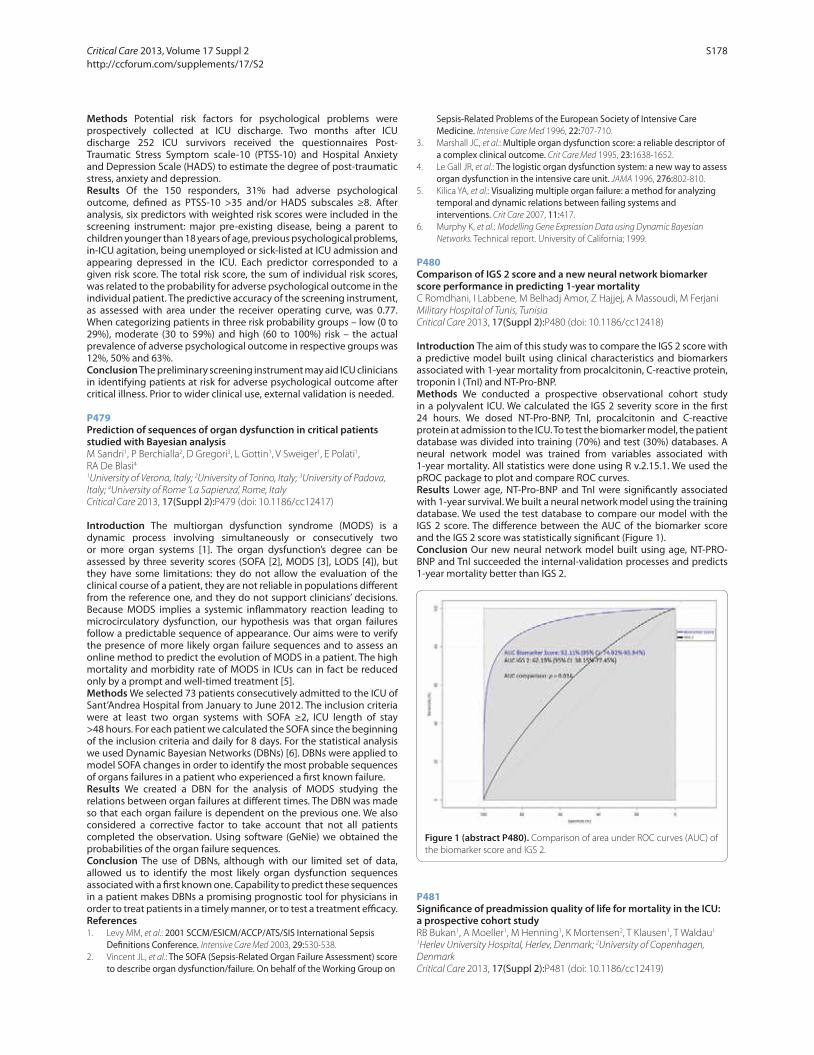
Methods Potential risk factors for psychological problems were prospectively collected at ICU discharge. Two months after ICU discharge 252 ICU survivors received the questionnaires Post-Traumatic Stress Symptom scale-10 (PTSS-10) and Hospital Anxiety and Depression Scale (HADS) to estimate the degree of post-traumatic stress, anxiety and depression.Results Of the 150 responders, 31% had adverse psychological outcome, defi ned as PTSS-10 >35 and/or HADS subscales ≥8. After analysis, six predictors with weighted risk scores were included in the screening instrument: major pre-existing disease, being a parent to children younger than 18 years of age, previous psychological problems, in-ICU agitation, being unemployed or sick-listed at ICU admission and appearing depressed in the ICU. Each predictor corresponded to a given risk score. The total risk score, the sum of individual risk scores, was related to the probability for adverse psychological outcome in the individual patient. The predictive accuracy of the screening instrument, as assessed with area under the receiver operating curve, was 0.77. When categorizing patients in three risk probability groups – low (0 to 29%), moderate (30 to 59%) and high (60 to 100%) risk – the actual prevalence of adverse psychological outcome in respective groups was 12%, 50% and 63%.Conclusion The preliminary screening instrument may aid ICU clinicians in identifying patients at risk for adverse psychological outcome after critical illness. Prior to wider clinical use, external validation is needed.
P479Prediction of sequences of organ dysfunction in critical patients studied with Bayesian analysisM Sandri1, P Berchialla2, D Gregori3, L Gottin1, V Sweiger1, E Polati1, RA De Blasi41University of Verona, Italy; 2University of Torino, Italy; 3University of Padova, Italy; 4University of Rome ‘La Sapienza’, Rome, ItalyCritical Care 2013, 17(Suppl 2):P479 (doi: 10.1186/cc12417)
Introduction The multiorgan dysfunction syndrome (MODS) is a dynamic process involving simultaneously or consecutively two or more organ systems [1]. The organ dysfunction’s degree can be assessed by three severity scores (SOFA [2], MODS [3], LODS [4]), but they have some limitations: they do not allow the evaluation of the clinical course of a patient, they are not reliable in populations diff erent from the reference one, and they do not support clinicians’ decisions. Because MODS implies a systemic infl ammatory reaction leading to microcirculatory dysfunction, our hypothesis was that organ failures follow a predictable sequence of appearance. Our aims were to verify the presence of more likely organ failure sequences and to assess an online method to predict the evolution of MODS in a patient. The high mortality and morbidity rate of MODS in ICUs can in fact be reduced only by a prompt and well-timed treatment [5].Methods We selected 73 patients consecutively admitted to the ICU of Sant’Andrea Hospital from January to June 2012. The inclusion criteria were at least two organ systems with SOFA ≥2, ICU length of stay >48 hours. For each patient we calculated the SOFA since the beginning of the inclusion criteria and daily for 8 days. For the statistical analysis we used Dynamic Bayesian Networks (DBNs) [6]. DBNs were applied to model SOFA changes in order to identify the most probable sequences of organs failures in a patient who experienced a fi rst known failure.Results We created a DBN for the analysis of MODS studying the relations between organ failures at diff erent times. The DBN was made so that each organ failure is dependent on the previous one. We also considered a corrective factor to take account that not all patients completed the observation. Using software (GeNie) we obtained the probabilities of the organ failure sequences.Conclusion The use of DBNs, although with our limited set of data, allowed us to identify the most likely organ dysfunction sequences associated with a fi rst known one. Capability to predict these sequences in a patient makes DBNs a promising prognostic tool for physicians in order to treat patients in a timely manner, or to test a treatment effi cacy.References1. Levy MM, et al.: 2001 SCCM/ESICM/ACCP/ATS/SIS International Sepsis
Defi nitions Conference. Intensive Care Med 2003, 29:530-538.2. Vincent JL, et al.: The SOFA (Sepsis-Related Organ Failure Assessment) score
to describe organ dysfunction/failure. On behalf of the Working Group on
Sepsis-Related Problems of the European Society of Intensive Care Medicine. Intensive Care Med 1996, 22:707-710.
3. Marshall JC, et al.: Multiple organ dysfunction score: a reliable descriptor of a complex clinical outcome. Crit Care Med 1995, 23:1638-1652.
4. Le Gall JR, et al.: The logistic organ dysfunction system: a new way to assess organ dysfunction in the intensive care unit. JAMA 1996, 276:802-810.
5. Kilica YA, et al.: Visualizing multiple organ failure: a method for analyzing temporal and dynamic relations between failing systems and interventions. Crit Care 2007, 11:417.
6. Murphy K, et al.: Modelling Gene Expression Data using Dynamic Bayesian Networks. Technical report. University of California; 1999.
P480Comparison of IGS 2 score and a new neural network biomarker score performance in predicting 1-year mortalityC Romdhani, I Labbene, M Belhadj Amor, Z Hajjej, A Massoudi, M FerjaniMilitary Hospital of Tunis, TunisiaCritical Care 2013, 17(Suppl 2):P480 (doi: 10.1186/cc12418)
Introduction The aim of this study was to compare the IGS 2 score with a predictive model built using clinical characteristics and biomarkers associated with 1-year mortality from procalcitonin, C-reactive protein, troponin I (TnI) and NT-Pro-BNP.Methods We conducted a prospective observational cohort study in a polyvalent ICU. We calculated the IGS 2 severity score in the fi rst 24 hours. We dosed NT-Pro-BNP, TnI, procalcitonin and C-reactive protein at admission to the ICU. To test the biomarker model, the patient database was divided into training (70%) and test (30%) databases. A neural network model was trained from variables associated with 1-year mortality. All statistics were done using R v.2.15.1. We used the pROC package to plot and compare ROC curves.Results Lower age, NT-Pro-BNP and TnI were signifi cantly associated with 1-year survival. We built a neural network model using the training database. We used the test database to compare our model with the IGS 2 score. The diff erence between the AUC of the biomarker score and the IGS 2 score was statistically signifi cant (Figure 1).Conclusion Our new neural network model built using age, NT-PRO-BNP and TnI succeeded the internal-validation processes and predicts 1-year mortality better than IGS 2.
P481Signifi cance of preadmission quality of life for mortality in the ICU: a prospective cohort studyRB Bukan1, A Moeller1, M Henning1, K Mortensen2, T Klausen1, T Waldau1
1Herlev University Hospital, Herlev, Denmark; 2University of Copenhagen, DenmarkCritical Care 2013, 17(Suppl 2):P481 (doi: 10.1186/cc12419)
Figure 1 (abstract P480). Comparison of area under ROC curves (AUC) of the biomarker score and IGS 2.
Critical Care 2013, Volume 17 Suppl 2 http://ccforum.com/supplements/17/S2
S178

Introduction Assessing whether a critically ill patient should be admitted to an ICU remains diffi cult and mortality amongst ICU patients is high. To render intensive care with no prospect of success is an immense emotional burden for both patient and relatives, and a great socioeconomic burden for society as well. Therefore, validated strategies that can help identify patients who will benefi t from intensive care are in demand. This study seeks to investigate whether preadmission quality of life can act as a predictor of mortality amongst patients admitted to the ICU.Methods All patients (>18 years) admitted to the ICU for more than 24 hours are included. In order to assess preadmission quality of life, the patient or close relatives complete the Short-Form 36 (SF-36) within 72 hours after ICU admission. Mortality is evaluated from ICU admission until 30 days hereafter. Logistic regression and receiver operating characteristic analyses are employed to assess predictive value for mortality using fi ve models: SF-36 Physical Component Summary (PCS) and APACHE II (model A), SF-36 PCS (model B), SF-36 General Health (GH) and APACHE II (model C), SF-36 GH (model D), and APACHE II (model E). Classifi cation tables are composed in order to assess sensitivity, specifi city, positive and negative predictive values and likelihood ratios.Results Preliminary results, based upon data from 175 included patients, reveal an ICU mortality of 12.6% and 30-day mortality of 22.9%. No patients were lost to follow-up. When the Physical Component of SF-36 (PCS) was used as an estimate of preadmission quality of life, the area under the curve for model B (AUC = 0.80) was comparable with model E (AUC = 0.81), and better than model A (AUC = 0.85). The General Health item of SF-36 (GH), used as an estimate of preadmission quality of life, gave an AUC = 0.76 (model D). All models were controlled for sex and age.Conclusion Preliminary results indicate that the SF-36 GH and the SF-36 PCS are as good as APACHE II to predict mortality 30 days after ICU admission. However, in order to conclude whether preadmission quality of life can contribute to triage, by successfully identifying patients suitable for intensive care, fi nal analyses, due in 2013, are awaited. These results will clarify whether future randomized controlled trials, in which preadmission quality of life acts as a supplement to triage, are justifi able.
P482Predicting physical function and mental health in trauma intensive care patients 2 years after hospitalisationL Aitken1, B Macfarlane1, W Chaboyer2, M Schuetz1, C Joyce1, AG Barnett3
1Princess Alexandra Hospital, Brisbane, Australia; 2Griffi th University, GoldCoast, Australia; 3Queensland University of Technology, Brisbane, AustraliaCritical Care 2013, 17(Suppl 2):P482 (doi: 10.1186/cc12420)
Introduction Long-term compromise after traumatic injury is signifi cant; however, few modifi able factors that infl uence outcome have been identifi ed. The aim of this study was to identify acute and early post-acute predictors of long-term recovery amenable to change through intervention.Methods Adults (>17 years) admitted to the ICU, Princess Alexandra Hospital, Australia following injury were prospectively followed. Data were collected on demographics, pre-injury health, injury characteristics and acute care factors. Psychosocial measures (self-effi cacy (SE), illness perception (IP), post-traumatic stress disorder (PTSD) symptoms and psychological distress) and health status (SF-36) were collected via questionnaire 1, 6, 12, and 24 months post injury. Outcomes of interest were the Physical Function (PF) and Mental Health (MH) subscales of the SF-36. Regression models were used to estimate predictors of physical function and mental health over a 2-year period. A subject-specifi c intercept in a mixed model was used to account for repeated data from participants over time.Results Participants (n = 123) were young (median 37, IQR 22 to 55 years), predominantly male (83%) and spent on average 3 days in the ICU and 3 weeks in hospital. Response rates were over 55% at each follow-up, with responders similar to nonresponders except for being generally older. PF and MH scores improved over time, although the averages remained below the Australian norms at 24 months.
Predictors of PF included IP (β = –1.5, 95% CI = –3.1 to –1.1, P <0.01), SE (β = 1.8, 95% CI = 1.3 to 2.6, P <0.01), hospital length of stay (β = –1.7, 95% CI = –2.0 to –0.8, P <0.01), never having been married (β = 1.8, 95% CI = 0.3 to 5.5, P = 0.03), and having injury insurance (β = –2.7, 95% CI = –6.9 to –1.9, P <0.01). Predictors of MH included PTSD symptoms (β = –2.4, 95% CI –3.4 to –1.4, P <0.01), psychological distress (β = –6.9, 95% CI = –8.9 to –5.2, P <0.01), SE (β = 0.6, 95% CI = 0.2 to 1.1, P <0.01), and unemployment (β = –2.3, 95% CI = –5.0 to –0.2, P = 0.04).Conclusion Trauma ICU patients experience compromised physical function and mental health 24 months after injury. Psychological distress, self-effi cacy and illness perception infl uence outcomes and are potentially amenable to change in response to interventions initiated during hospital stay.
P483Do ICU patients’ discharge characteristics change with Swiss Diagnosis Related Groups?G Rauchenstein-Ziltener, CA Rüst, P Fodor, S BlumenthalTiemli City Hospital, Zurich, SwitzerlandCritical Care 2013, 17(Suppl 2):P483 (doi: 10.1186/cc12421)
Introduction Swiss Diagnosis Related Groups (SwissDRG) have been eff ective since 1 January 2012. The infl uence of this new system on patients’ discharge characteristics from a large ICU is not known. With the introduction of the DRG we expect patients to be discharged after a shorter length of stay on the ICU and with higher severity of illness.Methods The ICU of the City Hospital Triemli in Zurich has an inter-disciplinary organization with surgical and internal medical patients, with a maximum occupancy of 18 beds and a center function for the surrounding hospitals. In this ongoing prospective observational study, we collect and analyze the anonymized data of all patients discharged from our ICU prior to and after the introduction of the SwissDRG. The primary endpoint was the length of stay on the ICU in hours. The secondary endpoints were the severity of illness of the patients at the time of discharge, detected by the scoring system SAPS II as well as measured by the number of readmissions to the ICU. Initially all patients were analyzed and in a second step only patients within percentiles 6 to 94 were considered. We also analyzed the subgroups of patients referred internally, patients sent back to referring hospital and patients regionalized to a homebase hospital. The statistics have been done with SPSS and P <0.05 was considered signifi cant.Results We present the results of an 18-month period, 9 months prior to and 9 months after the introduction of the SwissDRG. Data of 1,491 and 1,492 patients were analyzed, respectively. When all patients were included, we found prior to and after the introduction of the DRGs a comparable length of stay on the ICU (mean ± SD of 52.1 ± 2.2 hours vs. 50.82 ± 2.2 hours), no diff erence in the severity of illness at discharge detected by the SAPS II (mean ± SD of 27.9 ± 0.3 vs. 28.4 ± 0.3) and the number of readmissions (91 vs. 92). There was also no signifi cant diff erence when only percentiles 6 to 94 were included or when the three subgroups were analyzed.Conclusion Up to now, the introduction of the SwissDRG has no infl uence on patients’ discharge characteristics from a large ICU. Data assessment will continue and further data analysis has to be performed.
P484Do Diagnosis Related Groups change admission practice to a large Swiss ICU?K Homburg, CA Rüst, P Fodor, S BlumenthalTriemli City Hospital, Zurich, SwitzerlandCritical Care 2013, 17(Suppl 2):P484 (doi: 10.1186/cc12422)
Introduction The new Swiss fi nancing system by means of Diagnosis Related Groups (DRG), eff ective 1 January 2012, forms the basis of a performance-based system to reimburse general hospital services. There are only few data on the infl uence of DRG on ICU patients [1,2]. We expect that the introduction of the DRG in Switzerland will change the number of admissions from external hospitals to a large ICU with a centre function and will infl uence the severity of disease of the admitted patients.
Critical Care 2013, Volume 17 Suppl 2 http://ccforum.com/supplements/17/S2
S179
Methods The ICU of the Triemli City Hospital in Zurich has an inter-disciplinary organisation with surgical and internal medical patients, with a maximum occupancy of 18 beds and a centre function for the surrounding hospitals of the region. In this prospective ongoing observational study, we collect and analyse the anonymised data of all patients admitted to our ICU from an external hospital during 12 months prior to (1 January to 31 December 2011) and after (1 January to 31 December 2012) the introduction of the DRG in Switzerland. Exclusion criteria are admissions by the emergency department, self-assignments into the hospital and internal relocations. The primary endpoint is the number of admissions from an external hospital to our ICU. Secondary endpoints are the severity of the disease of the admitted patients, detected by the scoring systems SAPS II and APACHE II as well as the length of stay in external hospitals before admission. The statistical analysis is descriptive.Results We present the preliminary data for 10 months (in each case January to October) before and after the introduction of the DRG. We observed an increase of 9.2% (391 vs. 427 patients) of admissions to our ICU after the introduction of the DRG. The severity of disease determined by the SAPS II score is unchanged (mean 26.7 vs. 26.0 points, P = 0.466). The severity of disease determined by the APACHE II score is signifi cantly lower (15.4 vs. 14 points, P = 0.017). We also noted that after the introduction of the DRG the patients were earlier transferred from an external hospital to our ICU (mean time until transfer 29.9 vs. 18.7 hours), but this value was not signifi cant (P = 0.55).Conclusion Up to now the introduction of the DRG in Switzerland has had a complex infl uence on the number and the kind of patients admitted from an external hospital to an ICU with centre function. Data assessment and analysis will continue.References1. Ahmad M, et al.: Chest 1988, 93:176-179.2. Thomas F, et al.: Chest 1987, 91:418-423.
P485Variable cost of a Greek ICUE Tsigou1, D Karabatsou1, E Boutzouka1, V Psallida1, G Baltopoulos1, M Tsironi21A. Anargiroi Hospital, Athens, Greece; 2University of Peloponnisos, Sparti, GreeceCritical Care 2013, 17(Suppl 2):P485 (doi: 10.1186/cc12423)
Introduction The ICU consumes almost 20% of total hospital resources, despite accounting for less than 10% of hospital beds. Medical cost can be divided into a fi xed part, consisting of personnel and accommodation expenses, and a variable part, determined by the needs of each patient (medications, consumables, examinations). In order to rationalize medical cost, estimation of expenses must be real and individualized. The aim of this study was to assess the variable cost of critically ill patients in a new, seven-bed, adult, general ICU, using bottom-up costing methodology.Methods All 138 patients who were treated during 2011, and stayed for at least 24 hours, were included in the study. Data were retrospectively collected from patient records and included demographics, cause of admission, APACHE II score at admission, length of stay (LOS) and outcome. Cost was recorded for every day and for every patient, based on a hospital-specifi c cost catalogue and on national agreements. Data are presented as mean ± SEM. Analysis of data was carried out using Graph Pad Prism 5.0., applying Student’s t test.Results The age of participants (84 men and 54 women, 71 medical and 67 surgical) was 68.75 ± 1.18, APACHE II score was 18.64 ± 0.61, LOS was 18.46 ± 2.54, and mortality was 19%. The majority of the patients (61%) were mechanically ventilated. The total days of ICU stay were 2,548 and the total variable cost was €1,460,465 (€573.18 per patient and per day). Cost per patient was subdivided as follows: medication (including drugs, fl uids, blood products, nutrition): 56.49%, examinations (including laboratory, microbiological assays and diagnostic procedures): 22.23%, consumables: 21.26%. As for medication cost, the largest part was comprised of antibiotics (45.69%), followed by blood products (17.61%) and cardiovascular drugs (12.39%). Costs for medical patients were signifi cantly higher than those for surgical patients (P <0.0001).Conclusion The total average cost per patient and per day was found to be €573.18. Medication expenditure was responsible for the highest
variable cost of intensive care treatment. Among drugs, antibiotics accounted for the largest part. A medical cause of admission was correlated with higher costs.References1. McLaughlin AM, et al.: Intensive Care Med 2009, 35:2135-2140.2. Rossi C, et al.: Intensive Care Med 2006, 32:545-552.
P486Audit of the medical staffi ng of and referrals to a mixed adult ICU in a teaching hospitalA Needham, T Lyndham, D O’Neil, J Greiff University Hospitals of Leicester, UKCritical Care 2013, 17(Suppl 2):P486 (doi: 10.1186/cc12424)
Introduction Leicester Royal Infi rmary is a (funded) 15-bed ICU, annual admission 1,102 patients per year (ICNARC data 2011 to 2012). It houses a broad range of specialty, has over 1,000 beds and an emergency department (ED) responsible for 297 admissions over the 2011 to 2012 period. Unit guidelines state that a doctor should be immediately available to the unit at all times, ideally physically present on the unit. This audit looked in detail at each occasion a junior doctor left the unit, the timings, reason, and outcome of each episode.Methods Between 17 October 2011 and 14 November 2011, and between 2 July 2012 and 30 July 2012, junior doctors on the ICU completed a data-capture form after each occasion that they left the unit for a clinical need. Data collected were matched to the objectives. The 2011 audit results were communicated at the University Hospitals of Leicester Trust ICU Audit Meeting. A system was introduced so that daytime referrals were directed to the ICU consultant. The impact of this intervention was assessed by the 2012 re-audit.Results There were 105 occasions when a junior doctor left the unit in 2011, totalling 118 hours and 38 minutes (5 days), a continuation of the upward trend from the previous 2 years. In 2012 this reduced to 47 occasions totalling 40 hours and 55 minutes. Most attended referrals were during the daytime, 54% in 2011 and 68% in 2012. The majority of referrals originated from the ED/medicine, combined numbers 58% in 2011 and 38% in 2012. In 2011 only 30% of ED referrals had been discussed with a consultant of the referring team and only 32% of medical referrals. Just two of the 14 inappropriate ED referrals were discussed with the referring consultant, and none of the six inappropriate medical referrals were discussed with a medical consultant. In 2012 just 33% of ED/medical referrals had prior discussion with a consultant from the referring team. At the point of leaving, the ICU was left without a doctor on 22 occasions in 2011 and six occasions in the 2012 audit. Most occasions occurred at either night shift/weekend (86% in 2011/100% in 2012). Referral resulted in a patient being admitted to the ICU on 20 occasions in 2011 and 12 in 2012.Conclusion The latest audit follows introduction of a referral system directly to the ICU consultant and may account for the reduction in numbers of referrals attended by junior doctors. ED/medicine persist as the main source of referral to the ICU. Discussion with the referring team consultant may reduce inappropriate referrals. ICU staffi ng should not be reduced.
P487Impact of the time elapsed between ICU request and actual admission on mortality and length of stayGM Filho1, TA Silva1, AR Santana1, FB Soares1, LJ Almeida1, LG Godoy1, TA Rodrigues1, MO Maia2, JA Neto2, AP Amorim3, EB Moura2, FF Amorim1
1Escola Superior de Ciências da Saúde, Brasília, Brazil; 2Unidade de Terapia Intensiva Adulto do Hospital Santa Luzia, Brasilia, Brazil; 3Liga Academica de Medicina Intensiva do Distrito Federal (LIGAMI), Brasilia, BrazilCritical Care 2013, 17(Suppl 2):P487 (doi: 10.1186/cc12425)
Introduction Measures to ensure an appropriate early treatment for critically ill patients result in signifi cant decreases in mortality [1,2]. This study aims to evaluate the impact of the time elapsed from request until admission to the ICU on mortality and ICU length of stay (LOS).Methods A retrospective cohort study performed on patients in the ICU of Hospital Regional de Samambaia over a period of 4 years, from January 2008 to December 2011. The patients were allocated into two groups: patients who waited longer than 6 hours, long waiting period
Critical Care 2013, Volume 17 Suppl 2 http://ccforum.com/supplements/17/S2
S180

(LWP, n = 300); and patients whose waiting time was equal to or less than that period, short waiting period (SWP, n = 113).Results In total, 413 patients were included, 300 of which belonged to the LWP group (65.4%). For the entire cohort, the mean APACHE II score was 19 ± 7, the mean age was 52 ± 22 years, and 211 patients were male (51.1%). The LWP group did not show diff erence in the APACHE II score (19 ± 7 vs. 18 ± 8, P = 0.13), but was older (55 ± 20 vs. 49 ± 23, P = 0.01). LWP also had a higher incidence of primary bloodstream infection (23.8% vs. 10.4%, P = 0.01) and catheter-associated urinary tract infection (10.2% vs. 1.9%, P = 0.01). LWP patients had higher mortality (37.8% vs. 25.9%, P = 0.02) and longer ICU LOS (21 ± 47 vs. 14 ± 18 days, P = 0.01). Relative risk for death in the LWP was 1.74 (95% CI: 1.11 to 2.72).Conclusion Despite showing no signifi cant diff erences on APACHE II scores from the SWP group, patients from the LWP group presented greater incidence of primary bloodstream infection, catheter-associated urinary tract infection, higher mortality outcomes and longer ICU LOS.References1. Amorim FF, et al.: J Clin Med Res 2012, 4:410-414.2. Rivers E, et al.: N Engl J Med 2001, 345:1368-1377.
P488Medical problems: review from the major nuclear incidentsY Haraguchi1, Y Tomoyasu2, H Nishi3, M Hoshino4, M Sakai5, E Hoshino6
1Disaster Medicine Compendium Team, Japan, Tokyo, Japan; 2Shirahigebashi Hospital, Tokyo, Japan; 3Tokyo Disaster Medical Center, Tokyo, Japan; 4Shisei Hospital, Saitama, Japan; 5Tokyo Women’s Medical College, Tokyo, Japan; 6Chiba Nursing Association, Chiba, JapanCritical Care 2013, 17(Suppl 2):P488 (doi: 10.1186/cc12426)
Introduction Intensivists are expected to have many roles during and after a major disaster/catastrophe; that is, triage, intensive care, education for people, and so forth. The roles of intensivists against special disaster or nuclear disaster are studied based on actual experiences.Methods Several disasters are studied. The Fukushima Daiichi Nuclear plant explosion after the Higashinihon earthquake 2011 was medically reviewed based on the total 30-day stay on-site in addition to several days around the site. The Chernobyl incident 1986 was inspected 15 years after the incident. Other nuclear disasters are included.Results Many serious problems were revealed in the medical teams, which are as follows: inappropriate basic preparedness against large special disasters, including nuclear disaster; lack of appropriate education and training for medical teams against nuclear disaster – that is, most members of Japan DMAT or the disaster medical assistance team are still laypersons; incorrect standard/rules of Japan DMAT, which were excessively focused upon cure of the usual type of injury and planned short period or nearly 48 hours, which should be abandoned; and insuffi cient consideration to the weak/vulnerable people or CWAP, children, (pregnant) women, aged people, and the poor people/sicker patients. Many of them died because of an insuffi cient emergency transportation system from their contaminated houses or hospital.Conclusion In order to cope with the special disasters, such as NBC or nuclear, biological and chemical disaster, it is insuffi cient to take makeshift measures or use cheap tricks. Working out the systematization of disaster medicine, based upon the academic viewpoints and philosophy/reliability, is essential to protect the people and the nation.
P489Variation in acute care burden and supply across diverse urban settingsS Murthy1, S Austin2, H Wunsch3, NK Adhikari4, V Karir2, K Rowan5, ST Jacob6, J Salluh7, F Bozza8, B Du9, Y An10, B Lee2, F Wu2, C Oppong11, R Venkataraman12, V Velayutham13, D Angus2
1Hospital for Sick Children, Toronto, Canada; 2University of Pittsburgh, PA, USA; 3Columbia University, New York, NY, USA; 4University of Toronto, Canada; 5Intensive Care National Audit & Research Centre, London, UK; 6University of Washington, Seattle, WA, USA; 7D’OR Institute for Research and Education, Rio de Janeiro, Brazil; 8Oswaldo Cruz Foundation, Rio de Janeiro, Brazil; 9Peking Union Medical College Hospital, Beijing, China; 10Peking University People’s Hospital, Beijing, China; 11Komfo Anokye Teaching Hospital, Kumasi, Ghana; 12Apollo Hospital, Chennai, India; 13Stanley Medical College Hospital, Chennai, IndiaCritical Care 2013, 17(Suppl 2):P489 (doi: 10.1186/cc12427)
Introduction The World Bank has warned that the rapid growth of the world’s urban population can only be accommodated safely if cities adequately develop key infrastructure, such as the provision of acute care resources. Yet, even basic descriptive information on urban acute care supply and demand is extremely limited. We therefore conducted a pilot assessment across seven diverse urban settings across the world.Methods We selected a convenience sample of seven large cities with varying geographical and socioeconomic characteristics: Boston, Paris, Bogota, Recife, Liaocheng, Chennai, and Kumasi. To estimate acute care supply, we developed an instrument to collect data on acute and critical care infrastructure. We collected data from municipal authorities and local research collaborators. We expressed the burden of acute disease as the number of deaths due to acute illnesses, estimated from the 2008 Global Burden of Disease Study. Results were expressed as acute care supply and acute deaths per 100,000 population and acute care supply per 100 acute deaths.Results The supply of hospital beds varied from 72.4/100,000 population in Kumasi to 245.8/100,000 in Boston. ICU beds with capacity for invasive mechanical ventilation and intensive nursing services ranged from 0.4/100,000 in Kumasi to 19/100,000 population in Boston. The number of ambulances varied 70-fold between cities. The gap between cities widened when demand was estimated based on disease burden, with a 70-fold diff erence between cities in ICU beds/acute deaths. In general, most of the data were unavailable from municipal authorities.Conclusion The provision of acute care services, a key aspect of urban infrastructure, varied substantially across the seven diverse urban settings we studied. Furthermore, the local municipal authorities generally appeared to have little knowledge of their acute care infrastructure, with implications for future planning and development.
P490Diff erences in severity and resource utilization for medical and surgical ICU patientsBH Nathanson1, WT McGee2, E Lederman2, TL Higgins2
1OptiStatim LLC, Longmeadow, MA, USA; 2Baystate Medical Center, Springfi eld, MA, USACritical Care 2013, 17(Suppl 2):P490 (doi: 10.1186/cc12428)
Introduction Medical and surgical patients use the ICU diff erently. Resources may not always be allocated by severity of illness, but by custom or habit, particularly if diff erent groups administer bed control and triage. Specialty-specifi c diff erences may exist even when a single team controls triage. Variability in resource utilization has important implications for cost-containment and triage.Methods Patients admitted to a single, closed medical/surgical ICU with full-time intensivists and unifi ed triage control in a large, university-affi liated hospital were evaluated during 2011 to 2012. Patients who died in the ICU were excluded. The day of discharge (D/C) and severity using APACHE IV and its related Acute Physiology Score
Figure 1 (abstract P490).
Critical Care 2013, Volume 17 Suppl 2 http://ccforum.com/supplements/17/S2
S181

(APS) component were calculated daily for the fi rst 7 days. Trend was assessed across days by Cuzick’s test.Results A total of 719 surgical and 925 medical patients met inclusion criteria. In total, 20.2% of surgical and 21.3% of medical patients had an ICU LOS <1; P = 0.58. Admission severity was correlated with length of stay, P = 0.014 for both medical and surgical patients. Medical patients are sicker on admission and D/C from the ICU than surgical patients (P <0.05) (Figure 1).Conclusion ICU utilization diff ered by patient type even with unifi ed triage control within a single unit. Surgical patients were less severely ill on admission to and D/C from the ICU. A signifi cant percentage of medical and surgical patients are D/C within 1 day and may be more effi ciently served in a less resource-intensive environment. The reasons for the diff erences in ICU utilization for surgical versus medical patients require clarifi cation and may have implications for both resource utilization and cost.
P491Educational eff ectiveness of introductory training for the Rapid Response System in JapanT Kodama1, M Nakagawa2, E Kawamoto3, S Fujiwara4, H Imai3, S Fujitani5, K Atagi61The University of Texas Southwestern Medical Center at Dallas, TX, USA; 2Social Insurance Kinan Hospital, Tanabe-City, Japan; 3Mie University Hospital, Tsu-City, Japan; 4NHO Ureshino Medical Center, Ureshino-City, Japan; 5Tokyobay UrayasuIchikawa Medical Center, Urayasu-City, Japan; 6Osaka City General Hospital, Osaka-City, JapanCritical Care 2013, 17(Suppl 2):P491 (doi: 10.1186/cc12429)
Introduction Interest in safety and clinical outcomes of inpatients has been growing in Japan, because the 100,000 Lives Campaign was introduced under the Japanese Patient Safety Act in 2008. In this act, an introduction of the Rapid Response System (RRS) was one of the mainstreams to inpatients’ care. However, many Japanese healthcare providers cannot understand how to achieve the introduction of the RRS, because there are few who have knowledge of the system. Therefore, we developed a new introductory training course for the RRS. The educational eff ectiveness was analyzed through the surveillance questionnaires after the course.Methods The educational program includes a lecture series con cern-ing the outline and management methods, introduction of facilities that have already deployed, small group discussions, and teaching methods-of-training for the medical emergency team using a simulator. Evaluation was made in the fi ve-point scale by 82 participants (58 physicians, 16 nurses and eight other professions) throughout seven courses. The questionnaires are: A. understanding of RRS, B. knowledge acquisition about patient safety, C. expectation for decreasing the cardiopulmonary arrest by RRS, and D. expectation for decreasing the psychological burden by RRS.Results Seventy-three participants (89.0%) answered the question-naires. The numbers of participants who scored more than four points were as follows: A. was 71 (97.2%), B. was 70 (95.9%), C. was 64 (87.7%), and D. was 68 (93.2%), respectively. The majority of participants obtained the correct knowledge, and had a solid understanding for the RRS. It was evident that providing abundant material and didactic lectures traced from the introduction to management, and collecting and resolving the questions, promoted comprehension. However, there is a limitation of whether or not the participants introduce the RRS into their own institutions. It is essential to improve the course and continue to support the activities of the participants.Conclusion Our training course may promote the introduction and dissemination of the RRS in Japan.References1. Leape LL, et al.: JAMA 2005, 293:2384-2390.2. Jones D, et al.: N Engl J Med 2011, 365:139-146.
P492Standards of referrals and admissions of critically ill patients to the ICUA NgGlasgow Victoria Infi rmary Hospital, Glasgow, UKCritical Care 2013, 17(Suppl 2):P492 (doi: 10.1186/cc12430)
Introduction The management of emergency medical admissions has been a subject of recent clinical incidents. There was a high percentage of patients that were referred to the ICU by staff in training, and 21% of referrals were made by junior doctors. Consultant physicians had no knowledge of the case in 57% of referrals.Methods A prospective study of 21 cases of referrals and admissions to the ICU was conducted at the Glasgow Victoria Infi rmary Hospital from 8 to 21 September 2012. A questionnaire was produced relating to the referrals, admissions, seniority involvement, cause of referral, and time of patient review by the ICU consultant after ICU admission. They were distributed to specialist registrars and the ICU consultants. All data were electronically recorded into an Excel database. Questionnaires that were not completely fi lled were further investigated using patient clinical notes and contact with medical staff . Information that may identify a patient or clinician was removed from the questionnaire for confi dentiality purposes.Results Twenty-one complete questionnaires were collected. Fifty-seven percent (12/21) of cases involved admission to the ICU. Nine percent of the cases involved contacting either a specialist registrar or ICU consultant intensivist for assistance in practical procedures. Of the patients admitted to the ICU, 33% (4/12) were from medical wards, 33% were admitted from A&E. Consultants were the most common professionals who referred patients to critical care (48%; 10/21). Fourteen percent of cases (3/21) involved the referral of patients into ICU by a junior doctor, but only one of the referrals was accepted by the ICU intensivist. Consultants referred or were aware of the referral in 71% (15/21) of cases. Of admissions, 58% (7/12) were accepted by the ICU consultant and the remaining by the specialist registrars. All accepted were acknowledged by the ICU consultant. After admission all of the patients were reviewed by the ICU consultant and the time of review after admission was on average 1 hour 23 minutes (25 minutes to 3 hours 45 minutes).Conclusion There is still an issue with junior doctors referring patients to the ICU without the acknowledgement of consultant physicians, resulting in unnecessary admissions and decreased time that ICU trainees spend in the ICU. There are more appropriate ICU admissions when there is involvement with seniority. Contact with ICU staff to perform practical procedures outside the ICU and not about admissions should be explored further.Reference1. Gillbe C, Gunning K: Standards for Consultant Staffi ng of Intensive Care Units.
ICS & IBTICM Standards. Intensive Care Society; 2006.
P493Knowledge of confi dentiality, consent and information disclosure is not aff ected by position or experience in adult critical careM Lowings1, A Molokhia2, C Anderson3, S Cowman4, T Dixson5, K El-Boghdadly6, L Williams2
1St Thomas’ Hospital, London, UK; 2Lewisham University NHS Trust, London, UK; 3King’s College Hospital, London, UK; 4Great Ormond Street Hospital, London, UK; 5Queen Elizabeth Hospital, Woolwich, UK; 6Princess Royal University Hospital, Bromley, UKCritical Care 2013, 17(Suppl 2):P493 (doi: 10.1186/cc12431)
Introduction Teaching of medical ethical issues including confi den-tia lity and consent have long been a small part of the medical curriculum. These issues are more complex in an ICU where patients may lack capacity. Documents such as Good Medical Practice 1995, Confi dentiality 2009 and the Mental Capacity Act 2005 give guidance to medical professionals in these matters in the UK.Methods A questionnaire was distributed amongst staff in four ICUs in South London. Results were analysed according to level of experience and background (medical/nursing or allied health professional (AHP)).Results Of 225 questionnaires distributed, the response rate was 66% (31% doctors, 56% nurses and 13% AHP). Staff with either less than 1 year experience or greater than 10 years experience had the greatest exposure to the Mental Capacity Act and Data Protection Act, suggesting a gap in knowledge in staff with intermediate experience. Knowledge of the Caldicott principles were unaff ected by experience, with many experienced respondents having ‘No Idea’. The majority of respondents (unaff ected by experience) felt that
Critical Care 2013, Volume 17 Suppl 2 http://ccforum.com/supplements/17/S2
S182

when giving information to relatives face to face, relatives should be kept fully informed. When giving information over the telephone, most doctors felt the response should be tailored to the knowledge of the person being spoken to whilst nurses were split between tailoring the response, giving full information, setting up a password system and not giving any information at all. Most respondents felt date of birth and hospital number constituted ‘Patient Identifi able Information’. However, experienced staff did not appreciate the importance of unusual diagnosis and clinical photographs as also being able to identify patients. Similarly, the majority knew that the patient themselves identifi ed the ‘Next of Kin’ but 7% (unaff ected by experience) felt this was decided by the family and felt the family could decide on resuscitation status. When consent is required for an elective procedure in a patient who lacks capacity, doctors tended to have a better understanding of the need to delay the procedure where possible than nurses, the majority of which felt this could be decided by the next of kin or two consultant doctors. Most doctors felt that ‘Acting in the Patient’s Best Interests’ would mean doing what would give the patient the best outcome rather than doing what the patient would have wanted (unaff ected by experience). The majority of staff , on answering this questionnaire, felt that they lacked suffi cient knowledge on the subject and most felt annual reminders would be useful.Conclusion The ICU is an environment where issues of consent, confi dentiality and disclosure of information occur daily. Staff feel they lack knowledge in these areas that is unaff ected by their experience. We need to ensure that all staff have the necessary knowledge to deal with these situations.
P494Survey of alcohol-related admissions to critical care and the resource and fi nancial implicationsM Slattery, P Temblett, M Heatley, N Singatullina, B JonesABMU University Trust, Swansea, UKCritical Care 2013, 17(Suppl 2):P494 (doi: 10.1186/cc12432)
Introduction Alcohol-related hospital and ICU admissions are known to have a huge impact on healthcare resources in the UK. Excessive use of alcohol is independently associated with sepsis, septic shock and hospital mortality among ICU patients. This study assesses the relationship between alcohol abuse and intensive care resource utilisation in a mixed medical, surgical and neurosurgical ICU.Methods A prospective survey of emergency alcohol-related admissions over a 1-year period was undertaken at a tertiary university adult general and neurosurgical ICU. All patients were screened for acute and chronic alcohol abuse on admission. Acute alcohol abuse was defi ned as being intoxicated with alcohol at the time of admission and chronic alcohol abuse was defi ned as chronic alcohol use exceeding recommended UK national guidelines on consumption. The amount of alcohol consumption was obtained, diagnosis on admission, ICU and hospital mortality, length of stay, and total cost were recorded. All patients were screened for alcohol-related comorbidities. Comparative retrospective data were obtained for the same time period for nonalcohol-related emergency ICU admissions. Data were analyzed using SPSS.Results In total, 7.7% of patients were admitted with a history of acute/chronic alcohol excess. Sixty-seven per cent of alcohol-related admissions were due to acute alcohol excess. Neurosurgical patients admitted due to alcohol excess had higher ITU mortality than nonalcohol-related neurosurgical patients: 32.1% versus 14.39% (P = 0.02), respectively. Ninety-three per cent of alcohol-related neurosurgical admissions were caused by acute alcohol intoxication. The intensive care cost was signifi cantly higher for alcohol-related (£12,396 per patient) compared with nonalcohol-related neurosurgical admissions (£7,284 per patient). Of the medical patients admitted, 60% of these admissions were due to acute alcohol excess. The cost of intensive care treatment was lower for alcohol-related medical admissions.Conclusion This is one of the largest studies of alcohol-related admissions to critical care. Our survey confi rms that alcohol-related admissions to the ICU are commonplace; however, our frequency
is signifi cantly less than previously reported. Our study reveals interspecialty variations in demographic data, APACHE II scores, mortality and cost of admission. Neurosurgical alcohol-related admissions bear higher mortality and result in greater resource utilisation relative to nonalcohol-related neurosurgical admissions. Alcohol continues to burden both our patients and critical care.
P495Association of sleep disordered breathing with clinical trajectories in patients undergoing cardiac surgeryJ Roggenbach, B Tan, E Von der Leyen, A Weymann, M Karck, E Martin, S HoferUniversity of Heidelberg, GermanyCritical Care 2013, 17(Suppl 2):P495 (doi: 10.1186/cc12433)
Introduction The prevalence of sleep disordered breathing (SDB) is presumably high among individuals with cardiac diseases [1], nonetheless SDB remains predominantly undiagnosed. However, unrecognized SDB might have relevant impact on the postoperative course of patients undergoing cardiac surgery [2].Methods Polygraphic recordings of 181 patients, without previous diagnosis of SDB, undergoing standard cardiac surgical procedures with extracorporeal circulation were obtained during a preoperative night. The apnea–hypopnea index (AHI – the number of apneas, hypopneas per hour recorded) was determined and compared with clinical characteristics and postoperative course.Results The prevalence of SDB was considerably high among all examined patients. Median AHI was 20.8 (interquartile range, 10.6 to 36.4). Preoperative AHI was >30 in 32% of all examined individuals. During the fi rst three postoperative days, preoperative AHI >30 was associated with a prolonged weaning time, a reduced oxygenation index (arterial pO2/FiO2), an impaired kidney function, an augmented infl ammatory response and an overall increased length of stay in the ICU. The observed association of high preoperative AHI values with postoperative clinical characteristics remained statistically signifi cant throughout the fi rst three postoperative days.Conclusion Undiagnosed SDB is highly prevalent among cardiac surgical patients. Clinical trajectories of individuals with severe SDB are described by a prolonged recovery of pulmonary function, delayed weaning and a pronounced infl ammatory response after surgery. Screening for SDB might identify patients that are susceptible for a complicated postoperative course.References1. Schiza et al.: J Clin Sleep Med 2012, 8:21-26.2. Kaw et al.: Br J Anaesth 2012, 109:897-906.
P496Can massage therapy help cardiac patients? A literature review plus a case studyA FenlonSt Georges NHS Trust, London, UKCritical Care 2013, 17(Suppl 2):P496 (doi: 10.1186/cc12434)
Introduction A literature review was performed to assess whether massage benefi ts patients postoperatively following coronary bypass grafts (CABG) and or valve replacement/repair. A case study on a patient who had suff ered a hypoxic brain post cardiac arrest was conducted.Methods A review on MEDLINE and Cochrane using search terms massage, cardiac and ICU identifi ed nine research papers on the benefi ts of massage postoperatively for the aforementioned patient group. Other papers were listed but unrelated to cardiac surgery. None of the nine papers identifi ed for this review were ICU specifi c in the title but the ICU was mentioned in the main text body. For the purpose of this review the selected papers are researching the eff ects of massage on physiological parameters, anxiety, pain, calm and perceived stress indicators in the CABG and/or valve repair/replacement. Out of these nine papers, one is British (2002). Five are American (2006 to 2012), two are Brazilian (2010) and one is an Indian paper (2010). All papers are randomised control trials (RCTs). Papers written prior to 1999 were excluded from this literature review.
Critical Care 2013, Volume 17 Suppl 2 http://ccforum.com/supplements/17/S2
S183

Results Research from 1999 states there are methodological errors in prior papers and very few large-scale studies prior to this date, destabilising the validity and reliability of research from the papers written before 1999. The later research suggests that any change in the measured physiological parameters of blood pressure, heart rate and respiratory rate are insignifi cant (P >0.05). Pain, anxiety, rest and a calm score perform better with P >4.71 overall. Another American study showed that the length of stay is reduced if a patient receives healing touch postoperatively. However, in one RCT pain and anxiety increases and in some case SpO2 decreases. A case study was chosen by the author and the results support the benefi ts of massage with the cardiac patient group.Conclusion There appears to be some benefi ts but larger-scale studies are required within this and other ICUs in Britain. The author will be inquiring at her workplace whether further studies can be performed and whether she can initiate the research. She will identify further case studies and trial massage on select patients. The author recommends training for nurses interested in massage therapy so a bigger caseload can be identifi ed.
P497Ischemic stroke in diff erent ICU typesM Damasceno, F Rodriguez, R TuronHospital de Clinicas de Niterói, Rio de Janeiro, BrazilCritical Care 2013, 17(Suppl 2):P497 (doi: 10.1186/cc12435)
Introduction In our hospital there are a general intensive care unit (GICU) and a neurointensive care unit (NICU). Despite the preference for the NICU, in both there are admissions for ischemic stroke. There are diff erent staff for each ICU, with the same physician’s leadership. We have decided to evaluate the performance of both ICUs, analysing whether there are diff erences in results as some authors publish best results in specialized ICUs.Methods Using prospectively collected data, we undertook a retrospective evaluation of all patients admitted to the GICU and NICU of our hospital with the diagnosis of ischemic stroke, from December 2010 to November 2012. In both ICUs there are intensivists, but in the NICU the intensivists have special expertise in neuroscience. Data were collected from Epimedmonitor.Results A total of 3,854 admissions were registered in the period in both ICUs, with 257 (6.7%) being by ischemic stroke – 49 in GICU and 208 in NICU. Mean age of patients: 73 years in GICU and 70.1 years in NICU. Admissions from emergency unit: 44 (89.8%) GICU, 181 (87%) NICU. Mean SAPS 3 score: 50.7 (29 to 71) GICU, 50.6 (29 to 98) NICU. Patients admitted with infection: 6 (12.2%) GICU, 10 (4.81%) NICU. Mean Charlson comorbidity index points: 1.61 (median 1.0) GICU, 1.1 (median 1.0) NICU. Mean length of ICU stay: 4.8 days (median 3) GICU, 4.3 days (median 3) NICU. Mean length of hospital stay: 12.2 days (median 8) GICU, 14.5 days (median 8) NICU. Predicted hospital mortality (mean ± SD): 20.07% ± 13.48 GICU, 20.4% ± 13.83 NICU. Hospital mortality rate: 12.5% GICU, 5.39% NICU. Observed-to-expected (O/E) mortality ratios: 0.62 GICU, 0.26 NICU.Conclusion Despite the similar proportions numbers for patients in both ICUs, the mortality rate and the O/E mortality ratio for ischemic stroke were higher in patients of the GICU when compared with the NICU.References1. Varelas PN, et al.: Neurocrit Care 2008, 9:293-299.2. Berchad EM, et al.: Neurocrit Care 2008, 9:287-292.
P498Prevention of ventilator-associated pneumonia in the surgical ICU at Siriraj HospitalCH Pisitsak, O ChaiwatSiriraj Hospital, Bangkok, ThailandCritical Care 2013, 17(Suppl 2):P498 (doi: 10.1186/cc12436)
Introduction VAP has continued to be a major cause of morbidity and mortality in critically ill patients in Thailand for decades. Previous research found that the implementation of VAP care bundles and the
educational program can reduce VAP incidence in the ICU [1]. In this research we aimed to observe the reduction of VAP incidence after the implementation of VAP care bundles to ICU medical personnel.Methods Inclusion criteria: all adult surgical patients (>18 years old) who are on ventilatory support in the surgical ICU at Siriraj Hospital. There are two groups, divided into pre-educational group (group I) and post-educational group (group II) (n = 220/group). We also observed the adherence rate to VAP care bundles according to the educational program. The pretest and post-test to determine the effi cacy of the educational program were done. The VAP care bundles consisted of weaning according to weaning protocol, sedation vacation, head-of-bed elevation, measurement of cuff pressures four times/day, 2% chlorhexidine use for mouth care and emptying of ventilator circuit condensate.Results There were 45.38 and 25.25 episodes of VAP per 1,000 ventilator-days in group I and group II, respectively (P = 0.020). The incidence of VAP was 21.82% in group I and 9.09% in group II (P = 0.000). There was signifi cant reduction in the length of ventilatory support per person (group I = 2, group II = 1 (median), P = 0.013, 95% CI = 0.319 to 0.936) and mortality rate (group I = 15.5%, group II = 8.2%, P = 0.017). There was no signifi cant diff erence in LOI, LOH and ATB cost. The pretest scores were 15.53 and 17.53 on average from 40 medical personnel in group I and group II, respectively (P = 0.000). The head-of-bed elevation adherence rate was improved after the educational program (group I = 50.1%, group II = 70.36%, P = 0.017). But the adherence to other bundles was not improved. See Tables 1 and 2.
Table 1 (abstract P498). Impact of the educational program on outcomes
Outcome Pre-education Post-education P value
VAP 45.38 25.25 0.020
Ventday 2 1 0.013
LOI 3 3 0.054
LOH 17 17 0.960
ATB cost 2,842.50 2,098.00 0.994
% death 15.5 8.2 0.017
Table 2 (abstract P498). Adherence rate to VAP care bundles
Bundle Pre-education Post-education P value
Wean 95.04 96.08 0.723
Sedate 92.26 95.28 0.467
HOB 50.10 70.36 0.017
Cuff pressure 87.29 80.78 0.278
M care 89.60 80.86 0.204
Drain 95.42 99.42 0.130
Conclusion The educational program and the implementation of VAP care bundles can reduce the incidence of VAP, length of ventilatory support and mortality rate in the ICU.Reference1. Apisarnthanarak A, Pinitchai U, Thongphubeth K, Yeukyen C, Warren DK, Zack
JE, et al.: Eff ectiveness of an educational program to reduce ventilator-associated pneumonia in a tertiary care center in Thailand: a 4-year study. Clin Infect Dis 2007, 45:704-711.
P499Development of a severe infl uenza critical care curriculum and training materials for resource-limited settingsJV Diaz1, P Lister2, JR Ortiz3, NK Adhikari4, N Adhikari51World Health Organization, San Francisco, CA, USA; 2Great Ormond Street Hospital, London, UK; 3University of Washington, Seattle, WA, USA; 4Sunnybrook Health Sciences Centre and University of Toronto, Canada; 5Health Sciences Centre and University of Toronto, CanadaCritical Care 2013, 17(Suppl 2):P499 (doi: 10.1186/cc12437)
Critical Care 2013, Volume 17 Suppl 2 http://ccforum.com/supplements/17/S2
S184

Introduction The 2009 H1N1 pandemic caused surges of severely ill patients with viral pneumonia requiring ventilation and particularly aff ected high-dependency medical services such as ICUs in settings that lack suffi cient personnel and resources to provide optimal care. Ministries of Health of several countries asked the World Health Organization (WHO) for clinical management guidance.Methods We developed (2009 to 2011) and piloted (2011 to 2012) curriculum and training materials targeted at clinicians without formal critical care training who care for adult and paediatric patients with severe acute respiratory infections in ICUs in resource-limited settings.Results With contributions from 37 global experts, we developed a 3-day course including 14 learning units in early recognition, patho-physiology, oxygen therapy, infl uenza diagnostics, infection control, resuscitation of septic shock, antimicrobial therapy, monitoring, lung-protective ventilation for acute respiratory distress syndrome, sedation, weaning, preventive care, quality improvement, and ethics. Teaching techniques are appropriate for adult learners and include short slideshow-based lectures, interactive small-group role-play sessions and a toolkit with practical resources such as checklists. With funding from the WHO and local Ministries of Health, the training course was piloted among 67 clinician participants in the Caribbean and Indonesia (29 and 38, respectively) and taught by external critical care specialists. Participants’ daily evaluations rated all units as at least very good. Test scores to assess participant critical care knowledge improved signifi cantly from before the training to immediately after (Caribbean, 58 to 80%; Indonesia, 56 to 75%; P <0.001 for both).Conclusion It was feasible to develop and deliver an advanced critical care curriculum and related training materials in a short, interactive workshop for noncritical care trained clinicians in resource-limited settings. However, the small-scale programme was labour and time intensive. Widespread dissemination of these materials requires identifi cation of target countries, engagement of Ministries of Health, and secure funding. Longer-term evaluation is necessary to determine whether such programmes improve processes of care and clinical outcomes for critically ill patients.
P500Quality assurance in severe sepsis: an individualised audit/feedback system results in substantial improvements at a UK teaching hospitalM Simmonds, E Blyth, W Robson, M ChikhaniNottingham University Hospitals NHS Trust, Nottingham, UKCritical Care 2013, 17(Suppl 2):P500 (doi: 10.1186/cc12438)
Introduction Following our study of severe sepsis care across three centres [1], we aimed to introduce a rapid feedback mechanism into our rolling audit programme. Whilst previous audits raised awareness of severe sepsis, only whole organisation performance was reported and no feedback was given to individual clinicians. It is recognised that such feedback loops can improve clinical practice [2].Methods Patients admitted to critical care (58 beds, four units) with a primary admission diagnosis of infection were screened for severe sepsis. Pre-ICU care was then audited against the Surviving Sepsis Guidelines [3]. Time zero is defi ned as when criteria for severe sepsis were fi rst met. An individualised traffi c-light report was then generated and emailed to the patient’s consultant and other stakeholders involved in care (Figure 1). We aimed to report cases within 7 days of critical care admission. A cumulative report is generated monthly to track organisation-wide performance.
Results Since November 2011, 153 cases of severe sepsis have been audited and reported back to clinicians. Compliance with antibiotics in <1 hour has risen from 35 to 75% and compliance with the pre-ICU elements of the resuscitation bundle has risen from 20 to 70% (Figure 2). Feedback from clinicians has been encouraging as our reports highlight both positive and negative examples of practice.Conclusion Individualised feedback on sepsis care has led to substantial improvements in guideline compliance. This concept could be translated to other time-dependent patient pathways.References1. Simmonds MJR, Chikhani M, Smith P, et al.: Multi-departmental system
analysis is needed for evaluation of severe sepsis care: a multi-centre study [abstract]. Presented at the BMJ International Forum on Quality and Safety in Healthcare; 2011; Amsterdam.
2. Jamtvedt G, Young JM, Kristoff ersen DT, et al.: Audit and feedback: eff ects on professional practice and health care outcomes. Cochrane Database Syst Rev 2006, 2:CD000259.
3. Dellinger RP, Levy MM, Carlet JM, et al.: Surviving Sepsis Campaign: international guidelines for management of severe sepsis and septic shock: 2008. Crit Care Med 2008, 36:296-327.
P501Safety climate perception for staff nurses in an ICURL Rivera-Romero, M Torres-Campos, MJ Delgado-Amaya, E Curiel-Balsera, JG Quesada-GarcíaHospital Regional Carlos Haya, Málaga, SpainCritical Care 2013, 17(Suppl 2):P501 (doi: 10.1186/cc12439)
Introduction When we talk about safety culture, we speak of being aware that things can go wrong. We must be able to recognize mistakes and learn from them, sharing that information fairly and impartially to try to prevent its recurrence. Organizations such as the Agency for Healthcare Research and Quality (AHRQ) have developed tools to help organizations measure their safety culture and there is little information about our country.Methods A descriptive survey study. We sent the Spanish version of the questionnaire on patient safety culture (AHRQ) to the nursing staff of a polyvalent ICU of 42 beds in a tertiary hospital.Results The questionnaire was sent to 179 nurses, receiving correctly answered 88 surveys (response rate of 49.16%). On a scale of 0 to 10, 6.97 points was obtained to estimate the safety climate for staff respondents. The item best scored was teamwork in the unit (65.9%). Detected as a fortress, ‘communication between nurses at shift changes’ (76.1% positive responses). The worst rating was obtained in the section on human resources, followed by management support in the fi eld of patient safety.Conclusion The perception of safety culture in an ICU by nursing staff is far from optimal levels. The team work dimension was identifi ed as the most valued by workers, with the transmission of information on shift changes the most valued item.
Figure 1 (abstract P500). Example report.
Figure 2 (abstract P500). Compliance with pre-ICU resuscitation bundle.
Critical Care 2013, Volume 17 Suppl 2 http://ccforum.com/supplements/17/S2
S185

P502Physico-technical medicine: teaching technical skills to the intensivistJ Seifried, S Richter, J Guttmann, S SchumannUniversity Medical Center Freiburg, GermanyCritical Care 2013, 17(Suppl 2):P502 (doi: 10.1186/cc12440)
Introduction The requirements for the intensivist in handling medical technology are constantly growing. It appears necessary to acquire technological competences particularly within the fi elds of medical technology and physics. In the master’s degree program ‘MasterOnline Physico-Technical Medicine’, such technical authority is conveyed. To cope with the intensive vocational situation of the physician, this study course follows the blended-learning concept; that is, it is conceived as an online study course with small portions of intermittent presence phases. Within the fi rst year, technical basic skills such as ‘measurement technique’, ‘informatics’, and ‘advanced physics’ are covered. Subsequently, two of various advanced courses in diff erent fi elds of medical technology (‘technology in intensive care medicine’, ‘technology in surgery’, ‘technical cardiology’, ‘radiology’, and other) are selected.Methods In a survey, we evaluated the study course. Therefore, a questionnaire was distributed among all students including the topics course contents, learning materials, time management, supervision, and overall impression. The students were asked to score their agreement to the statements ‘content is well structured’, ‘content extent is appropriate’ and ‘content is relevant for medical purposes’ on a scale ranging from 1 (fully disagree) to 5 (fully agree).Results The students participated actively in this study course with highest motivation and large commitment. The students’ workload was in the targeted range of about 10 hours/week. Content structure was scored with 4.3 ± 0.1, content extent with 4.1 ± 0.2 and medical relevance with 4.3 ± 0.2.Conclusion The blended-learning concept fulfi lls the requirements for occupation-accompanying continued medical education, since it off ers the possibility to study self-employed accessing text documents, lecture recordings, and electronic lectures and to convert in concentrated presence phases this knowledge into practical exercises.
P503First-hour protocol clarifi es resource management and nursing staff education in the ICUM Arbelius-Iltanen, R Siren, J Heinilä, P Korhonen, T Sutinen, V Jalkanen, T Ahonen, S KarlssonTampere University Hospital, Tampere, FinlandCritical Care 2013, 17(Suppl 2):P503 (doi: 10.1186/cc12441)
Introduction The fi rst-hour protocol determines the patient-specifi c resources for the start of an ICU stay [1]. Staff resources are decided through triage. Task charts direct the start of intensive care. Our primary goal is to improve patient care.Methods A triage method (red, yellow, green) is used to manage ICU resources according to the severity of illness. For example, one doctor and one nurse would admit a stable (green) patient coming from the operating room for postoperative ICU care. A patient in septic shock with multiple organ disorder (red), on the other hand, would be admitted by a team of two doctors and three nurses. Each staff member has a task chart in a checking-list format. Also, an admission chart is used to improve data collection. The use of the protocol started as a pilot study in early 2012. Simulation education for staff members started in August 2012 and has included video recordings and debriefi ng of each simulated ICU admission. Primary goal-directed therapy goals have been mean arterial pressure (MAP >65 mmHg), SpO2 >94%, timing of the laboratory tests, start of antibiotics, and blood glucose level 6 to 8 mmol/l. Quality indicators have been followed from the data provided by The Finnish Intensive Care Consortium. Questionnaires for the staff members have been used to evaluate opinions about the fi rst-hour protocol.Results According to the questionnaire replies, 80% (n = 64/80) of our nurses estimate that the fi rst-hour protocol has improved the starting process of our patients’ intensive care. Twenty percent (n = 16/80) of the
nurses considered that the protocol has no eff ect, and none thought it to be adverse for patient care. Corresponding numbers for our ICU doctors were 87% (benefi cial n = 13/15), 13% (no eff ect n = 2/15) and 0% (adverse). Furthermore, 82.5% (n = 66/80) of the nurses replied that education of new nurse staff members has improved because of the fi rst-hour protocol. A total of 17.5% (n = 14/80) thought there has been no eff ect, and none considered the protocol harmful for education. For ICU doctors the protocol did not bring either clear educational advantages or disadvantages. The variable life-adjusted display curves (The Finnish Intensive Care Consortium) have shown improvement in our patient care after the implementation of the fi rst-hour protocol. However, we cannot determine whether it is a signifi cant factor in our intensive care results.Conclusion The fi rst-hour protocol has helped us in resource management, start of the patients’ intensive care and education of nursing staff .Reference1. Brown R, et al.: Aust Crit Care 2012, 25:178-187.
P504Infl uence of critical care nurse education and work environment on outcomes of mechanically ventilated older adultsDM Kelly1, LH Aiken2
1University of Pittsburgh, PA, USA; 2University of Pennsylvania, Philadelphia, PA, USACritical Care 2013, 17(Suppl 2):P504 (doi: 10.1186/cc12442)
Introduction Demand for critical care services is increasing yet a comprehensive understanding of how critical care nurses – the largest group of ICU direct care providers – impact outcomes remains unclear. The purpose of this study was to determine how critical care nurse education (hospital proportion of bachelor’s prepared ICU nurses) and ICU work environment infl uenced 30-day mortality of mechanically ventilated older adults.Methods A multi-state cross-sectional nurse survey was linked to hospital administrative data and Medicare claims (2006 to 2008). The fi nal sample included 55,159 mechanically ventilated older adults in 303 hospitals. Logistic regression modeling was employed to jointly assess the relationship of critical care nurse education, work environment and staffi ng on 30-day mortality while adjusting for hospital and patient characteristics and accounting for clustering.Results A 10% increase in the proportion of ICU nurses with a bachelor’s degree or higher was associated with 2% lower odds of death while controlling for patient and hospital characteristics. Patients cared for in better work environments experienced 11% lower odds of risk-adjusted death than those cared for in poorer ICU work environments.Conclusion Patients cared for in hospitals with a greater proportion of bachelor’s prepared ICU nurses and in better ICU work environments experienced signifi cantly lower odds of death. As the demand for critical care services increases, attention to the education level of ICU nurses and ICU work environment may be warranted to optimize currently available resources and potentially yield better outcomes.
P505Activity and case-mix changes in a medical ICU after the geographical transfer of a third-level university hospitalJC Cebrián, FM Monsalve, JB BonastreHospital Universitario y Politecnico La Fe, Valencia, SpainCritical Care 2013, 17(Suppl 2):P505 (doi: 10.1186/cc12443)
Introduction Information about big hospital geographical transfer is scarce in the medical literature. On 20 February 2011 our hospital (in fact, a big university complex) was transferred from their previous location in the north-center of our city towards a new southern peripheral, geographical location. This transfer has been done without any changes in assisted population or nursing/medical staff . The only change was a slight increase in bed number (21 to 24). Our aim is to analyze changes in activity indexes (length of stay, occupancy rate, and so forth) and case mix (origin, previous quality of life and NYHA score, main diagnostic groups, severity scores, in-ICU and in-hospital mortality).
Critical Care 2013, Volume 17 Suppl 2 http://ccforum.com/supplements/17/S2
S186

Methods To compare our number of admissions, related activity and case-mix indicators 1 year before and after the geographical change was done. We analyzed our whole number of patients admitted to the ICU. We used the chi-square test for categorical variables and one-way analysis of variance for quantitative data. Minitab and Statbas statistical programs were used. We plotted activity data using the Barber–Johnson 1 diagram.Results A total of 2,774 cases (63% males; mean age 61 years) were admitted to our ICU during the period (1 year before and after the transfer). No diff erences between both groups were founded in demographic data, Knaus score and NYHA status. Regarding their origin, we found more patients admitted from other hospital centers (20 vs. 29%; P <0.001). APACHE II score increased from 17.24 to 19.08% (P <0.001) and a slight increase change in SAPS 3 score was also found (52.29 to 53.75; P <0.01). Our in-ICU mortality remains lower (15.5 to 15.6%) whereas observed mortality decreased (22.37 to 19.88%; P <0.001). An increase in our neurologic patients has been the most consistent change regarding diagnostic groups. The activity indexes show a slightly decrease in occupancy rate (79.2 vs. 76.8).Conclusion According to the previous data our ICU seems to perform better in the new location with a decrease in the standardized mortality rate. On the other hand, we are admitting more patients transferred from other hospitals. A better occupancy rate was found.Reference1. Yates J: Hospital Beds: A Problem for Diagnostic and Management. Heinemann
Medical Books; 1982.
P506Are general wards suffi ciently staff ed to care for level 1 patients?G Rajendran, C Tjen, S Hutchinson, S FletcherNorfolk & Norwich University Hospital, Norwich, UKCritical Care 2013, 17(Suppl 2):P506 (doi: 10.1186/cc12444)
Introduction There are several defi nitions of level 1 (L1) care, all refer to a group at risk of clinical deterioration on the ward [1-3]. There is evidence that ward patients who become acutely unwell often receive suboptimal care [4]. A regional study commissioned by Norfolk, Suff olk & Cambridgeshire Critical Care Network (NSCCCN) found that a majority of ward patients may be of L1 dependency and death rates appear to be correlated with L1 status. We aim to examine the relationship between the ward distribution of illness acuity, staffi ng and patient outcome.Methods Data were collected as part of NSCCCN’s observational prevalence study in 2010. Ward surveys included acuity of illness, staffi ng levels and skill mix. Secondary data were obtained from the Patient Administration System. Emergency, oncology, paediatric and maternity units were excluded.Results Complete datasets were obtained from 1,402 patients in 22 wards in our university hospital over two seasons. This constitutes 98.3% of inpatients from those wards. The mean ward occupancy rate was 94% (10th to 90th percentile: 85% to 100%). At least one L1 acuity criterion was scored by 898 (64%) patients, with 25% from geriatrics followed by orthopaedics (17%) and general surgery (10%). Each ward had an average of eight qualifi ed nursing staff (range: 4 to 12) equating to an average staff :patient ratio (SPr) of 0.253. There was no correlation between ward occupancy and nursing staff (Pearson correlation, corr: 0.55), nor between prevalence of L1 criteria and staffi ng (corr: 0.34). The admission rate to intensive care was noted to be higher if the patients were nursed in a ward with lower than average SPr compared with higher SPr (2.7% vs. 1.2%, P = 0.058 Fisher’s exact), but this was not statistically signifi cant. Senior nursing (Band 6) staff were part of the skill mix on only nine of 44 ward surveys.Conclusion Better outcome with improved SPr may be unsurprising, although if proven conclusively would signifi cantly inform workforce planning. Lack of correlation between staffi ng levels and occupancy or acuity is also interesting given that we know L1 criteria are associated with worse outcome.References1. Levels of Critical Care for Adult Patients. Intensive Care Society; 2002.
[http://www.ics.ac.uk/professional/standards_safety_quality/standards_and_guidelines/levels_of_critical_care_for_adult_patients]
2. Acutely Ill Patients in Hospital. NICE Guideline 50. NICE; 2007.
[http://www.nice.org.uk/cg50]3. AUKUH Acuity/Dependency Tool. Association of UK University Hospitals; 2007.
[http://www.aukuh.org.uk/index.php/affi liate-groups/directors-of-nursing/patient-care-portfolio]
4. An Acute Problem? NCEPOD; 2005. [http://www.ncepod.org.uk/2005report/index.html]
P507Sleep quality among ICU workersG Burghi1, I Serkochian1, B Frache2, P Alzugaray2, K Goinheix1, M Rodriguez Verde3, H Bagnulo1, E Azoulay4
1Hospital Maciel, Montevideo, Uruguay; 2Sanatorio Americano, Montevideo, Uruguay; 3Hospital de Paysandú, Uruguay; 4Hôpital Saint Louis, Paris, FranceCritical Care 2013, 17(Suppl 2):P507 (doi: 10.1186/cc12445)
Introduction Prolonged shifts, workload, stress, and diff erent con-fl icts are associated with burnout, loss of psychological wellbeing, and probably with an inadequate sleep quality (ISQ). This relevant disturbance leads to deterioration of the work performance, may impair quality of care provided to patients and increases the incidence of serious adverse events. The objective was to determine the prevalence of ISQ and sleepiness among Uruguayan ICU workers, and to evaluate risk factors associated with ISQ.Methods A survey was conducted in six Uruguayan ICUs. The sleep quality was evaluated on the basis of the Pittsburgh score (PS), and the sleepiness was identifi ed by the Epworth scale. ISQ was defi ned as PS greater than 5 points and sleepiness by an Epworth scale higher than 6 points. ICU’s, patient’s, and clinician’s characteristics were assessed for their association with the prevalence of ISQ. All variables with P <0.2 in univariate analysis were included in a model of ordinal regression. P <0.05 was considered statistically signifi cant.Results The survey was completed by 129 ICU workers. The global prevalence of ISQ in ICU was 67.4%. ISQ was observed in 45% of physicians and 82% of nurses and nurses assistant (P <0.001). Sleep medication was used by 13.3% of the ICU team. Univariate analysis showed that ISQ was signifi cantly associated with sex (73% vs. 43%, P = 0.03 in women and men, respectively), marital status (84% vs. 61%, P = 0.01 in single and couple workers, respectively), more than 60 hours working in the last week (76% vs. 61%, P = 0.07) and less than 6 sleeping hours (95% vs. 54%, P <0.0001). Multivariable analysis demonstrated that a sleep duration less than 6 hours was independently associated with ISQ (OR = 24.5; 95% CI = 5.2 to 115.8; P <0.0001). Furthermore, pathologic sleepiness was present in 59.3% of ICU workers. Sleepiness was independently associated with use of sleep medication (OR = 5.9; 95% CI = 1.2 to 28.5; P = 0.025).Conclusion The prevalence of ISQ and sleepiness is very high among ICU workers. Those disturbances are independently associated with a sleep duration less than 6 hours, and sleep medication use, respectively. These results highlights that strategies to decrease ISQ and sleepiness in ICU clinicians are urgently needed to improve work performance, improve quality of care provided and prevent adverse events.
P508Work-related stress amongst doctors in intensive care, anaesthetics, accident and emergency and general medicineJI Tuthill, MS Ahmed, G Mathew, AC Bolton, AA MolokhiaUniversity Hospital Lewisham, London, UKCritical Care 2013, 17(Suppl 2):P508 (doi: 10.1186/cc12446)
Introduction Work-related stress is a potential problem among doctors and is associated with anxiety, depression, reduced job satisfaction, days off work, errors and near misses [1]. To compare stress levels between diff erent groups of doctors and identify causes of stress, we conducted a survey at University Hospital Lewisham using the UK Health and Safety Executive’s Management Standards (HSEMS). HSEMS is a validated tool developed to identify work conditions that warrant interventions to reduce stress levels across organisations [2].Methods We conducted an anonymous survey of doctors working in anaesthetics, intensive care, general medicine and accident and emergency (A&E) departments over 6 weeks using the HSEMS
Critical Care 2013, Volume 17 Suppl 2 http://ccforum.com/supplements/17/S2
S187

question naire. We also surveyed awareness of the Trust’s stress management services and whether staff had a designated supervisor or mentor. Results were analysed using the HSEMS Analysis Tool, which rates stressors with a score from 1 to 5 (5 represents the lowest amount of stress). We compared the Trust’s results against HSEMS national standards.Results Seventy-two doctors completed the survey. Lowest stress levels were found in doctors working in intensive care (n = 12, mean 3.63, SD 0.39). This was followed by medicine (n = 26, mean 3.55, SD 0.47), anaesthetics (n = 27, mean 3.40, SD 0.44), and A&E (n = 7, mean 3.11, SD 0.65), which had the highest stress levels. There was no signifi cant diff erence in stress levels between diff erent grades of doctors. When compared with HSEMS targets, staff relationships and peer support exceeded national standards. However, management of organisational change and demands at work need improvement. The majority of doctors (82%) had no idea what stress management services were provided by the Trust. Seventy-nine per cent of doctors had an allocated supervisor or mentor, 91% of those felt able to approach their supervisor.Conclusion These survey results provide reassurance that stress levels in intensive care compare well, despite critically unwell patients and higher mortality rates. We identifi ed areas that need improvement within the Trust and will present these results to all relevant departments. With the support of hospital management we will initiate HSEMS-validated measures to reduce stress.References1. Kerr et al.: HSE management standards and stress-related work outcomes.
Occup Med (Lond) 2009, 59:574-579.2. Health and Safety Executive Management Standards [http://www.hse.gov.
uk/stress/standards/index.htm]
P509Audit of obstetric critical care admissions in a high-risk populationK El-Boghdadly1, J Aron2, DN Onwochei11Princess Royal University Hospital, London, UK; 2University Hospital Lewisham, London, UKCritical Care 2013, 17(Suppl 2):P509 (doi: 10.1186/cc12447)
Introduction South-east London (SEL) presents unique challenges to healthcare providers due to its diverse demographic. The high levels of poverty, immigration and psychiatric illness impact delivery of obstetric care. These were identifi ed as risk factors for poor outcome in the latest CMACE report [1]. The Intensive Care National Audit and Research Centre (ICNARC) produced data on obstetric critical care admissions in 2007 [2]. We reviewed the obstetric critical care admissions in three SEL hospitals and compared this with the national average determined in the ICNARC and CMACE data.Methods All critical care admissions in three high-risk obstetric units in SEL (1 August 2009 to 31 July 2011) were screened for patients who were currently or recently pregnant. We compared local results with national data by ICNARC and CMACE.
Results There were 68 obstetric critical care admissions in the SEL hospitals within the audited time frame. The mean age was 30.05 in ICNARC data compared with 33.93 in SEL. Average APACHE II scores were lower in SEL compared with the ICNARC data, but length of stay was greater in SEL (2.72 days) compared with ICNARC (1.5 days). Haemorrhage was the most common reason for admission in SEL, whilst sepsis was the leading cause of death according to the latest CMACE report (Figure 1).Conclusion Data from national audits may guide protocol, but services must be tailored to local circumstances. SEL has unique population characteristics and obstetric critical care admissions diff er signifi cantly from national statistics; in particular, haemorrhage is over-represented in our region. Critical care services were generally required for a short period of time; during this period, routine postpartum care may be omitted as treatment priorities diff er. Dedicated critical care services on the labour ward may be a way to combine postnatal care with transient high-dependency requirements. This may enhance patient experience and prove cost-eff ective.References1. Centre for Maternal and Child Enquiries: Saving mothers’ lives: reviewing
maternal deaths to make motherhood safer: 2006–08. The Eighth Report on Confi dential Enquiries into Maternal Deaths in the United Kingdom. BJOG 2011, 118(Suppl 1):1-203.
2. Female Admissions (Aged 16–50 Years) to Adult, General Critical Care Units in England, Wales and Northern Ireland, Reported as ‘Currently Pregnant’ or ‘Recently Pregnant’ [http://www.oaa-anaes.ac.uk/assets/_managed/editor/File/Reports/ICNARC_obs_report_Oct2009.pdf ]
P510Infl uence of adverse drug events on the mortality and the length of hospital stay in ICUs in Japan: the JADE StudyY Ohta1, M Sakuma1, D Bates2, T Morimoto1
1Kinki University, Osaka, Japan; 2Harvard University, Boston, MA, USACritical Care 2013, 17(Suppl 2):P510 (doi: 10.1186/cc12448)
Introduction Adverse drug events (ADEs) are associated with a substantial increase in morbidity and mortality in any setting. Because patients in ICUs were critically ill with complex diseases and varied organ dysfunction, the incidence of ADEs on such patients is much more crucial than the counterparts. We thus assessed the nature of ADEs and their infl uence in ICUs.Methods We conducted a prospective cohort study at ICUs at three large tertiary-care hospitals in Japan. Trained research nurses reviewed all medical charts, incident reports and reconciliations from the pharmacy to identify suspected ADEs as well as the background of patients. ADEs are any injuries that result from the use of a drug. After suspected ADEs are collected by research nurses, physician reviewers independently evaluated them and classifi ed them as ADEs or rule violations. We used the validated methodology [1].Results We included 459 patients with 3,231 patient-days. The median age was 70 years and the median length of stay was 3 days. In total, 70 patients (15%) had at least one ADE during their stay in the ICU. The median ICU stay in patients who had ADEs was 14 days while 2 days in patients who had no ADEs (P <0.0001). The median length of the ADE onset days since admission was 3 days. Regarding the mortality, 73 patients (16%) were dead during their ICU stay: 12 deaths (17%) in patients who had ADEs and three of 12 deaths were caused by an ADE, and 61 deaths (16%) in counterparts (P = 0.8). There were no signifi cant diff erences of patients’ characteristics between patients with ADEs and without ADEs (Table 1).Conclusion ADEs were associated with longer stay and caused a part of death in ICU (4%) although they did not increase the mortality. Because the characteristics of patients were not associated with ADEs, early detection and intervention for ADEs could be important to improve the morbidity and reduce the death caused by ADEs in ICUs.Reference1. Morimoto T, et al.: Qual Saf Health Care 2004, 13:306-314.
Figure 1 (abstract P509). Causes for critical care admission in SEL, CMACE and ICNARC.
Critical Care 2013, Volume 17 Suppl 2 http://ccforum.com/supplements/17/S2
S188

P511Attitude and knowledge of intensive care staff concerning donation in Hungary: it is the fi rst step to changeA Smudla1, S Mihály2, J Fazakas1
1Semmelweis University, Budapest, Hungary; 2Hungarian National Blood Transfusion Service, Budapest, HungaryCritical Care 2013, 17(Suppl 2):P511 (doi: 10.1186/cc12449)
Introduction In Hungary, despite the high level of social support, the number of organ recoveries from deceased donors has not changed signifi cantly. The donation activity shows a positive relationship with the level of education of staff in ICUs as well as with their attitude towards transplantation. The aim of this cross-sectional study is to estimate the attitude and knowledge of intensive care specialists and nurses as regard donation and transplantation.Methods The self-completed questionnaire that consisted of 20 items was completed at the Congress of Hungarian Society of Anesthesiology and Intensive Therapy in 2011. Besides the epidemiological data, the intensive care specialists (n = 179) and nurses (n = 103) were asked about donation activity, participation in an organ donation course, self-reported knowledge of joining Eurotransplant, donor management, legislation, and transplantation. The data were analyzed by SPSS 17.0.Results A total of 53.6% of physicians and 16.7% of nurses attended an earlier organ donation course (P <0.01). The average age of those who participated in training was signifi cantly higher among doctors (P <0.01). Fifty-nine percent of doctors and 65.1% of nurses did not even want to participate in such training. Donation activity was higher among staff who joined training (P <0.01). Independently from accepting the presumed consent legislation (91.1%), 66% of physicians agreed with the hospital practice that requests the adult donor’s relatives to consent to organ recovery. This standpoint did not depend on donation activity, participation in an organ donation course, opinion about legislation and the nature of staff . A total 95.4% of participants consented to their organ retrieval after death. The staff who participated in an organ donation course had more
knowledge regarding the law and ethics of donation (P <0.01), donor management (P <0.01), living and deceased donor transplantation (P <0.01) and joining Eurotransplant (P <0.01). Older professionals had more information about all fi elds (P <0.01). Nurses had less knowledge concerning donor management (P <0.01), law and ethics (P <0.01) and deceased donor transplantation (P <0.01) than doctors.Conclusion Education about organ donation needs to be part of specialist training of intensive care staff , and refresher courses every fi fth year as well. The course should include knowledge regarding brain death, donor management and communication with family. This is the fi rst step to improve the number of transplantations.Reference1. Roels L, et al.: Transplant Int 2010, 23:842-850.
P512Specialist nurse for organ donation in an emergency department will increase organ donationT Neill, J Millar, P O’Connor, P GloverRoyal Victoria Hospital, Belfast, UKCritical Care 2013, 17(Suppl 2):P512 (doi: 10.1186/cc12450)
Introduction In the UK, three people die each day awaiting trans plan-tation, due to the unavailability of donor organs. Traditionally, donor identifi cation has been restricted to the ICU. However, following the UK Organ Donation Taskforce report in 2008 [1], a number of emergency departments (EDs) have been working with specialist nurses for organ donation (SN:OD) to identify potential donors and approach their families for consent in the ED. We present our initial experience after the introduction of a SN:OD to an Irish teaching hospital’s ED.Methods We conducted a retrospective review of deaths in our ED during a 28-month period. For those who died in the ED, case notes were reviewed to identify those suitable for organ donation. Referral and donation rates were compared in two cohorts, pre and post introduction of a SN:OD. Fisher’s exact test was used to assess diff erences between groups.Results Ninety-one deaths occurred in the study period. Following introduction of the SN:OD, referrals increased from zero to eight. Of the eight referred, three received consent and were transferred to the ICU, two of whom became successful donors. The number of missed potential donors fell from six to one (P = 0.009).Conclusion Introduction of a SN:OD and a clinical pathway has led to the identifi cation of previously missed potential organ donors in the ED. Several patients have subsequently been admitted to critical care solely to facilitate organ donation.Reference1. UK Organ Donation Taskforce: Organs for Transplants: A Report from the Organ
Donation Taskforce. Department of Health; 2008.
P513Sepsis in HIV patients admitted to the ICUP Vidal-Cortés1, P Lameiro-Flores1, M Mourelo-Fariña2, A Aller-Fernández2, R Gómez-López1, P Fernández-Ugidos1, M Alves-Pérez1, E Rodríguez-García1
1CHU Ourense, Spain; 2CHU A Coruña, SpainCritical Care 2013, 17(Suppl 2):P513 (doi: 10.1186/cc12451)
Introduction Our objective was to analyze septic HIV patients admitted to intensive care.Methods A retrospective study of HIV patients admitted to our ICU between January 2005 and December 2009. We identify patients admitted to the ICU with sepsis and analyze demographic factors, etiology, organ failure and outcome, and we compare immune status, frequency of organ failure and outcome between these patients and those admitted for other reasons. We use Student’s t test to compare quantitative variables, and the chi-square test for qualitative data.Results A total of 104 HIV patients were admitted to our ICU during the study period, 62 with sepsis (71% men, mean age: 40.59 ± 8.12). Of sepsis patients, 56.5% were admitted from the ER and 38.7% from a medical ward; 66.1% had history of intravenous drugs use, other comorbidities: COPD (9.7%), cirrhosis (8.1%), solid or hematologic malignancy (12.9%); 8.1% patients were unaware of their condition
Table 1 (abstract P510). Patient characteristics
Patients Patients with without ADEs ADEsCharacteristic (n = 70) (n = 389) P value
Age ≥65 years, n (%) 49 (70) 227 (58) 0.08
Male, n (%) 40 (57) 250 (64) 0.2
Department
Medicine 35 (50) 228 (59)
Surgery 35 (50) 161 (41) 0.2
Unconsciousness, GCS ≤8 13 (19) 91 (23) 0.4
Heart failure, NYHA = 4 37 (53) 216 (56) 0.7
Respiratory failure 44 (62) 262 (67) 0.5
Kidney failure 6 (9) 58 (15) 0.2
Clinical laboratory measurements, median (25%, 75%)
Blood urea nitrogen (mg/dl) 21 (15, 39) 19 (14, 31) 0.2
Creatinine (mg/dl) 0.8 (0.5, 1.5) 0.9 (0.7, 1.7) 0.8
Aspartate aminotransferase (IU/l) 27 (20, 47) 30 (22, 48) 0.5
Alanine transaminase (IU/l) 20 (13, 42) 20 (12, 33) 1.0
Total protein (g/dl) 6.7 (5.9, 7.4) 6.6 (5.9, 7.3) 0.8
Number of medications on admission ≥4, 38 (54) 223 (57) 0.6n (%)
Number of medications on admission, 5 (3, 7) 5 (4, 8) 0.7median (25%, 75%)
GCS, Glasgow Common Scale; NYHA, New York Heart Association.
Critical Care 2013, Volume 17 Suppl 2 http://ccforum.com/supplements/17/S2
S189

(2.4% in non-infected patients, P = 0.223) and 40.3% were under HAART (64.3% in patients admitted without infection, P = 0.016). Mean CD4 count at admission: 219.62 ± 353.93 cells/mm3 (vs. 370.22 ± 362.56, P = 0.048). Mean viral load: 4.57 ± 3.25 log (vs. 2.257 ± 1.96, P = 0.001). Of sepsis patients, 62.1% were on their CD4 nadir (vs. 34.5% in nonseptic patients, P = 0.009). Mean albumin levels were 2.3 ± 0.53 g/dl (vs. 2.92 ± 0.94, P <0.001). APACHE II at admission was 21.98 ± 7.97 (vs. 18.15 ± 8.47, P = 0.046). At admission, 52.8% were on severe sepsis and 44.3% on septic shock. The lung was the most frequent source of infection (65.6%) followed by CNS (16.4%), UTI (4.9%) and IE (4.9%). The most common pathogen isolated on these patients was S. pneumoniae (28.8%), followed by P. jirovecii (13.6%), toxoplasma (8.5%), E. coli (5.1%) and H. infl uenzae (5.1%). In total, 62.9% needed vasopressors (vs. 28.6% in non-infected patients, P = 0.001), 79% mechanical ventilation (vs. 42.85%, P <0.001) and 19.4% renal replacement (9.5% in no septic patients, P = 0.173). Mean ICU and hospital LOS was 10.43 ± 10.52 and 34.76 ± 29.64 days in septic patients versus 6.04 ± 8.45 (P = 0.026) and 20.54 ± 27.93 days (P = 0.016). ICU mortality: 33.9% (19% in nonseptic patients, P = 0.098), hospital mortality: 41.9% (vs. 23.8%, P = 0.057).Conclusion Sepsis is a common reason for admission to the ICU in HIV patients and is accompanied by high mortality. Pneumonia is the most frequent source of infection. Septic patients are less frequently under HAART and have a worse inmune status (lower CD4 count and higher viral load). Despite a higher APACHE II, and a higher need for hemodynamic and respiratory support, there is no statistically signifi cant diff erence in ICU and hospital mortality between septic and nonseptic patients.
P514Mortality related to respiratory failure among pediatric hematology patients requiring intensive careH Okuno, K Atagi, H ShimaokaOsaka City General Hospital, Osaka, JapanCritical Care 2013, 17(Suppl 2):P514 (doi: 10.1186/cc12452)
Introduction Although recent reports show an improvement in out-comes for pediatric hematology patients requiring intensive care [1,2], respiratory failure remains one of the major risks of pediatric mortality. This study was conducted to assess our hypothesis that mortality associated with respiratory failure is higher than that for other organ failures in pediatric hematology patients admitted to our ICU.Methods A retrospective study analyzed children with hematological disorders admitted to our ICU between April 2005 and June 2012. All of the included children required emergency admission and invasive mechanical ventilation. Those who did not need intubation, or required intubation only for therapeutic intervention and died within 24 hours of ICU admission were excluded. The survival group was defi ned as patients who were discharged from the ICU, and the nonsurvival group was defi ned as those who died in the ICU or within 7 days after discharge from the ICU. The PELOD score and PIM-II were applied as morbidity scoring systemsResults Twenty-seven patients, including 18 males and nine females, with a median age of 6.1 years (range, 0.2 to 16.6 years) were analyzed. Sixteen patients had leukemia, fi ve had hemophagocytic syndrome, six had solid tumors. The average predicted mortality rate was 31.3% in PIM-II. The survival group included 15 patients (56%) and the nonsurvival group included 12 patients (44%). When the survival group was compared with the nonsurvival group, there were no signifi cant diff erences in the systolic blood pressure (101.3 ± 13.9 mmHg vs. 92.8 ± 25.4 mmHg; P = 0.15), the proportion of patients requiring continuous renal replacement therapy (33.3% vs. 50.0%; P = 0.30), and PELOD score (15.5 ± 10.4 vs. 21.8 ± 15.4; P = 0.22). In the nonsurvival group, the PIM-II was higher than that in the survival group (27.9 ± 10.4 vs. 35.7 ± 9.0; P = 0.06); the PaO2/FiO2 (272.5 ± 136.7 vs. 153.3 ± 123.3; P = 0.03) and oxygenation index (6.7 ± 8.1 vs. 14.1 ± 9.5; P = 0.04) were signifi cantly worse in the nonsurvival group than in the survival group.Conclusion The data show that respiratory failure is more strongly associated with mortality than other organ failures in pediatric hematology patients requiring intensive care. These results also suggest that mechanical ventilation intervention in patients with respiratory failure must occur earlier to improve the outcomes for these patients.
References1. Tamburro RF, et al.: Pediatr Crit Care Med 2008, 9:270-277.2. Chima RS, et al.: Pediatr Crit Care Med 2012, 13:e336-e342.
P515Survival of critically ill patients with haematological malignancies compared with patients without haematological malignancyR Pugh1, P Hampshire2, P Hajimichael31Glan Clwyd Hospital, Rhyl, UK; 2Royal Liverpool University Hospital, Liverpool, UK; 3Christie Hospital, Manchester, UKCritical Care 2013, 17(Suppl 2):P515 (doi: 10.1186/cc12453)
Introduction Critically ill patients with haematological malignancies (HM) have high hospital mortality [1]. Severity of illness scores may underestimate mortality in such patients [2].Methods Data collection was conducted at three hospitals from 2008 to 2011. Patients with any active HM condition were matched with two control patients at two hospitals and with one control at Christie Hospital. Control patients had the same APACHE II (within 2 points) and admission diagnosis, but no HM. Readmissions and planned surgical cases were excluded.Results A total of 163 patients with HM were compared with 237 control patients. Seventy-four admissions with HM were identifi ed at two hospitals, and each was matched with two control patients. Eighty-nine admissions with HM from Christie Hospital were identifi ed. These were matched with 89 controls. Patients with HM spent signifi cantly longer in hospital before ICU admission (Table 1). Unit and hospital mortality rates were not statistically diff erent between patients with HM and without HM (Table 2).
Table 1 (abstract P515). Patient characteristics
Haematology Controls P value
Age, mean (SD) 56.5 (15.1) 57.1 (15.7) 0.717
APACHE II, mean (SD) 22.7 (6.1) 23.1 (6.0) 0.484
ICU LOS (median) 3.2 (1.8 to 6.2) 3.1 (1.3 to 8.4) 0.948
LOS prior (median) 6.5 (2 to 17) 2.0 (0 to 7) <0.0001
Table 2 (abstract P515). Unit and hospital mortality
Patients ICU mortality, Hospital mortality, (n) n (%) n (%)
Haematology 163 52 (31.9%) 81 (49.7%)
Controls 237 73 (30.8%) 99 (41.8%)
P value 0.827 0.126
Conclusion Unit mortality of critically ill patients with HM was similar to those without HM. Hospital mortality in patients with HM was higher than those without HM, although not statistically signifi cant. Severity of illness at presentation to critical care is the main determinant of outcome in patients with HM.References1. Hampshire PA, et al.: Crit Care 2009, 13:R137.2. Massion PB, et al.: Crit Care Med 2002, 30:2260-2270.
P516Outcome of critically ill patients with haematological malignancy admitted to the ICU as an emergencyT Lofaro1, S Easdale2, S Rowe2, M Ostermann2, R Carr2
1Queen Elisabeth Hospital, Woolwich, UK; 2Guys’ and St Thomas’ Hospital, London, UKCritical Care 2013, 17(Suppl 2):P516 (doi: 10.1186/cc12454)
Introduction ICU admission policies regarding patients with haemato-logical malignancy still vary despite data showing an acceptable prognosis. Our aim was to review ICU and 6-month outcomes in this
Critical Care 2013, Volume 17 Suppl 2 http://ccforum.com/supplements/17/S2
S190

group when requiring emergency admission to the ICU in a tertiary cancer centre.Methods A retrospective review of medical notes between 2004 and 2012.Results A total of 249 patients were admitted, of whom 54 had more than one admission. There were 310 episodes in total. Leukaemia n = 85; lymphoma n = 90; myeloma n = 36. We compared the characteristics of those who survived ICU admission with those who failed to survive to discharge from ICU. The two populations were similar (age 51 vs. 57; males 59% vs. 57%). Those who survived had a lower APACHE II score on admission (19 vs. 23; P <0.001), lower mean organ failure scores (1 vs. 2; P <0.05), lower requirements of inotropes (26% vs. 50%; P = 0.001), ventilation (31% vs. 64%; P = 0.001) and fi ltration (11% vs. 26%; P = 0.004). There was no diff erence in the prevalence of sepsis at the time of admission (64% vs. 70%). Both groups included patients with prior bone marrow transplant (38% vs. 40%). Of note, ICU and 6-month survival were 27% and 50%, respectively. These values are lower than those reported in the literature to date.Conclusion ICU and 6-month mortalities were 27% and 50%, respectively. Patients with haematological malignancy stand to benefi t from intensive care, and should be off ered admission based on clinical need.References1. Cuthberson et al.: The outcome of haematological malignancy in Scottish
intensive care units. J Intensive Care Soc 2008, 9:135-140.2. Evison JM, et al.: Intensive care unit admission in patients with
haematological disease: incidence, outcome and prognostic factors. Swiss Med Wkly 2001, 131:681-688.
3. Beed et al.: Intensive care management of patients with haematological malignancy. Conti Edu An Crit Care Pain 2010, 10:167-171.
4. Kleber et al.: Comorbidity as a prognostic variable in multiple myeloma: comparative evaluation of common comorbidity scores and use of a novel MM–comorbidity score. Blood Cancer J 2011, 1:e35.
5. McGrath S, et al.: ICU and 6-month outcome of oncology patients in the intensive care unit. QJM 2010, 103:397-403.
P517Eff ect of organizational structure of the ICU on the prognosis: open format versus semi-closed formatT Taniguchi, M OkajimaKanazawa University Hospital, Kanazawa, JapanCritical Care 2013, 17(Suppl 2):P517 (doi: 10.1186/cc12455)
Introduction In the year 2009, the organizational structure of the ICU in the Kanazawa University Hospital changed from an open to a semi-closed format ICU. The objective of this study was to evaluate the eff ect of this organizational change on outcome in high-risk surgical patients.Methods The medical records of all consecutive high-risk surgical patients admitted to the ICU from 2006 to 2009 (open format, n = 1,598) and from 2009 to 2012 (semi-closed format, n = 1,521) were reviewed. Parameters studied were mortality and ICU length of stay.Results Mortality of ICU patients was 9.9% in the open format group and 6.6% in the closed format group (P <0.05). The average length of hospital stay was 4.9 days in the open format group and 4.8 days in the closed format group.Conclusion Our results suggest that a semi-closed format is a more favorable setting than an open format to improve mortality in the ICU and to warrant safe outcome in this patient group.
P518Use of defaults on an electronic prescribing tool infl uences the type of fl uid received by patientsL Herbert, C Bordeaux, M Thomas, L Burrows, J Bewley, T GouldBristol Royal Infi rmary, Bristol, UKCritical Care 2013, 17(Suppl 2):P518 (doi: 10.1186/cc12456)
Introduction Many evidence-based interventions are not delivered to patients [1]. This may not be due to a clinician’s intentional decisions. The aim of this project was to compare the use of starch before and after removing it as an option from an e-prescribing template.
Methods Our e-prescribing software enables users to prescribe intravenous fl uids from a series of menus. One of these is a template that has several fl uids available to use as a bolus when instructed by a clinician. We removed starch as an option from the template in April 2009. Starch could still be prescribed elsewhere on the prescribing system. Data on the use of starch from November 2008 to November 2012 were analysed as the mean volume of starch infused per patient per month. The mean of each set of parameters was then compared using a Student’s t test.Results The mean volume of starch per patient administered before and after electronic prescription options were altered was 480 ml and 21 ml, respectively (P = 0.004). See Figure 1.Conclusion Despite clinicians intending to reduce the use of starch it was still regularly administered on our ICU. The removal of a default prescribing option dramatically reduced the volume of starch used whilst not restricting the ability to make a conscious choice to prescribe it. Adjusting default options has potential to infl uence clinical decisions and ensure more reliable, evidence-based care.Reference1. Schultz MJ, et al.: J Crit Care 2005, 20:199-204.
P519Automated, electronic monitoring for early detection of sepsisS Rauch1, M Fischer1, C Martin2, A Sablotzki31Alb Fils Kliniken, Göppingen, Germany; 2Fa. Löser, Leipzig, Germany; 3Klinikum Sankt Georg, Leipzig, GermanyCritical Care 2013, 17(Suppl 2):P519 (doi: 10.1186/cc12457)
Introduction Early detection of sepsis is important for a suffi cient treatment to reduce mortality. We hypothesized that using modifi ed systemic infl ammatory response syndrome criteria over 1 hour using an electronic software program facilitates the clinical diagnosis of sepsis.Methods After IRB approval and informed consent we enrolled in this prospective, observational, single-center study 1,119 consecutive patients (age 68.6 ± 16.4, female/male 476/649) admitted over a 6-month period to a surgical ICU. A total 149 of them met modifi ed systemic infl ammatory response criteria. Patients were monitored by an electronic software program using live data from the laboratory and bedside monitors to detect modifi ed systemic infl ammatory response syndrome criteria persisting over 1 hour. The physicians were blinded to the software program alerts that notifi ed in real time when modifi ed systemic infl ammatory response syndrome criteria were detected and persisted over 1 hour, but did not provide treatment recommendations.Results There was a total of 149 modifi ed systemic infl ammatory response syndrome criteria alerts. Seventy-four were confi rmed as true sepsis cases by physicians. The overall incidence of sepsis was 7%. Patients were categorized into length of stay <24 hours, 24 to 96 hours and >96 hours. The overall sensitivity of our system for detecting sepsis was 68% and the specifi city was 91%. The positive predictive value is 34% and the negative predictive value is 98%.Conclusion Real-time alerts using an automated, electronic monitoring of modifi ed systemic infl ammatory response syndrome criteria facili-tate the clinical diagnosis of sepsis.
Figure 1 (abstract P518). Starch administration between November 2008 and November 2012.
Critical Care 2013, Volume 17 Suppl 2 http://ccforum.com/supplements/17/S2
S191

P520Admission to the ICU overnight: is it really a bad thing?M Adams, P Dean, K MacDowall, P Stenhouse, A MackayVictoria Infi rmary, Glasgow, UKCritical Care 2013, 17(Suppl 2):P520 (doi: 10.1186/cc12458)
Introduction Admission to hospital overnight has been shown to increase mortality and decrease hospital length of stay [1]. The objective of this study was to determine whether this relationship is valid in patients admitted to our ICU, and whether length of stay was aff ected.Methods A retrospective data collection identifi ed 5,827 patients admitted to a fi ve-bed ICU from April 1994 to November 2012. Data regarding patient age, sex, APACHE II score and ICU admission date and time were collected along with the length of stay in the unit and hospital. Defi nitions of day and night were set to local ICU standards of 9:00 am to 8:00 pm. Patients were then separated into two groups and analysed using Analyse-It software for Excel.Results Crude ICU and hospital mortality rates in patients admitted during the day and overnight were examined. There was no signifi cant diff erence in unit mortality (day 22.3% vs. night 22.7%, OR = 1.02, 95% CI = 0.91 to 1.16, P = 0.718) or hospital mortality (day 30.7% vs. night 29.1%, OR = 0.93, 95% CI = 0.83 to 1.04, P = 0.203). The mean unit length of stay showed no diff erence in patients admitted during daytime compared with those admitted overnight (4.27 days vs. 4.09 days, P = 0.162). The mean hospital length of stay was decreased in patients admitted during daytime compared with patients admitted overnight (19.3 days vs. 21.7 days, P = 0.004). The average age of patients was less in those admitted out of hours (night 56.5 years vs. day 59.2 years, P = <.0001). There was no signifi cant diff erence in APACHE II scores of patients between the groups (day 19 vs. night 19, P = 0.580).Conclusion There is no signifi cant diff erence between the mortality of patients admitted overnight and patients admitted during the day to our unit. The hospital length of stay is increased in patients who are admitted overnight to intensive care; however, ICU length of stay is not aff ected. Adjustment for other confounders such as current bed occupancy and staffi ng ratios during the entire patient stay may help to understand the diff erences seen in the hospital length of stay.Reference1. Kuijsten HA, et al.: Intensive Care Med 2010, 36:1765-1771.
P521Assessing the quality of interdisciplinary roundsEC Ten Have, JE TullekenUniversitair Medical Center Groningen, the NetherlandsCritical Care 2013, 17(Suppl 2):P521 (doi: 10.1186/cc12459)
Introduction Interdisciplinary rounds (IDRs) in the ICU are increasingly recommended to support quality improvement and to reduce confl icts, but uncertainty exists about assessing the quality of IDRs. We developed, tested, and applied a scoring instrument to assess the quality of IDRs in ICUs.Methods A literature search was performed to identify criteria for instruments about assessing team processes in the ICU. Then, 10 videotaped patient presentations led by diff erent intensivists were analyzed by Delphi rounds. Appropriate and inappropriate behaviors were highlighted. The IDR-Assessment Scale was developed and statistically tested. The inter-rater reliability was evaluated by rating nine randomly selected videotaped patient presentations by three raters. Finally, the scale was applied to 98 videotaped patient presentations during 22 IDRs in three ICUs for adults in two hospitals in Groningen.Results The IDR-Assessment Scale had 19 quality indicators, subdivided into two domains: Patient Plan of Care, and Process. The domain Patient Plan of Care refl ects the technical performance from the initial identifi cation of a goal to the evaluative phase. The domain Process refl ects the team processes that are important to ensure that the appropriate plan of care is agreed, understood, and executed as planned by all care providers. Indicators were essential or supportive. The inter-rater reliability of nine videotaped patient presentations among three raters was satisfactory (κ = 0.85). The overall item
score correlations between three raters were excellent (r = 0.80 to 0.94). Internal consistency in 98 videotaped patient presentations was acceptable (α = 0.78). Application to 22 IDRs led by 14 diff erent intensivists in three ICUs in two hospitals demonstrated that indicators could be unambiguously rated. The staff and management of all three ICUs that were rated had considered their IDRs to be adequately performed, and they were surprised by these study results.Conclusion This study showed that the quality of IDRs can be reliably assessed for patient plan of care and process. The IDR-Assessment Scale had satisfactory inter-rater reliability, excellent overall item score correlations, and acceptable internal consistency. Our instrument may provide feedback for ICU professionals and managers to develop adjustments in quality of care. Testing the IDR-Assessment Scale in other ICUs may be required to establish general applicability.Reference1. Reader TW, et al.: Developing a team performance framework for the
intensive care unit. Crit Care Med 2009, 37:1787-1793.
P522Leadership training and quality improvement of interdisciplinary rounds in the ICUEC Ten Have, JE TullekenUniversitair Medical Center Groningen, the NetherlandsCritical Care 2013, 17(Suppl 2):P522 (doi: 10.1186/cc12460)
Introduction The development of patient-centered care by inter-disciplinary teams in the ICU has focused attention on leadership behavior. The purpose of this intervention study was to measure the eff ect of leadership training on the quality of performed inter-disciplinary rounds (IDRs) in the ICU.Methods In this nonrandomized intervention study, participants included nine intensive care medicine fellow trainees (intervention group) and 10 experienced intensivists (control group). Participants in the intervention and control groups previously were untrained in leading IDRs in the ICUs. After each participant led an IDR that was videotaped, the fellow trainees participated in a 1-day leadership training, which was consistent with principles of adult learning and behavioral modeling. After training, each fellow trainee led another IDR that was videotaped. Quality of the performed IDRs was measured by review of videotapes of the 19 IDRs lead by 19 intensivists, including 198 patient discussions subdivided into four ICUs, and assessment with the IDR-Assessment Scale.Results Comparison of the intervention versus control groups shows that the intervention group has more yes scores on the IDR-Assessment Scale than the control group. This diff erence was signifi cant in 12 of the, in total, 19 quality indicators.Conclusion Quality of leadership will be reliably trained and measured in the context of IDRs in ICUs. Training in a simulation environment, with real-life IDR scenarios including confl icting situations, and workplace-based feedback in the preparation and feedback phases, appears to be eff ective to train leadership behaviour.References1. Ten Have ECM, et al.: Assessing the quality of interdisciplinary rounds in the
intensive care unit. J Crit Care 2013, in press.2. Miller A, et al.: Uni- and interdisciplinary eff ects on round and handover
content in intensive care units. Hum Factors 2009, 51:339-353.3. Pronovost PJ, et al.: Perspective: physician leadership in quality. Acad Med
2009, 84:1651-1656.
P523Let’s go round again! Quality improvement through intentional roundingP Doyle, F Cox, R Tollyfi eld, A SerajHarefi eld Hospital, Harefi eld, UKCritical Care 2013, 17(Suppl 2):P523 (doi: 10.1186/cc12461)
Introduction Harefi eld Hospital is a 150-bed cardiothoracic tertiary referral centre with transplantation, artifi cial heart, ECMO and primary angioplasty services. Our 35-bed critical care department consists of 18 intensive therapy unit, seven recovery and 10 high-dependency
Critical Care 2013, Volume 17 Suppl 2 http://ccforum.com/supplements/17/S2
S192

beds. Intentional rounds or proactive patient rounds were recognised by the Royal College of Physicians and the Royal College of Nursing [1] as structured, evidence-based processes for nurses to carry out regular checks with individual patients at set intervals. The senior nursing team decided to adapt this initiative to the intensive care setting in order to address clinical challenges and provide guidance for shift leaders to focus on key elements of care.Methods Our intentional rounds, performed once per shift (twice daily), include two components. First, pressure area care – this component involves the shift leader checking whether key elements of pressure sore prevention have been performed. These include completion of the Waterlow risk assessment tool [2], noting the frequency of repositioning, use of lateral positioning and pressure-relieving pads. Second, renal replacement therapy rates – this element was identifi ed as an area for focus after we established that our haemofi ltration fl uid use per hour of therapy was twice that of a near identical clinical setting. This pattern continued even after adopting similar therapy guidelines. The shift leader was guided to check whether therapy rates had been adjusted in line with latest biochemical results.Results The incidence of pressure ulcers in the 4 months since the initiative began has averaged 2.25 per month compared with 7.8 per month prior to commencement of intentional rounding. Added to the rounding tool at the end of September 2012, RRT rates in the preceding 4 months averaged 31.5 ml/kg/hour over 24 hours, an 11.9% reduction from the previous average of 35.75 ml/kg/hour. If the pattern of RRT was to continue, this could equate to a cost saving of UK£40,000 per annum.Conclusion The use of a modifi ed targeted intentional rounding tool by the nursing shift leader can help ensure that best practice guidelines are adhered to. This strategy can improve patient outcomes and provide potentially signifi cant fi scal benefi ts.References1. RCP, RCN: Ward Rounds in Medicine. Principles for Best Practice. London: Royal
College of Physicians, Royal College of Nursing; 2012.2. Waterlow J: The importance of accurate risk assessment and appropriate
intervention in tissue viability. Br J Nurs 2009, 18:1090.
P524Prospective observational study of handover in a medical ICUA Mukhopadhyay1, B Leong1, A Lua2, R Aroos2, J Wong2, N Koh2, N Goh2, K See1, J Phua1, Y Kowitlawakul31National University Health System, Singapore; 2National University of Singapore, Singapore; 3Alice Lee School of Nursing, National University of Singapore, SingaporeCritical Care 2013, 17(Suppl 2):P524 (doi: 10.1186/cc12462)
Introduction Handovers are often associated with poor communi ca-tion. ICU patients with multiple complex problems are ideal to study naturally occurring handovers. However, few studies have been conducted in the ICU.Methods We conducted questionnaires of physicians and nurses involved and observed handovers in real time of medical ICU patients over 1 month.Results We interviewed 580 of 672 physicians and nurses involved (86.3%) and observed 90 real-time handovers (45 patients, 26.8%) of 168 patients. Mean duration of handover was 391.3 (± 263.6) seconds, 78.5% were face to face and 1.26 (± 1.75) distractions per handover were noted, person-to-person calling being the commonest mode of distraction (46.7%). Nurses received training during induction in signifi cantly higher numbers, covered allied specialties more and reviewed the patients early (all P <0.05). Perception of the relative importance of diff erent components of the handover varied signifi cantly between donors, recipients, physicians and nurses. Both physicians and nurses seldom (39.7%) reviewed the available electronic past medical records of the patient before handover, which in addition to training in handover and overall confi dence level in the management following handover are signifi cantly associated with better satisfaction in univariate analysis; only the confi dence level in patient management remained signifi cant after multivariate analysis. However, agreement between donor and recipient on overall satisfaction was poor (P >0.05). Nursing handovers were signifi cantly longer than physicians’
(572.08 ± 214.68 vs. 168.6 ± 97.27 seconds, P <0.001) but are also associated with higher distractions particularly during evening shifts.Conclusion A higher percentage of nurses received handover training; nursing handovers are longer and more inclusive of other components of patient management; perceived importance of components of handover varies among healthcare professionals; distractions are common during handovers and associated with longer duration, by nurses and in the evening shifts; and higher confi dence level in patient’s management following the handover is associated with better satisfaction.References1. Ye K, et al.: Handover in the emergency department: defi ciencies and
adverse eff ects. Emerg Med Australas 2007, 19:433-4412. Apker J, et al.: Communicating in the ‘gray zone’: perceptions about
emergency physician hospitalist handoff s and patient safety. Acad Emerg Med 2007, 14:884-894.
P525Impact of knowledge transfer through the implementation of a telemedicine program in a community hospital in BrazilCA Abreu Filho1, M Steinman1, A Andrade2, R Cal1, N Akamine1, J Teixeira2, E Silva1, A Kanamura1, M Cenderoglo1, C Lottenberg1
1Hospital Israelita Albert Einstein, São Paulo, Brazil; 2Hospital Municipal Dr. Moysés Deutsch, São Paulo, BrazilCritical Care 2013, 17(Suppl 2):P525 (doi: 10.1186/cc12463)
Introduction Emergency survival rates vary signifi cantly according to the quality of care, which depends on human and technological resources. Emergency and critical care medicine physicians must make fast decisions; the presence of experienced consultants improves survival. In developing countries, there is a shortage of skilled doctors. The aim is to describe the fi rst Brazilian initiative of real-time teleconferencing telemedicine (TM) providing 24/7 emergency department (ED) and ICU coverage.Methods Since May 2012 a TM program has been implemented at two hospitals in São Paulo, Brazil – Hospital Municipal Dr. Moysés Deutsch (HMMD), a public, secondary hospital, and Hospital Israelita Albert Einstein (HIAE), a tertiary private philanthropic entity – due to a partnership with the Brazilian Health Ministry. TM Central Command was located at HIAE with Endpoint 97 MXP Cisco® Solution and a mobile Intern MXP ISDN/IP Cisco® for the remote hospital (HMMD) via dedicated GB/sec connection. Imaging examinations were evaluated using PACS technology. Every recruited patient was assessed by the Central Command through TM with an experienced consultant.Results Over a 6-month period, 131 teleconsultations (114 patients) were done. Mean age was 50.1 years, 57.1% was male and mean APACHE II score was 24.3. A total of 64.8% originated from the ICU and 35.2% from the ED. Main consultation diagnoses were sepsis (31.3%); stroke (29.8%); survival from cardiac arrest (6.1%); trauma (6.1%); and acute myocardial infarction (5.3%). TM improved diagnosis in 14.5% and infl uenced the clinical management in 85.5% of the consultations. Invasive procedures were indicated in 61.1%. Life-saving procedures were TM related in seven patients (6.1%): stroke thrombolysis (n = 6) and limb amputation (n = 1). Seven patients (6.1%) were transferred and submitted to surgical procedures (heart surgery (n = 2), neurosurgery (n = 4) and liver transplantation (n = 1)). The majority of the patients remained at HMMD and were discharged.Conclusion A TM program is feasible to be implemented in a community hospital. The major benefi t is expertise medical transfer from the tertiary hospital to the community setting, improving diagnosis and management of critical care patients, and avoiding routine transfer to a major urban center.
P526Utilization of telemedicine to improve burn care in a developing countryJ Knittel, G FuzaylovMassachusetts General Hospital, Boston, MA, USACritical Care 2013, 17(Suppl 2):P526 (doi: 10.1186/cc12464)
Introduction Our objective is to present our experience from Shriner’s Hospital and Massachusetts General Hospital in Boston, MA, USA in
Critical Care 2013, Volume 17 Suppl 2 http://ccforum.com/supplements/17/S2
S193

using telemedicine to provide acute burn and critical care consultation on pediatric and adult burn patients in Lviv, Ukraine, as well as in triage and transport of critically ill patients from Lviv to a tertiary-care facility in the USA for further management.Methods Using a new telemedicine learning center established at City Hospital #8 in Lviv, Ukraine, consultations regarding acutely injured burn victims occurred between physicians in Ukraine and physicians at Shriners Hospital and Massachusetts General Hospital in Boston. After the initial presentation, each patient was reviewed on a daily basis by physicians in Boston. Skype, an Internet-based communication tool, was used in communication with the Burn Center in Lviv. Radiographic images were scanned and digitalized using an electronic scanner, and JPEG image compression was used to facilitate the transmission of radiographic images and patient charts. Informed consent and HIPPA guidelines were followed in transmitting any patient-related information.Results Since 2011 we have provided consultation on 14 patients in Lviv, Ukraine, ranging in age from 15 months to 63 years. Each patient had an average of six consultations. We present two of these cases as examples of the capabilities of our telemedicine program. The fi rst case involved a 15-month-old female with 40% TBSA from scald injury, where telemedicine was instrumental in the primary assessment as well as to arrange a direct assessment from a nearby burn surgeon. The second case resulted from a house fi re with multiple casualties, where physicians in Boston were able to utilize telemedicine to guide the initial resuscitation and airway management of three critically burned children, as well as to arrange for transport of one of the victims, an 11-year-old male with 87% TBSA, from Ukraine to the USA for acute management. Multiple diffi culties were overcome in implementing the system between the two countries including: time zone diff erences, language barrier, and diff erent approaches to patient care.Conclusion We have established a telemedicine program linking physicians in Boston, MA, USA with City Hospital #8 in Lviv, Ukraine to improve care in pediatric and adult burn patients. Our program has provided consultation on 14 patients since 2011, and it highlights the capabilities of telemedicine for acute consultation as well as triage and transport of critically ill patients to tertiary-care facilities.
P527Diff erent aspects of therapy limitation: a comparative study of the nurse’s viewO Szűcs1, G Élő1, L Szabó1, C Varga2, J Gál1, L Zubek1
1Semmelweis University, Budapest, Hungary; 2University of Kaposvár, HungaryCritical Care 2013, 17(Suppl 2):P527 (doi: 10.1186/cc12465)
Introduction During the last few years the frequency of end-of-life decisions (EOLD) signifi cantly increased in ICUs. The method of nurse involvement in making EOLD is diff erent worldwide [1,2]. The purpose of this study was to analyze opinions of nurses about therapy restriction. We have examined with a multicenter study the opinions of the medical stuff about end-of-life care in Hungarian ICUs.Methods We performed a questionnaire evaluation among physicians and nurses of ICUs about infl uencing factors of therapy restriction, the method of the decision-making process, and the frequency of diff erent EOLD. The questionnaire, containing 21 questions, was delivered electronically to Hungarian ICUs, and then we analyzed the responses anonymously. The retrieved 302 answers (191 physicians, 102 nurses) were analysed using a nonparametric Student’s test.Results A total 71% of the nurse responders work in university clinics, 2% in regional centrum, 24% in municipal hospital, 3% in other ICUs. The nurses found both human (2.72/5 vs. 1.98/5) and material (2.81/5 vs. 2.12/5) resources more restrictive factors during patient admission than physicians (P = 0.025, P = 0.0024). Nurses working in municipal hospital were more strongly infl uenced by lack of material and human resources (3.34/5, 3.3/5) than nurses working in university clinics (2.2/5, 2.43/5), P = 0.01, P = 0.025. Younger nurses (working between 6 and 10 years) were more interested in the patient’s or surrogate’s wishes than older nurses (working more than 10 years). Religion did not infl uence patient admission and forego therapy; however, religious nurses compared with atheists and nonpracticing believers preferred to prolong therapy against the patient’s will (P = 0.04). Nurses felt that
physicians slightly involved them in the end-of-life decision-making process (2.1/5 vs. 2.4/5 P = 0.0001).Conclusion We found that the workplace, level of medical attendance, godliness, work experience, and position in medical staff strongly infl uenced making EOLD. While limitation of the therapy should be team work, nurses felt their opinions were hardly taken into consideration, although nurses seemed to be more realistic in the decision-making process.References1. Prendergast et al.: A national survey of end-of-life care for critically ill
patients. Am J Respir Crit Care Med 1998, 158:1165-1167.2. Banbenishty et al.: Nurse involvement in end-of-life decision making: the
ETHICUS Study. Intensive Care Med 2006, 32:129-132.
P528Alternative to improve palliative care for all patients and families in critical care units: development and preliminary evaluation following MRC guidance of the King’s Psychosocial, Assessment and Care toolI Higginson, C Rumble, J Koff man, P Hopkins, S Heenen, W Prentice, R Burman, S Leonard, O Dampier, J Noble, M Morgan, C ShipmanKing’s Health Partners AHSC, London, UKCritical Care 2013, 17(Suppl 2):P528 (doi: 10.1186/cc12466)
Introduction More than one in fi ve people admitted to an ICU will die there. Research has highlighted concerns about support for patients and families and decision-making in this context [1,2]. Here, we describe the development and evaluation of a tool to improve palliative care in a 32-bed general ICU in a central London teaching hospital.Methods Medical Research Council guidance for complex interventions Phase 0 to I comprised literature review, theoretical modelling, obser-vation and qualitative interviews and focus groups with staff and families exploring concerns and views of interventions identifi ed in the literature review. Phase II comprised intervention development, implementation and evaluation of tool feasibility and eff ects using staff survey, observation, audit of records and relative survey.Results Phase I: 47 staff and 24 family members were interviewed. The short time between decisions for treatment withdrawal and death, plus concerns for support management, communication and decision-making, highlighted a need to ensure excellent psychosocial assessment for all. Phase II: as part of integrated care guidelines, we developed the King’s Psychosocial Assessment and Care tool (K-PACE). K-PACE is used for all patients entering the ICU, completed within 24 hours of admission. It contains psychosocial assessment of the family and patient needs, and identifi es key individuals for contact. Educational training was supported by K-PACE and was implemented in two waves. Post-implementation survey of 95 ICU staff found that most (80%) were aware of K-PACE. Eighty-two per cent of nurses but only 17% of doctors had completed the tool. In total, 158/213 (74%) family members responded to the survey (additionally three patients responded). There were high levels of satisfaction for symptom control and psychosocial care but concerns continued regarding explanation of treatment and care.Conclusion K-PACE is a feasible tool to improve the palliative care of patients and their families in the ICU. Further refi nement is needed and planned, with consideration of roll-out into the wider medical centre.References1. Azoulay E, et al.: Half the families of ICU patients experience inadequate
communication with physicians. Crit Care Med 2000, 28:3044-3049.2. Asch DA, et al.: Confl icts between physicians’ practices and patients’ wishes.
Am J Respir Crit Care Med 1995, 151:288-292.
P529End-of life thoughts in the ICU: results of a surveyA Yaguchi, M Namiki, N Saito, R Nagai, M Takeda, T Harada, R MoroiTokyo Women’s Medical University, Tokyo, JapanCritical Care 2013, 17(Suppl 2):P529 (doi: 10.1186/cc12467)
Introduction The decision of terminal care in the ICU is a very tough issue because the law, ethics, traditions and futility should
Critical Care 2013, Volume 17 Suppl 2 http://ccforum.com/supplements/17/S2
S194
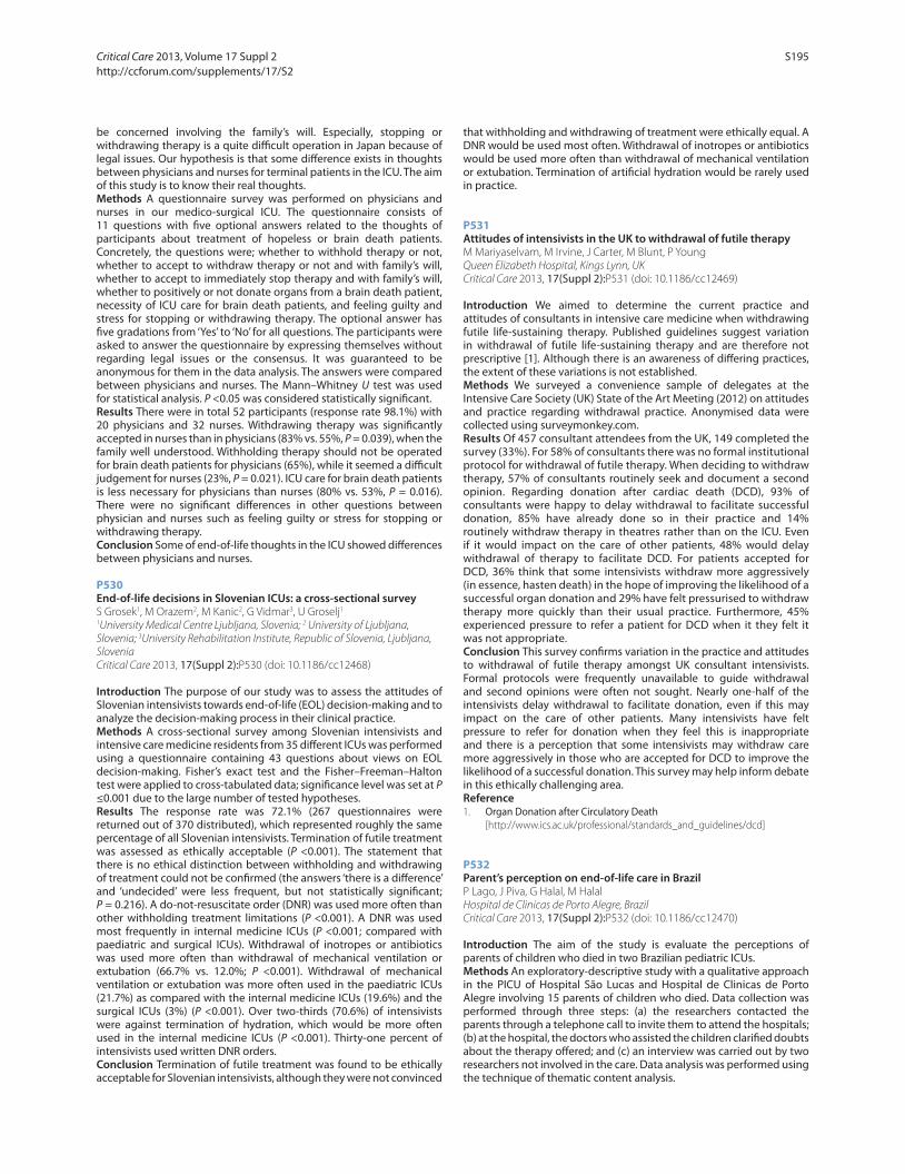
be concerned involving the family’s will. Especially, stopping or withdrawing therapy is a quite diffi cult operation in Japan because of legal issues. Our hypothesis is that some diff erence exists in thoughts between physicians and nurses for terminal patients in the ICU. The aim of this study is to know their real thoughts.Methods A questionnaire survey was performed on physicians and nurses in our medico-surgical ICU. The questionnaire consists of 11 questions with fi ve optional answers related to the thoughts of participants about treatment of hopeless or brain death patients. Concretely, the questions were; whether to withhold therapy or not, whether to accept to withdraw therapy or not and with family’s will, whether to accept to immediately stop therapy and with family’s will, whether to positively or not donate organs from a brain death patient, necessity of ICU care for brain death patients, and feeling guilty and stress for stopping or withdrawing therapy. The optional answer has fi ve gradations from ‘Yes’ to ‘No’ for all questions. The participants were asked to answer the questionnaire by expressing themselves without regarding legal issues or the consensus. It was guaranteed to be anonymous for them in the data analysis. The answers were compared between physicians and nurses. The Mann–Whitney U test was used for statistical analysis. P <0.05 was considered statistically signifi cant.Results There were in total 52 participants (response rate 98.1%) with 20 physicians and 32 nurses. Withdrawing therapy was signifi cantly accepted in nurses than in physicians (83% vs. 55%, P = 0.039), when the family well understood. Withholding therapy should not be operated for brain death patients for physicians (65%), while it seemed a diffi cult judgement for nurses (23%, P = 0.021). ICU care for brain death patients is less necessary for physicians than nurses (80% vs. 53%, P = 0.016). There were no signifi cant diff erences in other questions between physician and nurses such as feeling guilty or stress for stopping or withdrawing therapy.Conclusion Some of end-of-life thoughts in the ICU showed diff erences between physicians and nurses.
P530End-of-life decisions in Slovenian ICUs: a cross-sectional surveyS Grosek1, M Orazem2, M Kanic2, G Vidmar3, U Groselj11University Medical Centre Ljubljana, Slovenia; 2 University of Ljubljana, Slovenia; 3University Rehabilitation Institute, Republic of Slovenia, Ljubljana, SloveniaCritical Care 2013, 17(Suppl 2):P530 (doi: 10.1186/cc12468)
Introduction The purpose of our study was to assess the attitudes of Slovenian intensivists towards end-of-life (EOL) decision-making and to analyze the decision-making process in their clinical practice.Methods A cross-sectional survey among Slovenian intensivists and intensive care medicine residents from 35 diff erent ICUs was performed using a questionnaire containing 43 questions about views on EOL decision-making. Fisher’s exact test and the Fisher–Freeman–Halton test were applied to cross-tabulated data; signifi cance level was set at P ≤0.001 due to the large number of tested hypotheses.Results The response rate was 72.1% (267 questionnaires were returned out of 370 distributed), which represented roughly the same percentage of all Slovenian intensivists. Termination of futile treatment was assessed as ethically acceptable (P <0.001). The statement that there is no ethical distinction between withholding and withdrawing of treatment could not be confi rmed (the answers ‘there is a diff erence’ and ‘undecided’ were less frequent, but not statistically signifi cant; P = 0.216). A do-not-resuscitate order (DNR) was used more often than other withholding treatment limitations (P <0.001). A DNR was used most frequently in internal medicine ICUs (P <0.001; compared with paediatric and surgical ICUs). Withdrawal of inotropes or antibiotics was used more often than withdrawal of mechanical ventilation or extubation (66.7% vs. 12.0%; P <0.001). Withdrawal of mechanical ventilation or extubation was more often used in the paediatric ICUs (21.7%) as compared with the internal medicine ICUs (19.6%) and the surgical ICUs (3%) (P <0.001). Over two-thirds (70.6%) of intensivists were against termination of hydration, which would be more often used in the internal medicine ICUs (P <0.001). Thirty-one percent of intensivists used written DNR orders.Conclusion Termination of futile treatment was found to be ethically acceptable for Slovenian intensivists, although they were not convinced
that withholding and withdrawing of treatment were ethically equal. A DNR would be used most often. Withdrawal of inotropes or antibiotics would be used more often than withdrawal of mechanical ventilation or extubation. Termination of artifi cial hydration would be rarely used in practice.
P531Attitudes of intensivists in the UK to withdrawal of futile therapyM Mariyaselvam, M Irvine, J Carter, M Blunt, P YoungQueen Elizabeth Hospital, Kings Lynn, UKCritical Care 2013, 17(Suppl 2):P531 (doi: 10.1186/cc12469)
Introduction We aimed to determine the current practice and attitudes of consultants in intensive care medicine when withdrawing futile life-sustaining therapy. Published guidelines suggest variation in withdrawal of futile life-sustaining therapy and are therefore not prescriptive [1]. Although there is an awareness of diff ering practices, the extent of these variations is not established.Methods We surveyed a convenience sample of delegates at the Intensive Care Society (UK) State of the Art Meeting (2012) on attitudes and practice regarding withdrawal practice. Anonymised data were collected using surveymonkey.com.Results Of 457 consultant attendees from the UK, 149 completed the survey (33%). For 58% of consultants there was no formal institutional protocol for withdrawal of futile therapy. When deciding to withdraw therapy, 57% of consultants routinely seek and document a second opinion. Regarding donation after cardiac death (DCD), 93% of consultants were happy to delay withdrawal to facilitate successful donation, 85% have already done so in their practice and 14% routinely withdraw therapy in theatres rather than on the ICU. Even if it would impact on the care of other patients, 48% would delay withdrawal of therapy to facilitate DCD. For patients accepted for DCD, 36% think that some intensivists withdraw more aggressively (in essence, hasten death) in the hope of improving the likelihood of a successful organ donation and 29% have felt pressurised to withdraw therapy more quickly than their usual practice. Furthermore, 45% experienced pressure to refer a patient for DCD when it they felt it was not appropriate.Conclusion This survey confi rms variation in the practice and attitudes to withdrawal of futile therapy amongst UK consultant intensivists. Formal protocols were frequently unavailable to guide withdrawal and second opinions were often not sought. Nearly one-half of the intensivists delay withdrawal to facilitate donation, even if this may impact on the care of other patients. Many intensivists have felt pressure to refer for donation when they feel this is inappropriate and there is a perception that some intensivists may withdraw care more aggressively in those who are accepted for DCD to improve the likelihood of a successful donation. This survey may help inform debate in this ethically challenging area.Reference1. Organ Donation after Circulatory Death
[http://www.ics.ac.uk/professional/standards_and_guidelines/dcd]
P532Parent’s perception on end-of-life care in BrazilP Lago, J Piva, G Halal, M HalalHospital de Clinicas de Porto Alegre, BrazilCritical Care 2013, 17(Suppl 2):P532 (doi: 10.1186/cc12470)
Introduction The aim of the study is evaluate the perceptions of parents of children who died in two Brazilian pediatric ICUs.Methods An exploratory-descriptive study with a qualitative approach in the PICU of Hospital São Lucas and Hospital de Clinicas de Porto Alegre involving 15 parents of children who died. Data collection was performed through three steps: (a) the researchers contacted the parents through a telephone call to invite them to attend the hospitals; (b) at the hospital, the doctors who assisted the children clarifi ed doubts about the therapy off ered; and (c) an interview was carried out by two researchers not involved in the care. Data analysis was performed using the technique of thematic content analysis.
Critical Care 2013, Volume 17 Suppl 2 http://ccforum.com/supplements/17/S2
S195

Results and conclusion The research shows that the diffi culty of com-muni cation is a factor that impacts negatively on the grieving process. Moreover, it stresses the importance for parents to rediscuss the moment of their child’s death with health professionals.References1. Meyer E, Ritholz M, Burns J, Truog R: Improving the quality of end-of-life
care in the pediatric intensive care unit: parents priorities and recommendations. Pediatrics 2006, 3:469-457.
2. Meyer E: On speaking less and listening more during end-of-life conferences. Crit Care 2004, 32:1609-1611.
3. Piva, JP, Lago P, Othero J, Garcia PC: Evaluating end of life in ten Brazilian pediatric and adults intensive care units. J Med Ethics 2010, 36:344-348.
4. Meert, K et al.: Parents’ perspectives on physician-parent communication near the time of a child’s death in the pediatric intensive care unit. Pediatr Crit Care 2008, 9:2-7.
P533Getting critical care patients home for end-of-life careLS NielsenKolding Sygehus, Kolding, DenmarkCritical Care 2013, 17(Suppl 2):P533 (doi: 10.1186/cc12471)
Introduction Despite our eff orts in making patients healthy and going home, critical illness has a mortality, in Danish ICUs, of 10 to 12%. Approximately 90% of deaths in ICUs happen after life-sustaining therapy has been withheld or withdrawn. Although trying to provide patients and family with what we suppose is a good death, most patients would prefer dying at home, and sometimes patients and family ask for the possibility of doing that. The last 2 years we transferred seven patients from our unit to end-of-life care in their own home.Methods After making the decision of withholding or withdrawing intensive care therapy, the care of the patient changes from an active, medical, technological treatment to relief and care. In that period we determine whether the patient’s condition is stable enough to go home. We try to fi nd out if it is a wish for the patient to go home, and if the family has resources to take care of the patient at home. If that is the case, we start planning care at home, arrange for transportation, and contact the primary care physician and nurse. Due to the patient’s condition on the day of transferring, we plan following the patient by either a nurse or a doctor.Results Seven terminally ill patients wishing to go home for dying were transferred home. Diagnoses varied: end-stage lung disease, cancer, surgical complications. Ages ranged from 68 to 84 years. All patients survived transport home, and time at home varied from a few hours to 4 days. Later contacts with patients’ families indicated that both patient and family were grateful, and that they did not experience the patient having pain or dyspnea at home.Conclusion Sending critically ill patients home to die is not common. Anyhow, our experiences doing that are only positive. Terminally ill patients, awake to make a decision of their own, and in a condition making it possible, should have the choice to go home to die, with our help in logistics, planning and transportation.Reference1. Lusardi P, et al.: The going home initiative: getting critical care patients
home with hospice. Crit Care Nurse 2011, 31:46-57.
P534Project PIX (Post Intensive care eXercise): impact on physical fi tness and focus group analysis of quality of life following exercise rehabilitationJ Goodman1, W Walker2, J Wright1, G Danjoux1, S Howell3, D Martin4, S Bonner1
1James Cook University Hospital, Middlesbrough, UK; 2West Park Hospital, Darlington, UK; 3University of Leeds, UK; 4Teeside University, Middlesbrough, UKCritical Care 2013, 17(Suppl 2):P534 (doi: 10.1186/cc12472)
Introduction This study assessed the eff ect of a hospital-based aerobic exercise programme on physical fi tness and health-related quality of life (HRQoL) for survivors of ICU admission. Including the qualitative
arm of the study, we examined the patient experience after critical illness, their views of the exercise programme and the eff ects on their HRQoL.Methods A randomised controlled trial was undertaken in adult survivors of ICU admission. They were allocated to receive an 8-week in-hospital supervised aerobic programme consisting of two cycle ergometry and one unsupervised session per week (exercise group) or no exercise (control group). Primary outcomes were the anaerobic threshold (in ml O2/kg mass/minute), physical function and mental health scores (SF-36 questionnaire), measured at weeks 9 and 26. Participants were then allocated to focus groups where the interpretation of experiences was compared with outcomes from the PIX study.Results Fifty-nine patients were recruited to the study. The anaerobic threshold increased at week 9 in the exercise group by a clinically and statistically signifi cant amount of 2 ml O2/kg mass/minute (90% CI, 1 to 3 ml/kg/minute). There was further improvement in fi tness levels in both groups by week 26 (although no signifi cant diff erence between groups). No signifi cant diff erence in HRQOL measures between groups was demonstrated; however, the exercise group did show an improvement in their mental health scores. The focus groups centred on feelings of isolation, abandonment, vulnerability, dependency and reduced physical activity post hospital discharge. Many reported a lack of social inclusion as they did not have the energy or confi dence to venture outside. However, those in the exercise group felt that the rehabilitation programme was motivating, built up confi dence, improved fi tness, helped social interaction and gave them a sense of achievement.Conclusion The 8-week exercise intervention resulted in statistically signifi cant improvements in fi tness at 9 weeks while focus group participants highlighted the positive eff ects of the exercise intervention leading to enhanced energy levels, motivation and achievement. Psychological benefi ts of the exercise programme are apparent from the focus group, emphasising the important link between physical and mental health.
P535Feasibility of neuromuscular electrical stimulation in critically ill patientsJ Segers, G Hermans, F Bruyninckx, G Meyfroidt, D Langer, R GosselinkUZ/KU Leuven, BelgiumCritical Care 2013, 17(Suppl 2):P535 (doi: 10.1186/cc12473)
Introduction Survivors of critical illness often have a prolonged stay on the ICU. These patients may suff er from ICU-acquired weakness. It has been shown that reduction in muscle mass and muscle strength occurs early after admission to the ICU. However, in the very early stage on the ICU, patients are often sedated and not able to participate in any active mobilizations. Therefore the use of neuromuscular electrical stimulation (NMES) is becoming a treatment of interest in the ICU. The aim was to study the feasibility and safety of NMES in a surgical and medical ICU of a large, tertiary referral university hospital.Methods Fifty patients with an expected prolonged stay on the ICU of 5 more days (judged on day 3) with no trauma or neurological disease were included. They then received daily a NMES session (DUO 500; Gymna, Belgium) for 25 minutes on the quadriceps bilaterally during their entire stay on the ICU. The main outcome was the ability to produce a contraction of the quadriceps through NMES. The muscle contraction was quantifi ed on a 5-point scale: 1 (no contraction palpable and visible) up to 5 (contraction very well palpable and visible). Patients were classifi ed as responders when an adequate muscle bulk was obtained in ≥75% of the sessions. The potential factors associated with the feasibility were: gender, age, body mass index (BMI), diagnosis of sepsis, Barthel index prior to admission to the hospital, APACHE II score, Glasgow Coma Scale (GCS), fi ve questions for adequacy, stimulus intensity and leg edema. A multiple regression analysis was performed to identify the factors determining whether or not a contraction could be expected in a patient. Safety of NMES was assessed through heart rate, blood pressure, oxygen saturation and respiratory rate.Results In 48% of the patients we were able to achieve adequate muscle contractions in more than 75% of the sessions. GCS (P = 0.047), edema
Critical Care 2013, Volume 17 Suppl 2 http://ccforum.com/supplements/17/S2
S196

(P = 0.001) and sepsis (P = 0.010) were signifi cantly diff erent between responders and nonresponders. Responders had a lower mean GCS (7 ± 3 vs. 9 ± 3), lower amount of edema and were less likely to have had sepsis. In a multiple regression analysis, sepsis, edema, BMI and age explained 51% of the variance. As for safety, none of the parameters changed signifi cantly.Conclusion In patients with a better neurological condition, sepsis and/or leg edema it was more diffi cult to obtain an adequate quadriceps contraction with NMES. NMES is safe to apply on the ICU.
P536Follow-up and counseling service in trauma patients: needs and goals – a preliminary studyDL Grieco, M Biancone, SL Cutuli, G Pintaudi, MT Santantonio, ES Tanzarella, F Toni, AG De Belvis, MG Bocci, C Sandroni, M AntonelliCatholic University School of Medicine, Rome, ItalyCritical Care 2013, 17(Suppl 2):P536 (doi: 10.1186/cc12474)
Introduction Trauma is the most common cause of morbidity in young people. It has a high social impact both because of the high cost of the acute treatments and because of the physical and psychological consequences that it may cause. A prospective, observational, single-center study on quality of life 12 to 24 months after trauma was carried out. The aim of the study is to evaluate life quality after trauma and to identify the most important needs of the patients, in order to improve the level of care after an ICU stay and to implement a faster and more eff ective reintegration into the active and productive society.Methods All patients admitted to the 18-bed ICU of our university hospital between 5 May 2010 and 28 February 2011 because of a trauma who stayed in the ICU for at least 48 hours were eligible for the study. Patients were contacted and invited to attend a multidisciplinary interview. Data concerning family, working and economic status, main disabilities and medical needs were collected. The SF12 questionnaire was administered to defi ne physical component summary (PCS) and mental component summary (MCS) scores. SPSS 20.0 was used to perform statistical analysis.Results Sixty-two patients matched inclusion criteria and were contacted. Thirty-three patients (90.9% males; age 38.7 ± 14.3 years; mean SAPS II 33.75 ± 11.79) were interviewed. Mean ICU stay was 25.7 ± 15.3 days. Fifteen patients (45.5%) had undergone temporary tracheostomy. Twenty-six patients were employed at the moment of trauma: of these, 11 (42.3%) stated to be jobless and one was pensioned at follow-up. In the whole cohort, the unemployment rate increased from 12.1 to 45.6%. Mean PCS was 43.59 ± 6.43, mean MCS was 41.04 ± 7.69. At follow-up, the 14 employed patients had a lower MCS versus the 19 jobless patients (37.94 ± 7.14 vs. 43.33 ± 7.44; P = 0.04), while no diff erences concerning PCS were found. The three patients that were living on their own when the trauma occurred showed, at follow-up, a lower mean MCS score towards the rest of the cohort (32.2 ± 0.47 vs. 41.93 ± 7.51; P <0.001).Conclusion Trauma is associated with an increase of the unemployment rate in young people. Patients that are employed at follow-up seemed to have a worse mental health status towards the unemployed ones. Conversely, the employed patients did not show any trend to a lower physical health status. Thus a counseling service that provides psychological care would be useful to implement more eff ective work reintegration for trauma patients.
P537Post-traumatic stress disorder – are we doing all we can? A UK national-based surveyX WatsonAshford and St Peter’s Hospital, Surrey, UKCritical Care 2013, 17(Suppl 2):P537 (doi: 10.1186/cc12475)
Introduction Post-traumatic stress disorder (PTSD) is a well-recognised complication in patients discharged from the ICU. ICU clinics have been recommended to treat physical and psychosocial problems post discharge, with guidelines issued by the Department of Health UK [1]. Recent evidence has advocated the use of patient diaries to reduce the
incidence of new-onset PTSD [2]. Family support groups may also play a pivotal role [3]. We performed a national survey via the Intensive Care Society (ICS) to determine the provision of follow-up clinics, patient diary services and family support groups.Methods An online structured questionnaire was sent via the ICS to all ICU linkmen at 124 hospitals in the UK. Responses were received from 77 ICUs, a 62% response rate.Results Out of 77 ICUs, 37 (48%) run a follow-up clinic. The majority of clinics (51%) only invite patients who have been admitted for more than 3 to 4 days. Only 10 clinics (30%) receive funding from the ICU budget or PCT, with the majority (67%) receiving no funding at all. Only 44% (34) of ICUs use patient diaries, mostly as a nonfunded service (68%). Additionally, 91% of ICUs do not run family support groups; from the minority that do, these are mostly held quarterly and are largely not funded (55%).Conclusion This survey demonstrates that the provision of ICU clinics in the UK is not well established, with only 48% currently providing a regular service. Currently 67% of clinics are not funded and further resources should be employed so this service becomes an integral part of the ICU pathway. Despite recent evidence demonstrating that diaries reduce new-onset PTSD, only 44% of ICUs in our study provide this service. Ninety-one per cent of ICUs do not provide family support groups; similarly, it appears that fi nancial constraints are the limiting factor.References1. DH and Modernisation Agency: The National Outreach Report. London: NHS
Modernisation Agency; 2003.2. Jones C, et al.: Crit Care Med 2010, 14:168.3. Orcutt TA:. Nurs Crit Care 2010, 5:33-37.
P538Implementation of an ICU follow-up clinic: outcomes and patient satisfaction after 1 yearG De la CerdaQueens Hospital, London, UKCritical Care 2013, 17(Suppl 2):P538 (doi: 10.1186/cc12476)
Introduction The aim was to analyse the outcomes and patient satisfaction of a recently implemented ICU follow-up clinic. These clinics are National Institute for Clinical Excellence recommended [1].Methods A retrospective analysis of prospective collected data from January to December 2012. The clinic is run monthly by an ICU consultant and a critical care outreach sister. Criteria to be invited to the clinic are mechanical ventilation ≥3 days. Patients fi lled an anonymous satisfaction survey after the clinic.Results Our attendance rate is 50% (26 patients), which is similar to other series reported in the literature. Those patients who attended the clinic required a longer length of mechanical ventilation (5.3 days vs. 7.1) and a longer length of stay in the ICU (7.6 vs. 13) and in hospital (14 vs. 28). We identifi ed a wide range of physical and nonphysical morbidities on these patients (Figure 1). We referred them to the appropriate specialities. Patients were very satisfi ed with this new service (Figure 2).
Figure 1 (abstract P538). Physical and nonphysical morbidity.
Critical Care 2013, Volume 17 Suppl 2 http://ccforum.com/supplements/17/S2
S197
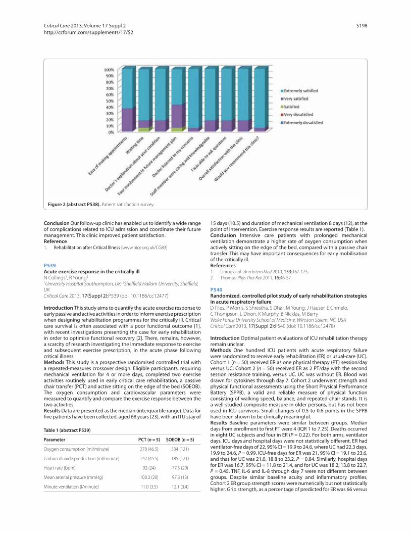
Conclusion Our follow-up clinic has enabled us to identify a wide range of complications related to ICU admission and coordinate their future management. This clinic improved patient satisfaction.Reference1. Rehabilitation after Critical Illness [www.nice.org.uk/CG83]
P539Acute exercise response in the critically illN Collings1, R Young2
1University Hospital Southampton, UK; 2Sheffi eld Hallam University, Sheffi eld, UKCritical Care 2013, 17(Suppl 2):P539 (doi: 10.1186/cc12477)
Introduction This study aims to quantify the acute exercise response to early passive and active activities in order to inform exercise prescription when designing rehabilitation programmes for the critically ill. Critical care survival is often associated with a poor functional outcome [1], with recent investigations presenting the case for early rehabilitation in order to optimise functional recovery [2]. There, remains, however, a scarcity of research investigating the immediate response to exercise and subsequent exercise prescription, in the acute phase following critical illness.Methods This study is a prospective randomised controlled trial with a repeated-measures crossover design. Eligible participants, requiring mechanical ventilation for 4 or more days, completed two exercise activities routinely used in early critical care rehabilitation, a passive chair transfer (PCT) and active sitting on the edge of the bed (SOEOB). The oxygen consumption and cardiovascular parameters were measured to quantify and compare the exercise response between the two activities.Results Data are presented as the median (interquartile range). Data for fi ve patients have been collected, aged 68 years (23), with an ITU stay of
15 days (10.5) and duration of mechanical ventilation 8 days (12), at the point of intervention. Exercise response results are reported (Table 1).Conclusion Intensive care patients with prolonged mechanical ventilation demonstrate a higher rate of oxygen consumption when actively sitting on the edge of the bed, compared with a passive chair transfer. This may have important consequences for early mobilisation of the critically ill.References1. Unroe et al.: Ann Intern Med 2010, 153:167-175.2. Thomas: Phys Ther Rev 2011, 16:46-57.
P540Randomized, controlled pilot study of early rehabilitation strategies in acute respiratory failureD Files, P Morris, S Shrestha, S Dhar, M Young, J Hauser, E Chmelo, C Thompson, L Dixon, K Murphy, B Nicklas, M BerryWake Forest University School of Medicine, Winston Salem, NC, USACritical Care 2013, 17(Suppl 2):P540 (doi: 10.1186/cc12478)
Introduction Optimal patient evaluations of ICU rehabilitation therapy remain unclear.Methods One hundred ICU patients with acute respiratory failure were randomized to receive early rehabilitation (ER) or usual-care (UC). Cohort 1 (n = 50) received ER as one physical therapy (PT) session/day versus UC; Cohort 2 (n = 50) received ER as 2 PT/day with the second session resistance training, versus UC. UC was without ER. Blood was drawn for cytokines through day 7. Cohort 2 underwent strength and physical functional assessments using the Short Physical Performance Battery (SPPB), a valid and reliable measure of physical function consisting of walking speed, balance, and repeated chair stands. It is a well-studied composite measure in older persons, but has not been used in ICU survivors. Small changes of 0.5 to 0.6 points in the SPPB have been shown to be clinically meaningful.Results Baseline parameters were similar between groups. Median days from enrollment to fi rst PT were 4 (IQR 1 to 7.25). Deaths occurred in eight UC subjects and four in ER (P = 0.22). For both arms, ventilator days, ICU days and hospital days were not statistically diff erent. ER had ventilator-free days of 22, 95% CI = 19.9 to 24.6, where UC had 22.3 days, 19.9 to 24.6, P = 0.99. ICU-free days for ER was 21, 95% CI = 19.1 to 23.6, and that for UC was 21.0, 18.8 to 23.2, P = 0.84. Similarly, hospital days for ER was 16.7, 95% CI = 11.8 to 21.4, and for UC was 18.2, 13.8 to 22.7, P = 0.45. TNF, IL-6 and IL-8 through day 7 were not diff erent between groups. Despite similar baseline acuity and infl ammatory profi les, Cohort 2 ER group strength scores were numerically but not statistically higher. Grip strength, as a percentage of predicted for ER was 66 versus
Figure 2 (abstract P538). Patient satisfaction survey.
Table 1 (abstract P539)
Parameter PCT (n = 5) SOEOB (n = 5)
Oxygen consumption (ml/minute) 270 (46.5) 334 (121)
Carbon dioxide production (ml/minute) 142 (45.5) 185 (121)
Heart rate (bpm) 92 (24) 77.5 (29)
Mean arterial pressure (mmHg) 100.3 (20) 97.3 (13)
Minute ventilation (l/minute) 11.0 (3.5) 12.1 (3.4)
Critical Care 2013, Volume 17 Suppl 2 http://ccforum.com/supplements/17/S2
S198

39 for UC, P = 0.06. Dynamometry for ER was 211 versus 181 lbs for UC, P = 0.124. Although the diff erence in SPPB values for ER versus UC (5 vs. 3, P = 0.172) was not statistically diff erent, it was greater than the minimal clinically signifi cant diff erence. There were no diff erences in adverse events.Conclusion In this pilot study, early ICU rehabilitation was safe, and was associated with numerically although not statistically shorter hospital stay, greater strength and improved functional scores. Particularly, the SPPB demonstrated discriminatory ability in groups of ICU survivors with low physical function. Future early ICU rehabilitation studies should consider ICU survivor assessments using the SPPB due to its ease, reproducibility and discriminatory ability following ICU and hospital discharge.Acknowledgement Supported by NIH grants 1R011186-01 and P30 AG21332.
P541Early results of a 6-week exercise programme in post-ICU patientsC Battle1, K James1, P Temblett1, H Hutchings2
1Morriston Hospital, ABMU Health Board, Swansea, UK; 2Swansea University, Swansea, UKCritical Care 2013, 17(Suppl 2):P541 (doi: 10.1186/cc12479)
Introduction The aim of this study was to investigate the eff ect of a 6-week exercise programme on outcomes in post-ICU patients. With improvements in intensive care medicine, increasing numbers of patients are surviving catastrophic illness [1]. Severe weakness is common in patients with prolonged critical illness and results in considerable morbidity, mortality and healthcare costs [2]. The NICE83 guidelines Rehabilitation in Critical Care recommend follow-up for post-ICU patients and that further research is needed in this fi eld [3].Methods Patients who have been discharged home from hospital following an ICU stay of 48 hours or more were recruited to the study. Patients were only excluded if they were not considered safe for exercise. Baseline measurements were completed prior to stratifi ed (age, gender, APACHE II score) random allocation to either the exercise or control group. Outcome measures included cardiopulmonary fi tness (6-minute walk test), balance (Berg Balance Scale), grip strength (JAMAR grip dynamometer) and hospital anxiety and depression (HAD score). The exercise group completed a 6-week supervised exercise programme, twice a week for up to 1 hour. In the seventh week, all patients repeated the baseline measurements. An unpaired Student’s t test was used to compare any diff erences between the control and exercise groups.Results At baseline measurements, there were no statistical diff erences in age, gender, length of stays or APACHE II scores between the two groups. Results indicate that the exercise group (n = 10) had signifi cantly greater improvements in cardiopulmonary fi tness (P <0.001) and balance (P <0.05) compared with the control group (n = 10). Greater improvements were also evident in anxiety, depression and grip strength in the exercise group, although not statistically signifi cant.Conclusion This pilot study highlights that a 6-week supervised exercise programme can signifi cantly improve cardiopulmonary fi tness and balance in post-ICU patients. Further recruitment to the study and 6-month/1-year follow-up is needed.References1. Baker C, et al.: Physical rehabilitation following critical illness. J Intensive Care
Soc 2008, 9:166-169.2. Jones C, et al.: Long term outcome from critical illness. Anaesth Intensive
Care Med 2006, 7:155-156.3. CG83 Critical Illness Rehabilitation: Guideline 2009 [www.nice.org.uk/CG83]
P542Recognition of the primary stressors aff ecting intensive care patients: a systematic reviewS Birch, S ElliotLeeds Teaching Hospitals, Leeds University Teaching Hospitals Trust, Leeds, UKCritical Care 2013, 17(Suppl 2):P542 (doi: 10.1186/cc12480)
Introduction Intensive care patients suff er psychological and physiological distress that may have debilitating and long-lasting eff ects [1-3]. Healthcare professionals are in a position to help avoid
or alleviate this stress [4]. To action this it is important to identify the main stressors from the patient’s perspective. A systematic review was performed to provide a list of what patients consider stressors in intensive care. These were then ranked in order to provide an identifi cation tool that can be used to shape appropriate care.Methods A systematic review was performed using MEDLINE, CINAHL, Psych INFO and Academic Search Complete. Grey literature was included and searches were not restricted to type of intensive care or country. Criteria were used to fi lter those articles that identifi ed the patients’ views of their stressor, not the patient experience. Eligible articles were critiqued using the Critical Appraisal Skills Programme for qualitative studies [5] and brought together using a narrative synthesis. All of the reviewed studies used a questionnaire as a means to identify what elements on the intensive care patients found stressful. A list of the top-10 stressors could then be expressed for each study and compared. From this information, a set of guidelines for best practice were devised.Results A total 1,424 articles were systematically assessed for suitability and applicability. Of these, 14 articles remained eligible for review. The stressors were ranked by their frequency in the individual studies’ top-10 lists. In rank order: 1, Tubes. 2, Pain. 3, Sleep Diffi culties. 4, Thirst. 5, Lack of Patient Understanding. Overall, stressors were found to be similar throughout all of the studies.Conclusion The review identifi ed a list of the most pertinent common stressors. Awareness of these and ranking in priority may enable plans of care to be instigated to eff ectively alleviate patient stress.References1. Jones C, et al.: Clin Intensive Care 1998, 9:199-205.2. Scragg P, et al.: Anaesthesia 2001, 56:9-14.3. Perrins J, et al.: Intensive Crit Care Nurs 1998, 14:108-116.4. Ballard KS: Issues Ment Health Nurs 1981, 3:89-108.5. Public Health Resource Unit: Critical Appraisal Skills Programme. 10 Questions
to Help You Make Sense of Qualitative Research. [http://www.medev.ac.uk/static/uploads/workshop_resources/166/166_Qualitative_Appraisal_Tool.pdf ]
P543Quality of care in the ICU from the perspective of relativesM Van Mol1, E Bakker2, A Rensen3, I Menheere3, L Verharen3
1Erasmus MC, Rotterdam, the Netherlands; 2OU, Heerlen, the Netherlands; 3HAN, Nijmegen, the NetherlandsCritical Care 2013, 17(Suppl 2):P543 (doi: 10.1186/cc12481)
Introduction This study describes the development and validation of the Consumer Quality Index Relatives in ICUs (CQI ‘R-ICU’), which aims to measure the satisfaction of relatives and to identify aspect of care that need improvement in the ICU in a reliable and valid way. According to the quality standards of the Dutch Society of Intensive Care, every ICU needs to record the satisfaction of relatives [1]. At this moment there is insuffi cient insight into the quality of care off ered to relatives on the ICU because an evidence-based Dutch measurement instrument is missing.Methods The CQI ‘R-ICU’ has been developed based on a scientifi c and standardised method [2]. A mixed design method is used, consisting of qualitative and quantitative survey studies. Factor analyses are carried out to determine the underlying structure of the newly developed questionnaire. Multiple regression analysis is used to explore the relationship between demographic variables and the perceived quality of care.Results In six hospitals the CQI ‘R-ICU’ is sent to relatives after receiving informed consent (n = 441), 55.1% of the respondents are the patient’s partner. Respondents seem to be most satisfi ed with the presence of a professional at fi rst entrance to the ICU. The highest need for improvement scores relate to information about meals, parking and other disciplines (for example, social worker, spiritual worker or psychologist). Factor analysis shows that quality of care is determined by four clusters of items: Support, Communication, General Information and Organisation. The reliability of the CQI ‘R-ICU’ is suffi ciently high, only Communication and Support are signifi cant predictors of total quality judgement of relatives (adj. R2 = 0.74). In addition, there is a signifi cant diff erence in mean total quality judgement between the six hospitals as well as between the four wards within Erasmus MC. None
Critical Care 2013, Volume 17 Suppl 2 http://ccforum.com/supplements/17/S2
S199

of the demographic variables such as sex, age, education, race and length of stay had an eff ect on perceived quality of care.Conclusion The CQI ‘R-ICU’ turned out to be a valid, reliable, sensitive and feasible instrument. Large-scale implementation is recommended.Acknowledgements Thanks to researchers of Ziekenhuis Gelderse Vallei, Kennemer Gasthuis, Catharinaziekenhuis, Ziekenhuis Rivieren-land and Scheperziekenhuis.References1. Vos et al.: Quality measurement at intensive care units: which indicators
should we use? J Crit Care 2007, 22:267-274.2. Sixma et al.: Handboek CQI Ontwikkeling: richtlijnen en voorschriften voor de
ontwikkeling van een CQI meetinstrument. Utrecht: Nivel; 2008.
P544Family satisfaction in the ICU: enhancing patient experienceE Kursumovic, J Bilinska, A MolokhiaUniversity Hospital Lewisham, London, UKCritical Care 2013, 17(Suppl 2):P544 (doi: 10.1186/cc12482)
Introduction Following recommendations made by the Scottish Intensive Care Society [1], we have introduced an annual Family Satisfaction Survey in the intensive care unit (FS-ICU), in anticipation that this will become an integral part of how we measure, improve and strive to advance patient care in the future. We aim to recognise factors impacting patient care and highlight areas for improvement. Improving understanding of patients’ needs is currently a UK national goal for CQUIN 2011/2012. An estimated £6.9 billion per year can be saved by reaching these goals [2] and a positive patient experience results in improved long-term outcomes and shorter hospital stays [2].Methods We performed a study to assess Family Satisfaction over a 10-week period in a 16-bed critical care unit (CCU). A modifi ed version of an FS-ICU published in US and Canadian studies [3] was distributed to up to two family members per patient. Five-point Likert scale responses were linearly transformed to give percentage scores. Higher values represented a greater degree of satisfaction.Results We received and analysed 32 completed surveys. Seventy-eight per cent of relatives reported that the treatment of the patient was excellent, including symptom control such as pain, breathlessness and agitation (72%, 69%, 100%, respectively). Ninety-one per cent felt the care and frequency of communication provided by nurses was excellent or very good, compared with 81% by doctors. Only 9% of family members were off ered spiritual support during their CCU experience. Forty-seven per cent felt they would have liked more involvement in the decision-making process.Conclusion The vast majority of relatives rated their overall experience on CCU as excellent or very good. The study highlighted two main areas for improvement: provision of spiritual support as well as family and patient involvement in the decision-making process. We will arrange multi-disciplinary teaching sessions focusing on the positive impact, and therefore importance of patient satisfaction. A poster providing information about available spiritual support will be displayed around our CCU. Further studies are required to evaluate these measures.
References1. Quality Indicators for Critical Care in Scotland [http://www.sicsag.scot.nhs.
uk/SICSQIG-report-2012-120209.pdf ]2. Using the Commissioning for Quality and Innovation (CQUIN) Payment
Framework – Guidance on National Goals for 2011/12 [http://www.dh.gov.uk/prod_consum_dh/groups/dh_digitalassets/@dh/@en/@ps/documents/digitalasset/dh_123009.pdf ]
3. Wall et al.: Refi nement, scoring, and validation of the Family Satisfaction in the Intensive Care Unit (FS-ICU) survey. Crit Care Med 2007, 35:271-279.
P545Family satisfaction with patient care in ICUs of nonacademic Brazilian public hospitalsJ Othero, AB Cavalcanti, JC Mouro, K Normilio-da-Silva, R Pusch, F Moreira, AA Kodama, O BerwangerHospital do Coração – HCor, São Paulo, BrazilCritical Care 2013, 17(Suppl 2):P545 (doi: 10.1186/cc12483)
Introduction We are conducting a cluster randomized trial with two parallel arms to evaluate strategies to improve family satisfaction with the care that themselves and their critically ill relatives receive in the ICUs of nonacademic Brazilian public hospitals. Here we report the results of the baseline phase of this trial.Methods In this baseline phase, we interviewed the family member most closely involved with the care of critically ill patients who stayed in the ICU for at least 72 hours. We applied a form with 24 questions divided into four domains: overall ICU experience, communication, decision-making, and questions related to end-of-life care for patients who died in the ICU. Each question scored from 0 (very poor) to 100 (excellent). The form was adapted from the Family Satisfaction with Care in the ICU (FS-ICU 24). As many questions assessed the quality of intensivist care or communication, the interview was applied by a psychologist or a nurse.Results Families of 564 patients were interviewed. A total 45/564 (8.3%) died in the ICU. Most respondents were satisfi ed with overall ICU experience (mean ± SD score 85.5 ± 11.9). However, family satisfaction with communication (67.8 ± 18.0) and decision-making (69.5 ± 21.1) resulted in somewhat lower scores. Most families of patients who died in the ICU (38/45 (82.6%)) considered that their relative’s life was neither extended nor shortened unnecessarily. Also, most of the families believed that their relative did not suff er or suff ered little in the ICU (37/46 (80.4%)) and felt supported by the healthcare team (40/46 (87.0%)).Conclusion Most families were satisfi ed with the care themselves and their critically ill relatives received in the ICU. Also, most relatives of patients who died in the ICU felt that end-of-life care was adequate. Although we believe there is much room for improvement in communication, decision-making and support critically ill patients and their families, as their baseline satisfaction with patient care is quite high, it may be hard to demonstrate substantial improvement after interventions.
Critical Care 2013, Volume 17 Suppl 2 http://ccforum.com/supplements/17/S2
S200