medical policy - multimarker serum testing related to
TRANSCRIPT

2
_______________________________________________________________________________
Medical Policy
Joint Medical Policies are a source for BCBSM and BCN medical policy information only. These documents are not to be used to determine benefits or reimbursement. Please reference the appropriate
certificate or contract for benefit information. _______________________________________________________________________________
*Current Policy Effective Date: 7/1/21
(See policy history boxes for previous effective dates)
Title: Multimarker Serum Testing Related to Ovarian Cancer (e.g., OVA1®, Overa™ and ROMA™ testing)
_______________________________________________________________________________
Description/Background Epithelial Ovarian Cancer The term epithelial ovarian cancer collectively includes high-grade serous epithelial ovarian, fallopian tubal, and peritoneal carcinomas due to their shared pathogenesis, clinical presentation, and treatment. We use epithelial ovarian cancer to refer to this group of malignancies in the discussion that follows. There is currently no serum biomarker that can distinguish between these types of carcinoma. An estimated 22,440 women in the United States are expected to be diagnosed in 2017 with ovarian cancer, and approximately 14,080 will die of the disease.1 The mortality rate depends on 3 variables: (1) patient characteristics; (2) tumor biology (grade, stage, type); and (3) treatment quality (nature of staging, surgery, and chemotherapy used).2 In particular, comprehensive staging and completeness of tumor resection appear to have a positive impact on patient outcome. In 1997, the Society of Surgical Oncology recommended ovarian cancer surgery and follow-up treatment be performed by physicians with ovarian cancer disease expertise.3 Numerous articles have been published on the application of this recommendation examining long- and short-term outcomes as well as process measures (e.g., types of treatment such as complete staging or tumor debulking). At least 2 meta-analyses have concluded that outcomes are improved when patients with ovarian cancer are treated by gynecologic oncologists.4,5 The available data are most convincing for patients with advanced-stage disease. Adult women presenting with an adnexal mass have an estimated 68% likelihood of having a benign lesion.6 About 6% have borderline tumors; 22%, invasive malignant lesions, and 3%, metastatic disease. Clinicians generally agree that women with masses that have a high likelihood of malignancy should undergo surgical staging by gynecologic oncologists. However, women with clearly benign masses do not require referral to a specialist. Criteria and tests that help differentiate benign from malignant pelvic masses are thus desirable.

3
In 2016, the American College of Obstetricians and Gynecologists updated a practice bulletin that addressed criteria for referring women with adnexal masses suspicious for ovarian cancer to gynecologic oncologists.7 Separate criteria were developed for premenopausal and postmenopausal women because the specificity and positive predictive value of cancer antigen 125 (CA 125) are higher in postmenopausal women. Prior guidance, which was based on expert opinion, recommended a CA 125 >200 U/mL for referring premenopausal women with an adnexal mass to a gynecologic oncologist. The current guidance advises using very elevated CA 125 levels with other clinical factors such as ultrasound findings, ascites, a nodular or fixed pelvic mass, or evidence of abdominal or distant metastasis for referral. The referral criteria for postmenopausal women are similar, except that a lower threshold for an elevated CA 125 test was used (35 U/mL) and nodular or fixed pelvic mass was an additional criterion. Three multimarker serum-based tests specific to ovarian cancer have been cleared by the Food and Drug Administration (FDA) with the intended use of triaging patients with adnexal masses. They are summarized in Table 1. The proposed use of the tests is to identify women with a substantial likelihood of malignant disease who may benefit from referral to a gynecologic oncology specialist. Patients with positive results may be considered candidates for referral to a gynecologic oncologist for treatment. The tests have been developed and evaluated only in patients with adnexal masses and planned surgical removal. Other potential uses, such as selecting patients to have surgery, screening asymptomatic patients, and monitoring treatment, have not been investigated. Furthermore, the tests are not intended to be used as stand-alone tests, but in conjunction with clinical assessment. Other multimarker panels and longitudinal screening algorithms are under development, but are not yet commercially available.8,9 Table 1. Summary of FDA-Approved Multimarker Serum-Based Tests Specific to Ovarian Cancer
Variables OVA1 Overa ROMA
Cleared 2009 2016 2011 Manufacturer Quest Diagnostics Vermillion Roche Diagnostics Biomarkers used
CA 125 II x x x β2-microglobulin x
Transferrin x x Transthyretin x
Apolipoprotein AI x x HE4 x x FSH x
Score range 0-10 0-10 0-10 Risk categorization
Premenopausal <5.0: low >5.0: high
<5.0: low >5.0: high
>1.3: high
Postmenopausal <4.4: low >4.4: high
>2.77: high
CA 125: cancer antigen 125; FDA: Food and Drug Administration; FSH: follicle stimulating hormone; HEA: human epididymis secretory protein 4.

4
Regulatory Status On July 16, 2009, the OVA1® test (Aspire Labs) was cleared for market by the U.S. Food and Drug Administration (FDA) through the 510(k) process. The intended use of OVA1® is as an aid to further assess the likelihood that malignancy is present when the physician’s independent clinical and radiological evaluation does not indicate malignancy. In March 2016, a second-generation test called Overa™, in which 2 of the 5 biomarkers in OVA1® are replaced with human epididymis secretory protein 4 and follicle stimulating hormone, was cleared for marketing by FDA through the 510(k) process. Similar to OVA1®, Overa™ generates a low or high risk of malignancy on a scale from 0 to 10. On September 1, 2011, the Risk of Ovarian Malignancy Algorithm (ROMA™ test, Fujirebio Diagnostics) was cleared for marketing by FDA through the 510(k) process. The intended use of ROMA™ is as an aid, in conjunction with clinical assessment, in assessing whether a premenopausal or postmenopausal woman who presents with an ovarian adnexal mass is at high or low likelihood of finding malignancy on surgery. FDA product code: ONX. Black Box Warning On December 10, 2011, the FDA amended its regulation for classifying ovarian adnexal mass assessment score test systems. The change required off-label risks be highlighted by using a black box warning.10 The warning is intended to mitigate the risk to health associated with off-label use as a screening test, stand-alone diagnostic test, or as a test to determine whether or not to proceed with surgery. Considering the history and currently unmet medical needs for ovarian cancer testing, the FDA concluded that there is a risk of off-label use of this device.7 To address this risk, the FDA requires that manufacturers provide notice concerning the risks of off-label uses in the labeling, advertising, and promotional material of ovarian adnexal mass assessment score test systems. Manufacturers must address the following risks:
• Women without adnexal pelvic masses (i.e., for cancer "screening") are not part of the intended use population for the ovarian adnexal mass assessment score test systems. Public health risks associated with false-positive results for ovarian cancer screening tests are well described in the medical literature and include morbidity or mortality associated with unneeded testing and surgery. The risk from false-negative screening results also includes morbidity and mortality due to failure to detect and treat ovarian malignancy.
• Analogous risks, adjusted for prevalence and types of disease, arise if test results are used to determine the need for surgery in patients who are known to have ovarian adnexal masses.
• If used outside the "OR" rule that is described in this special control guidance, results from ovarian adnexal mass assessment score test systems pose a risk for morbidity and mortality due to nonreferral for oncologic evaluation and treatment.
_

5
___________________________________________________________________________ Medical Policy Statement The safety and effectiveness of proteomics-based testing (e.g., OVA1®, Overa™ and ROMA™ tests) to identify women with adnexal masses who may benefit from referral to a gynecologic-oncology specialist have been established. These tests may be considered a useful (but not mandatory) diagnostic option in guiding referral to a gynecologic oncologist for women meeting defined criteria. Inclusionary and Exclusionary Guidelines (Clinically based guidelines that may support individual consideration and pre-authorization decisions) Inclusions: The proteomics-based OVA1® test, Overa™ and the ROMA™ (Risk of Ovarian Malignancy Algorithm [HE4 EIA + ARCHITECT CA 125 II]) tests are considered established when used as an aid to further assess the likelihood that malignancy is present when the physician’s (other than gynecologic oncologist) independent clinical and radiological preoperative evaluations do not indicate malignancy in a woman with an ovarian (adnexal) mass when ALL of the following criteria have been met: • The woman should be older than age 18 years; AND • Ovarian adnexal mass is present; AND • Surgery is planned for treatment of the mass; AND • The patient has not yet been referred to a gynecologic oncologist and referral to gynecologic
oncologist is being considered in the event of a positive test result. Exclusions: All other indications, including, but not limited to: • Screening for ovarian cancer; or • Selecting patients for surgery for an adnexal mass; or • Evaluation of patients with clinical or radiologic evidence of malignancy CPT/HCPCS Level II Codes (Note: The inclusion of a code in this list is not a guarantee of coverage. Please refer to the medical policy statement to determine the status of a given procedure) Established codes:
81500 81503 0003U Other codes (investigational, not medically necessary, etc.):
N/A ____________________________________________________________________________ Rationale Evidence reviews assess whether a medical test is clinically useful. A useful test provides information to make a clinical management decision that improves the net health outcome. That is, the balance of benefits and harms is better when the test is used to manage the condition than when another test or no test is used to manage the condition.

6
The first step in assessing a medical test is to formulate the clinical context and purpose of the test. The test must be technically reliable, clinically valid, and clinically useful for that purpose. The following is a summary of the key findings to date. MULTIMARKER SERUM TESTING RELATED TO OVARIAN CANCER Clinical Context and Test Purpose The purpose of multimarker serum testing of individuals over age 18 with an ovarian adnexal mass for which surgery is planned and not yet referred to an oncologist is to use the test as an aid to further assess the probability that malignancy is present, even when the physician’s independent clinical and radiologic evaluation does not indicate malignancy. The questions addressed in this evidence review are: (1) Is there evidence that multimarker serum testing of individuals described above has clinical validity? and (2) Does multimarker serum testing of such individuals change patient management in a way that improves outcomes as a result of testing? The following PICOs were used to select literature to inform this review. Population The relevant population of interest is individuals who:
• Are over age 18 • Have ovarian adnexal mass for which surgery is planned • Have not yet been referred to an oncologist • A physician’s independent clinical and radiologic evaluation does not indicate malignancy.
Interventions The relevant interventions are 3 commercially multimarker serum genetic tests (e.g., OVA1, Overa, ROMA). Multimarker serum testing for related to ovarian cancer may be performed at any point when an individual presents with an ovarian adnexal mass for which surgery is planned, to use in conjunction with physician’s independent clinical and radiologic evaluation to assess the probability that malignancy is present and aid in the decision of whether a referral to an oncologist is indicated. Most patients are likely to be tested in an outpatient setting. Comparators The comparator of interest is standard clinical assessment. Outcomes The potential beneficial outcomes of primary interest in the case of a true negative would be the avoidance of unnecessary surgery and its associated consequences (e.g., morbidity, mortality, resource utilization, patient anxiety). The potential harms from a false-positive could be inappropriate assessment and improper management of patients with ovarian malignancies, which could result in the following: inappropriate surgical decisions, high frequency of unnecessary further testing, and unnecessary patient anxiety. The potential harms from a false-negative could be a determination that the patient does not have ovarian malignancy, which would lead to a delay in surgery and tumor diagnosis. Off-label use of the test (e.g., in patients who have not already been identified as needing surgery for pelvic mass, or patients without reference to an independent clinical and radiologic

7
evaluation), might lead to a high frequency of unnecessary testing and surgery due to false-positive results, or to a delay in tumor diagnosis due to false-negative results. Study Selection Criteria Below are selection criteria for studies to assess whether a test is clinically valid. 1. Reported on the accuracy of the marketed version of the technology 2. Included a suitable reference standard 3. Patient/sample characteristics were described 4 Patient/sample selection criteria were described. Technically Reliable Assessment of technical reliability focuses on specific tests and operators and requires review of unpublished and often proprietary information. Review of specific tests, operators, and unpublished data are outside the scope of this evidence review and alternative sources exist. This evidence review focuses on the clinical validity and clinical utility. Clinically Valid A test must detect the presence or absence of a condition, the risk of developing a condition in the future, or treatment response (beneficial or adverse). OVA1 Test Descriptions of the developmental process for the OVA1 test have been published in U.S. Food and Drug Administration (FDA) documents and in a perspective by Fung (2010).11,12 Candidate biomarkers were selected based on initial studies using mass spectroscopy but were converted to standard immunoassays to improve analytic performance. Seven final markers were evaluated, none of which individually appeared to be highly specific for malignant ovarian disease. However, the choice of 5 of these (CA 125, prealbumin, apo AI, β2-microglobulin, transferrin) produced a composite profile that did appear to have discriminatory ability. The test, as cleared by FDA, is performed on a blood sample, which is to be sent to a reference laboratory for testing using the 5 immunoassays previously described. Results of the 5 determinations are entered manually into an Excel spreadsheet used by the OvaCalc software. This software contains an algorithm that combines the 5 discrete values into a single unitless numeric score from 0.0 to 10.0. Details of the algorithm appear proprietary, but the development is described as an empiric process, based on use of banked samples from academic partners, on a small prospective study of samples from Europe and using a designated subset of samples from the clinical study used to support submission to FDA. It appears at an undisclosed point in the developmental process as a result of interaction with FDA; separate cutpoints were developed for premenopausal and postmenopausal women. The clinical validity was evaluated in a prospective, double-blind, clinical study using 27 enrollment sites.11 The study was supported by the commercial sponsor of the test. Patients underwent a complete clinical evaluation before surgical intervention, and only patients with adnexal masses who had a planned surgical intervention were included. The study enrolled 743 patients, with 146 subjects used in the training set and 516 in the testing set. Seventy-four patients were excluded because of missing information or samples. The final prevalence of cancer in the population was 27%.
8
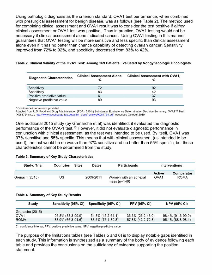
Using pathologic diagnosis as the criterion standard, OVA1 test performance, when combined with presurgical assessment for benign disease, was as follows (see Table 2). The method used for combining clinical assessment and OVA1 result was to consider the test positive if either clinical assessment or OVA1 test was positive. Thus in practice, OVA1 testing would not be necessary if clinical assessment alone indicated cancer. Using OVA1 testing in this manner guarantees that OVA1 testing will be more sensitive and less specific than clinical assessment alone even if it has no better than chance capability of detecting ovarian cancer. Sensitivity improved from 72% to 92%, and specificity decreased from 83% to 42%. Table 2. Clinical Validity of the OVA1 Testa Among 269 Patients Evaluated by Nongynecologic Oncologists
Diagnostic Characteristics Clinical Assessment Alone,
% Clinical Assessment with OVA1,
%
Sensitivity 72 92 Specificity 83 42 Positive predictive value 61 37 Negative predictive value 89 93
a Confidence intervals not provided Adapted from U.S. Food and Drug Administration (FDA). 510(k) Substantial Equivalence Determination Decision Summary: OVA1™ Test (K081754) n.d.; http://www.accessdata.fda.gov/cdrh_docs/reviews/K081754.pdf. Accessed October 2019. One additional 2015 study (by Grenarche et al) was identified; it evaluated the diagnostic performance of the OVA-1 test.13 However, it did not evaluate diagnostic performance in conjunction with clinical assessment, as the test was intended to be used. By itself, OVA1 was 97% sensitive and 55% specific. This means that with clinical assessment (as intended to be used), the test would be no worse than 97% sensitive and no better than 55% specific, but these characteristics cannot be determined from the study. Table 3. Summary of Key Study Characteristics
Study; Trial Countries Sites Dates Participants Interventions
Active Comparator Grenach (2015) US 2009-2011 Women with an adnexal
mass (n=146) OVA1 ROMA
Table 4. Summary of Key Study Results
Study Sensitivity (95% CI) Specificity (95% CI) PPV (95% CI) NPV (95% CI)
Grenache (2015) OVA1 96.8% (83.3-99.9) 54.8% (45.2-64.1) 36.6% (26.2-48.0) 98.4% (91.6-99.9) ROMA 83.9% (66.3-94.6) 83.5% (75.4-89.8) 57.8% (42.2-72.3) 95.1% (88.8-98.4)
CI: confidence interval; PPV: positive predictive value; NPV: negative predictive value. The purpose of the limitations tables (see Tables 5 and 6) is to display notable gaps identified in each study. This information is synthesized as a summary of the body of evidence following each table and provides the conclusions on the sufficiency of evidence supporting the position statement.

9
Table 5. Relevance Limitations
Study Populationa Interventionb Comparatorc Outcomesd Follow-Upe
FDA(k) OVA1 Test K081754
1. Some patients were not evaluated by a gynecologic oncologist; 3. Unclear how patients were recruited; 4. Enrollment was limited to patients with planned surgical intervention
Grenache et al (2015) 1. Patients were not evaluated by a gynecologic oncologist; 4. Enrollment included only patients with planned surgical intervention, due to the small number of women with malignant adnexal masses, the strength of conclusions was limited
The evidence limitations stated in this table are those notable in the current review; this is not a comprehensive limitations assessment. a Population key: 1. Intended use population unclear; 2. Clinical context is unclear; 3. Study population is unclear; 4. Study population not representative of intended use. b Intervention key: 1. Not clearly defined; 2. Version used unclear; 3. Delivery not similar intensity as comparator; 4. Not the intervention of interest. c Comparator key: 1. Not clearly defined; 2. Not standard or optimal; 3. Delivery not similar intensity as intervention; 4. Not delivered effectively. d Outcomes key: 1. Key health outcomes not addressed; 2. Physiologic measures, not validated surrogates; 3. No CONSORT reporting of harms; 4. Not establish and validated measurements; 5. Clinical significant difference not prespecified; 6. Clinical significant difference not supported. e Follow-Up key: 1. Not sufficient duration for benefit; 2. Not sufficient duration for harms. Table 6. Study Design and Conduct Limitations
Study Allocationa Blindingb Selective
Reportingc Follow-Upd Powere Statisticalf
FDA(k) OVA1 Test K081754
1. Subjects were not allocated randomly
1. 10% of subjects were eliminated due to missing information or lack of sample
Grenache et al (2015) 1. Subjects were not allocated randomly
1,2. Treatment assignment and outcome assessment were not blinded
The evidence limitations stated in this table are those notable in the current review; this is not a comprehensive limitations assessment. a Allocation key: 1. Participants not randomly allocated; 2. Allocation not concealed; 3. Allocation concealment unclear; 4. Inadequate control for selection bias. b Blinding key: 1. Not blinded to treatment assignment; 2. Not blinded outcome assessment; 3. Outcome assessed by treating physician. c Selective Reporting key: 1. Not registered; 2. Evidence of selective reporting; 3. Evidence of selective publication. d Follow-Up key: 1. High loss to follow-up or missing data; 2. Inadequate handling of missing data; 3. High number of crossovers; 4. Inadequate handling of crossovers; 5. Inappropriate exclusions; 6. Not intent to treat analysis (per protocol for noninferiority trials). e Power key: 1. Power calculations not reported; 2. Power not calculated for primary outcome; 3. Power not based on clinically important difference. f Statistical key: 1. Intervention is not appropriate for outcome type: (a) continuous; (b) binary; (c) time to event; 2. Intervention is not appropriate for multiple observations per patient; 3. Confidence intervals and/or p values not reported; 4.Comparative treatment effects not calculated. Overa Test

10
Descriptions of the developmental process for the Overa test have been published in FDA documents.14 The FDA documents do not provide details on how biomarkers were selected. The test, as cleared by the FDA, is performed on a blood sample, which is to be sent to a reference laboratory for testing using the 5 immunoassays previously described. Results of the 5 determinations are entered into a proprietary algorithm, called OvaCalc software (v4.0.0), which combines the 5 discrete values into a single unitless numeric score from 0.0 to 10.0. Clinical validity was evaluated in a nonconcurrent prospective study of 493 preoperatively collected serum specimens from premenopausal and postmenopausal women presenting with an adnexal mass requiring surgical intervention.14 Overa test scores were determined based on the analysis of archived serum specimens from a previous study,15 and the patient was stratified into a low- or high-risk group for finding malignancy on surgery. The analysis examined whether patient referral to a gynecologic oncologist was supported when dual assessment was determined to be positive (either Overa or clinical assessment was positive, or both were positive). A dual assessment was considered negative when both Overa and clinical assessment were negative. Using pathologic diagnosis as the criterion standard, Overa test performance, when combined with clinical assessment by nongynecologic oncologists, was as follows (see Table 7). The method used for combining clinical assessment and Overa test result was to consider the test positive if either clinical assessment or Overa test was positive. Using Overa testing in this manner guarantees that Overa testing will be more sensitive and less specific than clinical assessment alone, even if it has no better than chance capability of detecting ovarian cancer. Sensitivity improved from 74% to 94%, and specificity decreased from 93% to 65%. Table 7. Clinical Validity of the Overa Test Among 493 Patients Evaluated by Nongynecologic Oncologists
Diagnostic Characteristics Clinical Assessment Alone, % Clinical Assessment with OVA1, %
Sensitivity (95% CI) 74 (64 to 82) 94 (87 to 97) Specificity (95% CI) 93 (90 to 95) 65 (60 to 70) Positive predictive value (95% CI) 70 (62 to 77) 38 (35 to 41) Negative predictive value (95% CI) 94 (92 to 96) 98 (95 to 99) Prevalence 19 (92/493)
Adapted from U.S. Food and Drug Administration (FDA). 510(k) Substantial Equivalence Determination Decision Summary: OVA1™ Next Generation Test (K150588). n.d.; CI: confidence interval The purpose of the limitations tables (see Tables 8 and 9) is to display notable limitations identified in each study. This information is synthesized as a summary of the body of evidence following each table and provides the conclusions on the sufficiency of evidence supporting the position statement. Table 8. Relevance Limitations
Study Populationa Interventionb Comparatorc Outcomesd Follow-Upe
FDA 510(k) OVA1 K1505881
4. 70.3% of subjects were white
The evidence limitations stated in this table are those notable in the current review; this is not a comprehensive limitations assessment. a Population key: 1. Intended use population unclear; 2. Clinical context is unclear; 3. Study population is unclear; 4. Study population not representative of intended use. b Intervention key: 1. Not clearly defined; 2. Version used unclear; 3. Delivery not similar intensity as comparator; 4. Not the intervention of interest. c Comparator key: 1. Not clearly defined; 2. Not standard or optimal; 3. Delivery not similar intensity as intervention; 4. Not delivered effectively.

11
d Outcomes key: 1. Key health outcomes not addressed; 2. Physiologic measures, not validated surrogates; 3. No CONSORT reporting of harms; 4. Not establish and validated measurements; 5. Clinical significant difference not prespecified; 6. Clinical significant difference not supported. e Follow-Up key: 1. Not sufficient duration for benefit; 2. Not sufficient duration for harms. Table 9. Study Design and Conduct Limitations
Study Selectiona Blindingb Selective
Reportingc Delivery of
Testd Data Completenesse Statisticalf
FDA 510(k) OVA1 K1505881
1. Not described 1.Not described
1.Registration not described
1.Not described
1.Inadequate description of indeterminate and missing samples
None
The evidence limitations stated in this table are those notable in the current review; this is not a comprehensive limitations assessment. a Allocation key: 1. Participants not randomly allocated; 2. Allocation not concealed; 3. Allocation concealment unclear; 4. Inadequate control for selection bias. b Blinding key: 1. Not blinded to treatment assignment; 2. Not blinded outcome assessment; 3. Outcome assessed by treating physician. c Selective Reporting key: 1. Not registered; 2. Evidence of selective reporting; 3. Evidence of selective publication. d Test Delivery key: 1. Timing of delivery of index or reference test not described; 2. Timing of index and comparator tests not same; 3. Procedure for interpreting tests not described; 4. Expertise of evaluators not described. e Data Completeness: 1. Inadequate description of indeterminate and missing samples; 2. High number of samples excluded; 3. High loss to follow-up or missing data f Statistical key: 1. Intervention is not appropriate for outcome type: (a) continuous; (b) binary; (c) time to event; 2. Intervention is not appropriate for multiple observations per patient; 3. Confidence intervals and/or p values not reported; 4. Comparative treatment effects not calculated. ROMA Test Moore et all (2008) described the development of the ROMA test.16 The authors studied 9 biomarkers and chose human epididymis protein 4 (HE4) and CA 125 because these markers in tandem produced the best performance. The algorithm developed was subsequently modified to include menopausal status and was independently validated.17 Again, separate cutoffs were used for premenopausal and postmenopausal women. ROMA Compared with HE4 and CA 125 In 2014, Wang et al published a meta-analysis of studies evaluating the diagnostic accuracy of the ROMA test algorithm and comparing it to the performance of single markers HE4 and CA125.18 To be included in the meta-analysis, studies had to investigate both HE4 and CA125 or calculate ROMA, include women with ovarian cancer and benign gynecologic disease, use pathology diagnosis as the reference standard, and collect blood samples before treatment was initiated. A total of 32 studies met these inclusion criteria; 6 of these were conducted in the United States. Findings of the overall pooled analysis of diagnostic accuracy are presented in Table 10. Table 10. Characteristics of Systematic Reviews That Compared ROMA with HE4 and CA 125
Study Tests Evaluated (No. Studies) Study Populations Included Study Designs Included
Wang et al (2014)
CA 125 (28), HE4 (28), and ROMA (14)
Women with ovarian cancer and benign gynecologic disease
Blinded and unblinded
Dayyani et al (2016)
CA 125 (6), HE4 (6), and ROMA (6)
Women with ovarian cancer All
CA 125: cancer antigen 125; HE4: human epididymis secretory protein 4 Table 11. Meta-Analytic Findings for Diagnostic Performance of the ROMA Test vs. HE4 and CA 125
Test No. Studies Sensitivity (95% CI), % Specificity (95% CI), %
ROMA test 14 85.3 (81.2 to 88.6) 82.4 (77.4 to 86.5) Human epididymis secretory protein 4 28 76.3 (72.0 to 80.1) 93.6 (90.0 to 95.9) Cancer antigen 125 28 79.2 (74.0 to 83.6) 82.1 (76.6 to 86.5)

12
Adapted from Wang et al (2014).18 CI: confidence interval. CA 125: cancer antigen 125; HE4: human epididymis secretory protein 4. Findings were similar when diagnostic performance in premenopausal women and postmenopausal women were evaluated separately. ROMA had similar or higher sensitivity than HE4 and CA125, and HE4 had the highest specificity. In 2016, Dayyani et al conducted a meta-analysis comparing ROMA with HE4 and CA 125 in patients with suspected ovarian cancer.19 Six studies met the inclusion criteria, four of which were included in the 2014 Wang meta-analysis. Two studies were published in 2014 or later. Based on area under the curve analysis, ROMA had higher values than either HE4 (0.921; 95% CI, 0.855 to 0.960) or CA 125 alone (0.899; 95% CI, 0.835 to 0.943) and HE4 plus CA 125 (0.883; 95% CI, 0.771 to 0.950). Findings of the pooled analysis of diagnostic accuracy are shown in Table 12. Table 12. Meta-Analytic Findings for Diagnostic Performance of the ROMA Test vs. HE4 and CA 125
Test No. Studies Sensitivity (95% CI), % Specificity (95% CI), %
ROMA test 6 87.3 (75.2 to 94.0) 85.5 (71.9 to 93.2) Human epididymis secretory protein 4 6 68.2 (69.3 to 90.1) 85.1 (71.6 to 92.8) Cancer antigen 125 6 79.6 (66.3 to 88.5) 82.5 (82.5 to 91.9)
Adapted from Dayyani et al (2016).19 CI: confidence interval. CA 125: cancer antigen 125; HE4: human epididymis secretory protein 4. The point estimates for sensitivity and specificity were lower in pre- and postmenopausal women, with wider confidence intervals. Since the Wang et al (2014) and Dayyani et al (2016) meta-analyses, multiple studies have compared the use of the ROMA test to HE4 and CA 125 in various subgroups based on menopausal status, the cutoff value used, and different racial/ethnic background.20,21,22,23,24,25,26 These studies demonstrate that ROMA's sensitivity (range, 54.5% to 93%) and specificity (range, 75% to 96%) can vary importantly depending on variation in these factors. For example, in a few recent studies of racial/ethnic subpopulations, ROMA's sensitivity dramatically declined and was lowest when used in a sample of 274 African American women (54.5%; 95% CI 33.7-75.3)25 and when distinguishing between malignant/borderline vs. benign or between malignant and borderline/benign in a sample of 177 premenopausal Korean women (46.4% and 52.6%, respectively).24 On the other hand, specificity was highest (95.9%) in a subgroup of 104 postmenopausal women when using a "new optimal cutoff value" of 33.4% instead of 29.9%.22
ROMA compared with Risk Malignancy Index-I Chacon et al(2019) conducted a meta-analysis comparing ROMA with RMI for detecting ovarian cancer (Table 13).27 Among the 2662 women included in the meta-analysis, 50 percent were premenopausal and 50 percent were postmenopausal. Mean ovarian cancer prevalence was 29% in premenopausal women and 51% in postmenopausal women. The majority of studies were conducted at a single-center. Although pooled sensitivities for ROMA (Table 14) were similar to those reported in previous systematic reviews that compared ROMA to HE4 and CA 125, specificities for ROMA were somewhat lower in this meta-analysis (range of 82-85% in Wang et al 2014 and Dayyani et al 2016 meta-analyses compared with 75-78%). However, findings from this meta-analysis by Chacon et al (2019) should be interpreted with caution due to

13
important limitations including a high-risk of selection bias in most studies and significant unexplained statistical heterogeneity in the meta-analyses. Table 13. Characteristics of Chacon et al (2019) Systematic Review of ROMA compared with RMI
Study Dates Studies Participants N (Range) Design Risk of Bias
Chacon
et al (2019)
2011-2018
8 Patients in whom both ROMA and RMI, were calculated for predicting malignancy in adnexal masses
2,662 (50-1061)
Prospective (7) and
retrospective (1) cohort studies
Based on QUADAS-2 assessment, risk of bias was “high in most studies”, due to “selection bias in that they had selected only women who underwent surgery”
RMI: risk malignancy index. Table 14. Diagnostic Performance of ROMA Compared with RMI
Test Sensitivity (95% CI), % Specificity (95% CI), %
Premenopausal Postmenopausal Premenopausal Postmenopausal
ROMA 80% (70-88%) 87% (78–93%) 78% (69-85%) 75% (66–83%) RMI 73% (62–81%) 77% (65–86%) 89% (83–93%) 85% (73–92%)
CI: confidence interval; RMI: risk malignancy index. ROMA in Conjunction with Clinical Assessment The FDA labeling for ROMA, unlike that for OVA1, does not indicate how ROMA is to be used in conjunction with clinical assessment. All previously cited literature assessed ROMA as a stand-alone test for ovarian cancer and did not provide a comparison with clinical assessment alone. The study by Moore et al (2014) evaluated ROMA in conjunction with clinical assessment, using either a positive clinical assessment or a positive ROMA as a positive test (similar to the recommended usage for OVA1).28 Using this method of combining tests guarantees a higher sensitivity and lower specificity for the combined test than for either test alone. Used in this way, ROMA would only need to be given to patients with a negative clinical assessment. In this study, 461 women were enrolled, of whom 86 (19%) had a malignancy. Combined assessment improved sensitivity from 77.9% to 89.7%, but specificity worsened from 84.3% to 67.2% (see Table 15). Table 15. Summary of Key Study Characteristics
Study Countries Sites Dates Participants Comparison
ROMA Group Comparator Moore (2014) U.S. 13 2009-
2010 Women with an ovarian cyst or pelvic mass (n=461)
ICRA+ROMA ICRA
ICRA: Initial Cancer Risk Assessment. Table 16. Diagnostic Performance of the ROMA Test for All Malignancy
Diagnostic Characteristics Clinical Assessment Alone
(95% CI), % Clinical Assessment with ROMA
(95% CI), %
Sensitivity 77.9 (66.2 to 87.1) 89.7 (79.9 to 95.8) Specificity 84.3 (80.2 to 87.8) 67.2 (62.2 to 71.9) Positive predictive value 47.3 (37.8 to 57.0) 33.2 (26.4 to 40.5) Negative predictive value 95.5 (92.6 to 97.4) 97.3 (94.5 to 98.9)
14
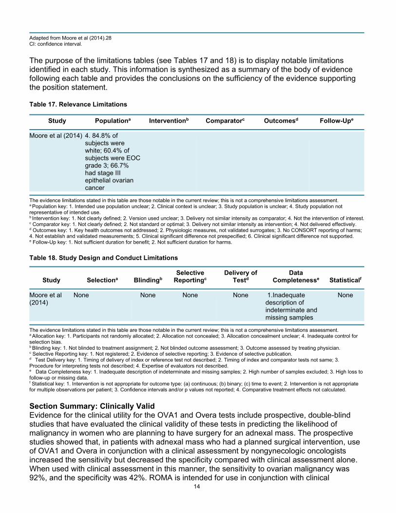
Adapted from Moore et al (2014).28 CI: confidence interval. The purpose of the limitations tables (see Tables 17 and 18) is to display notable limitations identified in each study. This information is synthesized as a summary of the body of evidence following each table and provides the conclusions on the sufficiency of the evidence supporting the position statement. Table 17. Relevance Limitations
Study Populationa Interventionb Comparatorc Outcomesd Follow-Upe
Moore et al (2014) 4. 84.8% of
subjects were white; 60.4% of subjects were EOC grade 3; 66.7% had stage III epithelial ovarian cancer
The evidence limitations stated in this table are those notable in the current review; this is not a comprehensive limitations assessment. a Population key: 1. Intended use population unclear; 2. Clinical context is unclear; 3. Study population is unclear; 4. Study population not representative of intended use. b Intervention key: 1. Not clearly defined; 2. Version used unclear; 3. Delivery not similar intensity as comparator; 4. Not the intervention of interest. c Comparator key: 1. Not clearly defined; 2. Not standard or optimal; 3. Delivery not similar intensity as intervention; 4. Not delivered effectively. d Outcomes key: 1. Key health outcomes not addressed; 2. Physiologic measures, not validated surrogates; 3. No CONSORT reporting of harms; 4. Not establish and validated measurements; 5. Clinical significant difference not prespecified; 6. Clinical significant difference not supported. e Follow-Up key: 1. Not sufficient duration for benefit; 2. Not sufficient duration for harms. Table 18. Study Design and Conduct Limitations
Study Selectiona Blindingb Selective
Reportingc Delivery of
Testd Data
Completenesse Statisticalf
Moore et al (2014)
None None None None 1.Inadequate description of indeterminate and missing samples
None
The evidence limitations stated in this table are those notable in the current review; this is not a comprehensive limitations assessment. a Allocation key: 1. Participants not randomly allocated; 2. Allocation not concealed; 3. Allocation concealment unclear; 4. Inadequate control for selection bias. b Blinding key: 1. Not blinded to treatment assignment; 2. Not blinded outcome assessment; 3. Outcome assessed by treating physician. c Selective Reporting key: 1. Not registered; 2. Evidence of selective reporting; 3. Evidence of selective publication. d Test Delivery key: 1. Timing of delivery of index or reference test not described; 2. Timing of index and comparator tests not same; 3. Procedure for interpreting tests not described; 4. Expertise of evaluators not described. e Data Completeness key: 1. Inadequate description of indeterminate and missing samples; 2. High number of samples excluded; 3. High loss to follow-up or missing data. f Statistical key: 1. Intervention is not appropriate for outcome type: (a) continuous; (b) binary; (c) time to event; 2. Intervention is not appropriate for multiple observations per patient; 3. Confidence intervals and/or p values not reported; 4. Comparative treatment effects not calculated. Section Summary: Clinically Valid Evidence for the clinical utility for the OVA1 and Overa tests include prospective, double-blind studies that have evaluated the clinical validity of these tests in predicting the likelihood of malignancy in women who are planning to have surgery for an adnexal mass. The prospective studies showed that, in patients with adnexal mass who had a planned surgical intervention, use of OVA1 and Overa in conjunction with a clinical assessment by nongynecologic oncologists increased the sensitivity but decreased the specificity compared with clinical assessment alone. When used with clinical assessment in this manner, the sensitivity to ovarian malignancy was 92%, and the specificity was 42%. ROMA is intended for use in conjunction with clinical

15
assessment. One study, which used clinical assessment and ROMA results, showed a sensitivity of 90% and a specificity of 67%. Two meta-analysis reported less than 90% sensitivity and specificity with ROMA testing. Clinical Useful A test is clinically useful if the use of the results informs management decisions that improve the net health outcome of care. The net health outcome can be improved if patients receive correct therapy, or more effective therapy, or avoid unnecessary therapy, or avoid unnecessary testing. The ideal study design to evaluate the clinical utility of multimarker serum-based test would be a randomized controlled trial comparing health outcomes (e.g., mortality) in patients managed using the tests with those managed according to best current clinical practices. According to the chain of logic, greater numbers of persons referred for initial surgical treatment with ovarian cancer should result in improved overall health outcomes. No randomized or nonrandomized studies with these comparisons were identified. OVA1, Overa, and ROMA, when used in conjunction with clinical assessment may improve the sensitivity for detection of malignancy, the specificity declines. In studies using either positive ROMA or clinical assessment as a positive test, sensitivity improved-but it was still less than 90%. It is uncertain whether there is meaningful clinical benefit from using a test that avoids a high number of referrals and does not contain sensitive data (even though incrementally better). Section Summary: Clinically Useful As no trials were identified that have compared health outcomes for patients managed with and without the use of FDA-cleared multimarker serum-based tests, there is no direct evidence of clinical usefulness. It is uncertain whether discrimination is sufficient to alter decision-making based on clinical assessment alone, thus offering a meaningful benefit to patients. Therefore, the chain of evidence supporting improved outcomes is incomplete. SUMMARY OF EVIDENCE The evidence for use of multimarker serum-based testing (OVA1 test, Overa or ROMA test) in conjunction with clinical assessment in patients who have adnexal masses undergoing surgery includes studies assessing the technical performance and diagnostic accuracy of the tests. Relevant outcomes are overall survival and test accuracy. Data has been presented that confirms the ROMA algorithm and OVA1 testing in the distinction of ovarian cancer from benign disease. The utility of this testing can be found in patients already going to surgery. The results of this testing along with the clinical picture allow for the appropriate triage of patients to a surgeon who is a generalist or to a gynecologist oncologist. SUPPLEMENTAL INFORMATION Clinical Input Received through Physician Specialty Societies and Academic Medical Centers Results of clinical input received by BCBSA in 2012 from subject matter experts revealed mixed support for the use of this test as a tool for triaging patients with an adnexal mass. Reviewers agreed that the evidence was insufficient to determine the impact of these tests on referral patterns. For indications other than triaging patients with an adnexal mass, there was a lack of support for use of these tests. PRACTICE GUIDELINES AND POSITION STATEMENTS

16
American College of Obstetricians and Gynecologists In 2017, with reaffirmation in 2019, the American College of Obstetricians and Gynecologists (ACOG) opinion on the role of the obstetrician-gynecologist in the early detection of epithelial ovarian cancer addressed. The opinion states that multimarker panels lack strong evidence for use in asymptomatic women without adnexal masses and do not improve early detection and survival rates in average-risk women. The Society for Gynecologic Oncology endorsed ACOG opinion in 2016, an ACOG Practice Bulletin addressing the evaluation and management of adnexal masses made a level B recommendation (based on limited or inconsistent scientific evidence) that consultation with or referral to a gynecologic oncologist is recommended for premenopausal or postmenopausal with an elevated score on a formal risk assessment test such as the multivariate index assay, risk of malignancy index, or the Risk of Ovarian Malignancy Algorithm, or 1 of the ultrasound-based scoring systems from the International Ovarian Tumor Analysis group. A level C recommendation (based on consensus and expert opinion) was given to using serum biomarker panels as an alternative to cancer antigen 125 (CA 125) level to decide about the referral to a gynecologic oncologist for an adnexal mass requiring surgery. National Institute for Health and Care Excellence The National Institute for Health and Care Excellence issued guidance in 2011 on the recognition and management of ovarian cancer.32 This guidance is currently being updated and is under review. National Comprehensive Cancer Network In 2020, The NCCN guidelines on ovarian cancer (v.2.2020) includes the following statement:33
“It has been suggested that specific biomarkers (serum HE4 [human epididymis secretory protein 4] and CA 125 [cancer antigen 125]) along with an algorithm (Risk of Ovarian Malignancy Algorithm [ROMA]) may be useful for determining whether a pelvic mass is malignant or benign. The FDA [Food and Drug Administration] has approved the use of HE4 and CA-125 for estimating the risk of ovarian cancer in women with a pelvic mass. Currently, the NCCN Panel does not recommend the use of these biomarkers for determining the status of an undiagnosed pelvic mass.”
Regarding the OVA1 test, the NCCN guidelines states: “The OVA1 test uses 5 markers (including transthyretin, apolipoprotein A1, transferrin, beta-2 microglobulin, and CA 125) to assess who should undergo surgery by an experienced gynecologic oncologist and who can have surgery in the community…. Based on data documenting an increased survival, NCCN Guidelines Panel Members recommend that all patients should undergo surgery by an experienced gynecologic oncologist (category 1).” U.S. Preventive Services Task Force Recommendations In 2018, the U.S. Preventive Services Task Force recommended against screening women for ovarian cancer (D recommendation). The task force has not addressed multimarker serum -based testing related to ovarian cancer. Ongoing and Unpublished Clinical Trials A search of ClinicalTrials.gov did not identify any ongoing or unpublished trials that would likely influence this review. Government Regulations

17
National: There is no national coverage determination on combined ovarian cancer biomarker tests. Requests would be reviewed on an individual consideration basis. Codes 81500, 81503 and 0003U have a fee schedule attached for January 2021. Local: There is no WPS local coverage determination on combined ovarian cancer biomarker tests. Requests would be reviewed on an individual consideration basis. (The above Medicare information is current as of the review date for this policy. However, the coverage issues and policies maintained by the Centers for Medicare & Medicare Services [CMS, formerly HCFA] are updated and/or revised periodically. Therefore, the most current CMS information may not be contained in this document. For the most current information, the reader should contact an official Medicare source.) ____________________________________________________________________________ Related Policies N/A ___________________________________________________________________________ References
1. Surveillance Epidemology and End Results (SEER) Program. SEER Stat Fact Sheets: Ovarian Cancer. n.d.; https://seer.cancer.gov/statfacts/html/ovary.html. Accessed October 2019.
2. du Bois A, Rochon J, Pfisterer J, et al. Variations in institutional infrastructure, physician specialization and experience, and outcome in ovarian cancer: a systematic review. Gynecol Oncol. Feb 2009;112(2):422-436. PMID 18990435
3. Hoskins W, Rice L, Rubin S. Ovarian cancer surgical practice guidelines. Society of Surgical Oncology practice guidelines. Oncology (Williston Park). Jun 1997;11(6):896-900, 903-894. PMID 9189944
4. Vernooij F, Heintz P, Witteveen E, et al. The outcomes of ovarian cancer treatment are better when provided by gynecologic oncologists and in specialized hospitals: a systematic review. Gynecol Oncol. Jun 2007;105(3):801- 812. PMID 17433422
5. Giede KC, Kieser K, Dodge J, et al. Who should operate on patients with ovarian cancer? An evidence-based review. Gynecol Oncol. Nov 2005;99(2):447-461. PMID 16126262
6. Van Holsbeke C, Van Belle V, Leone FP, et al. Prospective external validation of the 'ovarian crescent sign' as a single ultrasound parameter to distinguish between benign and malignant adnexal pathology. Ultrasound Obstet Gynecol. Jul 2010;36(1):81-87. PMID 20217895
7. Im SS, Gordon AN, Buttin BM, et al. Validation of referral guidelines for women with pelvic masses. Obstet Gynecol. Jan 2005;105(1):35-41. PMID 15625139
8. Simmons AR, Clarke CH, Badgwell DB, et al. Validation of a biomarker panel and longitudinal biomarker performance for early detection of ovarian cancer. Int J Gynecol Cancer. Jul 2016;26(6):1070-1077. PMID 27206285
9. Yanaranop M, Tiyayon J, Siricharoenthai S, et al. Rajavithi-ovarian cancer predictive score (R-OPS): A new scoring system for predicting ovarian malignancy in women presenting with a pelvic mass. Gynecol Oncol. Jun 2016;141(3):479-484. PMID 26996662
10. Guidance for Industry and FDA Staff - Class II Special Controls Guidance Document: Ovarian Adnexal Mass Assessment Score Test System. https://www.fda.gov/regulatory-

18
information/search-fda-guidance-documents/class-ii-special-controls-guidance-document-ovarian-adnexal-mass-assessment-score-test-system. Accessed October 2019.
11. U.S. Food and Drug Administration (FDA). 510(k) Substantial Equivalence Determination Decision Summary: OVA1TM Test (K081754) n.d.; https://www.accessdata.fda.gov/cdrh_docs/reviews/K081754.pdf. Accessed October 2019.
12. Fung ET. A recipe for proteomics diagnostic test development: the OVA1 test, from biomarker discovery to FDA clearance. Clin Chem. Feb 2010;56(2):327-329. PMID 20110452
13. Grenache DG, Heichman KA, Werner TL, et al. Clinical performance of two multi-marker blood tests for predicting malignancy in women with an adnexal mass. Clin Chim Acta. Jan 1 2015;438:358-363. PMID 25283731
14. U.S. Food and Drug Administration (FDA). 510(k) Substantial Equivalence Determination Decision Summary: OVA1TM Next Generation Test (K150588). n.d.; https://www.accessdata.fda.gov/cdrh_docs/reviews/K150588.pdf. Accessed October 2019.
15. Bristow RE, Smith A, Zhang Z, et al. Ovarian malignancy risk stratification of the adnexal mass using a multivariate index assay. Gynecol Oncol. Feb 2013;128(2):252-259. PMID 23178277
16. Moore RG, Brown AK, Miller MC, et al. The use of multiple novel tumor biomarkers for the detection of ovarian carcinoma in patients with a pelvic mass. Gynecol Oncol. Feb 2008;108(2):402-408. PMID 18061248
17. Moore RG, Miller MC, Disilvestro P, et al. Evaluation of the diagnostic accuracy of the risk of ovarian malignancy algorithm in women with a pelvic mass. Obstet Gynecol. Aug 2011;118(2 Pt 1):280-288. PMID 21775843
18. Wang J, Gao J, Yao H, et al. Diagnostic accuracy of serum HE4, CA125 and ROMA in patients with ovarian cancer: a meta-analysis. Tumour Biol. Jun 2014;35(6):6127-6138. PMID 24627132
19. Dayyani F, Uhlig S, Colson B, et al. Diagnostic performance of risk of ovarian malignancy algorithm against CA125 and HE4 in connection with ovarian cancer: a meta-analysis. Int J Gynecol Cancer. Nov 2016;26(9):1586-1593. PMID 27540691
20. Al Musalhi K, Al Kindi M, Al Aisary F, et al. Evaluation of HE4, CA-125, Risk of Ovarian Malignancy Algorithm (ROMA) and Risk of Malignancy Index (RMI) in the preoperative assessment of patients with adnexal mass. Oman Med J. Sep 2016;31(5):336-344. PMID 27602187
21. Cho HY, Park SH, Park YH, et al. Comparison of HE4, CA125, and risk of ovarian malignancy algorithm in the prediction of ovarian cancer in Korean women. J Korean Med Sci. Dec 2015;30(12):1777-1783. PMID 26713052
22. Terlikowska KM, Dobrzycka B, Witkowska AM, et al. Preoperative HE4, CA125 and ROMA in the differential diagnosis of benign and malignant adnexal masses. J Ovarian Res. Jul 19 2016;9(1):43. PMID 27436085
23. Shen Y, Zhao L, Lu S. Diagnostic performance of HE4 and ROMA among Chinese women. Clin. Chim. Acta, 2019 Oct 19. PMID 31626761
24. Shin KH, Kim HH, Kwon BS et al. Clinical Usefulness of Cancer Antigen (CA) 125, Human Epididymis 4, and CA72-4 Levels and Risk of Ovarian Malignancy Algorithm Values for Diagnosing Ovarian Tumors in Korean Patients With and Without Endometriosis. Ann Lab Med, 2019 Aug 23;40(1). PMID 31432638
25. Dunton C, Bullock RG, Fritsche H. Multivariate Index Assay Is Superior to CA125 and HE4 Testing in Detection of Ovarian Malignancy in African-American Women. Biomark Cancer, 2019 Jun 27;11:1179299X19853785. PMID 31236012
26. Han KH, Park NH, Kim JJ et al. The power of the Risk of Ovarian Malignancy Algorithm considering menopausal status: a comparison with CA 125 and HE4.. J Gynecol Oncol, 2019 Oct 3;30(6). PMID 31576682

19
27. Chacon E, Das J, Caballero C et al. Risk of Ovarian Malignancy Algorithm versus Risk Malignancy Index-I for Preoperative Assessment of Adnexal Masses: A Systematic Review and Meta-Analysis.. Gynecol. Obstet. Invest., 2019 Jul 17;1-8:1-8. PMID 31311023
28. Moore RG, Hawkins DM, Miller MC, et al. Combining clinical assessment and the Risk of Ovarian Malignancy Algorithm for the prediction of ovarian cancer. Gynecol Oncol. Dec 2014;135(3):547-551. PMID 25449569
29. American College of Obstetricians Gynecologists Committee on Gynecologic Practice. Committee Opinion No. 477: the role of the obstetrician-gynecologist in the early detection of epithelial ovarian cancer. Obstet Gynecol. Mar 2011;117(3):742-746. PMID 21343791
30. American College of Obstetricians Gynecologists Committee on Gynecologic Practice. Committee Opinion No. 716: the role of the obstetrician-gynecologist in the early detection of epithelial ovarian cancer in women at average risk:.https://www.acog.org/Clinical-Guidance-and-Publications/Committee-Opinions/Committee-on-Gynecologic-Practice/The-Role-of-the-Obstetrician-Gynecologist-in-the-Early-Detection-of-Epithelial-Ovarian-Cancer-in?IsMobileSet=false Accessed February 2021.
31. Society of Gynecologic Oncologists. Multiplex Serum Testing for Women with Pelvic Mass. 2013; https://www.sgo.org/newsroom/position-statements-2/multiplex-serum-testing-for-women-with-pelvic-mass/. Accessed February 2021.
32. National Center for Clinical Excellence (NICE). Ovarian cancer: recognition and initial management [CG122]. 2011; https://www.nice.org.uk/guidance/cg122. Accessed February 2021.
33. National Comprehensive Cancer Network (NCCN). NCCN Clinical Practice Guidelines in Oncology: Ovarian Cancer Including Fallopian Tub Cancer and Primary Peritoneal Cancer. Version 2.2020. https://www.nccn.org/professionals/physician_gls/pdf/ovarian.pdf Accessed February 2021.
34. Grossman DC, Curry SJ, Owens DK et al. Screening for Ovarian Cancer: US Preventive Services Task Force Recommendation Statement. JAMA, 2018 Feb 17;319(6). PMID 29450531
35. Blue Cross Blue Shield Association. Proteomics-based testing Related to Ovarian Cancer. Medical Policy Reference Manual. Policy #2.04.62. Issue 4:2015. Original policy date 4/8/10. Last review date January 2021.
36. HAYES GTE Report. OVA1 Ovarian Tumor Triage Test. Lansdale, PA: HAYES, Inc. August 21, 2014. Last updated August 2015.
37. HAYES Medical Technology Directory. Architect HE4 Assay for Ovarian Cancer Detection. Lansdale, PA: HAYES, Inc. Published December 2012. Last updated December 2015.
38. BCBSA TEC Assessment Program. Multianalyte Testing for the Evaluation of Adnexal Masses. Volume 27, Number 8, April 2013.
The articles reviewed in this research include those obtained in an Internet based literature search for relevant medical references through February 2021, the date the research was completed.

20
Joint BCBSM/BCN Medical Policy History
Policy Effective Date
BCBSM Signature Date
BCN Signature Date
Comments
9/1/10 7/22/10 6/29/10 Joint policy established
7/1/12 5/15/12 5/15/12 Policy updated; status changed from experimental and investigational to established. Added information regarding the ROMA™ test. Title changed from “Evaluation of Ovarian (Adnexal) Masses (OVA1) by Proteomics-based Testing to Evaluation of Ovarian (Adnexal) Masses By Proteomics-Based Testing (e.g., OVA1™ and ROMA™ Testing). Added additional references and rationale to include ROMA testing. Reformatted to match BCBSA policy.
9/1/13 4/16/13 4/23/13 Routine update. Policy status changed to experimental/ investigational based on new evidence and BCBSA policy position change. References updated.
1/1/15 10/21/14 11/7/14 Policy status changed back to established for patients meeting FDA guidelines.
7/1/16 4/19/16 6/23/16 Routine policy maintenance. Updated references and rationale section. No change in policy status.
7/1/17 4/18/17 4/18/17 Policy title changed to Multimarker Serum Based Testing Related to Ovarian Cancer to mirror BCBSA policy of same title. Code 0003U added to policy. Added references and rationale. No change in policy status.
7/1/18 4/17/18 4/17/18 Rationale updated, added reference # 1, 10, 12, 16 and 27. Added Overa to policy, policy statement and title.
7/1/19 4/16/19 Routine policy maintenance. No change in policy status.
7/1/20 4/14/20 Routine policy maintenance. Added references 23-27. No change in policy status.

21
7/1/21 4/20/21 Routine policy maintenance. No change in policy status.
Next Review Date: 2nd Qtr. 2022

22
BLUE CARE NETWORK BENEFIT COVERAGE POLICY: EVALUATION OF OVARIAN (ADNEXAL) MASSES BY MULTIMARKER SERUM -
BASED TESTING (E.G., OVA1®, OVERA™ AND ROMA™ TESTING)
I. Coverage Determination:
Commercial HMO (includes Self-Funded groups unless otherwise specified)
Covered; criteria apply
BCNA (Medicare Advantage)
See government section
BCN65 (Medicare Complementary)
Covered if primary Medicare covers the service.
II. Administrative Guidelines:
• The member's contract must be active at the time the service is rendered. • The service must be authorized by the member's PCP except for Self-Referral Option
(SRO) members seeking Tier 2 coverage. • Services must be performed by a BCN-contracted provider, if available, except for
Self-Referral Option (SRO) members seeking Tier 2 coverage. • Payment is based on BCN payment rules, individual certificate and certificate riders. • Appropriate copayments will apply. Refer to certificate and applicable riders for
detailed information. • CPT - HCPCS codes are used for descriptive purposes only and are not a guarantee
of coverage.