medical imaging of the upper limb x rays. how to read x -ray
TRANSCRIPT
Medical Imaging of the Upper Limb
X rays
How to read X -Ray
X rays
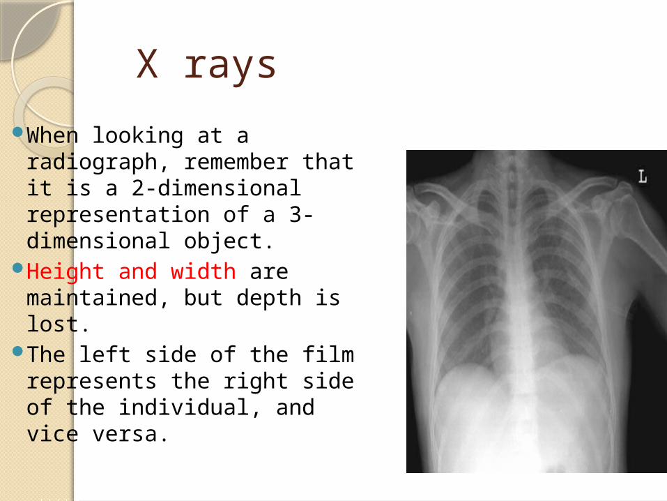
When looking at a radiograph, remember that it is a 2-dimensional representation of a 3-dimensional object.
Height and width are maintained, but depth is lost.
The left side of the film represents the right side of the individual, and vice versa.
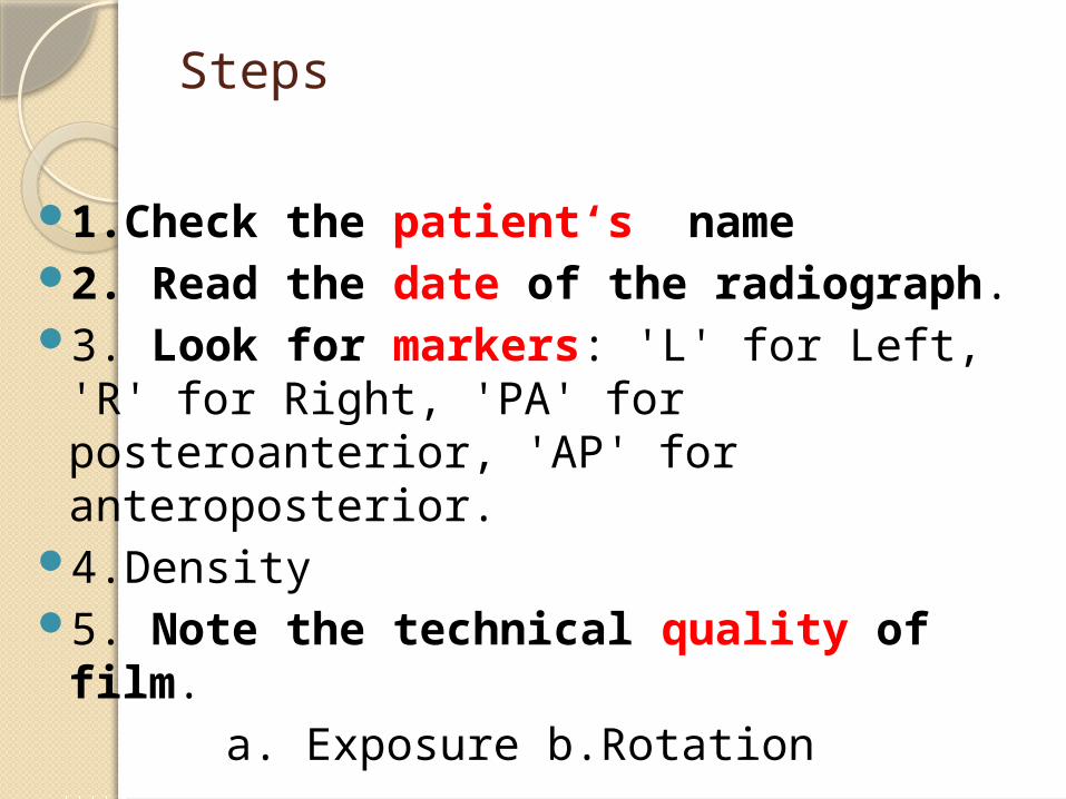
Steps
1.Check the patient‘s name2. Read the date of the radiograph.3. Look for markers: 'L' for Left, 'R'
for Right, 'PA' for posteroanterior, 'AP' for anteroposterior.
4.Density5. Note the technical quality of
film. a. Exposure b.Rotation
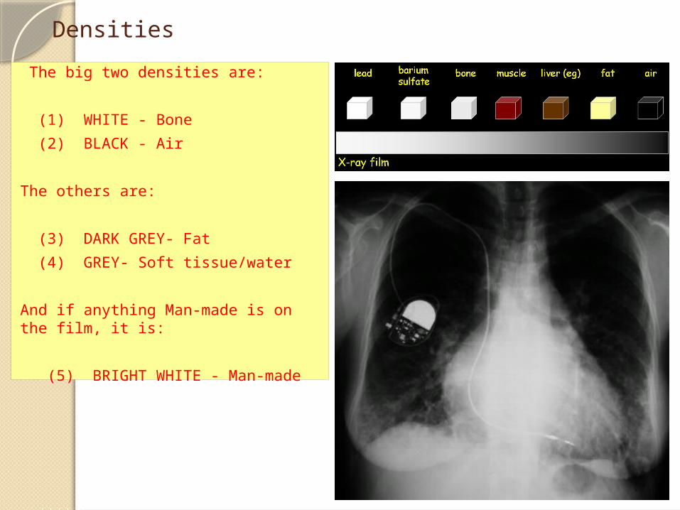
Densities
The big two densities are:
(1) WHITE - Bone
(2) BLACK - Air
The others are:
(3) DARK GREY- Fat
(4) GREY- Soft tissue/water
And if anything Man-made is on the film, it is:
(5) BRIGHT WHITE - Man-made

Techniques - Projection
•P-A (relation of x-ray beam to patient)
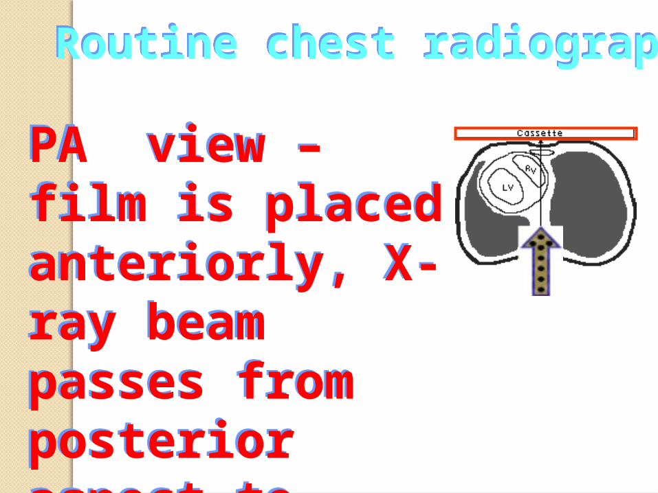
Routine chest radiographRoutine chest radiograph
PA view – film is placed anteriorly, X-ray beam passes from posterior aspect to anterior side.
PA view – film is placed anteriorly, X-ray beam passes from posterior aspect to anterior side.
The standard view of the chest is the posteroanterior radiograph, or "PA chest."
This film is taken with the patient upright, in full inspiration (breathed in all the way), and the x-ray beam radiating horizontally 6 feet away from the film.

AP viewAP view
An AP film, enlarges the shadow of the heart and makes the posterior ribs appear more horizontal.
An AP film, enlarges the shadow of the heart and makes the posterior ribs appear more horizontal.
Usually obtained with a portable x-ray machine from very sick patients, those unable to stand, and infants.
AP radiographs are generally taken at shorter distance from the film compared to PA radiographs.
The farther away the x-ray source is from the film, the sharper and less magnified the image
Since AP radigraphs are taken from shorter distances, they appear more magnified and less sharp compared to standard PA films.
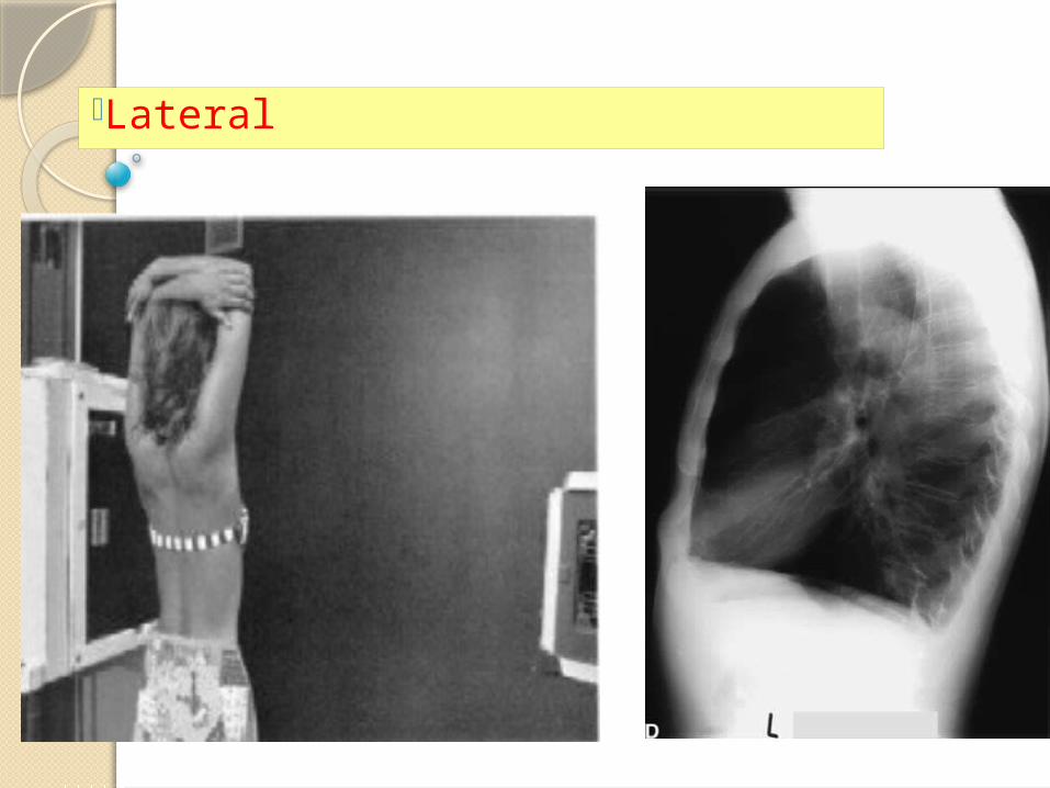
•Lateral
Medical Imaging of the Upper Limb
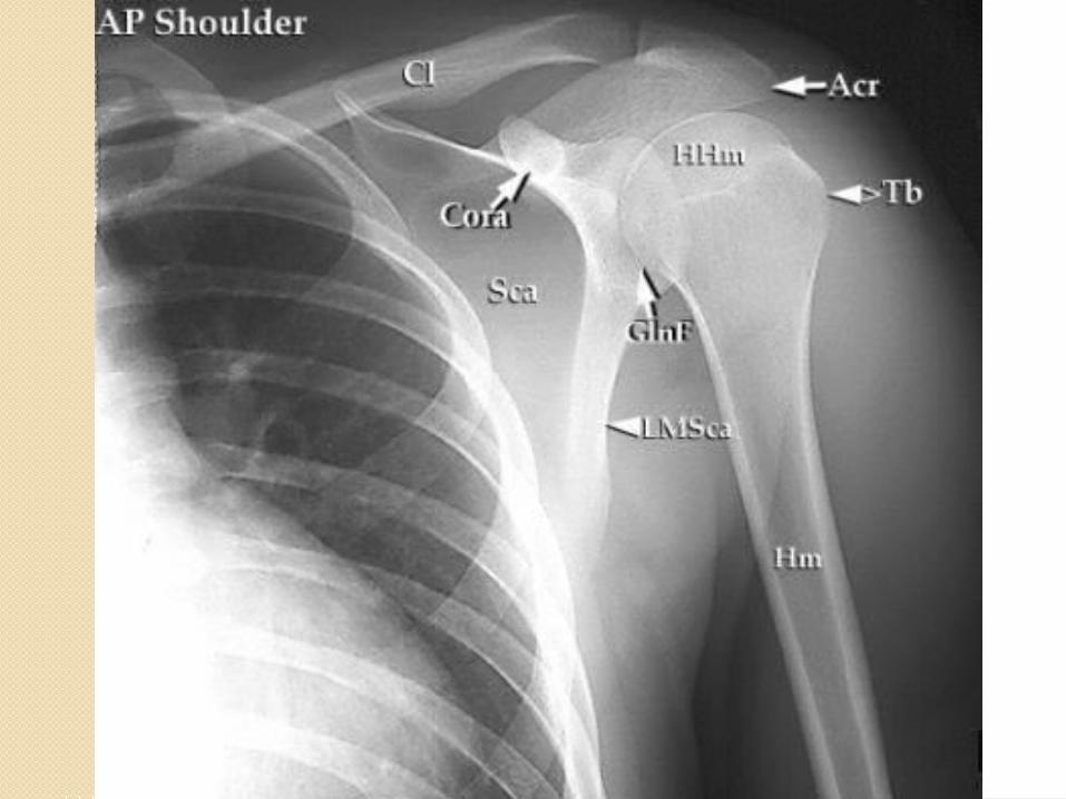
Radiological examinations of the upper limb focus mainly on bony structures, because muscles, tendons, and nerves are not well visualized.
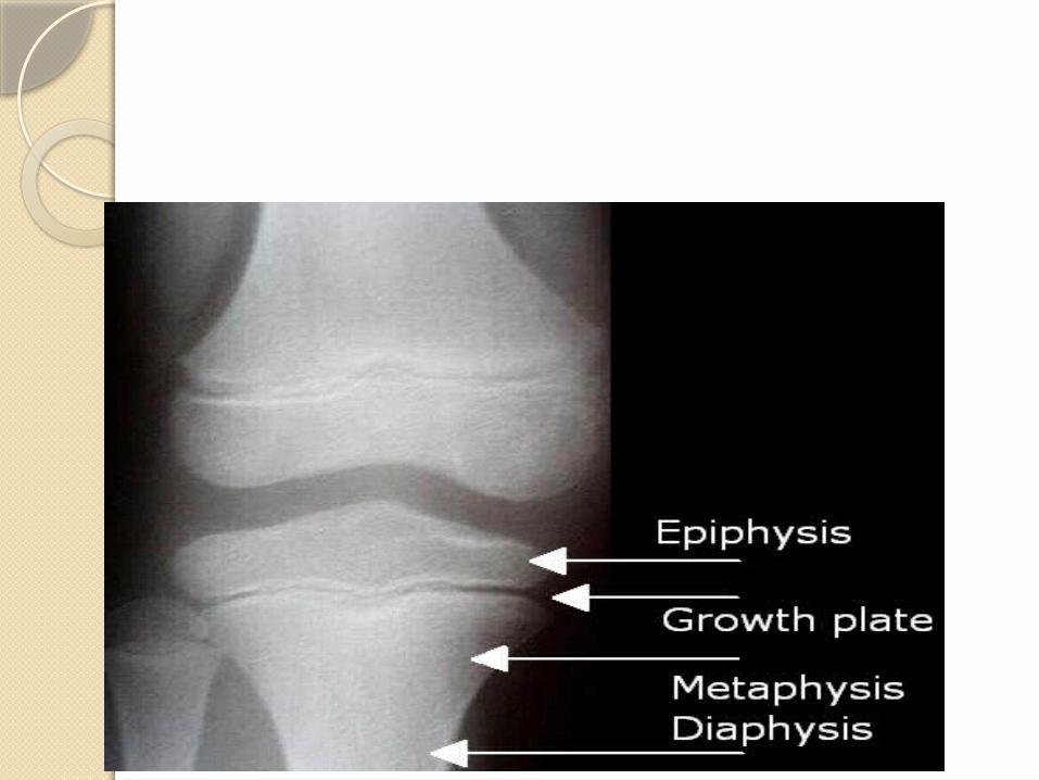
ImportantWhen examining radiographs of
the upper limb, it is essential to know the median times of appearance of postnatal ossification centers and when fusion of epiphyses is radiographically complete in males and females.
Without such knowledge, an epiphysial line could be mistaken for a fracture.
TopicsClavicleShoulder DislocationHumerusElbowForearmDistal RadiusScaphoid
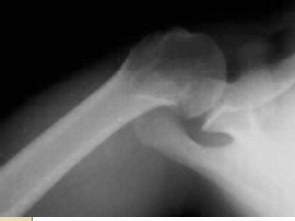
Normal axillary view

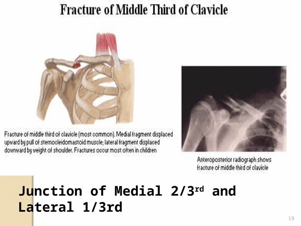
CLAVICLE
19
Junction of Medial 2/3rd and Lateral 1/3rd
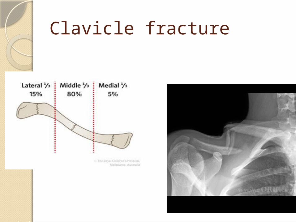
Clavicle fracture
Shoulder dislocations
Most commonly dislocated large joint
Anterior in 97%Mechanism: force on
abducted/externally rotated shoulder
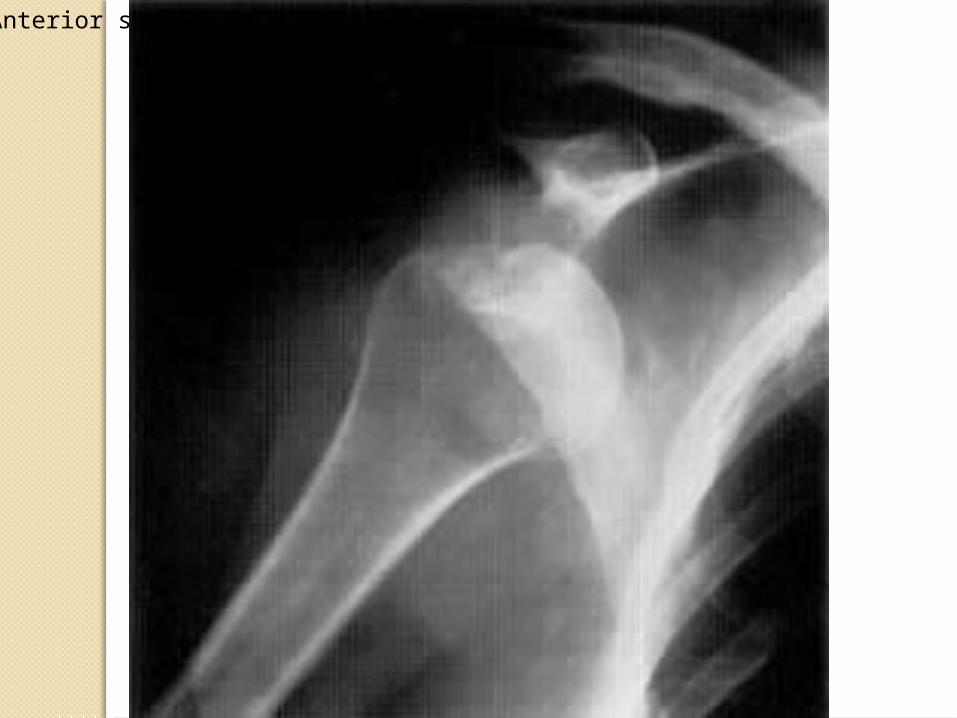
Anterior shoulder dislocation

HUMERUS
24
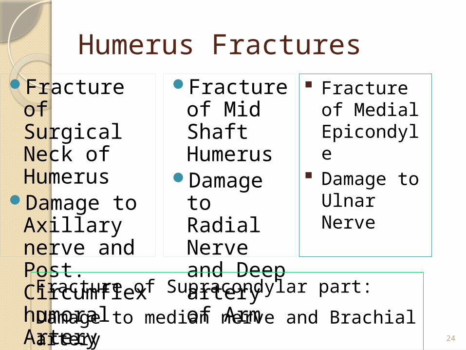
Humerus Fractures Fracture of
Surgical Neck of Humerus
Damage to Axillary nerve and Post. Circumflex humoral Artery
Fracture of Mid Shaft Humerus
Damage to Radial Nerve and Deep artery of Arm
Fracture of Medial Epicondyle
Damage to Ulnar Nerve
Fracture of Supracondylar part:
Damage to median nerve and Brachial artery
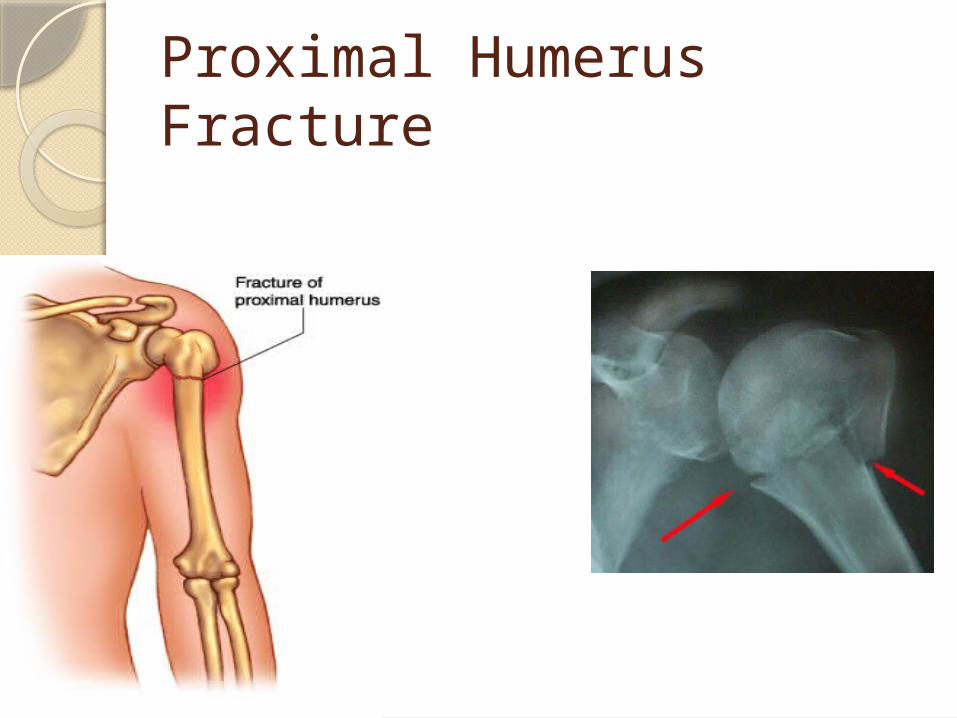
Proximal Humerus Fracture
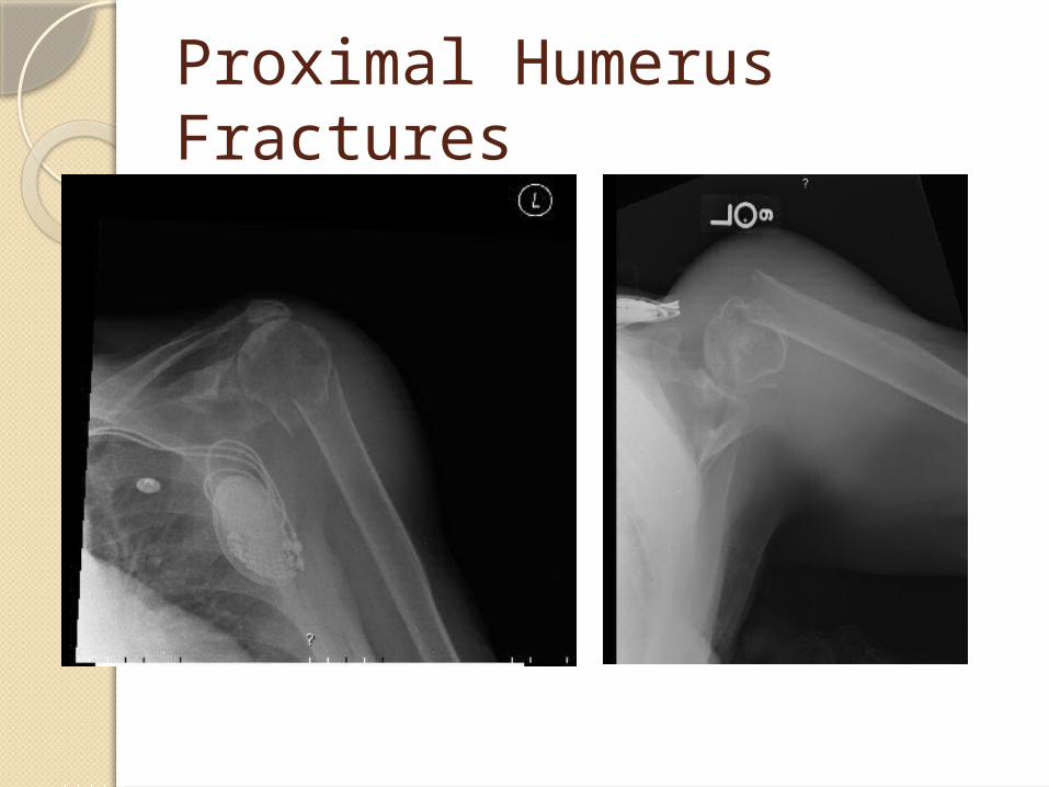
Proximal Humerus Fractures
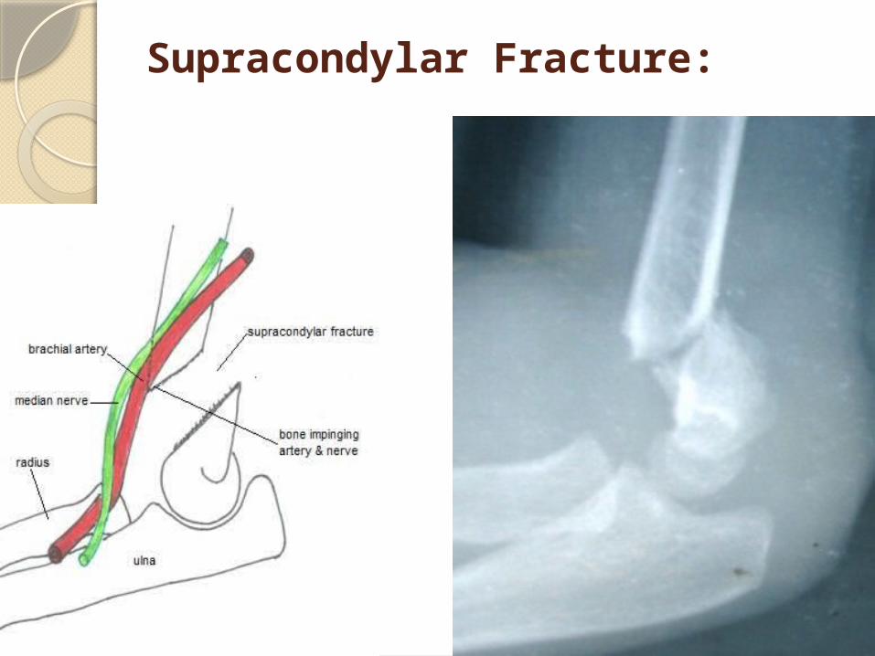
Supracondylar Fracture:
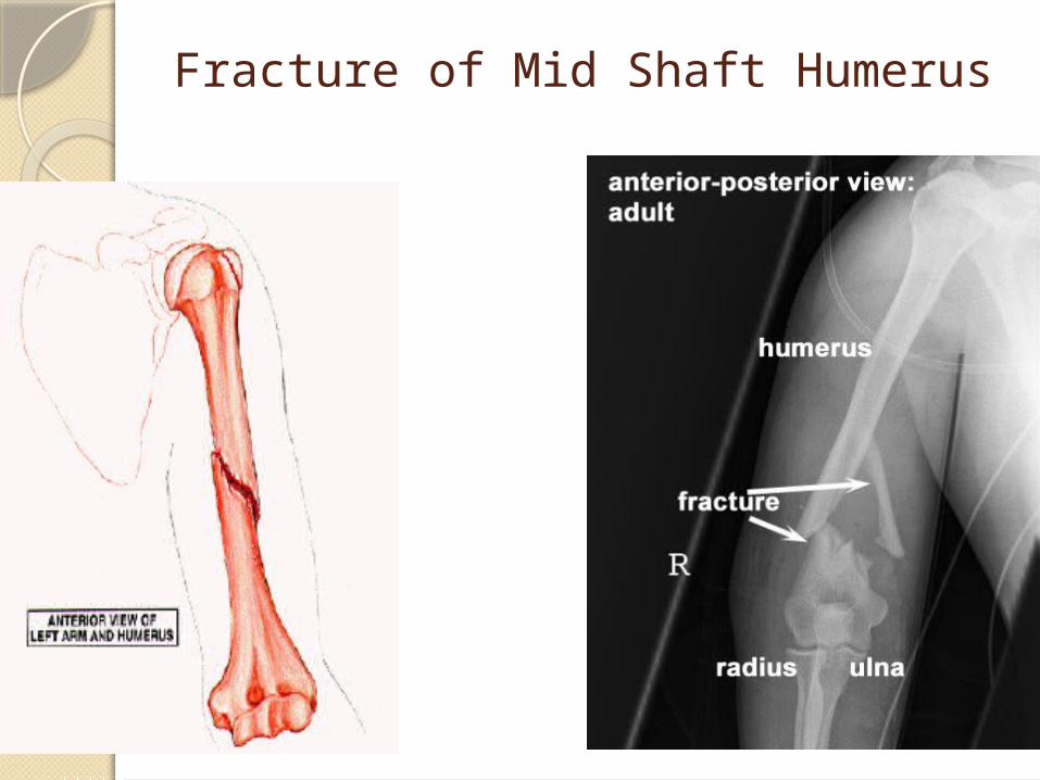
Fracture of Mid Shaft Humerus
Elbow traumaFracturesDislocationsLigament sprainsLook for compartment syndromeRule out neurovascular injury

Radius and ulna
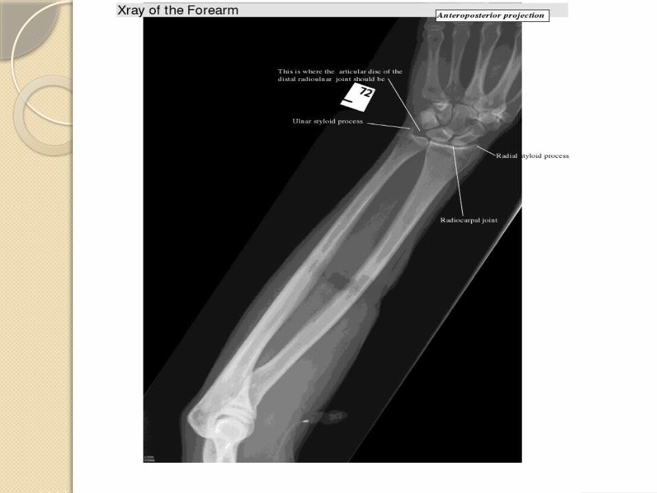
34
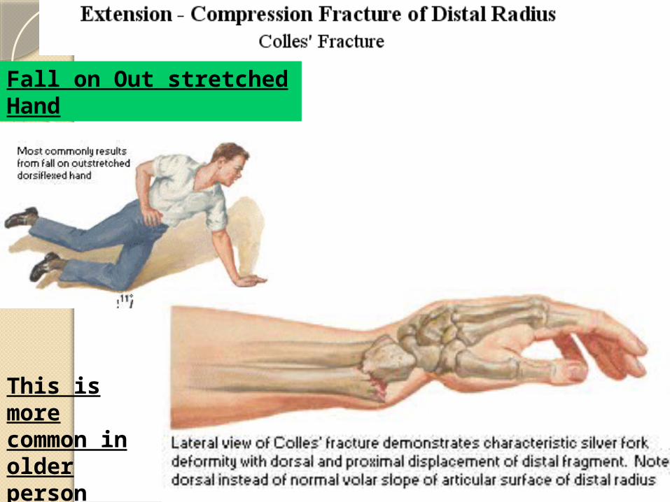
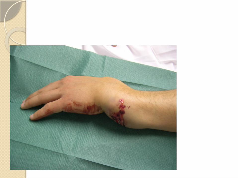
Fall on Out stretched Hand
This is more common in older person
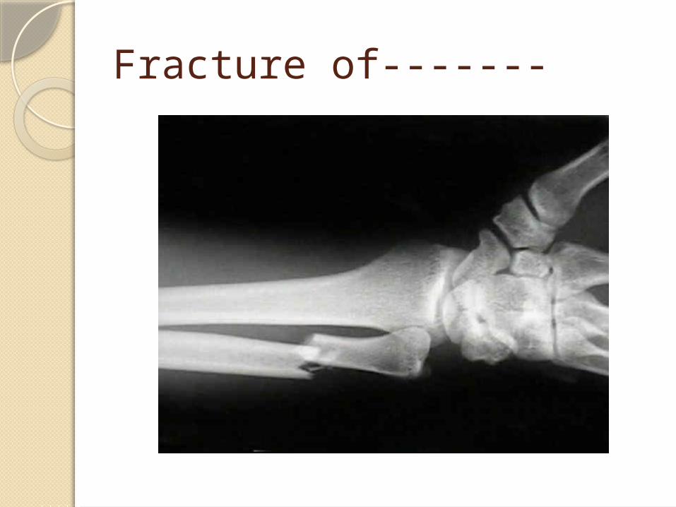
Fracture of-------

Wrist
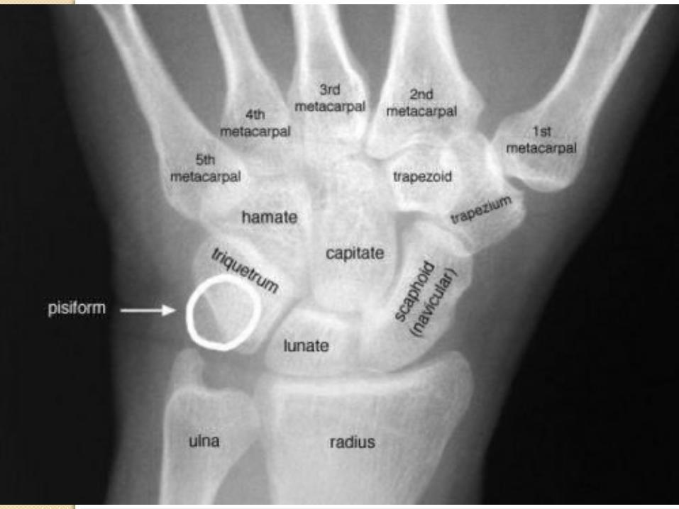
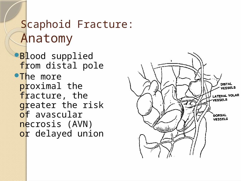
Scaphoid Fracture:Anatomy
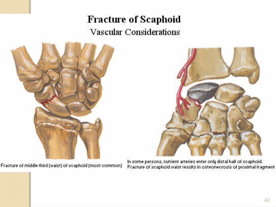
Blood supplied from distal pole
The more proximal the fracture, the greater the risk of avascular necrosis (AVN) or delayed union
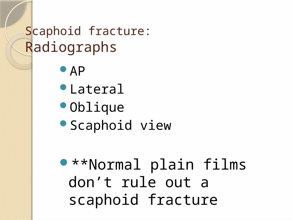
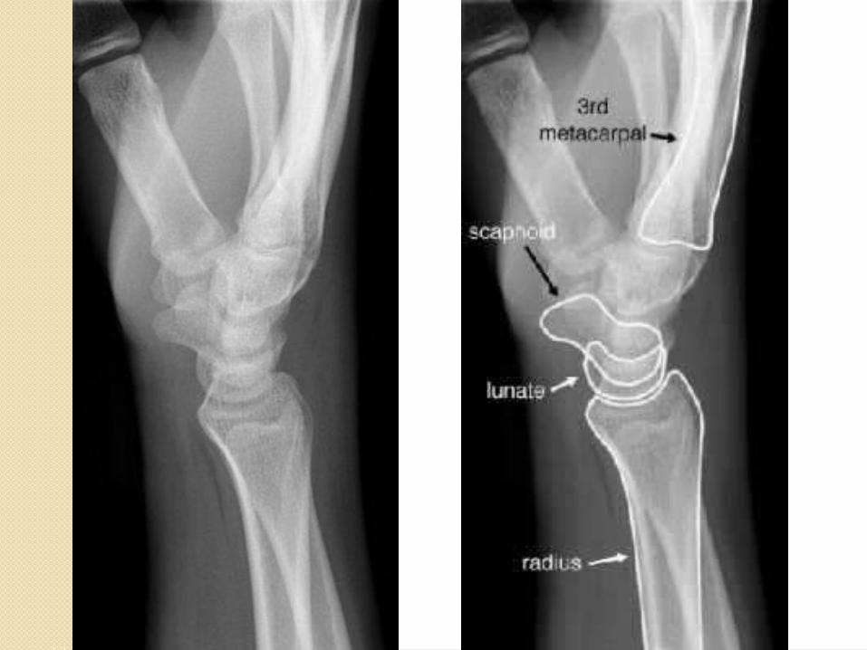
Scaphoid fracture: Radiographs
APLateralObliqueScaphoid view
**Normal plain films don’t rule out a scaphoid fracture
42