mediators of mast cells in bullous pemphigoid and dermatitis
TRANSCRIPT

Research ArticleMediators of Mast Cells in Bullous Pemphigoid andDermatitis Herpetiformis
Agnieszka Zebrowska,1 Malgorzata Wagrowska-Danilewicz,2
Marian Danilewicz,2 Olga Stasikowska-Kanicka,2 Lilianna Kulczycka-Siennicka,1
Anna Wozniacka,1 and Elzbieta Waszczykowska1
1 Department of Dermatology and Venereology, Medical University of Lodz, Plac Hallera 1, 90-497 Lodz, Poland2 Laboratory of Nephropathology of Medical University of Lodz, Pomorska 251, 92-213 Lodz, Poland
Correspondence should be addressed to Agnieszka Zebrowska; [email protected]
Received 29 June 2014; Revised 1 October 2014; Accepted 3 October 2014; Published 21 October 2014
Academic Editor: Magdalena Klink
Copyright © 2014 Agnieszka Zebrowska et al. This is an open access article distributed under the Creative Commons AttributionLicense, which permits unrestricted use, distribution, and reproduction in any medium, provided the original work is properlycited.
Bullous pemphigoid (BP) and dermatitis herpetiformis (DH) are skin diseases associated with inflammation. However, few findingsexist concerning the role of mast cells in autoimmune blistering disease. Skin biopsies were taken from 27 BP and 14 DH patients,as well as 20 healthy individuals. Immunohistochemistry was used to identify the localization and mast cell expression of TNF𝛼and MMP9 in skin lesions and perilesional skin. The serum concentrations of TNF𝛼, MMP9, chymase, tryptase, PAF, and IL-4were measured by immunoassay. TNF𝛼 and MMP9 expression in the epidermis and in inflammatory influxed cells in the dermiswas detected in skin biopsies from patients. Although these mediators were found to be expressed in the perilesional skin of allpatients, the level was much lower than that in lesional skin. Increased serum PAF levels were observed in BP patients. Mast cellsmay play an essential role in activating inflammation, which ultimately contributes to the tissue damage observed in BP and DH.Our findings suggest that differences in the pattern of cytokine expression directly contribute to variations in cellular infiltrationin DH and BP.
1. Introduction
Dermatitis herpetiformis (DH) is one of the subepidermalautoimmune bullous diseases, which is characterized byskin and intestinal lesions. Skin lesions include polymorphiceruption accompanied by severe pruritus. Intestinal lesionsare characterized by atrophy of intestinal villi resulting fromimmunological process [1]. Diagnosis of DH is established onthe basis of a direct immunofluorescence test (DIF) revealinggranular deposits of IgA in the papillae and the presence ofcirculating IgA antibodies directed against the endomysiumand/or tissue and epidermal transglutaminase (tTG, eTG)[2, 3]. Skin lesions in DH are histologically characterized byneutrophilic infiltrates leading to destruction of basementmembrane zone (BMZ) proteins and anchoring fibers andblister formation [4–6].
Bullous pemphigoid (BP) is a blistering disease character-ized by inflammatory infiltrate in the dermis and the presence
of IgG and C3 deposits along the BMZ and circulating IgGautoantibodies. Autoantibodies bound to autoantigens, theglycoproteins BPAG1 (230 kD) and BPAG2 (180 kD), local-ized in the basement membrane of the epidermis activate aseries of immunological and enzymatic phenomena leadingto the destruction of basement membrane components andthe formation of blisters, as observed in DH [7, 8].
In the dermis, inflammatory infiltrates formed by eosin-ophils and neutrophils and bound in vivo deposits, can beobserved along the basementmembrane or at the top of papil-lae. Ultrastructural studies have also confirmed the presenceof intensive inflammatory infiltrates at the dermoepidermaljunction, as well as destruction of hemidesmosomes andcomponents of the extracellular matrix [9].
Infiltrate formation is preceded by early accumulation ofleukocytes, depending on the activity of adhesion molecules.The binding of autoantibodies leads to the activation of ker-atinocytes, which release cytokines, as well as the activation of
Hindawi Publishing CorporationMediators of InflammationVolume 2014, Article ID 936545, 10 pageshttp://dx.doi.org/10.1155/2014/936545

2 Mediators of Inflammation
metalloproteinases and theC5 component of the complement[10, 11]. Many mediators are important chemoattractants forboth eosinophils and neutrophils [12, 13].
Mast cells are a source of many mediators, cytokines,and enzymes which may affect the course of inflammation inthe skin in different ways. One mediator which plays a veryimportant role in the development skin lesions in autoim-mune skin diseases is tumor necrosis factor 𝛼 (TNF𝛼). Othercytokines derived from mast cells also appear to be involvedin the inflammatory process in skin. In the course of BP andDH, an important role is played by mast cell mediators suchas histamine, tryptase, and chymase. Evidence suggests thatmetalloproteinase (MMP9 in particular), leukotrienes (LT),platelet activating factor (PAF), and heparin derived frommast cells play a role in the inflammatory process involvedin blister formation [14]. Recent data on the role of cytokinesand mediators from mast cells in blister formation shouldbe taken into account in the planning of new methods fortreating these diseases. The aim of the study was to evaluatethe expression of these markers of mast cells in skin lesionsand the perilesional area, as well as in the serum of patientswith DH and BP.
2. Materials and Methods
2.1. Patients. The study included 61 persons: 27 untreated pa-tients with BP (range: 58 to 84 years, average: 68.5) and 14with DH (range: 18 to 70 years, average: 49.8) in an activestage of the disease. The control group comprised 20 healthyindividuals in total. The mean age of control group number1 (10 patients), for BP patients, was 71.6 years (range 50 to80 years) while the mean age of control group number 2 (10patients), DHpatients, was 42 years (range 19 to 49 years).Thecontrol groups consisted of unrelated volunteers matched forsex and age.
In all DH cases, histological examination revealed per-ivascular neutrophilic infiltrates, the presence of Pierrard’sabscesses, and small subepidermal blisters. In most samples,large unilocular blisters displaying multiple neutrophilicpapillary microabscesses were found. All histopathologicalfindings according to Ackerman were fully developed [15].Direct immunofluorescence tests revealed the presence ofgranular deposits of IgA in skin papillae and all indirectimmunofluorescence tests were positive for IgAEmA (Oe-sophagus monkey IgAEmA, Medizinische Labordiagnos-tica). Immunoassay (Celikey, Pharmacia & Upjohn) revealedthe presence of anti-tissue transglutaminase antibodies in8/14 cases. Diagnosis of DH was established based on clinicalpresentation and results of histological and immunologicalexamination.
Twenty-seven patients (16 women, 11 men; mean age 68.5years; range: 58–84) with BP were included in the study.Pemphigoid was diagnosed based on clinical picture andhistological and immunological findings.The patients were atan active stage of the disease; 22 of the 27 patients presentedwith skin blisters, vesicles, and itching papules, whereas therest had only small vesicles and urticarial papules. In all cases,the histopathology findings were fully developed according
to Ackerman et al. [15]. In all patients, direct immunoflu-orescence assay revealed IgG/C3 linear deposits along theBMZ. In the salt split test, deposits were observed in theepidermal part of the blister. Indirect immunofluorescenceassay revealed circulating IgG antibodies to be present inall patients. According to ELISA, anti-Nc16 autoantibodies(MBL, Nagoya, Japan) were present in the serum of 21out of 27 patients. The clinical diagnosis was supported bytypical histological features of BP, including the presence ofneutrophilic infiltrates, eosinophils, and lymphocytes (as wellas subepidermal blisters in 22 cases).
All the patients gave their informed written consentbefore entering the study. The study protocol (RNN/132/07/KB) was approved by the Local Ethical Committee of theMedical University of Lodz.
Tissue Specimens. The biopsies were taken from the skin ofthe buttock or trunk before administration of any (topicalor systemic) treatment. Skin lesions lasted between 2 weeksand 4 months. Biopsy specimens were taken from skin of thebuttock from healthy volunteers.
2.2. Immunohistochemistry. Paraffin-embedded sections, 3-4 𝜇m thick, were used for routine H&E staining and forimmunoperoxidase immunohistochemical examinationwiththeDAKOEnVision detection system.The following primarymonoclonal antibodies were used: anti-TNF𝛼 (R&D, UK)and matrix metalloproteinase 9 (MMP9) (Novocastra).
For immunohistochemistry, the paraffin-embedded sec-tions were placed on adhesive plates and dried at 56∘C for24 hours and were later deparaffinated in a series of xylenesand alcohols with decreasing concentrations. The activityof endogenous peroxidase was inhibited with 3% hydrogenperoxide solution in methanol for 5 minutes.
In order to retrieve the antigenicity of tissues and allowthem to react with antibodies, specific procedures were usedfor each tested antibody, according to the manufacturer’sinstructions. After incubation with diluted antibodies for60 minutes at room temperature, they were washed withTris buffer twice. DAKO EnVision double-step visualizationsystem was then applied in order to visualize the antigen-antibody reaction. In the case of a positive immunohisto-chemical reaction, cellular nuclei were stained with Meyerhaematoxylin for 2 minutes. After dehydration and process-ing through a series of acetones and xylenes, the sectionswerefixed in Canadian balm.
2.3. SemiquantitativeAnalysis. In each specimen, the stainingintensity of MMP9 and TNF𝛼 in inflammatory infiltrateswas recorded by two independent observers in 7–10 adjacenthigh power fields. They were graded from 0 (staining notdetectable), 1 (minimal immunostaining in some cells), 2(weak immunostaining intensity in all cells), and 3 (strongstaining in all cells). The mean grade was calculated byaveraging the grades assigned by the two authors and approx-imating the arithmetical mean to the nearest unity.
2.4. Morphometry. Histological morphometry of MMP9 andTNF𝛼 immunoexpression by epithelial and inflammatory

Mediators of Inflammation 3
cells was performed by using an image analysis system. Thehardware comprised a PC with Indeo Fast card (frame grab-ber, true colour, real time) (Indeo, Taiwan) and colour videocamera (Panasonic, Japan) linked to a Jenaval microscope(Carl Zeiss, Germany). The software used was MultiScan8.08 (Computer Scanning Systems, Poland). The followingvalues were calculated: the number of objects (semiautomaticfunction) and the surface area of the structure based on astereological grid, with a regulated number of points.
The percentage of immunopositive inflammatory cellswas estimated by counting 100–120 inflammatory cells in 7–10 adjacent high power fields on each slide using the semi-automatic function. The staining in the epithelial cells wasmeasured using the point-countingmethod, based onWeibel[16].The point spacing was 16 𝜇m.The total number of pointsin the gridwas 169, and total areawas 36864 sq.𝜇m.Using thisgrid, 7–10 randomly selected adjacent fields of the epitheliumwere investigated. The percentage of positive staining wascalculated as the percentage of the number of points overlyingpositive areas with regard to the total number of pointscounted.
2.5. StatisticalMethods. Median levels of TNF andMMP9 forpatients with BP and DH in lesions (L) and surrounding (S)were compared using the nonparametric Mann-Whitney Utest. Results were considered statistically significant if 𝑃 <0.05.
2.6. Serum Chemokine Levels. In order to determine the con-centrations of the examined protein in the serum, theenzyme-linked immunoassay method was used: chymase:ELISA Kit for Human Chymase 1 Mast Cell (Uscn LifeScience), tryptase: ELISA Kit for Human Tryptase (Uscn LifeScience), TNF𝛼: Human TNF𝛼 ELISA Kit (Gen-Probe Dia-clone), IL-4: Human IL-4 ELISA Kit (Gen-Probe Diaclone),PAF: ELISA Kit for Human Platelet Activating Factor (PAF)(Uscn Life Science), andMMP9 (Quantikine, R&D Systems).
Chymase, tryptase, TNF𝛼, PAF, MMP9, and IL-4 levelswere measured in serum in all patients and healthy controlsundergoing skin biopsy. Samples of 5cc venous blood weredrawn from the ulnar vein, and, after centrifugation, theserum was stored at −20∘C for an immunoassay.
2.7. StatisticalMethods. All data was presented asmedian andrange.NonparametricKruskal-Wallis analysis was performedfollowed by a median test. Significance at 𝑃 < 0.05 was takenas statistically significant. Statistical analysis was carried outusing Statistica 10 software (Statsoft Polska).
3. Results
3.1. Serum Chemokine Levels. PAF levels were statisticallyhigher in BPpatients (218.19 +/− 29.874) than healthy subjects29.87 +/− 2.973 (𝑃 = 0.004981). No differences were foundbetween DH patients and the control group (𝑃 = 0.614416),Figure 1.
MMP9 levels were significantly higher in BP patients(364.79 +/− 41.383) and patients with dermatitis herpeti-formis (243.10 +/− 77.110) compared to healthy subjects (52.61+/− 0.779): 𝑃 = 0.000002 and 𝑃 = 0.029907, respectively,Figure 1.
The chymase (34.05 +/− 0.209 versus 24.30 +/− 3.438versus 32.10 +/− 0.823), tryptase (19.79 +/− 1.983 versus 20.74+/− 6.083 versus 19.33 +/− 3.231), IL-4 (68.66 +/− 6.653versus 71.50 +/− 20.219 versus 66.98 +/− 10.850), and TNF𝛼(57.26 +/− 1.263 versus 63.57 +/− 3.318 versus 62.11 +/− 2.558)levels were similar in BP and DH patients, as well as healthycontrols, (Figure 1).
3.2. Expression of TNF𝛼 in Skin. Moderate expression ofTNF𝛼 was found in inflammation cells, the basal kerati-nocytes, and blister fluid (Figure 2). TNF𝛼 expression wasalso observed in BP (17/27) and DH (9/14) perilesional skin(Figure 2). Immunohistochemistry revealed TNF𝛼 expres-sion to be absent in the skin biopsies from control patients(Figure 2).
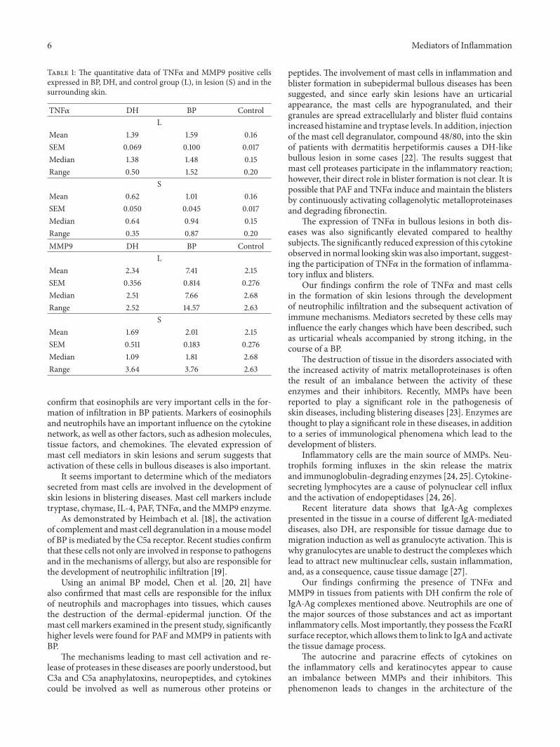
Morphometric analysis revealed that TNF𝛼 expressionwas significantly higher in lesions than the surrounding skin:in BP 1.59 ± 0.100 versus 1.01 ± 0.045 (𝑃 = 0.000005)and DH 1.39 ± 0.069 versus 0.62 ± 0.050 (𝑃 = 0.004998)(Table 1) (Figure 3). Expression was significantly higher inBP and DH skin lesions versus control group (1.59 ± 0.100versus 0.16 ± 0.017 and 1.39 ± 0.069 versus 0.16 ± 0.017,resp., 𝑃 = 0.000004 and 0.007028) and in BP perilesionalskin versus control group (1.01 ± 0.045 versus 0.16 ± 0.017,𝑃 = 0.0000001) (Figure 4).
3.3. Expression of MMP9 in Skin. Moderate expression ofMMP9 was found in inflammation cells in BP patients, aswell as in the epidermal basal cells in DH patients (Figure 2).MMP9 expression was also observed in BP and DH patientsin perilesional skin. Immunohistochemistry found MMP9expression to be absent in the skin biopsies from controlpatients (Figure 2).
Morphometric analysis revealed that MMP9 expressionwas significantly higher in lesions than the surrounding skinin patients with BP (7.41 ± 0.814 versus 2.01 ± 0.183, 𝑃 =0.000045) (Table 1) and in those with DH (2.34 ± 0.356versus 1.69 ± 0.511). Expression was significantly higher inskin lesions in the BP group compared to the control group(7.41 ± 0.814 versus 2.15 ± 0.276). No differences were foundbetween the expression of MMP9 in DH skin lesions versusperilesional skin (𝑃 = 0.128206) (Figures 3 and 4).
4. Discussion
D’Auria et al. [17] have studied the correlation between theenzymes myeloperoxidase, a product specific for granulo-cytes, and tryptase, a proteolytic enzyme synthesized andreleased by mast cells, with the levels of various cytokines inthe blister fluid. They note a positive correlation between thelevels of tryptase, IL-8, and RANTES, showing high activityof cytokines in relation to influx of eosinophils. A few studies

4 Mediators of Inflammation
101214161820222426283032343638
0
5
10
15
20
25
30
35
40
45
50
55
0
20
40
60
80
100
120
140
160
180
0
100
200
300
400
500
0
100
200
300
400
500
600
700
800
900
C DH BP
Con
cent
ratio
n of
chym
ase i
n se
rum
(pg/
mL)
Con
cent
ratio
n of
tryp
tase
in se
rum
(pg/
mL)
Con
cent
ratio
n of
IL- 4
in se
rum
(pg/
mL)
Con
cent
ratio
n of
MM
P9 in
seru
m (p
g/m
L)
Con
cent
ratio
n of
PA
F in
seru
m (p
g/m
L)
0
10
20
30
40
50
60
70
80
Median Min–max Min–max25%–75%
Median25%–75%
C DH BP
C DH BP C DH BP
C DH BP
C DH BP
Con
cent
ratio
n of
TN
F𝛼in
seru
m (p
g/m
L)
P < 0.01 P < 0.001
P < 0.05
Figure 1: Serum levels of examined mediators in BP, DH, and control groups.

Mediators of Inflammation 5
15𝜇m
(a)
15𝜇m
(b)
15𝜇m
(c)
15𝜇m
(d)
15𝜇m
(e)
15𝜇m
(f)
15𝜇m
(g)
Figure 2: Immunoexpression of TNF𝛼 and MMP9 in epidermis and influx, 400x, immunohistochemistry. (a) Immunoexpression of TNF𝛼in epidermis and influx, skin lesions (BP). (b) Immunoexpression of TNF𝛼 in epidermis, skin lesion (DH). (c) Immunoexpression of TNF𝛼in epidermis, perilesional skin (BP). (d) Immunoexpression of TNF𝛼 in epidermis, perilesional skin (DH). (e) Negative immunoexpressionof TNF𝛼, normal skin. (f) Immunoexpression of MMP9 in epidermis and influx, skin lesions (BP). (g) Almost negative immunoexpressionof MMP9, normal skin.
6 Mediators of Inflammation
Table 1: The quantitative data of TNF𝛼 and MMP9 positive cellsexpressed in BP, DH, and control group (L), in lesion (S) and in thesurrounding skin.
TNF𝛼 DH BP ControlL
Mean 1.39 1.59 0.16SEM 0.069 0.100 0.017Median 1.38 1.48 0.15Range 0.50 1.52 0.20
SMean 0.62 1.01 0.16SEM 0.050 0.045 0.017Median 0.64 0.94 0.15Range 0.35 0.87 0.20MMP9 DH BP Control
LMean 2.34 7.41 2.15SEM 0.356 0.814 0.276Median 2.51 7.66 2.68Range 2.52 14.57 2.63
SMean 1.69 2.01 2.15SEM 0.511 0.183 0.276Median 1.09 1.81 2.68Range 3.64 3.76 2.63
confirm that eosinophils are very important cells in the for-mation of infiltration in BP patients. Markers of eosinophilsand neutrophils have an important influence on the cytokinenetwork, as well as other factors, such as adhesion molecules,tissue factors, and chemokines. The elevated expression ofmast cell mediators in skin lesions and serum suggests thatactivation of these cells in bullous diseases is also important.
It seems important to determine which of the mediatorssecreted from mast cells are involved in the development ofskin lesions in blistering diseases. Mast cell markers includetryptase, chymase, IL-4, PAF, TNF𝛼, and theMMP9 enzyme.
As demonstrated by Heimbach et al. [18], the activationof complement andmast cell degranulation in amousemodelof BP is mediated by the C5a receptor. Recent studies confirmthat these cells not only are involved in response to pathogensand in the mechanisms of allergy, but also are responsible forthe development of neutrophilic infiltration [19].
Using an animal BP model, Chen et al. [20, 21] havealso confirmed that mast cells are responsible for the influxof neutrophils and macrophages into tissues, which causesthe destruction of the dermal-epidermal junction. Of themast cell markers examined in the present study, significantlyhigher levels were found for PAF andMMP9 in patients withBP.
The mechanisms leading to mast cell activation and re-lease of proteases in these diseases are poorly understood, butC3a and C5a anaphylatoxins, neuropeptides, and cytokinescould be involved as well as numerous other proteins or
peptides. The involvement of mast cells in inflammation andblister formation in subepidermal bullous diseases has beensuggested, and since early skin lesions have an urticarialappearance, the mast cells are hypogranulated, and theirgranules are spread extracellularly and blister fluid containsincreased histamine and tryptase levels. In addition, injectionof the mast cell degranulator, compound 48/80, into the skinof patients with dermatitis herpetiformis causes a DH-likebullous lesion in some cases [22]. The results suggest thatmast cell proteases participate in the inflammatory reaction;however, their direct role in blister formation is not clear. It ispossible that PAF and TNF𝛼 induce andmaintain the blistersby continuously activating collagenolytic metalloproteinasesand degrading fibronectin.
The expression of TNF𝛼 in bullous lesions in both dis-eases was also significantly elevated compared to healthysubjects.The significantly reduced expression of this cytokineobserved in normal looking skin was also important, suggest-ing the participation of TNF𝛼 in the formation of inflamma-tory influx and blisters.
Our findings confirm the role of TNF𝛼 and mast cellsin the formation of skin lesions through the developmentof neutrophilic infiltration and the subsequent activation ofimmune mechanisms. Mediators secreted by these cells mayinfluence the early changes which have been described, suchas urticarial wheals accompanied by strong itching, in thecourse of a BP.
The destruction of tissue in the disorders associated withthe increased activity of matrix metalloproteinases is oftenthe result of an imbalance between the activity of theseenzymes and their inhibitors. Recently, MMPs have beenreported to play a significant role in the pathogenesis ofskin diseases, including blistering diseases [23]. Enzymes arethought to play a significant role in these diseases, in additionto a series of immunological phenomena which lead to thedevelopment of blisters.
Inflammatory cells are the main source of MMPs. Neu-trophils forming influxes in the skin release the matrixand immunoglobulin-degrading enzymes [24, 25]. Cytokine-secreting lymphocytes are a cause of polynuclear cell influxand the activation of endopeptidases [24, 26].
Recent literature data shows that IgA-Ag complexespresented in the tissue in a course of different IgA-mediateddiseases, also DH, are responsible for tissue damage due tomigration induction as well as granulocyte activation. This iswhy granulocytes are unable to destruct the complexes whichlead to attract new multinuclear cells, sustain inflammation,and, as a consequence, cause tissue damage [27].
Our findings confirming the presence of TNF𝛼 andMMP9 in tissues from patients with DH confirm the role ofIgA-Ag complexes mentioned above. Neutrophils are one ofthe major sources of those substances and act as importantinflammatory cells. Most importantly, they possess the Fc𝛼RIsurface receptor, which allows them to link to IgA and activatethe tissue damage process.
The autocrine and paracrine effects of cytokines onthe inflammatory cells and keratinocytes appear to causean imbalance between MMPs and their inhibitors. Thisphenomenon leads to changes in the architecture of the
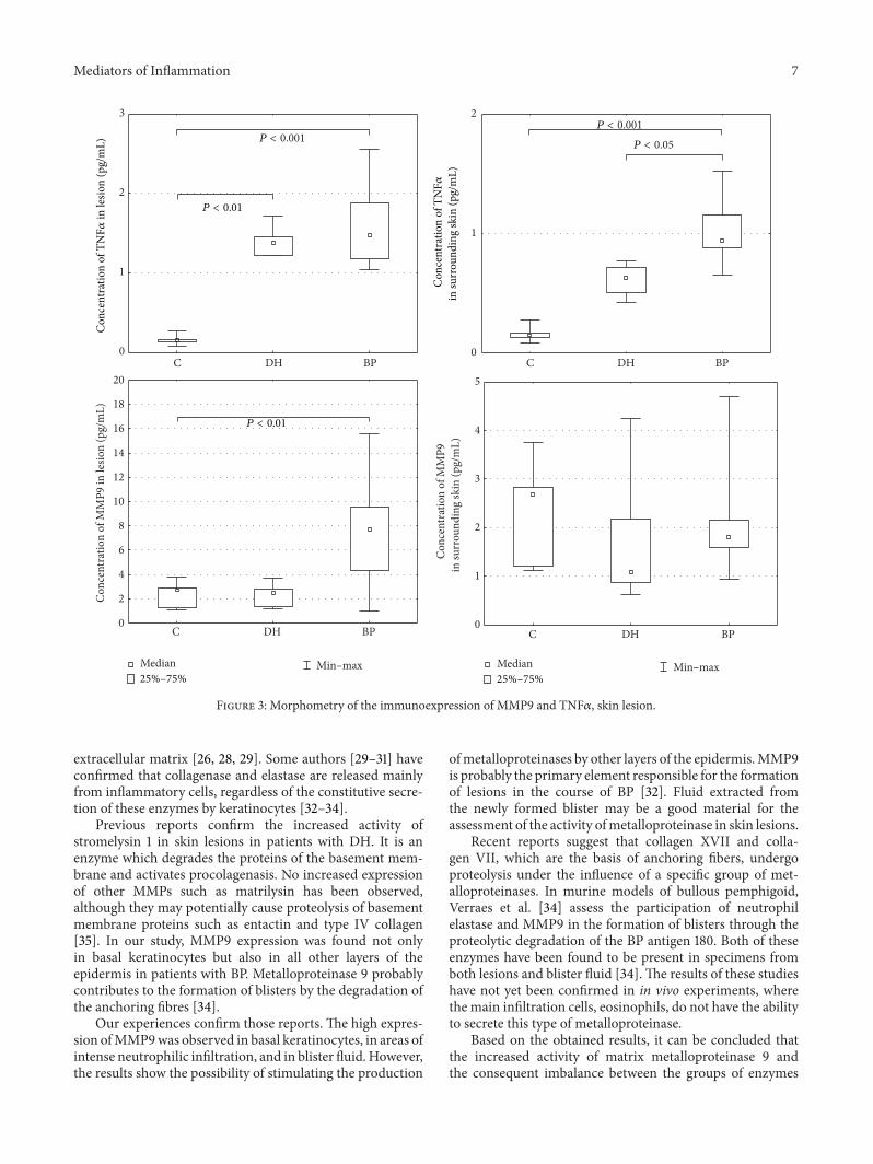
Mediators of Inflammation 7
0
1
2
3
0
1
2
0
2
4
6
8
10
12
14
16
18
20
0
1
2
3
4
5
Con
cent
ratio
n of
MM
P9 in
lesio
n (p
g/m
L)
Con
cent
ratio
n of
MM
P9in
surr
ound
ing
skin
(pg/
mL)
Con
cent
ratio
n of
TN
F𝛼in
lesio
n (p
g/m
L)
C DH BP C DH BP
C DH BP C DH BP
P < 0.01
P < 0.01
P < 0.001P < 0.001
P < 0.05
Con
cent
ratio
n of
TN
F𝛼in
surr
ound
ing
skin
(pg/
mL)
Median Min–max25%–75%
Median Min–max25%–75%
Figure 3: Morphometry of the immunoexpression of MMP9 and TNF𝛼, skin lesion.
extracellular matrix [26, 28, 29]. Some authors [29–31] haveconfirmed that collagenase and elastase are released mainlyfrom inflammatory cells, regardless of the constitutive secre-tion of these enzymes by keratinocytes [32–34].
Previous reports confirm the increased activity ofstromelysin 1 in skin lesions in patients with DH. It is anenzyme which degrades the proteins of the basement mem-brane and activates procolagenasis. No increased expressionof other MMPs such as matrilysin has been observed,although they may potentially cause proteolysis of basementmembrane proteins such as entactin and type IV collagen[35]. In our study, MMP9 expression was found not onlyin basal keratinocytes but also in all other layers of theepidermis in patients with BP. Metalloproteinase 9 probablycontributes to the formation of blisters by the degradation ofthe anchoring fibres [34].
Our experiences confirm those reports. The high expres-sion ofMMP9was observed in basal keratinocytes, in areas ofintense neutrophilic infiltration, and in blister fluid.However,the results show the possibility of stimulating the production
ofmetalloproteinases by other layers of the epidermis.MMP9is probably the primary element responsible for the formationof lesions in the course of BP [32]. Fluid extracted fromthe newly formed blister may be a good material for theassessment of the activity ofmetalloproteinase in skin lesions.
Recent reports suggest that collagen XVII and colla-gen VII, which are the basis of anchoring fibers, undergoproteolysis under the influence of a specific group of met-alloproteinases. In murine models of bullous pemphigoid,Verraes et al. [34] assess the participation of neutrophilelastase and MMP9 in the formation of blisters through theproteolytic degradation of the BP antigen 180. Both of theseenzymes have been found to be present in specimens fromboth lesions and blister fluid [34]. The results of these studieshave not yet been confirmed in in vivo experiments, wherethe main infiltration cells, eosinophils, do not have the abilityto secrete this type of metalloproteinase.
Based on the obtained results, it can be concluded thatthe increased activity of matrix metalloproteinase 9 andthe consequent imbalance between the groups of enzymes

8 Mediators of Inflammation
0
1
2
0
1
2
3
0
1
2
3
4
5
0
2
4
6
8
10
12
14
16
18
20
Con
cent
ratio
n of
MM
P9 in
DH
pat
ient
s (pg
/mL)
Con
cent
ratio
n of
MM
P9 in
BP
patie
nts (
pg/m
L)
In lesion In surrounding skin In lesion In surrounding skin
In lesion In surrounding skin
Con
cent
ratio
n of
TN
F𝛼in
DH
pat
ient
s (pg
/mL)
P < 0.01 P < 0.001
P < 0.001
In lesion In surrounding skin
Con
cent
ratio
n of
TN
F𝛼in
BP
patie
nts (
pg/m
L)
Median Min–max25%–75%
Median Min–max25%–75%
Figure 4: Morphometry of the immunoexpression of MMP9 and TNF𝛼, perilesional skin.
are responsible for tissue destruction in the course of BP.Considering the increasing number of studies concerningthe participation of MMPs in the pathology of skin diseases,research regarding the therapeutic use ofMMP inhibitors canbe expected in the near future.
The findings of the present study demonstrate increasedexpression of TNF𝛼 in skin lesions, both in the case of bul-lous pemphigoid and in dermatitis herpetiformis. This canactivate the production of other inflammatory mediators.Elevated PAF and MMP9 levels in the sera of patients mayindicate the activation of mast cells in the process of blisterformation in these diseases. In the present study, we inves-tigated the relative contribution of mediators of mast cellsin the immunopathogenesis of BP and DH skin lesions. Weconclude that mast cells are active participants in eventsthat mediate tissue damage in autoimmune disease. Disease-associated increases in mast cell numbers accompanied bymast cell degranulation and elaboration of numerous mastcell mediators at sites of inflammation are commonlyobserved in many human autoimmune diseases, such as
multiple sclerosis and rheumatoid arthritis. In animal mod-els, treatment with mast cell stabilizing drugs or mast cellablation can result in diminished disease [35].
Conflict of Interests
The authors declare that there is no conflict of interestsregarding the publication of this paper.
Acknowledgments
The study was funded byMedical University of Lodz researchprojects no. 503/1-152-01/503-01 andThePolish Science Com-mittee Grant no. 4746/B/PO1/2009/37.
References
[1] S. Karpati, “Dermatitis herpetiformis: close to unravelling adisease,” Journal of Dermatological Science, vol. 34, no. 2, pp. 83–90, 2004.
Mediators of Inflammation 9
[2] M. Sardy, S. Karpati, B.Merkl,M. Paulsson, andN. Smyth, “Epi-dermal transglutaminase (TGase 3) is the autoantigen of der-matitis herpetiformis,” The Journal of Experimental Medicine,vol. 195, no. 6, pp. 747–757, 2002.
[3] W. Dieterich, E. Laag, L. Bruckner-Tuderman et al., “Antibodiesto tissue transglutaminase as serologic markers in patients withdermatitis herpetiformis,” Journal of Investigative Dermatology,vol. 113, no. 1, pp. 133–136, 1999.
[4] J. D. Hendrix, K. L. Mangum, J. J. Zone, and W. R. Gammon,“Cutaneous IgA deposits in bullous diseases function as ligandsto mediate adherence of activated neutrophils,” Journal ofInvestigative Dermatology, vol. 94, no. 5, pp. 667–672, 1990.
[5] M. V. Dahl, R. J. Falk, R. Carpenter, and A. F. Michael, “Mem-brane attack complex of complement in dermatitis herpeti-formis,” Archives of Dermatology, vol. 121, no. 1, pp. 70–72, 1985.
[6] K. Airola, M. Vaalamo, T. Reunala, and U. K. Saarialho-Kere,“Enhanced expression of interstitial collagenase, stromelysin-1, and urokinase plasminogen activator in lesions of dermatitisherpetiformis,” Journal of InvestigativeDermatology, vol. 105, no.2, pp. 184–189, 1995.
[7] R. F. Ghohestani, J. Novotney, M. Chaudhary, and R. S. Agah,“Bullous pemphigoid: from the bedside to the research labora-tory,” Clinics in Dermatology, vol. 19, no. 6, pp. 690–696, 2001.
[8] J. R. Stanley, “Cell adhesion molecules as targets of autoanti-bodies in pemphigus and pemphigoid, bullous diseases due todefective epidermal cell adhesion,” Advances in Immunology,vol. 53, pp. 291–326, 1993.
[9] R. E. Jordon, E. H. Beutner, E. Witebsky, G. Blumental, W. L.Hale, and W. F. Lever, “Basement zone antibodies in bullouspemphigoid,” Journal of the American Medical Association, vol.200, no. 9, pp. 751–756, 1967.
[10] E. Schmidt, S. Reimer, N. Kruse et al., “Autoantibodies to BP180associated with bullous pemphigoid release interleukin-6 andinterleukin-8 from cultured human keratinocytes,” Journal ofInvestigative Dermatology, vol. 115, no. 5, pp. 842–848, 2000.
[11] S. A. Grando, B. T. Glukhenky, G. N. Drannik, E. V. Epshtein, A.P. Kostromin, and T. A. Korostash, “Mediators of inflammationin blister fluids from patients with pemphigus vulgaris andbullous pemphigoid,” Archives of Dermatology, vol. 125, no. 7,pp. 925–930, 1989.
[12] M. Baggioloni, “Chemokines in pathology and medicine,” Jour-nal of Internal Medicine, vol. 250, no. 2, pp. 91–104, 2001.
[13] M. M.Wong and E. N. Fish, “Chemokines: attractive mediatorsof the immune response,” Seminars in Immunology, vol. 15, no.1, pp. 5–14, 2003.
[14] B. U. Wintroub, M. C. Mihm, E. J. Goetzl, N. A. Soter, and K.F. Austen, “Morphologic and functional evidence for release ofmast-cell products in bullous pemphigoid,” The New EnglandJournal of Medicine, vol. 298, no. 8, pp. 417–421, 1978.
[15] A. B. Ackerman, N. Chongchitnant, J. Sanchex et al., HistologicDiagnosis of Inflammatory Skin Disease: A Algorithmic MethodBased on Pattern Analysis, Williams &Wilkins, Baltimore, Md,USA, 2nd edition, 1997.
[16] E. R. Weibel, “Point counting methods,” in Stereological Meth-ods, E. R. Weibel, Ed., vol. 1, pp. 101–159, Academic Press,London, UK, 1979.
[17] L. D’Auria, M. Pietravalle, P. Cordiali-Fei, and F. Ameglio, “In-creased tryptase and myeloperoxidase levels in blister fluids ofpatients with bullous pemphigoid: correlations with cytokines,adhesion molecules and anti-basement membrane zone anti-bodies,” Experimental Dermatology, vol. 9, no. 2, pp. 131–137,2000.
[18] L. Heimbach, Z. Li, P. Berkowitz et al., “The C5a receptor onmast cells is critical for the autoimmune skin-blistering diseasebullous pemphigoid,” The Journal of Biological Chemistry, vol.286, no. 17, pp. 15003–15009, 2011.
[19] B. A.Walker, M. A. Jacobson, D. A. Knight et al., “Adenosine A3receptor expression and function in eosinophils,” AmericanJournal of Respiratory Cell and Molecular Biology, vol. 16, no. 5,pp. 531–537, 1997.
[20] R. Chen, J. A. Fairley, M.-L. Zhao et al., “Macrophages, butnot T and B lymphocytes, are critical for subepidermal blisterformation in experimental bullous pemphigoid: macrophage-mediated neutrophil infiltration depends on mast cell activa-tion,” Journal of Immunology, vol. 169, no. 7, pp. 3987–3992, 2002.
[21] R. Chen, G. Ning,M.-L. Zhao et al., “Mast cells play a key role inneutrophil recruitment in experimental bullous pemphigoid,”The Journal of Clinical Investigation, vol. 108, no. 8, pp. 1151–1158,2001.
[22] K. Brockow, D. Abeck, K. Hermann, and J. Ring, “Tryptase con-centration in skin blister fluid from patients with bullous skinconditions,” Archives of Dermatological Research, vol. 288, no.12, pp. 771–773, 1996.
[23] Y. Niimi, R. Pawankar, and S. Kawana, “Increased expressionof matrix metalloproteinase-2, matrix metalloproteinase-9 andmatrix metalloproteinase-13 in lesional skin of bullous pem-phigoid,” International Archives of Allergy and Immunology, vol.139, no. 2, pp. 104–113, 2006.
[24] H. Nagase, “Activation mechanisms of matrix metallopro-teinases,” Biological Chemistry, vol. 378, no. 3-4, pp. 151–160,1997.
[25] D. Reinhardt, H.H. Sigusch, J. Henße, S. C. Tyagi, R. Korfer, andH. R. Figulla, “Cardiac remodelling in end stage heart failure:upregulation of matrix metalloproteinase (MMP) irrespectiveof the underlying disease, and evidence for a direct inhibitoryeffect of ACE inhibitors onMMP,”Heart, vol. 88, no. 5, pp. 525–530, 2002.
[26] J. J. Reynolds, “Collagenases and tissues inhibitors of metal-loproteinases: functional balance in tissue degradation,” OralDiseases, vol. 2, no. 1, pp. 70–76, 1996.
[27] L. P. van der Steen, J. E. Bakema, A. Sesarman et al., “BlockingFc𝛼 receptor I on granulocytes prevents tissue damage inducedby IgA autoantibodies,”The Journal of Immunology, vol. 189, no.4, pp. 1594–1601, 2012.
[28] J. J. Reynolds andM. C.Meikle, “The functional balance of met-alloproteinases and inhibitors in tissue degradation: relevanceto oral pathologies,” Journal of the Royal College of Surgeons ofEdinburgh, vol. 42, no. 3, pp. 154–160, 1997.
[29] K. J. Leco, R. Khokha, N. Pavloff, S. P. Hawkes, and D. R.Edwards, “Tissue inhibitor of metalloproteinases-3 (TIMP-3)is an extracellular matrix-associated protein with a distinctivepattern of expression in mouse cells and tissues,”The Journal ofBiological Chemistry, vol. 269, no. 12, pp. 9352–9360, 1994.
[30] M. Larry and W. Corcoran, “Regulation of monocyte/macro-phage metalloproteinase production by cytokines,” Journal ofPeriodontology, vol. 64, no. 5, pp. 467–473, 1993.
[31] L. S. Lohmander, L. A. Hoerrner, and M.W. Lark, “Metallopro-teinases, tissue inhibitor, and proteoglycan fragments in kneesynovial fluid in human osteoarthritis,” Arthritis and Rheuma-tism, vol. 36, no. 2, pp. 181–189, 1993.
[32] A. I. Oikarinen, J. J. Zone, A. R. Ahmed, U. Kiistala, and J. Uitto,“Demonstration of collagenase and elastase activities in theblister fluids from bullous skin diseases. Comparison between

10 Mediators of Inflammation
dermatitis herpetiformis and bullous pemphigoid,” Journal ofInvestigative Dermatology, vol. 81, no. 3, pp. 261–266, 1983.
[33] H. G.Welgus, E. A. Bauer, and G. P. Stricklin, “Elevated levels ofhuman collagenase inhibitor in blister fluids of diverse etiology,”Journal of Investigative Dermatology, vol. 87, no. 5, pp. 592–596,1986.
[34] S. Verraes, W. Hornebeck, M. Polette, L. Borradori, and P.Bernard, “Respective contribution of neutrophil elastase andmatrix metalloproteinase 9 in the degradation of BP180 (typeXVII collagen) in human bullous pemphigoid,” Journal ofInvestigative Dermatology, vol. 117, no. 5, pp. 1091–1096, 2001.
[35] M. A. Brown and J. K. Hatfield, “Mast cells are important mod-ifiers of autoimmune disease: with so much evidence, why isthere still controversy?” Frontiers in Immunology, vol. 7, no. 3,article 147, 2012.

Submit your manuscripts athttp://www.hindawi.com
Stem CellsInternational
Hindawi Publishing Corporationhttp://www.hindawi.com Volume 2014
Hindawi Publishing Corporationhttp://www.hindawi.com Volume 2014
MEDIATORSINFLAMMATION
of
Hindawi Publishing Corporationhttp://www.hindawi.com Volume 2014
Behavioural Neurology
EndocrinologyInternational Journal of
Hindawi Publishing Corporationhttp://www.hindawi.com Volume 2014
Hindawi Publishing Corporationhttp://www.hindawi.com Volume 2014
Disease Markers
Hindawi Publishing Corporationhttp://www.hindawi.com Volume 2014
BioMed Research International
OncologyJournal of
Hindawi Publishing Corporationhttp://www.hindawi.com Volume 2014
Hindawi Publishing Corporationhttp://www.hindawi.com Volume 2014
Oxidative Medicine and Cellular Longevity
Hindawi Publishing Corporationhttp://www.hindawi.com Volume 2014
PPAR Research
The Scientific World JournalHindawi Publishing Corporation http://www.hindawi.com Volume 2014
Immunology ResearchHindawi Publishing Corporationhttp://www.hindawi.com Volume 2014
Journal of
ObesityJournal of
Hindawi Publishing Corporationhttp://www.hindawi.com Volume 2014
Hindawi Publishing Corporationhttp://www.hindawi.com Volume 2014
Computational and Mathematical Methods in Medicine
OphthalmologyJournal of
Hindawi Publishing Corporationhttp://www.hindawi.com Volume 2014
Diabetes ResearchJournal of
Hindawi Publishing Corporationhttp://www.hindawi.com Volume 2014
Hindawi Publishing Corporationhttp://www.hindawi.com Volume 2014
Research and TreatmentAIDS
Hindawi Publishing Corporationhttp://www.hindawi.com Volume 2014
Gastroenterology Research and Practice
Hindawi Publishing Corporationhttp://www.hindawi.com Volume 2014
Parkinson’s Disease
Evidence-Based Complementary and Alternative Medicine
Volume 2014Hindawi Publishing Corporationhttp://www.hindawi.com