mechanisms of tumour development - iarc · pdf filemechanisms of tumour development ......
TRANSCRIPT
Mechanisms of tumour development Mechanisms of tumour development Mechanisms of tumour development
The phenotypic changes which a cell undergoes in theprocess of malignant transformation is a reflection of thesequential acquisition of genetic alterations. This multi-stepprocess is not an abrupt transition from normal to malignantgrowth, but may take place over 20 years or more. The muta-tion of critical genes, including suppressor genes, oncogenesand genes involved in DNA repair, leads to genetic instabilityand progressive loss of differentiation. Tumours enlargebecause cancer cells lack the ability to balance cell divisionby cell death (apoptosis) and by forming their own vascularsystem (angiogenesis). The transformed cells lose their abili-ty to interact with each other and exhibit uncontrolledgrowth, invade neighbouring tissues and eventually spreadthrough the blood stream or the lymphatic system to distantorgans.
333
WCR-S3.Q (composition) 27/01/03 9:26 Page 1

Cancer arises from a single cellMalignant tumours (or “cancers”) aredescribed as monoclonal, meaning thateach tumour arises from a single cell. Thedevelopment of a malignant tumour froma normal cell usually occurs over a con-siderable fraction of our lifetime. Such along period is reflected, for example, bythe difference between the age at whichpeople start smoking and the age atwhich diagnosis of lung cancer mostoften occurs. The long “latent period” inlung cancer and almost all other malig-nancies is not explicable on the basis of asingle-step transition from a normal cellto malignant one. Rather, the tumour isthe outcome of an evolutionary processinvolving successive generations of cells,which are progressively further advancedtowards cancerous growth [1].Human histopathological observationssupport this scenario, and a range of pre-malignant lesions have been identified[2]. Likewise, in experimental animals,
specific cell populations may be identifiedas marking a commitment towards malig-nancy, and these may be exploited as anearly indicator in the context of carcino-gen testing [3]. Thus, wholly on morpho-logical grounds, cancer may be perceivedas the outcome of a complex biologicalprocess.
Multiple steps are required for a can-cer to ariseAnimal “models” of cancer development,most commonly involving treatment ofrodents with carcinogenic chemicals orother cancer-inducing agents, have pro-vided clear evidence that specific stagesin malignant transformation can occur dis-cretely [4]. Chemicals which cause cancerin animals without the need for othertreatment are sometimes called “com-plete carcinogens” (although “carcino-gens” would be appropriate). Most suchcarcinogens cause damage to DNA of
cells or tissues exposed to them. DNA-damaging activity may be identified on thebasis of defined protocols (sometimescalled “short-term tests”, to emphasizetheir difference from chronic lifetimebioassay in rodents). Chemicals whichexhibit mutagenic activity in short-termtests, which typically involve sensitivebacterial strains and cell-free extracts tocatalyse metabolism of the test com-pound, are characterized as “genotoxic”[5]. Genotoxic agents may be completecarcinogens, but can also act as “initiatingagents”. After a single treatment with aninitiating agent, tumour growth may befacilitated by chemicals (or treatments)which stimulate cell proliferation, some-times by inducing mild toxic damage inexposed tissue. These agents are termed“promoters” (Table 3.1). As well as thesegenotoxic chemicals, a range of non-geno-toxic agents can cause cancer in humansand/or experimental animals [6].
MULTISTAGE CARCINOGENESIS
SUMMARY
> Tumours consist of cells whose growthand morphological characteristics aremarkedly different from those of normalcells. Criteria for malignancy includeincreased cell proliferation, loss of differ-entiation, infiltrative growth and metasta-sis to other organs.
> Malignant transformation is a multistageprocess, typically a progression frombenign lesions (e.g. adenoma) to malig-nant tumours (e.g. carcinoma). This evo-lution of malignant cells is caused by thesequential accumulation of alterationsin genes responsible for the control ofcellular proliferation, cell death and themaintenance of genetic integrity.
> The development of cancer may be initi-ated by environmental agents (chemicalcarcinogens, radiation, viruses) andinherited genetic factors (germlinemutations).
Fig. 3.1 Carcinogenesis is a multistage process involving multiple genetic and epigenetic events in proto-oncogenes, tumour suppressor genes and anti-metastasis genes.
During this process the cell develops :- Defects in terminal differentiation- Defects in growth control- Resistance to cytotoxicity- Defects in programmed cell death
These steps are caused by :- Activation of proto-oncogenes- Inactivation of tumour suppressor genes- Inactivation of genomic stability genes
INITIATEDCELL
VIRUS
CHEMICALS
NORMALCELL
PRE-NEOPLASTIC
LESIONMALIGNANT
TUMOURCLINICALCANCER
CANCERMETASTASIS
Geneticchange
Geneticchange
Geneticchange
Geneticchange
Selectiveclonal
expansion
84 Mechanisms of tumour development
RADIATION
WCR-S3.Q (composition) 27/01/03 9:26 Page 84

The stages in tumorigenesis have beendesignated “initiation”, which encom-passes damage to, and then division ofexposed cells such that their growthpotential is changed irreversibly, and“progression”, denoting multiple roundsof cell replication mediating the gradualtransition of an initiated cell towardsautonomous, cancerous, growth. Ultimatespread of malignant cells resulting in mul-tiple tumour sites has been termed“metastasis”. The unequivocal identifica-tion by the mid-1970s of these variousphases was one indication that carcino-genesis is a multistage process. Arguably,the greatest achievement of cancerresearch during the last decades of the20th century has been the elucidation ofmultistage carcinogenesis at the molecu-lar genetic level.
The molecular basis of tumourpathologyIn a seminal publication, Vogelstein andcolleagues [7] provided evidence thatthe different stages in the cellular evo-lution of colon cancer in humans, histo-logically identified as hyperplasia,early-stage adenoma, late-stage adeno-ma etc., could be identified with specif-ic successive genetic changes (Fig.3.2). The genetic changes includedoncogene activation by mutation atspecific sites and loss of chromosomalregions (necessarily involving multiplegenes) which were subsequently shownto be the location of tumour suppressorgenes. Since that initial description,knowledge of the molecular geneticbasis for human colon cancer has beenmassively extended (Colorectal cancer,p198). For most tumours, the geneticchanges are not inherited from our par-ents but arise in a previously normalcell. The progeny of this cell after celldivision carry the same genetic changebut the surrounding cells remain nor-mal. Because these genetic changesaffect only the cancer cells, they arenot passed on to the children of cancerpatients. However, in a minority ofcases some critical changes are inherit-ed, giving a familial predisposition tocolon or other cancers.
Commonality and heterogeneityThe molecular biological basis of multi-stage carcinogenesis initially describedfor colon cancer appears to have appli-cation to all tumour types, althoughthere is marked variation in the extentto which genes relevant to particulartumours have been identified [8]. Somegenes, and the corresponding changeassociated with tumorigenesis (muta-tion, overexpression, deletion and/or
amplification) are common to a numberof tumour types. However, each tumourtype is associated with a distinctive setof gene alterations. The genes in ques-tion are discussed under the subhead-ing Pathology and genetics for each ofthe tumour types included in Chapter 5.Such enumeration of relevant genesnecessitates a degree of simplification.There is clear heterogeneity betweenindividual tumours of the same type. In
Factor Cancer site/cancer
Hormones Estrogens, progesterone Uterus, mammary glandGonadotrophins Ovary, testis, pituitaryTestosterone Prostate gland
Pharmaceutical Oral contraceptives Liverproducts Anabolic steroids Liver
Analgesics Renal pelvis
Miscellaneous Bile acids Small intestinesubstances Saturated fatty acids Colon
Salt StomachTobacco Oral cavity, lung, bladder etc. Saccharin, uracil, melamine, Urinary bladdertetraphthalic acid and other xenobiotics causing urinary stonesDichlorobenzene, trimethylpentane Kidney(lead-free gasoline), perchloroethyl-eneButylated hydroxyanisole, propionic StomachacidNitrilotriacetate Kidney
Table 3.1 Promoting agents: non-genotoxic agents that facilitate carcinogenesis by stimulating cell division.Tobacco smoke also contains genotoxic carcinogens.
Fig. 3.2 The original Vogelstein model for the genetic and histological evolution of colon cancer.(Colorectal cancer, p198).
CHROMOSOME: 5qALTERATION: MutationGENE: FAP
12pMutation
KRAS
18qLossDCC?
17pLossp53
Otheralterations
DNA hypomethylation
Normalepithelium
Hyperproliferativeepithelium
Early adenoma
Intermediateadenoma
Carcinoma MetastasisLate
adenoma
Multistage carcinogenesis 85
WCR-S3.Q (composition) 29/01/03 13:58 Page 85

other words, not every tumour will nec-essarily exhibit all the genetic changesestablished for the tumour type in ques-tion. Moreover, there is often markedheterogeneity within an individualtumour: adjacent cells differ. Mappingand identification of genes involved inmalignant transformation has been amajor component of the study of themolecular mechanisms of carcinogene-sis.
Multiple genetic changes requiredThe emergence of a malignant cell popu-lation is understood to be the cumulativeeffect of multiple (perhaps five, ten ormore) genetic changes, such changesbeing accumulated in the course of theevolution of the cell from normal to malig-nant. The genes designated as oncogenesand tumour suppressor genes (Oncogenes
and tumour suppressor genes, p96) havebeen identified in terms of their biologicalfunction [9]. Such genes are among thosethat facilitate transmission of growth con-trol signals from the cell membrane to thenucleus (that is, signal transduction), thatmediate cell division, differentiation or celldeath and, perhaps most critical of all,that maintain the integrity of genetic infor-mation by DNA repair and similar process-es (Carcinogen activation and DNA repair,p89). Since mutations are normally infre-quent events, it seems unlikely that in thecourse of a human lifetime a cell wouldacquire all the mutations necessary forcancer to develop, unless at some pointthe developing cell lost its ability to pro-tect itself against mutation and gainedwhat is called a “mutator” phenotype [10].Thus, alterations in gene structure andexpression which bring about carcinogen-
esis are being progressively identified[11]. As noted earlier, members of somecancer-susceptible families inherit muta-tions in particular genes that contribute tocancer development, and hence to theirindividual risk of disease. However, withmost cancers, the genetic change criticalto carcinogenesis results from damage toDNA by chemicals, radiation and viruses(Fig. 3.1). This damage is not entirely andperhaps not predominantly produced byexogenous agents but by natural process-es, such as the production of reactive oxy-gen species or the spontaneous deamina-tion of the 5-methylcytosine naturallypresent in DNA [13]. Furthermore, asshown as the second step in Fig. 3.2, bio-logical change that is heritable may resultfrom non-genetic processes including themodulation of gene expression by hyper-methylation [12].
Fig. 3.3 Histological representation of the pathogenesis of colorectal cancer. Phenotypic changes in the morphology of the colonic mucosa reflect the sequen-tial acquisition of genetic alterations.
Dysplasia in hamartoma
Flat cancer
Cancer in mixed hyperplastic adenomatous polyps (MHAP)
Ulcerative colitis-associated colorectal carcinoma
Flat adenoma
MHAP/Serrated adenoma
Flat dysplasia
Normal Early adenoma Intermediate adenoma Late adenoma
Loss of mismatch repair
Cancer
RER+ cancer(ReplicationError Positive)
Juvenile polyp
Peutz-Jeghers polyp
86 Mechanisms of tumour development
WCR-S3.Q (composition) 27/01/03 9:27 Page 86

AgeingApart from multistage development, cer-tain other processes are fundamental tomalignant disease. Principal amongstthese is ageing, which can be consideredboth in relation to the whole individual, and
also at the cellular level. In humans, as wellas in other mammals, the incidence ofcancer rises dramatically with age. Anexponential increase occurs from mid-life[14]. Passage of time is also critical to cellbiology. Normal cells do not divide indefi-nitely due to senescence (Box: Telomeresand Telomerase, p108). Senescent cellscannot be stimulated to divide further,become resistant to apoptotic cell deathand acquire differentiated functions.Senescence may be an anti-cancer mech-anism that limits accumulation of muta-tions. However, when maintained in cul-ture, cells treated with carcinogenic chem-icals or infected with oncogenic virusesmay avoid senescence and proliferateindefinitely. Such cell populations aredescribed as being “transformed” and
when further maintained in culture, once-normal cells acquire the same characteris-tics as cells cultured from malignanttumours. These and various other alter-ations in growth characteristics are recog-nized as the experimental counterpart ofmultistage carcinogenesis through whichtumours develop in intact animals orhumans. The genetic basis for senescence,and its relationship to malignancy, is a sub-ject of intense investigation [15].
Preventing cancerThe significance of multistage carcino-genesis extends beyond facilitatingunderstanding of how a transition fromnormal to malignant cell growth occurs.The fundamental cellular studies outlinedearlier provide a basis for preventing can-
Fig. 3.4 Severe intraepithelial neoplasia (dyspla-sia) in the epithelium of an intrahepatic large bileduct, a condition caused by hepatolithiasis.
Multistage carcinogenesis 87
Trials of agents for chemopreventiveactivity which are based on assessmentof malignant disease are almost unman-ageable because of the long period oftime (perhaps decades) potentiallyinvolved. Attention has therefore beenfocused on lesions, either cellular ormolecular, demonstrated to be valid indi-cators of the subsequent development ofmalignancy. A trial may then evaluate theeffect of the putative chemopreventiveagent on such precursor lesions.
The best-validated precursor lesions arebenign tumours, such as colorectal ade-nomas. It is established that adenomanumber, size, and severity of dysplasia arepredictive factors for colorectal cancerincidence. It has been estimated that 2-5% of all colorectal adenomas progress toadenocarcinomas if not removed or treat-ed. The risk is greater for large andseverely dysplastic polyps. Cancer risk isdecreased by polyp removal, and a strongcorrelation exists between the relativeprevalence of adenomas and cancersacross populations (Winawer SJ et al., N
Engl J Med, 328: 901-906, 1993). Severalepidemiological studies have shown thatregular use of aspirin or related drugs isassociated with a reduced adenoma inci-dence (IARC Handbooks of CancerPrevention. Vol. 1, Lyon, 1997). This pro-vides further confirmation that adenomasare precursor lesions for colon cancer,since aspirin is known to reduce the inci-dence of malignant colon cancer.
Potential precursor lesions of carcinogene-sis include both phenotypic and genotypicmarkers (Miller AB et al. Biomarkers inCancer Chemoprevention, IARC ScientificPublications 154, Lyon, 2001). Thus oralleukoplakia is a recognized precursor forcancer of the oral cavity. Histological mod-ulation of a precancer (often called intraep-ithelial neoplasia) has been used as a pre-cursor lesion in prevention trials (Kelloff GJet al., Cancer Epidemiol Biomarkers Prev, 9:127-137, 2000). Additionally, geneticlesions such as progressive genomic insta-bility as measured by loss of heterozygosi-ty or amplification at specific microsatelliteloci, have been considered (Califano J et al.Cancer Res, 56: 2488-2492, 1996). Otherpotential precursor endpoints include pro-liferation and differentiation markers, spe-
cific gene and general chromosomal dam-age, cell growth regulatory molecules,and biochemical activities (e.g. enzymeinhibition). Serum proteins are of specialinterest because of their availability. Thusprostate-specific antigen (PSA) is beingused as a “surrogate” marker for prostatecancer. It is expected that the number andvariety of biomarkers for precursorlesions will continue to expand In parallelwith the advances in understanding of thegenetic and cellular basis of carcinogene-sis.
PRECURSOR LESIONS IN CHEMOPREVENTION TRIALS
Fig. 3.6 Tubular adenoma of the colon is a precur-sor lesion for colorectal cancer.
WCR-S3.Q (composition) 27/01/03 9:27 Page 87

cer (see chapter 4). The fact that partic-ular patterns of cell morphology andgrowth precede emergence of anunequivocally malignant cell populationis the basis of secondary prevention ofcancer.Examples include detection of polyps inthe large bowel (Fig. 3.5) and of morpho-logical change which is the basis of thePapanicolaou smear test for early detec-tion of cervical cancer. Moreover, dietaryor pharmaceutical interventions calculat-ed to prevent or reverse such lesions arethe basis of chemoprevention [16]. Mostimportantly, knowledge of the geneticbasis underlying tumour growth shouldprovide new criteria for individual deter-mination of diagnosis and prognosis. The
mechanisms now known to operate inthe proliferation of cancer cells provide abasis for the development of new, moreefficient therapies without the side-effects that currently often afflict cancerpatients [17].
1. Foulds L, ed. (1969) Neoplastic Development, Vol. 1,London, Academic Press.
2. Correa P (1996) Morphology and natural history of can-cer precursors. In: Schottenfeld D, Fraumeni JF, eds,Cancer Epidemiology and Prevention, New York, OxfordUniversity Press, 45-64.
3. Ito N, Imaida K, Asamoto M, Shirai T (2000) Earlydetection of carcinogenic substances and modifiers in rats.Mutat Res, 462 : 209-217.
4. Weinstein IB (1982) Carcinogenesis as a multistageprocess—experimental evidence. In: Bartsch H, ArmstongB, eds, Host Factors in Human Carcinogenesis (IARCScientific Publications No. 39) Lyon, IARCPress, 9-25.
5. Vainio H, Magee PN, McGregor DB, McMichael AJ, eds(1992) Mechanisms of Carcinogenesis in Risk Identification(IARC Scientific Publications No. 116), Lyon, IARCPress.
6. Yamasaki H, Ashby J, Bignami M, Jongen W, LinnainmaaK, Newbold RF, Nguyen-Ba G, Parodi S, Rivedal E,Schiffmann D, Simons JW, Vasseur P (1996) Nongenotoxiccarcinogens: development of detection methods based on
mechanisms: a European project. Mutat Res, 353: 47-63.
7. Vogelstein B, Fearon ER, Hamilton SR, Kern SE,Preisinger AC, Leppert M, Nakamura Y, White R, Smits AM,Bos JL (1988) Genetic alterations during colorectal-tumordevelopment. N Engl J Med, 319: 525-532.
8. Balmain A, Harris CC (2000) Carcinogenesis in mouseand human cells: parallels and paradoxes. Carcinogenesis,21: 371-377.
9. Evan GI, Vousden KH (2001) Proliferation, cell cycleand apoptosis in cancer. Nature, 411: 342-348.
10. Loeb LA (2001) A mutator phenotype in cancer.Cancer Res, 61: 3230-3239.
11. Hahn WC, Counter CM, Lundberg AS, BeijersbergenRL, Brooks MW, Weinberg RA (1999) Creation of humantumour cells with defined genetic elements. Nature, 400:464-468.
12. Esteller M, Corn PG, Baylin SB, Herman JG (2001) Agene hypermethylation profile of human cancer. CancerRes, 61: 3225-3229.
13. Marnett LJ, Plastaras JP (2001) Endogenous DNAdamage and mutation. Trends Genet, 17: 214-221.
14. Armitage P, Doll R (1954) The age distribution of can-cer and a multistage theory of carcinogenesis. Br J Cancer,8: 1-12.
15. Wynford-Thomas D (1999) Cellular senescence andcancer. J Pathol, 187: 100-111.
16. Bartsch H (2000) Studies on biomarkers in canceretiology and prevention: a summary and challenge of 20years of interdisciplinary research. Mutat Res, 462: 255-279.
17. Kallioniemi OP, Wagner U, Kononen J, Sauter G(2001) Tissue microarray technology for high-throughputmolecular profiling of cancer. Hum Mol Genet, 10: 657-662.
REFERENCES
Fig. 3.5 Pedunculated hyperplastic polyp of thecolon.
88 Mechanisms of tumour development
WCR-S3.Q (composition) 27/01/03 9:27 Page 88

Experimental studies in rodents and incultured cells have led to the classifica-tion of chemical carcinogens into twobroad classes: genotoxic and non-geno-toxic. Genotoxic carcinogens alter thestructure of DNA, mostly by covalentbinding to nucleophilic sites. Theselesions, that is, the chemical entity ofcarcinogen bound to DNA, are calledDNA “adducts”. The replication of DNAcontaining unrepaired adducts mayresult either in the generation ofsequence changes (mutations) in thenewly synthesized daughter strands ofDNA or in DNA rearrangements evidentas chromosome aberrations.This critical, irreversible genetic eventcan thus result in fixation of the originalstructural change in DNA as permanent,transmissible, genetic damage, or in theloss of ge netic information throughalterations in chromosomes. Such heri-tab le change has the potential to per-turb growth control in the affected cell,and is sometimes referred to as the “ini-tiation” step of the tumorigenic process(Fig. 3.7).
Carcinogen activationThe first indication that certain cancerswere associated with exposure to chemi-cals arose from observations by clini-cians in the 18th and 19th centuries. Thefield of experimental chemical carcino-genesis started in 1915 with the experi-ments of Yamagiwa and Ichikawa, whoshowed that application of tar to the earsof rabbits induced skin tumours. In the1940s, experiments on mouse skindemonstrated the stepwise evolution ofcancer and allowed the characterizationof two classes of agents, initiators andpromoters [1]. Most chemical carcino-gens are subject to metabolism thatresults in their elimination, but in thecourse of which reactive intermediatesare generated. Such metabolic activationresults in the modification of cellularmacromolecules (nucleic acids and pro-teins) [2]. Accordingly, mutagenicitytests using bacteria and mammalian cellsin culture were developed and are exten-sively used to identify potential carcino-gens. Not all chemicals known to causecancer, however, can be demonstrated tobind to DNA and hence be classified as“genotoxic”.Activation of chemical carcinogens in mam-malian tissue mostly occurs through oxida-tion by microsomal mono-oxygenases(cytochromes P450, phase I enzymes).Cytochromes P450 are located in theendoplasmic reticulum (internal mem-branes of the cell) and constitute a super-family of proteins; about 50 are nowknown in humans. The oxidation productsare substrates for other families ofenzymes (transferases, phase II enzymes)which link the carcinogen residues to aglutathione, acetyl, glucuronide or sulfategroup; the resulting conjugates arehydrophilic and thus can be easily excret-ed. Carcinogenic electrophilic metabo-lites arise as by-products of these meta-bolic reactions. The metabolic pathwaysare well characterized for the majorclasses of chemical carcinogens (Fig.3.8), including polycyclic aromatic hydro-
carbons, aromatic amines, N-nitro-samines, aflatoxins and vinyl halides,which yield electrophilic species throughphase I activation [3]. Other metabolicpathways are known. For example,dihaloalkanes are activated to carcino-genic metabolites by glutathione trans-ferases. Understanding of carcinogen-DNA inter-actions (Fig. 3.9) has resulted largelyfrom the development of sensitive andspecific methods for determining DNAadducts [4]. The most frequently usedmethods include immunoassays usingadduct-specific anti-sera or antibodies,32P-postlabelling, fluorescence spec-troscopy, electrochemical detection andmass spectrometry. Measurement of car-cinogen-DNA adducts in rodents hasrevealed correlations between the con-centration of the carcinogen in the envi-ronment, DNA adduct levels in tissueswhere tumours may arise and cancerincidence. It is therefore accepted thatDNA adducts may be used as indicators
CARCINOGEN ACTIVATION AND DNA REPAIR
SUMMARY
>Many chemical carcinogens requirespontaneous or enzymatic activation toproduce reactive intermediates whichbind to DNA. The resulting carcinogen-DNA adducts may be eliminated fromDNA by various enzyme-mediated repairprocesses.
>In cells and tissues with deficient DNArepair, replication of carcinogen-dam-aged DNA may result in the mutation ofgenes that regulate cell growth and dif-ferentiation in target cell populations.Such genetic alterations typically leadto progressive genetic instability result-ing in uncontrolled growth, loss of dif-ferentiation, invasion and metastasis.
Fig. 3.7 Critical stages in the process of initiationby genotoxic chemicals.
PROCARCINOGEN
INITIATED CELLSNORMAL CELLS
Metabolic activationDetoxification
No or error-proneDNA repairDNA repair
Cell replicationwith no
DNA sequence changes
Cell replication withDNA sequence changes
(gene mutations)
Covalent binding toDNA, RNA, proteins
Promutagenic DNAadducts
Ultimate carcinogen
Carcinogen activation and DNA repair 89
WCR-S3.Q (composition) 27/01/03 9:27 Page 89

of the effective biological exposure, andhence of carcinogenic risk in humans [5].However, analysis of DNA adducts inhuman cells and tissues remains diffi-cult, due to the very low levels of adductspresent in DNA (typically, one adduct per107-108 parent nucleotides).Activities of the enzymes involved in car-cinogen metabolism vary greatly betweenindividuals due to induction and inhibitionprocesses or to gene polymorphisms thatcan affect activity. These variations canaffect the formation of carcinogen-DNA
adducts, together with other geneticdeterminants that regulate DNA repair orcell cycle control, for example, and thusaffect the outcome of exposure to DNA-damaging agents and influence cancerrisk in different individuals [6]. Many stud-ies have sought to correlate genetic poly-morphisms, adduct levels and cancer riskin human populations (Genetic suscepti-bility, p71). These studies have hithertoprovided some correlations for risk pre-diction at the population level. However,due to the great number of enzymes and
polymorphisms involved, large-scale stud-ies and high throughput assays (based onDNA microchips, for example) will berequired to fully elucidate the complexnature of such gene-environment interac-tions.
Mutational spectraAdducts of DNA and proteins can be usedas early markers of exposure to carcino-gens as indicated. However, becauseadducts only persist for a short time (typ-ically, for a few hours or days for DNA
90 Mechanisms of tumour development
Fig. 3.8 Carcinogen activation by mammalian enzymes: reactions catalysed during metabolism of benzo[a]pyrene and NNK (4-(methylnitrosamino)-1-(3-pyridyl)-1-butanone), both contained in tobacco, and of aflatoxin B1, produce reactive intermediates (ultimate carcinogens, in box), which bind to DNA. Otherreaction pathways leading to the formation of glucuronides and other esters, which are excreted, are not shown. 1. Benzo{a}pyrene-7, 8-diol-9, 10-epoxide; 2. 4-(methyl-nitrosamino)-1-(3-pyridyl)-1-butanol; 3. Diazohydroxide; 4. Diazohydroxide; 5. Aflatoxin B1-8,9-oxide; 6. 2,3-Dihydro-2-(N7- guanyl)-3-hydroxyaflatoxin B1.
WCR-S3.Q (composition) 27/01/03 9:27 Page 90

adducts, a few weeks or months for albu-min or haemoglobin adducts), their use-fulness as exposure markers is limited.Mutations in specific genes can be usedas longer-term “biomarkers” of early bio-logical effects or of disease [7]. Indeed,gene mutation patterns are probably theonly biological marker that can be char-acteristic of a past exposure to a carcino-genic agent or mixture. Study of suchmutations will increasingly assist in theidentification of etiologic agents, in riskprediction and in cancer prevention stud-ies. Mutation spectra can be analysedeither in normal tissues (including bloodcells) or in tumour tissues. Analysis ofmutations in normal tissues remains diffi-cult, because the mutant cell or DNAmust be identified against a backgroundof a very large excess of non-mutant cellsor DNA, and a selection or an enrichmentstep is required. In contrast, mutations intumour cells often favour growth and areamplified due to clonal expansion of thetumour cell population. A few genes are suitable markers(“reporters”) of mutation induction inexperimental animals and in humans.Thus the hypoxanthine-guanine phospho-
ribosyl-transferase gene HPRT, when inac-tivated by mutation, renders cells resist-ant to growth inhibition by 6-thioguanine;such mutant cells can therefore be isolat-ed by culture in the presence of thisagent. Studies in humans have associatedincreases in the frequency of HPRT muta-tions (measured in circulating lympho-cytes) with exposure to environmentalgenotoxic agents. However, in contrast toobservations made in rodents, in whichmutation profiles often reflect the rela-tively extreme DNA damage that inducedthem, characteristic HPRT mutation spec-tra (i.e. the types and positions of thebase changes within the DNA sequence ofthe HPRT gene) are more difficult toobserve in humans. The identification of oncogenes andtumour suppressor genes (Oncogenes andtumour suppressor genes, p96) has led tothe characterization of gene mutationswhich are more directly associated withcarcinogenesis. The RAS family of onco-genes was among the first that was rec-ognized as being mutated in a wide varietyof human cancers. p53 is the most com-monly altered tumour suppressor gene inhuman cancer, being mutated in over 50%
of almost all tumour types. A large data-base of p53 mutations has been generat-ed. Mutational spectra have been identi-fied that provide evidence for the directaction of environmental carcinogens inthe development of certain cancers (i.e. inthese cases, cancer can be linked causal-ly to past exposure to a defined carcino-genic agent). These mutations, whichcould in principle be used to identify expo-sure to particular agents, have beentermed “signature” mutations. They resultfrom the formation of specific DNAadducts. For example, p53 mutationscharacteristic of the known or suspectedetiological agent occur in lung cancer(attributable to benzo[a]pyrene in tobaccosmoke) and hepatocellular carcinomas(due to aflatoxin B1 in contaminated food)(Box: Geographic variation in mutationpatterns, p102). In general, however, it isoften not practical to obtain DNA fromhealthy tissue to analyse for potentiallytumorigenic mutations, as invasive meth-ods of sampling are required. Fortunately,the protein products of the mutated genesand, even the mutated DNA itself, can bedetected and measured in body fluids orsecretions, such as blood plasma, thathave been in contact with the malignanttissue.Presumed signature mutations have alsobeen identified in “normal” tissues (non-pathological but probably containing initi-ated cells) from exposed individuals. Forexample, the p53 mutation associatedwith exposure to aflatoxin B1 has beenfound in liver tissue and in plasma DNAfrom healthy subjects (without cancer)who have consumed food contaminatedwith aflatoxins. Therefore, mutations incancer genes could be used, in certaincases, as early indicators of risk beforedisease diagnosis.
DNA repairThe 3 x 10⁹ nucleotides of the DNA withineach human cell are constantly exposedto an array of damaging agents of bothenvironmental origin, exemplified by sun-light and tobacco smoke, and of endoge-nous origin, including water and oxygen[8] (Table 3.2). This scenario necessitatesconstant surveillance so that damaged
Carcinogen activation and DNA repair 91
Fig. 3.9 Common DNA damaging agents, examples of DNA lesions induced by these agents and the mostimportant DNA repair mechanism responsible for the removal of these lesions.
X-raysOxygen radicals
Alkylating agentsSpontaneous reactions
UracilAbasic site
8-OxoguanineSingle-strand break
A-G MismatchT-C Mismatch
InsertionDeletion
UV lightPolycyclic aromatic
hydrocarbons
6-4 PhotoproductBulky adduct
Cyclobutane pyrimidine-dimer
X-raysAnti-tumour agents
(cisplatin, mitomycin C)
Interstrand cross-linkDouble-strand break
Replication errors
Base-excisionrepair
UG
Nucleotide-excisionrepair
Recombinationalrepair (homologous
or end-joining)
Mismatch repair
G
TT
GG
CT
AG
CT
REPAIR PROCESS
DAMAGING AGENT
WCR-S3.Q (composition) 27/01/03 9:27 Page 91

nucleotides may be removed andreplaced before their presence in a DNAstrand at the time of replication leads tothe generation of mutations [9].Restoration of normal DNA structure isachieved in human cells by one of severalDNA repair enzymes that cut out thedamaged or inappropriate bases andreplace them with the normal nucleotidesequence. This type of cellular responseis referred to as “excision repair” andthere are two major repair pathwayswhich function in this manner: “base exci-sion repair” which works mainly on modi-fications caused by endogenous agentsand “nucleotide excision repair” whichremoves lesions caused by environmentalmutagens. UV light is probably the mostcommon exogenous mutagen to whichhuman cells are exposed and the impor-tance of the nucleotide excision repairpathway in protecting against UV-inducedcarcinogenesis is clearly demonstrated inthe inherited disorder xeroderma pigmen-tosum. Individuals who have this diseaselack one of the enzymes involved innucleotide excision repair and have a1,000 times greater risk of developing skincancer following exposure to sunlight thannormal individuals. The genes in questionhave been named XPA, XPB, etc. [10].One of the great achievements of the lasttwo decades has been the isolation andcharacterization of the genes, and theirprotein products, involved in base excisionrepair and nucleotide excision repair. Ithas become apparent that certain pro-teins so identified are not exclusivelyinvolved in DNA repair but play an integralpart in other cellular processes such asDNA replication and recombination.
Excision repairThe first step in both base excision repairand nucleotide excision repair is therecognition of a modification in DNA byenzymes that detect either specific formsof damage or a distortion in the DNAhelix. Recognition of damage is followedby an excision step in which DNA con-taining the modified nucleotide isremoved. Gap-filling DNA synthesis andligation of the free ends complete therepair process.
Nucleotide excision repair may occur inthe non-transcribed (non-protein-coding)regions of DNA (Fig. 3.10, steps I to V). Adistortion in DNA is recognized, probablyby the XPC-hHR23B protein (I). An openbubble structure is then formed around
the lesion in a reaction that uses the ATP-dependent helicase activities of XPB andXPD (two of the subunits of TFIIH) andalso involves XPA and RPA (II-III). TheXPG and ERCC1-XPF nucleases exciseand release a 24- to 32-residue oligonu-
92 Mechanisms of tumour development
Fig. 3.10 Nucleotide excision repair (NER). Two NER pathways are predominant for removal of UV light-and carcinogen-damaged DNA. In global genome NER, the lesion is recognized by the proteins XPC andhHR23B while in transcription-coupled NER of protein-coding genes, the lesion is recognized when itstalls RNA polymerase II. Following recognition, both pathways are similar. The XPB and XPD helicasesof the multi-subunit transcription factor TFIIH unwind DNA around the lesion (II). Single-stranded bindingprotein RPA stabilizes the intermediate structure (III). XPG and ERCC1-XPF cleave the borders of the dam-aged strand, generating a 24-32 base oligonucleotide containing the lesion (IV). The DNA replicationmachinery then fills in the gap (V).
WCR-S3.Q (composition) 27/01/03 9:27 Page 92

cleotide (IV) and the gap is filled in byPCNA-dependent polymerases (POL)epsilon and delta and sealed by a DNA lig-ase, presumed to be LIG1 (V). Nucleotideexcision repair in regions which are tran-scribed (and hence code for proteins)requires the action of TFIIH [11].
DNA base excision repair (Fig. 3.11, stepsI to VI or steps III to IX) involves theremoval of a single base by cleavage ofthe sugar-base bond by a damage-specificDNA glycosylase (e.g. hNth1 or uracil DNAglycosylase) and incision by anapurinic/apyrimidinic nuclease (human
AP1) [12]. Gap-filling may proceed byreplacement of a single base or by resyn-thesis of several bases in the damagedstrand (depending on the pathwayemployed).More complex and unusual forms of dam-age to DNA, such as double strand breaks,clustered sites of base damage and non-coding lesions that block the normal repli-cation machinery are dealt with by alter-native mechanisms. Inherited human dis-eases in which the patient shows extremesensitivity to ionizing radiation and alteredprocessing of strand breaks, such as atax-ia telangiectasia and Nijmegen breakagesyndrome, constitute useful models tostudy the repair enzymes involved in theseprocesses. Indeed, if elucidation of baseexcision repair and nucleotide excisionrepair was the great achievement of thelate 1990s, then understanding strand
Carcinogen activation and DNA repair 93
Fig. 3.11 Stages of base excision repair. Many glycosylases, each of which deals with a relatively narrowspectrum of lesions, are involved. The glycosylase compresses the DNA backbone to flip the suspectbase out of the DNA helix. Inside the glycosylase, the damaged base is cleaved, producing an “abasic”site (I). APE1 endonuclease cleaves the DNA strand at the abasic site (II). In the repair of single-strand-ed breaks, poly(ADP-ribose)polymerase (PARP) and polynucleotide kinase (PNK) may be involved. In the“short-patch” pathway, DNA polymerase β fills the single nucleotide gap and the remaining nick is sealedby DNA ligase 3. The “long-patch” pathway requires the proliferating cell nuclear antigen (PCNA) andpolymerases β, ε and δ fill the gap of 2-10 nucleotides. Flap endonuclease (FEN-1) is required to removethe flap of DNA containing the damage and the strand is sealed by DNA ligase 3.
APE1
I
II III
IV
V
VI
VII
VIII
IX
DNA polβ
XRCC1
+dGTP
DNAligase 3
DNAligase 1
FEN1
PCNA
PNK
PARP
OHP
P
XRCC1
DNA pol δ/ε+dNTPs
DNAglycosylase
LONG-PATCH BASE EXCISION REPAIR(Minor pathway)
SHORT-PATCH BASE EXCISION REPAIR(Main pathway)
Reactive oxygen speciesMethylation, deamination
X-rays(single-stranded break)
Spontaneous hydrolysis(abasic site)
Fig. 3.12 In the human genome there arenumerous places where short sequences of DNAare repeated many times. These are calledmicrosatellites. In DNA from a patient with hered-itary nonpolyposis colorectal cancer, there arechanges in the number of repeats in themicrosatellites. Note the difference in themicrosatellite pattern between normal (N) andtumour tissue (T) from the same patient. Thismicrosatellite instability is caused by errors inpost-replicative DNA mismatch repair.
WCR-S3.Q (composition) 27/01/03 9:27 Page 93

break repair will probably be the greatachievement of the next decade. This willhave important consequences. Certaincancers are often treated with radiother-apy (Radiotherapy, p277) and a small per-centage of patients show considerablesensitivity to their treatment, with theresult that treatment schedules are
reduced to try to avoid adverse reactions.A better understanding of the possiblecauses of this radiosensitivity, includingcharacterization of the enzymes involvedin the repair of DNA damage produced byionizing radiation, may lead to better tai-loring of radiotherapy doses to individualpatients.
Other repair pathwaysHuman cells, in common with othereukaryotic and prokaryotic cells, can alsoperform one very specific form of damagereversal, the conversion of the methylatedadduct, O6-methylguanine, in DNA back tothe normal base (Fig. 3.14). O6-Methylgua-nine is a miscoding lesion: both RNA andDNA polymerases “read” it incorrectlywhen they transcribe or replicate a DNAtemplate containing it. As this modifiedbase can pair with both the base cytosine(its correct partner) and the base thymine(an incorrect partner), its presence in DNAcan give rise to transition mutations bymispairing of relevant bases. A specificprotein, O6-alkylguanine-DNA-alkyltrans-ferase, catalyses transfer of the methylgroup from the guanine base to a cysteineamino acid residue located at the activesite of the protein [13]. This error-freeprocess restores the DNA to its originalstate but results in the inactivation of therepair protein. Consequently, repair can besaturated when cells are exposed to highdoses of alkylating agents and synthesis ofthe transferase protein is required beforerepair can continue. Mismatched bases in DNA arising fromerrors in DNA replication, for instance gua-nine paired with thymine rather than cyto-sine, are repaired by several pathwaysinvolving either specific glycosylases,
94 Mechanisms of tumour development
Table 3.2 Spectra of p53 mutations caused by environmental carcinogens or endogenous mechanisms.
Agent Mutation hotspot Type of mutation Tumours associated(> = changes to)
Benzo[a]pyrene Codons 157, 158, 248, 273 G>T transversions Lung, larynx(tobacco smoke)
4-Aminobiphenyl Codons 280, 285 G>C transversions Bladder(aromatic dyes, tobacco smoke) G>A transitions
Aflatoxin B1 Codon 249 AGG>AGT Hepatocellular carcinoma(arginine > serine)
Ultraviolet (UV) Codons 177-179, 278 C>T transitions Skin cancer CC>TT transitions (not melanoma)
Vinyl chloride Several codons A>T transversions Angiosarcoma of the liver
Endogenous mechanism Codons 175, 248, 273, 282 C>T transitions Colon, stomach(enhanced by nitric oxide) at CpG dinucleotides Brain cancers
Fig. 3.13 Mismatch repair pathways: after DNA synthesis, base pairing mistakes that have escaped theediting function of DNA polymerase are recognized by mismatch repair proteins.
SINGLE BASE MISPAIRS INSERTION OR DELETION LOOPS
hMutSα
hMSH6
hMSH6
hMSH2
hMSH2hMSH2hMLH1
hPMS2
hMLH1
hPMS2
hMLH1
CTAGG TTTAGATCCGGAT
CTAGGCCTAGATCCGGAT
C ACACACAG TG TG TG T
C ACACACAGTGTGTG T
CA
hPMS2
hMutLα hMutSαor hMutSβ
hMSH6
hMSH2 hMLH1
hPMS2
hMutLα
WCR-S3.Q (composition) 27/01/03 9:27 Page 94

which remove the mismatched bases, orlong-patch mismatch repair involvinghomologues of the bacterial genes MUTSand MUTL (Fig. 3.13). Insertion or deletionloops at microsatellite sequences can berecognized by hMutSα (a heterodimer ofhMSH2 and hMSH6) or hMutSβ (a het-erodimer of hMSH2 and hMSH3).Subsequent recruitment of hMutLα (a het-erodimer of hMLH1 and hPMS2) to thealtered DNA targets the area for repair,which requires excision, resynthesis, andligation. Single nucleotide mispairingevents require hMutSα function for recog-nition. One important requirement of suchrepair processes is that they are able todistinguish the correct base from theincorrect one in the mispair. Since bothbases are normal constituents of DNA, thiscannot be achieved by an enzyme that
scans the DNA for a lesion or structurethat is not a normal constituent of theDNA. Defects in at least four of the geneswhose products are involved in mismatchrepair, namely hMSH2, hMLH1, hPMS1and hPMS2, have been associated withhereditary nonpolyposis colorectal cancer.This is one of the most common geneticdiseases and affects as many as 1 in 200individuals and may account for 4-13% ofall colorectal cancers (Colorectal cancer,p198). Affected individuals also developtumours of the endometrium, ovary andother organs. The DNA of hereditary non-polyposis colorectal cancer tumours ischaracterized by instabilities in simplemono-, di- and trinucleotide repeats whichare common in the human genome (Fig.3.12). This instability is also seen in certainsporadic colorectal tumour cells and arises
directly from alterations in the proteinsinvolved in mismatch repair [14]. Generallyspeaking, genomic instability is consideredan indicator of, and fundamental to thenature of, malignant cell growth.
Carcinogen activation and DNA repair 95
1. Miller EC, Miller JA (1979) Milestones in chemical car-cinogenesis. Semin Oncol, 6: 445-460.
2. Miller JA, Miller EC (1977) Ultimate chemical carcino-gens as reactive mutagenic electrophiles. In: Hiatt HH,Watson, JD, Winsten, JA eds, Origins of Human Cancer(Book B), Cold Spring Harbor, Cold Spring HarborLaboratory, 605-627.
3. Guengerich FP (2000) Metabolism of chemical car-cinogens. Carcinogenesis, 21: 345-351.
4. Hemminki K, Dipple A, Shuker DEG, Kadlubar FF,Segerbäck D, Bartsch H, eds (1994) DNA Adducts.Identification and Biological Significance (IARC ScientificPublications No. 125), Lyon, IARCPress.
5. Toniolo P, Boffetta P, Shuker DEG, Rothman N, Hulka B,Pearce N, eds (1997) Application of Biomarkers in CancerEpidemiology (IARC Scientific Publications No. 142), Lyon,IARCPress.
6. Vineis P, Malats N, Lang M, d'Errico A, Caporaso N,Cuzick J, Boffetta P, eds (1999) Metabolic Polymorphismsand Susceptibility to Cancer (IARC Scientific PublicationsNo. 148), Lyon, IARCPress.
7. McGregor DB, Rice JM, Venitt S, eds (1999) The Use ofShort- and Medium-Term Tests for Carcinogens and Dataon Genetic Effects in Carcinogenic Hazard Evaluation(IARC Scientific Publications No. 146), Lyon, IARCPress.
8. Friedberg EC, Walker GC, Siede W, eds (1995) DNARepair and Mutagenesis, Washington DC, ASM Press.
9. Lindahl T (2000) Suppression of spontaneous mutage-nesis in human cells by DNA base excision-repair. MutatRes, 462: 129-135.
10. de Boer J, Hoeijmakers JH (2000) Nucleotide excisionrepair and human syndromes. Carcinogenesis, 21: 453-460.
11. Benhamou S, Sarasin A (2000) Variability innucleotide excision repair and cancer risk: a review. MutatRes, 462: 149-158.
12. Cadet J, Bourdat AG, D'Ham C, Duarte V, GasparuttoD, Romieu A, Ravanat JL (2000) Oxidative base damage toDNA: specificity of base excision repair enzymes. MutatRes, 462: 121-128.
13. Pegg AE (2000) Repair of O6-alkylguanine by alkyl-transferases. Mutat Res, 462: 83-100.
14. Pedroni M, Sala E, Scarselli A, Borghi F , Menigatti M,Benatti P, Percesepe A, Rossi G, Foroni M, Losi L, DiGregorio C, De Pol A, Nascimbeni R, Di Betta E, Salerni B,de Leon MP, Roncucci L (2001) Microsatellite instabilityand mismatch-repair protein expression in hereditary andsporadic colorectal carcinogenesis. Cancer Res, 61: 896-899.
REFERENCESA comprehensive listing of human DNA repair genes:http://www.sciencemag.org/cgi/content/abstract/291/5507/1284
DNA Repair Interest Group (NCI):http://www.nih.gov:80/sigs/dna-rep/
WEBSITES
Fig. 3.14 The repair of O6-methylguanine by O6-alkylguanine-DNA-alkyltransferase.
Me
Me
CysMGMT
MGMTCys
WCR-S3.Q (composition) 27/01/03 9:27 Page 95

DefinitionsThe multi-step nature of carcinogenesishas long been recognized (Multistage car-cinogenesis, p84). Over the past 20 years,experimental studies in animals andmolecular pathological studies have con-verged to establish the notion that eachstep in malignant transformation is deter-mined by a limited number of alterationsin a small subset of the several thousandsof cellular genes [1]. The terms “onco-gene” and “tumour suppressor gene” arecommonly used to identify the sets ofgenes involved in such sequences ofevents [2]. Both groups of genes areextremely diverse in terms of nature andfunction. An oncogene is a gene whosefunction is activated in cancer. This can beachieved by a number of simple molecular
mechanisms, including point mutationsthat constitutively activate an enzyme,deletions that remove negative regulatoryregions from proteins, or increasedexpression resulting from promoter dereg-ulation or from multiplication of the num-ber of copies of the gene (a phenomenoncalled “amplification” [3]). Activation of anoncogene is a dominant mechanism, sincealteration of a single allele is sufficient toconfer a gain of function for cancer onsetor progression. The non-activated coun-terpart of an oncogene is sometimescalled a “proto-oncogene”. A proto-onco-gene is in fact a “normal” gene in allrespects, often with important functionsin the control of the signalling of cell pro-liferation, differentiation, motility or sur-vival.A tumour suppressor gene is a genewhose alteration during carcinogenesisresults in the loss of a functional propertyessential for the maintenance of normalcell proliferation. Loss of function of atumour suppressor gene is typically arecessive mechanism. Indeed, in manyinstances both copies of the gene need tobe inactivated in order to switch off thecorresponding function. Inactivation oftumour suppressor genes proceeds byloss of alleles (most often through the lossof entire chromosomal sections encom-passing several dozen genes), small dele-tions or insertions that scramble the read-ing frame of the gene, transcriptionalsilencing by alteration of the promoterregion, or point mutations that change thenature of residues that are crucial for theactivity of the corresponding protein.Recently, it has emerged that tumour sup-pressor genes can be conveniently sub-classified into two major groups. Thegenes of the first group are nicknamed“gatekeepers”. Their products control thegates on the pathways of cell proliferation.Typically, gatekeeper genes are negativeregulators of the cell cycle, acting as“brakes” to control cell division. The genesof the second group are called “caretak-ers”, as their primary function is not to
control the speed or timing of cell divisionbut rather its accuracy. Caretaker genesare usually involved in DNA repair and inthe control of genomic stability. Theirinactivation does not enhance cell prolif-eration per se but primes the cell for rapidacquisition of further genetic changes [4]. The combined activation of oncogenesand inactivation of tumour suppressorgenes drive the progression of cancer. Themost evident biological consequences ofthese alterations are autonomous cell pro-liferation, increased ability to acquiregenetic alterations due to deregulatedDNA repair, ability to grow in adverse con-ditions due to decreased apoptosis,(Apoptosis, p113) capacity to invade tis-sues locally and to form distant metas-tases, and ability to activate the formationof new blood vessels (a process calledangiogenesis). Together, these five biolog-ical phenomena may be caricatured aspieces of the “cancer jigsaw” [5] (Fig.3.15). None alone is sufficient in itself, butcancer arises when they interact togetherinto a chain of coordinated events thatprofoundly modifies the normal cellularpattern of growth and development.
ONCOGENES AND TUMOUR SUPPRESSOR GENES
96 Mechanisms of tumour development
SUMMARY
> Human cells become malignant throughthe activation of oncogenes and inactivation of tumour suppressorgenes. The pattern of genes involvedvaries markedly at different organ sites.
> Oncogenes stimulate cell proliferationand may be overexpressed by geneamplification (e.g. MYC). In addition,oncogenes may be activated by muta-tions (e.g. the RAS gene family).
> Tumour suppressor genes are typicallyinactivated by gene mutations in oneallele (gene copy), followed by loss ofthe intact allele during cell replication(two-hit mechanism). This leads to lossof expression and abolition of the sup-pressor function, which is particularlyimportant in cell cycle control.
> Mutational inactivation of suppressorgenes in germ cells is the underlyingcause of most inherited tumour syndromes. The same type of mutationmay arise through mutations occurringduring an individual’s lifetime.
Fig. 3.15 The cancer jigsaw: multiple functionsmust be altered for tumorigenesis to occur.
Autonomousgrowth
Unlimitedreplicative potential
InvasivenessGenetic instability
Angiogenesis
WCR-S3.Q (composition) 27/01/03 9:27 Page 96

Common human oncogenesMany common proto-oncogenes encodecomponents of the molecular cascadesthat regulate the cellular response tomitogenic signals [6]. They include growthfactors (e.g. TGFA), growth factor recep-tors (e.g. the receptors for epidermalgrowth factor, EGF and its close homo-logue, ERBB2), receptor-coupled signaltransduction molecules (in particular, sev-eral small guanosine triphosphate (GTP)-binding proteins located on the inner faceof the cell membrane, such as the variousmembers of the RAS family), kinases(SRC, ABL, RAF1), regulatory subunits ofcell cycle kinases (CCND1 and CCNA),phosphatases (CDC25B), anti-apoptoticmolecules (BCL2) and transcription fac-tors (MYC, MYB, FOS, JUN). The cumber-some nomenclature of these genes (Box:Naming genes and proteins, p101) owesmuch to the way they were discovered andidentified. The SRC gene, for example, wasthe first oncogene identified, in 1976, as amodified version of a cellular gene incor-porated in the genome of a highly trans-formant chicken retrovirus, the Rous sar-coma virus. The MYC gene was also origi-nally identified in the genome of an avianretrovirus inducing promyelocyticleukaemia. The RAS genes were first iden-tified as activated genes capable of induc-ing the formation of rat sarcomas, andvarious members of the family were foundin different murine retroviruses, such asthe Harvey sarcoma virus (HRAS) and theKirsten sarcoma virus (KRAS).The most commonly activated oncogenesin human cancers are ERBB2 (in breastand ovarian cancers), members of the RASfamily (in particular KRAS in lung, colorec-tal and pancreatic cancers, and MYC (in alarge variety of tumours such as cancers ofthe breast and oesophagus and in someforms of acute and chronic leukaemia).These three examples give an excellentillustration of the diversity of the mecha-nisms of oncogene activation and of theirconsequences for cell growth and division.
ERBB2In the case of ERBB2, oncogenic activationis almost always the result of amplificationof the normal gene [7] (Fig. 3.16). This
gene is located within a region of thegenome which is amplified in about 27% ofadvanced breast cancers, leading to aspectacular increase in the density of themolecule at the cell surface. ERBB2encodes a transmembrane protein withthe structure of a cell-surface receptor, theintracellular portion of which carries atyrosine kinase activity. Overexpression ofERBB2 leads to constitutive activation ofthe growth-promoting tyrosine phosphory-lation signal. The elucidation of this mech-anism has led to the development of neu-tralizing antibodies and specific chemicalinhibitors of tyrosine kinase activity astherapeutic approaches to the blocking ofERBB2 action.
RASThe RAS genes are located one step down-stream of ERBB2 in growth signalling cas-cades. The protein products of the RASgenes are small proteins anchored at thecytoplasmic side of the plasma membraneby a lipidic moiety. They indirectly interact
with activated tyrosine kinases and act as“amplifiers” to increase the strength of thesignal generated by the activation of cell-surface receptors [8]. In their active form,ras proteins bind guanosine triphosphate(GTP) and catalyse its hydrolysis intoguanosine diphosphate (GDP) returning totheir inactive form. Oncogenic forms ofactivated RAS genes often carry missensemutations at a limited number of codonswithin the GTP-binding site of the enzyme,making it unable to hydrolyse GTP and thustrapping it in the active form. Activation ofRAS genes thus induces the cell to behaveas if the upstream, Ras-coupled receptorswere being constantly stimulated.
MYCThe MYC oncogene may be seen as a pro-totype of the family of molecules which liesat the receiving end of the signal transduc-tion cascades. MYC encodes a transcrip-tion factor which is rapidly activated aftergrowth stimulation and which is requiredfor the cell to enter into cycle [9].
Oncogenes and tumour suppressor genes 97
Fig. 3.17 In cell cultures, activation of a single oncogene may result in a changed morphology from“normal” (A) to “transformed” (B) and this often corresponds to a change in growth properties. Malignanttransformation appears to require the co-operation of at least three genes.
Fig. 3.16 Analysis of the status of the ERBB2 oncogene by fluorescent in situ hybridization (FISH) with arhodamine-labelled ERBB2 probe (pink). In breast tumour cells without amplification of the gene, eachnucleus possesses two copies of ERBB2 (A). In tumour cells with high-level amplification of the gene,numerous signals are evident in each nucleus (B).
A
A
B
B
WCR-S3.Q (composition) 6/02/03 9:00 Page 97

Myc transactivates a number of other cel-lular genes and has a wide spectrum ofmolecular effects (a phenomenon thatmay explain why Myc is activated in manydifferent types of cancer cells).Activation of Myc often proceeds throughamplification of the region containing thegene on chromosome 8, but Myc is alsocommonly activated by chromosomaltranslocation in some forms of B-cellleukaemia (Leukaemia, p242).
BCL2The BCL2 gene (activated in B cell lym-phomas) exemplifies another kind ofoncogene. Initially identified as a genelocated within a chromosomal breakpointin some forms of leukaemia, BCL2 wasfound to encode a protein capable ofextending the life span of a cell by pre-venting the onset of programmed celldeath, or apoptosis [10] (Apoptosis,p113). Biochemical studies have revealedthat BCL2 encodes a regulator of the per-meability of the mitochondrial mem-brane. Mitochondrial damage and cyto-plasmic leakage of mitochondrial compo-nents is one of the important signals thatlead a cell to apoptosis. By helping tokeep the mitochondrial permeabilitypores closed, Bcl-2 protein prevents thisleakage and thus allows the survival ofcells that would otherwise have beeneliminated by a physiological process.
Tumour suppressor genes: history of aconceptWhereas the study of retroviruses andgene transfection experiments were thekeys to the discovery of oncogenes,tumour suppressor genes were identifiedthrough the study of large DNA virusesand the analysis of familial tumour syn-dromes.
RetinoblastomaIn 1971, Knudsen proposed the now popu-lar “two hits” hypothesis to explain theinheritance of retinoblastoma, a rarechildhood tumour type [11,12] (Geneticsusceptibility, p71). He postulated that, ina familial setting, individuals may inheritonly one normal copy of the gene (local-ized by linkage studies to chromosome13q14), the other being either lost, par-tially deleted or otherwise inactivated.Consequently, these individuals would justneed one additional mutagenic step toswitch off the remaining copy of the gene,thus totally losing the corresponding func-tion (Fig. 3.18). The very same type of can-cer may also occur in a sporadic manner,but in this case it would require two con-secutive “hits” (mutagenic events) to inac-tivate the two copies of the gene in thesame cell. This theory paved the way forthe modern concept of recessive tumoursuppressor genes. In 1988, the generesponsible for familial retinoblastomawas identified [13]. The RB1 gene encodesa protein that binds and inactivates tran-scription factors that are essential for theprogression of the cell cycle, thus fulfillingthe functions of a molecular “brake” oncell division.
Large DNA virusesIn parallel with events previously outlined,it became evident that many DNA virusesassociated with cancer encode complexviral proteins that are capable of seques-tering and inactivating cellular proteins[14]. This is the case of a tumorigenicsimian virus, SV40, of several adenomaand polyoma viruses and of oncogenicforms of human papillomaviruses. In thecase of SV40, the virus encodes a largeprotein (called LT for Large Tumour anti-gen) which binds two cellular proteins, the
98 Mechanisms of tumour development
Fig. 3.19 Many types of biological stress lead to a p53-mediated response.
Binding to p53-interacting proteinsInduction of p53 target genes
Cytotoxic drugs
CytokinesRibonucleotidedepletion
Microtubule depletion
Growth factordepletion
Senescence
Hypoxia
UV
Redox changes
X-raysγ-rays
Temperature?
Activation or accumulation of p53 protein
Fig. 3.18 The retinoblastoma gene is a paradigmfor tumour suppressor genes: if a child inherits amutation or deletion of one copy (“allele”) of theretinoblastoma gene, the remaining normal copytends to be lost at a high frequency in cells of theretina, resulting in loss of function and in the for-mation of tumours. The diagram shows loss of thewhole normal chromosome but the normal allelecan also be lost by mutation, deletion, gene con-version or mitotic recombination.
Proliferation
Nonmalignant cellsProliferating retinoblastoma cells
Child heterozygousfor RB1
NormalParent
Normalchromosome 13
RB1
Chromosome 13with deletionof RB1
Parentheterozygousfor RB1
Somatic mutation with high frequencyin retinal cell with loss of
normal chromosome
WCR-S3.Q (composition) 6/02/03 9:00 Page 98

product of the RB1 gene (pRb) and anubiquitous protein that was conservativelycalled p53. In the case of oncogenichuman papillomaviruses, the virusesencode two distinct proteins, E7 (whichneutralizes pRb) and E6 (which neutralizesp53). Thus it was suggested that pRb andp53 might have similar, complementaryfunctions, operating jointly in the controlof cell division. The “missing link” in this conceptual edi-fice was the discovery of alterations in thegene encoding p53. This was achieved in1989, when it emerged that the p53 genewas often mutated and/or deleted inmany forms of cancers [15]. In 1991,inherited loss of p53 was found to beassociated with a rare familial syndromeof multiple cancers, the Li-Fraumeni syn-drome, in which afflicted family memberssuffer vastly increased incidence of manytumour types [16]. Today, about 215 fami-lies worldwide affected by this syndromehave been described and the p53 muta-tions they exhibit are compiled in a data-base maintained at IARC.
Tumour suppressor genes and familialcancer syndromesMost familial cancer syndromes areinherited as a recessive trait, and cor-respond to the constitutive inactivationof an important tumour suppressorgene, as described above in the case offamilial retinoblastoma. Over the past15 years, many loci containing tumoursuppressor genes have been identifiedby linkage studies in cancer-prone fam-ilies.
Colorectal cancerIn colorectal cancers, two different famil-ial cancer syndromes have been found tobe associated with the constitutive alter-ation of two distinct sets of tumour sup-pressor genes (Colorectal cancer, p198).Patients with familial adenomatous poly-posis, a disease that predisposes to theearly occurrence of colon cancer, oftencarry alterations in one copy of the ade-nomatous polyposis coli (APC) gene [17].This gene plays a central role in a sig-nalling cascade that couples cell-surfacereceptors, calcium-dependent adhesion
molecules and transcription factors thatregulate cell proliferation. Loss of APCfunction sets these transcription factorsfree, an event that favours not only theformation of polyps but also their trans-formation into adenomas and carcino-mas.
Breast cancerTwo genes have been identified asinvolved in familial breast cancer risk,BRCA1 and BRCA2 [18]. These genesencode large proteins with complexfunctions in many aspects of cell regu-lation, such as cell cycle control andDNA repair. However, how their inacti-vation contributes to the onset ordevelopment of breast cancer is stilllargely unknown.
OthersIn the case of hereditary Wilms tumours, arare type of kidney cancer, the gene identi-fied encodes a protein essential for the cor-rect differentiation of the nephron. This veryspecific role may explain why the hereditaryloss of this gene does not seem to be asso-ciated with cancers at any other site.
This short overview gives only a few exam-ples of the diversity of tumour suppressorgenes, and there is little doubt that manystill remain to be identified. Given thebreadth of the concept of “tumour suppres-sors”, many genes encoding components ofstress response pathways have the poten-tial to behave in this fashion (as their alter-ation may prevent cells from mounting anadequate response to genotoxic, potentially
Oncogenes and tumour suppressor genes 99
Fig. 3.20 Accumulation of p53 in human epidermis after exposure to sunlight. Unexposed skin shows noimmunostaining against p53 protein (A). Exposed skin (B) shows a dense dark nuclear coloration of epi-dermal cells due to positive immunostaining for p53 protein.
A B
Fig. 3.21 Multiple response pathways are triggered by the accumulation of p53 in the cell nucleus.
Cell cycle arrest
Replication/Transcription/
RepairApoptosis
14-3-3 σ p21 p53R2RPA
TFIIH
Bcl-2Bax
IGF/BP3Killer/DR5
PAG608PIG3
Unknownproteins?
Transcriptional activationTranscriptional repression
Protein interactions
G2
cdc25 Cdk PCNA
G1 G1/S
WCR-S3.Q (composition) 27/01/03 9:27 Page 99

oncogenic forms of stress). The genesresponsible for complex inherited diseasessuch as ataxia telangiectasia or xerodermapigmentosum (Carcinogen activation andDNA repair, p89) belong to this category[19]. Alteration of such genes results inmany defects, including hypersensitivity toradiation and therefore to the developmentof cancers such as skin tumours.
Tumour suppressor genes and spo-radic cancers Many of the tumour suppressor genesassociated with familial cancer syndromes
are also mutated at variable rates in manyforms of sporadic cancer. However, two ofthem, p53 and CDKN2A, are very com-monly altered in almost every kind ofhuman cancer.
p53, the guardian of the genomeThe p53 gene encodes a phosphoproteinof molecular weight 53,000 daltons,which accumulates in the nucleus inresponse to various forms of stress, inparticular, DNA damage (Fig. 3.20). In thiscontext, p53 acts as a transcriptional reg-ulator, increasing or decreasing the
expression of several dozen genesinvolved in cell cycle control, in the induc-tion of apoptosis, in DNA repair and in dif-ferentiation control. Together these genesexert complex, anti-proliferative effects(Fig. 3.21). Essentially, when cells are sub-jected to tolerable levels of DNA-damag-ing agents, activation of p53 will result incell cycle arrest, temporarily removing thecells from the proliferative pool or mediat-ing differentiation. However, when facedwith highly damaging levels of genotoxicstress, p53 will induce apoptosis, a pro-grammed form of suicide that eliminatescells with potentially oncogenic alter-ations. This complex role in the protectionof the cell from DNA damage has resultedin p53 being described as the “guardian ofthe genome” [20]. Loss of this function bymutation, as often occurs during carcino-genesis, will allow cells with damagedDNA to remain in the proliferative popula-tion, a situation that is essential for theexpansion of a clone of cancer cells. The p53 gene differs from most othertumour suppressors in its mode of inacti-vation in human cancers. Whereas mosttumour suppressors are altered by loss ofalleles or inactivating deletions or inser-tions, p53 is commonly the target ofpoint mutations within the portion of thegene that encodes the DNA-bindingdomain of the protein (Fig. 3.22). Thesemutations prevent the correct folding ofthis protein domain, and therefore dis-rupt the interactions of p53 with its spe-cific DNA targets. However, the mutantproteins are often extremely stable andtherefore accumulate to high levels with-in the nucleus of cancer cells. This accu-mulated protein can often be detected byimmunohistochemistry in primary tumoursas well as in distant metastases. Althoughnot all mutations induce accumulation ofthe protein, p53 accumulation provides aconvenient tool for pathologists to assessthe possibility of a p53 dysfunction incancer specimens [21].Mutation is not the only way to alter p53protein in cancer. In cervical cancers,p53 gene mutations are infrequent, butthe protein is inactivated by binding ofthe viral protein E6 which is produced byhuman papillomavirus. This protein cre-
100 Mechanisms of tumour development
Fig. 3.22 Molecular modelling of part of the p53 protein (DNA-binding domain), showing its interactionwith DNA. The amino acids labelled (arginine 175, 248, 273) are important for maintaining biologicalactivity and are among the “hotspots” for mutations in cancer. The zinc atom is required for stabilizingthe complex three-dimensional structure of the p53 oligomer.
Fig. 3.23 Generally, a single segment of DNA codes for a single protein. However, the p16 and p14ARF
proteins are both encoded by a single region of DNA. P = promoter.
Inhibitor ofcyclin D/CDK4
complexes
Inhibitor of p53-Mdm2 complex
formation
Exon 1 β
p16INK4A
CDKN2A/INK4Agene
p14ARF
Exon 1 α Exon 2 Exon 3
WCR-S3.Q (composition) 27/01/03 9:27 Page 100

ates a molecular bridge between p53 andthe protein degradation machinery,resulting in the rapid degradation andeffective elimination of p53 protein. Thisinteraction plays an important role in cer-vical cancer (Cancers of the femalereproductive tract, p215). In normal cells,the degradation of p53 is regulated bythe Mdm2 protein. Mdm2 (“murine dou-ble minute gene 2”) was originally identi-fied in the mouse as the product of agene amplified in aberrant chromosomefragments called “double minute chromo-somes”. Amplification of MDM2 is com-mon in osteosarcomas and is sometimesdetected in other cancers, such as carci-nomas or brain tumours. MDM2 thusbehaves as an oncogene, since its activa-tion by amplification causes the inactiva-tion of a tumour suppressor gene [22]. The p53 gene (and its product) is one ofthe most studied genes in human cancer.In the 20 years since its discovery in1979, more than 15,000 publicationshave addressed its structure, functionand alteration in cancers. There havebeen many attempts to exploit thisknowledge in the development of newtherapies based on the control of p53activity in cancer cells. Experimentalgene therapy has shown that it may bepossible to restore p53 function in cellsthat have lost the gene. More recently,drugs designed to specifically target and
restore the function of mutant p53 haveshown promising results in experimentalsystems. As knowledge of the p53 path-way improves, it is anticipated that thiscentral molecular event in human cancerwill provide a basis for developing newforms of therapy.
CDKN2A: one locus, two genesCDKN2, or “cyclin dependent kinaseinhibitor 2”, is known under several names,including INK4A (inhibitor of kinase 4A)and MTS1 (multiple tumour suppressor 1).The CDKN2A locus is located at theextremity of the short arm of chromosome9, the letter “A” serving to distinguish itfrom the CDKN2B gene, which is locatedjust 20 kilobases away. This gene is unique in that it contains twodistinct reading frames, with two differentpromoters, the same DNA being used tosynthesize two proteins that do not have asingle amino acid sequence in common[23] (Fig. 3.23). The first reading frame tobe discovered encodes p16, an inhibitor ofcyclin-dependent kinases 4 and 6, whichassociates with cyclin D1 in G1 phase ofthe cell cycle (The cell cycle, p104). Thep16 protein is thus an archetypal cell cycle“brake”, its loss leading to increased cellproliferation and, more specifically, toescape from replicative senescence andextended cellular life span. The other read-ing frame, named p14ARF for “alternative
reading frame” (often called by the samename as its mouse homologue, p19ARF), issynthesized from a different portion of theCDKN2A locus but shares one exon (exon2) with p16. However, although the DNAsequence encoding the two products isidentical, p16 and p14ARF use differentreading frames of exon 2, such that theiramino acid sequences are completely dif-ferent. p14ARF is a protein that controlsMdm2, which in turn regulates p53 proteinstability. Activation of p14ARF blocks Mdm2and therefore results in p53 protein accu-mulation and activation. Thus the CDKN2Alocus behaves as two unrelated but inter-locked genes. The first gene, encodingp16, directly controls cell cycle progres-sion and senescence. The second, encod-ing p14ARF, controls p53 and all its down-stream anti-proliferative functions. The CDKN2A locus is often altered by lossof alleles (which removes both p16 andp14ARF), by mutation (most frequently inexon 2, common to both gene products),and by hypermethylation. Increasedmethylation of specific regions of the DNAwithin the promoters and some of the cod-ing regions prevents adequate transcrip-tion and decreases the levels of proteinsynthesized. Loss of expression due tohypermethylation may be the most fre-quent way of altering the CDKN2A locus inmany forms of cancers, particularly carci-nomas.
Oncogenes and tumour suppressor genes 101
Conventionally, a gene (that is, a specif-ic segment of DNA) is identified by a sin-gle name, in upper case and italicized(e.g. the oncogene RAS) that is indica-tive of the character or function of theprotein encoded, which is designated bythe same name, in lower case (ras in thecase of the present example). Proteinsare also named by reference to theirmolecular weight, with the correspon-ding gene in superscript (e.g. p21WAF1 ).The names of genes are often based onacronyms, and are generally the prerog-
ative of the successful investigator.Identification of a novel gene is often fol-lowed by discovery of structurally relatedor “homologous” genes (and correspon-ding proteins) and these may be givennames closely related to the first memberof the “family” identified. Such anapproach to nomenclature may be inade-quate, for example, in those instances inwhich a single DNA segment encodesmultiple proteins through alternativesplicing of messenger RNA. Multiplenames for the same gene or protein mayarise because of independent discovery(and hence, naming) by different investi-gators. Thus, the cyclin-dependent kinase
inhibitor WAF1 is also known asCDKN1A, CAP20, MDA-6, PIC-1 and SDI-1. In scientific writing, all such namesare given in the first instance, afterwhich a single name is used consistent-ly in any one document. The latest esti-mates from the Human Genome Projectsuggest that there may be about 30,000human genes.Conventions for the naming of genesand proteins are subject to internationalagreement and are continuously subjectto review (HUGO Gene NomenclatureCommittee, http://www.gene.ucl.ac.uk/nomenclature/).
NAMING GENES AND PROTEINS
WCR-S3.Q (composition) 27/01/03 9:27 Page 101

Lesions in the p16INK4A-cyclin D, CDK4-pRband p14ARF-Mdm2-p53 pathways occur sofrequently in cancer, regardless of patientage or tumour type, that they appear to befundamental to malignancy [24].
Prospects for the molecular analysis ofcancerMore than 200 genes that are altered atvariable proportions in different human
cancer types have been characterized.Most of these have a powerful impacton tumour growth. However, it is verylikely that many critical genes with lesspenetrant phenotypes remain to beidentified. In particular, the genesinvolved in stress reponses, in the con-trol of oxygen metabolism and in thedetoxification of xenobiotics are all can-didates for a role as cofactors in the
cancer process. Moreover, many biolog-ical alterations leading to cancer maynot be detectable at the DNA level.Cancer-causing changes may resultfrom modification of RNA levels or pro-cessing, and of protein structure andfunction through a variety of epigeneticphenomena. The systematic profiling ofgene expression in cancer cells willprobably reveal a whole new set of
102 Mechanisms of tumour development
Mutations in cancer genes are the directconsequence of attack on DNA by exoge-nous or endogenous agents or of errors inDNA repair systems. By analysing thetype and the sequence context of suchmutations, it is possible to form hypothe-ses regarding the nature of the mutagenicmechanism involved. The most interest-ing genes in this respect are those alteredby missense point mutations, such asmembers of the RAS family, CDKN2A/INK4A, and, in particular, the p53 gene.
The p53 gene is the most frequentlymutated gene in human cancer, with over16,000 mutations reported and compiledin a database maintained at IARC(http://www.iarc.fr/p53). The diversity ofthese mutations allows the identificationof patterns which vary depending on thetumour type, the geographic origin andthe risk factors involved. These are oftenspecific for particular agents that havecaused these mutations. Thus p53 genemutations in cancers may be seen as “fin-gerprints” left by carcinogens in thehuman genome, which may help to identi-fy the particular carcinogen involved.
A typical example of such a “fingerprint”is the mutation at codon 249 observed inliver cancers of patients from sub-Saharan Africa and Eastern Asia. In theseregions, liver cancer is a consequence ofchronic infection by hepatitis viruses andof dietary poisoning with aflatoxins, a
class of mycotoxins which contaminatestraditional diets (groundnuts) (Food con-taminants, p43). Experiments in animalsand in cell culture have shown that aflatox-ins can directly induce the mutation atcodon 249. This particular mutation is notfound in liver cancers in areas of the world,such as the USA, where exposure to afla-toxins is low.
Specific mutations have also beenobserved in lung cancers from smokers(due to tobacco carcinogens). In skin can-cers, the mutations bear typical chemicalsignatures of the damage inflicted to DNAby exposure to solar ultraviolet radiation. Inother instances, exemplified by patterns ofmutation in breast cancer, marked differ-
ences have been observed between geo-graphical areas, which may provide infor-mation on the nature of risk factorsinvolved.
In many other cancers, mutation patternsalso vary from one region of the world toanother. This variability may give cluesabout the genetic heterogeneity of popu-lations, as well as about the diversity ofagents involved in causing cancers. Forexample, in oesophageal cancers, muta-tion types widely differ between high-inci-dence and low-incidence regions, sug-gesting that specific mutagens are atwork in causing the excess incidenceseen in some parts of the world, such asNorthern Iran and Central China.
GEOGRAPHIC VARIATION INMUTATION PATTERNS
Fig. 3.24 Geographic variations in the prevalence of p53 gene mutations in breast cancers.
WCR-S3.Q (composition) 27/01/03 9:27 Page 102

Oncogenes and tumour suppressor genes 103
1. Fearon ER, Vogelstein B (1990) A genetic model for colo-rectal tumorigenesis. Cell, 61: 759-767.
2. Weinberg RA (1995) The molecular basis of oncogenesand tumor suppressor genes. Ann N Y Acad Sci, 758: 331-338.
3. Savelyeva L, Schwab M (2001) Amplification of onco-genes revisited: from expression profiling to clinical appli-cation. Cancer Lett, 167: 115-123.
4. Kinzler KW, Vogelstein B (1997) Cancer-susceptibilitygenes. Gatekeepers and caretakers. Nature, 386: 761, 763.
5. Hanahan D, Weinberg RA (2000) The hallmarks of can-cer. Cell, 100: 57-70.
6. Hunter T (1991) Cooperation between oncogenes. Cell,64: 249-270.
7. Hung MC, Lau YK (1999) Basic science of HER-2/neu:a review. Semin Oncol, 26: 51-59.
8. Wiesmuller L, Wittinghofer F (1994) Signal transductionpathways involving Ras. Mini review. Cell Signal, 6: 247-267.
9. Henriksson M, Luscher B (1996) Proteins of the Mycnetwork: essential regulators of cell growth and differenti-ation. Adv Cancer Res, 68: 109-182.
10. Antonsson B, Martinou JC (2000) The Bcl-2 proteinfamily. Exp Cell Res, 256: 50-57.
11. Knudson AG (1996) Hereditary cancer: two hits revis-ited. J Cancer Res Clin Oncol, 122: 135-140.
12. Knudson AG, Jr. (1971) Mutation and cancer: statisti-cal study of retinoblastoma. Proc Natl Acad Sci U S A, 68:820-823.
13. Bartek J, Bartkova J, Lukas J (1997) The retinoblas-toma protein pathway in cell cycle control and cancer. ExpCell Res, 237: 1-6.
14. Lukas J, Muller H, Bartkova J, Spitkovsky D, KjerulffAA, Jansen-Durr P, Strauss M, Bartek J (1994) DNA tumorvirus oncoproteins and retinoblastoma gene mutations
share the ability to relieve the cell's requirement for cyclinD1 function in G1. J Cell Biol, 125: 625-638.
15. Baker SJ, Fearon ER, Nigro JM, Hamilton SR,Preisinger AC, Jessup JM, vanTuinen P, Ledbetter DH,Barker DF, Nakamura Y (1989) Chromosome 17 deletionsand p53 gene mutations in colorectal carcinomas.Science, 244: 217-221.
16. Birch JM (1994) Li-Fraumeni syndrome. Eur J Cancer,30A: 1935-1941.
17. Bresalier RS (1997) The gatekeeper has many keys:dissecting the function of the APC gene. Gastroenterology,113: 2009-2010.
18. Zheng L, Li S, Boyer TG, Lee WH (2000) Lessonslearned from BRCA1 and BRCA2. Oncogene, 19: 6159-6175.
19. Shiloh Y, Rotman G (1996) Ataxia-telangiectasia andthe ATM gene: linking neurodegeneration, immunodeficien-cy, and cancer to cell cycle checkpoints. J Clin Immunol,16: 254-260.
20. Lane DP (1992) Cancer. p53, guardian of thegenome. Nature, 358: 15-16.
21. Hainaut P, Hollstein M (2000) p53 and human can-cer: the first ten thousand mutations. Adv Cancer Res, 77:81-137.
22. Momand J, Jung D, Wilczynski S, Niland J (1998) TheMDM2 gene amplification database. Nucleic Acids Res,26: 3453-3459.
23. Chin L, Pomerantz J, DePinho RA (1998) TheINK4a/ARF tumor suppressor: one gene--two products--two pathways. Trends Biochem Sci, 23: 291-296.
24. Sherr CJ (2000) The Pezcoller lecture: cancer cellcycles revisited. Cancer Res, 60: 3689-3695.
REFERENCESAmerican Tissue Type Collection: http://www.atcc.org/
Centers for Disease Control and Prevention, Atlanta: http://www.cdc.gov/
Cancer Genome Anatomy Project:http://www.ncbi.nlm.nih.gov/ncicgap/
European Bioinformatics Institute: http://www.ebi.ac.uk/
HotMolecBase (Weizmann Institute, Israel): http://bioinformatics.weizmann.ac.il/hotmolecbase/
IARC p53 database:http://www.iarc.fr/p53/
Kyoto encyclopedia of genes and genomes:http://www.genome.ad.jp/kegg/kegg.html
OMIM (Online Mendelian Inheritance in Man): http://www3.ncbi.nlm.nih.gov/omim/
Protein Data Bank (a protein structure database):http://www.rcsb.org/pdb/
WEBSITES
potential cancer genes altered throughless conspicuous mechanisms. Apartfrom expression studies, the whole terri-
tory of protein structure, dynamics andinteractions is still largely unexplored.An understanding of such processes will
provide a basis for the development ofnew means for treatment and diagnosis.
083 A 125 WORLD CANCER 27/06/03 12:05 Page 103

Classically, the “cell cycle” refers to theset of ordered molecular and cellularprocesses during which genetic materialis replicated and segregates between twonewly generated daughter cells via theprocess of mitosis. The cell cycle can bedivided into two phases of major morpho-logical and biochemical change: M phase(“mitosis”), during which division is evi-dent morphologically and S phase (“syn-thesis”), during which DNA is replicated.These two phases are separated by so-called G (“gap”) phases. G1 precedes Sphase and G2 precedes M phase. During progression through this divisioncycle, the cell has to resolve a number ofcritical challenges. These include ensuringthat sufficient ribonucleotides are avail-able to complete DNA synthesis, proof-reading, editing and correcting the newly-synthesized DNA; that genetic material isnot replicated more than once; that thespatial organization of the mitotic spindleapparatus is operational; that the packingand the condensation of chromosomes isoptimal; and that there is equal distribu-
tion of cellular materials between thedaughter cells. Moreover, immediatelybefore or after the cell cycle, various fac-tors interact to determine whether the celldivides again or whether the cell becomescommitted to a programme of differentia-tion or of cell death. Therefore, the term“cell cycle” is often used in a broad senseto refer to, as well as the basic, self-repli-cating cellular process, a number of con-nected processes which determine pre-and post-mitotic commitments. Thesemay include the commitment to stopdividing in order to enter a quiescentstate, to undergo senescence or differen-tiation, or to leave the quiescent state tore-enter mitosis.
Molecular architecture of the cell cycleThe molecular ordering of the cell cycle isa complex biological process dependentupon the sequential activation and inacti-vation of molecular effectors at specificpoints of the cycle. Most current knowl-edge of these processes stems fromexperiments carried out in the oocyte ofthe frog, Xenopus laevis, or in yeast, eitherSaccharomyces cerevisiae (budding yeast)or Schizosaccharomyces pombe (fissionyeast). The Xenopus oocyte is, by many cri-teria, one of the easiest cells to manipulatein the laboratory. Its large size (over a mil-limetre in diameter) means that cell cycleprogression can be monitored visually insingle cells. Microinjections can be per-formed for the purpose of interfering withspecific functions of the biochemicalmachinery of the cell cycle. The Xenopusoocyte has proven to be an invaluable toolin the study of the biochemistry of the cellcycle, allowing, among other findings, theelucidation of the composition and regula-tion of maturation promoting factor (MPF),a complex enzyme comprising a kinase(p34cdc2) and a regulatory subunit (cyclinB), which drives progression from G2 to Mphase [1]. In contrast, the exceptionalgenetic plasticity of yeast has allowed theidentification of scores of mutants withdefects in cell cycle progression; in mam-
malian cells, these mutations would havebeen lethal and it would therefore havebeen impossible to characterize them.These mutants were called “cdc”, for celldivision cycle mutants, and many of themhave been accorded wider recognitionthrough the application of their names tothe mammalian homologues correspon-ding to the yeast genes.
THE CELL CYCLE
104 Mechanisms of tumour development
SUMMARY
> The control of cell division is critical to normal tissue structure and function. Itis regulated by a complex interplay ofmany genes that control the cell cycle,with DNA replication (S phase) and mitosis as major checkpoints.
> The cell cycle is tightly regulated to minimize transmission of genetic damageto subsequent cell generations.
> Progression through the cell cycle is primarily controlled by cyclins, asso-ciated kinases and their inhibitors.Retinoblastoma (RB) and p53 are majorsuppressor genes involved in the G1/Scheckpoint control.
> Cancer may be perceived as the conse-quence of loss of cell cycle control andprogressive genetic instability.
Fig. 3.25 Proliferating cells in the basal parts ofthe colonic crypts, visualized by immunohisto-chemistry (stained brown).
Fig. 3.26 A human osteosarcoma cell nucleusduring mitosis. Cell division proceeds clockwisefrom upper right through interphase, prophase(centre), prometaphase, metaphase, anaphaseand telophase. During the cycle, the chromo-somes are replicated, segregated and distributedequally between the two daughter cells.
WCR-S3.Q (composition) 27/01/03 9:27 Page 104

One of the earliest genes to be identifiedin this way was cdc2. Isolated in S.pombe, cdc2 was determined to be ableto correct a G2 cell cycle arrest defect.The product of this gene, a serine-threo-nine kinase of molecular weight 32-34,000 daltons, was subsequently shownto be the yeast homologue of the kinasecontained in the Xenopus MPF. Thisenzyme became the paradigm of a classof enzymes now called cyclin-dependentkinases, or CDKs. In their active form,CDKs form heterodimers with cyclins, aclass of molecules synthesized in a time-dependent manner during the cell cycle.The progression of the cell cycle dependsupon the sequential activation and inacti-vation of cyclin/CDK complexes [2], aprocess which requires the synthesis ofcyclins, the formation of a complexbetween a specific cyclin and a CDK andpost-translational modification of theCDK to convert the enzyme to an activeform (Fig. 3.27). Progression through the cell cycle asmediated by cyclins is, in turn, deter-mined by factors categorized as havingeither regulatory (upstream) or effector(downstream) roles. Upstream ofcyclin/CDKs are regulatory factorscalled cyclin-dependent kinase inhibitors(CDKIs), that regulate the assembly and
the activity of cyclin/CDK complexes.Downstream of cyclin/CDKs are effectormolecules, essentially transcription fac-tors, which control the synthesis of pro-teins that mediate the molecular and cel-lular changes occurring during eachphase.CDKIs are small proteins that form com-plexes with both CDKs and cyclins [3].Their role is primarily to inhibit the activi-ties of cyclin/CDK complexes and to neg-atively regulate cell cycle progression.They constitute the receiving end of manyof the molecular cascades signallinggrowth promotion or suppression ofgrowth. Thus CDKIs may be considered asthe interface between the cell cyclemachinery and the network of molecularpathways which signal proliferation,death or stress responses. However, byvirtue of their complexing properties,some CDKIs also play a positive role incell cycle progression by facilitating theassembly of cyclin/CDK complexes. Forexample, p21, the product of the CDKN1Agene (also known as WAF1/CIP1), pro-motes the assembly of cyclin D/cdk2complexes in G1 at a stoichiometric 1:1ratio, but inhibits the activities of thesecomplexes when expressed at higher lev-els. There are three main families ofCDKIs, each with distinct structural and
functional properties: the WAF1/CIP1family (p21), the KIP family (p27, p57) andthe INK4 family (p16, p15, p18) (Fig.3.27).Downstream effectors of cyclin/CDKsinclude proteins mediating three mainfunctional categories: (1) those involvedin the control of the enzymes responsiblefor DNA replication, proof-reading andrepair, (2) those involved in chromosomeand chromatin remodelling and in thecontrol of genomic integrity and (3) thoseinvolved in the mechanics of cell division(including the formation of the centro-some and the mitotic spindle, and in theresorption of the nuclear membrane).These processes require the coordinatedsynthesis of hundreds of cellular pro-teins. Transcription factors of the E2Ffamily play a critical role in the control ofgene transcription during cell cycle pro-gression (Fig. 3.28). In G1, factors of theE2F family are bound to their DNA tar-gets but are maintained in a transcrip-tionally inactive state by the binding ofproteins of the retinoblastoma (pRb) pro-tein family. At the G1/S transition, thesequential phosphorylation of pRb byseveral cyclin/CDKs dissociates pRbfrom the complexes, allowing E2Fs tointeract with transcription co-activatorsand to initiate mRNA synthesis [4].
The cell cycle 105
Table 3.3 Cell cycle regulatory genes commonly altered in human cancers.
Gene (chromosome) Product Type of alteration Role in cell cycle Involvement in cancer
p53 (17p13) p53 Mutations, deletions Control of p21, 14-3-3σ, etc. Altered in over 50% of all cancers
CDKN2A (9p22) p16 and p19ARF Mutations, deletions, Inhibition of CDK4 and 6 Altered in 30-60% of all hypermethylation cancers
RB1 (13q14) pRb Deletions Inhibition of E2Fs Lost in retinoblastomas, altered in 5-10% of other cancers.
CCND1 Cyclin D1 Amplification Progression into G1 10-40% of many carcinomas
CDC25A, CDC25B cdc25 Overexpression Progression in G1, G2 10-50% of many carcinomas
KIP1 p27 Down-regulation Progression in G1/S Breast, colon and prostate cancers
WCR-S3.Q (composition) 27/01/03 9:27 Page 105

Through this mechanism, E2Fs exert adual function both as transcriptionalrepressors in G1, when bound to pRb, andas transcriptional activators in G1/S andin S phase, after dissociation of pRb fromthe complex. Recent observations suggestthat transcriptional repression by E2Fs isessential to prevent the premature activa-tion of cell cycle effectors, which wouldscramble the temporal sequence ofmolecular events and preclude cell cycleprogression.
Cell cycle checkpointsThe notion of “cell cycle checkpoints” isalso derived from early studies in Xenopusoocytes and in yeast mutants. In S. cere-visiae, commitment to the mitotic cyclerequires the crossing of a “restrictionpoint” called the start transition. Failure tocross this transition results in cells beingblocked in the G1 phase of the cycle.Another control point has been clearlyidentified after S phase, at the transitionbetween G2 and M phases. Cells unable tocross this checkpoint may remain blockedin a pre-mitotic, tetraploid state.Physiologically, this checkpoint is active in
germ cells during the second division ofmeiosis: cells that have undergone thefirst, asymmetric division of the meioticcycle arrest in G2 until completing the sec-ond division, which is triggered by fertiliza-tion. This concept of “cell cycle check-points” was later extended to all mam-malian cells [5]. It is now common to envis-age the mammalian cell cycle as a succes-sion of checkpoints that have to be negoti-ated in order for division to be achieved.There is no clear agreement on how manysuch checkpoints exist in the mammaliancell cycle, or on their exact position.
Control of cdk1 at G2/M transitionThe regulation of the complex betweencdk1 (also called p34cdc2) and cyclin Bexemplifies how different factors co-oper-ate to control the activation ofcyclin/CDK complexes at a cell cyclecheckpoint. This activation processrequires co-operation between three lev-els of regulation: association between thetwo partners of the complex, post-trans-lational modifications of the kinase and ofthe cyclin, and escape from the negativeregulation exerted by the CDKIs.
In early G2, cdk1 is in an inactive form.Its activation requires first associationwith cyclin B, followed by post-transla-tional modification of the kinase itself.This modification includes phosphoryla-tion of a conserved threonine residue(Thr161) by a kinase complex called CAK(CDK-activating kinase), as well asdephosphorylation of two residues local-ized within the active site of the enzyme,a threonine (Thr14) and a tyrosine(Tyr15). The removal of these phosphategroups is carried out by the dual-speci-ficity phosphatases of the cdc25 group,comprising three isoforms in humans (A,B and C). Activation of these phos-phatases is therefore crucial for the acti-vation of cyclin B/cdk1 complexes. Thephosphatase is directly controlled by anumber of regulators, including plk1(polo-like kinase), an activating kinase,pp2A, (protein phosphatase 2A), aninhibitory phosphatase and 14-3-3s, asignal transduction molecule which com-plexes with cdc25, sequesters it in thecytoplasm and thus prevents it fromdephosphorylating its nuclear targets. Ofcourse, the action of cdc25 phos-phatases is counteracted by kinases thatrestore the phosphorylation of Thr14 andTyr15, named wee1 and mik1 [6]. Following the activation process out-lined above, the cyclin B/cdk1 complexis potentially able to catalyse transfer ofphosphates to substrate proteins.However, in order to achieve this, it hasto escape the control exerted by CDKIs,such as p21. The function of this CDKI isitself controlled by several activators,including BRCA1, the product of a breastcancer susceptibility gene (Oncogenesand tumour suppressor genes, p96). Thep21 protein is removed from the com-plex by a still poorly understood phos-phorylation process, which also drivesrapid degradation of the protein by theproteasome. This leaves the cyclinB/cdk1 complex ready to function, aftera final step of autophosphorylation, inwhich cdk1 phosphorylates cyclin B. Thecomplex is now fully active and ready tophosphorylate many different sub-strates, such as nuclear lamins, duringentry into mitosis.
106 Mechanisms of tumour development
Fig. 3.27 The progression of the cell cycle depends upon the sequential activation and inactivation ofcyclin/CDK complexes. This process requires the synthesis of cyclins, the formation of a complexbetween a specific cyclin and a CDK, and modification of the CDK to convert this enzyme to an activeform. The enzyme’s activity may be disrupted by a specific inhibitor, a CDKI.
WAF1/CIP1
WAF1/CIP1
WAF1/CIP1
CDK2A
CDK2B
INK4D
WAF1/CIP1
WAF1/CIP1
KIP1
KIP1
KIP2
KIP2
WCR-S3.Q (composition) 27/01/03 9:27 Page 106
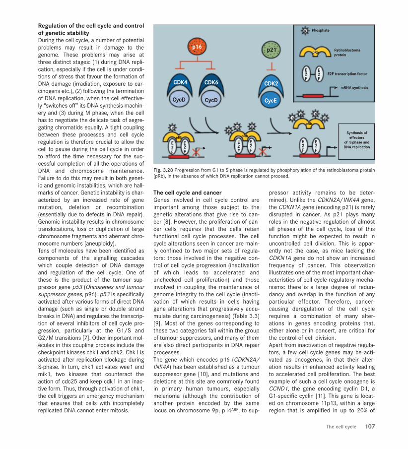
Regulation of the cell cycle and controlof genetic stabilityDuring the cell cycle, a number of potentialproblems may result in damage to thegenome. These problems may arise atthree distinct stages: (1) during DNA repli-cation, especially if the cell is under condi-tions of stress that favour the formation ofDNA damage (irradiation, exposure to car-cinogens etc.), (2) following the terminationof DNA replication, when the cell effective-ly “switches off” its DNA synthesis machin-ery and (3) during M phase, when the cellhas to negotiate the delicate task of segre-gating chromatids equally. A tight couplingbetween these processes and cell cycleregulation is therefore crucial to allow thecell to pause during the cell cycle in orderto afford the time necessary for the suc-cessful completion of all the operations ofDNA and chromosome maintenance.Failure to do this may result in both genet-ic and genomic instabilities, which are hall-marks of cancer. Genetic instability is char-acterized by an increased rate of genemutation, deletion or recombination(essentially due to defects in DNA repair).Genomic instability results in chromosometranslocations, loss or duplication of largechromosome fragments and aberrant chro-mosome numbers (aneuploidy).Tens of molecules have been identified ascomponents of the signalling cascadeswhich couple detection of DNA damageand regulation of the cell cycle. One ofthese is the product of the tumour sup-pressor gene p53 (Oncogenes and tumoursuppressor genes, p96). p53 is specificallyactivated after various forms of direct DNAdamage (such as single or double strandbreaks in DNA) and regulates the transcrip-tion of several inhibitors of cell cycle pro-gression, particularly at the G1/S andG2/M transitions [7]. Other important mol-ecules in this coupling process include thecheckpoint kinases chk1 and chk2. Chk1 isactivated after replication blockage duringS-phase. In turn, chk1 activates wee1 andmik1, two kinases that counteract theaction of cdc25 and keep cdk1 in an inac-tive form. Thus, through activation of chk1,the cell triggers an emergency mechanismthat ensures that cells with incompletelyreplicated DNA cannot enter mitosis.
The cell cycle and cancerGenes involved in cell cycle control areimportant among those subject to thegenetic alterations that give rise to can-cer [8]. However, the proliferation of can-cer cells requires that the cells retainfunctional cell cycle processes. The cellcycle alterations seen in cancer are main-ly confined to two major sets of regula-tors: those involved in the negative con-trol of cell cycle progression (inactivationof which leads to accelerated andunchecked cell proliferation) and thoseinvolved in coupling the maintenance ofgenome integrity to the cell cycle (inacti-vation of which results in cells havinggene alterations that progressively accu-mulate during carcinogenesis) (Table 3.3)[9]. Most of the genes corresponding tothese two categories fall within the groupof tumour suppressors, and many of themare also direct participants in DNA repairprocesses.The gene which encodes p16 (CDKN2A/INK4A) has been established as a tumoursuppressor gene [10], and mutations anddeletions at this site are commonly foundin primary human tumours, especiallymelanoma (although the contribution ofanother protein encoded by the samelocus on chromosome 9p, p14ARF, to sup-
pressor activity remains to be deter-mined). Unlike the CDKN2A/INK4A gene,the CDKN1A gene (encoding p21) is rarelydisrupted in cancer. As p21 plays manyroles in the negative regulation of almostall phases of the cell cycle, loss of thisfunction might be expected to result inuncontrolled cell division. This is appar-ently not the case, as mice lacking theCDKN1A gene do not show an increasedfrequency of cancer. This observationillustrates one of the most important char-acteristics of cell cycle regulatory mecha-nisms: there is a large degree of redun-dancy and overlap in the function of anyparticular effector. Therefore, cancer-causing deregulation of the cell cyclerequires a combination of many alter-ations in genes encoding proteins that,either alone or in concert, are critical forthe control of cell division.Apart from inactivation of negative regula-tors, a few cell cycle genes may be acti-vated as oncogenes, in that their alter-ation results in enhanced activity leadingto accelerated cell proliferation. The bestexample of such a cell cycle oncogene isCCND1, the gene encoding cyclin D1, aG1-specific cyclin [11]. This gene is locat-ed on chromosome 11p13, within a largeregion that is amplified in up to 20% of
The cell cycle 107
Fig. 3.28 Progression from G1 to S phase is regulated by phosphorylation of the retinoblastoma protein(pRb), in the absence of which DNA replication cannot proceed.
WCR-S3.Q (composition) 27/01/03 9:27 Page 107

several carcinomas (e.g. breast, head andneck, oesophageal and lung cancers).There is also limited evidence for tran-scriptional activation of cyclin A (an S-
phase cyclin) and for activating mutationsof CDK4 (one of the partners of cyclin D1)in some cancers. Indeed, the high com-plexity of cell cycle effectors provides an
extremely diverse range of possibilities forcancer-associated alterations. In thisrespect, cancer can be seen as, funda-mentally, a disease of the cell cycle.
108 Mechanisms of tumour development
1. Hunt T (1989) Maturation promoting factor, cyclin andthe control of M-phase. Curr Opin Cell Biol, 1: 268-274.
2. Pines J (1995) Cyclins and cyclin-dependent kinases: abiochemical view. Biochem J, 308 (Pt 3): 697-711.
3. Sherr CJ, Roberts JM (1999) CDK inhibitors: positiveand negative regulators of G1-phase progression. GenesDev, 13: 1501-1512.
4. Weinberg RA (1995) The retinoblastoma protein andcell cycle control. Cell, 81: 323-330.
5. Hartwell LH, Weinert TA (1989) Checkpoints: controlsthat ensure the order of cell cycle events. Science, 246:629-634.
6. Zeng Y, Forbes KC, Wu Z, Moreno S, Piwnica-Worms H,Enoch T (1998) Replication checkpoint requires phospho-rylation of the phosphatase Cdc25 by Cds1 or Chk1.Nature, 395: 507-510.
7. Hainaut P, Hollstein M (2000) p53 and human cancer:the first ten thousand mutations. Adv Cancer Res, 77: 81-137.
8. Hartwell LH, Kastan MB (1994) Cell cycle control andcancer. Science, 266: 1821-1828.
9. Kinzler KW, Vogelstein B (1997) Cancer-susceptibilitygenes. Gatekeepers and caretakers. Nature, 386: 761, 763.
10. Strohmaier H, Spruck CH, Kaiser P, Won KA, SangfeltO, Reed SI (2001) Human F-box protein hCdc4 targetscyclin E for proteolysis and is mutated in a breast cancercell line. Nature, 413: 316-322.
11. Schuuring E (1995) The involvement of the chromo-some 11q13 region in human malignancies: cyclin D1 andEMS1 are two new candidate oncogenes--a review. Gene,159: 83-96.
REFERENCESAnimation of the phases of the cell cycle and of mitosis:http://www.cellsalive.com/
Nature Reviews, “Focus on cell division”:http://www.nature.com/ncb/celldivision/
The Forsburg laboratory home pages, a guide to the cellcycle and DNA replication in S. pombe:http://pingu.salk.edu/~forsburg/lab.html
WEBSITES
The ends of eukaryotic chromosomes arereferred to as telomeres. These containmany copies of a repetitive DNAsequence, which in vertebrates is thehexanucleotide TTAGGG. The telomeres ofnormal human somatic cells shorten by50 to 150 base pairs every time cell divi-sion occurs. This appears to act as a celldivision counting mechanism: when acell's telomeres have shortened below acritical length, the cell exits permanentlyfrom the cell cycle. Normal cells thushave a limited proliferative capacity, andthis acts as a major barrier against car-cinogenesis. Cells that have accumulatedsome carcinogenic changes are unable toform clinically significant cancers unlessthis proliferation barrier is breached.More than 85% of all cancers achieve thisby expressing an enzyme, telomerase,that synthesizes new telomeric DNA toreplace the sequences lost during celldivision (Shay JW, Bacchetti S, Eur JCancer, 33A: 787-791, 1997).
Telomerase assays have not yet enteredroutine clinical practice, but there is con-siderable interest in their possible use forcancer diagnosis and prognosis. For exam-ple, telomerase assays of urine sedimentsmay be useful for diagnosis of urinary tractcancer (Kinoshita H et al., J Natl CancerInst, 89: 724-730, 1997), and telomeraseactivity levels may be a predictor of out-come in neuroblastoma (Hiyama E et al.,Nature Medicine, 1: 249-255, 1995).
The catalytic subunit of human telomerase,hTERT, was cloned in 1997 (Lingner J, CechTR, Curr Opin Genet Dev 8: 226-232, 1998). Ithas subsequently been shown that geneticmanipulations of hTERT which result in inhi-bition of telomerase activity in tumour cellslimit their proliferation and often result in celldeath. This raises the possibility that telom-erase inhibitors may be a very useful form oftherapy for many or most types of cancer.However, in tumours with long telomeres, itmay take many cell divisions before telom-erase inhibitors exert an anti-tumour effect.When such drugs are developed they willtherefore need to be carefully integrated withother anticancer treatments.
A potential challenge facing telomeraseresearch is the finding that some cancersmaintain their telomeres by a mechanismthat does not involve telomerase,referred to as alternative lengthening oftelomeres, ALT (Bryan TM et al., NatureMedicine, 3: 1271-1274, 1997; Reddel RR, J Clin Invest, 108: 665-667, 2001).
TELOMERES AND TELOMERASE
Fig. 3.29 Telomeres contain repetitive DNAsequences that cap the ends of chromosomes.Quantitative fluorescence in situ hybridizationanalysis of human metaphase chromosomespreads is shown, using oligonucleotide probesspecific for telomere (white) and centromere (red)DNA sequences, and the DNA dye DAPI (blue). From the laboratory of Drs J.W. Shay and W.E.Wright.
WCR-S3.Q (composition) 27/01/03 9:27 Page 108

In complex organisms, neighbouring cellsbehave and function in harmony for thebenefit of the whole organism through theoperation of cell-cell communication.During evolution, various types of intercel-lular communication have developed,which in mammals take two forms: (1)humoral communication and (2) cell con-tact-mediated communication (Fig. 3.30).Humoral communication is typically medi-ated by molecules, such as growth factorsand hormones, excreted from certain cellsand received by receptors of other cells.Intercellular communication based ondirect cell-cell contact is mediated by var-ious junctions, including adherence junc-tions, desmosomes and gap junctions.During multistage carcinogenesis, genescritically involved in cell growth arealtered [1]. Most such genes are known tobe directly or indirectly involved in thecontrol of cell replication (The cell cycle,p104) or in the death of individual cells [2](Apoptosis, p113). Genes involved in inter-cellular communication control cellulargrowth at another level. These genes func-tion to maintain cell growth in harmonywith that of the surrounding tissue. Sincemost cancer cells do not proliferate in har-
mony with normal neighbouring cells, it isnot surprising that the function of genesinvolved in intercellular communicationmechanisms is disrupted in manytumours. Thus, several oncogenes (Onco-genes and tumour suppressor genes, p96)encode products involved in humoral inter-cellular communication: c-erb, c-erbB2and c-SIS [3]. It has also become clearthat cell contact-mediated intercellularcommunication plays a crucial role in cellgrowth control [4] and genes involved areoften classified as tumour suppressorgenes [5]. Cell adhesion molecules arealso involved in cell-cell recognition. Thereare several lines of evidence which sug-gest that aberrant functions of cell adhe-sion may be involved in tumour invasionand metastasis [6].
Gap junctional intercellular communi-cation and cancerGap junctional intercellular communica-tion is the only means by which cellsexchange signals directly from the interi-or of one cell to the interior of surround-ing cells [7]. Since the extent to whichtumour cells deviate from cells whichexhibit tissue homeostasis is fundamen-
tal to the nature of malignancy, it has longbeen postulated that gap junctional inter-cellular communication is disturbed incancer. The first confirmatory evidencewas the observation that of a reducedlevel of gap junctional intercellular com-munication in one tumour type [8]. Thisphenomenon has now been observed inalmost all tumours [4]. Cell lines estab-lished from tumours, as well as cellstransformed in vitro, usually exhibitimpaired function in respect of gap junc-tional intercellular communication. Gapjunctional intercellular communicationbetween transformed cells and neigh-bouring normal counterparts is selective-ly defective in murine embryonicBALB/c3T3 cells (Fig. 3.31). A lack of het-erologous gap junctional intercellularcommunication between transformed andnormal cells has been observed using ratliver epithelial cell lines and rat livertumour in vivo. It appears that reducedgap junctional intercellular communica-tion is common to many tumour cells.Further studies with multistage models ofrat liver and mouse skin carcinogenesishave revealed that there is, in general, aprogressive decrease in the level of gap
CELL-CELL COMMUNICATION
SUMMARY
> Cells communicate by means of secret-ed molecules which affect neighbouringcells carrying appropriate receptors,and also by direct cell contact, includingspecifically includes gap junctions.
> Cell contact-mediated communicationthrough gap junctions is controlled byconnexin genes and is often disrupted incancer. This may contribute to uncon-trolled and autonomous growth.
> Interventions restoring gap junctioncommunication may provide a basis fortherapy.
Fig. 3.30 Relationships between cells are maintained by different types of intercellular communication,which may (B) or may not (A) require cell contact.
A. Humoral communication
B. Cell contact-mediated communication
1.Cell adhesion 2. Gap junction
Cell-cell communication 109
WCR-S3.Q (composition) 27/01/03 9:27 Page 109

junctional intercellular communicationduring carcinogenesis and tumour pro-gression.Another line of evidence, that implies acausal role for blockage of intercellularcommunication in carcinogenesis, is thatagents or genes involved in carcinogenesishave been shown to modulate gap junc-tional intercellular communication. Themouse skin tumour-promoting agent 12-O-tetradecanoylphorbol 13-acetate (TPA)inhibits gap junctional intercellular commu-nication. Many other tumour-promotingagents inhibit gap junctional intercellularcommunication [9]. In addition to suchchemicals, other tumour-promoting stimuli,such as partial hepatectomy and skinwounding, have been demonstrated toinhibit gap junctional intercellular commu-nication. Activation of various oncogenes,including those which encode src, SV-40 Tantigen, c-erbB2/neu, raf, fps and ras, alsoresults in inhibition of gap junctional inter-cellular communication. Conversely, somechemopreventive agents enhance gap junc-tional intercellular communication [10].
110 Mechanisms of tumour development
Fig. 3.31 Selective gap junctional communication: cells transformed by a chemical carcinogen (spindle-shaped and criss-crossed) communicate among themselves, but not with their surrounding non-trans-formed counterparts. When a gap junction-diffusible fluorescent dye is microinjected into a single cell(marked with star) of a transformed focus there is communication between transformed cells but not withthe surrounding non-transformed cells (A, B). Injection of the dye into a non-transformed cell which islocated near a transformed focus results in communication between non-transformed cells, but not withtransformed cells (C, D).
Fluorescence micrographs
A B
C D
Phase-contrast micrographs
Injection into non-transformed cell
Injection into transformed cell
Fig. 3.32 Role of cell-cell interaction in gene therapy. In a tumour cell population, only a few cells can be reached by vectors carrying the HSV-tk gene.Expression of the tk gene (orange) makes these cells sensitive to ganciclovir: they produce phosphorylated ganciclovir, which is toxic. As phosphorylated gan-ciclovir cannot pass through the cell membrane, theoretically only cells expressing the tk gene will die as a result of ganciclovir treatment. Transmembrane dif-fusion of phosphorylated ganciclovir from cytoplasm to cytoplasm can induce a bystander effect sufficient to eradicate a tumour cell population, even if only afew cells express the tk gene [13].
Ganciclovir(GCV)
Bystandereffect
No bystandereffectGCV-P
Transfected cellscarrying the HSV-tk gene
Tumour cell population
Movement of GVC-Pfrom cytoplasm tocytoplasm in gap
junctions
GCV-P cannot pass throughthe cell membrane
Cells carrying the tk gene arekilled by the toxic phosphorylated
GCV (GCV-P) they produce
GCV-P
WCR-S3.Q (composition) 27/01/03 9:27 Page 110
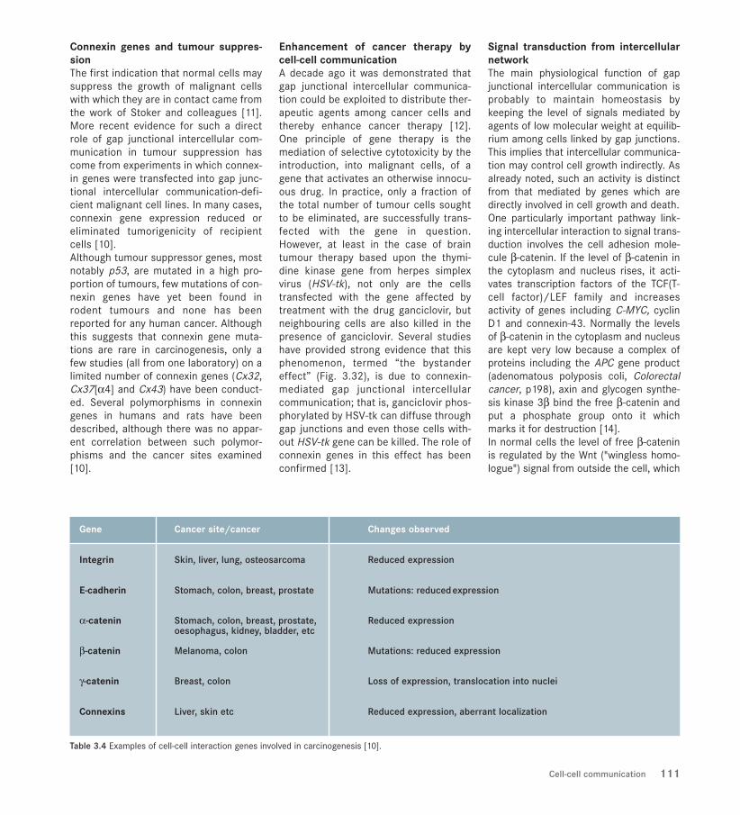
111Cell-cell communication
Table 3.4 Examples of cell-cell interaction genes involved in carcinogenesis [10].
Gene Cancer site/cancer Changes observed
Integrin Skin, liver, lung, osteosarcoma Reduced expression
E-cadherin Stomach, colon, breast, prostate Mutations: reduced expression
α-catenin Stomach, colon, breast, prostate, Reduced expressionoesophagus, kidney, bladder, etc
β-catenin Melanoma, colon Mutations: reduced expression
γ-catenin Breast, colon Loss of expression, translocation into nuclei
Connexins Liver, skin etc Reduced expression, aberrant localization
Connexin genes and tumour suppres-sionThe first indication that normal cells maysuppress the growth of malignant cellswith which they are in contact came fromthe work of Stoker and colleagues [11].More recent evidence for such a directrole of gap junctional intercellular com-munication in tumour suppression hascome from experiments in which connex-in genes were transfected into gap junc-tional intercellular communication-defi-cient malignant cell lines. In many cases,connexin gene expression reduced oreliminated tumorigenicity of recipientcells [10].Although tumour suppressor genes, mostnotably p53, are mutated in a high pro-portion of tumours, few mutations of con-nexin genes have yet been found inrodent tumours and none has beenreported for any human cancer. Althoughthis suggests that connexin gene muta-tions are rare in carcinogenesis, only afew studies (all from one laboratory) on alimited number of connexin genes (Cx32,Cx37[α4] and Cx43) have been conduct-ed. Several polymorphisms in connexingenes in humans and rats have beendescribed, although there was no appar-ent correlation between such polymor-phisms and the cancer sites examined[10].
Enhancement of cancer therapy bycell-cell communicationA decade ago it was demonstrated thatgap junctional intercellular communica-tion could be exploited to distribute ther-apeutic agents among cancer cells andthereby enhance cancer therapy [12].One principle of gene therapy is themediation of selective cytotoxicity by theintroduction, into malignant cells, of agene that activates an otherwise innocu-ous drug. In practice, only a fraction ofthe total number of tumour cells soughtto be eliminated, are successfully trans-fected with the gene in question.However, at least in the case of braintumour therapy based upon the thymi-dine kinase gene from herpes simplexvirus (HSV-tk), not only are the cellstransfected with the gene affected bytreatment with the drug ganciclovir, butneighbouring cells are also killed in thepresence of ganciclovir. Several studieshave provided strong evidence that thisphenomenon, termed “the bystandereffect” (Fig. 3.32), is due to connexin-mediated gap junctional intercellularcommunication; that is, ganciclovir phos-phorylated by HSV-tk can diffuse throughgap junctions and even those cells with-out HSV-tk gene can be killed. The role ofconnexin genes in this effect has beenconfirmed [13].
Signal transduction from intercellularnetworkThe main physiological function of gapjunctional intercellular communication isprobably to maintain homeostasis bykeeping the level of signals mediated byagents of low molecular weight at equilib-rium among cells linked by gap junctions.This implies that intercellular communica-tion may control cell growth indirectly. Asalready noted, such an activity is distinctfrom that mediated by genes which aredirectly involved in cell growth and death. One particularly important pathway link-ing intercellular interaction to signal trans-duction involves the cell adhesion mole-cule β-catenin. If the level of β-catenin inthe cytoplasm and nucleus rises, it acti-vates transcription factors of the TCF(T-cell factor)/LEF family and increasesactivity of genes including C-MYC, cyclinD1 and connexin-43. Normally the levelsof β-catenin in the cytoplasm and nucleusare kept very low because a complex ofproteins including the APC gene product(adenomatous polyposis coli, Colorectalcancer, p198), axin and glycogen synthe-sis kinase 3β bind the free β-catenin andput a phosphate group onto it whichmarks it for destruction [14]. In normal cells the level of free β-cateninis regulated by the Wnt ("wingless homo-logue") signal from outside the cell, which
WCR-S3.Q (composition) 27/01/03 9:27 Page 111

112 Mechanisms of tumour development
REFERENCES1. Fearon ER, Vogelstein B (1990) A genetic model for colorectal tumorigenesis. Cell, 61: 759-767.
2. Kinzler KW, Vogelstein B (1997) Cancer-susceptibilitygenes. Gatekeepers and caretakers. Nature, 386: 761, 763.
3. Heldin CH (1996) Protein tyrosine kinase receptors.Cancer Surv , 27: 7-24.
4. Krutovskikh V, Yamasaki H (1997) The role of gap junc-tional intercellular communication (GJIC) disorders inexperimental and human carcinogenesis. HistolHistopathol, 12: 761-768.
5. Hirohashi S (1998) Inactivation of the E-cadherin-medi-ated cell adhesion system in human cancers. Am J Pathol,153: 333-339.
6. Birchmeier EJ, Behrens J (1994) Cadherin expression incarcinoma: Role in the formation of cell junctions and pre-vention of invasiveness. Biochim Biophys Acta, 1198: 11-26.
7. Bruzzone R, White TW, Paul DL (1996) Connectionswith connexins: the molecular basis of direct intercellularsignaling. Eur J Biochem, 238: 1-27.
8. Loewenstein WR, Kanno Y (1966) Intercellular commu-nication and the control of tissue growth: lack of commu-nication between cancer cells. Nature, 209: 1248-1249.
9. Trosko JE, Chang CC, Madhukar BV, Klaunig JE (1990)Chemical, oncogene and growth factor inhibition gap junc-tional intercellular communication: an integrative hypothe-sis of carcinogenesis. Pathobiology, 58: 265-278.
10. Yamasaki H, Omori Y, Zaidan-Dagli ML, Mironov N,Mesnil M, Krutovskikh V (1999) Genetic and epigeneticchanges of intercellular communication genes during mul-tistage carcinogenesis. Cancer Detect Prev, 23: 273-279.
11. Stoker MG (1967) Transfer of growth inhibitionbetween normal and virus-transformed cells:
autoradiographic studies using marked cells. J Cell Sci, 2:293-304.
12. Yamasaki H, Katoh F (1988) Novel method for selectivekilling of transformed rodent cells through intercellularcommunication, with possible therapeutic applications.Cancer Res, 48: 3203-3207.
13. Mesnil M, Yamasaki H (2000) Bystander effect in her-pes simplex virus-thymidine kinase/ganciclovir cancergene therapy: role of gap-junctional intercellular communi-cation. Cancer Res, 60: 3989-3999.
14. Behrens J, Jerchow BA, Wurtele M, Grimm J, AsbrandC, Wirtz R, Kuhl M, Wedlich D, Birchmeier W (1998)Functional interaction of an axin homolog, conductin, withbeta-catenin, APC, and GSK3beta. Science, 280: 596-599.
increases the level by transiently reducingthe activity of the kinase. However, muta-tions in either the APC gene or in the partof the β-catenin (CTNNB1) gene whichcodes for the part of the molecule which
accepts the phosphate allow the levels ofβ-catenin to rise; when this happens theTCF/LEF-controlled genes are permanent-ly activated. The fact that mutations of theCTNNB1, AXIN1, AXIN2 and APC genes
occur in many cancers, including those ofthe colon, breast and endometrium,shows the importance of this pathway andemphasizes the connection between cell-cell contact and signal transduction.
WCR-S3.Q (composition) 27/01/03 9:27 Page 112

Apoptosis 113
Apoptosis is a mode of cell death thatfacilitates such fundamental processes asdevelopment (for example, by removal ofunwanted tissue during embryogenesis)and the immune response (for example,by elimination of self-reactive T cells).This type of cell death is distinguishedfrom necrosis both morphologically (Fig.3.33) and functionally. Specifically, apop-tosis involves single cells rather thanareas of tissue and does not provokeinflammation. Tissue homeostasis isdependent on controlled elimination ofunwanted cells, often in the context of acontinuum in which specialization andmaturation is ultimately succeeded bycell death in what may be regarded as thefinal phase of differentiation. Apart fromelimination in a physiological context,cells that have been lethally exposed tocytotoxic drugs or radiation may be sub-ject to apoptosis. The process of apoptosis can bedescribed by reference to distinct phases,termed “regulation”, “effector” and
“engulfing” respectively [1]. The regulato-ry phase includes all the signalling path-ways that culminate in commitment tocell death. Some of these pathways regu-late only cell death, but many of themhave overlapping roles in the control ofcell proliferation, differentiation, respons-es to stress and homeostasis. Critical toapoptosis signalling are the “initiator”caspases (including caspase-8, caspase-9and caspase-10) whose role is to activatethe more abundant “effector” caspases(including caspase-3 and caspase-7)which, in turn, brings about the morpho-logical change indicative of apoptosis.Finally, the engulfing process involves therecognition of cellular “remains” and theirelimination by the engulfing activity ofsurrounding cells. Identification of genes mediating apopto-sis in human cells has been critically
dependent on definition of the ced genesin the nematode Caenorhabditis elegans,members of this gene family being vari-ously homologous to human BCL2 (whichsuppresses apoptosis), APAF-1 (whichmediates caspase activation) and thecaspases themselves (proteases whichmediate cell death). The centrality ofapoptosis to cancer biology is indicatedby excess tumorigenesis in BCL2-trans-genic and p53-deficient mice. An appreci-ation of apoptosis provides a basis for thefurther development of novel and conven-tional cancer therapy
The role of cell death in tumourgrowthApoptosis, or lack of it, may be critical totumorigenesis [2]. BCL2, a gene mediat-ing resistance to apoptotic stimuli, wasdiscovered at the t(14:18) chromosomal
APOPTOSIS
SUMMARY
> The term apoptosis refers to a type ofcell death that occurs both physiologi-cally and in response to external stimuli,including X-rays and anticancer drugs.
> Apoptotic cell death is characterized bydistinctive morphological changes dif-ferent from those occurring duringnecrosis, which follows ischaemic injuryor toxic damage.
> Apoptosis is regulated by several dis-tinct signalling pathways. Dysregulationof apoptosis may result in disorderedcell growth and thereby contribute tocarcinogenesis.
> Selective induction of apoptosis in tumourcells is among current strategies for thedevelopment of novel cancer therapies.
Fig. 3.33 Apoptosis and necrosis are distinguished by characteristic morphological changes.
APOPTOSIS
Shrinking/rounding up,
fragmentationof cell and
nucleus
Condensation andfragmentation of
chromatin
Engulfing byneighbouring cell
as ‘apoptotic-body’
NECROSIS
Organelle disruptionand break-
down,cell swelling
Membraneblebbing,residual
‘ghost’ cell
WCR-S3.Q (composition) 29/01/03 14:06 Page 113

114 Mechanisms of tumour development
translocation in low grade B cell non-Hodgkin lymphoma. It thus becameapparent that neoplastic cell expansioncould be attributable to decreased celldeath rather than rapid proliferation.Defects in apoptosis allow neoplasticcells to survive beyond senescence,thereby providing protection from hypoxiaand oxidative stress as the tumour massexpands. Growth of tumours, specificallyin response to chemical carcinogens, hasbeen correlated with altered rates ofapoptosis in affected tissues as cell pop-ulations with altered proliferative activityemerge. Paradoxically, growth of somecancers, specifically including breast, hasbeen positively correlated with increasingapoptosis [3].
Interrelationships between mitogenicand apoptotic pathwaysA dynamic relationship between regula-tion of growth/mitosis and apoptosis maybe demonstrated using a variety of rele-vant signalling pathways. Many differingpromoters of cell proliferation have beenfound to possess pro-apoptotic activity[4]. Thus, ectopic expression of the C-MYConcogene (normally associated with prolif-erative activity) causes apoptosis in cul-tured cells subjected to serum deprivation(which otherwise prevents proliferation).Oncogenes that stimulate mitogenesiscan also activate apoptosis. These includeoncogenic RAS, MYC and E2F. Mutationsin E2F that prevent its interaction with theretinoblastoma protein (pRb) accelerate Sphase entry and apoptosis. A function ofpRb is to suppress apoptosis: pRb-defi-cient cells seem to be more susceptible top53-induced apoptosis. Agents such as radiation or cytotoxic drugscause cell cycle arrest and/or cell death[5]. The DNA damage caused by radiationor drugs is detected by various means (Fig.3.34). DNA-dependent protein kinase andthe ataxia-telangiectasia mutated gene(ATM) (as well as the related ATR protein)bind to damaged DNA and initiate phos-phorylation cascades to transmit damagesignals. DNA-dependent protein kinase isbelieved to play a key role in the responseto double-stranded DNA breaks. ATM playsan important part in the response to DNA
damage caused by ionizing radiation, con-trolling the initial phosphorylation of pro-teins such as p53, Mdm2, BRCA1, Chk2and Nbs1. Other sensors of DNA damageinclude mammalian homologues of thePCNA-like yeast proteins Rad1, Rad9 andHus1, as well as the yeast homologue ofreplication factor C, Rad17. Specific mole-cules detect nucleotide mismatch or inap-propriate methylation. Following exposureof mammalian cells to DNA-damagingagents, p53 is activated and among many“targets” consequently upregulated are thecyclin-dependent kinase inhibitor p21(which causes G1 arrest) and Bax (whichinduces apoptosis). Thus, the tumour sup-pressor gene p53 mediates two responsesto DNA damage by radiation or cytotoxicdrugs: cell cycle arrest at the G1 phase ofthe cell cycle and apoptosis (Oncogenesand tumour suppressor genes, p96). Theserine/threonine kinase Chk2 is also ableto positively interact with p53 and BRCA1.Chk2 and the functionally related Chk1kinase appear to have a role in the inhibi-tion of entry into mitosis via inhibition ofthe phosphatase Cdc25 (The cell cycle,p104).
The regulatory phaseTwo major apoptotic signalling pathwayshave been identified in mammalian cells(Fig. 3.37). The “extrinsic” pathway
Fig. 3.36 Apoptotic cells in an adenoma, visual-ized by immunohistochemistry (red). Apoptosis isrestricted to single cells, unlike necrosis, whichtypically involves groups of cells. Apoptosis doesnot produce an inflammatory response.
Fig. 3.34 Response to DNA damage is mediated by several signalling pathways and may include apoptosis.
DNA DAMAGE
ATM + ATR
Chk1
CELL CYCLEARREST
APOPTOSIS TRANSCRIPTION DNA REPAIR
Cdk
Cdc25 p2114-3-3θ
cAblNbs1BRCA1p53Mdm2 Chk2
Rad17Rad9, Rad1, Hus1
REPLICATION STRESS
Fig. 3.35 Apoptotic cell death requires gap junc-tional intercellular communication. Expressionand subcellular location of connexin 43 in healthy(A) and in apoptotic (B) rat bladder carcinomacells. Arrows indicate location of connexin 43 inareas of intercellular contact between apoptotic(B) and non-apoptotic (A) cells. Counterstaining ofDNA with propidium iodide reveals fragmentationof the nucleus typical of apoptosis (B).
WCR-S3.Q (composition) 27/01/03 9:27 Page 114

Apoptosis 115
depends upon the conformational changein certain cell surface receptors followingthe binding of respective ligands. The“intrinsic” pathway involves mitochondrialfunction and is initiated by growth factordeprivation, corticosteroids or DNA dam-age induced by radiation or cytotoxicdrugs.
Cell surface receptorsApoptosis may be induced by signallingmolecules, usually polypeptides such asgrowth factors or related molecules,which bind to “death” receptors on thecell surface [6]. Such cell death was ini-tially investigated in relation to theimmune response, but has much wider
ramifications. The best-characterizedreceptors belong to the tumour necrosisfactor (TNF) receptor gene superfamily[7]. In addition to a ligand-bindingdomain, death receptors contain homolo-gous cytoplasmic sequence termed the“death domain”. Members of the familyinclude Fas/APO-1/CD95 and TNF-1receptor (which binds TNFα). Activationof the Fas (or CD95) receptor by its spe-cific ligand (FasL or CD95L) results in aconformational change such that the“death domain” interacts with the adap-tor molecule FADD which then binds pro-caspase-8. In some cell types, drug-induced apoptosis is associated with Fasactivation. Ultraviolet irradiation directly
activates the Fas receptor in the absenceof ligand. TRAIL (TNF-related apoptosis-inducing ligand, Apo-2L) has 28% aminoacid identity to FasL. TRAIL induces celldeath only in tumorigenic or transformedcells and not in normal cells [8].
The regulation of apoptosis by BCL2 family genesWhile the members of the “death recep-tor” family and their ligands have struc-tural elements in common, agents andstimuli initiating the mitochondrial path-way to apoptosis are diverse. Common tothese stimuli, however, is a change inmitochondrial function, often mediated bymembers of the BCL2 family [9]. Inhumans, at least 16 homologues of BCL2have been identified. Several family mem-bers (including Bcl-2, Bcl-xL, Bcl-W) sup-press apoptosis, while others induceapoptosis and may be subdivided on thebasis of their ability to dimerize with Bcl-2 protein (Bad, Bik, Bid) or not (Bax, Bak).Phosphorylation of Bad protein by a spe-cific (Akt/PKB) and other kinases pre-vents dimerization with Bcl-2 and pro-motes cell survival. At least two distinctmechanisms of action are recognized: thebinding of Bcl-2 (or other members of thefamily) with either pro- or anti-apoptoticmembers of the Bcl-2 family or the for-mation of pores in mitochondrial mem-branes. Bcl-xL is a potent death suppres-sor that is upregulated in some tumourtypes. Bax is a death promoter that isinactivated in certain types of colon can-cer, stomach cancer and in haematopoi-etic malignancies. By dint of relevantbinding sites, Bax is under the direct tran-scriptional control of p53.
Involvement of mitochondriaApoptosis induced by cytotoxic drugs isaccompanied by critical changes in mito-chondria. Such apoptotic stimuli inducetranslocation of Bax from cytosol to mito-chondria, which induces release ofcytochrome c. Loss of transmembranepotential follows cytochrome c releaseand is dependent on caspase activation(see below), whereas cytochrome crelease is not. Bcl-2 and Bcl-xL residechiefly in the outer mitochondrial mem-
Fig. 3.37 Apoptosis occurs when specific proteases (caspases) digest critical proteins in the cell. Thecaspases are normally present as inactive procaspases. Two pathways lead to their activation. The deathreceptor pathway (at the top and left side of the figure) is triggered when ligands bind to death receptorssuch as CD95/Fas. The mitochondrial pathway is triggered by internal insults such as DNA damage aswell as by extracellular signals. In both pathways, procaspases are brought together. They then cleaveeach other to release active caspase. The binding of ligand (FasL or CD95L) to CD95 brings procaspase8 molecules together; release of mitochondrial components bring procaspases 9 together. The activecaspase 8 and 9 then activate other procaspases such as procaspase 3.
Bid
c-FLIP
FADD
CD95L
Truncated bid
Procaspase-8
Procaspase-3
Apoptosome
Caspase-3
Apoptotic substrates
Caspase-8
BclxL
Bcl2
Bax
p53
DNA damage
CD95
Procaspase-9
Cytochrome c
Apaf-1Smac/DIABLO
Apoptosis-inducingfactor (AIF)
Inhibitors ofapoptosis (IAPs)
WCR-S3.Q (composition) 27/01/03 9:27 Page 115

116 Mechanisms of tumour development
brane. Bcl-2, Bcl-xL and Bax can form ionchannels when they are added to synthet-ic membranes, and this may be related totheir impact on mitochondrial biology [10].In the cytosol after release from mito-chondria, cytochrome c activates the cas-pases through formation of a complex (the“apoptosome”) with Apaf-1 (apoptotic-protease activating factor-1), procaspase-9 and ATP. It appears that Bcl-2/Bcl-xL
may suppress apoptosis by either prevent-ing release of cytochrome c or interferingwith caspase activation by cytochrome cand Apaf-1. Sustained production of nitricoxide (NO) may cause the release of mito-chondrial cytochrome c into the cyto-plasm and thus contribute to the activa-
tion of caspases. However, nitric oxide isinvolved in several aspects of apoptosisand may act both as a promoter andinhibitor depending on conditions [11].
The effector and engulfing phasesIn mammals at least 13 proteases whichmediate the breakdown of cell structureduring apoptosis have been identified andare designated caspases-1 through -13[12]. All possess an active site cysteineand cleave substrates after aspartic acidresidues. They exist as inactive zymogens,but are activated by different processeswhich most often involve cleavage of theirpro-forms (designated procaspase-8, etc.)at particular sites, thereby generating sub-units which form active proteases consist-ing of two large and two small subunits.Proteolytic cascades may occur withsome caspases operating as upstream ini-tiators (which have large N-terminalprodomains and are activated by protein-protein interaction) and others beingdownstream effectors (activated by pro-tease cleavage). As noted earlier, at leasttwo pathways of caspase activation canbe discerned: one involving FADD or simi-lar protein-protein complexes and theother mediated by release of cytochromec. In the former, affinity labelling suggeststhat caspase-8 activates caspases-3 and -7 and that caspase-3 in turn may activatecaspase-6. On the other hand, release ofcytochrome c into the cytoplasm results inthe activation of caspase-9 which in turnactivates caspase-3. Though the intrinsic pathway to caspase-3activation may be distinguished from theextrinsic pathway (i.e. that activated byFas, etc.), some interaction is demonstra-ble. Thus, caspase-9 is able to activatecaspase-8. Nonetheless, the pathways areseparate to the extent that caspase-8 nullanimals are resistant to Fas- or TNF-induced apoptosis while still susceptibleto chemotherapeutic drugs; cells deficientin caspase-9 are sensitive to killing byFas/TNF but show resistance to drugsand dexamethasone. Finally, death ofsome cells may occur independently ofcaspase-3. Caspases-3, -7 and –9 areinactivated by proteins of the inhibitor ofapoptosis family (IAPs) which are suppres-
sors conserved throughout evolution. TheIAP protein “survivin” is overexpressed ina large proportion of human cancers.Little is known about the involvement ofcaspase mutations in cancer.
Caspase substrates and late stages ofapoptosisApoptosis was initially defined by refer-ence to specific morphological change. Infact, both mitosis and apoptosis are char-acterized by a loss of substrate attach-ment, condensation of chromatin andphosphorylation and disassembly ofnuclear lamins. These changes are nowattributable to caspase activation and itsconsequences.Most of the more than 60 known caspasesubstrates are specifically cleaved by cas-pase-3 and caspase-3 can process pro-caspases-2, -6, -7 and -9 [13]. Despite themultiplicity of substrates, protease activi-ty mediated by caspases is specific andseems likely to account for much of themorphological change associated withapoptosis. Caspases cleave key compo-nents of the cytoskeleton, including actinas well as nuclear lamins and other struc-tural proteins. Classes of enzymes cleavedby caspases cover proteins involved inDNA metabolism and repair exemplifiedby poly(ADP-ribose) polymerase and DNA-dependent protein kinase. Other classesof substrates include various kinases, pro-teins in signal transduction pathways andproteins involved in cell cycle control,exemplified by pRb. Cleavage of somesubstrates is cell-type specific. Caspaseactivity accounts for internucleosomalcleavage of DNA, one of the first charac-terized biochemical indicators of apopto-sis. ICAD/DFF-45 is a binding partner andinhibitor of the CAD (caspase-activatedDNAase) endonuclease, and cleavage ofICAD by caspase-3 relieves the inhibitionand promotes the endonuclease activityof CAD.
Therapeutic implicationsIn theory, knowledge of critical signallingor effector pathways which bring aboutapoptosis provides a basis for therapeuticintervention, including the development ofnovel drugs to activate particular path-
Fig. 3.38 Neuroblastoma cells treated with ioni-zing radiation undergo apoptosis. The TUNEL assaywas used to visualize apoptotic cells (green),before (A) and 24 hours after (B) treatment with X-rays (5 Gray). Close-up shows that the nuclei of theapoptotic cells are fragmented (C).
A
B
C
WCR-S3.Q (composition) 27/01/03 9:27 Page 116

117Apoptosis
ways. Several options are under investiga-tion [14]. More immediately, attempts arebeing made to exploit knowledge of apop-totic processes to increase the efficacy orspecificity of currently available therapy.Simple answers have not emerged. Thus,for example, relatively increased expres-sion of Bcl-2 (which, under many experi-mental conditions, inhibits apoptosis) is notnecessarily indicative of poor prognosisand the reverse appears true for some
tumour types. In experimental systems,cells acquiring apoptosis defects (e.g. p53mutations) can more readily survive hypox-ic stress and the effects of cytotoxic drugs[15]. However, clinical studies have notconsistently established that mutation ofp53 is associated with poor response tochemotherapy [16].The function of Bcl-2 family members maybe subject to interference by small mole-cules [17]. In preclinical animal models,
suppression of Bcl -2 by an antisenseoligonucleotide has been shown to retardtumour growth and the approach is cur-rently subject to clinical trial. Likewise,antisense oligonucleotides directed at“survivin” are being evaluated. The possi-bility of using recombinant TRAIL toinduce apoptosis in malignant cells isunder investigation. TRAIL is implicated asthe basis of all-trans-retinoic treatment ofpromyelocytic leukaemia [18]. Also note-
In complex multicellular organisms, cellproliferation, differentiation and survivalare regulated by a number of extracellularhormones, growth factors and cytokines.These molecules are ligands for cellularreceptors and communicate with the nucle-us of the cell through a network of intracel-lular signalling pathways. In cancer cells,key components of these signal transduc-tion pathways may be subverted by proto-oncogenes through over-expression ormutation, leading to unregulated cell sig-nalling and cellular proliferation. Because anumber of these components may be pref-erentially over-expressed or mutated inhuman cancers, the cell signalling cascadeprovides a variety of targets for anticancertherapy (Adjei AA, Current PharmaceuticalDesign, 6: 471-488, 2000).
Different approaches have been used toattack these targets and include classicalcytotoxic agents as well as small moleculedrug inhibitors. In addition, antisenseoligonucleotides, vaccines, antibodies,ribozymes and gene therapy approacheshave been utilized.
The diagram illustrates cell signallingpathways that are targeted by anticanceragents currently undergoing clinical test-
ing. The drug Gleevec is already in clinicaluse (Leukaemia, p242). It is hoped that infuture, a combination of agents targetingparallel pathways, as well as combinationswith classical cytotoxic agents will improvethe outcome of cancer patients. Classes of agents and their potential tar-gets include:
Inhibitors of ligands, such as recombinanthuman antibody to VEGF (rHu mAbVEGF)
Receptors, anti-receptor antibodies andtyrosine kinase receptor inhibitorsRAS farnesyltransferase inhibitorsRAF inhibitorsMEK inhibitorsRapamycin analoguesProtein kinase C (PKC) inhibitorsInhibitors of protein degradationInhibitors of protein trafficking
DRUGS TARGETING SIGNALTRANSDUCTION PATHWAYS
Signaling pathways targeted by anticancer agents. PI3K = phosphoinositide-3-kinase; PLC = phos-pholipase C; PKC = protein kinase C; MEK = mitogen-activated protein kinase kinase; ERK = extracellular sig-nal-regulated kinase; Akt = protein kinase B (PKB); BAD = Bcl-XL/Bcl-2-associated death protein; VEGF = vas-cular endothelial growth factor; HER = human epidermal growth factor receptor family; PDGF = plateletderived growth factor; FGF = fibroblast growth factor; SOS = son of sevenless guanine nucleotide exchangeprotein; GRB = growth factor receptor-bound protein.
Cell membrane
Intr
acel
lula
r m
esse
nger
s
Ligand
Receptor
APOPTOSISNUCLEAR TRANSCRIPTION
DNA
PI3K
Akt
BAD P7056K
PLC
DAG
PKCBclxL
GRB SOS Ras
Raf
MEK
ERK
>
> > > > > >
>
>
WCR-S3.Q (composition) 6/02/03 9:03 Page 117

118 Mechanisms of tumour development
worthy is the development of caspaseinhibitors for the treatment of certaindegenerative (non-cancerous) diseasescharacterized by excess apoptosis.Drugs shown to induce apoptosis specifi-cally include chemopreventive agents(Chemoprevention, p151), exemplified by
4-hydroxyphenylretinamide. Butyrate, ashort-chain fatty acid produced by bacter-ial fermentation of dietary fibre, inhibitscell growth in vitro and promotes differen-tiation; it also induces apoptosis. Bothroles may contribute to its prevention ofcolorectal cancer. Moreover, cyclo-oxyge-
nase enzyme (COX-2) expression maymodulate intestinal apoptosis via changesin Bcl-2 expression. Aspirin and similardrugs which inhibit COX-2 may promoteapoptosis and prevent tumour formation.
1. Strasser A, O'Connor L, Dixit VM (2000) Apoptosis sig-naling. Annu Rev Biochem, 69: 217-245.
2. Kaufmann SH, Gores GJ (2000) Apoptosis in cancer:cause and cure. Bioessays, 22: 1007-1017.
3. Parton M, Dowsett M, Smith I (2001) Studies of apop-tosis in breast cancer. BMJ, 322: 1528-1532.
4. Choisy-Rossi C, Yonish-Rouach E (1998) Apoptosis andthe cell cycle: the p53 connection. Cell Death Differ, 5:129-131.
5. Rich T, Allen RL, Wyllie AH (2000) Defying death afterDNA damage. Nature, 407: 777-783.
6. Peter ME, Krammer PH (1998) Mechanisms of CD95(APO-1/Fas)-mediated apoptosis. Curr Opin Immunol, 10:545-551.
7. Yeh WC, Hakem R, Woo M, Mak TW (1999) Gene tar-geting in the analysis of mammalian apoptosis and TNFreceptor superfamily signaling. Immunol Rev, 169: 283-302.
8. Griffith TS, Lynch DH (1998) TRAIL: a molecule withmultiple receptors and control mechanisms. Curr OpinImmunol, 10: 559-563.
9. Gross A, McDonnell JM, Korsmeyer SJ (1999) BCL-2family members and the mitochondria in apoptosis. GenesDev, 13: 1899-1911.
10. Matsuyama S, Llopis J, Deveraux QL, Tsien RY, ReedJC (2000) Changes in intramitochondrial and cytosolic pH:
early events that modulate caspase activation during apop-tosis. Nat Cell Biol, 2: 318-325.
11. Chung HT, Pae HO, Choi BM, Billiar TR, Kim YM (2001)Nitric oxide as a bioregulator of apoptosis. BiochemBiophys Res Commun, 282: 1075-1079.
12. Kumar S (1999) Regulation of caspase activation inapoptosis: implications in pathogenesis and treatment ofdisease. Clin Exp Pharmacol Physiol, 26: 295-303.
13. Porter AG, Janicke RU (1999) Emerging roles of cas-pase-3 in apoptosis. Cell Death Differ, 6: 99-104.
14. Nicholson DW (2000) From bench to clinic with apop-tosis-based therapeutic agents. Nature, 407: 810-816.
15. Zhou BB, Elledge SJ (2000) The DNA damageresponse: putting checkpoints in perspective. Nature, 408:433-439.
16. Brown JM, Wouters BG (1999) Apoptosis, p53, andtumor cell sensitivity to anticancer agents. Cancer Res, 59:1391-1399.
17. Zheng TS (2001) Death by design: the big debut ofsmall molecules. Nat Cell Biol, 3: E43-E46.
18. Altucci L, Rossin A, Raffelsberger W, Reitmair A,Chomienne C, Gronemeyer H (2001) Retinoic acid-inducedapoptosis in leukemia cells is mediated by paracrine actionof tumor-selective death ligand TRAIL. Nat Med, 7: 680-686.
REFERENCESThe European Cell Death Organization:http://www.ecdo.dote.hu/
WEBSITE
WCR-S3.Q (composition) 27/01/03 9:27 Page 118

Invasion and metastasis 119
Metastasis (from the Greek meaning“change in location”) refers to growth ofsecondary tumours at sites distant from aprimary neoplasm. Metastasis thus dis-tinguishes benign from malignant lesionsand is the ultimate step in the multistageprocess of tumour progression.Metastatic growth is the major cause oftreatment failure and the death of cancerpatients. Although secondary tumoursmay arise by shedding of cells within bodycavities, the term metastasis is generallyreserved for the dissemination of tumourcells via the blood or lymphatics. Spreadin the cerebrospinal fluid andtranscoelomic passage may also occur.Most (60-70%) cancer patients have overtor occult metastases at diagnosis, andthe prognosis of the majority of thesepatients is poor (Box: TNM Classificationof Malignant Tumours, p124).There is a critical need to identify reliableindicators of metastatic potential, sinceclinical detection of metastatic spread is
synonymous with poor prognosis. Currentmethods of detecting new tumours,including computed tomography (CT)scans or magnetic resonance imaging(MRI), ultrasound, or measurement of cir-culating markers such as carcinoembry-onic antigen (CEA), prostate-specific anti-gen (PSA) or cancer antigen 125 (CA125)are not sufficiently sensitive to detectmicrometastases. A greater understand-ing of the molecular mechanisms ofmetastasis is required. It is clear thatmetastatic growth may reflect both gainand loss of function, and indeed thesearch for “metastasis suppressor” geneshas been more fruitful than identificationof genes which specifically and reliablypotentiate metastasis [1].
The genetics of metastasisWith the publication of the humangenome sequence, and various major ini-tiatives such as the Cancer GenomeProject in the UK and the Cancer GenomeAnatomy Project in the USA, the searchfor genes selectively upregulated, mutat-
INVASION AND METASTASIS
SUMMARY
> The ability of tumour cells to invade and colonize distant sites is a major feature distinguishing benign growths from malignant cancer.
>Most human tumours lead to death throughwidespread metastasis rather than theadverse local effects of the primary neo-plasm.
> Often, metastatic spread first involves regional lymph nodes, followed byhaematogenous spread throughout thebody. Metastases may become clinicallymanifest several years after surgical resec-tion of the primary tumour.
> Current methods are inadequate for the routine detection of micrometastases andthe search for effective, selective therapies directed toward metastaticgrowth remains a major challenge.
Fig. 3.41 The stages in the metastatic process, illustrated in relation to the spread of a primary tumourfrom a surface epithelium to the liver.
1.Localized tumour 2.Breakthrough 3.Invasion
7.Metastasis6.Extravasation5.Lodgement
4.Transport
• Growth • Angiogenesis • Protease activation
• Selection
• Adhesion• Protease production
• Proliferation • Angiogenesis• Protease activation/production
• Metastasis of metastases
• Decreased cell-cell adhesion• Increased protease production• Increased cell-matrix adhesion
Fig. 3.40 The hypoxia hypothesis suggests thatthe progression of malignant tumours to ametastatic phenotype is mediated by deficiency ofoxygen and resulting tumour necrosis.
Malignant tumour
Growth
Viable cells
Zone of sublethalhypoxia
Necrosis
Angiogenicfactors
Cytokines
Growthfactors
Inflammatorycells
Rapid accumulationof genetic damagein sublethal zone
mediated by oxygenfree radicals
(NO= nitric oxide)
Reperfusion of sub-lethal zone containing
cells with varyingdegrees of genetic
damage. Some haveacquired a metastatic
phenotype
Metastasis
WCR-S3.Q (composition) 27/01/03 9:27 Page 119

120 Mechanisms of tumour development
ed or lost in metastatic cancers (Table3.5) has gained momentum. It is now pos-sible, using laser capture microdissectionand serial analysis of gene expression(SAGE), to isolate invasive cancer cellsand compare their gene or protein expres-sion with non-invasive or normal cellsfrom the same patient [2]. Prior to this,transfection of chromosomes or DNAfrom metastatic to non-metastatic cells(or vice versa), subtractive hybridiza-tion/differential display PCR, cDNA arrayand other strategies resulted in identifica-tion of some genes specifically linked to
metastasis, although many others so iden-tified are also associated with tumourgrowth or developmental processes.The events which lead to cancer metasta-sis include changes in cell-cell and cell-matrix adhesion, alterations in cell shape,deformability and motility, invasion of sur-rounding normal tissues, gaining accessto lymphatic or vascular channels, dis-semination via blood or lymph, survival ofhost defence mechanisms, extravasationand colonization of secondary sites (Fig.3.41). There are now many features ofcancer cells recognized to potentiate
metastasis, and a great deal is knownabout the cellular and molecular eventsthat underlie the process. However theability to predict which patient has occultmicrometastases, and the discovery ofeffective, selective therapies for metasta-tic disease, remain major challenges inoncology.
The biology of metastasisGrowth of tumours beyond a few millime-tres in diameter cannot progress withoutneovascularization, and there is a growingappreciation of how this phenomenon islinked to metastasis [3]. Many geneticchanges associated with malignant pro-gression (mutation of HRAS, over-expres-sion of ERBB2 oncogenes, loss of p53)induce an angiogenic phenotype (devel-oping blood vessels) via induction ofcytokines, such as vascular endothelialgrowth factor (VEGF-A). VEGF-A is alsoupregulated by hypoxia in tumours, partlyby host cells such as macrophages. Thepresence of hypoxic areas is a character-istic of solid tumours and has been relat-ed to poor response to conventional ther-apies (Fig. 3.40). In addition, activation ofepithelial growth factor receptor (EGFR)and other oncogenic signalling pathwayscan also upregulate VEGF-C, a knownlymphangiogenic cytokine [4]. The recep-tors for these cytokines (Flk-1 and Flk-4)are expressed on tumour vasculature,and both (in addition to acting as potentmitogens for endothelial cells) alsoenhance vessel permeability. Thus activa-tion of these signalling pathways maypotentiate both vascular and lymphaticinvasion and tumour spread. Basic fibrob-last growth factor (bFGF) is often upregu-lated in cancers, particularly at the inva-sive edge where tumour cells interactwith host cells [5].Epithelial cells are normally bounded bybasement membranes which separatethem from the underlying stroma andmesenchymal compartments. Breachingthis barrier is the first step in the transi-tion from carcinoma in situ to invasiveand potentially metastatic carcinoma.Basement membrane is composed of avariety of structural proteins includingcollagen IV (the major component),
Fig. 3.42 Multiple metastatic growths of an intes-tinal carcinoma in the liver.
Fig. 3.43 Multiple metastases to the brain from alung carcinoma.
Gene Cancer type(s) Mechanism
nm23 Family (H1-6) of Breast Cell migration?nucleoside diphosphate (liver, ovary, melanoma) Signalling via G proteins,kinases microtubule assembly
PTEN/MMAC1 Prostate, glioma, breast Migration, focal adhesions
KAI1/CD82/C33 Prostate, stomach, colon, Cell-cell adhesion, motilitybreast, pancreas, lung
CAD1/E-cadherin Many adenocarcinomas Cell-cell adhesion, epithelial organization
MKK4/SEK1 Prostate Cellular response to stress?
KiSS-1 Melanoma, breast cancer Signal transduction? Regulation of MMP-9?
BRMS1 Breast Cell communication, motility
DPC4 Colon, pancreas ?
Table 3.5 Putative metastasis suppressor genes.
WCR-S3.Q (composition) 27/01/03 9:27 Page 120

Invasion and metastasis 121
laminin, entactin and also heparan sulfateproteoglycans. Interactions of tumourcells with the basement membrane havebeen considered to comprise three steps,which can readily be demonstrated invitro: adhesion, matrix dissolution/prote-olysis and migration [6].Epithelial cells are normally polarized andfirmly attached to each other via desmo-somes, tight junctions and intercellularadhesion molecules such as E-cadherin,and also bound to the basement mem-brane via other adhesion moleculesincluding integrins. Changes in cell-celland cell-matrix adhesive interactions arecommon in invasive cancer (Cell-cell com-munication, p109). Indeed, E-cadherinmay be designated a tumour suppressorgene, since its loss or functional inactiva-tion is one of the most common charac-teristics of metastatic cancer, and its re-introduction into cells can reverse themalignant phenotype. The adenomatouspolyposis coli gene (APC), which is mutat-ed in many inherited and sporadic coloncancers, normally regulates the expres-sion of β-catenin, a protein which inter-acts with E-cadherin. Mutations in APC(or β-catenin) increase cellular levels ofthe latter and facilitate interactions withtranscription factors such as T-cell fac-tor/lymphoid enhancer factor (TCF/LEF)which drive the expression of genesinvolved in inhibiting apoptosis and stim-ulating cell proliferation. Other genescommonly lost in cancers (e.g. DCC,Deleted in Colon Carcinoma) also encodeadhesion molecules.
IntegrinsIntegrins are heterodimeric proteins thatmediate adhesion between cells and theextracellular matrix or other cellular ele-ments. Ligand specificity is determined bythe subunit composition; many integrinsbind multiple substrates and others aremore selective. Far from being an inert“glue”, they are capable of transmittingimportant signals regulating cell survival,differentiation and migration [7]. Many dif-ferences in integrin expression betweenbenign and malignant cells have been doc-umented, but the patterns are complex. Inaddition, their expression and binding
affinity can be profoundly influenced bythe local microenvironment and solublefactors, enabling the tumour cell torespond to different conditions encoun-tered throughout the metastatic cascade.
Other molecules involved in adhesionOther adhesion molecules implicated incancer progression include selectins suchas sialyl Lex and members of theimmunoglobulin superfamily, includingintercellular adhesion molecules (ICAM-1,ICAM-2, VECAM and PECAM). The latterare upregulated on activated endothelialcells, and can interact with integrins onleukocytes and circulating tumour cells,assisting their arrest and extravasation.CD44 is another adhesion molecule uti-
lized during lymphocyte “homing”, and achange from the standard “epithelial” pat-tern to expression of splice variants asso-ciated with haematopoietic cells hasbeen proposed to assist carcinoma cellsin haematogenous dissemination. Throm-bospondin may mediate adhesion bet-ween circulating tumour cells, plateletsand endothelial cells, promoting emboliza-tion (vessel obstruction) and arrest.Tumour cells then gain access to the sub-endothelial basement membrane whenendothelial cells retract in response tothese emboli, and can adhere to exposedproteins. Synthetic peptides containingsequences of amino acids which competewith binding to laminin or fibronectin caninhibit colonization of the lung by intra-
Fig. 3.44 MRI scan showing skeletal metastases in a patient with a primary prostatic carcinoma (frontand back views). Some of the larger metastases are marked by arrows. Note the numerous metastasesin the ribs and in the spine.
WCR-S3.Q (composition) 27/01/03 9:28 Page 121

122 Mechanisms of tumour development
venously injected cells in experimentalmodels.
RHOThe RHO gene family of small GTP-hydrolysing proteins contains severalmembers known to be involved in cellmigration via regulation of actomyosin-based cytoskeletal filament contractionand the turnover of sites of adhesion.Overexpression of RhoC alone inmelanoma cells is sufficent to induce ahighly metastatic phenotype [8].
Enzyme functions in invasion andmetastasisInvasive tumour cells show increasedexpression of many enzymes due toupregulation of genes, enhanced activa-tion of pro-enzymes or reduced expres-sion of inhibitors such as tissue inhibitorsof metalloproteinases (TIMPs). In addi-tion, tumour cells may also induce expres-sion of enzymes by neighbouring hostcells and “hijack” these to potentiateinvasion.
Matrix metalloproteinasesOne important group is the matrix metal-loproteinases (MMP). Different cancersmay show different patterns of expres-sion; for instance squamous carcinomasfrequently have high levels of gelatinase B(MMP-9), stromelysins 1-3 (MMP-3,MMP-10 and MMP-11, normally stromalenzymes, but also expressed by thesecarcinomas) and matrilysin (MMP-7).Adenocarcinomas such as breast mayhave increased levels of gelatinase A(MMP-2) and colon carcinomas common-ly overexpress MMP-7. In addition, MT1-MMP, which activates MMP-2, is oftenupregulated in tumour and/or neighbour-ing host tissues. The major substrate ofthe gelatinases is collagen IV, a majorcomponent of the basement membrane,whereas the stromelysins prefer laminin,fibronectin and proteoglycans, and canalso activate procollagenase (MMP-1),which in turn degrades the fibrillar colla-gens of the interstitial tissues. Urokinaseplasminogen activator (uPA) is also fre-quently upregulated in cancer. It controlsthe synthesis of plasmin, which degrades
laminin and also activates gelatinases.Thus, upregulation of these enzymes incancers leads to proteolytic cascades andpotential for invasion of the basementmembrane and stroma. Metalloproteinases also contribute totumour growth and metastasis by othermeans [9]. During angiogenesis, “inva-sion” of capillary sprouts requires localproteolysis (mediated in part by upregu-lated MMP-2 and MMP-9 together withuPA) and in addition MMP-9 has beenimplicated in the “angiogenic switch” byreleasing VEGF from sequestration in theextracellular matrix [10]. Furthermore,these proteases can contribute to thesustained growth of tumours by theectodomain cleavage of membrane-bound pro-forms of growth factors, andthe release of peptides which are mito-genic and chemotactic for tumour cells.
HeparanaseApart from the structural proteins cleavedby metalloproteinases in the basementmembrane and extracellular matrix, theother major components are glycos-aminoglycans, predominantly heparansulfate proteoglycan (HSPG). Heparanaseis an important enzyme which degradesthe heparan sulfate side-chains of HSPGsand, like the proteases described above,not only assists in the breakdown of extra-cellular matrix and basement membrane,but is also involved in the regulation ofgrowth factor and cytokine activity. Basicfibroblast growth factor (bFGF, anotherpotent mitogen and chemotactic factorfor endothelial cells) and other heparin-binding growth factors are sequestered byheparan sulfate, providing a localizeddepot available for release by heparanase.Similarly, uPA and tissue plasminogenactivator (tPA) can be released fromheparan sulfate by heparanase, furtherpotentiating proteolytic and mitogeniccascades.
Tissue-specific growth factorsFinally, it is possible that release of tissue-specific growth factors may play a role inorgan selectivity of metastasis. For exam-ple, colorectal carcinoma cells over-expressing EGFR have a predilection for
Fig. 3.45 Location of metastases at autopsy forsome common cancers, indicating that the site ofmetastasis is not random.
Table 3.6 Some sites of metastasis which are notexplicable by circulatory anatomy.
Primary tumour Site of metastasis
Bronchial cancer Adrenal (often bilateral)
Breast ductal Livercarcinoma
Breast lobular Diffuse peritonealcarcinoma seeding
Breast Bone, ovary
Lung Brain
Ocular melanoma Liver
Prostate Bone
Melanoma Brain
WCR-S3.Q (composition) 27/01/03 9:28 Page 122

Invasion and metastasis 123
growth in the liver where there are highconcentrations of its ligands. All of theserequire proteolytic cleavage for activation.Other enzymes which have been implicat-ed in metastasis include the cysteine pro-teinases, notably cathepsins B and D. Formost of the enzymes described, there areactive research programmes seekingselective inhibitors (some of which havereached phase II and III clinical trials) toprevent or treat metastatic disease. Motility, coupled with proteolysis, is thebasis of tumour cell invasion, and is alsoimportant during intravasation andextravasation of blood and lymphatic ves-sels. Many motility factors have beendescribed which may be tumour- or host-derived. Many growth factors, such as
transforming growth factor alpha, epider-mal growth factor and platelet-derivedgrowth factor, can induce chemotacticresponses in tumour cells expressing thecognate receptors. Scatter factor (alsoknown as hepatocyte growth factor, HGF)is a potent host-derived motility factor,and tumour cells themselves secrete avariety of autocrine motility factors includ-ing autotaxin and neuroleukin/phospho-hexose isomerase.
Organ preference of metastasesThe organ distribution of metastasesdepends on the type and location of theprimary tumour, with 50-60% of the sec-ondary sites being dictated by theanatomical route followed by the dissemi-
nating cells. Most metastases occur inthe first capillary bed or lymph nodeencountered. The number of involvednodes is a key prognostic factor for manycancers, and this has led to efforts toidentify “sentinel” lymph nodes in orderto improve predictions of cancer spread.Lymphatic channels present less of achallenge to tumour cell entry than capil-laries since they have scanty basementmembrane. Once in the lymphatics,tumour cells are carried to the subcapsu-lar sinus of draining nodes, where theymay arrest and grow, succumb to hostdefences, or leave the node via the effer-ent lymphatics. The propensity for atumour cell to generate a lymphaticmetastasis may depend upon its ability to
Table 3.7 Therapeutic agents directed towards stroma-tumour interactions.
Target Example of agent Comments
Adhesion/attachment RGD-toxin constructs and RGD-targeted Have not reached clinical trialsgene therapy
Anti-avfl3 monoclonal antibody Cytostasis in patients; anti-tumour and (Vitaxin, Medi522) anti-angiogenic in animal models
Proteolysis Matrix metalloproteinase inhibitors Cytostatic in patients; rare occurence of tumour partial regressions; stromal fibrosis;activity seen in multiple animal models andin combination with chemotherapy; new agents with varied MMP specificity under development
Motility No selective agents
Signal pathways Squalamine (NHE-3 inhibitor) Selective for endothelial cells
PDGFR, KDR and EGFR small molecule Active in vitro in animal models; preclinicalinhibitors activity in combinations; phase I trials
completed for several agents, some tumour stabilization or regression
Anti-EGFR monoclonal antibody Neutralizing antibody; active in vitro in (C225) animal models; phase I trials ongoing
Anti-VEGF antibody Blocking antibody; active in vitro in animal models; preclinical activity alone and in combination; phase I-III trials ongoing
CAI (non-voltage-gated Ca++ uptake Active in vitro in animal models; preclinicalinhibitor) activity in combinations; phase I trials of single
agents and combinations, some tumour stabilization or regression
Extracellular matrix Pirfenidone Suppresses stromal/inflammatory cellRemodelling by stromal expression of TGF-βPhase I trials for pulmonary fibrosis
WCR-S3.Q (composition) 27/01/03 9:28 Page 123

124 Mechanisms of tumour development
The TNM system for the classification ofmalignant tumours (http://tnm.uicc.org/)is a form of clinical shorthand used todescribe the anatomic extent (staging) ofa cancer in terms of:T - the primary tumour N - regional lymph nodesM - distant metastasesThese components are given a numberthat reflects the absence or presence andextent of the disease. For example, atumour of the colon that is classified asT2N1M0 would have extended into thecolon’s muscular wall, spread to 1 to 3regional lymph nodes but without evi-dence of distant metastasis. Evaluationby the TNM system can therefore help inthe planning of treatment by the oncolo-gist and in monitoring the efficacy of thistreatment, as well as giving some indica-tion of prognosis. Moreover, the use of astandardized system facilitates the dis-semination of information in the clinicalcommunity.The TNM system was developed by PierreDenoix (President of the UICC, 1973-1978)between 1943 and 1952 (Sobin LH, TNM –principles, history and relation to otherprognostic factors. Cancer, 91:1589-92,2001). In 1968, a series of brochures pub-lished by UICC describing the classifica-tion of cancers at 23 body sites were com-bined to produce the Livre de Poche,which has been subject to regular re-edi-tion, enlargement and revision over sub-sequent years. In order to preventunwanted variations in the classificationby its users, in 1982 it was agreed that a
single international TNM classificationshould be formulated. This is achieved viameetings of experts that update existingclassifications, as well as develop newones. The present TNM edition (Eds. SobinLH and Wittekind Ch, TNM Classification ofMalignant Tumours, 6th Edition, Wiley,2002) contains guidelines for classificationand staging that correspond exactly withthose of the 6th edition of the AJCC CancerStaging Manual (2002). TNM, now the mostwidely used system to classify tumourspread, is published in 12 languages and isaccompanied by an illustrated TNM Atlas(Eds. Hermanek P et al., 4th Edition,Springer-Verlag, 1997), a TNM MobileEdition (Wiley, 2002) and a TNM supple-ment (Eds. Wittekind Ch et al., TNMSupplement 2001. A Commentary on
Uniform Use, 2nd Edition, Wiley 2001)with rules and explanations.The challenge for the future is the incor-poration into TNM of information fromnew diagnostic and imaging technologies(such as endoscopic ultrasound, magnet-ic resonance imaging, sentinel node bio-psy, immunohistochemistry and poly-merase chain reaction). There is anexpanding array of known and potentialprognostic factors (Eds. Gospodarowicz Met al., Prognostic Factors in Cancer, Wiley,2001) with which TNM could be integrat-ed to form a comprehensive prognosticsystem. Such integration could potential-ly be exploited to enhance the predictionof prognosis, and individualize cancerpatient treatment.
TNM CLASSIFICATION OFMALIGNANT TUMOURS
T = primary tumourTX Primary tumour cannot be assessedT0 No evidence of primary tumourTis Carcinoma in situT1 Tumour invades submucosaT2 Tumour invades muscularis propriaT3 Tumour invades through muscularis propria into subserosa or into non-
peritonealized pericolic or perirectal tissuesT4 Tumour directly invades other organs or structures and/or
perforates visceral peritoneum
N = regional lymph nodesNX Regional lymph nodes cannot be assessedN0 No regional lymph node metastasisN1 Metastasis in 1 to 3 regional lymph nodesN2 Metastasis in 4 or more regional lymph nodes
M = distant metastasisMX Distant metastasis cannot be assessedM0 No distant metastasisM1 Distant metastasis
Table 3.8 TNM classification of cancer of the colon and rectum.
adhere to reticular fibres in the subcapsu-lar sinus. These fibres contain laminin,fibronectin and collagen IV, and differentintegrins expressed by different tumourcells may be responsible for adhesion tothese structures and to the lymphaticendothelial cells [11].Sarcomas tend to metastasize to lungsbecause the venous drainage returns
there; colon carcinoma cells enter theportal circulation which delivers cells tothe liver, and so on (Fig. 3.45). However, anon-random element in metastatic pat-terns has long been recognized (Table3.6). Stephen Paget developed the “seedand soil” hypothesis in 1889, based on hisobservations from autopsies of over 700women with breast cancer. He proposed
that specific cancer cells (the seed) hadan affinity for certain organs (the soil).In experimental systems there are manyexamples showing that primary tumoursare heterogeneous, and that cloned cellscan vary in their ability to metastasize todifferent sites. Some of the patternsrelate to the ability of malignant cells toadhere to the endothelial cells in target
WCR-S3.Q (composition) 27/01/03 9:28 Page 124

Invasion and metastasis 125
1. Yoshida BA, Sokoloff MM, Welch DR, Rinker-SchaefferCW (2000) Metastasis-suppressor genes: a review and per-spective on an emerging field. J Natl Cancer Inst, 92: 1717-1730.
2. Simone NL, Paweletz CP, Charboneau L, Petricoin EF,Liotta LA (2000) Laser capture microdissection: Beyondfunctional genomics to proteomics. Mol Diagn, 5: 301-307.
3. Fidler IJ (2000) Angiogenesis and cancer metastasis.Cancer J Sci Am, 6 Suppl 2: S134-S141.
4. Eccles SA (2000) Cell biology of lymphatic metastasis.The potential role of c-erbB oncogene signalling. RecentResults Cancer Res, 157: 41-54.
5. Fidler IJ (1999) Critical determinants of cancer metas-tasis: rationale for therapy. Cancer Chemother Pharmacol,43 Suppl: S3-10.
6. Stracke ML, Liotta LA (1992) Multi-step cascade oftumor cell metastasis. In Vivo, 6: 309-316.
7. Berman AE, Kozlova NI (2000) Integrins: structure andfunctions. Membr Cell Biol, 13: 207-244.
8. Ridley A (2000) Molecular switches in metastasis.Nature, 406: 466-467.
9. McCawley LJ, Matrisian LM (2000) Matrix metallopro-teinases: multifunctional contributors to tumor progres-sion. Mol Med Today, 6: 149-156.
10. Bergers G, Brekken R, McMahon G, Vu TH, Itoh T,Tamaki K, Tanzawa K, Thorpe P, Itohara S, Werb Z, HanahanD (2000) Matrix metalloproteinase-9 triggers the angio-genic switch during carcinogenesis. Nat Cell Biol, 2: 737-744.
11. Brodt P (1991) Adhesion mechanisms in lymphaticmetastasis. Cancer Metastasis Rev, 10: 23-32.
REFERENCESThe Metastasis Research Society (UK):http://www.metastasis.icr.ac.uk
WEBSITE
organs, and to respond to local growthfactors once they have extravasated. Itused to be thought that escape from theprimary tumour and survival in the circu-lation were the major rate-limiting stepsfor successful metastasis. However, whilethere is a good deal of attrition at these
stages, both in experimental models andin man, many tumour cells reach distantsites but may remain dormant, either dueto lack of appropriate growth factors, ortheir failure to induce neoangiogenesis.Indeed, using sensitive assays such asimmunocytochemistry and polymerase
chain reaction (PCR), individual tumourcells (or specific genetic markers) can befound in blood, nodes, bone marrow, bodyfluids etc, but the significance of “posi-tive” results, and whether they can beused to predict subsequent overt metas-tases is not yet established.
WCR-S3.Q (composition) 6/02/03 9:05 Page 125