mechanisms of assembly and cellular interactions for the bacterial
TRANSCRIPT

Mechanisms of Assembly and CellularInteractions for the Bacterial Genotoxin CDTDragana Nesic, C. Erec Stebbins
*
Laboratory of Structural Microbiology, The Rockefeller University, New York, New York, United States of America
Many bacterial pathogens that cause different illnesses employ the cytolethal distending toxin (CDT) to induce hostcell DNA damage, leading to cell cycle arrest or apoptosis. CDT is a tripartite holotoxin that consists of a DNase I familynuclease (CdtB) bound to two ricin-like lectin domains (CdtA and CdtC). Through the use of structure-basedmutagenesis, biochemical and cellular toxicity assays, we have examined several key structural elements of the CdtAand CdtC subunits for their importance to toxin assembly, cell surface binding, and activity. CdtA and CdtC possess N-and C-terminal nonglobular polypeptides that extensively interact with each other and CdtB, and we have determinedthe contribution of each to toxin stability and activity. We have also functionally characterized two key bindingelements of the holotoxin revealed from its crystal structure. One is an aromatic cluster in CdtA, and the other is a longand deep groove that is formed at the interface of CdtA and CdtC. We demonstrate that mutations of the aromaticpatch or groove residues impair toxin binding to HeLa cells and that cell surface binding is tightly correlated withintoxication of cultured cells. These results establish several structure-based hypotheses for the assembly and functionof this toxin family.
Citation: Nesic D, Stebbins CE (2005) Mechanisms of assembly and cellular interactions for the bacterial genotoxin CDT. PLoS Pathog 1(3): e28.
Introduction
The cytolethal distending toxin (CDT) is a tripartitebacterial toxin that targets many types of eukaryotic cells.Most of the cells intoxicated with CDT predominantly arrestin G2/M transition of the cell cycle; they slowly distend over aperiod of 2�5 d, and eventually die [1�4]. The effect onlymphocyte cultures is somewhat faster and different, sincethey do not distend but undergo apoptosis [5�7]. CDT activityhas been detected among many pathogenic gram-negativebacteria that can cause a panoply of diseases such aschancroid, endocarditis, diarrhea, periodontal disease, andchronic hepatitis [1�4].
A major advancement in the understanding of the actionof CDT was taken with the discovery that CdtB (cytolethaldistending toxin B) shares weak active site sequencesimilarity with DNase I�like nucleases and that the mutationof predicted active site residues leads to a loss of CDTactivity [8,9]. Subsequent studies demonstrated that CdtB hasweak nuclease activity in vitro [10,11] and that CDT inflictsdamage to eukaryotic DNA in vivo [4,5,9,10,12�15]. Thecrystal structure of the CDT holotoxin from Haemophilusducreyi revealed that CdtB is indeed a DNase I�like nucleasein which all key residues involved in the catalysis andbinding to DNA are conserved [16]. A growing amount ofevidence supports a model of CDT intoxication by whichCdtB delivery into the cells is mediated by CdtA (cytolethaldistending toxin A) and CdtC (cytolethal distending toxin C)[17�19]. Once inside the cell, CdtB can translocate to thenucleus and induce DNA lesions, which will activate DNAdamage response cascades and cause cell cycle arrest[18�20].
Very limited data are available about the function of CdtAand especially of CdtC. Only recently have several groupsadapted different binding assays to study the interactions ofCdtA and CdtC with eukaryotic cells. Most of these assays
find that both CdtA and CdtC are capable of binding to thecellular membrane [21�24], and some suggested that theymight share the same receptor [22,23]. Reports indicatingthat antibodies raised against CdtC were protective againstCDT toxicity corroborated these findings [25]. On thecontrary, Mao and DiRienzo [11] were not able to detectCdtC on the cell surfaces by using immunofluorescenttechniques. The crystal structure of CDT holotoxin uncov-ered that both CdtA and CdtC are lectin-type structures,similar to each other and to the binding component of planttoxin ricin [16]. This realization, together with the position-ing of lectin subunits, and the presence of two notablesurface elements at their interface, an aromatic patch andan adjacent deep groove surface, suggest that the role ofCdtA and CdtC is to interact with the cell surface and toenable translocation of the holotoxin across the plasmamembrane [16].In the present report, we have used structure-guided
mutagenesis, biochemical and cellular assays, to examinethe main structural features of CdtA and CdtC. We havediscovered nonglobular interactions critical for the holotoxinassembly, stability, and cytotoxicity. Mutational analysis ofthe surface exposed residues of the aromatic patch and thegroove shows that these two elements are critical to cellsurface binding and toxicity.
Received August 12, 2005; Accepted October 12, 2005; Published November 18,2005DOI: 10.1371/journal.ppat.0010028
Copyright: � 2005 Nesic and Stebbins. This is an open-access article distributedunder the terms of the Creative Commons Attribution License, which permitsunrestricted use, distribution, and reproduction in any medium, provided theoriginal author and source are credited.
Abbreviations: CDT, cytolethal distending toxin; DTT, dithiothreitol
Editor: Jorge Galan, Yale University, United States of America
* To whom correspondence should be addressed. E-mail: [email protected]
PLoS Pathogens | www.plospathogens.org November 2005 | Volume 1 | Issue 3 | e280214

Results
The Role of CdtA Nonglobular InteractionsWe recently determined the crystal structure of Haemophi-
lus ducreyi CDT [16]. The structure revealed that the toxin iscomposed of three different subunits (Figure 1A): CdtA andCdtC, which both exhibit a lectin-type fold, and CdtB, whichis a DNase I-like nuclease. All three subunits interactintimately with each other, forming three extensive globularprotein-protein interfaces. In addition, CdtA and CdtC eachhave extended, nonglobular polypeptides at their N- and C-termini, and in both proteins these regions interact withother elements of the holotoxin to cement the assembly of theternary complex. These four nonglobular extensions, or‘‘tails,’’ account for nearly a third of the surface area buriedupon complex formation. These tails, therefore, likelycontribute significantly to the stability of the ternarycomplex. By creating a series of deletion mutants, we haveexamined the role of the CdtA and CdtC ‘‘tails’’ in holotoxinassembly and activity.
The N-terminus of CdtA is particularly interesting.Residues 18�56 are disordered and invisible in the crystalstructure, and we sought to determine their contribution tothe assembly and activity of the holotoxin. The amino acids56�67 of CdtA make only minor contacts with other subunitswithin a single holotoxin complex, but instead interact withthe groove of an adjacent holotoxin complex in the crystals[16]. The amino acids 67�75 of CdtA, however, do contactCdtB and CdtC in the appropriate complex in the crystal andtherefore represent a more realistic structural interaction.Therefore, we created three N-terminal deletions of CdtA(D18�56, D18�67, D18�75) and examined them for complexassembly and toxin activity.
All mutants with N-terminal deletions of CdtA weresuccessfully purified and refolded into CDT complex. Thecomplex integrity in all cases was preserved during ion-exchange chromatography (Figure 2). The refolded com-plexes were loaded on a cation-exchange column (SPSepharose Fast Flow; GE Healthcare, Piscataway, New Jersey,United States) at 40 mM NaCl concentration and eluted bygradual increase of salt concentration (from 0 mM to 500 mMNaCl). The CDT holotoxin, wild-type and all mutants, elutesbetween 100 mM and 150 mM NaCl. In addition, in some
preparations even in the case of the wild-type complex, weobserved a second peak eluting at concentrations higher than200 mM NaCl. This peak was composed of the CdtB subunitonly, the only CDT subunit that binds to SP Sepharose underthese conditions (data not shown). That is not unexpected asthe calculated pI of CdtB is fairly basic at 8.5, whereas CdtAand CdtC have acidic pIs of 5.58 and 6.21, respectively. It ispossible that single peak of CdtB is due to excess CdtBprotein in the refolding reactions that refolded apart fromCdtA and CdtC, which then binds more strongly to thecation-exchange resin than the holotoxin with the acidiclectin domains. It is also possible that the cation-exchangecolumn destabilizes the CDT holotoxin to some degree,leading to an increase in the ‘‘CdtB peak.’’ CdtA and CdtCwere present in the flow through of the column as well, andthis is what we observed with CDT holotoxin with D18�75truncation of CdtA (Figure 2D). Moreover, when refolded inthe absence of CdtB, CdtA and CdtC do not bind the resinunder these conditions (data not shown).On a Superdex 200 gel filtration column (GE Healthcare),
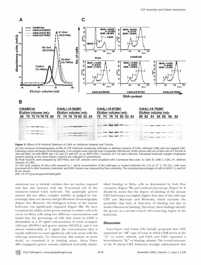
using a gel filtration buffer containing 200 mM NaCl, 20 mMHEPES (pH 7.5), 2.5 mM dithiothreitol (DTT) (Figure 3A), allCdtA deletion mutants that were refolded into a CDTholotoxin were shifted significantly to a lower molecularweight in gel filtration (Figure 3A). At the same time, all threesubunits appear in a stoichiometric ratio in the peakfractions (Figure 3B). Moreover, all of these deletions ofCdtA do not reduce the toxicity of the holotoxin, as measuredby cell cycle arrest of holotoxin-treated cells (Figure 3C). Oneexplanation for the discrepancy between the results forbiochemical assembly and activity of the CdtA N-terminaldeletion mutants would be to postulate an enhancedstabilization of the ternary complex upon interaction with acell surface ligand. Indeed, gel filtration chromatography is amore demanding assay for complex stability than otherchromatographic methods, and the three CdtA deletionmutants discussed above all co-purified by ion-exchangechromatography (Figure 2).As an alternative to demonstrate complex formation, we
have performed co-immunoprecipitation experiments. AMyc-tagged CdtC subunit was refolded together with wild-type CdtB and wild-type or deletion mutants of CdtA andsubjected to ion-exchange and size exclusion chromatogra-phy. Gel filtration fractions were then subjected to immuno-precipitation with anti-MYC antibody. They were all able toco-immunoprecipitate, demonstrating that the subunits werestill able to form complexes (Figure 4A) despite theirdisassembly during gel filtration chromatography. Nonspe-cific binding to protein G Sepharose was not detected (Figure4B). All of these experiments suggest that contacts that theresidues 18�75 of N-terminus CdtA make with other subunitscontribute to the holotoxin stability, but they are not criticalfor complex formation and toxin activity.The C-terminus of CdtA extends to CdtC and binds by
forming an intermolecular b-sheet, whereas the C-terminalextension of CdtC interacts with both CdtA and CdtB (Figure1A). The CdtA C-terminus possesses a more extensiveinteraction than the N-terminal region of this subunit, andit might be expected that deletions of this region would havea more pronounced effect. This is indeed the case, as removalof this polypeptide (CdtAD215�223) leads to an inability of
PLoS Pathogens | www.plospathogens.org November 2005 | Volume 1 | Issue 3 | e280215
CDT Assembly and Cellular Interactions
Synopsis
The cytolethal distending toxin is used by many bacteria to damagethe DNA of infected organisms. This DNA damage prevents cellsfrom dividing and eventually leads to cell death, which raises thepossibility that this genomic damage may be a contributing factorto carcinogenesis. The cytolethal distending toxin is composed ofthree proteins that form a tightly associated complex. Aftersecretion by the bacterium, two proteins in this complex adhereto the cell surface and achieve the delivery of the third protein intothe cell, where it causes DNA lesions. This report examines how thistoxin is assembled and how it adheres to host cell surfaces. A set ofmolecular features on the toxin is shown to be critical for this celladherence and for the ability of the cytolethal distending toxin toinhibit cell division. These results tie together for the first timeaspects of the molecular structure of the cytolethal distending toxinand its ability to adhere to host cell surfaces, contributing tomechanistic understanding of the activity of this genotoxin.

the complex to co-refold and to a precipitation of CdtA (datanot shown).
The Role of CdtC Nonglobular InteractionsWe also made a series of deletion mutants of the CdtC N-
and C- terminal tails, which demonstrate that the non-globular interactions of CdtC with both CdtA and CdtBstrongly contribute to toxin assembly, stability, and activity.
The N-terminal 13 amino acids of CdtC interact with activesite residues of CdtB, and we have previously shown that theseinteractions play an autoinhibitory role in holotoxin activityin vitro [16]. We have previously shown that CdtCD(21�35)assembles into a biochemically stable holotoxin, and it is asactive as the wild-type complex in toxicity assays [16]. Aslightly larger deletion, CdtCD(21�39), which removes severalresidues that make a small number of additional contacts toCdtB and CdtA, destabilizes the complex. The mutantcomplex still co-purifies by cation-exchange chromatography,but there is a significant decrease in the holotoxin stability(Figure 6C). This is also evident by gel filtration chromatog-raphy (Figure 5A and 5B). This leads to impaired toxicity of
the mutant holotoxin as determined by analysis of cell cycleof holotoxin-treated cells (Figure 5C).As the C-terminal tail of CdtC interacts with both CdtA and
CdtB, it is expected that its removal would underminecomplex stability and decrease toxicity. CdtCD179�186,which deletes residues at the C-terminus that are not visiblein the crystals, partially destabilizes the holotoxin assembly, asjudged by gel filtration and ion-exchange chromatography(Figures 5A and 6D), and significantly reduces toxicity againstcells (Figure 5C). It is not possible with these data todetermine whether the decrease in toxicity is due to partialdestabilization of the ternary complex or to a role of theCdtC C-terminus in receptor binding. A larger C-terminaldeletion, CdtCD169�186, presents a much more forcefulphenotype, as it does not productively refold, indicating thatthe loss of residues 169�178, which make contact with bothCdtA and CdtB, more significantly destabilizes the assemblyof the ternary complex (data not shown).
Structural Basis of Cell Surface BindingCdtA and CdtC have many similarities to the ricin B-chain
in fold and in placement with respect to the active subunit
Figure 1. Structure of CDT and Mutational Locations
Four alternative orientations of the crystal structure of the H. ducreyi CDT are shown as a ribbon cartoon tracing the three polypeptide chains. CdtA,CdtB, and CdtC are shown in blue, red, and green, respectively. Red dots indicate CdtB peptide not modeled due to disorder. N, NH2-terminus; C, COOH-terminus. The final image shows a partially transparent surface illustration focusing on the groove (red) and aromatic patch (yellow). Residues mutatedfor cell binding studies are indicated in white.DOI: 10.1371/journal.ppat.0010028.g001
PLoS Pathogens | www.plospathogens.org November 2005 | Volume 1 | Issue 3 | e280216
CDT Assembly and Cellular Interactions

within the holotoxin. Therefore, in analogy to ricin, it may beexpected that these two subunits of CDT will have a functionin host cell binding and internalization of the toxin. Severalreports indicate that these subunits can adhere to cellsurfaces [11,21�23]. The crystal structure shows that CdtAand CdtC together contribute toward the formation of twonotable surfaces, a cluster of highly conserved aromaticresidues, and a long and deep groove that is formed at theinterface of two subunits (Figure 1B). These may representregions responsible for cell contact and attachment. Toaddress this possibility, we have altered these molecularsurfaces by mutagenesis and examined mutant holotoxins forbinding to cells and effectiveness to induce cell cycle arrest.
In order to be able to determine if different mutantholotoxins interact with the cells, we have developed afluorescence-based binding assay. The holotoxin was fluo-rescently labeled with Alexa Fluor 488 fluorescent dye(Molecular Probes, Eugene, Oregon, United States). TheAlexa Fluor 488 reactive dye has a tetrafluorophenyl estermoiety that reacts efficiently with primary amines of proteinsto form stable dye-protein conjugates. It was necessary to testthe activity of CDT-Alexa Fluor conjugate in a cellularintoxication assay, since there are reports suggesting thatbiotin labeling of CdtA disrupted complex formation and ledto a reduction in toxicity [22]. Dye-conjugated CDT was aspotent as unconjugated toxin and caused a comparable level
of G2/M arrest of HeLa cells (Figure 7A). This enabled us totest the binding of fluorescently labeled holotoxin to HeLacells by flow cytometry and confocal microscopy. Flowcytometry gave a quantitative evaluation of holotoxin bind-ing, whereas confocal microscopy provided qualitative visual-ization of the cytometry results, while also revealing thelocalization of bound holotoxin. The labeled holotoxinspecifically binds to cells in a manner easily detectable overbackground.We have previously demonstrated that a quadruple
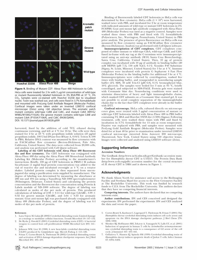
aromatic patch mutant (W91G/W98G/W100G/Y102A) is in-capable of intoxicating cells [16] (Figure 8B) and postulatedthat this prominent structural element could be involved incell surface interactions. Using a fluorescence-based cellbinding assay, we were able to show that this mutant is indeedincapable of binding to HeLa cells. By both flow cytometryand confocal microscopy, we were not able to detect anysignificant binding of the aromatic patch mutant abovebackground (Figures 7B and 9). This shows that the aromaticpatch residues are vital for cell surface attachment. A triplearomatic mutant (W98G/W100G/Y102A) behaves in the sameway as the quadruple (Figure 8B).We also mutagenized the extended groove formed by CdtA
and CdtC to assess its importance. The groove mutantcontains two mutations in CdtA (P103A and Y106A) andtwo in CdtC (R43K and Q49A). The rationale for these
Figure 2. Cation-Exchange Chromatography of the CDT Holotoxin Containing N-Terminal Deletions of CdtA
Wild-type (A) or mutant CDT holotoxins [(B) holotoxin with CdtA (D18–56), (C) holotoxin with CdtA (D18–67), (D) holotoxin with CdtA (D18–75)] wererun on Fast Flow SP Sepharose columns using an AKTA FPLC. Refolded CDT complexes were loaded and examined in a salt gradient as described inMaterials and Methods. Individual fractions (5 ml) or pooled material from the elution peak were collected and examined by SDS-PAGE. Proteins werestained with Coomassie blue stain. Images of the gels are presented inside corresponding chromatograms. L, loaded material; fr, fraction number; Fl,flowthrough; P, pooled material from elution peak. Absorbance was measured at 280 nm.DOI: 10.1371/journal.ppat.0010028.g002
PLoS Pathogens | www.plospathogens.org November 2005 | Volume 1 | Issue 3 | e280217
CDT Assembly and Cellular Interactions
mutations was to include residues that are surface exposedand that also interact with the N-terminal tail of thesymmetry-related CdtA molecule. The quadruple groovemutant did not affect complex stability as judged by ion-exchange (data not shown) and gel filtration chromatography(Figure 8A). However, the biological activity of the mutantholotoxin was significantly impaired (Figure 8B). We haveexamined the ability of the groove mutant to induce cell cyclearrest on HeLa cells using two different concentrations andfound that the percentage of cells that arrest in G2/M isdiminished at a 10 ng/ml concentration of toxin (comparewild-type [68.98%] and groove mutant [48.52%]), and it wasalmost undetectable at 1 ng/ml (the concentration that isusually sufficient to cause significant cell cycle arrest with thewild-type holotoxin). To characterize this mutant in moredetail, we examined it in binding assays. Alexa Fluor488�conjugated groove mutant exhibited noticeably dimin-
ished binding to HeLa cells as determined by both flowcytometry (Figure 7B) and confocal microscopy (Figure 9). Itshould be noted that the degree of labeling of the mutantCDT holotoxins was slightly higher than that of the wild-typeCDT (see Materials and Methods), which excludes thepossibility that lack of detection of binding was due toweaker fluorescent labeling. Therefore, these findings identifythe groove as a second critical cell-contacting region of theholotoxin.
Discussion
Lara-Tejero and Galan [18] initially proposed that CDTrepresents an ‘‘AB’’ type of toxin in which CdtB serves as the‘‘A,’’ or active, subunit, and CdtA and CdtC represent aheterodimeric ‘‘B,’’ or binding, subunit. The crystal structureof the H. ducreyi CDT holotoxin strongly substantiated that
Figure 3. Effects of N-Terminal Deletions of CdtA on Holotoxin Integrity and Toxicity
(A) Size exclusion chromatography profile of CDT holotoxin containing wild-type or deletion mutants of CdtA, wild-type CdtB, and myc-tagged CdtC.Following cation-exchange chromatography, 2-ml samples were injected onto a Superdex 200 HiLoad 16/60 column and run at flow rate of 2 ml/min in200 mM NaCl, 20 mM HEPES (pH 7.5), and 2.5 mM DTT on an AKTA FPLC. Fractions of 2 ml were collected. Calculated molecular weights of globularproteins eluting at the same elution volumes are indicated in parentheses.(B) Peak fractions were analyzed by SDS-PAGE, and CDT subunits were visualized with Coomassie blue stain. A, CdtA; B, CdtB; C, CdtC; A*, deletionmutants of CdtA.(C) Cell cycle analysis of HeLa cells exposed to 1 ng/ml concentration of the wild-type or mutant holotoxin for 3 hr at 37 8C, 5% CO2. Cells wereprocessed 48 hr after holotoxin treatment, and DNA content was measured by flow cytometry. The calculated percentages of cells in G0/G1, S, and G2/M are shown.DOI: 10.1371/journal.ppat.0010028.g003
PLoS Pathogens | www.plospathogens.org November 2005 | Volume 1 | Issue 3 | e280218
CDT Assembly and Cellular Interactions

hypothesis [16]. By analogy to other AB toxins [26], it has beenproposed that CDT travels from the plasma membranethrough an endocytic pathway and then enters the Golginetwork and endoplasmic reticulum, after which CdtBtranslocates directly into the nucleus [27], or is delivered byretrograde transport into the cytosol, and then translocatesinto the nucleus [17�20]. In order to try to elucidate the role
of CdtA and CdtC in the toxin assembly and cellular binding,we have addressed the contribution of several criticalstructural elements of these molecules by biochemical andcellular assays.Using deletion mutagenesis, we examined the role of
several nonglobular interactions in toxin stability andfunction. We found that contacts of C-terminal tails of CdtA
Figure 4. Immunoprecipitation of CDT Complexes Containing Deletion Mutants of CdtA
(A) The complexes (10 lg each) were immunoprecipitated with 10 lg anti-myc antibody (myc tag was fused to C-terminus of CdtC) in 150 mM NaCl, 20mM HEPES (pH 7.5), 0.1% Nonidet P40 substitute, followed by exposure to protein G Sepharose beads, and subjected to nondenaturing SDS-PAGE.(B) Nonspecific binding of CDT complexes to protein G Sepharose was not detected under the same experimental conditions. A, CdtA; B, CdtB; C, CdtC;A*, truncated CdtA; A1, holotoxin with CdtA D18�56; A2, holotoxin with CdtA D18�67; A3, holotoxin with CdtA D18�75; WT, wild-type; Ab, only anti-myc antibody, no CDT; IgG (HþL), nonreduced immunoglobulin G; IgG H, immunoglobulin G heavy chain; IgG L, immunoglobulin G light chain; SN,supernatant.DOI: 10.1371/journal.ppat.0010028.g004
Figure 5. Effects of N- and C-Terminal Deletions of CdtC on Holotoxin Integrity and Toxicity
(A) Size exclusion chromatography of recombinant holotoxin containing different CdtC mutants. After cation-exchange chromatography, 2-ml CDTholotoxin samples were injected onto a Superdex 200 HR 10/30 column and run at flow rate of 0.5 ml/min in 200 mM NaCl, 20 mM HEPES (pH 7.5), 2.5mM DTT on an AKTA FPLC. Fractions of 1 ml were collected.(B) Following elution from the gel filtration column, several peak fractions were subjected to SDS-PAGE and CDT subunits visualized with Coomassieblue stain. A, CdtA; B, CdtB; C, cdtC; C*, deletion mutants of CdtC.(C) Cell cycle analysis of HeLa cells exposed to 1 ng/ml concentration of either wild-type or mutant holotoxin for 3 hr at 37 8C, 5% CO2. Cells wereprocessed 48 hr after holotoxin treatment, and DNA content was measured by flow cytometry. The calculated percentages of cells in G0/G1, S, and G2/M are shown.DOI: 10.1371/journal.ppat.0010028.g005
PLoS Pathogens | www.plospathogens.org November 2005 | Volume 1 | Issue 3 | e280219
CDT Assembly and Cellular Interactions

and CdtC with other subunits are vital for toxin assembly.Their removal results in the poor refolding of the holotoxin,destabilization, and, as a consequence, inefficient cellularintoxication. The N-terminal tail of CdtC is deeply insertedinto the active site of CdtB, which may contribute to overallstability of the complex. Interestingly, the truncation of 35amino acids from the N-terminus of CdtC does not disturbcomplex stability, but removal of just four more residuesleads to destabilization of the holotoxin and impairedtoxicity. On the other hand, the N-terminal tail of CdtAmakes only a few contacts with other subunits, which appearto be inconsequential for the CdtA function in the holotoxin.Although removal of up to 75 residues from N-terminal tail ofCdtA destabilizes the complex under gel filtration conditions,truncated mutants are still as potent as the wild-typeholotoxin against target cells. This finding is in agreementwith previous observations that deletion 19�49 of closelyrelated Actinobacillus actinomycetemcomitans CdtA [28] or, inanother report, the first 59 residues [29] are dispensable forholotoxin activity. Moreover, several groups have demon-strated that some preparations of holotoxin actually containtruncated CdtA [25,30]. In other bacterial pathogens, theregion N-terminal to ricin fold of CdtA is very diverse and
much longer than in H. ducreyi. It is possible that these long N-terminal tails play a role in the assembly and translocation ofthe holotoxin in bacterial cells.Recently, several studies examined the binding of CDT
subunits to target cells and addressed the role of both CdtAand CdtC. The results were somewhat conflicting in theconclusion of what contribution CdtC made to this process.While several groups have shown that both CdtA and CdtCcan bind to the surface of HeLa cells and that preincubationwith CdtA-CdtC complex can inhibit subsequent intoxicationwith holotoxin [21�24], others have observed only binding ofCdtA to Chinese hamster ovary cells [11]. Furthermore, it wassuggested that CdtA and CdtC might bind to the same cellularreceptor [22,23]. The crystal structure of H. ducreyi CDTholotoxin demonstrated that both CdtA and CdtC are lectin-type structures, and they both contribute to the formation oftwo prominent molecular surfaces that could be involved incellular interactions. The first is a region composed of highlyconserved, surface-exposed aromatic residues. We havepreviously shown that mutations of four CdtA aromaticresidues (W91G, W98G, W100G, Y102A) abolish the holotoxinactivity [16]. Here, using fluorescence-based binding experi-ments, we were able to demonstrate that these residues are
Figure 6. Cation-Exchange Chromatography of CDT Holotoxin Containing N- and C-Terminal Deletions of CdtC
Wild-type (A) or mutant CDT holotoxins [(B) holotoxin with CdtC (D21�35), (C) holotoxin with CdtC (D21�39), (D) holotoxin with CdtC (D179�186)] wererun on Fast Flow SP Sepharose columns (20 ml) using an AKTA FPLC. Refolded CDT complexes were loaded on an SP Sepharose column in buffercontaining 40 mM NaCl, 20 mM HEPES (pH 7.5), 2.5 mM DTT and analyzed by a gradient in salt concentration as described in Materials and Methods.Individual fractions (5 ml) or pooled material (P) from elution peak were collected and examined by SDS-PAGE. Proteins were stained with Coomassieblue dye. Images of the gels are presented inside corresponding chromatograms. The peak that elutes between 100 and 150 mM NaCl concentration(P1) contains an intact CDT holotoxin, and the second peak (P2) that elutes at concentrations higher than 200 mM NaCl contains only the CdtB subunit.A, CdtA; B, CdtB; C, CdtC; C*, deletion mutants of CdtC; L, loaded material; fr, fraction number; Fl, flowthrough; P, pooled material from elution peak.Absorbance was measured at 280 nm. In A, P1 contains fractions 14�22, and P2 contains fractions 31�34; in D, P1 contains fractions 20�22, and P2contains fractions 32�34.DOI: 10.1371/journal.ppat.0010028.g006
PLoS Pathogens | www.plospathogens.org November 2005 | Volume 1 | Issue 3 | e280220
CDT Assembly and Cellular Interactions

indeed involved in the cell surface binding. While binding offluorescently labeled wild-type holotoxin to HeLa cells wasreadily detectable by flow cytometry and confocal micro-scopy, binding of an Alexa Fluor 488�conjugated aromaticmutant was undetectable in both assays. It appears that thedisruption of only three aromatic residues of CdtA (W98,W100, Y102) is sufficient to abolish cellular interactions andabrogate holotoxin activity. It was recently shown thatEscherichia coli CdtA-II and CdtC-II can interact withglycoproteins, and it was postulated that the putative cellularreceptor would most probably have an N-linked fucose-containing structure [23]. It has been suggested that thearomatic patch could be a potential carbohydrate-interactingregion [23,31], although the lipophilic nature of this cluster ofaromatic residues suggests that it may also serve to interactwith components of the plasma membrane. Indeed, it hasbeen recently shown that interaction of CDT with the cells issensitive to cholesterol depletion, and it was thereforesuggested that cholesterol-rich lipid rafts could be involvedin CDT binding [27].
We have also identified an additional cell-binding regionon the surface of the CDT holotoxin. It is represented by agroove that is formed by CdtA and CdtC along theirmolecular surfaces. Mutations of four surface-exposed groove
residues (two in CdtA:P103A/Y106A and two in CdtC:R43K/Q49A) significantly decrease binding to HeLa cells, whichresults in the decrease in CDT toxicity. The effect of thesemutations was less severe than that of mutations of aromaticresidues, representing an intermediate phenotype. By in-creasing concentrations of the groove mutant, it was possibleto induce more robust cell cycle arrest. In contrast, aromaticmutants were completely incapable of causing any effect ontreated cells. This could suggest that these two regions areinvolved in two independent binding events with the cellsurface.Our findings are an important step toward a better
understanding of the molecular mechanisms underlyingCDT holotoxin assembly and interaction with the target cellsurfaces. Understanding toxin assembly and activity alsoprovides potential targets for pharmacological disruption ofCDT activity. Finally, the identification of host cell surfacebinding mutants of CDT provides important tools for theidentification of host cell receptors mediating toxin entry.
Materials and Methods
Production of wild-type and mutant H. ducreyi holotoxin. H. ducreyiCDT subunits were cloned, purified, and holotoxin complex
Figure 7. The Activity of CDT Holotoxin Labeled with Alexa Fluor 488
(A) The toxicity of CDT holotoxin labeled with Alexa Fluor 488. HeLa cells were treated with 1 ng/ml (black) or 10 ng/ml (gray) concentration ofunconjugated or Alexa Fluor 488�conjugated CDT for 3 hr at 378C, 5% CO2. Cells were processed 48 hr after holotoxin treatment, and DNA content wasmeasured by flow cytometry. The calculated percentages of cells in G0/G1, S, and G2/M are shown.(B) Binding of CDT-Alexa Fluor 488 to cells. Harvested HeLa cells were exposed for 2 hr to 5 and 10 lg/ml concentration of wild-type or mutant CDT-Alexa Fluor 488. The histogram shows the binding of 5 or 10 lg/ml concentration of wild-type and mutant CDT-Alexa Fluor 488 conjugates to HeLacells. Mock represents cells in buffer only (2% FCS in PBS), and control is goat anti-mouse IgG conjugated with Alexa Fluor 488, which does not bind toHeLa cells. The level of fluorescence was analyzed by flow cytometry. The relative levels of fluorescent labeling of wild-type and mutant CDT holotoxinwas maintained to be nearly equivalent, with the mutant holotoxins (groove and aromatic patch) possessing a slightly higher level of labeling than thewild-type (Materials and Methods).DOI: 10.1371/journal.ppat.0010028.g007
PLoS Pathogens | www.plospathogens.org November 2005 | Volume 1 | Issue 3 | e280221
CDT Assembly and Cellular Interactions

reconstituted as previously described [16]. CdtA(18�223),CdtB(23�283), and CdtC(21�186) were cloned by PCR from genomicDNA (ATCC [American Type Culture Collection] number 700724D,Manassas, Virginia, United States) as an N-terminal hexahistidinefusion proteins in E. coli such that the predicted N-terminal secretionsignals were removed [32]. cdtA was cloned in pET-28a vector(Novagen, Madison, Wisconsin, United States) with engineered in-frame rhinovirus 3C protease recognition sequence at the 59 end ofcdtA. cdtB was cloned into an engineered version of the pac28 vector[33], which also contains a 3C protease recognition sequence betweenthe hexahistidine tag and the cdtB. cdtC was cloned in the pET-21avector (Novagen), which has an insertion with a hexahistidine tag andan engineered 3C protease recognition site. The three subunits wereexpressed separately in BL-21 E. coli cells by the addition of 0.1 mMisopropyl-b-D-thiogalactopyranoside (IPTG; Gold Biotechnology,Inc., St. Louis, Missouri, United States) for 4 hr at 37 8C, 200 rpm.The subunits were purified under denaturing conditions (8 M urea,10 mM Tris [pH 8.0], 0.1 M Na-phosphate) using nickel chelatingaffinity resin (GE Healthcare). Each subunit was eluted from nickelchelating affinity resin with the elution buffer that contained 500 mMimidazole (pH 8.0), 8 M urea, 10 mM Tris (pH 8.0), 0.1 M Na-phosphate. The usual yields per liter of bacterial culture were 50 mgof CdtA, 60 mg of CdtB, and 200 mg of CdtC. The CDT holotoxin wasreconstituted by co-refolding all three subunits together via dialysisat 48C into a native buffer consisting of 20 mM HEPES (pH 7.5), 200mM NaCl, 2.5 mM DTT, 5% glycerol, and 2 mM EDTA. The totalprotein concentration during refolding was maintained under 0.1mg/ml (20 mg of protein in 200 ml of denaturing buffer per 4 L ofnative buffer), and the native buffer was changed four times within a24-hr period. The proteins were separated from the affinity tagthrough site-specific proteolytic cleavage with rhinovirus 3C protease(2 lg/ml 3C protease fused to a GST tag) for 12 hr at 4 8C. Theprotease was subsequently removed by passing the material through aFast Flow GST-Sepharose column (GE Healthcare). The CDTholotoxin was further purified by cation-exchange chromatography(SP Sepharose Fast Flow, 20 ml bead volume; GE Healthcare) using anAKTA FPLC (GE Healthcare). Refolded material was diluted fivetimes to lower the salt concentration to 40 mM NaCl prior to loadingthe SP Sepharose column. After equilibrating the column withwashing buffer (20 mM HEPES [pH 7.5], 2.5 mM DTT), the columnwas subjected to a gradient increase in salt concentration (from 0 to500 mM NaCl in 20 mM HEPES [pH 7.5], 2.5 mM DTT during 150 min
at flow rate of 3 ml/min). The remaining material was bumped fromthe column with 1 M NaCl, 20 mM HEPES (pH 7.5), 2.5 mM DTT.Individual fractions (5 ml) or pooled material from elution peak wascollected and examined by SDS-PAGE. As a final step in thepurification, and at the same time as a test of complex integrity,fractions eluted from the SP Sepharose column that contained intactCDT holotoxin were pooled together, concentrated using CentriconPlus-20 (Millipore, Billerica, Massachusetts, United States), and runon a gel filtration column (Superdex 200 HiLoad 16/60 [120 ml] orSuperdex 200 HR 10/30 [25 ml]; GE Health), as indicated in the figurelegends. Gel filtration buffers contained 200 mM NaCl, 20 mMHEPES(pH 7.5), 2.5 mM DTT; a 2-ml sample was injected, and 2-ml (fromSuperdex 200 HiLoad 16/60) or 1-ml (from Superdex 200 HR 10/30)fractions were collected. The peak fractions were examined by SDS-PAGE, and proteins were visualized by staining with Coomassie bluestaining solution (50% methanol, 0.05% Coomassie brilliant blue R-50, 10% acetic acid in water) followed by destaining solution (5%methanol, 7% acetic acid in water).
The deletion mutants were generated by PCR and inserted into thesame vector as the wild-type genes. The amino acid substitutions wereintroduced into cdt genes by PCR using Pfu DNA Polymerase(Stratagene, La Jolla, California, United States) and primers contain-ing the appropriate base changes. The template plasmid wassubsequently removed by digestion with DpnI prior to transforma-tion. Myc tag sequence (EQKLISEEDL) was fused in-frame to the 39end of the wild-type cdtC gene in two subsequent PCRs using the pET-21a-CdtC plasmid as template, a T7 promoter primer as a forwardprimer in both reactions, and myc1R (59-GAGTTTCTGCTCGCTACCCTGATTTCTTCG-39) and myc2R (59-TTACAGATCCTCTTCAGAGATGAGTTTCTGCTCGCTACC-39) as reverse primers. ThePCR product was initially cloned into the pCR2.1 TOPO vector(Invitrogen, Carlsbad, California, United States), and after sequenceverification subcloned into the pET21a. Purification of the mutantproteins and assembly into the mutant CDT holotoxin wereperformed as previously described for the wild-type complex.
Cell cycle analysis/cellular intoxication assay. As a measure of toxinpotency and cellular intoxication, we have examined by flowcytometry the cell cycle progression of HeLa cells treated with theCDT holotoxin. HeLa cells (23 106 cells) were treated with indicatedconcentrations of wild-type or mutant CDT holotoxin for 3 hr at 378C, 5% CO2, and were subsequently washed of toxin and maintainedin culture media. At 48 hr after treatment with CDT, cells were
Figure 8. Effect of the Aromatic Patch and Groove Mutants on the Holotoxin Assembly and Toxicity
(A) Elution profile of the wild-type and groove mutant of the CDT holotoxin during size exclusion chromatography. Samples were loaded on a Superdex200 HiLoad 16/60 column at a flow rate of 2 ml/min in 200 mM NaCl, 20 mM HEPES (pH 7.5), 2.5 mM DTT on AKTA FPLC. Peak fractions (2 ml) wereanalyzed by SDS-PAGE (15% gel), and CDT subunits were visualized with Coomassie blue staining. SDS-PAGE of the CDT holotoxin groove mutant isshown inside gel filtration chromatogram. Elution volumes of wild-type and mutant holotoxin are shown.(B) Cell cycle analysis of HeLa cells exposed to 1 ng/ml (gray) or 10 ng/ml (black) concentration of either wild-type or mutant holotoxin for 3 hr at 37 8C,5% CO2. Cells were processed 48 hr after holotoxin treatment, and DNA content was measured by flow cytometry, as detailed in Materials and Methods.The calculate percentages of cells in G0/G1, S, and G2/M are shown. CdtABC 4m Aromatic mutant (CdtA: W91G/W98G/W100G/Y102A); CdtABC 3mAromatic mutant (CdtA: W98G/W100G/Y102A); CdtABC groove mutant (CdtA: P103A/Y106A, CdtC: R43K/Q49A).DOI: 10.1371/journal.ppat.0010028.g008
PLoS Pathogens | www.plospathogens.org November 2005 | Volume 1 | Issue 3 | e280222
CDT Assembly and Cellular Interactions
harvested, fixed by the addition of cold 70% ethanol duringcontinuous vortexing, and left at 4 8C for 16 hr. The cells were thenstained for 2 hr at 23 8C with propidium iodide solution (25 lg/mlpropidium iodide, 100 U/ml DNase-free RNase A, 0.04% Triton X-100in PBS). Relative DNA content was measured by flow cytometry withan FACSCalibur flow cytometer (Becton-Dickinson, Palo Alto,California, United States). The data were collected from 20,000 cells,and analysis was performed with Cell Quest software.
Labeling of the CDT holotoxin with Alexa Fluor 488 fluorescentdye and flow cytometry. The CDT holotoxin was conjugated withAlexa Fluor 488 dye using the Alexa Fluor 488 Monoclonal AntibodyLabeling Kit (Molecular Probes) according to the manufacturer’sinstructions. Briefly, 100 lg of CDT holotoxin in PBS/0.1 M sodiumbicarbonate (1 mg/ml final protein concentration) was added to thevial of reactive dye and incubated overnight at 4 8C on a rotatorshaker. Labeled protein complex is then separated from uncon-jugated dye using a purification resin supplied by manufacturer. Thedegree of labeling was determined by measuring the absorbance at280 nm and 494 nm using a NanoDrop ND-1000 spectrophotometer(Wilmington, Delaware, United States) and calculating the proteinconcentration and the fluorescent dye concentration using Protein &Labels module of ND-1000 software. The degree of labeling wascalculated as moles of dye per mole of protein. This producedcalculations of labeling of 2.98 6 0.11 for wild-type CdtABC, 3.99 60.32 for groove mutant CdtABC, and 5.64 6 0.33 for aromaticmutant. Goat anti-mouse IgG was purchased already conjugated withAlexa 488 (Molecular Probes), and the degree of labeling was 6.4moles of dye per mole of protein.
Binding of fluorescently labeled CDT holotoxin to HeLa cells wasdetermined by flow cytometry. HeLa cells (1 3 106) were harvested,washed twice with PBS, and incubated for 2 hr at room temperaturewith indicated amounts of wild-type or mutant CDT holotoxin in 2%FCS/PBS. Goat anti-mouse IgG antibody conjugated with Alexa Fluor488 (Molecular Probes) was used as a negative control. Samples werewashed three times with PBS and fixed with 1% formaldehyde(Polysciences, Inc., Warrington, Pennsylvania, United States) in PBSprior analysis. The presence of green fluorescence on HeLa cells wasmeasured by flow cytometry using an FACSCalibur flow cytometer(Becton-Dickinson). Analysis was performed with Cell Quest software.
Immunoprecipitation of CDT complexes. CDT complexes com-posed of either mutant or wild-type CdtA, wild-type CdtB, and CdtCfused in-frame with myc tag at the C-terminus were immunoprecipi-tated using an anti-myc antibody (9E10; Santa Cruz Biotechnology,Santa Cruz, California, United States). Then, 10 lg of proteincomplex was incubated with 10 lg of antibody in binding buffer (20mM HEPES [pH 7.5], 150 mM NaCl, 0.1% Nonidet P40 Substitute[Sigma, St. Louis, Missouri, United States]) for 3 hr at 4 8C. Immunecomplexes were incubated with protein G Sepharose 4 Fast Flow(Molecular Probes) in the binding buffer for additional 3 hr at 4 8C.Immunoprecipitates were collected by centrifugation, washed fivetimes with binding buffer, and resuspended in nonreducing samplebuffer (2% SDS, 50 mM Tris-Cl [pH 6.8], 0.1% bromophenol blue,10% glycerol). The samples were boiled for 3 min at 95 8C, brieflycentrifuged, and subjected to SDS-PAGE. Protein gels were stainedwith Coomassie blue dye. Nonreducing conditions were used tominimize dissociation of heavy and light immunoglobulin chains,which masked CDT subunits on a Coomassie blue�stained gel. It wasnot possible to avoid complete separation of the immunoglobulinchains due to the fact that CDT complexes were already in the bufferwith DTT.
Confocal microscopy. HeLa cells, cultured directly on microscopecover glass, were treated with 5 lg/ml concentration of mutant orwild-type CDT holotoxin labeled with Alexa Fluor 488 in PBS buffercontaining 3% BSA and Hoechst 33258 dye (1:500) (Sigma). Followingtreatment, cells were washed three times with PBS and fixed byincubation in 3.7% formaldehyde for 15 min at room temperature.Fixative was replaced with PBS, and samples were mounted withProLong Gold Antifade Reagent (Molecular Probes). Samples weredried for at least 48 hr prior to examination under inverted LSM510confocal microscope (inverted Zeiss Axiovert 200 microscope,Thornwood, New York, United States) using 360 objective lenses.Images were collected and analyzed using LSM510 software (Zeiss).
Supporting InformationAccession Numbers
The GenBank (http://www.ncbi.nlm.nih.gov/Genbank) accession num-ber for Haemophilus ducreyi CDT is U53215. The Protein Data Bank(http://www.rcsb.org/pdb) accession number for the crystal structureof H. ducreyi CDT is 1SR4 and is shown in Figure 1.
Acknowledgments
We thank Alison North for assistance and access to the BioImagingFacility and Svetlana Mazel for access to the Flow Cytometry Facilityat The Rockefeller University. This work was funded by researchfunds to C.E.S. from The Rockefeller University. The authors declarethat they have no competing financial interests.
Competing interests. The authors have declared that no competinginterests exist.
Author contributions. DN and CES conceived and designed theexperiments. DN performed the experiments. DN and CES analyzedthe data and wrote the paper. &
References1. Lara-Tejero M, Galan JE (2002) Cytolethal distending toxin: Limited damage
as a strategy to modulate cellular functions. Trends Microbiol 10: 147–152.2. De Rycke J, Oswald E (2001) Cytolethal distending toxin (CDT): A bacterial
weapon to control host cell proliferation? FEMS Microbiol Lett 203: 141–148.
3. Johnson WM, Lior H (1988) A new heat-labile cytolethal distending toxin(CLDT) produced by Campylobacter spp. Microb Pathog 4: 115–126.
4. Frisan T, Cortes-Bratti X, Thelestam M (2002) Cytolethal distending toxinsand activation of DNA damage-dependent checkpoint responses. Int J MedMicrobiol 291: 495–499.
5. Cortes-Bratti X, Karlsson C, Lagergard T, Thelestam M, Frisan T (2001) TheHaemophilus ducreyi cytolethal distending toxin induces cell cycle arrest andapoptosis via the DNA damage checkpoint pathways. J Biol Chem 276:5296–5302.
6. Shenker BJ, Hoffmaster RH, Zekavat A, Yamaguchi N, Lally ET, et al. (2001)Induction of apoptosis in human T cells by Actinobacillus actinomycetemcomi-tans cytolethal distending toxin is a consequence of G2 arrest of the cellcycle. J Immunol 167: 435–441.
7. Gelfanova V, Hansen EJ, Spinola SM (1999) Cytolethal distending toxin ofHaemophilus ducreyi induces apoptotic death of Jurkat T cells. Infect Immun67: 6394–6402.
Figure 9. Binding of Mutant CDT- Alexa Fluor 488 Holotoxin to Cells
HeLa cells were treated for 2 hr with 5 lg/ml concentration of wild-typeor mutant fluorescently labeled holotoxin in 3% BSA/PBS at 37 8C, 5%CO2. Samples were co-stained with Hoechst 33258 dye to visualizednuclei. Toxin was washed out, and cells were fixed in 3.7% formaldehydeand mounted with ProLong Gold Antifade Reagent (Molecular Probes).Confocal images were taken under an inverted LSM510 confocalmicroscope (Zeiss) using 360 objective lenses. The aromatic patchmutant contains wild-type CdtB and CdtC, and mutant CdtA (W91G/W98G/W100G/Y102A); the groove mutant contains wild-type CdtB andmutant CdtA (P103A/Y106A), and CdtC (R43K/Q49A).DOI: 10.1371/journal.ppat.0010028.g009
PLoS Pathogens | www.plospathogens.org November 2005 | Volume 1 | Issue 3 | e280223
CDT Assembly and Cellular Interactions

8. Elwell CA, Dreyfus LA (2000) DNase I homologous residues in CdtB arecritical for cytolethal distending toxin-mediated cell cycle arrest. MolMicrobiol 37: 952–963.
9. Lara-Tejero M, Galan JE (2000) A bacterial toxin that controls cell cycleprogression as a deoxyribonuclease I-like protein. Science 290: 354–357.
10. Elwell C, Chao K, Patel K, Dreyfus L (2001) Escherichia coli CdtB mediatescytolethal distending toxin cell cycle arrest. Infect Immun 69: 3418–3422.
11. Mao X, DiRienzo JM (2002) Functional studies of the recombinant subunitsof a cytolethal distending holotoxin. Cell Microbiol 4: 245–255.
12. Cortes-Bratti X, Frisan T, Thelestam M (2001) The cytolethal distendingtoxins induce DNA damage and cell cycle arrest. Toxicon 39: 1729–1736.
13. Hassane DC, Lee RB, Mendenhall MD, Pickett CL (2001) Cytolethaldistending toxin demonstrates genotoxic activity in a yeast model. InfectImmun 69: 5752–5759.
14. Alby F, Mazars R, de Rycke J, Guillou E, Baldin V, et al. (2001) Study of thecytolethal distending toxin (CDT)-activated cell cycle checkpoint. Involve-ment of the CHK2 kinase. FEBS Lett 491: 261–265.
15. Frisan T, Cortes-Bratti X, Chaves-Olarte E, Stenerlow B, Thelestam M(2003) The Haemophilus ducreyi cytolethal distending toxin induces DNAdouble-strand breaks and promotes ATM-dependent activation of RhoA.Cell Microbiol 5: 695–707.
16. Nesic D, Hsu Y, Stebbins CE (2004) Assembly and function of a bacterialgenotoxin. Nature 429: 429–433.
17. Cortes-Bratti X, Chaves-Olarte E, Lagergard T, Thelestam M (2000) Cellularinternalization of cytolethal distending toxin from Haemophilus ducreyi.Infect Immun 68: 6903–6911.
18. Lara-Tejero M, Galan JE (2001) CdtA, CdtB, and CdtC form a tripartitecomplex that is required for cytolethal distending toxin activity. InfectImmun 69: 4358–4365.
19. Deng K, Latimer JL, Lewis DA, Hansen EJ (2001) Investigation of theinteraction among the components of the cytolethal distending toxin ofHaemophilus ducreyi. Biochem Biophys Res Commun 285: 609–615.
20. Lewis DA, Stevens MK, Latimer JL, Ward CK, Deng K, et al. (2001)Characterization of Haemophilus ducreyi cdtA, cdtB, and cdtC mutants in invitro and in vivo systems. Infect Immun 69: 5626–5634.
21. Deng K, Hansen EJ (2003) A CdtA-CdtC complex can block killing of HeLacells by Haemophilus ducreyi cytolethal distending toxin. Infect Immun 71:6633–6640.
22. Lee RB, Hassane DC, Cottle DL, Pickett CL (2003) Interactions ofCampylobacter jejuni cytolethal distending toxin subunits CdtA and CdtCwith HeLa cells. Infect Immun 71: 4883–4890.
23. McSweeney LA, Dreyfus LA (2005) Carbohydrate-binding specificity of theEscherichia coli cytolethal distending toxin CdtA-II and CdtC-II subunits.Infect Immun 73: 2051–2060.
24. Akifusa S, Heywood W, Nair SP, Stenbeck G, Henderson B (2005)Mechanism of internalization of the cytolethal distending toxin ofActinobacillus actinomycetemcomitans. Microbiology 151: 1395–1402.
25. Frisk A, Lebens M, Johansson C, Ahmed H, Svensson L, et al. (2001) Therole of different protein components from the Haemophilus ducreyicytolethal distending toxin in the generation of cell toxicity. MicrobPathog 30: 313–324.
26. Sandvig K, Grimmer S, Iversen TG, Rodal K, Torgersen ML, et al. (2000)Ricin transport into cells: Studies of endocytosis and intracellular trans-port. Int J Med Microbiol 290: 415–420.
27. Guerra L, Teter K, Lilley BN, Stenerlow B, Holmes RK, et al. (2005) Cellularinternalization of cytolethal distending toxin: A new end to a knownpathway. Cell Microbiol 7: 921–934.
28. Saiki K, Gomi T, Konishi K (2004) Deletion and purification studies toelucidate the structure of the Actinobacillus actinomycetemcomitans cytolethaldistending toxin. J Biochem (Tokyo) 136: 335–342.
29. Shenker BJ, Besack D, McKay T, Pankoski L, Zekavat A, et al. (2005)Induction of cell cycle arrest in lymphocytes by Actinobacillus actino-mycetemcomitans cytolethal distending toxin requires three subunits formaximum activity. J Immunol 174: 2228–2234.
30. Shenker BJ, Besack D, McKay T, Pankoski L, Zekavat A, et al. (2004)Actinobacillus actinomycetemcomitans cytolethal distending toxin (Cdt): Evi-dence that the holotoxin is composed of three subunits: CdtA, CdtB, andCdtC. J Immunol 172: 410–417.
31. Rini JM (1995) X-ray crystal structures of animal lectins. Curr Opin StructBiol 5: 617–621.
32. Cope LD, Lumbley S, Latimer JL, Klesney-Tait J, Stevens MK, et al. (1997) Adiffusible cytotoxin of Haemophilus ducreyi. Proc Natl Acad Sci U S A 94:4056–4061.
33. Kholod N, Mustelin T (2001) Novel vectors for co-expression of twoproteins in E. coli. Biotechniques 31: 322–323, 326–328.
PLoS Pathogens | www.plospathogens.org November 2005 | Volume 1 | Issue 3 | e280224
CDT Assembly and Cellular Interactions