mechanisms of action of docosahexaenoic acid in the
TRANSCRIPT

ABSTRACT: This review describes (from both the animal andhuman literature) the biological consequences of losses in ner-vous system docosahexaenoate (DHA). It then concentrates onbiological mechanisms that may serve to explain changes inbrain and retinal function. Brief consideration is given to ac-tions of DHA as a nonesterified fatty acid and as a docosanoidor other bioactive molecule. The role of DHA-phospholipids inregulating G-protein signaling is presented in the context ofstudies with rhodopsin. It is clear that the visual pigment re-sponds to the degree of unsaturation of the membrane lipids. Atthe cell biological level, DHA is shown to have a protective rolein a cell culture model of apoptosis in relation to its effects inincreasing cellular phosphatidylserine (PS); also, the loss ofDHA leads to a loss in PS. Thus, through its effects on PS, DHAmay play an important role in the regulation of cell signalingand in cell proliferation. Finally, progress has been made re-cently in nuclear magnetic resonance studies to delineate dif-ferences in molecular structure and order in biomembranes dueto subtle changes in the degree of phospholipid unsaturation.
Paper no. L8776 in Lipids 36, 945–959 (September 2001)
DHA COMPOSITION
In the 1960s, the very high level of docosahexaenoic acid(DHA, 22:6n3) in the mammalian brain was already appreci-ated (1,2) although the first description by Thudichum (3) wasnearly a century earlier [see review by Salem et al. (4)]. Yabu-uchi and O’Brien (5) described the positional distribution ofbrain phosphoglycerides in 1968, detailing both the high con-centration of DHA in position sn-2 and its concentration inthe aminophospholipids, phosphatidylserine (PS), and phos-phatidylethanolamine (PE). By the early 1970s, the very highconcentration in brain synaptosomal plasma membranes (6)and synaptic vesicles (7) was described by Breckenridge andco-workers. Table 1 presents the DHA composition in the
aminophospholipids of brain and other selected mammaliantissues (1,6–18). It is apparent that the DHA content of thenervous system is very high. The retina not only contains avery high level of DHA in the rod outer segment (ROS) mem-branes, but also contains a very considerable amount of di-DHA species (19) as well as ones with DHA coupled to otherhighly unsaturated fatty acids (HUFA). The sperm is anothercompartment enriched in DHA. Every mammalian cell con-tains DHA, and phospholipids of internal organs and muscleshave a significant content. Human milk contains a relativelylow content of DHA with a higher percentage in phospho-lipids than triglycerides, the main lipid component of milk.
It has long been known that when an adult mammal con-sumes a diet low in DHA and its n-3 precursors, the nervoussystem content of DHA is much less altered than are other or-gans, i.e., DHA is said to be tenaciously retained once neuraldevelopment has occurred (for reviews, see Refs. 4,20). How-ever, animal studies have shown that when n-3 fat sources areinadequate during early neural development, then the levelsof brain and retinal DHA decline (4,20–24). This has alsobeen confirmed in autopsy studies of human infants that werefed a vegetable oil-based formula with low n-3 fat sources vs.breast-feeding in which preformed DHA was present (25–27).This has naturally led to an interrogation of the functionalconsequences of neural DHA loss.
ANIMAL STUDIES
Representative studies in the animal literature (28–42) con-cerning the n-3 fatty acid deficiency syndrome are presentedin Table 2. Only studies that focus on neural functions, no-tably brain and retinal functions, have been included here.Typically, these studies involve a two-generation diet regi-men in which the mother is raised on an n-3-deficient diet andher offspring are then studied. Such treatment has generallybeen found to be necessary to induce a marked decline inbrain and retinal DHA; a decline of 50–80% is typical ofthose associated with a change in neural function. A varietyof different tasks show impairment, including those in boththe visual and olfactory modalities (Table 2). In addition todecrements in performance in simple associative learningtypes of tasks, losses in spatial memory (39) and olfactory setlearning have been reported recently (43). Thus, the loss inbrain DHA may be said to affect cognition, at least to the ex-tent that it can be ascertained in the rat.
Copyright © 2001 by AOCS Press 945 Lipids, Vol. 36, no. 9 (2001)
*To whom correspondence should be addressed at 12420 Parklawn Dr.,Room 150, Rockville, MD 20852. E-mail: [email protected]: AA, arachidonic acid; DHA, docosahexaenoic acid; DPAn-6,docosapentaenoic acid; DROSS, dipolar recoupling on-axis with scaling andshape preservation; HUFA, highly unsaturated fatty acids; LCP, long-chainpolyunsaturates; α-LNA, α-linolenate; LO, lipoxygenase; M, metarhodopsin;MAS, magic angle spinning; NMR, nuclear magnetic resonance; NOESY,nuclear Overhauser enhancement spectroscopy; PC, phosphatidylcholine;PDE, phosphodiesterase; PE, phosphatidylethanolamine; PS, phosphatidylser-ine; ROS, rod outer segment.
Mechanisms of Action of Docosahexaenoic Acid in the Nervous System
Norman Salem, Jr.*, Burton Litman, Hee-Yong Kim, and Klaus GawrischLaboratory of Membrane Biochemistry and Biophysics, National Institute on Alcohol Abuse and Alcoholism,
National Institutes of Health, Rockville, Maryland

DHA RECOVERY
It has long been known that once depleted, the brain recoversits DHA rather slowly (44,45). A recent study in rats providedthe time courses of DHA recovery and the reciprocal declinein docosapentaenoic acid (DPAn-6, 22:5n-6) in the retina,brain, liver, and serum when the rats were repleted with a dietcontaining both α-linolenate (LNA) and DHA (46). The half-times for brain and retinal recovery of DHA were 2.9 and 2.1wk, respectively, even though the liver and plasma half-times
were only 0.3 and 0.5 wk, respectively. This suggests a ratherslow transport of DHA into the brain/retina even in the caseof a DHA-deficient nervous system (47).
It could be hypothesized then that if the functional conse-quences of dietary n-3 fatty acid deficiency were due to theloss in DHA, at least some neural functions may be restoredas the neuronal and retinal DHA level is restored. Others maynot be reversible due to missed opportunities in sequential de-velopment or changes in structural features of the brain (48).The first such study of functional recovery by Connor and
946 N. SALEM ET AL.
Lipids, Vol. 36, no. 9 (2001)
TABLE 1Docosahexaenoic Acid (DHA) Content of Aminophospholipids in Various Mammalian Tissues
Phospholipid class (% DHA)
Ref Species Tissue Fraction Phosphatidylserines Phosphatidylethanolamines
1 Human Brain Gray matter 36.6 24.31 Human Brain White matter 5.6 3.48 Bovine Brain Gray matter 28.7 —8 Bovine Brain White matter 7.6 —6 Rat Brain Synaptic plasma membrane 34.1 32.47 Rat Brain Synaptic vesicles 37.0 30.69 Human Retina — 18.5 22.2
10 Bovine Retina Rod outer segment 37.7 38.711 Ram Sperm — — 40.512 Bovine Sperm — — 37.813 Rat Heart — — 23.414 Rat Liver Plasma membrane 14.1 6.915 Rat Muscle — — 36.116 Human Platelet — 2.1 4.117 Human Erythrocyte (infant) — — 5.918 Human Milk — — 0.08
TABLE 2Animal Studies of Effects of Low n-3 Fatty Acid Dietson Neural Functions
Task Reference
Rodent studiesReduced amplitude of a- and b-waves Wheeler and Benolken, 1975 (28)Y-maze performance Lamptey and Walker, 1976 (29)Active avoidance task Mills et al., 1988 (30)Brightness discrimination Yamamoto et al., 1991 (31)Shock avoidance Bourre et al., 1989 (32)Death after neurotoxin Bourre et al., 1989 (32)Exploratory activity Enslen et al., 1991 (33)Scopolamine-induced locomotion Nakashima et al., 1993 (34)Age of eye opening (mice) Wainwright et al., 1991 (35)Morris water maze (mice) Nakashima et al., 1993 (34)Electroretinogram, a-wave,peak-to-peak Weisinger et al., 1996 (36,37)
Delayed acquisition of olfactorydiscrimination Sheaff-Greiner, et al., 1999 (38)
Spatial task acquisition and memory Moriguchi et al., 2000 (39)
Cat studyElectroretinogram, a- and b-waveimplicit time Pawlosky et al., 1997 (40)
Primate studiesReduced visual acuity, longerimplicit time Connor and Neuringer, 1984 (41)
Impaired recovery of dark-adaptation Neuringer et al., 1986 (42)

Neuringer (49) indicated that electroretinographic changesassociated with low retinal DHA persisted after DHA reple-tion. However, Moriguchi et al. (50) recently presented evi-dence that spatial task acquisition and memory are reversibleand, to a first approximation, correlate well with the level ofbrain DHA. However, Weisinger et al. (51) reported thatwhen n-3-deficient guinea pigs were subsequently given adiet containing LNA, changes in mean arterial blood pressureand electroretinographic changes in the a-wave were not re-versed even when the DHA levels were indistinguishablefrom the control levels. Thus, it appears that there may be nogeneral answer to the question of reversibility of losses infunction due to DHA losses in early development; the answerwill depend on the type of function involved.
HUMAN STUDIES
As mentioned above, formula-feeding of infants has been asso-ciated with a loss in brain DHA with respect to the level inthose breast-fed (25–27). The pre-existing animal literaturewould predict that formula-fed infants would have a functionaldeficit if the neural DHA loss were of a sufficient magnitude.Of course, there are many differences between breast-feedingand formula-feeding; these involve not only differences in nu-
trients, but also maternal contact and care, and the associationof breast-feeding with socioeconomic factors. Nevertheless, itappears that the DHA variable can explain a good portion ofthe benefits associated with breast-feeding.
Studies of preterm infants have generally shown a benefitwhen DHA is added to the formula in controlled experiments(Table 3) (52–59). The studies included here are limited to con-trolled studies of formula-feeding with or without addition ofDHA or DHA plus arachidonic acid (AA). Also, only those inwhich neural outcomes were included are listed; studies ofgrowth and other anthropometric measures are not included.Studies of preterm infants (52–58) indicated, with one excep-tion (59), that there was a benefit to adding long-chain polyun-saturates (LCP) to formulas that contain only the 18-carbon es-sential fatty acids found in vegetable oils. In addition, a meta-analysis of visual acuity differences in premature infants at 2and 4 mon of age found a benefit of LCP of 0.47 and 0.28 oc-taves, respectively (60). These observations, in combinationwith studies indicating the safety of the ingredients used to sup-ply these nutrients, lead us to conclude that preterm infant for-mulas must contain DHA/AA.
Studies of full-term infants are listed in Table 4 (61–72). Inhalf of these studies, the LCP supplement supported an in-creased visual acuity (61,63,67), neurodevelopmental score
NEURAL MECHANISMS OF DHA ACTION 947
Lipids, Vol. 36, no. 9 (2001)
TABLE 4Studies of Formula Supplementation with DHA or DHA and AA on Retinal and Brain Function in Full-Term Infantsa
Authors Reference Year Outcome tested Results Age
Makrides et al. 61 1995 VEP LCP > F LCP = BF 16, 30 wkAgostoni et al. 62 1995 Brunet-Lezineb LCP > F LCP = BF 4 monCarlson et al. 63 1996 FPL LCP > F LCP = BF 2 monAgostoni et al. 64 1997 Brunet-Lezine LCP = F LCP = BF 24 monAuestad et al. 65 1997 FPL, VEP LCP = F LCP < BF 2, 4, 6, 9, 12 monHornby Jorgensen et al. 66 1998 VEP LCP = F LCP = BF 4 monBirch et al. 67 1998 VEP LCP > F LCP = BF 6, 17, 52 wkWillatts et al. 68 1998 Means-end problem solving LCP > F — 10 monScott et al. 69 1998 MCDI LCP < F — 14 monLucas et al. 70 1999 Bayley MDI, PDI LCP = F LCP = BF 18 monBirch et al. 71 2000 Bayley MDI LCP > F — 18 monMakrides et al. 72 2000 VEP, Bayley MDI LCP = F LCP < BF 34 wk, 2 yraVEP, visual evoked potential (log MAR); MCDI, Minnesota Child Development Inventory; PDI, psychomotor development index; MDI, mental developmentindex. For other abbreviations see Table 3.bBrunet-Lezine derived from Gesell test for psychomotor development.
TABLE 3Effect of Formula Supplementation with Docosahexaenoic Acid (DHA) or DHAand Arachidonic Acid (AA) on Brain and Retinal Function in Preterm Infantsa
Authors Reference Year Outcome tested Results Age
Uauy et al. 52 1990 ERG threshold, Vmax LCP > F LCP = BF 36 wk PCABirch et al. 53 1992 VEP, FPL LCP > F LCP = BF 36, 57 wk PCACarlson et al. 54 1993 FPL LCP > F 2, 4 monCarlson et al. 55 1996 FPL LCP > F 2 monWerkman and Carlson 56 1996 Fagan NPT LCP > F 6.5, 9, 12 monCarlson and Werkman 57 1996 Fagan NPT LCP > F 12 monFaldella et al. 58 1996 ERG latency LCP > F LCP = BF 52 wk PCABougle et al. 59 1999 Motor nerve conduction LCP < F LCP < BF 30 daERG, electroretinogram; LCP, long-chain polyunsaturates, i.e., DHA or AA/DHA; F, formula-fed; BF, breast-fed; PCA, post-conceptional age; VEP, visual evoked potential (log MAR, where MAR = minimum angle of resolution); FPL, forced choicepreferential looking (Teller cards); NPT, novel preference test or Fagan test of infant intelligence.

(62,71), or problem-solving ability (68). In five of the trials, noeffect was observed for the LCP supplement (64–66,70,72). Inone trial, infants with the LCP supplement appeared to performmore poorly in a vocabulary test administered to 14-mon-oldchildren (69). However, a recent trial with a larger number ofpreterm infants reported a positive effect of LCP-supplementedformula on vocabulary scores (73). San Giovanni et al. (74)performed a meta-analysis of the trials involving visual acuityand concluded that there was a 0.32 octave difference in visualacuity when supplemented and unsupplemented formulagroups were compared, with the DHA-fed groups having thehigher acuity. A somewhat larger difference (0.49 octaves) wasobserved when breast-fed infants were compared with unsup-plemented formula-fed infants.
In most of these trials, a rather low level (0.1–0.35% of totalfatty acids) of DHA supplement was given, corresponding to a“Western” level of DHA in milk. In a recent review, Jensen(75) calculated the average for DHA content in mature milksin Western and non-Western women to be 0.45 and 0.88% oftotal fatty acids, respectively. Thus, it is likely that more of thetrials would have observed a benefit of DHA if given at ahigher level that is more consistent with the range of present-day worldwide human milk values. Viewed from this perspec-tive, it is rather surprising that some trials can succeed indemonstrating a benefit of a fat component that is only 0.1–0.2% of the total fatty acids. This suggests that these LCP are po-tent and essential nutrients for optimal development.
It was also of interest to note that Jensen (75) found theratios of AA/DHA to be very close to 1 in his averages ofhuman milk from both Western and non-Western women. Theaverage levels found in non-Western women of ~0.9% each AAand DHA may be a good starting point for future research. Thisis believed by some to represent a better standard than that ofWestern women because the composition is strongly influencedby the diet and Westerners have in the last century or two shiftedtheir consumption of fats toward n-6 fats and away from n-3 fatsdue to the availability of linoleic-rich vegetable oils. Estimatesof the Paleolithic diet indicate a much greater intake of LCP anda ratio of n-3/n-6 fats close to 1 (76). Thus, human infants likelyreceived a much higher intake of DHA and other LCP from theirmother’s milk during human evolution. In modern times, thisensured supply of DHA during neurodevelopment has been ab-rogated by formula-feeding and by a very low maternal intakeof n-3 fats in many modern women. Given the present state ofknowledge from human and animal studies of changes in neuralfunction associated with a low DHA status, coupled with bio-chemical and nutritional studies indicating the loss of DHA inboth peripheral tissues and the nervous system when preformedDHA is not fed and the safety of ingredients (70,77) used to sup-ply DHA, it is clear that a prudent course of action would be tosupply sources of preformed DHA in the infant diet.
MECHANISMS OF ACTION OF DHA
From the above, it should be clear that many effects of DHAstatus have been observed relating to physiologic and behav-
ioral functions of the nervous system. What has been unclearare the mechanisms underlying DHA function. Perhaps themost perplexing aspect of this question relates to the phenome-nal degree of specificity that is apparent in this effect. It mustbe recalled that these studies did not involve essential fatty aciddeficiency and that there were adequate and often excessiveamounts of n-6 fats present, usually in the form of linoleic acid(LA). There is a well-known reciprocal replacement of DHAwith DPAn-6, in this case in the brain (78) and retina(37,46,49). These two fatty acids differ only with respect to theabsence of the ∆-19 double bond in the DPAn-6 molecule; bothare 22-carbon HUFA with their first five double bonds in thesame positions with respect to the carboxyl end of the mole-cule. There is little in the modern disciplines of biochemistry,biophysics, and neuroscience to offer a conceptual frameworkto understand this extraordinary specificity.
The first and most reasonable hypothesis was that a cy-clooxygenase or lipoxygenase (LO) product of DHA but notDPAn-6 was produced that had an important function in the cen-tral nervous system. This hypothesis was explored extensivelyafter the early reports that cyclooxygenase products of DHAwere produced in the rainbow trout gill (79). Early investiga-tions indicated that products made by rat brain were sensitive toLO inhibitors (80–83). A correction of the trout gill work indi-cated that the DHA products were not prostaglandins but ratherLO products (84). Aveldano and Sprecher (85) observed thatplatelet LO produced a monohydroxylated form of DHA, andBazan et al. (86) found a similar product after incubations withrat retinas. However, Kim and co-workers (80,81) demonstratedthat the brain DHA products were a racemic mixture and thusunlikely to be enzymatic products. They also demonstrated thatmany of the products observed in the brain were a result of thefailure to remove platelets and other blood cells by perfusion ofthe brain before in vitro experiments. Apparently, what wasbeing measured in vitro may have corresponded to the low levelof nonenzymatic fatty acid peroxidation that is known to occur.This is not to deny the existence of LO in the capillary beds inthe brain, because Moore et al. (87) demonstrated 12-S-LO ac-tivity in a microvessel fraction. Also, Sawazaki et al. (82) founda 12-LO product of AA and DHA in the rat pineal gland andZhang et al. (88) subsequently observed that the formation ofthese products is regulated by the light–dark cycle and mela-tonin through the modulation of both 12-LO (88) and cytosolicphopholipase A2 expression (89). They also reported that n-3fatty acid deficiency had profound effects not only on the pineallipid profile but also on pineal biochemical activity, resulting insignificantly fewer LO products (90).
It is still quite possible that a “magic bullet” type of mole-cule may be found for DHA, i.e., a function for the nonesteri-fied fatty acid or a metabolite that is extremely potent. Physio-logic experiments, for example, have demonstrated that DHAor an anandamide analog of DHA has a potent effect on the K+
channel (91–93). Leaf and co-workers showed that the nones-terified form of DHA has a potent effect on Na+ (94–96) andCa2+ channels (96,97). Also, synaptic transmission (98) andlong-term potentiation (99,100) in the hippocampus as well as
948 N. SALEM ET AL.
Lipids, Vol. 36, no. 9 (2001)

N-methyl-D-aspartate responses in the cerebral cortex (101) arealtered by DHA. However, what is not clear is whether theseactions of DHA and its analogs are operative in vivo. More-over, the substrate specificity required to explain the n-3 defi-ciency syndrome has generally not been found in these studies.
The failure of this initial hypothesis led to proposals thatare based on the concept that the active form of DHA is in theform of a phospholipid (102–104). Little progress was madeon this intractable problem until workers focused on this hy-pothesis. Several approaches have been used including cellbiological, biochemical, and biophysical attacks. Examplesof each of these will be summarized in turn below with refer-ence to apoptosis, protein-lipid interactions, and the physicalstate and membrane properties of DHA-phospholipids.
The first topic that will be taken up is G-protein signalingwith a focus on rhodopsin. This line of inquiry is central to anunderstanding of the function of DHA-lipids in the visual sys-tem; it also serves as a model of other G-protein-coupled sig-naling receptor systems that helps us to understand how DHAmay function in the brain.
PROTEIN-LIPID INTERACTIONS: G-PROTEIN SIGNALING
Intercellular signaling is initiated through the activation ofligand-specific receptors imbedded in the lipid bilayers ofcellular membranes. An understanding of the factors that gov-ern the efficiency of signaling processes requires elucidatinghow the lipid composition of the membrane modulates the in-teractions of these receptors with the other membrane-boundprotein components in the signaling pathway. To elucidate therole of n-3 fatty acids in the nervous system and visualprocess, the phospholipid acyl chain dependence of severalsteps in the visual transduction pathway was studied (105).This was accomplished by purifying several components ofthe visual transduction system and reconstituting them inphospholipid vesicles of defined lipid composition (106).
The visual transduction pathway is initiated by the absorp-tion of a photon by rhodopsin, a prototypical member of thefamily of G-protein-coupled receptors that includes manyneurotransmitter receptors such as those for serotonin anddopamine. Metarhodopsin (M)II is the conformation of pho-toactivated rhodopsin that binds and activates the visual G-protein, Gt, which in turn activates a cGMP-specific phospho-diesterase (PDE) (105). Hydrolysis of cGMP by the PDE re-sults in the closing of cGMP-gated channels in the ROSplasma membrane, changing the transmembrane potential andinitiating the neuronal response to light. In n-3-deficient ani-mals, a reduced amplitude and delayed response are observedin the leading portion of the a-wave of electroretinograms(37,40,41). This portion of the a-wave is associated with thetransduction pathway. To determine whether these observa-tions can be linked to changes in membrane composition, weexamined the bilayer dependence of MII formation, the ki-netics and extent of MII-Gt complex formation, and resultingPDE activity as a function of phospholipid acyl chain compo-
sition and cholesterol content of the bilayer. These topics willbe treated in turn below.
Keq measures the extent of MII formation, which repre-sents the formation of the activated ligand-bound receptorstate, for the MI-MII equilibrium. This parameter dependscritically on the level of acyl chain unsaturation (Fig. 1)(107). For both mixed-chain phosphatidylcholines (PC) andsymmetrically substituted PC, the highest levels of MII for-mation were seen in DHA-containing bilayers. The additionof 30 mol/100 mol phospholipid to these systems lowered thelevel of MII formation. However, the lowest percentage re-ductions were obtained in the DHA-containing systems, sug-gesting that DHA-containing phospholipids are best able tobuffer the inhibitory effects of cholesterol.
The first amplification step in the visual cascade is the ac-tivation of Gt. The initial step in this activation is the bindingof Gt to MII and is characterized by the association constant,Ka. This process is also affected by acyl chain composition.The value of Ka in 18:0,22:6-PC is more than two timesgreater than that in 18:0,18:1-PC, indicating that twice asmuch MII-Gt complex is formed in the DHA phospholipidthan in the monounsaturated bilayer (Niu, S., Mitchell, D.S.,and Litman, B.J. unpublished results). The number of Gt mol-ecules activated in the two bilayers should be proportional tothe amount of complex formed, suggesting that at equivalentlevels of MII and Gt, a higher signal amplitude will be ob-served in the DHA-containing bilayer.
Another important aspect of signaling is the response time.This aspect of signaling was addressed by measuring the ki-netics of both MII and MII-Gt formation in several bilayers.An important characterizing parameter in these measurementsis the ratio of the rate of formation of MII-Gt to that of MII.This parameter represents the lag time in appearance of the
NEURAL MECHANISMS OF DHA ACTION 949
Lipids, Vol. 36, no. 9 (2001)
FIG. 1. The compositional dependence of the metarhodopsin MI↔MIIequilibrium constant, Keq, determined for rhodopsin in a series ofcompositionally defined bilayers varying in degree of unsaturation at37°C. Source: Reference 107. M, metarhodopsin; PC, phosphatidyl-choline.

complex after MII has formed and is a measure of the effi-ciency of the interaction of the receptor and Gt protein. Thisratio is 1.4 in the native ROS disk membrane, indicating arapid complex formation after the appearance of MII (106).Complex formation in 18:0,22:6-PC and 18:0,18:1-PC ischaracterized by ratios of 3.5 and 4.9, respectively. Althoughthe DHA PC phospholipid is not as efficient as the disk sys-tem, it does provide for more efficient MII-Gt formation thanthe less unsaturated bilayer. Some of the enhanced efficiencyof MII-Gt formation in the disk membrane may be attribut-able to the more complex mixture of phospholipid classes thatcontribute a net negative surface charge to the membrane.
The overall measure of the signaling pathway is the dose-response curve, generated by determining the level of PDEactivity brought about by increasing levels of rhodopsin acti-vation. In these experiments, both Gt and PDE were reassoci-ated with rhodopsin-containing vesicles. At light exposurelevels at which 1 in 1000 rhodopsin molecules was activated,ROS disks yielded 87% of their maximal PDE activity. Undersimilar light exposure conditions, 59 and 26% of maximal
disk activity was obtained in 16:0,22:6-PC and 16:0,18:1-PC,respectively (106). Although not reaching the same activityas native disk membranes, the DHA-containing bilayer yieldstwice the activity of the monounsaturated bilayer. Thus, in theintegrated function of the pathway, the DHA-containing bi-layer yields higher activity levels than the monounsaturatedbilayer.
Recent studies suggest that lateral domain formation mayplay a critical role in the requirements for DHA-containingphospholipids (Fig. 2) (108). Fluorescence energy transfer ex-periments have provided evidence of the formation of lateraldomains in reconstituted membranes consisting of di22:6-PC,di16:0-PC, cholesterol, and rhodopsin. In these domains, thelipid composition around rhodopsin is highly enriched indi22:6-PC, whereas the di16:0-PC is highly enriched in choles-terol. Domain formation requires the presence of bothrhodopsin and cholesterol, indicating the complex nature of theinteractions that drive lateral segregation of the constituents ofthis system. If the other components of the signaling pathwayhave the same preferable partitioning into a DHA-rich lipid do-main as rhodopsin, then this would raise their local concentra-tion and provide a greater efficiency for the signaling pathway.
The results presented here demonstrate that the visual sig-naling pathway is greatly dependent on the acyl chain com-position of the bilayer. The steps in this signaling process in-volve unimolecular conformation changes required forrhodopsin activation and several protein–protein interactionsfor the activation of Gt and PDE. Both of these types ofprocesses were enhanced in bilayers containing DHA. Thestudies reported here were carried out in PC bilayers of vary-ing acyl chain composition. The disk membrane contains~42–45% of both PC and PE and ~10–12% PS. The differ-ences in activity observed between the pure PC system andnative disk membrane might be attributable to the lack of asurface potential supplied by the presence of PS or perhapsspecific properties contributed by the PE. It should be notedthat both PE and PS contain the highest levels of symmetri-cally substituted di-DHA species in the retina. Despite thesedifferences, the results reported here suggest an explanationfor the observations in the electroretinograms of n-3-deficientanimals. The delay in the development of the leading edge ofthe a-wave is likely related to the increased lag time observedin the formation of the MII-Gt complex, whereas the reducedamplitude may be explained on the basis of the reduced asso-ciation constant observed for MII-Gt complex formation.
ANTIAPOPTOTIC EFFECT OF DOCOSAHEXAENOICACID
Next, we turn to a consideration of the effects of DHA at a cellbiological level. Unlike AA, DHA is not easily released fromneuronal membranes, but instead is retained by membranephospholipids (109–111). In contrast, astroglia cells, which areknown to support neuronal survival, release this fatty acid read-ily (110–113). This suggests that DHA fatty acid may act as atrophic factor, and enrichment of this fatty acid in neuronal
950 N. SALEM ET AL.
Lipids, Vol. 36, no. 9 (2001)
FIG. 2. Domain structure of rhodopsin-lipid model membranes. Fluores-cence energy transfer studies of model membranes composed of a 3:7:3mixture of di22:6-phosphatidylcholine (PC)/di16:0-PC/cholesterol andvarying levels of rhodopsin show the presence of lateral domains. Thesedomains are composed of a rhodopsin-containing region highly enrichedin di22:6-PC and a second region highly enriched in di16:0-PC and cho-lesterol. The formation of these domains requires the presence of bothrhodopsin and cholesterol, demonstrating the complex nature of the mol-ecular interaction responsible for domain formation. These include arhodopsin preference for docosahexaenoic acid (DHA) acyl chains and apreference of cholesterol for saturated acyl chains (108).

membranes may be an important aspect in neuronal survival.In neuronal membranes, DHA is highly enriched inaminophospholipids, especially PS (1,2,4,5–10,114). We andothers have previously demonstrated that the enrichment ofDHA in cell membranes increases PS synthesis and, con-versely, that depletion of this fatty acid by an n-3-deficient dietor by chronic ethanol exposure decreases the accumulation ofPS (115–118). Considering the fact that PS is the major nega-tively charged phospholipid class in many mammalian cellmembranes and many of the signaling proteins such as proteinkinases are influenced by PS (119–121), this alteration of PScontent may have significant implications for cellular function.
In contrast to the well-documented apoptotic effect ofDHA (122–127), only a few studies have indicated an anti-apoptotic function for this fatty acid (128–131). In each case,an antiapoptotic effect was observed only after preincubationwith DHA before the induction of apoptosis. Because DHAis prone to oxidation during the incubation period and muchof the apoptotic effect is mediated through oxidative stress(126,127), it is difficult to successfully enrich cultured cellswith DHA without accompanying lipid peroxidation. There-fore, to observe the antiapoptotic effect of DHA, it is crucialthat an antioxidant such as vitamin E be added along withDHA to the culture medium (130,131).
In studies by Kim et al. (131) in which PC-12 or Neuro-2A cells were exposed to AA (1–25 µΜ) during serum depri-vation, apoptosis determined by genomic DNA fragmenta-tion decreased in a dose-dependent manner, but treatmentwith DHA or oleic acid had no effect. The insensitivity of theprotective effect of AA to indomethacin or nordihydroguai-aretic acid indicated that the observed protective effect of AAwas not mediated by either cyclooxygenase or LO deriva-tives, but rather through the direct action of AA (132). In con-trast to the effect of AA, DHA became protective only after aprolonged period of incubation. In Neuro-2A cells, the pro-tective effect required at least 24 h of enrichment, and alonger incubation led to an enhancement of the protective ac-tion of DHA. During this period, DHA was steadily incorpo-rated into PS, and total cellular PS was increased. The protec-tive effect is related to the extent of cellular PS accumulation.When cells were enriched with DHA in a serine-free medium,the PS content did not increase significantly and the antiapop-totic effect was diminished significantly.
Caspase-3 activity, which has been shown to mediatemammalian apoptosis, increased as the starvation proceeded,with the exception of cells enriched with DHA. Both AA- andDHA-treated cells initially showed less caspase-3 activity incomparison to nonenriched control or oleic acid-enrichedcells. However, prolonged serum starvation abolished theprotective effect of AA, and only DHA-treated cells main-tained caspase-3 activity at a level similar to that of controlcells kept in the 5% serum medium. The DNA fragmentationdata and DNA ladder formation obtained after at least 48 h ofserum starvation showed consistent results (Fig. 3). It wasalso observed that the 17-kDa active fragment of caspase-3increased under serum-free conditions, and supplementation
of Neuro-2A cells with DHA before serum starvation effec-tively prevented the increase of the 17-kDa fragment (131).
Enrichment of cells with DHA also altered expression ofvarious proteins at the gene level because the levels of mRNAfor caspase-3 decreased, whereas mRNA of Raf-1 increased(131,132). DHA has been shown to affect transcriptional activ-ities through nuclear hormone receptors such as the peroxisomeproliferator-activated receptor (133) or the retinoid X receptor(134). The antiapoptotic effect of DHA enrichment suggeststhat ensuring the survival of neuronal cells may be one of thereasons for the high level of DHA in brain. Alterations of themembrane PS content by DHA will influence not only the re-ceptor activities but also the translocation of various signalingproteins as well as their activation (118–120,131). Althoughthe release of DHA in neuronal cells may be minimal, it maybe possible to reach a local concentration of intracellular DHAsufficient to activate nuclear receptors involved in transcrip-tional activity. It is likely that the antiapoptotic effect of DHAis the result of multiple regulations at various signaling stages,ranging from the plasma membrane to nuclear events, most ofwhich have yet to be discovered.
BIOPHYSICAL PROPERTIES OF POLYUNSATURATEDLIPID MEMBRANES
As referred to above, there has been a lack of a conceptual basisin biophysics in which a meaningful difference in functioncould be predicted with respect to a DHA- vs. a DPAn-6-containing lipid. In fact, there have been few biophysical stud-ies that discern appreciable differences in the properties of vari-ous polyunsaturated species. It has often been said that intro-duction of a double bond into a saturated lipid results in a largechange in physical properties. Introduction of a second doublebond also results in an additional effect; however, it is of a muchsmaller magnitude than that of the first double bond. Thereafter,introduction of additional double bonds (for a total of three ormore) has little effect. However, the n-3 fatty acid deficiencysyndrome described above indicates that there are significantbiological effects measurable in the whole organism, e.g., by
NEURAL MECHANISMS OF DHA ACTION 951
Lipids, Vol. 36, no. 9 (2001)
FIG. 3. Protection from DNA fragmentation after the enrichment of cellswith docosahexaenoic acid for 48 h.

behavioral or physiologic means, when DPAn-6 is substitutedfor DHA. Because these are pentaenoic and hexaenoic lipids, itwould appear that a physical basis must exist for the differentialbiological function of various highly unsaturated fatty acids. Re-cent studies have begun to demonstrate that all highly unsatu-rated lipids indeed do not have the same physical properties.Some of the techniques and research approaches as well as dataobtained from these investigations are presented below.
MAGIC ANGLE SPINNING (MAS)
Solid-state nuclear magnetic resonance (NMR) spectroscopyon models of polyunsaturated membranes enables the mea-surement of dozens of parameters, probing every segment ofthe lipid bilayer with atomic resolution. This became possibleafter improving the performance of magic angle spinning(MAS) probes in combination with very high magnetic fieldstrength. MAS NMR reduces the linewidth of lipid reso-nances to ~10 Hz (135). That is equivalent to or better thanresolution of resonances for very small unilamellar liposomesthat tumble rapidly enough to eliminate anisotropic interac-tions. However, in contrast to studies of liposomes, for MASNMR, the membranes are not required to have small radii ofcurvature, and water content does not matter as long as thelipids remain in the liquid-crystalline state. Just a few mil-ligrams of sample is sufficient to provide very high signal-to-noise ratios as evidenced by the spectra in Figure 4 (136).
NUCLEAR OVERHAUSER ENHANCEMENT SPECTROSCOPY (NOESY)
The excellent resolution of lipid resonances allows applica-tion of techniques that probe magnetization transfer between
protons such as nuclear Overhauser enhancement spec-troscopy (NOESY), well known for its important contribu-tion to the structural determination of soluble proteins (137).
Magnetization transfer is the result of interactions betweenthe magnetic dipoles of the protons in lipids. Rates of transferbecome observable when the protons approach each other towithin distances ≤5 Å (138). In the two-dimensional NOESYcontour plot shown in Figure 5 (139), the peaks along the di-agonal correspond to the one-dimensional resonances shownin Figure 4. The rate of magnetization transfer is reflected bythe intensity of the off-diagonal crosspeaks. The surprisingobservation has been that magnetization is transferred be-tween all lipid resonances, but at different rates. Even themost distant protons such as methyl groups of the cholineheadgroup and methyl groups at the end of lipid hydrocarbonchains exchange magnetization. For a long time, such surpris-ing transfers were ascribed to a process called spin diffusion.Spin diffusion relays magnetization via coordinated flip-flopsof magnetization along the proton network of a lipid mole-cule. In a series of experiments on protonated lipids in deuter-ated matrices, we demonstrated recently that this interpreta-tion is incorrect (140). In the biologically relevant liquid-crystalline state, membranes are truly disordered to the pointthat lipid headgroups and ends of hydrocarbon chains meetonce in a while, albeit with lower probability than membranesegments that are located closer to each other. The experi-ments also demonstrated that the unexpected long-range con-tacts are not taking place within one lipid molecule, but rep-
952 N. SALEM ET AL.
Lipids, Vol. 36, no. 9 (2001)
FIG. 4. 1H magic angle spinning (MAS) nuclear magnetic resonance(NMR) spectrum of 16:0,18:1 PC in 50 wt% D2O recorded at ambienttemperature and a spinning speed of 10 kHz. Signal assignment is pro-vided by numbers in the spectrum and the formula. Source: Reference137.
FIG. 5. Two-dimensional nuclear Overhauser enhancement spec-troscopy (NOESY) magic angle spinning (MAS) nuclear magnetic reso-nance (NMR) spectrum of an 18:0-22:6 phosphatidylcholine(PC)/18:0d35-22:6 phosphatidylethanolamine (PE)/18:0d35-22:6 phos-phatidylserine (PS)/cholesterold7 (4:4:1:3, by vol) mixture.

resent magnetization transfer to neighboring lipid moleculesthat surround the emitter (141).
Experiments on sn-1 chain perdeuterated 1-stearoyl-2-do-cosahexaenoyl-sn-glycero-3-phosphocholine (18:0d35,22:6-PC) not only confirmed that polyunsaturated chains in mem-branes are similarly disordered, but also offered evidence thatconformational disorder of the remaining degrees of freedomof a polyunsaturated chain is higher (135,142). Furthermore,the intensity of cross- and diagonal peaks as a function ofmixing times allows judgment about motional correlationtimes in the polyunsaturated membranes on the time scalefrom pico- to milliseconds (138). The spin-lattice and spin-spin relaxation rates of polyunsaturated chains are lower, in-dicating more rapid motions of these protons. This result issurprising because polyunsaturated chains have long beenperceived as rigid and bulky owing to the loss of degrees offreedom.
DIPOLAR RECOUPLING ON-AXIS WITH SCALINGAND SHAPE PRESERVATION (DROSS)
MAS NMR techniques with application of radio-frequencypulses, which are synchronized with the phase of the spinningrotor, enable recoupling of anisotropic interactions, e.g., themagnetic dipolar interaction between 1H and 13C nuclei. Thestrength of this interaction depends on the time-averaged ori-entation of the 1H-13C bond vector with respect to the lipidbilayer normal.
In the two-dimensional dipolar recoupling on-axis withscaling and shape preservation (DROSS) experiments (Fig.6), this technique allows assignment of order parameters toresolved 13C resonances of lipid segments (143). Conductingthis experiment on 13C nuclei benefits from the much greaterresolution of 13C chemical shifts. Throughout the molecule,>20 order parameters can be assigned to specific regionswithin the polyunsaturated lipid, without isotopic labeling.
The experiments showed unambiguously that order param-eters of double bond 1H-13C vectors are close to 0. Because
of differences in bond geometry between saturated and unsat-urated hydrocarbon chains, this result was not unexpected.However, the very-low-order parameters for the five 1H-13Cvectors of the methylene groups that are sandwiched betweendouble bonds were surprising. These order parameters mustbe low as a result of crankshaft-like coordinated motionswithin the DHA chain. Such motions are likely because oflower potential barriers for rotations about the vinyl-methy-lene bonds compared with C-C bonds in saturated chains(144). This enables DHA chains to adapt to looped confor-mations that have shorter length and larger area per molecule(145).
2H NMR
Although the DROSS technique allows determination of as-signed order parameters, for technical reasons, the precisionin order parameter determination remains about one order ofmagnitude lower than the precision of order parameters mea-sured by the classical approach, i.e., analysis of effectivequadrupolar splittings in the 2H NMR spectra of deuteratedlipids (146). Quadrupole splittings are measured with a reso-lution of ~50 Hz, which corresponds to a precision for orderparameters of ∆S = ±0.0004. Such small changes are of rele-vance, e.g., to detect the level of stress that is caused in thelipid matrix as the result of a conformational change of amembrane protein (147). We conducted preliminary 2H NMRexperiments on perdeuterated DHA, incorporated at low con-centration into bilayers of 18:0,18:1-PC (Fig. 7). Partial as-signment of the order parameters to segments of the DHAchain was achieved by taking advantage of the small differ-ences in chemical shift between the deuterium resonances inMAS experiments with partial recoupling of the quadrupolarinteraction (Gawrisch, K., Safley, A.M., and Polozov, I.V., un-published data). The results fully confirmed the observationsfrom the less precise DROSS experiment. Order parametersof all methylene segments between double bonds in the hy-
NEURAL MECHANISMS OF DHA ACTION 953
Lipids, Vol. 36, no. 9 (2001)
FIG. 6. Extracted columns of a two-dimensional dipolar recoupling on-axis with scaling and shape preservation (DROSS) spectrum correspond-ing to resonance signals of double bonds (129.83 ppm), methylenegroups between the double bonds (27.36 kHz), and the C-17 methy-lene group of stearic acid (24.39 ppm). The peak doublets are the resultof recoupling of dipolar interactions between the 1H and 13C nuclei.The value of order parameters is directly proportional to the magnitudeof splittings.
FIG. 7. 2H nuclear magnetic resonance (NMR) spectrum of perdeuter-ated docosahexaenoic acid in an 18:0,18:1-PC matrix.
drocarbon chain, as well as the order of the majority of thedouble bonds, are very low. Only the two methylene segmentsnear the carboxyl group of DHA have order parameters thatare comparable to values of more saturated chains.
X-RAY DIFFRACTION
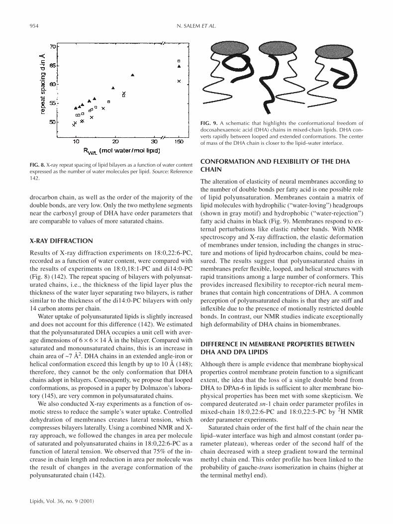
Results of X-ray diffraction experiments on 18:0,22:6-PC,recorded as a function of water content, were compared withthe results of experiments on 18:0,18:1-PC and di14:0-PC(Fig. 8) (142). The repeat spacing of bilayers with polyunsat-urated chains, i.e., the thickness of the lipid layer plus thethickness of the water layer separating two bilayers, is rathersimilar to the thickness of the di14:0-PC bilayers with only14 carbon atoms per chain.
Water uptake of polyunsaturated lipids is slightly increasedand does not account for this difference (142). We estimatedthat the polyunsaturated DHA occupies a unit cell with aver-age dimensions of 6 × 6 × 14 Å in the bilayer. Compared withsaturated and monounsaturated chains, this is an increase inchain area of ~7 Å2. DHA chains in an extended angle-iron orhelical conformation exceed this length by up to 10 Å (148);therefore, they cannot be the only conformation that DHAchains adopt in bilayers. Consequently, we propose that loopedconformations, as proposed in a paper by Dolmazon’s labora-tory (145), are very common in polyunsaturated chains.
We also conducted X-ray experiments as a function of os-motic stress to reduce the sample’s water uptake. Controlleddehydration of membranes creates lateral tension, whichcompresses bilayers laterally. Using a combined NMR and X-ray approach, we followed the changes in area per moleculeof saturated and polyunsaturated chains in 18:0,22:6-PC as afunction of lateral tension. We observed that 75% of the in-crease in chain length and reduction in area per molecule wasthe result of changes in the average conformation of thepolyunsaturated chain (142).
CONFORMATION AND FLEXIBILITY OF THE DHACHAIN
The alteration of elasticity of neural membranes according tothe number of double bonds per fatty acid is one possible roleof lipid polyunsaturation. Membranes contain a matrix oflipid molecules with hydrophilic (“water-loving”) headgroups(shown in gray motif) and hydrophobic (“water-rejection”)fatty acid chains in black (Fig. 9). Membranes respond to ex-ternal perturbations like elastic rubber bands. With NMRspectroscopy and X-ray diffraction, the elastic deformationof membranes under tension, including the changes in struc-ture and motions of lipid hydrocarbon chains, could be mea-sured. The results suggest that polyunsaturated chains inmembranes prefer flexible, looped, and helical structures withrapid transitions among a large number of conformers. Thisprovides increased flexibility to receptor-rich neural mem-branes that contain high concentrations of DHA. A commonperception of polyunsaturated chains is that they are stiff andinflexible due to the presence of motionally restricted doublebonds. In contrast, our NMR studies indicate exceptionallyhigh deformability of DHA chains in biomembranes.
DIFFERENCE IN MEMBRANE PROPERTIES BETWEENDHA AND DPA LIPIDS
Although there is ample evidence that membrane biophysicalproperties control membrane protein function to a significantextent, the idea that the loss of a single double bond fromDHA to DPAn-6 in lipids is sufficient to alter membrane bio-physical properties has been met with some skepticism. Wecompared deuterated sn-1 chain order parameter profiles inmixed-chain 18:0,22:6-PC and 18:0,22:5-PC by 2H NMRorder parameter experiments.
Saturated chain order of the first half of the chain near thelipid–water interface was high and almost constant (order pa-rameter plateau), whereas order of the second half of thechain decreased with a steep gradient toward the terminalmethyl chain end. This order profile has been linked to theprobability of gauche-trans isomerization in chains (higher atthe terminal methyl end).
954 N. SALEM ET AL.
Lipids, Vol. 36, no. 9 (2001)
FIG. 8. X-ray repeat spacing of lipid bilayers as a function of water contentexpressed as the number of water molecules per lipid. Source: Reference142.
FIG. 9. A schematic that highlights the conformational freedom ofdocosahexaenoic acid (DHA) chains in mixed-chain lipids. DHA con-verts rapidly between looped and extended conformations. The centerof mass of the DHA chain is closer to the lipid–water interface.

Changes in hydrocarbon chain length and area per mole-cule are reflected in order parameter changes. An increase ofaverage chain order by ∆S = +0.002, which can be easily re-solved, corresponds to an increase of average bilayer thick-ness of 0.1 Å and a decrease of area per molecule of 0.2 Å2.Compared with monounsaturated bilayers, the order of sn-1chains that are paired with polyunsaturated chains in the sn-2position is lower, mostly for the second half of the chain, witha maximal decrease by ∆S = −0.018 near carbon atom num-ber 13 for 18:0,22:6-PC (149). We propose that this decreasereflects formation of short looped chain conformations (videinfra) with a higher density of polyunsaturated chain seg-ments near the lipid–water interface. As a result of this redis-tribution of DHA chain density, the lower segments of the sat-urated chain have more freedom for movement and lowerchain order parameters in the second half of the chain. Thesurprising observation has been that the loss of a single dou-ble bond from DPAn-6 to DHA in 18:0,22:5-PC results inorder parameters that are much closer to those of a matrixwith monounsaturated oleic acid chains in position sn-2.
In summary, assigned order parameters and relaxationtimes related to molecular motions were measured using novelMAS NMR approaches and classical 2H NMR order parame-ter studies on chain perdeuterated, polyunsaturated lipids. Incomparison to saturated chains, DHA order parameters werelow, reflecting both a change in bond geometry and an increasein chain dynamics. The loss of a single double bond near theterminal methyl group of hydrocarbon chains has a significantinfluence on lipid matrix properties; therefore, membranes richin DHA have unique properties. We speculate that the differ-ences are of importance for the function of receptor proteinsin retinal and synaptosomal membranes.
REFERENCES
1. O’Brien, J.S., and Sampson, E.L. (1965) Fatty Acid and Alde-hyde Composition of the Major Brain Lipids in Normal GrayMatter, White Matter and Myelin, J. Lipid Res. 4, 545–551.
2. Svennerholm, L. (1968) Distribution and Fatty Acid Composi-tion of Phosphoglycerides in Normal Human Brain, J. LipidRes. 9, 570–579.
3. Thudichum, J.L.W. (1962) A Treatise on the Chemical Consti-tution of the Brain, pp. 7, 8, 87, 90, Archon Books, Hamden.
4. Salem, N., Jr., Kim, H.Y., and Yergey, J.A. (1986) Docosa-hexaenoic Acid: Membrane Function and Metabolism, inHealth Effects of Polyunsaturated Fatty Acids in Seafoods(Simopoulos, A.P., Kifer, R.R., and Martin, R.E., eds.), pp.263–317, Academic Press, New York.
5. Yabuuchi, H., and O’Brien, J.S. (1968) Positional Distributionof Fatty Acids in Glycerophosphatides of Bovine Gray Matter,J. Lipid Res. 9, 65–67.
6. Breckenridge, W.C., Gombos, G., and Morgan, I.G. (1972) TheLipid Composition of Adult Rat Brain Synaptosomal Mem-branes, Biochem. Biophys. Acta 266, 695–707.
7. Breckenridge, W.C., Morgan, I.G., Zanetta, J.P., and Vincen-don, G. (1973) Adult Rat Brain Synaptic Vesicles II, Biochim.Biophys. Acta 211, 681–686.
8. Salem, N., Jr., Serpentino, P., Puskin, J.S., and Abood, L.G.(1980) Preparation and Spectroscopic Characterization of Mo-
lecular Species of Brain Phosphatidylserines, Chem. Phys.Lipids 27, 289–304.
9. Anderson, R.E. (1970) Lipids of Ocular Tissues IV. A Com-parison of the Phospholipids from the Retina of Six Mam-malian Species, Exp. Eye Res. 10, 399–341.
10. Anderson, R.E., and Sperling, L. (1971) Lipids of Ocular Tis-sue VII. Positional Distribution of Fatty Acids in the Phospho-lipids of Bovine ROSs, Arch. Biochem. Biophys. 144, 673–677.
11. Niell, A.R., and Masters, C.J. (1973) Metabolism of FattyAcids by Ovine Spermatozoa, J. Reprod. Fertil. 34, 279.
12. Christie, W.W. (1978) The Composition, Structure and Func-tion of Lipids in the Tissue of Ruminant Animals, Prog. LipidRes. 17, 111–205.
13. Gudbjarnason, S., Doell, B., and Oskarsdottir, G. (1978) Do-cosahexaenoic Acid in Cardiac Metabolism and Function, ActaBiol. Med. Germ. 37, 777–784.
14. Van Hoeven, R.P., Emmelot, P., Krol, J.H., and Oomen-Meule-mans, E.P.M. (1975) Studies on Plasma Membranes. XXII.Fatty Acid Profiles of Lipid Classes in Plasma Membranes ofRat and Mouse Livers and Hepatomas, Biochim. Biophys. Acta380, 1–11.
15. Tinoco, J., Babcock, R., Hincenbergs, I., Medwadowski, B.,and Miljanich, P. (1978) Linolenic Acid Deficiency: Changesin Fatty Acid Patterns in Female and Male Rats Raised on aLinolenic Acid-Deficient Diet for Two Generations, Lipids 13,6–17.
16. Mori, T.A., Coddle, J.P., Vandongen, R., and Beilin, L.J.(1987) New Findings in the Fatty Acid Composition of Indi-vidual Platelet Phospholipids in Man After Dietary Fish OilSupplementation, Lipids 22, 744–750.
17. Carlson, S.E., Rhodes, P.G., and Ferguson, M.G. (1986) Do-cosahexaenoic Acid Status of Preterm Infants at Birth and Fol-lowing Feeding with Human Milk or Formula, Am. J. Clin.Nutr. 44, 798–804.
18. Lammi-Keefe, C.J., and Jensen, R.G. (1984) Lipids in HumanMilk: A Review. 2: Composition and Fat Soluble Vitamins, J.Pediatr. Gastroenterol. Nutr. 3, 172–198.
19. Stinson, A.M., Wiegand, R.D., and Anderson, R.E. (1991)Fatty Acid and Molecular Species Compositions of Phospho-lipids and Diacylglycerols, Exp. Eye Res. 52, 213–218.
20. Salem, N., Jr. (1989) Omega-3 Fatty Acids: Molecular andBiochemical Aspects, in Current Topics in Nutrition and Dis-ease: New Protective Roles for Selected Nutrients (Spiller,G.A., and Scala, J., eds.), Vol. 22, pp. 109–228, Alan R. Liss,New York.
21. Tinoco, J. (1982) Dietary Requirements and Functions ofAlpha-Linolenic Acid in Animals, Prog. Lipid Res. 21, 1–45.
22. Okuyama, H., Kobayashi, T., and Watanabe, S. (1997) DietaryFatty Acids—The n-6/n-3 Balance and Chronic Elderly Dis-eases. Excess Linoleic Acid and Relative n-3 Deficiency Syn-drome Seen in Japan, Prog. Lipid Res. 35, 409–457.
23. Hamosh, M., and Salem, N., Jr. (1998) Long-Chain Polyunsat-urated Fatty Acids, Biol. Neonate 74, 106–120.
24. Neuringer, M. (2000) Infant Vision and Retinal Function in Stud-ies of Dietary Long-Chain Polyunsaturated Fatty Acids: Meth-ods, Results, and Implications, Am. J. Clin. Nutr. 71, 256S–267S.
25. Farquharson, J., Cockburn, F., Patrick, W.A., Jamieson, E.C.,and Logan, R.W. (1992) Infant Cerebral Cortex PhospholipidFatty-Acid Composition and Diet, Lancet 340, 810–813.
26. Makrides, M., Neumann, M.A., Byard, R.W., Simmer, K., andGibson, R.A. (1994) Fatty Acid Composition of Brain, Retina,and Erythrocytes in Breast- and Formula-Fed Infants, Am. J.Clin. Nutr. 60, 189–194.
27. Jamieson, E.C., Farquharson, J., Logan, R.W., Howatson,A.G., Patrick, W.J.A., Weaver, L.T., and Cockburn, F. (1999)Infant Cerebellar Gray and White Matter Fatty Acids in Rela-tion to Age and Diet, Lipids 34, 1065–1071.
NEURAL MECHANISMS OF DHA ACTION 955
Lipids, Vol. 36, no. 9 (2001)

28. Wheeler, T.G., and Benolken, R.M. (1975) Visual Membranes:Specificity of Fatty Acid Precursors for the Electrical Responseto Illumination, Science 188, 1312–1314.
29. Lamptey, M.S., and Walker, B.L. (1976) A Possible EssentialRole for Dietary Linolenic Acid in the Development of theYoung Rat, J. Nutr. 106, 86–93.
30. Mills, D.E., Ward, R.P., and Young, C. (1988) Effect of Prena-tal and Early Postnatal Fatty Acid Supplementation on Behav-ior, Nutr. Res. 8, 273–286.
31. Yamamoto, N., Okaniwa, Y., Mori, S., Nomura, M., andOkuyama, H. (1991) Effects of a High-Linoleate and a High-Alpha-Linolenate Diet on the Learning Ability of Aged Rats,J. Gerontol. 46, B17–B22.
32. Bourre, J.M., Francois, M., Youyou, A., Dumont, O., Piciotti,M., Pascal, G., and Durand, G. (1989) The Effects of DietaryAlpha-Linolenic Acid on the Composition of Nerve Mem-branes, Enzymatic Activity, Amplitude of Electrophysiologi-cal Parameters, Resistance to Poisons and Performance ofLearning Tasks in Rats, J. Nutr. 119, 1880–1892.
33. Enslen, M., Milon, H., and Molnoe, A. (1991) Effect of LowIntake of n-3 Fatty Acids During Development on Brain Phos-pholipid Fatty Acid Composition and Exploratory Behavior inRats, Lipids 26, 203–208.
34. Nakashima, Y., Yuasa, S., Hukamizu, Y., Okuyama, H., Ohhara,T., Kameyama, T., and Nabeshima, T. (1993) Effect of a HighLinoleate and a High Alpha-Linolenate Diet on General Behav-ior and Drug Sensitivity in Mice, J. Lipid Res. 34, 239–247.
35. Wainwright, P.E., Huang, Y.S., Bulman-Fleming, B., Mills,D.E., and McCutcheon, D. (1991) The Role of n-3 EssentialFatty Acids in Brain and Behavioral Development: A Cross-Fostering Study in the Mouse, Lipids 26, 37–45.
36. Weisinger, H.S., Vingrys, A.J., and Sinclair, A.J. (1996) TheEffect of Dietary n-3 Deficiency on the Electroretinogram ofthe Guinea Pig, Ann. Nutr. Metab. 40, 91–98.
37. Weisinger, H.S., Vingrys, A.J., and Sinclair, A.J. (1996) TheEffect of Docosahexaenoic Acid on the Electroretinogram ofthe Guinea Pig, Lipids 31, 65–70.
38. Sheaff-Griener, R., Moriguchi, T., Hutton, A., Slotnik, B.M.,and Salem, N., Jr. (1999) Rats with Low Levels of Brain Do-cosahexaenoic Acid Show Impaired Performance in Olfactory-Based and Spatial Learning Tasks, Lipids 34, S239–S243.
39. Moriguchi, T., Sheaff-Griener, R., and Salem, N., Jr. (2000)Behavioral Deficits Associated with Dietary Induction of De-creased Brain Docosahexaenoic Acid Concentration, J. Neu-rochem. 75, 2563–2573.
40. Pawlosky, R.J., Denkins, Y., Ward, G., and Salem, N., Jr.(1997) Retinal and Brain Accretion of Long-Chain Polyunsat-urated Fatty Acids in Developing Felines: The Effects of CornOil-Based Maternal Diets, Am. J. Clin. Nutr. 65, 465–472.
41. Connor, W.E., and Neuringer, M. (1988) The Effect of n-3Fatty Acid Deficiency and Repletion upon the Fatty Acid Com-position and Function of the Brain and Retina, in BiologicalMembranes: Abbreviations in Membrane and Function Struc-ture (Karnovsky, M.L., Leaf, A., and Bolls, L.C., eds.), pp.275–276, Alan R. Liss, New York.
42. Neuringer, M., Connor, W.E., Lin, D.S., Barstad, L., and Luck,S. (1986) Biochemical and Functional Effects of Prenatal andPostnatal ω-3 Fatty Acid Deficiency on Retina and Brain inRhesus Monkeys, Proc. Natl. Acad. Sci. USA 83, 4021–4025.
43. Greiner, R., Catalan, J.N., Moriguchi, T., Slotnick, B., andSalem, N., Jr. (2000) DHA-Deficient Rats Exhibit LearningDeficits in Olfactory-Based Behavioral Tasks, paper presentedat PUFA in Maternal and Child Health, a conference sponsoredby the American Oil Chemists’ Society, Kansas City, MO, p. 24(abstr.).
44. Bourre, J.M., Pascal, G., Durand, G., Masson, M., Dumont, O.,and Piciotti, M. (1984) Alterations in the Fatty Acid Composi-
tion of Rat Brain Cells (neurons, astrocytes, and oligodendro-cytes) and of Subcellular Fractions (myelin and synaptosomes)Induced by a Diet Devoid of n-3 Fatty Acids, J. Neurochem.43, 342–348.
45. Youyou, A., Durand, G., Pascal, G., Piciotti, M., Dumont, O.,and Bourre, J.M. (1986) Recovery of Altered Fatty Acid Com-position Induced by a Diet Devoid of n-3 Fatty Acids inMyelin, Synaptosomes, Mitochondria, and Microsomes of De-veloping Rat Brain, J. Neurochem. 46, 224–228.
46. Moriguchi, T., Loewke, J., Garrison, M., Nicklay-Catalan, J.,and Salem, N., Jr. (2001) Reversal of Docosahexaenoic AcidDeficiency in the Rat Brain, Retina, Liver and Serum, J. LipidRes. 42, 1–9.
47. Contreras, M.A., Sheaf-Greiner, R., Chang, M.C.J., Myers, C.S.,Salem, N., Jr., and Rapoport, S.I. (2000) Nutritional Deprivationof α-Linolenic Acid Decreases but Does Not Abolish Turnoverand Availability of Unacylated Docosahexaenoic Acid and Do-cosahexaenoyl-CoA in Rat Brain, J. Neurochem. 75, 2392–2400.
48. Ahmad, A., Moriguchi, T., and Salem, N., Jr. Pediatr. Neurol.,in press.
49. Connor, W.E., and Neuringer, M. (1988) The Effect of n-3Fatty Acid Deficiency and Repletion upon the Fatty Acid Com-position and Function of Brain and Retina, in Biological Mem-branes: Aberrations in Membrane Structure and Function(Karnovsky, M.L., Leaf, A., and Bolls, L.C., eds.), pp.275–294, Alan R. Liss, New York.
50. Moriguchi, T., Loewke, J., and Salem, N., Jr. (2000) SpatialTask Performance Depends upon the Level of Brain Docosa-hexaenoic Acid, paper presented at PUFA in Maternal andChild Health, a conference sponsored by the American OilChemists’ Society, Kansas City, MO, p. 29 (abstr.).
51. Weisinger, H.S., Armitage, J.A., Sinclair, A.J., Vingrys, A.J.,Burns, P.L., and Weisinger, R.S. (2001) Perinatal Omega-3Fatty Acid Deficiency Affects Blood Pressure Later in Life,Nat. Med. 7, 258–259.
52. Uauy, R.D., Birch, D.G., Birch, E.E., Tyson, J.E., and Hoff-man, D.R. (1990) Effect of Dietary Omega-3 Fatty Acids onRetinal Function of Very-Low-Birth-Weight Neonates, Pedi-atr. Res. 28, 485–492.
53. Birch, E.E., Birch, D.G., Hoffman, D.R., and Uauy, R. (1992)Dietary Essential Fatty Acid Supply and Visual Acuity Devel-opment, Invest. Ophthalmol. Vis. Sci. 32, 3242–3253.
54. Carlson, S.E., Werkman, S.H., Rhodes, P.G., and Tolley, E.A.(1993) Visual-Acuity Development in Healthy Preterm Infants:Effect of Marine-Oil Supplementation, Am.. J. Clin. Nutr. 58,35–42.
55. Carlson, S.E., Werkman, S.H., and Tolley, E.A. (1996) Effectof Long-Chain n-3 Fatty Acid Supplementation on Visual Acu-ity and Growth of Preterm Infants With and Without Bron-chopulmonary Dysplasia, Am. J. Clin. Nutr. 63, 687–697.
56. Werkman, S.H., and Carlson, S.E. (1996) A Randomized Trialof Visual Attention of Preterm Infants Fed DocosahexaenoicAcid Until Nine Months, Lipids 31, 91–97.
57. Carlson, S.E., and Werkman, S.H. (1996) A Randomized Trialof Visual Attention of Preterm Infants Fed DocosahexaenoicAcid Until Two Months, Lipids 31, 85–90.
58. Faldella, G., Govoni, M., Alessandroni, R., Marchiani, E.,Salvioli, G.P., Biagi, P.L., and Spanò, C. (1996) Visual EvokedPotentials and Dietary Long Chain Polyunsaturated Fatty Acidsin Preterm Infants, Arch. Dis. Child. Fetal Neonatal 75,F108–F112.
59. Bougle, D., Denise, P., Vimard, F., Nouvelot, A., Penneillo,M.J., and Guillois, B. (1999) Early Neurological and Neu-ropsychological Development of the Preterm Infant andPolyunsaturated Fatty Acids Supply, Clin. Neurophysiol. 110,1363–1370.
60. San Giovanni, J.P., Parra-Cabrera, S., Colditz, G.A., Berkey,
956 N. SALEM ET AL.
Lipids, Vol. 36, no. 9 (2001)

C.S., and Dwyer, J.T. (2000) Meta-Analysis of Dietary Essen-tial Fatty Acids and Long Chain Polyunsaturated Fatty Acidsas They Relate to Visual Resolution Acuity in Healthy PretermInfants, Pediatrics 6, 1292–1298.
61. Makrides, M., Neumann, M., Simmer, K., Pater, J., and Gib-son, R.A. (1995) Are Long-Chain Polyunsaturated Fatty AcidsEssential Nutrients in Infancy? Lancet 345, 1463–1468.
62. Agostoni, C., Trojan, S., Bellù, R., Riva, E., and Giovannini,M. (1995) Neurodevelopmental Quotient of Healthy Term In-fants at 4 Months and Feeding Practice: The Role of Long-Chain Polyunsaturated Fatty Acids, Pediatr. Res. 38, 262–266.
63. Carlson, S.E., Ford, A.J., Werkman, S.H., Peeples, J.M., andKoo, W.W.K (1996) Visual Acuity and Fatty Acid Status ofTerm Infants Fed Human Milk and Formulas With and With-out Docosahexaenoate and Arachidonate from Egg YolkLecithin, Pediatr. Res. 39, 882–888.
64. Agostoni, C., Trojan, S., Bellù, R., Riva, E., Bruzzese, M.G.,and Giovannini, M. (1997) Developmental Quotient at 24Months and Fatty Acid Composition of Diet in Early Infancy:A Follow-Up Study, Arch. Dis. Child. 76, 421–424.
65. Auestad, N., Montalto, M.B., Hall, R.T., Fitzgerald, K.M.,Wheeler, R.E., Connor, W.E., Neuringer, M., Connor, S.L.,Taylor, J.A., and Hartmann, E.E. (1997) Visual Acuity, Ery-throcyte Fatty Acid Composition, and Growth in Term InfantsFed Formulas with Long Chain Polyunsaturated Fatty Acidsfor One Year, Pediatr. Res. 41, 1–10.
66. Hornby Jorgensen, M., Holmer, G., Lund, P., Hernell, O., andMichaelsen, K.F. (1998) Effect of Formula Supplemented withDocosahexaenoic Acid and Gamma-Linolenic Acid on FattyAcid Status and Visual Acuity in Term Infants, J. Pediatr. Gas-troenterol. Nutr. 26, 412–421.
67. Birch, E.E., Hoffman, D.R., Uauy, R., Birch, D.G., andPrestidge, C. (1998) Visual Acuity and the Essentiality of Do-cosahexaenoic Acid and Arachidonic Acid in the Diet of TermInfants, Pediatr. Res. 44, 201–209.
68. Willatts, P., Forsyth, J.S., DiModugno, M.K., Varma, S., andColvin, M. (1998) Effect of Long-Chain Polyunsaturated FattyAcids in Infant Formula on Problem Solving at 10 Months ofAge, Lancet 352, 688–691.
69. Scott, D.T., Janowsky, J.S., Carroll, R.E., Taylor, J.A., Aue-stad, N., and Montalto, M.B. (1998) Formula Supplementationwith Long-Chain Polyunsaturated Fatty Acids: Are There De-velopmental Benefits? Pediatrics 102, E59.
70. Lucas, A., Stafford, M., Morley, R., Abbott, R., Stephenson,T., MacFadyen, U., Elias-Jones, A., and Clements, H. (1999)Efficacy and Safety of Long-Chain Polyunsaturated Fatty AcidSupplementation of Infant-Formula Milk: A Randomised Trial,Lancet 354, 1948–1954.
71. Birch, E.E., Garfield, S., Hoffman, D.R., Uauy, R., and Birch,D.G. (2000) A Randomized Controlled Trial of Early DietarySupply of Long-Chain Polyunsaturated Fatty Acids and Men-tal Development in Term Infants, Dev. Med. Child. Neurol. 42,174–181.
72. Makrides, M., Neumann, M.A., Simmer, K., and Gibson, R.A.(2000) A Critical Appraisal of the Role of Dietary Long-ChainPolyunsaturated Fatty Acids on Neural Indices of Term Infants:A Randomized, Controlled Trial, Pediatrics 105, 32–38.
73. O’Connor, D., Hall, R., Adamkin, D., Auestad, N., Castillo, M.,Connor, W.E., Connor, S.L., Fitzgerald, K., Groh-Wargo, S.,Hartmann, E.E., et al. (2001) Growth and Development inPreterm Infants Fed Long-Chain Polyunsaturated Fatty Acids: AProspective Randomized Control Trial, Pediatrics 108, 359–371.
74. San Giovanni, J.P., Berkey, C.S., Dwyer, J.T., and Colditz,G.A. (2000) Dietary Essential Fatty Acids, Long-ChainPolyunsaturated Fatty Acids, and Visual Resolution Acuity inHealthy Fullterm Infants: A Systematic Review, Early Hum.Dev. 57, 165–188.
75. Jensen, R.G. (1999) Lipids in Human Milk, Lipids 34,1243–1272.
76. Eaton, S.B., Eaton, S.B., Sinclair, A.J., Cordain, L., and Mann,N.J. (1998) Dietary Intake of Long-Chain Polyunsaturated FattyAcids During the Paleolithic, World Rev. Nutr. Diet. 83, 12–23.
77. Vanderhoof, J., Gross, S., Hegyi, T., Clandinin, T., Porcelli, P.,DeCristofaro, J., Rhodes, T., Tsang, R., Shattuck, K., Cowett,R., et al. (1999) Evaluation of Long-Chain PolyunsaturatedFatty Acid Supplemented Formula on Growth, Tolerance andPlasma Lipids in Preterm Infants up to 48 Weeks Postconcep-tional Age, J. Pediatr. 29, 318–326.
78. Galli, C., Trzeciak, H.I., and Paoletti, R. (1971) Effects of DietaryFatty Acids on the Fatty Acid Composition of BrainEthanolamine Phosphoglyceride: Reciprocal Replacement of n-6and n-3 Polyunsaturated Fatty Acids, Biochim. Biophys. Acta248, 449.
79. Mai, J., Goswami, S.K., Bruckner, G., and Kinsella, J.E. (1981)A New Prostaglandin, C22-PGF4α Synthesized from Docosa-hexaenoic Acid (C22:6n3) by Trout Gill, Prostaglandins 21,691–698.
80. Kim, H.Y., Sawazaki, S., and Salem, N., Jr. (1991) Lipoxy-genation in Rat Brain? Biochem. Biophys. Res. Commun. 174,729–734.
81. Kim, H.Y., Karanian, J.W., Shingu, T., and Salem, N., Jr.(1990) Stereochemical Analysis of Hydroxylated Docosa-hexaenoates Produced by Human Platelets and Rat Brain Ho-mogenate, Prostaglandins 40, 473–490.
82. Sawazaki, S., Salem, N., Jr., and Kim, H.Y. (1994) Lipoxy-genation of Docosahexaenoic Acid by the Rat Pineal Body, J.Neurochem. 62, 2437–2447.
83. Karanian, J.W., Kim, H.Y., and Salem, N., Jr. (1996) TheStructure-Activity Relationship of Lipoxygenase Products ofLong-Chain Polyunsaturated Fatty Acids: Effects on HumanPlatelet Aggregation, Lipids 31, S305–S308.
84. German, B., Bruckner, G., and Kinsella, J. (1983) EvidenceAgainst a PGF4α Prostaglandin Structure in Trout Tissue—ACorrection, Prostaglandins 26, 207–210.
85. Aveldano, M.I., and Sprecher, H. (1983) Synthesis of HydroxyFatty Acids from 4,7,10,13,16,19-[1-14C]DocosahexaenoicAcid by Human Platelets, J. Biol. Chem. 258, 9339–9343.
86. Bazan, N.G., Birkle, D.L., and Reddy, T.S. (1984) Docosa-hexaenoic Acid (22:6, n-3) Is Metabolized to LipoxygenaseReaction Products in the Retina, Biochem. Biophys. Res. Com-mun. 125, 741–747.
87. Moore, S.A., Giordano, M.J., Kim, H.Y., Salem, N., Jr., andSpector, A.A. (1991) Brain Microvessel 12-Hydroxyeicosatet-raenoic Acid Is the (S) Enantiomer and Is Lipoxygenase De-rived, J. Neurochem. 57, 922–929.
88. Zhang, H., Akbar, M., and Kim, H.Y. (1999) Melatonin: AnEndogenous Negative Modulator of 12-Lipoxygenase in theRat Pineal Gland, Biochem. J. 344, 487–493.
89. Li, B., Zhang, H., Akbar, M., and Kim, H.Y. (2000) NegativeRegulation of Cytosolic Phospholipase A2 by Melatonin in theRat Pineal Gland, Biochem. J. 351, 709–716.
90. Zhang, H., Hamilton, J.H., Salem, N., Jr., and Kim, H.Y.(1998) n-3 Fatty Acid Deficiency in the Rat Pineal Gland: Ef-fects on Phospholipid Molecular Species Composition and En-dogenous 12-HETE and Melatonin Levels, J. Lipid Res. 39,1397–1403.
91. Poling, J.S., Karanian, J.W., Salem, N., Jr., and Vicini, S. (1995)Time- and Voltage-Dependent Block of Delayed Rectifier Potas-sium Channels by Docosahexaenoic Acid, Mol. Pharmacol. 47,381–390.
92. Poling, J.S., Vicini, S., Rogawski, M.A., and Salem, N., Jr.(1996) Docosahexaenoic Acid Block of Neuronal Voltage-Gated K+ Channels: Subunit Selective Antagonism by Zinc,Neuropharmacology 35, 969–982.
NEURAL MECHANISMS OF DHA ACTION 957
Lipids, Vol. 36, no. 9 (2001)

93. Poling, J.S., Rogawski, M.A., Salem, N., Jr., and Vicini, S.(1996) Anandamide, an Endogenous Cannabinoid, InhibitsShaker-Related Voltage-Gated K+ Channels, Neuropharmacol-ogy 35, 983–991.
94. Xiao, Y.F., Kang, J.X., Morgan, J.P., and Leaf, A. (1995)Blocking Effects of Polyunsaturated Fatty Acids on Na+ Chan-nels of Neonatal Rat Ventricular Myocytes, Proc. Natl. Acad.Sci. USA 92, 11000–11004.
95. Kang, J.X., and Leaf, A. (1996) The Cardiac AntiarrhymthicEffects of Polyunsaturated Fatty Acids, Lipids 31, S41–S44.
96. Vreugdenhil, M., Bruehl, C., Voskuyl, R.A., Kang, J.X., Leaf,A., and Wadman, W.J. (1996) Polyunsaturated Fatty AcidsModulate Sodium and Calcium Currents in CA1 Neurons,Proc. Natl. Acad. Sci. USA 93, 12559–12563.
97. Hallaq, H., Smith, T.W., and Leaf, A. (1992) Modulation ofDihydropyridine Channels in Heart Cells by Fish Oil FattyAcids, Proc. Natl. Acad. Sci. USA 89, 1760–1764.
98. Young, C., Gean, P.-W., Chiou, L.-C., and Shen, Y.-Z. (2000)Docosahexaenoic Acid Inhibits Synaptic Transmission andEpileptiform Activity in the Rat Hippocampus, Synapse 37,90–94.
99. Young, C., Gean, P.-W., Wu, S-P., Lin, C.-H., and Shen, Y.-Z.(1998) Cancellation of Low Frequency Stimulation-InducedLong-Term Depression by Docosahexaenoic Acid in the RatHippocampus, Neurosci. Lett. 247, 198–200.
100. Itokazu, N., Ikegaya, Y., Nishikawa, M., and Matsuki, N. (2000)Bidirectional Actions of Docosahexaenoic Acid on Hippocam-pal Neurotransmissions in vivo, Brain Res. 862, 211–216.
101. Nishikawa, M., Kimura, S., and Akaike, N. (1994) FacilatoryEffect of Docosahexaenoic Acid on N-Methyl-D-Aspartate Re-sponses in Pyramidal Neurons of Rat Cerebral Cortex, J. Phys-iol. 475, 83–93.
102. Salem, N., Jr., and Niebylski, C.D. (1992) An Evaluation ofAlternative Hypotheses Involved in the Biological Function ofDocosahexaenoic Acid in the Nervous System, in EssentialFatty Acids and Eicosanoids (Sinclair, A., and Gibson, R.,eds.), pp. 84–86, American Oil Chemists’ Society, Champaign.
103. Salem, N., Jr., and Niebylski, C.D. (1995) The Nervous Sys-tem Has an Absolute Molecular Species Requirement forProper Function, Mol. Membr. Biol. 12, 131–134.
104. Mitchell, D.C., Gawrisch, K., Litman, B.J., and Salem, N., Jr.(1998) Why Is Docosahexaenoic Acid Essential for NervousSystem Function? Biochem. Soc. Trans. 26, 365–370.
105. Litman, B.J., and Mitchell, D.C. (1996) Rhodopsin Structureand Function, in Biomembranes (Lee, A.G., ed.), Vol. 2A, pp.1–32, Jai Press, Greenwich.
106. Litman, B.J., Niu, S.L., Polozova, A., and Mitchell, D.C.(2001) The Role of Docosahexaenoic Acid Containing Phos-pholipids in Modulating G Protein-Coupled Signaling Path-ways: Visual Transduction, J. Mol. Neurosci. 16:237–242.
107. Litman, B.J., and Mitchell, D.C. (1996) A Role for Phospho-lipid Polyunsaturation in Modulating Membrane Protein Func-tion, Lipids 31, S193–S197.
108. Polozova, A., and Litman, B.J. (2000) Cholesterol DependentRecruitment of Di22:6-PC by a G Protein-Coupled Receptorinto Lateral Domains, Biophys. J. 79, 2632–2643.
109. Kim, H.Y., Edsall, L., and Ma, Y.C. (1996) Specificity ofPolyunsaturated Fatty Acid Release from Rat Brain Synapto-somes, Lipids 31, S229–S233.
110. Kim, H.Y., and Edsall, L. (1999) The Role of DocosahexaenoicAcid (DHA) in Neuronal Signaling, Lipids 34, S249–S250.
111. Kim, H.Y., Edsall, L., Garcia, M., and Zhang, H. (1999) TheRelease of Polyunsaturated Fatty Acids and Their Lipoxygena-tion in the Brain, Adv. Exp. Med. Biol. 447, 75–85.
112. Garcia, M.C., and Kim, H.Y. (1997) Mobilization of Arachi-donate and Docosahexaenoate by Stimulation of the 5-HT2aReceptor in Rat C6 Glioma Cells, Brain Res. 768, 43–48.
113. Moore, S.A. (1993) Cerebral Endothelium and Astrocytes Co-operate in Supplying Docosahexaenoic Acid to Neurons, Adv.Exp. Med. Biol. 331, 229–233.
114. Hitzemann, R. (1981) Developmental Changes in the FattyAcids of Synaptic Membrane Phospholipids: Effect of ProteinMalnutrition, Neurochem. Res. 6, 935–946.
115. Garcia, M.C., Ward, G., Ma, Y.C., Salem, N., Jr., and Kim,H.Y. (1998) Effect of Docosahexaenoic Acid on the Synthesisof Phosphatidylserine in Rat Brain Microsomes and C6 GliomaCells, J. Neurochem. 70, 24–30.
116. Kim, H.Y., and Hamilton, J. (2000) Accumulation of Docosa-hexaenoic Acid in Phosphatidylserine Is Selectively Inhibitedby Chronic Ethanol Exposure in C-6 Glioma Cells, Lipids 35,187–195.
117. Hamilton, J., Greiner, R., Salem, N., Jr., and Kim, H.Y. (2000)n-3 Fatty Acid Deficiency Decreases Phosphatidylserine Accu-mulation Selectively in Neuronal Tissues, Lipids 35, 863–869.
118. Green, P., and Yavin, E. (1995) Modulation of Fetal Rat Brainand Liver Phospholipid Content by Intraamniotic Ethyl Do-cosahexaenoate Administration, J. Neurochem. 65, 2555–2560.
119. Mosior, M., and Newton, A.C. (1998) Mechanism of the Ap-parent Cooperativity in the Interaction of Protein Kinase C withPhosphatidylserine, Biochemistry 37, 17271–17279.
120. Ghosh, S., Strum, J.C., Sciorra, V.A., Daniel, L., and Bell,R.M.. (1996) Raf-1 Kinase Possesses Distinct Binding Do-mains for Phosphatidylserine and Phosphatidic Acid. Phospha-tidic Acid Regulates the Translocation of Raf-1 in 12-O-Tetradecanoylphorbol-13-Acetate-Stimulated Madin-DarbyCanine Kidney Cells, J. Biol. Chem. 271, 8472–8480.
121. Leevers, S.J., Paterson, H.F., and Marshall, C.J. (1994) Re-quirement for Ras in Raf Activation Is Overcome by TargetingRaf to the Plasma Membrane, Nature 369, 411–414.
122. Calviello, G., Palozza, P., Piccioni, E., Maggiano, N., Frattucci,A., Franceschelli, P., and Bartoli, G.M. (1998) Dietary Supple-mentation with Eicosapentaenoic and Docosahexaenoic AcidInhibits Growth of Morris Hepatocarcinoma 3924A in Rats:Effects on Proliferation and Apoptosis, Int. J. Cancer 75,699–705.
123. Diep, Q.N., Touyz, R.M., and Schiffrin, E.L. (2000) Docosa-hexaenoic Acid, a Peroxisomal Proliferator Activated Recep-tor-Alpha Ligand, Induces Apoptosis in Vascular Smooth Mus-cle Cells by Stimulation of p38 Mitogen-Activated Protein Ki-nase, Hypertension 36, 851–855.
124. Albino, A.P., Juan, G., Traganos, F., Reinhart, L., Connolly, J.,Rose, D.P., and Daraynkiewicz, Z. (2000) Cell Cycle Arrestand Apoptosis of Melanoma Cells by Docosahexaenoic Acid:Association with Decreased pRb Phosphorylation, Cancer Res.60, 4139–4145.
125. Minami, M., and Noguchi, M. (1996) Effects of Low-DoseEicosapentaenoic Acid, Docosahexaenoic Acid and Dietary Faton the Incidence, Growth and Cell Kinetics of Mammary Car-cinomas in Rats, Oncology 53, 398–405.
126. Tsai, W.S., Nagawa, H., Kaizaki, S., Tsuruo, T., and Muto, T.(1998) Inhibitory Effects of n-3 Polyunsaturated Fatty Acidson Sigmoid Colon Cancer Transformants, J. Gastroenterol. 33,206–212.
127. Chen, Z.Y., and Istfan, N.W. (2000) Docosahexaenoic Acid Isa Potent Inducer of Apoptosis in HT-29 Colon Cancer Cells,Prostaglandins Leukot. Essent. Fatty Acids 63, 301–308.
128. Rotstein, N., Aveldano, M., Barrantes, F., Roccamo, A., andPoliti, L. (1997) Apoptosis of Retinal Photoreceptors DuringDevelopment in vitro: Protective Effect of DocosahexaenoicAcid, J. Neurochem. 6, 504–513.
129. Kishida, E., Yano, M., Kasahara, M., and Masuzawa, Y. (1998)Distinctive Inhibitory Activity of Docosahexaenoic AcidAgainst Sphingosine-Induced Apoptosis, Biochim. Biophys.Acta 1391, 401–408.
958 N. SALEM ET AL.
Lipids, Vol. 36, no. 9 (2001)

130. Yano, M., Kishida, E., Iwasaki, M., Kojo, S., and Masuzawa,Y. (2000) Docosahexaenoic Acid and Vitamin E Can ReduceHuman Monocytic U937 Cell Apoptosis Induced by TumorNecrosis Factor, J. Nutr. 130, 1095–1101.
131. Kim, H.Y., Akbar, M., Lau, A., and Edsall, L. (2000) Inhibi-tion of Neuronal Apoptosis by Docosahexaenoic Acid (DHA):Role of Phosphatidylserine in Antiapoptotic Effect, J. Biol.Chem. 275, 35215–35223.
132. Kim, H.Y., Akbar, M., and Kim, K. (2001) Inhibition of Neu-ronal Apoptosis by Polyunsaturated Fatty Acids, J. Mol. Neu-rosci. 16, 223–227.
133. Lin, Q., Ruuska, S.E,, Shaw, N.S., Dong, D., and Noy, N.(1999) Ligand Selectivity of the Peroxisome Proliferator-Activated Receptor Alpha, Biochemistry 38, 185–190.
134. de Urquiza, A.M., Liu, S., Sjoberg, M., Zetterstrom, R.H., Grif-fiths, W., Sjovall, J., and Perlmann, T. (2000) DocosahexaenoicAcid, a Ligand for the Retinoid X Receptor in Mouse Brain,Science 290, 2140–2144.
135. Holte, L.L., and Gawrisch, K. (1997) Determining Ethanol Dis-tribution in Phospholipid Multilayers with MAS-NOESYSpectra, Biochemistry 36, 4669–4674.
136. Yau, W.M., and Gawrisch, K. (2000) Lateral Lipid DiffusionDominates NOESY Cross-Relaxation in Membranes, J. Am.Chem. Soc. 122, 3971–3972.
137. Wüthrich, K. (1986) NMR of Proteins and Nucleic Acids, JohnWiley & Sons, New York.
138. Feller, S.E., Huster, D., and Gawrisch, K. (1999) Interpretationof NOESY Cross-Relaxation Rates from Molecular DynamicsSimulation of a Lipid Bilayer, J. Am. Chem. Soc. 121,8963–8964.
139. Huster, D., Arnold, K., and Gawrisch, K. (1998) Influence of Do-cosahexaenoic Acid and Cholesterol on Lateral Lipid Organiza-tion in Phospholipid Mixtures, Biochemistry 37, 17299–17308.
140. Huster, D., and Gawrisch, K. (1999) NOESY NMR CrosspeaksBetween Lipid Headgroups and Hydrocarbon Chains: SpinDiffusion or Molecular Disorder? J. Am. Chem. Soc. 121,1992–1993.
141. Huster, D., Arnold, K., and Gawrisch, K. (1999) Investigationof Lipid Organization in Biological Membranes by Two-Dimensional Nuclear Overhauser Enhancement Spectroscopy,J. Phys. Chem. B 103, 243–251.
142. Koenig, B.W., Strey, H.H., and Gawrisch, K. (1997) Mem-brane Lateral Compressibility Determined by NMR and X-RayDiffraction: Effect of Acyl Chain Polyunsaturation, Biophys. J.73, 1954–1966.
143. Gross, J.D., Warschawski, D.E., and Griffin, R.G. (1997) Dipo-lar Recoupling in MAS NMR: A Probe for Segmental Order inLipid Bilayers, J. Am. Chem. Soc. 119, 796–802.
144. Rabinovich, A.L., and Ripatti, P.O. (1991) On the Conforma-tional, Physical Properties and Functions of PolyunsaturatedAcyl Chains, Biochim. Biophys. Acta 1085, 53–62.
145. Albrand, M., Pageaux, J.F., Lagarde, M., and Dolmazon, R.(1994) Conformational-Analysis of Isolated DocosahexaenoicAcid (22/6 n-3) and Its 14-(S) and 11-(S) Hydroxy Derivativesby Force-Field Calculations, Chem. Phys. Lipids 72, 7–17.
146. Davis, J.H. (1983) The Description of Membrane Lipid Con-formation, Order and Dynamics by 2H-NMR, Biochim. Bio-phys. Acta 737, 117–171.
147. Holte, L.L., Separovic, F., and Gawrisch, K. (1996) NuclearMagnetic Resonance Investigation of Hydrocarbon ChainPacking in Bilayers of Polyunsaturated Phospholipids, Lipids31, S199–S203.
148. Applegate, K.R., and Glomset, J.A. (1991) Effect of AcylChain Unsaturation on the Conformation of Model Diacylglyc-erols: A Computer Modeling Study, J. Lipid Res. 32,1635–1644.
149. Holte, L.L., Peter, S.A., Sinwell, T.M., and Gawrisch, K. (1995)2H Nuclear Magnetic Resonance Order Parameter Profiles Sug-gest a Change of Molecular Shape for Phosphatidylcholines Con-taining a Polyunsaturated Acyl Chain. Biophys. J. 68, 2396–2403.
[Received March 20, 2001; accepted June 20, 2001]
NEURAL MECHANISMS OF DHA ACTION 959
Lipids, Vol. 36, no. 9 (2001)