mechanisms and evolution of magnetotactic...
TRANSCRIPT

Mechanisms and Evolution of Magnetotactic Bacteria
Thesis by
Cody Nash
In Partial Fulfillment of the Requirements
for the Degree of
Doctor of Philosophy
California Institute of Technology
Pasadena, California
2008
(Defended May 22, 2008)

ii
© 2008
Cody Nash
All Rights Reserved

iii
Acknowledgements
I would like to thank first and foremost, my advisor, Joseph Kirschvink. Without
his support and encouragement I couldn’t have done this. Joe exemplifies the finest
quality of a scientist — the ability to get others excited about learning. I thank Bob Kopp
for being a friend and for the engaging discussions and so much assistance with learning
magnetometry. It was through Bob’s recognition that he could use some of my
discoveries for his own work that I began to understand how I could contribute as a
scientist.
I thank Arash Komeili and Betty Bertani for teaching me everything I know about
genetics. I would like to thank Dianne Newman for letting me be a part of her lab, and
Victoria Orphan and Ken Nealson for giving me the opportunity to work in their labs as
well.
Quite a number of people have assisted me over the years in my field work, which
is yet another chapter in Joe’s quest to find a magnetotactic Archaea. Before I got here
Megan Andrews laid the groundwork, and countless Ge1 students over the decades have
gone with Joe to Mono Lake to search for unusual bacteria. None of this would have
been possible without assistance from Tom Crowe, SNARL, and Dan Dillingess for
providing boating, sampling, and SCUBA expertise regarding Mono Lake. I thank Clint
Dodd, Michael Shearn, and M. David Henry for helping me re-acquaint myself with
SCUBA. I want to thank Radu Popa for his assistance over the years on this project.
Along the way Radu and I discovered thermophilic magnetotactic bacteria and he has let
me run with it.

iv
I also want to thank Mom for never giving up on me, my father and grandparents
who saw me start this journey, Patty and Robert for being family through it all, Elizabeth
Bent who made so many problems smaller, Sara Egner who always cheered me up, and
last but not least Dala, who never left me alone.

v
Abstract (350 Words)
Magnetotactic bacteria (MB) contain intracellular magnetic crystals of iron oxides
and/or iron sulfides. These crystals and the membranes which enclose them are together
known as magnetosomes. The crystals formed by MB fall into a narrow size range and
have species-specific crystal morphologies. Magnetosomes are physically connected to
the rest of the cell by actin-like filaments that are thought to allow the MB to take
advantage of their passive orientation in the Earth’s magnetic field to navigate more
efficiently across chemical gradients. The large excess of crystals in most strains
suggests that magnetosomes may also function as an iron reservoir or as a redox battery.
This thesis describes a number of investigations of the MB. First, a set of genes
was identified as being conserved uniquely among the MB by comparative genomics.
This method was validated by finding many of the genes already known to be involved in
magnetotaxis. Many additional genes were identified and some of these genes were
found to cluster together. Three of these clusters were genetically interrupted to
determine their role in magnetite biomineralization.
Second, a transposon mutagenesis was undertaken to identify genes necessary for
the magnetic phenotype of MB. Out of 5809 mutants screened, nineteen were found to
be non- or partially magnetic. Fourteen of these have insertion sites in genes known to be
involved in magnetotaxis. Five more were found to have insertions in previously
unsuspected genes. The mutant phenotypes of the five mutants include the complete
absence of magnetosomes, elongate crystals, reduced numbers of crystals and incomplete
mineralization. These mutant strains were used to develop ferromagnetic resonance
theory of isolated single-domain particles and biogenic particle identification.
vi
Third, MB were discovered in hot springs and in hyper-saline, hyper-alkaline
Mono Lake, CA. This extends the environmental range of MB to astro- and
paleobiologically relevant environments. Magnetotactic Archaea were tentatively
identified from Mono Lake, CA and are the first magnetotactic representatives of that
domain.

vii
Table of Contents
Summary 1
Chapter 1: Genomics 3
1.1. Comparative genomics of the MS-1 and MC-1 genome 3
1.1.1. Introduction 4
1.1.2. Methods 7
1.1.3. Results 8
1.1.4. Conclusions 18
1.2. Targeted interruptions of genomically predicted genes 19
1.2.1. Introduction 19
1.2.2. Methods 22
1.2.3. Results and Discussion 23
Chapter 2: Transposon mutagenesis analysis of magnetotactic biomineralization 28
2.1. Introduction 28
2.2. Methods 29
2.3. Results and Discussion 33
2.4. Conclusions 48
Chapter 3: Environmental Microbiology 52
3.1. Extremophilic magnetotactic bacteria and archaea 52
3.2. Degenerate PCR recovery of mamB from LA arboretum magnetic
cocci
63
References 71
Appendices 76
Introduction to appendices 76
Ferromagnetic resonance spectroscopy for assessment of magnetic anisotropy
and magnetostatic interactions: A case study of mutant magnetotactic bacteria
77
Chains, clumps, and strings: Magnetofossil taphonomy with ferromagnetic
resonance spectroscopy
92
The paleoproterozoic snowball Earth: A climate disaster triggered by the
evolution of oxygenic photosynthesis
108

viii
Experimental observation of magnetosome chain collapse in magnetotactic
bacteria: Sedimentological, paleomagnetic, and evolutionary implications
114
Bugbuster—survivability of living bacteria upon shock compression 127

1 Summary
This thesis set out to be an exploration of the magnetotactic bacteria. Going into
it my interests were evolutionary and planetary. How long had magnetotactic bacteria
been around? Which planet did they come from? The advent of nanotechnology in
society made me wonder what technologies could be enabled by understanding how
magnetotactic bacteria make their crystals. Could we make a better battery? Or a better
hard drive? How about better contrast agents for magnetic resonance imaging? Or better
standards for basic research in magnetism? Along the way through graduate school I
became aware of the ways in which magnetotactic bacteria can be of use in geobiological
investigations. The preservation of the crystals from the magnetosomes in the rock
record summons dreams of a bacterial fossil record — a new set of calibration points for
our evolutionary models. Their efficient migration back and forth through redox
gradients means they may be important in the cycling of the iron, sulfur, phosphorus, and
nitrates that are found stored in most of them. And really so little is known about them.
Can they use the iron in their magnetosomes for other cellular processes?
So I began by looking at the genomes of the magnetotactic bacteria, looking to
see which genes are correlated with the magnetotactic phenotype. After finding a far
larger set than expected, I began genetic manipulations of the genes predicted by the
genomic analysis. The targeted interruption of three different operons resulted in no
significant effect. This may be because the genes are not involved in the magnetic
phenotype, but could be due to a number of other reasons. The genes may not be
necessary under the conditions assayed or the interruptions may not have disrupted the

2 expression of downstream genes which are necessary. This work constitutes the first
chapter of my thesis.
Then I learned about transposon mutagenesis and set out to see how many genes
could be discovered by randomly knocking them out. This is the work presented in
Chapter 2. This work consisted of generating 5809 mutants and screening them with a
magnetic and PCR assay to detect non-magnetic mutants that weren’t due to the
spontaneous loss of the mamAB cluster. Nineteen non-magnetic mutants were found,
five of which have transposon insertion sites in genes outside the mam clusters. These
five mutants are insertions in a predicted radical SAM protein, a hydrolase, a
transcriptional regulator, an indole-pyruvate oxidoreductase, and a hypothetical protein.
Their phenotypes are, respectively, no magnetosome vesicles, elongate crystals, fewer
crystals, 1–2 crystals, and poorly formed crystals. Two of the mutants were the subject of
additional work described in the appendices.
Finally, two studies concerning magnetotactic bacteria in the environment are
presented in Chapter 3. The first documents a search for magnetotactic archaea, for
which the first sequences are presented. In the course of this search we discovered MB
for the first time in hot springs and present the first work describing MB from the hyper-
saline, hyper-alkaline Mono Lake, CA. This extends the relevance of MB to the geo- and
astrobiologically relevant environments of geothermal features and ancient, evaporitic
basins. Also presented is the first fragment of a gene known to be involved in
magnetotaxis from the environment — part of the mamB gene recovered from magnetic
cocci in the LA arboretum using magnetic column purification and degenerate primers.

3 Chapter One: Genomics
1.1. Comparative genomics of the MS-1 and MC-1 genome
Abstract
The biologically controlled mineralization of magnetite (BCMM) within lipid-
bilayer-bounded vesicles (magnetosomes) by some bacteria is the biophysical basis of
their magnetotactic behavior and is under genetic control. In an effort to identify genes
involved in this process we compared the genomes of two magnetotactic bacteria,
Magnetospirillum magnetotacticum MS-1 and magnetic coccus strain MC-1, to each
other and to the NCBI protein database. Our comparison focused on finding proteins
conserved particularly between these magnetotactic bacteria. We define a conserved
gene in MS-1 as a gene (i.e., gene A) with the highest sequence similarity to a gene from
MC-1, gene B, where gene B also best matches a gene from MS-1. In MS-1 and MC-1
we found 115 and 91 conserved open reading frames (ORFs), respectively, showing that
the two species use similar systems for BCMM. The conserved ORFs include 15 of the
20 genes whose products are reported to localize to the magnetosome [Grünberg et al.,
2004] and 2 of the 3 genes reported to be involved in BCMM based on transposon
mutagenesis studies [Komeili et al., 2004; Nakamura et al., 1995]. They also include a
significant number of ORFs involved in functions consistent with models of
magnetotaxis, including glycoprotein synthesis, redox reactions, inorganic ion transport,
and signal transduction. We elaborate upon previous models of magnetotaxis with
pathways deduced from the conserved ORFs and find support for the hypothesis that
BCMM is a metabolic strategy as well as a navigational one.

4 1.1.1. Introduction
The highly defined nature of biologically controlled mineralization of magnetite
(BCMM) implies strict genetic control, especially as no abiological method for
synthesizing such particles has been discovered after decades of industrial and scientific
effort [Thomas-Keprta et al., 2000]. Modern bacterial BCMM produces magnetite as
individual crystals bound within lipid-bilayer membranes (magnetosomes) and organized
into chains [Balkwill et al., 1980]. Magnetosome chains are thought to be used to take
advantage of the Earth’s magnetic field for navigation of chemical gradients [Frankel and
Bazylinski, 1994; Kirschvink, 1980], though they may also function as a redox reservoir
[Vali and Kirschvink, 1991], and/or be a metabolic by-product [Guerin and Blakemore,
1992].
The wide but sparse distribution of BCMM among the domain Bacteria suggests
that it evolved multiple times [Delong et al., 1993; Kawaguchi et al., 1995] or has been
laterally transferred. The same distribution argues against the less parsimonious
hypothesis that BCMM is ancestral to the Bacteria and most bacteria have lost it. If it did
evolve multiple times we would expect different genes to be involved in each system,
whereas if it had been laterally transferred or was ancestral we would expect to find
orthologous genes in each system. Protein separation studies indicate there are more than
18 different proteins specific to the magnetosome membrane [Grünberg et al., 2004] and
a transposon mutagenesis study suggests there may be as many as 60 different genes
involved in BCMM [Wahyudi et al., 2001]. Only a few of these genes and proteins have
been characterized. Previous studies have shown many of the genes whose products
appear to localize to the magnetosome membrane are part of a gene cluster present in

5 three different BCMM bacteria (magnetic coccus strain MC-1, Magnetospirillum
magnetotacticum MS-1, and M. gryphiswaldense MSR-1) [Grünberg et al., 2001;
Grünberg et al., 2004]. Most of the genes in this cluster are reported as having much less
similarity to genes from non-BCMM organisms than to genes from other BCMM
organisms (i.e., they appear conserved among BCMM organisms). The conservation of
these genes among the magnetospirilla and the magnetic cocci indicates that they are
using the same genetic system for BCMM.
The presence of conserved genes and gene clusters associated with the
magnetosome membrane, the indication that there are many undiscovered genes
involved, and the availability of the genomes of two BCMM bacteria (MS-1 and MC-1)
prompted us to search these genomes for other conserved genes and clusters. We
hypothesized that these genomes would contain genes that would be more similar to each
other than to any other non-BCMM organism due to conserved BCMM functionality
arising from a common evolutionary origin. MS-1 and MC-1 are well suited to this
analysis because they share few other traits besides BCMM that they do not also share
with other α-proteobacteria. They differ in habitat, cell shape, flagellar location,
aerotactic behavior, magnetite crystal form, and the guanine-cytosine (GC) content of
their genomes (see Table 1). They are also phylogenetically distinct based on 16S rRNA
sequences, with MC-1 branching off from the base of the α-Proteobacteria and MS-1
branching off deeper within the phylum. The non-BCMM α-proteobacteria genomes in
particular, as well as the rest of the Genbank database, act as a filter to identify common
genes (e.g., metabolic and information processing) which are not conserved only among
BCMM bacteria and thus not likely to be a unique part of the BCMM apparatus.

6
Table 1: Comparison of MS-1 and MC-1 Traits
Quality MS-1 MC-1 Cell Shape Spirillum [Blakemore et al.,
1979] Coccus [Meldrum et al., 1993]
Flagellar Location Polar [Frankel et al., 1997] Bilophotrichous [Frankel et al., 1997]
Aerotactic Behavior Temporal [Frankel et al., 1997]
2-Way Switch [Frankel et al., 1997]
Magnetite Crystal Form
Cubo-Octahedral [Mann et al., 1984]
Hexahedral [Meldrum et al., 1993]
Habitat Freshwater sediment [Blakemore et al., 1979]
Estuary sediment [Meldrum et al., 1993]
GC content 63.0% [Sakane and Yokota, 1994]
52–57% [Dean and Bazylinski, 1999]
Through the use of this comparative genomic strategy, called differential genome
comparison [Raymond et al., 2002], we have identified a set of open reading frames
(ORFs) likely involved in BCMM. The validity of the technique is supported by the fact
that we find most of the genes which localize to the magnetosome membrane to be
conserved. Conserved metabolic ORFs support the hypothesis that BCMM is more than
just a navigational strategy. Conserved glycoprotein synthesis ORFs support the
hypothesis that BCMM is a matrix-vesicle type biomineralization system. This set of
ORFs can act as a library of targets for future investigations of the evolution and process
of BCMM.

7 1.1.2. Methods and Definitions
Genome comparisons were made using the blastp program of the BLAST package
[Altschul et al., 1997] without the simple sequence filter and with an expectancy cutoff of
10-5. Translated ORF models for the MS-1 and MC-1 genomes were obtained from the
National Center for Biotechnology (NCBI, www.ncbi.nih.gov). Sequences of MS-1
DNA published and deposited in Genbank earlier are 98–100% consistent with the draft
sequence (data not shown). An analysis of GC content versus contig size indicates that
the larger contigs have the expected composition (data not shown). We only use 316 of
the 3880 available contigs, those which have 20x coverage or greater and thus have the
highest sequence quality. These contigs provide > 95% coverage of the genome.
Comparisons were made to Genbank’s non-redundant (nr) protein database, available
from the NCBI website (www.ncbi.nlm.nih.gov).
Conservancy for a given ORF was calculated as the difference in BLAST bit
scores between the ORF’s closest match in a given target genome and its closest match in
the nr database, excluding hits to other BCMM bacteria.
A conserved gene, gene A, in MS-1 is defined as best matching a gene from MC-
1, gene B, where gene B best matches a gene from MS-1. Conserved genes are likewise
defined for MC-1.
Genes and ORFs were mapped by contig and scaffold information obtained from
the DOE JGI. Note that contigs are fractions of the genome sequence determined from
connecting overlapping sequencing runs. Scaffolds are sequences of oriented contigs
with gaps between the contigs.

8 Operons were defined as a sequence of ORFs transcribed in the same direction
without a gap larger than 200 base pairs between any of the ORFs.
1.1.3. Results and Discussion
1.1.3.1 Conserved ORFs and clusters
Out of 4280 ORF models in MS-1, we found 115 ORFs fitting our definition of
conserved. In MC-1 we found 91 conserved ORFs out of 3677 total ORF models. The
conserved ORFs are arranged in 53 clusters and operons in MS-1 (see Figure 1) and 51
clusters and operons in MC-1. A number of conserved ORFs were found in the same
clusters in both genomes, with common functionality predicted in both genomes (see
Table 2). Several of these clusters were duplicated in one or both of the genomes.

9
Figure 1: Map of conserved ORFs in MS-1. Spikes on the outer rim represent conserved
genes, with length scaled to conservancy. Ticks represent 100kb. Scaffold position and
orientation determined by restriction mapping ([Bertani et al., 2001] and unpublished
data).

10 Table 2: Conserved Clusters
MS-1 ORF
MC-1 ORF COG Cons
210479 2713 Hypothetical protein 26 210481 2715 Predicted carbamoyl transferase, NodU family 12 210485 3211 Glycosyltransferase 76 28368 1292 ABC-type phosphate transport system, permease component 23 28369 1291 ABC-type phosphate transport system, ATPase component 8 29670 184 TRAP-type C4-dicarboxylate transport system, periplasmic component 75 29671 185 Adenylate cyclase, family 3 (some proteins contain HAMP domain) 195 29464 916 Methyl-accepting chemotaxis protein 298 29465 915 Chemotaxis response regulator: CheY-like receiver and methylesterase 65 29466 914 Methylase of chemotaxis methyl-accepting proteins 150 29467 913 Chemotaxis protein histidine kinase and related kinases 366 29468 912 Response regulators: CheY-like receiver and winged-helix DNA-binding 49.4 29469 911 Response regulator: CheY-like receiver and a GGDEF 130 29470 910 Chemotaxis signal transduction protein 47.3 29439 1756 Thiol-disulfide isomerase and thioredoxins 17 29440 1755 Predicted permeases 60 29366 1519 Hypothetical protein 172 29367 1520 Permeases of the major facilitator superfamily 485 28051 3295 Membrane-fusion protein 636 28052 3296 Membrane-fusion protein 380 28053 3297 Membrane-fusion protein 135 28054 3298 Outer membrane protein 362 27932 3297 Membrane-fusion protein 17 27933 3296 Membrane-fusion protein 101 27934 3295 Membrane-fusion protein 178 27993 2747 Membrane-fusion protein 51 27994 2746 ABC-type bacteriocin/lantibiotic exporters 320 27995 2745 ABC-type bacteriocin/lantibiotic exporters 283 27944 2613 Hypothetical protein 94 27945 2612 Adenylate cyclase, family 3 (some proteins contain HAMP domain) 212 29034 281 Hypothetical protein (MamI) 79.7 29035 267 Trypsin-like serine proteases, typically periplasmic (MamE) 170 29037 273 Actin-like ATPase involved in cell morphogenesis (MamK) 126 29039 274 Predicted Co/Zn/Cd cation transporters (MamM) 120 29041 275 Predicted permeases (MamO) 283.1 29042 276 Trypsin-like serine proteases, typically periplasmic (MamP) 161 29043 277 FOG: TPR repeat (MamA) 70.7 29046 1531 Predicted Co/Zn/Cd cation transporters (MamB) 120 29047 1530 Hypothetical protein (MamS) 79.3 29048 1529 Cytochrome c, mono- and diheme variants (MamT) 115 29050 1531 Predicted Co/Zn/Cd cation transporters 41 28108 267 Trypsin-like serine proteases, typically periplasmic (MamE duplication) 122 28110 275 Trypsin-like serine proteases, typically periplasmic (MamO duplication) 41.9

11 The distribution of conserved ORFs among COG families is significantly different
from that of a random sample of their respective genomes. Both sets of conserved ORFs
contain more cell envelope biogenesis, cell motility/secretion, inorganic ion
transport/metabolism, carbohydrate transport/metabolism, energy production, and signal
transduction ORFs. Both sets were also deficient in housekeeping ORFs (translation and
amino acid/nucleic acid/lipid metabolism).
Finding a large set of ORFs conserved particularly between MS-1 and MC-1
supports the hypothesis that they use similar mechanisms to carry out BCMM. The
presence of a large number of the conserved ORFs in clusters present in both bacteria,
sometimes with the order of ORFs conserved as well, and the fact that the distribution of
predicted functions is non-random lends support to the hypothesis that these ORFs
function together in a larger framework.
1.1.3.2. Previously documented genes
Fifteen of the genes previously cloned and sequenced from magnetotactic bacteria
are found to be conserved, all of which come from the mam cluster (see Table 3). Two
bacterioferritin genes, RubisCO, superoxide dismutase sodB, phaZ1, and mms16 are not
found in MC-1. The genes magA, recA, RNase HII, mpsA, cytochromes cb1, c-550 and
a1, and a nitrate reductase gene cluster (nap) are present in both genomes but not
conserved. Further, eight putative transposases located around the mam cluster in MSR-1
are either not conserved or absent from the MC-1 genome.

12 Table 3: Conservation of Cloned Magnetospirillum BCMM Genes
Gene Source MC-1 ORF
Bit Score NR best hit
Bit Score Cons.
MagA AMB-1 2765 169 Unknown 264 -95
MamA MSR-1 277 150 O-linked N-acetylglucosamine transferase 82.8 67.2
MamB MSR-1 1531 287 Cation efflux family protein 166 121 MamC MSR-1 269 45.1 Hypothetical protein 54.3 -9.2 MamD MSR-1 1518 71.2 Heavy-chain fibroin 70.1 1.1 MamE MSR-1 267 218 Probable serine protease 168 50 MamF MSR-1 283 81.6 - - 81.6 MamG MSR-1 - - Fibroin heavy chain precursor-like protein 65.5 -65.5 MamH MSR-1 266 414 Transporter, putative 162 252 MamI MSR-1 281 42 - - 42 MamJ MSR-1 - - Hypothetical protein 95.9 -95.9 MamK MSR-1 273 357 Unknown 208 149 MamL MSR-1 - - - - - MamM MSR-1 274 289 Predicted Co/Zn/Cd cation transporters 147 142 MamN MSR-1 - - ArsB, Arsenical pump membrane protein 189 -189 MamO MSR-1 275 290 Serine protease 90.9 199.1 MamP MSR-1 276 141 - - 141 MamQ MSR-1 1533 81.3 LemA 90.1 -8.8 MamR MSR-1 - - - - - MamS MSR-1 1530 69.3 - - 69.3 MamT MSR-1 1529 114 - - 114 MamU MSR-1 - - Hypothetical protein 159 -159 MM22 MSR-1 - - Hypothetical protein 61.6 -61.6 Mms16 AMB-1 - - Hypothetical protein 164 -164 Mms6 AMB-1 - - Fibroin heavy chain precursor-like protein 58.9 -58.9 MpsA AMB-1 125 308 Acetyl-CoA carboxylase alpha subunit 444 -136
The fact that the conserved ORFs contain 15 of the 25 genes reported to encode
magnetosome-specific proteins supports the hypothesis that conserved ORFs are
associated with BCMM. The absence of the putatively magnetosome-specific gene
mms16 from MC-1 is consistent with recent observations of Mms16’s association with
poly-hydroxy alkanoate metabolism as opposed to magnetosomes [Grünberg et al., 2004;

13 Handrick et al., 2004]. The first gene shown by mutagenesis to be necessary for BCMM,
magA [Nakamura et al., 1995], is present in MC-1, but it is not conserved. MagA is an
iron-transporter, so it is likely that while necessary for BCMM in MS-1, its use is not
restricted to BCMM. MpsA is present in MC-1, but the Magnetospirillum MpsA is most
similar to an acetyl-CoA carboxylase subunit from Rhodosprillim rubrum, arguing that
the gene plays a broader role in the cell that has been exapted to BCMM in the
magnetotactic bacteria.
1.1.3.3. BCMM model
The predicted functions of the conserved ORFs fit into previous models of BCMM
[Frankel et al., 1983; Kirschvink and Lowenstam, 1979; Mann et al., 1990]. In these
models BCMM is divided into distinct processes: magnetosome membrane formation,
iron uptake, low-density ferrihydrite formation, dehydration to a higher-density
ferrihydrite, and finally conversion to magnetite. We do not expect all of these processes
to be conserved because they will have other uses besides BCMM such that they will be
found in non-BCMM species. Iron uptake is necessary for many cellular activities, and
membrane budding and formation are essential to cell division. There are several
processes which we do expect to find conserved, namely controls of crystal morphology,
ultrastructures for maintaining the magnetosomes in a chain, mechanisms for transport
into and out of the magnetosomes, and regulatory elements for all of these activities.

14 1.1.3.3.1 Iron transport and redox control
Iron transport within the cell may be carried out by MagA, a known iron
transporter in BCMM bacteria [Nakamura et al., 1995], though it is not conserved and
may not function specifically for BCMM. MagA is involved in iron-efflux, and may be
widely used to prevent metals from building up to toxic levels in the cytoplasm. Both
MS-1 and MC-1 have non-conserved ORFs for the feo ferrous iron transporter for
transport across the cytoplasmic membrane as well as several non-conserved ferric
uptake regulators. Conserved predicted metal transporters are found only in the mam
cluster. In the mam cluster there are four conserved predicted cobalt/zinc/cadmium
cation transport ORFs. Recent work has shown cobalt can be incorporated into the
magnetosomes of three strains of magnetospirilla [Staniland et al., 2008]. These
transporters may function for iron transport in the magnetospirilla, but one or more still
have retained vestigial cobalt transport capacity. One of the conserved metal transporters
is the mamB gene, which is known to specifically localize to the magnetosome
membrane. There are several other conserved ORFs and ORF clusters encoding
predicted transporters, but the molecule to be transported was either non-metallic
(phosphate and sulfate) or not predicted.
BCMM involves the conversion of iron to a low-density ferrihydrite through a
series of metabolic reactions. Ferric iron is first reduced, a process which primarily
occurs in the periplasm in MS-1, possibly as an energy-conserving process [Guerin and
Blakemore, 1992; Paoletti and Blakemore, 1988] linked to magnetite synthesis in MS-1
[Noguchi et al., 1999]. The majority of ferrous iron is presumed to be associated with the
cell wall [Ofer et al., 1984], where it becomes re-oxidized to low-density 2-line

15 ferrihydrite. The ferrous iron may be re-oxidized by coupling to either denitrification
[Yamazaki et al., 1995] or aerobic respiration [Guerin and Blakemore, 1992].
Magnetotaxis would be an advantageous trait for navigating across environmental redox
gradients to facilitate such redox cycling of iron for metabolic purposes. Note that the
purity of BCMM magnetite may be a result of this redox processing. The ORFs
associated with the above processes include a highly similar (38–85%), but not conserved
nap-type nitrate reductase (contig 3771 in MS-1, 404 in MC-1), two other nitroreductase
family ORFs, and two hydrogenases.
1.1.3.3.2 Magnetite synthesis
After redox processing the ferrihydrite is transported to the magnetosome, not by
a ferritin storage protein[Vali and Kirschvink, 1991], but by an unknown mechanism,
perhaps by a conserved TolC-like transport complex. Once the low-density phase is in
the magnetosome it is partially dehydrated to form a higher-density, crystalline
ferrihydrite, and then converted to magnetite. All three phases have been observed
within the magnetosome in MS-1 [Frankel et al., 1983; Mann et al., 1984]. The
conversion to magnetite is thought to occur due to a ferrous iron ion bonding with the
crystalline ferrihydrite, which then collapses into a soluble intermediate. This
intermediate is further dehydrated and crystallizes as magnetite. Such conversions can
spontaneously occur given proper pH, Eh, and ionic conditions [Abe et al., 1999; Mann et
al., 1990] which could be regulated by the numerous conserved permeases and
transporters. Magnetite may thus be synthesized through carefully regulated transport of
the amorphous phase and ferrous iron ions to the magnetosome.

16 The control of crystal shape during magnetite synthesis appears to be carried out by
matrices of modified proteins. Glycoprotein matrices have been implicated in
morphological control in biomineralization in a variety of systems where the matrices are
thought to provide nucleation sites and constraints on crystal growth by binding to the
crystal itself (matrix-vesicle type biomineralization) [Boskey, 1998]. Conserved ORFs
encode glycosyltransferases, sugar epimerases, and D-alanine exporters, all of which are
members of glycoprotein synthesis and cross-linking pathways. The conserved mms6,
mamC, and mamD ORFs have been shown to be tightly associated with the magnetite
crystals within the magnetosome [Arakaki et al., 2003]. Further, mms6 has been shown
to control crystal shape in magnetite precipitation experiments [Arakaki et al., 2003].
The sequence similarity of these three ORFs with fibroin is very interesting as
glycoprotein/fibroin matrices are a major component of other biomineralization systems
[Levi-Kalisman et al., 2001; Pereira-Mouries et al., 2002].
The magnetosomes exist as chains in MS-1 and MC-1 and therefore must have some
structure for maintaining the chain orientation of the magnetite crystals, as otherwise the
chains would collapse upon themselves into a lower energy state [Kirschvink, 1982]. The
mam cluster contains a conserved mreB -like gene (actin-homolog, contig 3824 ORF 4 in
MS-1, ORF 619 in MC-1) which may be used for supporting magnetosome chains. This
has been demonstrated in AMB-1 [Komeili et al., 2006].

17 1.1.3.3.3. Regulation of BCMM
Some aspects of BCMM are not unique to BCMM (e.g., iron uptake, vesicle
formation, and chemotaxis) and would not be expected to be conserved but would be
expected to have conserved regulatory systems (If they are distinct from other regulatory
systems). Approximately one fifth of the conserved ORFs are associated with
chemotactic and cyclic nucleotide mono-phosphate signal transduction mechanisms,
common methods for regulation of choice of metabolic pathways and response to
external chemical signals. The conserved signal transduction elements include cheB,
cheR, cheW, and cheY elements, as well as several histidine kinases and methyl-accepting
chemotaxis proteins. There are also conserved elements of a cAMP signaling pathway,
including adenyl cyclases and phosphodiesterases. These conserved signaling ORFs may
also be used for regulation of conditions within the magnetosome and/or of choice of
position in the environmental redox gradient appropriate to their energy needs.
1.1.3.4 Qualifications
It is important to note that not all ORFs involved in BCMM would be expected to
turn up in our analysis. The aspect of a protein that makes it specific to BCMM may be
of such short sequence length that other variations in sequence would hide the short
conserved signal. Also some ORFs may not be specific to BCMM and are used for other
functions which MS-1 and MC-1 share with other bacteria such that the ORFs do not
appear conserved. Most importantly one must not make too much of the absence of
ORFs from one or both genomes until the final assembly is complete, as rearrangements
and gap closure will likely reveal more ORFs. Another danger is that there are sequenced

18 organisms which are not currently known to carry out BCMM but actually do, which
would mask out ORFs important to BCMM. There is recent evidence that a close relative
of the sequenced Shewanella oneidensis, S. putrefaciens, creates intracellular, membrane-
bounded iron oxides [Glasauer et al., 2002]. Other qualifications to note are: (1) by only
comparing protein sequences we may have missed important similarities at the nucleotide
level, such as promoter regions or ORFs not identified during draft analysis, and (2)
protein function similarity and sequence similarity are not always correlated.
1.1.4. Conclusions
• We have identified a large set of conserved ORFs which fit into previous BCMM
models.
• The presence and nature of the conserved ORFs indicates that MS-1 and MC-1
are using largely similar genetic mechanisms for BCMM and magnetotaxis.
• The finding of conserved metabolic ORFs lends support to the idea that BCMM is
not just for navigational purposes but is also a metabolic strategy.
• These metabolic ORFs also lend support to evolutionary speculations where
BCMM arose first as a metabolic strategy and was later exapted for navigation.
The conserved ORFs and the model developed will hopefully provide a useful
framework for further elucidation of the mechanisms and evolution of BCMM. These
ORFs prompt specific computational, genetic, and biochemical experiments which could
accelerate the pace of discovery of BCMM mechanisms. It will be interesting to see if
other BCMM organisms with different cellular structures and/or different habitats use
similar genetic machinery.

19 Chapter One: Genomics
1.2. Targeted interruptions of genomically predicted genes
1.2.1. Introduction
To test the predictions generated by the comparison of the MS-1 and MC-1
genomes I undertook targeted interruptions of three conserved operons to see if this
affected the phenotype. By inserting a drug resistance marker into the first gene of the
operon I hoped to disrupt the synthesis of magnetite. Below are descriptions of the three
operons selected for targeted interruptions.
Chemotaxis Pathway
There is a cluster of seven conserved genes in both MS-1 and MC-1 which
encodes many components of a chemotaxis pathway (see Figure 2). Chemotaxis
pathways are a means by which bacteria seek their optimal growth conditions, and the
conservation of this chemotaxis pathway may be the result of similar growth
requirements for MS-1 and MC-1. This seems unlikely as there are many other alpha-
proteobacterial genomes sequenced which are phylogenetically between MS-1 and MC-1
and from organisms which share similar metabolic drives (e.g., microaerophily).
Chemotaxis pathways are able to convey temporal information for transcriptional
regulation and are able to interact with other protein complexes besides the flagellar
motor. As such this chemotaxis pathway may be a mechanism for regulating the state of
the magnetosome (e.g., redox poise or ionic concentrations). Another possibility is that
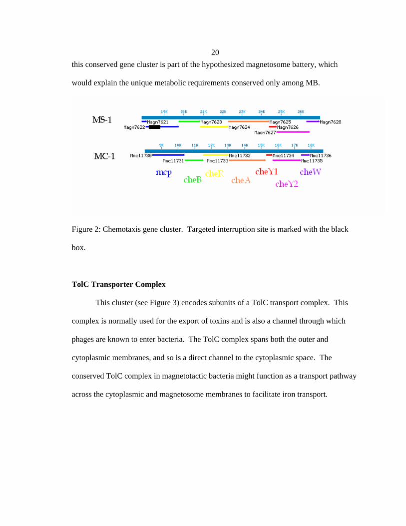
20 this conserved gene cluster is part of the hypothesized magnetosome battery, which
would explain the unique metabolic requirements conserved only among MB.
Figure 2: Chemotaxis gene cluster. Targeted interruption site is marked with the black
box.
TolC Transporter Complex
This cluster (see Figure 3) encodes subunits of a TolC transport complex. This
complex is normally used for the export of toxins and is also a channel through which
phages are known to enter bacteria. The TolC complex spans both the outer and
cytoplasmic membranes, and so is a direct channel to the cytoplasmic space. The
conserved TolC complex in magnetotactic bacteria might function as a transport pathway
across the cytoplasmic and magnetosome membranes to facilitate iron transport.

21
Figure 3: Cluster encoding predicted TolC transport complex. Targeted Interruption sites
are marked with black boxes.
Hydrogenases 1 and 2
Large and small subunits of Hydrogenases 1 and 2 are conserved between MS-1
and MC-1 (See Figure 4). Hydrogenase 1 (HyaA and HyaB) is used by other bacteria for
H2 uptake, and Hydrogenase 2 (HybA and HybB) generates H2 during fermentation of
formate [Maness and Weaver, 2001]. It is not yet clear whether MS-1 or MC-1 are
capable of hydrogen metabolism. It has been shown that there are several substrate-
specific metabolisms (iron, nitrate, oxygen) which affect magnetite production, and it
may be that hydrogen is another one.

22
Figure 4: Conserved hydrogenase gene cluster. Targeted interruption site is marked with
the black box.
1.2.2. Methods
Construction of Targeted Insertion Strains
Approximately 500 bp of the target gene was PCR amplified from AMB-1 DNA,
purified and cloned into the TOPO vector (QIAGEN, USA). The target sequence was
digested out of the TOPO vector with SpeI and NotI, purified, and ligated into the pAK31
vector. This plasmid was cloned into E. coli DH5α and transformed into donor strain E.
coli wm3064. The donor strain was then mated with AMB-1 and transconjugants were
selected for with kanamycin. Proper insertion was checked by PCR using a forward
primer for the start of the interrupted gene and reverse primer reading out of the insertion.
Magnetization Assay
For the magnetization assay, stocks stored at -80˚ C were used to inoculate 1.5 ml
of AMB-1 media without added iron in Eppendorf tubes and incubated at 30˚ C. After
three days (late log phase) these cultures were added to 13.5 ml of AMB-1 medium

23 without added iron and allowed to grow overnight. The culture was centrifuged and
resuspended in 1.5 ml of the original supernatant. 0.35 ml of the concentrated culture
was then added to 10 ml of N2 flushed AMB-1 media in Balsch tubes. Then 10 mM
ferric quinate and air were added to achieve the specific test conditions. Cultures were
then incubated at 30˚ C with shaking. Magnetization of the TI strains was checked by
Cmag, the ratio of absorbance at 400 nm of cells aligned by external magnet parallel and
perpendicular to the line of sight in the spectrometer.
1.2.3. Results and Discussion
None of the targeted interruptions caused a change in phenotype. Time course
assays were carried out under varying iron and oxygen concentrations with no significant
difference being found in magnetization (see Figure 5, 6, 7). This may be due to
redundancy in the pathways in which these genes are involved; selection for genetic
adaptation in the case of lethality of the TIs, the lack of their necessity for BCMM in our
laboratory conditions, or these genes may not be involved in BCMM at all. If the genes
are not involved in BCMM it raises the question why these genes are so well conserved
among the magnetotactic bacteria. Since the targeted interruptions were undertaken, the
hydrogenase genes have been found in new, non-BCMM genomes, further indicating that
they are not necessary for BCMM. This demonstrates the necessity of testing of
genomically generated hypotheses.
A more recent comparison using four MB genomes and 426 non-MB
genomes found only 28 genes conserved among the MB [Richter et al., 2007]. This
analysis used the complete genomes of MC-1 and Magnetospirillum magneticum AMB-

24 1, and the draft genomes of MS-1 and M. gryphiswaldense MSR-1. Richter et al. found
15 of the mam genes conserved as well as 13 genes outside of the mam cluster. We
found 13 of their 15 mam genes conserved in our study — we did not find the short
proteins mamQ or mamC conserved. We only found 7 of the 13 genes they found outside
of the mam cluster. This includes mtxA, mmsF, and mamX as well as two predicted
hemerythrins, a mamH-like gene, and a phage.
There are a number of differences in the methods that could account for the
different set of genes found between this study and Richter et al.’s 2007 study. First, they
used expectation values to compare BLAST hits. Expectation values are based in part on
the size of the database used for searching, and thus will change from one search to the
next. Our use of bit scores, which aren’t tied to database size, facilitate consistent
comparisons. Ideally one would use an alignment and scoring algorithm with a rigorous
statistical base, such as HMMER (http://hmmer.wustl.edu). The second, and perhaps
greatest difference, is the way they searched the non-magnetotactic genomes. They
restricted their conserved genes (their “MTB-related genes”) to not having a BLAST hit
outside of the MB with an E-value less than 1e-50. This eliminates many of the proteins
identified in our study as they were found to have significant similarity to proteins
outside of the MB, though not as high a similarity to other MB proteins. When they
included significant hits (i.e., E-value < 1e-80) to Rhodospirillum rubrum, the
phylogenetically closest non-MB genome to the magnetospirilla, they found only one
gene conserved. Finally, their use of twice as many MB genomes and non-MB genomes
in their analysis likely eliminated some of the genes we identified in our more limited
analysis.

25
Magnetization, 50 uM Iron, 0.5% Oxygen
0.91
1.11.21.31.41.51.61.71.8
0 500 1000 1500 2000
Time (Min)
Cm
ag
wtchehydtolC
Figure 5: Time course of acquisition of magnetization by TI strains (che: Chemotaxis
cluster, hyd: Hydrogenase cluster, tolC: TolC cluster) versus wild-type (wt). High iron
concentration and low oxygen allowed the cultures to become very magnetic. Error bars
are the average of 3 subcultures of a culture passaged under the same conditions.

26
Magnetization, 20 uM Iron, 2% Oxygen
0.98
1
1.02
1.04
1.06
1.08
1.1
1.12
0 500 1000 1500 2000 2500
Time (Min)
Cm
ag
WTCheHydTolC
Figure 6: Time course of acquisition of magnetization by TI strains (che: Chemotaxis
cluster, hyd: Hydrogenase cluster, tolC: TolC cluster) versus wild-type (wt). Lower iron
and higher oxygen reduced the total magnetization acquired and delayed the onset of
magnetization. Error bars are the average of 3 subcultures of a culture passaged under
the same conditions.

27
Magnetization, 6 uM Iron, 0.5% Oxygen
0.991
1.011.021.031.041.051.061.071.081.091.1
0 100 200 300 400 500
Time (Min)
Cm
ag
WTCheHydTolC
Figure 7: Time course of acquisition of magnetization by TI strains (che: Chemotaxis
cluster, hyd: Hydrogenase cluster, tolC: TolC cluster) versus wild-type (wt). Very low
iron and low oxygen reduced the total magnetization acquired without delaying the onset
of magnetization. Error bars are the average of 3 subcultures of a culture passaged under
the same conditions.

28 Chapter Two: Genetics
2. Transposon Mutagenesis Analysis of Magnetotactic Biomineralization
Cody Z. Nash, Arash Komeili, Robert E. Kopp, Atsuko Kobayashi, Hojatollah Vali,
Dianne K. Newman, and Joseph L. Kirschvink
2.1. Introduction
Magnetotactic bacteria (MB) are distinguished from other bacteria by their ability
to synthesize intracellular magnetic crystals in the single-domain size range. In
laboratory strains of MB the crystals are mainly composed of the iron oxide magnetite
(Fe3O4) with minor amounts of other iron oxides. These crystals occur within lipid-
membrane-bounded spaces termed magnetosomes, which have a distinct population of
proteins from that of the other cell membranes. These crystals are generally chemically
pure, have no crystallographic defects, and are arranged into chains. Different strains of
magnetotactic bacteria form different shapes of crystals, and some strains produce the
magnetic iron sulfide Greigite. Within each strain the shape and composition of crystals
is uniform, which suggests that there is genetic control over their synthesis.
A number of studies have identified the genes involved by using transposon
mutagenesis. In this technique bacteria are mutated by introducing a transposon to them
through conjugation with a donor strain. The transposon carries a drug resistance marker
which allows the transconjugants to be isolated. The resulting population can then be
screened in various ways to isolate mutants in a specific phenotype. The gene
responsible can then be discovered by determining the insertion site of the transposon.

29 Previous studies have demonstrated the role of several genes necessary for the
magnetotactic phenotype, including iron transporters, flagellum, and regulatory genes.
In this study we screened through 5809 mutants to identify more of the genes
involved in the process. Nineteen non-magnetic or partially magnetic mutants were
identified. Fourteen of these were in the mam cluster, an island of genes which encode
many of the proteins found in abundance in the magnetosome membrane. The other five
mutants have insertions in genes outside of the mam cluster. The necessity of genetic
machinery outside of the mam cluster indicates that magnetotaxis may not be as easily
laterally transferred as has been previously speculated. It also suggests a number of
previously unsuspected pathways may be involved in the magnetotactic phenotype.
2.2. Methods
Mutagenesis
To generate mutants, the Mariner transposon was introduced to Magnetospirillum
magneticum AMB-1 through conjugation with a donor strain, Escherichia coli β2155, as
previously described [Komeili et al., 2004]. Selection was done with Kanamycin at 5
μg/ml. Transconjugation rates of up to 5e-05 were achieved. Timing was critical to
prevent spontaneous mutants.

30 Magnetic Screen
Mutants were screened by growing them in 96-well plates and, after grown,
placing them over an array of magnets, as previously described [Komeili et al., 2004].
Magnetic mutants were pulled to the sides of the wells, while non-magnetic mutants
remained at the bottom (See Figure 1).
Figure 1: Magnetic screen for non-magnetic mutants. (a) A plate of transconjugants
placed over an array of magnets to pull the MB to the side of the wells. (b) Close-up of a
magnetic and non-magnetic mutant
PCR Screen
Due to the high rate of spontaneous mutation in strain AMB-1, non-magnetic
mutants were checked for deletions in the mam cluster. Mutants were initially screened
for the presence of mamA, and then along 16 kb of the mam cluster. PCR was used to
amplify 1–3 kb sections of the mam cluster to determine their presence, absence, or
change in length. Amplifications were carried out with Promega 2X PCR mix (Promega
Corporation, Madison, WI, USA) with 30 cycles and a 56˚ C annealing temperature.

31
List of Primers for PCR screen:
MamA-up 5' GGCGAATTCATGTCTAGCAAGCCGTCGGA 3'
MamA-down 5' GGCGGATCCGACCGAGGCCCCTTCGTCAAGT 3'
ClBeg 5' GTTCAGGTCGGTGGGTTTTT 3'
ORF2-2 5' GTTCGGCAAGACCGAAATTA 3'
ORF2-1 5' GCGTGGAAGAGGTCGAGA 3'
ORF5-2 5' CTTGTATCTCCGGCTTCTCG 3'
ORF5-1 5' CGTCAATGTGGCCATGTATT 3'
ORF7d 5' ACATTGCCCTTGACCACATT 3'
ORF7c 5' CGAGAAGTTCCTGCATTTCC 3'
AQ2 5' ACGGCCAGATCGTACTTTTG 3'
AQ1 5' GGGAATCGCCTATGTGAAGA 3'
ORF11-2 5' GCGACAGATTTTCCAAAACG 3'
ORF11-1 5' ACGTCAAGTCGATCCAGGTC 3'
ClEnd 5' CTCGCAAACACTCAAGACACTCAG 3'
Magnetization Measurements
For time-course experiments, magnetization was measured by absorbance at 400
nm wavelength. Magnetic cultures have a higher absorbance when they are aligned
parallel to the line of sight than when aligned perpendicularly. Alignment was induced
by placing a large stir bar magnet adjacent to the culture during measurement. The ratio
of parallel to perpendicular absorbance is defined as Cmag.

32 For moment per cell measurements, 1600 mT isothermal remnant magnetization
was measured on 1.5 ml cultures in Eppendorf tubes with a 2G Enterprises SQUID
magnetometer. Culture density was determined by direct cell counts.
TEM Imaging
Conventional TEM and HAADF/STEM/EDX analysis was carried out on a
Tecnai G2 F20 Twin (FEI, Holland), as described previously [Kobayashi et al., 2006].
Cryo-TEM was carried out with a JEOL JEM-2000FX TEM as previously described
[Komeili et al., 2004].
Complementation
The pAK4 vector, a modified pBBR1MCS4 vector containing a tac promoter,
was used as the base for complementation plasmids. Sequences to be complemented
were PCR amplified with the high fidelity polymerase EHF (Roche, Switzerland), ligated
into the pAK4 backbone, cloned into DH5α λpir, transformed into donor strain wm3064,
and then into the mutant strain. Selection was done with Ampicillin or Carbenicillin.
Targeted Interruption
The pWM91 vector was used as the base for targeted interruptions [Metcalf et al.,
1996]. 1 kb target sites were amplified with ThermoPol polymerase (NEB, USA), ligated
into the pWM91 backbone, cloned into DH5α λpir, transformed into donor strain
wm3064, and then inserted into the chromosome of wild-type Magnetospirillum

33 magneticum AMB-1 via homologous recombination. Selection was done with
Kanamycin.
Strains and Plasmids Used:
Magnetospirillum magneticum AMB-1 [Matsunaga et al., 1991]
E. coli DH5a cloning strain for vector construction [Sambrook et al., 1989]
E. coli wm3064 donor strain for mutagenesis [Dehio and Meyer, 1997]
E. coli b2155 for mutagenesis
PSC189 — hyperactive mariner transposon [Chiang and Rubin, 2002]
pAK [Komeili et al., 2004]
2.3. Results and Discussion
Out of 5809 mutants screened, 192 were initially found to be non-magnetic. Most
of these were found to be spontaneous mutants during the PCR screen. The rate of
spontaneous mutation decreased from 11% to 0.5% when care was taken to minimize the
amount of time in stationary phase the AMB-1 cultures experienced prior to mating. This
is consistent with previous work showing a correlation between time in stationary phase
and spontaneous mutation in MB, probably due to native transposon activity [Schübbe et
al., 2003; Ullrich et al., 2005]. 19 of the 192 were found to be non- or partially magnetic
and not due to deletions of all or part of the mamAB cluster based on the PCR assay. Of
the 19 non-magnetic mutants, 14 had insertion sites inside the mamAB cluster in six
different genes (see Figure 2). The five mutants with insertion sites outside of the
mamAB cluster are discussed individually below.

34
Figure 2: Map of insertion sites in the mamAB cluster in AMB-1. Locus numbers are
inside the genes, map position below the scale, gene names below the scale, and insertion
sites are marked with the arrowheads.
MNM9
Mutant MNM9 has a transposon insertion site 99088 base pairs from the origin of
the chromosome, near the beginning of predicted gene amb0089. Magnetic
measurements show that it is completely non-magnetic. HAADF TEM showed an
absence of magnetite (See Figure 3a). Cryo-TEM also shows an absence of magnetite,
but further shows that MNM9 lacks magnetosomes entirely, membranes and all (See
Figure 3b & 3c).

35
Figure 3: (a) HAADF TEM of MNM9 showing absence of inclusions. (b) and (c) Cryo-
TEM of MNM9 showing complete absence of magnetosomes. (d) Cryo-TEM of wild-
type AMB-1 for comparison
Complementation of MNM9 with amb0089; amb0089 and amb0090; or amb0089,
amb0090, and amb0091 failed to recover any of the wild-type phenotype. Targeted
interruption of amb0089 failed to alter the wild-type phenotype. This suggests that

36 MNM9’s phenotype may be due to a spontaneous mutation in some other part of the
chromosome, although large-scale deletions in the mamAB cluster were ruled out by the
PCR screen. The discovery and development of efficient native promoters in AMB-1
may allow better complementation in the future [Yoshino and Matsunaga, 2005]. The
failure of the targeted interruption to replicate the MNM9 phenotype may be because the
interruption did not replicate necessary polar effects.
The predicted gene amb0089 is a predicted Fe-S oxidoreductase and falls into the
Radical SAM protein superfamily. These proteins have an iron-sulfur center which
generates organic radicals from the S-adenosylmethionine (SAM) molecule (for reviews,
see [Fontecave et al., 2004; Layer et al., 2004]). Radical SAM proteins can act in a
regulatory role by catalyzing the methylation of DNA, hormones, neurotransmitters, and
signal transduction systems. They also play a role in numerous biosynthetic pathways by
catalyzing the addition of parts of the SAM molecule to the substrate. Many of the
predicted genes near amb0089 which code in the same direction belong to the sialic acid
biosynthesis pathway (See Figure 4). Sialic acid is a terminal sugar residue on
glycoproteins which is expressed on the outer surface of membranes in eukaryotes to
regulate vesicle transport. Sialic acid is also commonly used by pathogenic bacteria to
evade an immune response. What sialic acid is doing in AMB-1 is still a mystery.

37
Figure 4: Location of transposon insertion for MNM9. Locus numbers are inside the
genes, map position below the scale, predicted product below the scale, and the insertion
site is marked with the arrowhead.
MNM13
Mutant MNM13 has an insertion site at 197516 bp along the AMB-1
chromosome, in the middle of gene amb0172. Magnetization assays showed it to have a
weakly magnetic phenotype. Cryo-TEM shows MNM13 to have isolated, elongate
crystals (See Figure 5) in contrast to the equant crystals produced by wild-type AMB-1.
Figure 5: Cryo-TEM of MNM13 showing isolated, elongate crystals

38
Complementation of gene amb0172 or amb0172 and amb0173 failed to change
the phenotype of MNM13. Gene amb0172 is a predicted hydrolase or acyltransferase,
and the adjacent genes coding in the same direction are a hypothetical protein and a
predicted aldehyde dehydrogenase (see Figure 6).
Figure 6: Transposon insertion site for MNM13. Locus numbers are inside the genes,
map position below the scale, predicted product below the scale, and the insertion site is
marked with the arrowhead.
MNM16
MNM16 has its transposon insertion site at 225328 bp along the AMB-1
chromosome near the end of predicted gene amb0200. Magnetization assays indicated it
was partially magnetic (see Figure 7) and conventional TEM revealed fewer crystals
scattered along the magnetosome chain, with each crystal appearing normal (see Figure
8).

39
Magnetization
1
1.1
1.2
1.3
1.4
1.5
1.6
1.7
1.8
0 5 10 15 20 25
Hours
Cm
ag
wt 1% O2wt 0% O216 1% O216 0% O2
Figure 7: Magnetization assays of MNM16 under 0% and 1% O2 in headspace, as
compared to wild-type. Error bars are the average of 3 subcultures of a culture passaged
under the same conditions.
Figure 8: TEM of MNM16 showing fewer crystals than in wild type
Complementation of gene amb0200 did not have any significant effect on the
phenotype, while complementation of genes amb0200 through amb0196 (on the

40 complementary strand) partially restored the phenotype both in Cmag and IRM
measurements of magnetization (see Figures 9 and 10).
16 comp 1%
11.051.1
1.151.2
1.251.3
1.351.4
0 2 4 6 8 10
hrs
cmag
apq.116pB16P
16 comp 0%
1
1.05
1.1
1.15
1.2
1.25
1.3
0 2 4 6 8 10
hrs
cmag
apq.116pB16P
Figure 9: Time course of complementation of MNM16. (a) Experiments with 1% O2 in
headspace of Balsch tubes. (b) Experiments with 0% O2 in headspace of Balsch tubes.
16pB is mutant mnm16 carrying an empty complementation vector to confer Ampicillin
resistance. 16P is complemented for the mutated gene and downstream genes (see text
for discussion). APQ.1 is wild-type AMB-1 carrying the Ampicillin and Kanamycin
resistance carrying plasmid APQ.1. Growth curves are from single cultures.

41
Complementation
0.00E+00
2.00E-14
4.00E-14
6.00E-14
8.00E-14
1.00E-13
1.20E-13
16pB 16G 16P 18pB 18G 18P 20pB 20P APQ.1
Mom
ent/C
ell
Figure 10: Complementation assays for MNM16, MNM18, and MNM20. 16pB, 18pB,
and 20pB are mutants mnm16, 18, and 20 carrying an empty complementation vector to
confer Ampicillin resistance. 16G and 18G are mnm16 and 18 with only the mutated
gene complemented. 16P, 18P, and 20P are complemented for the mutated gene and
downstream genes (see text for discussion). APQ.1 is wild-type AMB-1 carrying the
Ampicillin and Kanamycin resistance carrying plasmid APQ.1. Error bars are the
average of 3 subcultures of a culture passaged under the same conditions.
MNM16’s insertion is in a predicted transcriptional regulator. Complementation
of this gene alone did not significantly change the phenotype, while complementation of
amb0200 and four of the downstream genes did have a significant effect. This suggests
that one of the downstream genes is responsible for the phenotype. These genes are
predicted to be of various and uncharacterized functions (see Figure 11), and in that way

42 resemble the genes of the mamAB cluster. This diversity of predicted function may be
how a new pathway appears in annotation.
The reduced number of crystals seen in MNM16 is similar to that seen in AMB-1
when mamA is knocked out [Komeili et al., 2004]. It would be very interesting to see if
the expression of the mam genes is affected in the same manner in both MNM16 and
mamA deletions.
Figure 11: Transposon insertion site of MNM16 and nearby genes. Locus numbers are
inside the genes, map position below the scale, predicted product below the scale, and the
insertion site is marked with the arrowhead.
MNM18
The transposon insertion site of MNM18 is at base pair 3494968 in gene
amb3233. MNM18 is partially magnetic (see Figure 12) and is the only mutant to display
a growth defect. TEM shows that MNM18 produces 1–2 equant crystals per cell (See
Figure 13).

43
Growth Curves of Wild-type AMB-1 and MNM18
0
0.05
0.1
0.15
0.2
0.25
0 5 10 15 20 25 30 35
Hours
Abs
orba
nce
at 4
00 n
m
Figure 12: Growth curves of MNM18 versus wild-type. Wild-type is in black, MNM18
is in light grey. Curves are not averaged so that the individual behavior of each culture
can be examined. The 3 wild-type and mnm18 cultures are subcultures of a single culture
passaged under the same conditions as the experiment.
44
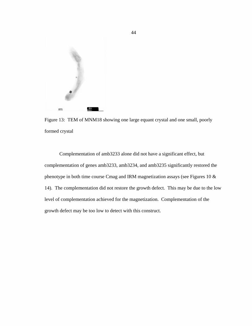
Figure 13: TEM of MNM18 showing one large equant crystal and one small, poorly
formed crystal
Complementation of amb3233 alone did not have a significant effect, but
complementation of genes amb3233, amb3234, and amb3235 significantly restored the
phenotype in both time course Cmag and IRM magnetization assays (see Figures 10 &
14). The complementation did not restore the growth defect. This may be due to the low
level of complementation achieved for the magnetization. Complementation of the
growth defect may be too low to detect with this construct.

45
18 comp 1%
11.051.1
1.151.2
1.251.3
1.351.4
0 2 4 6 8 10
hrs
cmag
apq.118pB18P
18 comp 0%
1
1.05
1.1
1.15
1.2
1.25
1.3
0 2 4 6 8 10
hrs
cmag
apq.118pB18P
Figure 14: Time course of complementation of MNM18. (a) Experiments with 1% O2 in
headspace of Balsch tubes. (b) Experiments with 0% O2 in headspace of Balsch tubes.
18pB is mutant mnm16 carrying an empty complementation vector to confer Ampicillin
resistance. 18P is complemented for the mutated gene and downstream genes (see text
for discussion). APQ.1 is wild-type AMB-1 carrying the Ampicillin and Kanamycin
resistance carrying plasmid APQ.1. Growth curves are from single cultures.

46 The gene amb3233 is a predicted to encode the α and β subunits of a
pyruvate/ferredoxin oxidoreductase. The downstream genes are the γ-subunit and a cheY
gene, and the upstream gene is a predicted transcriptional regulator (see Figure 15). The
genes flanking this cluster code on the opposite strand. The growth defect of MNM18 is
consistent with a disruption of metabolic genes.
Figure 15: Insertion site of MNM18. Locus numbers are inside the genes, map position
below the scale, predicted product below the scale, and the insertion site is marked with
the arrowhead.
MNM20
MNM20 has its insertion site at position 553134, in gene amb0516.
Magnetization assays show MNM20 to be partially magnetic and HAADF TEM shows
that it has many poorly formed and amorphous crystals (see Figure 16).
MNM20 sometimes grows without any defect in magnetization, and it is not clear
what growth conditions are necessary to produce the mutant phenotype. It is possible
that the frequent spontaneous mutations AMB-1 experiences are able to revert MNM20
to the wild-type phenotype.

47
Figure 16: HAADF TEM images of MNM20 showing many poorly formed and
amorphous crystals
The gene amb0516 is a hypothetical protein with few matches outside of the
Magnetospirilla. The downstream gene amb0517 is even more unusual in that it is
similar to many eukaryotic proteins. This gene cluster (see Figure 17) again displays the

48 trait of genes with various and poorly characterized functions, suggesting involvement in
a poorly understood pathway.
Figure 17: Insertion site of MNM20. Locus numbers are inside the genes, map position
below the scale, predicted product below the scale, and the insertion site is marked with
the arrowhead.
2.4. Conclusions
Out of 5809 mutants screened, 19 were identified as non- or partially magnetic
and did not have large deletions in the mamAB cluster. Fourteen of these are insertions in
the mamAB cluster, demonstrating its role in the magnetic phenotype of magnetotactic
bacteria. Five mutants have insertions in genes outside the mamAB cluster, suggesting
that the genetic systems necessary for the wild-type magnetic phenotype are not restricted
to a few clusters of genes. The difference between the genes identified in this
mutagenesis and others may be due to the different transposons used. In comparing this
study with other transposon mutagenesis studies in magnetotactic bacteria, this is the only
one to use a PCR screen, which may account for the lower total number of non-magnetic
mutants seen (see Table 1).

49 The absence of insertions in the mamGFDC cluster, another cluster of genes
which encode magnetosomal proteins, is consistent with recent results that these genes
are not necessary for a magnetic phenotype [Scheffel et al., 2008]. The absence of
insertions in the mamJ and mamK genes is also consistent with recent work showing that
these genes affect magnetosome localization but not magnetite formation in the
magnetospirilla [Komeili et al., 2006; Scheffel et al., 2006].
There are a number of methods which future investigators could use to examine
the role of these genes in magnetotaxis. One could test recently developed high-
efficiency native promoters to try to improve the complementation of the mutants
[Yoshino and Matsunaga, 2005]. Another route to conclusively testing the role of these
genes would be to carry out knock outs of the genes, which would eliminate the
possibility of read-through from the drug resistance marker inserted during targeted
interruptions. Targeted interruptions can also generate incomplete versions of the protein
they interrupt, either from the fragment before or after the insertion. These incomplete
versions may retain full functionality in the conditions assayed or may interfere with
other cellular processes to mask any effect on magnetite biomineralization. A knock out
avoids these possibilities.
Finally, another way to examine these mutants is to look at the mRNA levels of
the relevant genes. Through reverse transcriptase PCR (RT PCR) one could check the
expression levels of mam genes to see which, if any of the mutants has altered regulation
of these genes known to be involved in magnetotaxis. RT PCR would also allow one to
determine how effective complementation is relative to the mutant expression levels of
the mutated genes. It could also be used to compare expression levels of the mam genes

50 between mutants, targeted interruptions, and knock outs to see if the latter two methods
are replicating the mutant phenotype at the RNA level.

51 Table 1: Transposon Mutagenesis Studies in Magnetotactic Bacteria
Strain Transposon Mutants Screened
Non-magnetic
Insertion sites identified
Ref
AMB-1 Mini-Tn5 118 5 amb3990 (magA) [Matsunaga et al., 1992; Nakamura et al., 1995]
AMB-1 Mini-Tn5 5762 69 amb0192, amb0291, amb0503, amb0521, amb0676, amb0741, amb0759, amb1309, amb1394, amb1482, amb1692, amb1722, amb1790, amb2051, amb2087, amb2504, amb2554, amb2611, amb2660, amb2765, amb2922, amb3184, amb3268, amb3279, amb3295, amb3450, amb3458, amb3672, amb3734, amb3742, amb3766, amb4107, amb4111, amb4543
[Matsunaga et al., 2005; Wahyudi et al., 2001]
MSR-1 Mini-Tn5 ? 2 AAX11190 (CheY) (~amb0983)
[Li et al., 2005]
AMB-1 Mariner 700 2 amb0968 (mamN), amb0972 (mamQ)
[Komeili et al., 2004]
AMB-1 Mariner 5809 19 amb0089, amb0172, amb0200, amb0516, amb3233, amb0963 (mamE), amb0967 (mamM), amb0968 (mamN), amb0969 (mamO), amb0971 (mamA), amb0972 (mamQ)
This Study

52 Chapter 3: Environmental Microbiology
3.1. Extremophilic Magnetotactic Bacteria and Archaea
Nash, C. Z.1, Popa, R.2, Kobayashi, A.3, and J. L. Kirschvink1
1Division of Geological and Planetary Sciences, California Institute of Technology,
Pasadena, CA, 91125, USA. 2Portland State University, Portland, OR, 97207, USA.
3Photonics Research Institute, National Institute of Advanced Industrial Science and
Technology, Osaka, Japan.
Magnetotactic bacteria (MB) form nanoscale crystals of the ferrimagnetic
minerals magnetite (Fe3O4) or greigite (Fe3S4) inside intracellular lipid-membrane-
bounded vesicles termed magnetosomes. All reports since their initial discovery
[Blakemore, 1975] have been in freshwater, marine, and soil environments at near
neutral pH and temperatures below 30° C [Bazylinski and Frankel, 2004]. Only two
of the 150+ 16S rDNA sequences of MB lie outside of the bacterial phylum
Proteobacteria. These two belong to the genus Magnetobacterium in the phylum
Nitrospirae. In order to better understand the diversity and environmental range
of MB we examined samples from two extreme environments. Here we report the
discovery of several magnetotactic extremophiles: a thermophile from Little Hot
Creek (LHC), (37.69º N, 118.84º W), and four from the hyper-alkaline, hyper-saline
waters of Mono Lake (37.98º N 119.12º W). Analysis of the 16S rRNA gene indicates
that the thermophilic organism groups with the genus Magnetobacterium in the
phylum Nitrospirae, whereas one species of MB from Mono Lake (MonoEub) lies
with the γ-proteobacteria. We also report the first evidence for magnetotactic
Archaea, in three clones from Mono Lake: ML1, ML3, and ML4. Our findings

53 extend the environmental range of these fossil-forming organisms to include
environments expected to have existed on early Earth and will facilitate geo- and
astrobiological investigations. Our findings also extend the phylogenetic breadth of
magnetotactic microorganisms to include all three domains of life. We anticipate
these organisms to be targets for further analysis to investigate the evolution and
origins of magnetotaxis.
We conducted an initial survey of the LHC site by taking 10 microliter samples
with a pipette and checking them microscopically for a response to a magnetic field. We
checked a range of temperatures from 40o to 80º C at every spring within the group. We
only observed magnetotactic bacteria at one freshwater spring, in 45º–55º C mats
adjacent to the main flow channel. These microbial mats were ~ 1 cm thick, dominated
by a red layer on the surface. We observed MB throughout these mats, but in higher
densities ~ 5 mm from the surface. Microscopic examination of biofilms from similar hot
springs in central California revealed the presence of MB in microbial mats at
temperatures up to 58º C.
We collected small cores of mats and sediments that were kept at 50º C during
transport to, and in, the lab. We observed MB bacteria in these samples over several
months, indicating that they are able to live at these temperatures. Using the magnetic
racetrack technique [Wolfe et al., 1987], we then isolated pellets of MB. We amplified,
cloned, and sequenced DNA using standard techniques, employing either universal
bacterial or archaeal primers. RFLP analysis of 6–10 clones from each sample showed
they were homogenous. Phylogenetic analysis of the sequences obtained was carried out

54 with ARB (see Figure 1). The LHC sequences belong to a group of Nitrospirae that
contains two other MB, as well as clones from other hot springs (see Figure 2).
Samples from Mono Lake (pH of 9.8 and salinity 80.8 g/l) were collected from the
shore to depths up to 40 m using a gravity corer or SCUBA. Cores were stored at room
temperature and MB were observed over a period of months. MB were extracted and
analyzed as with the LHC samples. The closest relatives of the archaeal sequences are
clones from deep-sea hydrothermal surveys, while the closest relatives to the MonoEub
sequence are from Mono Lake and other basic, salty locales (see Figures 3–5).
HRTEM and HAADF/STEM/EDX examination (Tecnai F20 G2Twin) of these
organisms revealed prismatic or bullet-shaped magnetite crystals organized in string-like
bundles; no Greigite magnetosomes were detected (see Figures 6 and 7). The ability of
these bacteria to form magnetite in extremes of temperature, salinity and alkalinity may
prove useful in manufacturing magnetic nanoparticles for many existing applications
[Šafarík and Šafaríková, 2002].
As the deepest branches of the tree of life are composed of thermophiles, these
discoveries, coupled with the wide phyletic distribution of magnetite-precipitating
organisms shown in Figure 1, indicate that magnetotaxis could be a very ancient trait,
perhaps even pre-dating the divergence of the Bacterial, Archaeal, and Eukaryal
Domains.
Sediments of hydrothermal origin are known in the geological record as far back as
3.5 Ga [Brasier et al., 2002], so there is the potential for tracing the fossil remains of
these bacteria into Archean time. Currently unequivocal magnetofossils have been found
in sediments dating back to the Cretaceous, and potential ones as old as the

55 Paleoproterozoic [Chang et al., 1989; Kopp and Kirschvink, 2008]. The ability to identify
these biomarkers in environments found on the early Earth and thought to exist elsewhere
in the universe provides a new tool to look for ancient and alien life.
Author Contributions C.Z.N., R.P., and J.L.K. performed the sampling. C.Z.N.
performed the molecular and phylogenetic work. A.K.K. performed the electron
microscopy. All authors discussed the results and commented on the manuscript.
Figure 1: Small subunit rRNA phylogenetic tree. Groups containing species which
synthesize magnetic minerals highlighted in grey and labelled. Sequences obtained from
this study shown in dark grey.

56
Figure 2: Phylogenetic tree of 16S rRNA sequences closest to the magnetotactic bacterial
sequence obtained from Little Hot Creek, CA. They fall into the Nitrospirae phylum, a
diverse group of nitrite oxidizing, sulfate reducing organisms from contaminated and
thermal environments. The closest clone to our sequences, BcrEubac, is from the outflow
channel of Octopus Springs, Yellowstone National Park, WY [Kopczynski et al., 1994].
Saltmarsh clone LCP-6 is an unpublished sequence from a contaminated site. The next
two closest relatives are both MB from lakes in Bavaria [Flies et al., 2005; Spring et al.,
1993]. Note the closest isolates are Thermodesulfovibrio spp. from hot springs in
Yellowstone and Iceland. Tree was constructed using ARB, with alignments manually
curated and sequences added to the tree in interactive parsimony mode [Ludwig et al.,
2004].

57
Figure 3: Phylogenetic tree of 16S rRNA sequences closest to the magnetotactic bacterial
sequence obtained from Mono Lake, CA, marked with the asterisk. Note the closest
isolates are Marinospirillum minutulum and M. megaterium, both from a low oxygen,
high nitrogen, moderate salinity environment — a fermented brine [Satomi et al., 1998].
Tree was constructed using ARB, with alignments manually curated and sequences added
to the tree in interactive parsimony mode [Ludwig et al., 2004].

58
Figure 4: Phylogenetic tree of 16S rRNA sequences closest to the first putative archaeal
sequence obtained from Mono Lake, CA, marked with the asterisk. There are no isolates
closely related to ML1. The nearby clones, UncArc16 and AhaVC21, are both from deep
sea hydrothermal vent samples and belong to marine benthic group B [Reysenbach et al.,
2000; Teske et al., 2002]. This tree was constructed using ARB, with alignments
manually curated and sequences added to the tree in interactive parsimony mode [Ludwig
et al., 2004].

59
Figure 5: Phylogenetic tree of 16S rRNA sequences closest to the other putative archaeal
sequences obtained from Mono Lake, CA, marked with the asterisk. There are no
isolates closely related to either ML3 or ML4. ML3’s neighbors, VC2.1 Arc6 and an
unidentified archaeon are both from deep sea hydrothermal vent samples [Reysenbach et
al., 2000; Takai and Horikoshi, 1999]. ML4’s neighbors are single clone from the
methanogenic zone of a contaminated sediment (WCHD3-16, [Dojka et al., 1998]) and a

60 dominant clone from high salinity, deep sea brines (KTK4A, [Eder et al., 1999]). The
nearest isolates are Thermoplasma acidophilum and Picrophilus oshimae, both meso- to
thermophilic acidophiles [Golyshina and Timmis, 2005]. This tree was constructed using
ARB, with alignments manually curated and sequences added to the tree in interactive
parsimony mode [Ludwig et al., 2004].

61
Figure 6: HAADF/EDX of ML and LHC samples. (A) HAADF image of Mono Lake
sample. Region boxed in red analyzed with EDX. (B) EDX Analysis of sample from
Mono Lake shows Fe and O, suggesting magnetite. (C) HAADF image of LHC sample.
(D) EDX analysis of sample from LHC shows Fe and O, suggesting magnetite.

62
Figure 7: HRTEM/FFT of LHC sample. (A) HRTEM from one of LHC crystals in 2C
showing lattice spacing characteristic of magnetite (B) Fast Fourier Transform pattern of
the crystal in A confirms magnetite.

63 Chapter 3: Environmental Microbiology
3.2. Degenerate PCR recovery of mamB fragment from LA Arboretum magnetic
cocci.
3.2.1. Introduction
The phylogenetic distribution of MB includes all of the phyla Nitrospirae,
Proteobacteria, and possibly even Firmicutes. Genes which are known to be involved in
magnetosome formation have so far only been retrieved from three magnetospirilla and
one magnetic coccus (strain MC-1), all of which are in the α-Proteobacteria. This project
was an effort to retrieve some of these genes from other MB.
For this we chose two targets. The first was a magnetic cocci found in the Los
Angeles Arboretum tentatively named ARB-1 [Cox et al., 2002]. ARB-1 was chosen
both because of its great abundance in the environment and because of its phylogenetic
position. ARB-1 is distant from both MC-1 and the magnetospirilla, yet still within the
magnetic coccus clade of the α-Proteobacteria.
The second target is Desulfovibrio magneticus RS-1 [Sakaguchi et al., 1993;
Sakaguchi et al., 2002]. RS-1 is a δ-Proteobacteria and the only MB in pure culture
which is not an α-Proteobacteria.
The genes chosen were mamA, mamB, and mamC. These genes were chosen
because of their presence in both the magnetospirilla and MC-1 and their position at the
beginning of the mam cluster, which encodes many of the proteins that are abundant in
the magnetosome membrane. More recent work on mamA and mamC has demonstrated

64 that these genes affect the number and shape of crystals in the magnetospirilla, but are not
necessary for magnetite formation [Komeili et al., 2004; Scheffel et al., 2008].
3.2.2. Methods
Enrichment of ARB-1
Sediment samples were collected from Lake Baldwin in the Los Angeles
Arboretum in Arcadia, CA. Magnetic cocci were observed to be in the highest
abundance in the most sapropelic muds, based on hanging drop observations. MB were
enriched by placing magnets on the outside of the sample bottles, shaking the jar until all
sediment was suspended and then allowing it to settle for 2 hours. 1 ml of water and
magnetic material was collected from each the north and south magnets and run over
superparamagnetic columns (Miltenyi Biotec, Germany). 10 ml aliquots of 0.2 μm
filtered sample water were used to rinse non-MB from the column; washing was carried
out five times until no bacteria were observed in the elution. The magnets were then
removed from the column and two 10 ml aliquots of 0.2 μm filtered sample water were
flushed through to collect the MB. Samples were centrifuged and re-suspended in 1 ml
of the original supernatant. DNA was then extracted from the sample using an Ultraclean
Soil DNA Kit (Mo Bio Laboratories, USA).
RS-1
Desulfovibrio magneticus RS-1 was obtained from the DSMZ (Germany) and
cultured anaerobically as described previously [Sakaguchi et al., 2002]. DNA was
extracted using a standard phenol-chloroform extraction [Sambrook et al., 1989].

65
Degenerate PCR
Degenerate primers were designed based on the mamA, mamB, and mamC
sequences from MS-1 and MC-1 using the CODEHOP software [Rose et al., 1998].
Various PCR conditions were tested. Promega 2X reaction solution was used with 1–10
μL of template DNA, with 30–50 amplification cycles, with annealing temperatures of
52–58˚ C. Products were purified with QIAGEN gel extraction kit, cloned with TOPO
TA 2.1 cloning vector into DH5α cells, and minipreped with QIAGEN miniprep kit for
sequencing.
Primers Used:
Adeg4 5' TGAATGATGATTATCGTCAGGTGTATTATmgngayaargg 3'
Adeg5 5' GATTATCGTCAGGTGTATTATCGTgayaarggnat 3'
Adeg6R 5' CCATGCCCAGACGATAATGAayrttraartt 3'
Adeg7R 5' CTGAAAGGCATCAATCgcytcrtcrwa 3'
Bdeg1 5' CCCTGAAAATTTCCAACAGAccngcngayga 3'
Bdeg2 5' CGGCGATTATGGCGaaygcntggga 3'
Bdeg3 5' GCGATTATGGCGAACgcntgggayaa 3'
Bdeg4 5' GGCGAACGCGtgggayaaymg 3'
Bdeg5R 5' CATCGGAACGGTTAtcccangcrtt 3'
Bdeg6R 5' AACGCATCGGAACGGttrtcccangc 3'
Bdeg7R 5' GAGGAAAACGCATCGGAAckrttrtccca 3'
Bdeg8R 5' CCAAAGGTGGCAAAAATCacnccnaycat 3'

66 Bdeg9R 5' CCGGGGAGGCAtccatnarncc 3'
Cdeg1 5' ACCAACACAGAAGCGGTGathgayacngg 3'
Cdeg2 5' CAACACAGAAGCGGTGATTgayacnggnaa 3'
Cdeg3 5' AACACAGAAGCGGTGATTGATacnggnaarga 3'
Cdeg4 5' AAGCGGTGATTGATACCggnaargarrc 3'
Cdeg5R 5' TGATCCATGCCATAATCCcangcrtaytt 3'
Cdeg6R 5' TGATCCATGCCATAATcccangcrtayt 3'
Cdeg7R 5' TGATCCATGCCATAAtcccangcrta 3'
3.2.3. Results and Discussion
After magnets were left on the shaken sample jar for two hours, 1 ml samples
were taken. Approximately 103 magnetic cocci were observed in the 10 μL sub-samples,
or ~ 105 total magnetic cocci (see Figure 8a). After running onto the magnetic column
and flushing five times, no bacteria were visible in the elutant (see Figure 8b–c). The
magnets were removed from the column and washed twice, yielding ~ 104 magnetic cocci
per ml (see Figure 8d). The sample was centrifuged and resuspended in 1/20th of the
supernatant (see Figure 8e) and then checked for non-magnetotactic cells (see Figure 9).

67
Figure 8: Enrichment of magnetic cocci: (a) un-enriched sample; (b) 1st negative
selection; (c) 5th negative selection; (d) positive selection; (e) 20x concentrated positive
selection

68
Figure 9: Checking sample for non-magnetotactic bacteria. The applied field is towards
the top left of the image. Note the population of south-seeking MB, as previously
observed [Cox et al., 2002].
Degenerate PCR
Numerous products were obtained, but by screening the PCR products for the
expected size most false positives could be excluded. The rest were examined by
sequencing. Some of the false positives obtained on sequencing had no similarity to any
genes from MB at either the DNA or protein level and included fragments which best
matched a xanthine/uracil permease from Pseudomonas fluorescens, a hypothetical

69 protein from Nostoc punctiformis, and a glycosyltransferase from Geobacter
metallireducens. The degenerate primers for mamA repeatedly recovered fragments that
were > 95% identical to sequences from the magnetospirilla. This is likely due to
magnetospirilla being present in the magnetically enriched samples from which DNA
was extracted. It is also possible the arboretum magnetic cocci have Magnetospirillum
versions of their mamA gene. This could be determined through larger scale sequencing
or probing for the mamA gene and the 16S rRNA gene with fluorescent probes. The
degenerate primers for mamC failed to amplify any products of the expected size.
The primer Bdeg1 with either Bdeg5R or Bdeg7R was able to retrieve a 247 bp
sequence whose translation is 80% similar to the MC-1 MamB and 73% similar to the
MS-1 MamB protein. MamB is a protein known to localize to the magnetosome
[Grünberg et al., 2001] and is predicted to be a heavy metal transporter similar to the
cobalt/zinc/cadmium transporter CzcD. The arboretum MamB sequence groups with the
MamB genes from magnetotactic bacteria, and broadly follows the 16S rDNA gene
phylogeny (see Figure 10). Note that although it broadly follows the 16S tree, there are
clear instances of LGT: Porphyromonas has an archaeal version, Methanococcus and
Pyrococcus have a bacterial version. Also note MS-1 has one close duplicate and MC-1
has two duplicates.

70
Figure 10: Phylogeny of MamB and CzcD proteins. This is a strict-consensus tree of
100 bootstrap replicates of a Neighbor-Joining/Distance tree using the Kimura 2-
parameter correction with 301 characters in the alignment, made using the PHYLIP
package. Branches with less than 90% bootstrap support were collapsed. The scale is
arbitrary, only branching order is informative.

71 References
Abe, M., et al. (1999), Magnetite Film Growth at 30°C on Organic Monomolecular
Layer, Mimicking Bacterial Magnetosome Synthesis, Journal of Applied Physics, 85(8), 5705–5707.
Altschul, S. F., et al. (1997), Gapped BLAST and PSI-BLAST: A New Generation of Protein Database Search Programs, Nucleic Acids Research, 25(17), 3389–3402.
Arakaki, A., et al. (2003), A Novel Protein Tightly Bound to Bacterial Magnetic Particles in Magnetospirillum magneticum strain AMB-1, Journal of Biological Chemistry, 278(10), 8745–50.
Balkwill, D. L., et al. (1980), Ultrastructure of a Magnetotactic Spirillum, Journal of Bacteriology, 141(3), 1399–1408.
Bazylinski, D. A., and R. B. Frankel (2004), Magnetosome Formation in Prokaryotes, Nature Reviews Microbiology, 2, 217–230.
Bertani, L. E., et al. (2001), Physical and genetic characterization of the genome of Magnetospirillum magnetotacticum, strain MS-1, Gene, 264(2), 257–263.
Blakemore, R. (1975), Magnetotactic Bacteria, Science, 190(4212), 377–379. Blakemore, R. P., et al. (1979), Isolation and Pure Culture of a Freshwater Magnetic
Spirillum in Chemically Defined Medium, Journal of Bacteriology, 140(2), 720–729.
Boskey, A. L. (1998), Biomineralization: Conflicts, challenges, and opportunities, Journal of Cellular Biochemistry, 30–31, 83–91.
Brasier, M. D., et al. (2002), Questioning the evidence for Earth's oldest fossils, Nature, 416(6876), 76–81.
Chang, S. B. R., et al. (1989), Biogenic Magnetite in Stromatolites .2. Occurrence in Ancient Sedimentary Environments, Precambrian Research, 43(4), 305–315.
Chiang, S. L., and E. J. Rubin (2002), Construction of a mariner-based transposon for epitope-tagging and genomic targeting, Gene, 296(1–2), 179–85.
Cox, B. L., et al. (2002), Organization and elemental analysis of P-, S-, and Fe-rich inclusions in a population of freshwater magnetococci, Geomicrobiology Journal, 19(4), 387–406.
Dean, A. J., and D. A. Bazylinski (1999), Genome analysis of several marine, magnetotactic bacterial strains by pulsed-field gel electrophoresis, Current Microbiology, 39(4), 219–225.
Dehio, C., and M. Meyer (1997), Maintenance of broad-host-range incompatibility group P and group Q plasmids and transposition of Tn5 in Bartonella henselae following conjugal plasmid transfer from Escherichia coli., Journal of Bacteriology, 179(2), 538–40.
Delong, E. F., et al. (1993), Multiple Evolutionary Origins of Magnetotaxis in Bacteria, Science, 259(5096), 803–806.
Dojka, M. A., et al. (1998), Microbial diversity in a hydrocarbon- and chlorinated-solvent-contaminated aquifer undergoing intrinsic bioremediation, Applied and Environmental Microbiology, 64(10), 3869–3877.

72 Eder, W., et al. (1999), Novel 16S rRNA gene sequences retrieved from highly saline
brine sediments of Kebrit Deep, Red Sea, Archives of Microbiology, 172(4), 213–218.
Flies, C. B., et al. (2005), Diversity and vertical distribution of magnetotactic bacteria along chemical gradients in freshwater microcosms, FEMS Microbiology Ecology, 52(2), 185–195.
Fontecave, M., et al. (2004), S-adenosylmethionine: nothing goes to waste, Trends in Biochemical Sciences, 29(5), 243–249.
Frankel, R. B., et al. (1983), Fe3O4 Precipitation in Magnetotactic Bacteria, Biochimica Et Biophysica Acta, 763(2), 147–159.
Frankel, R. B., and D. A. Bazylinski (1994), Magnetotaxis and Magnetic Particles in Bacteria, Hyperfine Interactions, 90(1–4), 135–142.
Frankel, R. B., et al. (1997), Magneto-aerotaxis in marine coccoid bacteria, Biophysical Journal, 73(2), 994–1000.
Glasauer, S., et al. (2002), Intracellular iron minerals in a dissimilatory iron-reducing bacterium, Science, 295(5552), 117–119.
Golyshina, O. V., and K. N. Timmis (2005), Ferroplasma and relatives, recently discovered cell wall-lacking archaea making a living in extremely acid, heavy metal-rich environments, Environmental Microbiology, 7(9), 1277–1288.
Grünberg, K., et al. (2001), A Large Gene Cluster Encoding Several Magnetosome Proteins is Conserved in Different Species of Magnetotactic Bacteria, Applied and Environmental Microbiology, 67(10), 4573–4582.
Grünberg, K., et al. (2004), Biochemical and proteomic analysis of the magnetosome membrane in Magnetospirillum gryphiswaldense, Applied and Environmental Microbiology, 70(2), 1040–1050.
Guerin, W. F., and R. P. Blakemore (1992), Redox Cycling of Iron Supports Growth and Magnetite Synthesis by Aquaspirillum magnetotacticum, Applied and Environmental Microbiology, 58(4), 1102–1109.
Handrick, R., et al. (2004), Unraveling the function of the Rhodospirillum rubrum activator of polyhydroxybutyrate (PHB) degradation: the activator is a PHB-granule-bound protein (phasin), Journal of Bacteriology, 186(8), 2466–2475.
Kawaguchi, R., et al. (1995), Phylogenetic Analysis of a Novel Sulfate-Reducing Magnetic Bacterium, RS-1, Demonstrates its Membership of the δ-Proteobacteria, FEMS Microbiology Letters, 126(3), 277–282.
Kirschvink, J. L., and H. A. Lowenstam (1979), Mineralization and Magnetization of Chiton Teeth — Paleomagnetic, Sedimentologic, and Biologic Implications of Organic Magnetite, Earth and Planetary Science Letters, 44(2), 193–204.
Kirschvink, J. L. (1980), South-Seeking Magnetic Bacteria, Journal of Experimental Biology, 86(JUN), 345–347.
Kirschvink, J. L. (1982), Paleomagnetic Evidence for Fossil Biogenic Magnetite in Western Crete, Earth and Planetary Science Letters, 59(2), 388–392.
Kobayashi, A., et al. (2006), Experimental observation of magnetosome chain collapse in magnetotactic bacteria: Sedimentological, paleomagnetic, and evolutionary implications, Earth and Planetary Science Letters, 245(3–4), 538–550.

73 Komeili, A., et al. (2004), Magnetosome vesicles are present before magnetite formation,
and MamA is required for their activation, Proceedings of the National Academy of Sciences of the United States of America, 101(11), 3839–3844.
Komeili, A., et al. (2006), Magnetosomes are cell membrane invaginations organized by the actin-like protein MamK, Science, 311(5758), 242–245.
Kopczynski, E. D., et al. (1994), Recognition of chimeric small subunit ribosomal DNAs composed of genes from uncultivated microorganisms, Applied and Environmental Microbiology, 60(2), 746–748.
Kopp, R. E., and J. L. Kirschvink (2008), The identification and biogeochemical interpretation of fossil magnetotactic bacteria, Earth-Science Reviews, 86(1–4), 42–61.
Layer, G., et al. (2004), Structure and function of radical SAM enzymes, Current Opinion in Chemical Biology, 8(5), 468–476.
Levi-Kalisman, Y., et al. (2001), Structure of the nacreous organic matrix of a bivalve mollusk shell examined in the hydrated state using Cryo-TEM, Journal of Structural Biology, 135(1), 8–17.
Li, F., et al. (2005), Cloning and functional analysis of the sequences flanking mini-Tn5 in the magnetosomes deleted mutant NM6 of Magnetospirillum gryphiswaldense MSR-1, Science in China Series C-Life Sciences, 48(6), 574–584.
Ludwig, W., et al. (2004), ARB: a software environment for sequence data, Nucleic Acids Research, 32(4), 1363–1371.
Maness, P. C., and P. F. Weaver (2001), Evidence for three distinct hydrogenase activities in Rhodospirillum rubrum, Applied Microbiology and Biotechnology, 57(5–6), 751–6.
Mann, S., et al. (1984), Structure, Morphology and Crystal-Growth of Bacterial Magnetite, Nature, 310(5976), 405–407.
Mann, S., et al. (1990), Magnetotactic Bacteria — Microbiology, Biomineralization, Paleomagnetism and Biotechnology, Advances in Microbial Physiology, 31, 125–181.
Matsunaga, T., et al. (1991), Magnetite Formation by a Magnetic Bacterium Capable of Growing Aerobically, Applied Microbiology and Biotechnology, 35(5), 651–655.
Matsunaga, T., et al. (1992), Gene-Transfer in Magnetic Bacteria — Transposon Mutagenesis and Cloning of Genomic DNA Fragments Required for Magnetosome Synthesis, Journal of Bacteriology, 174(9), 2748–2753.
Matsunaga, T., et al. (2005), Complete genome sequence of the facultative anaerobic magnetotactic bacterium Magnetospirillum sp strain AMB-1, DNA Research, 12(3), 157–166.
Meldrum, F. C., et al. (1993), Electron-Microscopy Study of Magnetosomes in a Cultured Coccoid Magnetotactic Bacterium, Proceedings of the Royal Society of London Series B—Biological Sciences, 251(1332), 231–236.
Metcalf, W. W., et al. (1996), Conditionally Replicative and Conjugative Plasmids Carrying lacZa for Cloning, Mutagenesis, and Allele Replacement in Bacteria, Plasmid, 35, 1–13.

74 Nakamura, C., et al. (1995), An Iron-Regulated Gene, magA, Encoding an Iron Transport
Protein of Magnetospirillum sp. Strain AMB-1, Journal of Biological Chemistry, 270(47), 28392–28396.
Noguchi, Y., et al. (1999), Iron reductase for magnetite synthesis in the magnetotactic bacterium Magnetospirillum magnetotacticum, Journal of Bacteriology, 181(7), 2142–2147.
Ofer, S., et al. (1984), Magnetosome Dynamics in Magnetotactic Bacteria, Biophysical Journal, 46(1), 57–64.
Paoletti, L. C., and R. P. Blakemore (1988), Iron Reduction by Aquaspirillum magnetotacticum, Current Microbiology, 17(6), 339–342.
Pereira-Mouries, L., et al. (2002), Soluble silk-like organic matrix in the nacreous layer of the bivalve Pinctada maxima, European Journal of Biochemistry, 269(20), 4994–5003.
Raymond, J., et al. (2002), Whole-Genome Analysis of Photosynthetic Prokaryotes, Science, 298, 1616–1620.
Reysenbach, A. L., et al. (2000), Novel bacterial and archaeal lineages from an in situ growth chamber deployed at a Mid-Atlantic Ridge hydrothermal vent, Applied and Environmental Microbiology, 66(9), 3798–3806.
Richter, M., et al. (2007), Comparative genome analysis of four magnetotactic bacteria reveals a complex set of group-specific genes implicated in magnetosome biomineralization and function, Journal of Bacteriology, 189(13), 4899–4910.
Rose, T. M., et al. (1998), Consensus-degenerate hybrid oligonucleotide primers for amplification of distantly related sequences, Nucleic Acids Research, 26(7), 1628–1635.
Šafarík , I., and M. Šafaríková (2002), Magnetic Nanoparticles and Biosciences, Monatshefte Fur Chemie, 133(6), 737–759.
Sakaguchi, T., et al. (1993), Magnetite Formation by a Sulfate-Reducing Bacterium, Nature, 365(6441), 47–49.
Sakaguchi, T., et al. (2002), Desulfovibrio magneticus sp. nov., a novel sulfate-reducing bacterium that produces intracellular single-domain-sized magnetite particles, International Journal of Systematic and Evolutionary Microbiology, 52, 215–221.
Sakane, T., and A. Yokota (1994), Chemotaxonomic Investigation of Heterotrophic, Aerobic and Microaerophilic Spirilla, the Genera Aquaspirillum, Magnetospirillum and Oceanospirillum, Systematic and Applied Microbiology, 17(1), 128–134.
Sambrook, J., et al. (1989), Molecular cloning: a laboratory manual, 2nd ed., Cold Spring Harbor Laboratory Press, Cold Spring Harbor, NY.
Satomi, M., et al. (1998), Marinospirillum gen. nov., with descriptions of Marinospirillum megaterium sp. nov., isolated from kusaya gravy, and transfer of Oceanospirillum minutulum to Marinospirillum minutulum comb. nov, International Journal of Systematic Bacteriology, 48, 1341–1348.
Scheffel, A., et al. (2006), An acidic protein aligns magnetosomes along a filamentous structure in magnetotactic bacteria, Nature, 440(7080), 110–114.
Scheffel, A., et al. (2008), The major magnetosome proteins MamGFDC are not essential for magnetite biomineralization in Magnetospirillum gryphiswaldense but

75 regulate the size of magnetosome crystals, Journal of Bacteriology, 190(1), 377–386.
Schübbe, S., et al. (2003), Characterization of a Spontaneous Nonmagnetic Mutant of Magnetospirillum gryphiswaldense Reveals a Large Deletion Comprising a Putative Magnetosome Island, Journal of Bacteriology, 5779–5790.
Spring, S., et al. (1993), Dominating Role of an Unusual Magnetotactic Bacterium in the Microaerobic Zone of a Fresh Water Sediment, Applied and Environmental Microbiology, 59(8), 2397–2403.
Staniland, S., et al. (2008), Controlled cobalt doping of magnetosomes in vivo, Nature Nanotechnology, 3(3), 158–162.
Takai, K., and K. Horikoshi (1999), Genetic diversity of archaea in deep-sea hydrothermal vent environments, Genetics, 152(4), 1285–1297.
Teske, A., et al. (2002), Microbial diversity of hydrothermal sediments in the Guaymas Basin: Evidence for anaerobic methanotrophic communities, Applied and Environmental Microbiology, 68(4), 1994–2007.
Thomas-Keprta, K. L., et al. (2000), Elongated prismatic magnetite crystals in ALH84001 carbonate globules: Potential Martian magnetofossils, Geochimica Et Cosmochimica Acta, 64(23), 4049–4081.
Ullrich, S., et al. (2005), A hypervariable 130 kilobase genomic region of Magnetospirillum gryphiswaldense comprises a magnetosome island which undergoes frequent rearrangements during stationary growth, Journal of Bacteriology, 187(21), 7176–7184.
Vali, H., and J. L. Kirschvink (1991), Observations of Magnetosome Organization, Surface Structure, and Iron Biomineralization of Undescribed Magnetic Bacteria: Evolutionary Speculations, in Iron Biominerals, edited by R. Blakemore, Plenum Press, New York, pp. 97–116.
Wahyudi, A. T., et al. (2001), Isolation of Magnetospirillum magneticum AMB-1 Mutants Defective in Bacterial Magnetic Particle Synthesis by Transposon Mutagenesis, Applied Biochemistry and Biotechnology, 91–3, 147–154.
Wolfe, R. S., et al. (1987), A Capillary Racetrack Method for Isolation of Magnetotactic Bacteria, FEMS Microbiology Ecology, 45(1), 31–35.
Yamazaki, T., et al. (1995), Nitrite Reductase from the Magnetotactic Bacterium Magnetospirillum magnetotacticum — a Novel Cytochrome-cd1 with Fe(II)-Nitrite Oxidoreductase Activity, European Journal of Biochemistry, 233(2), 665–671.
Yoshino, T., and T. Matsunaga (2005), Development of efficient expression system for protein display on bacterial magnetic particles, Biochemical and Biophysical Research Communications, 338(4), 1678–1681.