mechanism of the 5-hydroxytryptamine 2a receptor … of the 5-hydroxytryptamine 2a receptor-mediated...
TRANSCRIPT
Mechanism of the 5-hydroxytryptamine 2Areceptor-mediated facilitation of synapticactivity in prefrontal cortexJean-Claude Beıque*†, Mays Imad‡, Ljiljana Mladenovic§, Jay A. Gingrich¶, and Rodrigo Andrade*§�
§Department of Pharmacology, *Department of Psychiatry and Behavioral Neurosciences, and ‡Cellular and Clinical Neurobiology Training Program,Wayne State University School of Medicine, Detroit, MI 48201; and ¶Department of Psychiatry, College of Physicians and Surgeons,Columbia University, New York, NY 10032
Edited by Solomon H. Snyder, Johns Hopkins University School of Medicine, Baltimore, MD, and approved April 23, 2007 (received for reviewJanuary 17, 2007)
Classic hallucinogens such as lysergic acid diethylamide arethought to elicit their psychotropic actions via serotonin receptorsof the 5-hydroxytryptamine 2A subtype (5-HT2AR). One likely sitefor these effects is the prefrontal cortex (PFC). Previous studieshave shown that activation of 5-HT2ARs in this region results in arobust increase in spontaneous glutamatergic synaptic activity,and these results have led to the widely held idea that hallucino-gens elicit their effect by modulating synaptic transmission withinthe PFC. Here, we combine cellular and molecular biological ap-proaches, including single-cell 5-HT2ARs inactivation and 5-HT2ARrescue over a 5-HT2AR knockout genetic background, to distinguishbetween competing hypotheses accounting for these effects. Theresults from these experiments do not support the idea that5-HT2ARs elicit the release of an excitatory retrograde messengernor that they activate thalamocortical afferents, the two dominanthypotheses. Rather, they suggest that 5-HT2ARs facilitate intrinsicnetworks within the PFC. Consistent with this idea, we locate adiscrete subpopulation of pyramidal cells that is strongly excitedby 5-HT2AR activation.
gene gun � in vitro electrophysiology � organotypic slices � serotonin �hallucinogen
The idea that classic hallucinogens such as lysergic aciddiethylamide and psylocibin act by interfering with seroto-
nergic neurotransmission can be traced to the middle of the 20thcentury (1). It was, however, not until the 1980s that serotoninreceptors of the 5-hydroxytryptamine 2A subtype (5-HT2AR)were identified as the molecular target for these agents (refs. 2,3; reviewed in refs. 4, 5). Subsequent brain imaging studies inhuman subjects have extended these findings to identify theprefrontal cortex (PFC), which is highly enriched in thesereceptors, as a key brain region in mediating the effects ofhallucinogens (6, 7). These findings have led to the now widelyaccepted view that activation of 5-HT2AR in the prefrontal is akey biological step leading to the psychological effects of hallu-cinogens (5, 8).
Our understanding of the mechanisms by which 5-HT2ARactivation elicits the sensory and behavioral manifestation ofhallucinogens would be enriched by a precise understanding ofhow these receptors modulate cellular and network excitabilityin the PFC. To that effect, a number of studies have addressedthe electrophysiological effects signaled by 5-HT2ARs in thisregion. There is general concordance that the most robustcellular effect observed in pyramidal cell of the PFC on stimu-lation of 5-HT2ARs involves an increase in both the frequencyand amplitude of glutamatergic spontaneous excitatory postsyn-aptic potentials/spontaneous excitatory postsynaptic currents(sEPSCs) (9–14). This observation thus points to 5-HT2ARs aspowerful modulators of the excitability of PFC networks andreconciles evidence implicating both glutamatergic and seroto-nergic systems in the actions of hallucinogens (15).
Although multiple mechanistic interpretations have beenproposed to account for the effect of 5-HT2AR activation onglutamatergic synaptic activity in the PFC, the now dominantview holds that 5-HT2ARs, which are overwhelmingly locatedpostsynaptically on pyramidal neurons in this region, trigger therelease of glutamate from thalamocortical fibers by mean of ayet-unidentified retrograde messenger (11, 14, 16) (but see ref.17). From a conceptual standpoint, this idea, which has come todominate the field (e.g., refs. 5, 14, 15, 18), forms a very attractivehypothesis that integrates results from a cellular level into thebroader context of the thalamocortical gating hypothesis ofpsychotomimetic hallucinogens and schizophrenia.
Despite its conceptual attractiveness, this hypothesis has notbeen rigorously tested, partly because of the unknown identity ofthe postulated retrograde messenger. Here, we use variousmolecular and cellular strategies to directly test different aspectsof this hypothesis. Our results are inconsistent with this view andinstead indicate that 5-HT2ARs lead to an increase in glutama-tergic recurrent network activity in the PFC.
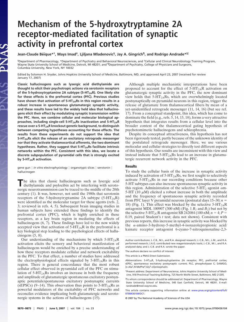
ResultsTo study the cellular basis of the increase in synaptic activityinduced by activation of 5-HT2ARs, we first sought to selectivelyactivate 5-HT2ARs in our recording conditions because other5-HT subtypes can also increase spontaneous synaptic activity inthis region. Administration of the selective 5-HT2 agonist �m-5-HT (10 �M) elicited a robust increase in both the amplitudeand the frequency of spontaneous synaptic activity recordedfrom PFC layer V pyramidal neurons (postnatal days 15–30; n �18) (Fig. 1). This effect was blocked by the selective 5-HT2ARantagonist MDL 100907 (300 nM) (Fig. 1 B1 and B2) but not bythe selective 5-HT2CR antagonist SB 242084 (100 nM; n � 4; P �0.19, paired Student’s t test; data not shown). Consistent withprevious reports, this increase in synaptic activity was blocked bythe �-amino-3-hydroxy-5-methyl-4-isoxazolepropionic acid/kainate receptor antagonist 6-cyano-7-nitroquinoxaline-2,3-
Author contributions: J.-C.B., M.I., and R.A. designed research; J.-C.B., M.I., L.M., and R.A.performed research; J.A.G. contributed new reagents/analytic tools; J.-C.B., M.I., and R.A.analyzed data; and J.-C.B. and R.A. wrote the paper.
The authors declare no conflict of interest.
This article is a PNAS Direct Submission.
Abbreviations: 5-HT2AR, 5-hydroxytryptamine 2A receptor; PFC, prefrontal cortex;sEPSC, spontaneous excitatory postsynaptic current; PLC, phospholipase C; DAMGO,[D-Ala2,N-MePhe4,Gly5-ol]enkephalin.
†Present address: Department of Neuroscience, Johns Hopkins University School of Medi-cine, 918 Preclinical Teaching Building, 725 North Wolfe Street, Baltimore, MD 21205.
�To whom correspondence should be addressed at: Department of Pharmacology, WayneState University School of Medicine, 540 East Canfield, Detroit, MI 48201. E-mail:[email protected].
This article contains supporting information online at www.pnas.org/cgi/content/full/0700436104/DC1.
© 2007 by The National Academy of Sciences of the USA
9870–9875 � PNAS � June 5, 2007 � vol. 104 � no. 23 www.pnas.org�cgi�doi�10.1073�pnas.0700436104
dione (30 �M; n � 3; data not shown) and by tetrodotoxin (1�M; n � 10 cells; data not shown). In prefrontal cortical slices,administration of tetrodotoxin reduces the frequency of synapticevents indicating that a subset of neurons is spontaneously active(12). Together, these results recapitulate previous findings show-ing that activation of 5-HT2ARs in PFC induces an increase inglutamate-mediated sEPSCs recorded from layer V pyramidalneurons.
Given that cortical pyramidal neurons are extensively inter-connected, the simplest explanation for these observationswould be that activation of 5-HT2ARs excites a subset of pyra-midal neurons (presumably of layer V because they are highlyenriched in 5-HT2ARs) whose activity is then detected in therecorded neuron as an increase in sEPSCs. This interpretation,however, has been generally rejected because 5-HT2ARs havenot been found to excite (i.e., induce action potential firing)pyramidal neurons of PFC (4, 9, 11). This is contrary to thesituation that prevails for muscarinic receptor activation, which,in addition to inducing a robust increase in sEPSCs in PFC,readily depolarizes and excites layer V pyramidal neurons (19).We thus next sought to address several alternative mechanismsthat could account for this 5-HT2AR-mediated increase in sEPSCs.
Activation of 5-HT2AR Does Not Change Glutamate Release Probabil-ities or the Number or Function of Synaptic �-Amino-3-hydroxy-5-methyl-4-isoxazolepropionic Acid Receptors. First, a direct action of5-HT2ARs on synaptic terminals could increase the probability of
release of glutamate and thus contribute to the increase infrequency of sEPSCs. However, administration of �m-5-HT didnot increase the frequency of mEPSCs (n � 13) [see supportinginformation (SI) Fig. 6], a measure of presynaptic releaseprobabilities. This finding is consistent with the observation that5-HT2ARs are located predominantly on postsynaptic dendriticstructures and are generally undetectable at glutamate releasingterminals (18). Second, the amplitude of mEPSCs was un-changed by administration of �m-5-HT (SI Fig. 6). Thesefindings, in conjunction with previous reports using similar,although not identical, conditions and approaches (9, 11, 12)indicate that neither an increase in the probability of release noran increase in the number or function of synaptic �-amino-3-hydroxy-5-methyl-4-isoxazolepropionic acid receptors is likely tocontribute significantly to the effect of �m-5-HT on sEPSCs.
Single-Cell Inactivation of 5-HT2AR-Mediated Signaling Does Not Blockthe 5-HT2AR-Mediated Increase in Spontaneous Synaptic Activity. Wenext sought to determine whether activation of postsynaptic5-HT2ARs elicited an increase in sEPSCs by inducing the releaseof a retrograde messenger that, in turn, would induce glutamaterelease from excitatory terminals, as was previously proposed(11, 14, 16). To this end, we envisioned a set of cellular andmolecular strategies to be implemented by biolistic transfectionof neurons (particle-mediated gene transfer) in organotypicslices of PFC. Organotypic slices from cortex maintain theirgeneral synaptic architecture for several days in culture (�2weeks) and biolistically transfected neurons can routinely berecorded from such slices (20, 21). Recordings from layer Vpyramidal neurons showed that administration of �m-5-HT toslices maintained in culture for up to 5–7 days induced anincrease in the frequency and amplitude of sEPSCS (n � 40) (seeSI Fig. 7) that was blocked by MDL 100907 (300 nM; n � 3; datanot shown). Although the magnitude of this effect was generallysmaller than that observed in acute slices, these observationsindicate that this preparation can be used to address the mech-anism underlying the 5-HT2AR-mediated increase in sEPSCs.
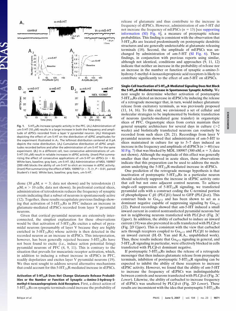
One prediction of the retrograde message hypothesis is thatinactivation of postsynaptic 5-HT2ARs in a particular neuronshould selectively suppress the increase in sEPSC activity ontothat cell but not onto adjacent control neurons. To achievesingle-cell suppression of 5-HT2AR signaling, we transfectedpyramidal cells with a construct coding the C-terminal portionof phospholipase C �1 (PLC�1) fused to GFP (PLC�-ct). Thisconstruct binds to G�q/11 and has been shown to act as adominant negative capable of suppressing signaling by G�q/11(22). Paired recordings showed that �m-5-HT induced a smallinward current in control nontransfected pyramidal neurons butnot in neighboring neurons transfected with PLC�-ct (Fig. 2CUpper). In addition, the ability of carbachol to induce an inwardcurrent (19) was also prevented in cells transfected with PLC�-ct(Fig. 2D Upper). This is consistent with the view that carbacholacts through receptors coupled to G�q-11 and PLC�1 to inducean inward current (H.-D. Yan and R.A., unpublished work).Thus, these results indicate that G�q/11 signaling in general, and5-HT2AR signaling in particular, were effectively blocked in cellstransfected with PLC�-ct dominant negative.
If postsynaptic 5-HT2ARs induce the release of a retrogrademessenger that then induces glutamate release from presynapticterminals, inhibition of postsynaptic 5-HT2AR signaling can beexpected to inhibit the ability of these receptors to increasesEPSC activity. However, we found that the ability of �m-5-HTto increase the frequency of sEPSCs was indistinguishablebetween controls and neurons transfected with PLC�-ct (Fig. 2CLower). Likewise, the ability of carbachol to increase frequencyof sEPSCs was unaltered by PLC�-ct (Fig. 2D Lower). Theseresults are inconsistent with the idea that postsynaptic 5-HT2ARs
Fig. 1. 5-HT2ARs increase synaptic activity in the PFC. (A1) Administration of�m-5-HT (10 �M) results in a large increase in both the frequency and ampli-tude of sEPSCs recorded from a layer V pyramidal neuron. (A2) Histogramdepicting the effect of �m-5-HT on the distribution of sEPSC amplitudes forthe experiment illustrated in A1. The leftmost distribution centered at 0 pAdepicts the noise distribution. (A3) Cumulative distribution of sEPSC ampli-tudes recorded before and after the administration of �m-5-HT for the sameexperiment. (B1) In a different cell, two consecutive administrations of �m-5-HT (10 �M) result in reliable increases in sEPSC activity. (Inset) Plot summa-rizing the effect of consecutive applications of �m-5-HT on sEPSCs (n � 6).White bars, baseline; gray bars, �m-5-HT. (B2) Administration of MDL 100907(300 nM) blocks the ability of �m-5-HT to elicit an increase in sEPSC activity.(Inset) Plot summarizing the effect of MDL 100907 (n � 7). (*, P � 0.01, pairedStudent’s t test). White bars, baseline; gray bars, �m-5-HT.
Beıque et al. PNAS � June 5, 2007 � vol. 104 � no. 23 � 9871
NEU
ROSC
IEN
CE
signal the release of a retrograde messenger capable of inducingglutamate release.
Single-Cell Rescue of 5-HT2AR Signaling Rescues the Ability of �m-5-HTto Signal an Inward Current but Not to Increase Synaptic Activity. Inthe previous experiments, the pyramidal neurons whose5-HT2ARs were inactivated by transfection with PLC�-ct weresurrounded by neurons presumably expressing control levels offunctional 5-HT2ARs. Activation of 5-HT2ARs in these untrans-fected neurons could, in principle, have released a retrogrademessenger capable of diffusing away from the site of release toact on excitatory terminals releasing glutamate onto the trans-fected neuron. To control for this possibility, we sought to carryout essentially the reverse experiments, i.e., to record from aneuron that expresses 5-HT2ARs but that is surrounded byneurons devoid of 5-HT2ARs. To this end, we used biolistictransfection procedures to rescue expression of 5-HT2ARs inprefrontal cortical neurons derived from 5-HT2AR knockout(5-HT2AR�/�) mice (23). We reasoned that if 5-HT2ARs signaledthe increase in sEPSCs by the release of a retrograde messenger,then we should be able to selectively rescue the ability of�m-5-HT to increase sEPSCs only in cells whose expression of5-HT2ARs has been rescued.
We first characterized the effect of �m-5-HT on acute slicesderived from wild-type and 5-HT2AR�/� mice. In wild-type mice,�m-5-HT increases sEPSCs frequency and this effect wasblocked by the selective 5-HT2A antagonist MDL 100907 (300
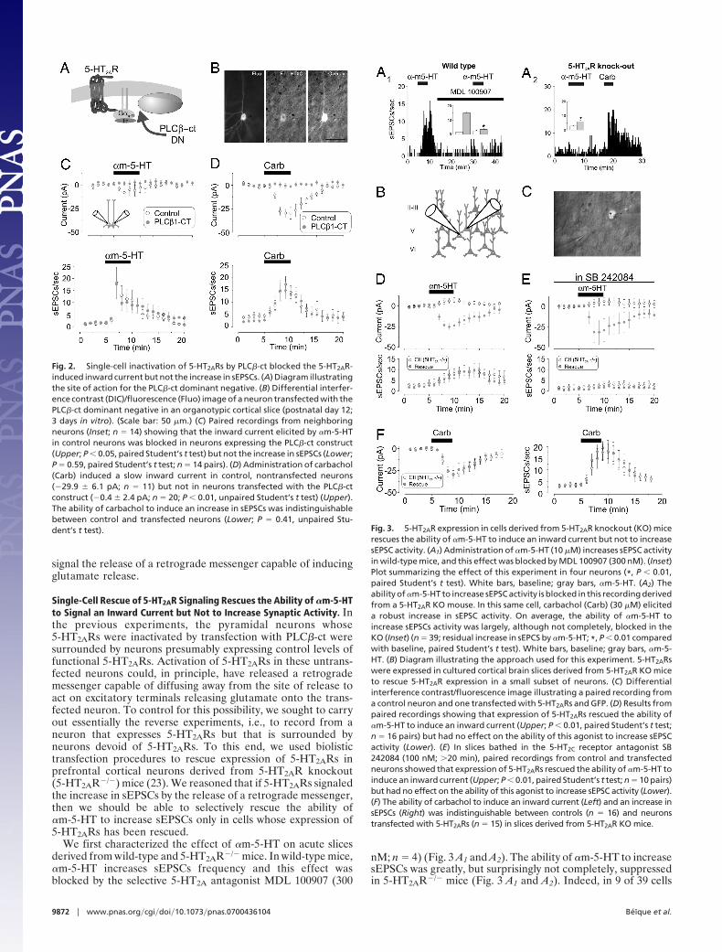
nM; n � 4) (Fig. 3 A1 and A2). The ability of �m-5-HT to increasesEPSCs was greatly, but surprisingly not completely, suppressedin 5-HT2AR�/� mice (Fig. 3 A1 and A2). Indeed, in 9 of 39 cells
Fig. 2. Single-cell inactivation of 5-HT2ARs by PLC�-ct blocked the 5-HT2AR-induced inward current but not the increase in sEPSCs. (A) Diagram illustratingthe site of action for the PLC�-ct dominant negative. (B) Differential interfer-ence contrast (DIC)/fluorescence (Fluo) image of a neuron transfected with thePLC�-ct dominant negative in an organotypic cortical slice (postnatal day 12;3 days in vitro). (Scale bar: 50 �m.) (C) Paired recordings from neighboringneurons (Inset; n � 14) showing that the inward current elicited by �m-5-HTin control neurons was blocked in neurons expressing the PLC�-ct construct(Upper; P � 0.05, paired Student’s t test) but not the increase in sEPSCs (Lower;P � 0.59, paired Student’s t test; n � 14 pairs). (D) Administration of carbachol(Carb) induced a slow inward current in control, nontransfected neurons(�29.9 � 6.1 pA; n � 11) but not in neurons transfected with the PLC�-ctconstruct (�0.4 � 2.4 pA; n � 20; P � 0.01, unpaired Student’s t test) (Upper).The ability of carbachol to induce an increase in sEPSCs was indistinguishablebetween control and transfected neurons (Lower; P � 0.41, unpaired Stu-dent’s t test). Fig. 3. 5-HT2AR expression in cells derived from 5-HT2AR knockout (KO) mice
rescues the ability of �m-5-HT to induce an inward current but not to increasesEPSC activity. (A1) Administration of �m-5-HT (10 �M) increases sEPSC activityin wild-type mice, and this effect was blocked by MDL 100907 (300 nM). (Inset)Plot summarizing the effect of this experiment in four neurons (*, P � 0.01,paired Student’s t test). White bars, baseline; gray bars, �m-5-HT. (A2) Theability of �m-5-HT to increase sEPSC activity is blocked in this recording derivedfrom a 5-HT2AR KO mouse. In this same cell, carbachol (Carb) (30 �M) eliciteda robust increase in sEPSC activity. On average, the ability of �m-5-HT toincrease sEPSCs activity was largely, although not completely, blocked in theKO (Inset) (n � 39; residual increase in sEPCS by �m-5-HT; *, P � 0.01 comparedwith baseline, paired Student’s t test). White bars, baseline; gray bars, �m-5-HT. (B) Diagram illustrating the approach used for this experiment. 5-HT2ARswere expressed in cultured cortical brain slices derived from 5-HT2AR KO miceto rescue 5-HT2AR expression in a small subset of neurons. (C) Differentialinterference contrast/fluorescence image illustrating a paired recording froma control neuron and one transfected with 5-HT2ARs and GFP. (D) Results frompaired recordings showing that expression of 5-HT2ARs rescued the ability of�m-5-HT to induce an inward current (Upper; P � 0.01, paired Student’s t test;n � 16 pairs) but had no effect on the ability of this agonist to increase sEPSCactivity (Lower). (E) In slices bathed in the 5-HT2C receptor antagonist SB242084 (100 nM; �20 min), paired recordings from control and transfectedneurons showed that expression of 5-HT2ARs rescued the ability of �m-5-HT toinduce an inward current (Upper; P � 0.01, paired Student’s t test; n � 10 pairs)but had no effect on the ability of this agonist to increase sEPSC activity (Lower).(F) The ability of carbachol to induce an inward current (Left) and an increase insEPSCs (Right) was indistinguishable between controls (n � 16) and neuronstransfected with 5-HT2ARs (n � 15) in slices derived from 5-HT2AR KO mice.
9872 � www.pnas.org�cgi�doi�10.1073�pnas.0700436104 Beıque et al.
tested from 5-HT2AR�/� mice, a generally small, but clearlydistinguishable, increase in the frequency of sEPSCs could bedetected after administration of �m-5-HT. This residual re-sponse to �m-5-HT was blocked by the selective 5-HT2C receptorantagonist SB 242084 (100 nM; n � 4; P � 0.05, paired Student’st test; data not shown). Because the effect of �m-5-HT appearedto be entirely mediated by 5-HT2ARs in wild-type mice, weinterpreted this finding as reflecting a possible small adaptiveup-regulation of 5-HT2C receptors in the 5-HT2AR�/� mice.However, because this residual increase in sEPSC activity wasvery small, these findings validate the use of this knockout mouseas a suitable physiological null background.
A key requirement for this experiment is that 5-HT2ARfunction in pyramidal neurons be effectively ‘‘rescued’’ by thetransfection of 5-HT2ARs. We thus monitored the ability of5-HT2AR activation to elicit an inward current in pyramidalneurons. Paired recordings in slices derived from 5-HT2AR�/�
mice showed that �m-5-HT did not induce an inward current incontrol (nontransfected) cells but elicited a clear inward currentin neighboring neurons transfected with 5-HT2ARs (Fig. 3DUpper). Thus, 5-HT2AR-mediated signaling was effectively res-cued in 5-HT2AR�/� cells transfected with 5-HT2ARs.
If activation of 5-HT2AR were to result in the generation of aretrograde messenger capable of inducing the release of gluta-mate, the ability of �m-5-HT to increase the frequency ofsEPSCs should be rescued in 5-HT2AR�/� cells transfected with5-HT2ARs. We found that �m-5-HT elicited a small increase insEPSC frequency in control untransfected cells from5-HT2AR�/� mice and this small increase was of similar magni-tude in neighboring neurons transfected with 5-HT2ARs (Fig. 3DLower; n � 16 pairs).
Admittedly, the interpretation of this experiment is compli-cated by the small increase in sEPSCs induced by �m-5-HT inslices derived from 5-HT2AR�/� mice. We had earlier attributedthis residual response to a possible up-regulation of 5-HT2Creceptor function in 5-HT2AR�/� mice. To resolve this remainingambiguity, we repeated the rescue experiment in the presence ofthe 5-HT2C receptor antagonist SB 242084 (100 nM) (Fig. 3E).Consistent with our observation in acute slices, in the presenceSB 242084, �m-5-HT did not increase the frequency of sEPSCsin control (untransfected) neurons in cultured slices from5-HT2AR�/� mice (Fig. 3E Lower; n � 10 pairs). Importantly,�m-5-HT also did not increase the frequency of sEPSCs inneighboring cells rescued with 5-HT2ARs in these conditions(Fig. 3E Lower; n � 10 pairs). Again, 5-HT2ARs were functionalin the transfected neurons in these experiments because �m-5-HT induced a robust inward current only in transfectedneurons (Fig. 3E Upper). Finally, this general failure to observechanges in the frequency of sEPSCs was likely not the result ofnonspecific changes brought by the transfection procedure orfrom 5-HT2AR gene deletion per se, because the ability ofcarbachol to induce an inward current and to increase thefrequency of sEPSCs was indistinguishable between control cellsand cells transfected with 5-HT2ARs (Fig. 3F). Together, theseresults indicate that expression of 5-HT2ARs in pyramidal cellsfrom 5-HT2AR�/� mice effectively rescued the ability of5-HT2ARs to signal an inward current, but not the ability toincrease the frequency of sEPSCs. As such, they failed to supportthe retrograde message hypothesis.
GTP�S Selectively Renders Irreversible the 5-HT2AR-Induced InwardCurrent but Not the Increase in sEPSCs. To control for a potentialconfound resulting from the culturing procedure, we sought analternative strategy to test for the presence of a retrogrademessenger induced by activation of 5-HT2ARs in acute slices. Wereasoned that a manipulation that would alter G protein-mediated signaling in the recorded cell should equally disruptthe ability of 5-HT2ARs to induce an inward current and the
release of a retrograde messenger. To test this prediction, weused the poorly hydrolysable GTP analog GTP�S, a compoundthat limits the rate of GTP hydrolysis by G� subunits and thusrenders G protein-mediated responses irreversible. A previousstudy has successfully used this strategy to disclose a G protein-mediated release of a cannabinoid retrograde messenger (24).
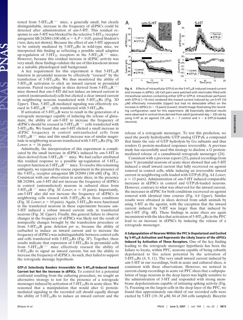
Consistent with a previous report (25), paired recordings fromlayer V pyramidal neurons of acute slices showed that �m-5-HTinduced a small inward current that fully recovered on agonistremoval in control cells, while inducing an irreversible inwardcurrent in neighboring cells loaded with GTP�S (Fig. 4A Lower;n � 13 pairs). Administration of �m-5-HT induced comparableincreases in sEPSCs in control and GTP�S-loaded neurons.However, contrary to what was observed for the inward current,the increases in sEPSC for both conditions recovered on agonistremoval with identical time courses (Fig. 4A Upper). Similarresults were obtained in slices derived from adult animals byusing 5-HT as the agonist, with the exception that the inwardcurrent induced by 5-HT was larger than that induced by�m-5-HT (Fig. 4B). These findings in acute slices are againinconsistent with the idea that activation of 5-HT2ARs in the PFClead to an increase in sEPSCs by inducing the release of aretrograde messenger.
A Subpopulation of Neurons Within the PFC Is Depolarized and Excitedby 5-HT2AR Activation and Represents the Likely Source of the sEPSCsInduced by Activation of These Receptors. One of the key findingleading to the retrograde messenger hypothesis has been thefailure to locate, within PFC, neurons capable of being robustlydepolarized to fire action potential by the activation of5-HT2ARs (4, 9, 11). The very small inward current induced by�m-5-HT in our recordings, both in acute and cultured slices, isconsistent with these observations. However, we noticed incurrent-clamp recordings in acute rat PFC slices that a subpopu-lation of large neurons in the deep layers was highly sensitive tothe administration of 5-HT and responded with strong mem-brane depolarizations capable of initiating spiking activity (Fig.5). Focusing on the largest cells in the deep layer of the PFC, wefound that approximately one-third of our recorded cells wereexcited by 5-HT (10–30 �M; 84 of 260 cells sampled). Biocytin
Fig. 4. Effects of intracellular GTP�S on the 5-HT2AR-induced inward currentand increase in sEPSCs. (A) Cell pairs were patched with electrodes filled withintracellular solution containing either GTP or GTP�S. Intracellular perfusionwith GTP�S (�10 min) rendered the inward current induced by �m-5-HT (10�M) effectively irreversible (Upper) but had no detectable effect on theincrease in sEPSCs (n � 13 pairs) (Lower). (Inset) Image illustrating the record-ing configuration used for this experiment. (B) Essentially identical resultswere observed in cortical slices derived from adult (postnatal day � 33) rats byusing 5-HT as an agonist (10 �M, n � 7 control and n � 6 GTP�S-loadedneurons).
Beıque et al. PNAS � June 5, 2007 � vol. 104 � no. 23 � 9873
NEU
ROSC
IEN
CE
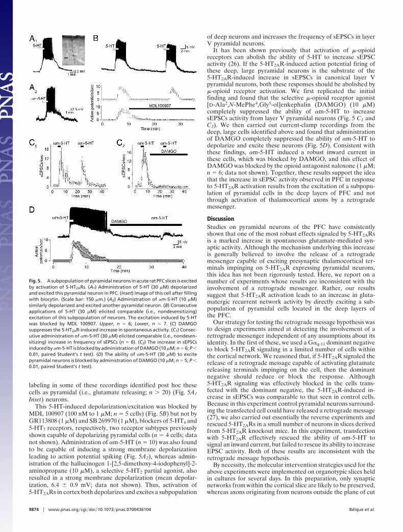
labeling in some of these recordings identified post hoc thesecells as pyramidal (i.e., glutamate releasing; n � 20) (Fig. 5A1
Inset) neurons.This 5-HT-induced depolarization/excitation was blocked by
MDL 100907 (100 nM to 1 �M; n � 5 cells) (Fig. 5B) but not byGR113808 (1 �M) and SB 269970 (1 �M), blockers of 5-HT4 and5-HT7 receptors, respectively, two receptor subtypes previouslyshown capable of depolarizing pyramidal cells (n � 4 cells; datanot shown). Administration of �m-5-HT (n � 10) was also foundto be capable of inducing a strong membrane depolarizationleading to action potential spiking (Fig. 5A2), whereas admin-istration of the hallucinogen 1-[2,5-dimethoxy-4-iodophenyl]-2-aminopropane (10 �M), a selective 5-HT2 partial agonist, alsoresulted in a strong membrane depolarization (mean depolar-ization, 6.4 � 0.9 mV; data not shown). Thus, activation of5-HT2ARs in cortex both depolarizes and excites a subpopulation
of deep neurons and increases the frequency of sEPSCs in layerV pyramidal neurons.
It has been shown previously that activation of �-opioidreceptors can abolish the ability of 5-HT to increase sEPSCactivity (26). If the 5-HT2AR-induced action potential firing ofthese deep, large pyramidal neurons is the substrate of the5-HT2AR-induced increase in sEPSCs in canonical layer Vpyramidal neurons, both these responses should be abolished by�-opioid receptor activation. We first replicated the initialfinding and found that the selective �-opioid receptor agonist[D-Ala2,N-MePhe4,Gly5-ol]enkephalin (DAMGO) (10 �M)completely suppressed the ability of �m-5-HT to increasesEPSCs activity from layer V pyramidal neurons (Fig. 5 C1 andC2). We then carried out current-clamp recordings from thedeep, large cells identified above and found that administrationof DAMGO completely suppressed the ability of �m-5-HT todepolarize and excite these neurons (Fig. 5D). Consistent withthese findings, �m-5-HT induced a robust inward current inthese cells, which was blocked by DAMGO, and this effect ofDAMGO was blocked by the opioid antagonist naloxone (1 �M;n � 6; data not shown). Together, these results support the ideathat the increase in sEPSC activity observed in PFC in responseto 5-HT2AR activation results from the excitation of a subpopu-lation of pyramidal cells in the deep layers of PFC and notthrough activation of thalamocortical axons by a retrogrademessenger.
DiscussionStudies on pyramidal neurons of the PFC have consistentlyshown that one of the most robust effects signaled by 5-HT2ARsis a marked increase in spontaneous glutamate-mediated syn-aptic activity. Although the mechanism underlying this increaseis generally believed to involve the release of a retrogrademessenger capable of exciting presynaptic thalamocortical ter-minals impinging on 5-HT2AR expressing pyramidal neurons,this idea has not been rigorously tested. Here, we report on anumber of experiments whose results are inconsistent with theinvolvement of a retrograde messenger. Rather, our resultssuggest that 5-HT2AR activation leads to an increase in gluta-matergic recurrent network activity by directly exciting a sub-population of pyramidal cells located in the deep layers ofthe PFC.
Our strategy for testing the retrograde message hypothesis wasto design experiments aimed at detecting the involvement of aretrograde messenger independent of any assumptions about itsidentity. In the first of these, we used a G�q-11 dominant negativeto block 5-HT2AR signaling in a limited number of cells withinthe cortical network. We reasoned that, if 5-HT2AR signaled therelease of a retrograde message capable of activating glutamatereleasing terminals impinging on the cell, then the dominantnegative should reduce or block the response. Although5-HT2AR signaling was effectively blocked in the cells trans-fected with the dominant negative, the 5-HT2AR-induced in-crease in sEPSCs was comparable to that seen in control cells.Because in this experiment control pyramidal neurons surround-ing the transfected cell could have released a retrograde message(27), we also carried out essentially the reverse experiments andrescued 5-HT2ARs in a small number of neurons in slices derivedfrom 5-HT2AR knockout mice. In this experiment, transfectionwith 5-HT2AR effectively rescued the ability of �m-5-HT tosignal an inward current, but failed to rescue its ability to increaseEPSC activity. Both of these results are inconsistent with theretrograde message hypothesis.
By necessity, the molecular intervention strategies used for theabove experiments were implemented on organotypic slices heldin cultures for several days. In this preparation, only synapticnetworks from within the cortical slice are likely to be preserved,whereas axons originating from neurons outside the plane of cut
Fig. 5. A subpopulation of pyramidal neurons in acute rat PFC slices is excitedby activation of 5-HT2ARs. (A1) Administration of 5-HT (30 �M) depolarizedand excited this pyramidal neuron in PFC. (Inset) Image of this cell after fillingwith biocytin. (Scale bar: 150 �m.) (A2) Administration of �m-5-HT (10 �M)similarly depolarized and excited another pyramidal neuron. (B) Consecutiveapplications of 5-HT (30 �M) elicited comparable (i.e., nondesensitizing)excitation of this subpopulation of neurons. The excitation induced by 5-HTwas blocked by MDL 100907. Upper, n � 6; Lower, n � 7. (C) DAMGOsuppresses the 5-HT2AR-induced increase in spontaneous activity. (C1) Consec-utive administration of �m-5-HT (30 �M) elicited comparable (i.e., nondesen-sitizing) increase in frequency of sEPSCs (n � 6). (C2) The increase in sEPSCsinduced by �m-5-HT is blocked by administration of DAMGO (10 �M; n � 6; P �0.01, paired Student’s t test). (D) The ability of �m-5-HT (30 �M) to excitepyramidal neurons is blocked by administration of DAMGO (10 �M; n � 5; P �0.01, paired Student’s t test).
9874 � www.pnas.org�cgi�doi�10.1073�pnas.0700436104 Beıque et al.
can be expected to have degenerated. Thus, it remains possiblethat the culturing procedure resulted in functional changesimpacting the ability of 5-HT2AR activation to trigger the releaseof a retrograde messenger. To test this possibility, we tookadvantage of the ability of GTP�S to allow for the irreversibleactivation of G protein signaling cascades. Intracellular injectionof GTP�S, however, had no effect on the ability of �m-5-HT toelicit an increase in sEPSCs while rendering the inward currentelicited by 5-HT2AR activation effectively irreversible. As such,these results again failed to support the idea that postsynaptic5-HT2ARs in pyramidal cell of the rat PFC regulate sEPSCs byreleasing a retrograde message.
Historically, acceptance of the retrograde messenger hypoth-esis emerged from the failure of simpler mechanisms to explainthe increase the ability of 5-HT2ARs to increase sEPSC activity.Specifically, 5-HT2AR activation did not appear to excite pyra-midal neurons (4, 9, 11, 25) but involved an increase in therelease of glutamate by afferent presynaptic terminals (9, 11, 12,14). As such, the retrograde messenger hypothesis did seem tooffer the only possible explanation for the phenomenon. Wehave now identified a discrete subpopulation of neurons in thePFC that is excited by 5-HT2AR activations and that can, inprinciple, represent the cellular elements responsible for theincrease in sEPSCs. Although it is difficult to test experimentallythis conjecture, previous studies have reported that �-opioidagonist suppress the ability of 5-HT2ARs to increase sEPSCactivity. Because these receptors are expressed at very low levelsin cortex, it has also been suggested this effect could help identifythe presynaptic cellular elements mediating the 5-HT2AR induceincrease in glutamate release. We found that activation of�-opioid receptors completely blocked the ability of 5-HT2ARsto excite this subpopulation of pyramidal cells. These physio-logical results are consistent with the presence of scattered�-opioid receptor expressing cells in the frontal cortex, espe-cially in the deep layers (28, 29). As such, these results supportthe view that 5-HT2ARs in PFC enhance the overall excitabilityof PFC network by regulating the properties of a key subpopu-lation of pyramidal neurons.
Although 5-HT2ARs are expressed robustly in the PFC, theyare also expressed in other regions of the brain (30) where theymediate membrane depolarization and neuronal excitation (31).
However, selective rescue of 5-HT2AR expression in cortex issufficient to rescue 5-HT2AR-induced head shaking, a behavioralproxy for hallucinogenic activity (32) and as such provideexperimental support to the idea that cortical 5-HT2ARs mediatethe psychotropic effects of hallucinogens. Interestingly, activa-tion of �-opioid receptors suppresses the 5-HT2AR-inducedexcitation of the subpopulation of neurons identified in thecurrent study, the increase in sEPSCs recorded from canonicallayer V neurons, and the 1-[2,5-dimethoxy-4-iodophenyl]-2-aminopropane-induced head shaking behavior (33). This con-gruence across different levels of biological organization sup-ports the idea that the 5-HT2AR-mediated increase in glutamatesynaptic activity analyzed in the current work bears a causalrelationship to the mechanism of action of hallucinogens.
The results outlined in the current work lead to a reconcep-tualization of the mechanism of action of hallucinogens awayfrom the idea that they facilitate thalamocortical excitatorysynaptic transmission and toward the idea that they directlymodulate recurrent intrinsic networks, perhaps regulating thegain of recurrent circuits in the PFC. A corollary implication ofthis view hold that excessive stimulation of 5-HT2ARs, such asduring hallucinogen use, might destabilize PFC recurrent circuitsand thus give rise to the sensory manifestation of hallucinogens.The present results thus suggest that the ‘‘breakdown’’ of corticalfunction brought by hallucinogens does not result from anexcessive stimulation of thalamocortical innervation but ratherfrom altered function of PFC intrinsic circuitry. Future studieswill be required to further dissect the behavioral consequencespredicted by this model.
MethodsAcute slices containing PFC were prepared following standardprocedures, and cultured slices were prepared as previouslydescribed. Whole-cell voltage- and current-clamp recordingswere obtained from layer V pyramidal neurons following stan-dard procedures. These procedures are described in greaterdetail in SI Methods.
This study was supported by National Institutes of Health GrantMH43985 (to R.A.) and by a grant from the Mental Illness ResearchAssociation (to J.-C.B.). J.-C.B. was in receipt of a postdoctoral fellow-ship from the Canadian Institutes for Health Research.
1. Wooley DW, Shaw E (1954) Proc Natl Acad Sci USA 40:228–231.2. Glennon RA, Titeler M, McKenney JD (1984) Life Sci 35:2505–2511.3. Titeler M, Lyon RA, Glennon RA (1988) Psychopharmacology (Berl) 94:213–216.4. Aghajanian GK, Marek GJ (1999) Neuropsychopharmacology 21:16S–23S.5. Nichols DE (2004) Pharmacol Ther 101:131–181.6. Vollenweider FX, Vollenweider-Scherpenhuyzen MF, Babler A, Vogel H, Hell
D (1998) NeuroReport 9:3897–3902.7. Vollenweider FX, Leenders KL, Scharfetter C, Maguire P, Stadelmann O,
Angst J (1997) Neuropsychopharmacology 16:357–372.8. Vollenweider FX, Geyer MA (2001) Brain Res Bull 56:495–507.9. Aghajanian GK, Marek GJ (1997) Neuropharmacology 36:589–599.
10. Marek GJ, Aghajanian GK (1999) Eur J Pharmacol 367:197–206.11. Zhou FM, Hablitz JJ (1999) J Neurophysiol 82:2989–2999.12. Beıque JC, Chapin-Penick EM, Mladenovic L, Andrade R (2004) J Physiol
556:739–754.13. Lambe EK, Goldman-Rakic PS, Aghajanian GK (2000) Cereb Cortex 10:974–980.14. Lambe EK, Aghajanian GK (2001) J Neurosci 21:9955–9963.15. Aghajanian GK, Marek GJ (2000) Brain Res Brain Res Rev 31:302–312.16. Marek GJ, Wright RA, Gewirtz JC, Schoepp DD (2001) Neuroscience 105:379–
392.17. Martin-Ruiz R, Puig MV, Celada P, Shapiro DA, Roth BL, Mengod G, Artigas
F (2001) J Neurosci 21:9856–9866.18. Miner LA, Backstrom JR, Sanders-Bush E, Sesack SR (2003) Neuroscience
116:107–117.
19. Haj-Dahmane S, Andrade R (1996) J Neurosci 16:3848–3861.20. Beıque JC, Andrade R (2003) J Physiol 546:859–867.21. Villalobos C, Shakkottai VG, Chandy KG, Michelhaugh SK, Andrade R (2004)
J Neurosci 24:3537–3542.22. Kammermeier PJ, Ikeda SR (1999) Neuron 22:819–829.23. Weisstaub NV, Zhou M, Lira A, Lambe E, Gonzalez-Maeso J, Hornung JP,
Sibille E, Underwood M, Itohara S, Dauer WT, et al. (2006) Science 313:536–540.
24. Haj-Dahmane S, Shen RY (2005) J Neurosci 25:896–905.25. Beıque JC, Campbell B, Perring P, Hamblin MW, Walker P, Mladenovic L,
Andrade R (2004) J Neurosci 24:4807–4817.26. Marek GJ, Aghajanian GK (1998) Neuroscience 86:485–497.27. Wilson RI, Nicoll RA (2001) Nature 410:588–592.28. Mansour A, Khachaturian H, Lewis ME, Akil H, Watson SJ (1987) J Neurosci
7:2445–2464.29. Mansour A, Fox CA, Burke S, Meng F, Thompson RC, Akil H, Watson SJ
(1994) J Comp Neurol 350:412–438.30. Lopez-Gimenez JF, Mengod G, Palacios JM, Vilaro MT (1997) Naunyn
Schmiedebergs Arch Pharmacol 356:446–454.31. Barnes NM, Sharp T (1999) Neuropharmacology 38:1083–1152.32. Gonzalez-Maeso J, Weisstaub NV, Zhou M, Chan P, Ivic L, Ang R, Lira A,
Bradley-Moore M, Ge Y, Zhou Q, et al. (2007) Neuron 53:439–452.33. Marek GJ (2003) Eur J Pharmacol 474:77–83.
Beıque et al. PNAS � June 5, 2007 � vol. 104 � no. 23 � 9875
NEU
ROSC
IEN
CE