mechanism, inhibition and spectroscopy of isoprenoid
TRANSCRIPT
MECHANISM, INHIBITION AND SPECTROSCOPY
OF ISOPRENOID BIOSYNTHESIS ENZYMES
BY
JIKUN LI
DISSERTATION
Submitted in partial fulfillment of the requirements for the degree of Doctor of Philosophy in Biophysics and Computational Biology
in the Graduate College of the University of Illinois at Urbana-Champaign, 2014
Urbana, Illinois
Doctoral Committee:
Professor Eric Oldfield, Chair and Director of Research Professor Martin Gruebele Professor Chad Rienstra Professor James Imlay
ii
Abstract
Isoprenoids (or terpenes) are a large class of compounds with great physiological
importance in all kingdoms of life. The research in this work explored the mechanism and
inhibition of several enzymes in two important pathways for isoprenoid biosynthesis.
The first pair of enzyme are IspG and IspH, which constitute the last two steps in the
MEP/DOXP (or non-mevalonate) pathway, which some bacteria, protozoa and plants rely on to
produce the basic building blocks of isoprenoids, isopentenyl pyrophosphate (IPP) and
dimethylallyl pyrophosphate (DMAPP). IspG and IspH are both 4Fe-4S cluster-containing
proteins with a unique iron whose ligand is not a cysteine residue. Continuous-wave and pulsed
electron paramagnetic resonance (EPR) experiments, with the help of isotope-labeled substrates,
revealed the electronic structures of reaction intermediates and inhibitors bound to the iron-
sulfur clusters. Diverse binding modes of ligands to the clusters were discovered. In addition to
IspG and IspH, another 4Fe-4S protein, NadA (in a biosynthetic pathway to NAD) was also
mechanistically studied.
The second pair of enzymes are FPPS and SQS, the enzymes responsible for the first two
steps of the biosynthetic pathway of sterols in animals, bacteria and protozoa. We focused on the
inhibition and structure aspects of the FPPS and SQS enzymes of the protozoan pathogen
Trypanosoma cruzi. For a series of bisphosphonate inhibitors, structure-activity relationship was
explored, as well as their cell-killing capability.
iii
Acknowledgements
The work in this dissertation would not have been possible without the efforts of my co-
workers in the Oldfield lab. Dr. Ke Wang, Dr. Kai Li and Dr. Yonghui Zhang synthesized most of
the inhibitors and substrates. Dr. Weixue Wang, along with Dr. Mark Nigles in the Illinois EPR
Center, trained me for various EPR experimental techniques. Dr. Rong Cao, Dr. Fu-Yang Lin and
Dr. Yi-Liang Liu trained me for various molecular biology experimental methods and X-ray
crystallography. Francisco Guerra and Dr. Wei Zhu have been greatly helpful co-workers in
projects on which we collaborated.
Collaborators outside UIUC have also been indispensable. Prof. Michael Groll and Dr.
Ingrid Span at TU-Munich has worked with our group for the crystal structures of metalloprotein
complexes. Prof. Yong Zhang at Stevens Institute of technology taught me basic quantum
chemistry and performed quantum calculations in various research projects. Prof. Tatyana
Smirnova at North Carolina State University provided instrument and guidance on EPR
experiments. Dr. Rey-Ting Guo’s group at Tianjin Institute of Industrial Biology performed X-ray
crystallography of SQS-inhibitor complexes. Prof. Roberto Docampo’s group at University of
Georgia performed cell-killing assays that are important part of the research on FPPS/SQS
inhibitors.
I also thank my committee members, including Prof. Martin Gruebele, Chad Rienstra and
James Imlay, as well as Prof. So Hirata who was in my prelim committee.
Finally, I am heartily grateful to my advisor, Prof. Eric Oldfield, for years of great patience,
wisdom and support guiding me toward this dissertation.
iv
Table of Contents
Chapter 1. Introduction ............................................................................................................................. 1
1.1. Isoprenoids and Isoprenoid Biosynthesis .................................................................................... 1
1.2. The MEP Pathway, IspG and IspH ............................................................................................... 3
1.3. 4Fe-4S Protein Beyond IspG/H: Quinolinate Synthase .............................................................. 4
1.4. Prenyl-Transferases FPPS and SQS .............................................................................................. 6
1.5. Overall Research Strategies ........................................................................................................... 7
1.6. Tables and Figures .......................................................................................................................... 9
Chapter 2. Mechanism of Action of 4Fe-4S Protein IspG ................................................................... 15
2.1. Notes and Acknowledgements ................................................................................................... 15
2.2. Introduction ................................................................................................................................... 16
2.3. Results and Discussion ................................................................................................................. 16
2.4. Conclusions .................................................................................................................................... 20
2.5. Materials and Methods ................................................................................................................. 20
2.6. Tables and Figures ........................................................................................................................ 26
Chapter 3. Mechanism of Action of 4Fe-4S Protein IspH ................................................................... 33
3.1. Notes and Acknowledgements ................................................................................................... 33
3.2. Introduction ................................................................................................................................... 34
3.3. Results and Discussion ................................................................................................................. 34
3.4. Conclusions .................................................................................................................................... 41
3.5. Materials and Methods ................................................................................................................. 41
3.6. Tables and Figures ........................................................................................................................ 44
Chapter 4. Diverse Inhibitor Binding Modes of IspH ......................................................................... 58
4.1. Notes and Acknowledgements ................................................................................................... 58
4.2. Introduction ................................................................................................................................... 58
4.3. Results and Discussion ................................................................................................................. 59
4.4. Conclusions .................................................................................................................................... 68
4.5. Materials and Methods ................................................................................................................. 69
4.6 Tables and Figures ......................................................................................................................... 72
v
Chapter 5. Preliminary Study of Mechanism of 4Fe-4S Protein Quinolinate Synthase ................. 88
5.1. Notes and Acknowledgements ................................................................................................... 88
5.2. Introduction ................................................................................................................................... 88
5.3. Results and Discussion ................................................................................................................. 89
5.4. Conclusions .................................................................................................................................... 91
5.5. Materials and Methods ................................................................................................................. 92
5.6. Tables and Figures ........................................................................................................................ 95
Chapter 6. Lipophilic Bisphosphonates Targeting Squalene Synthase and Farnesyl Pyrophosphate Synthase ....................................................................................................................... 101
6.1. Notes and Acknowledgements ................................................................................................. 101
6.2. Introduction ................................................................................................................................. 102
6.3. Results and Discussion ............................................................................................................... 103
6.4. Conclusions .................................................................................................................................. 106
6.5. Materials and Methods ............................................................................................................... 106
6.6. Tables and Figures ...................................................................................................................... 110
Bibliography ........................................................................................................................................... 118
1
Chapter 1. Introduction
1.1. Isoprenoids and Isoprenoid Biosynthesis
Isoprenoids, also known as terpenoids, are a large class of compounds present in all
kingdoms of life. To name a few of them: Retinol is a form of vitamin A essential for vision;
staphyloxanthin is a pigment giving Staphylococcus aureus its characteristic gold color as well as
an antioxidant against host defense; ergosterol is an essential component of the cell membrane of
several parasitic protozoa; and cholesterol, probably the best-known isoprenoid, acts as a
structural component of animal cell membranes, as well as a precursor to certain hormones.
Overall, there are over 65,000 known compounds belonging to this category (1), diverse in
chemical structures and physiological functions.
It is therefore natural that living organisms have a myriad of biosynthetic pathways leading
towards isoprenoids. However, what remains invariant for the isoprenoids are their common
basic building blocks, two five-carbon pyrophosphates: isopentenyl pyrophosphate (IPP) and
dimethylallyl pyrophosphate (DMAPP) (2), which provide them with the basic isoprene unit.
Organisms carry out catabolism from various carbon sources, which are converted to the basic C-
5 building blocks, and the C-5 building blocks are in turn converted to different end products.
Therefore, IPP and DMAPP form the point where many biosynthetic pathways converge and
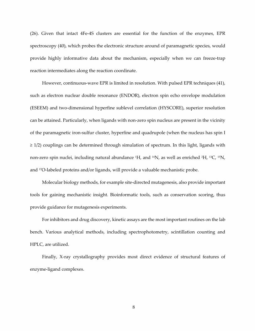
must pass through, as shown in Figure 1.1.
With the overarching goal being the development of new inhibitors against the enzymes in
these pathways, the main targets in this research have been set at two pairs of enzymes
2
immediately upstream and downstream of the IPP/DMAPP “chokepoint”, as highlighted in
Figure 1.1.
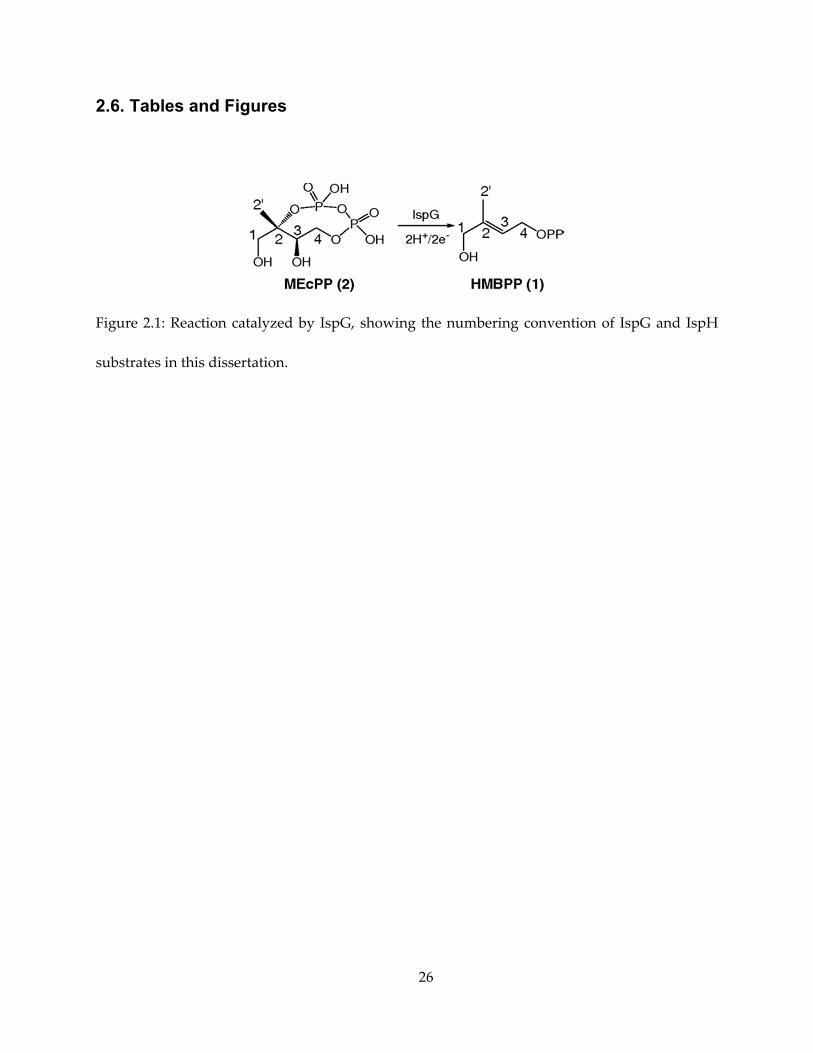
The first pair of enzymes targeted, (E)-4-hydroxy-3-methyl-but-2-enyl diphosphate
(HMBPP) synthase (IspG, also known as GcpE) and HMBPP reductase (IspH or LytB), catalyze
the last two steps of the 2-C-methyl-D-erythritol 4-phosphate/1-deoxy-D-xylulose 5-phosphate
(MEP, DOXP, or non-mevalonate) pathway that converts metabolites from catabolism to IPP and
DMAPP in certain organisms. Both enzymes contain 4Fe-4S clusters that are essential for the
2H+/2e- reduction of their respective substrates. Both clusters have three iron atoms attached to
the protein via a cysteine ligand, while the “unique” fourth iron has a non-cysteine ligand. We
elucidated the mechanisms of both enzymes by trapping reaction intermediates and probing the
electronic structure of these intermediates with continuous-wave EPR (electron paramagnetic
resonance) and pulsed EPR spectroscopies. In addition, we found inhibitors for the enzymes,
some of which are effective against both IspG and IspH.
The second pair of enzymes, farnesyl diphosphate synthase (FPPS, also called IspA) and
squalene synthase (SQS), catalyze the first two steps in the pathways from IPP and DMAPP to
various sterols, e.g. cholesterol in animals and ergosterol in fungi and protozoa. This research
investigated the inhibition of both enzymes by a series of lipophilic bisphosphonates, some of
which are also shown to inhibit the growth of the protozoan Trypanosoma cruzi (the cause of the
tropical Chagas disease) in its pathologically relevant phase of life cycle.
3
1.2. The MEP Pathway, IspG and IspH
In different organisms, IPP and DMAPP are synthesized in two distinct, non-homologous
pathways – the “classical” mevalonate pathway (also dubbed MVA pathway or HMG-CoA
pathway) discovered in 1950s, and the MEP/non-mevalonate pathway (3) discovered more
recently (1990s). The distribution of the two pathways in various organisms is complex (4), but to
summarize crudely, the mevalonate pathway is more prevalent in archaea, yeasts and animals,
the presence of the non-mevalonate pathway is biased towards bacteria and the apicomplexa
group of parasitic protists, and plants tend have both pathways.
Humans rely solely on the mevalonate pathway for the synthesis of IPP and DMAPP, while
many bacterial and protozoan pathogens produce the essential building blocks IPP and DMAPP
exclusively through the non-mevalonate pathway. Such a pathway distribution presents an
opportunity for development of anti-infective drugs with the MEP pathway as an antibiotic
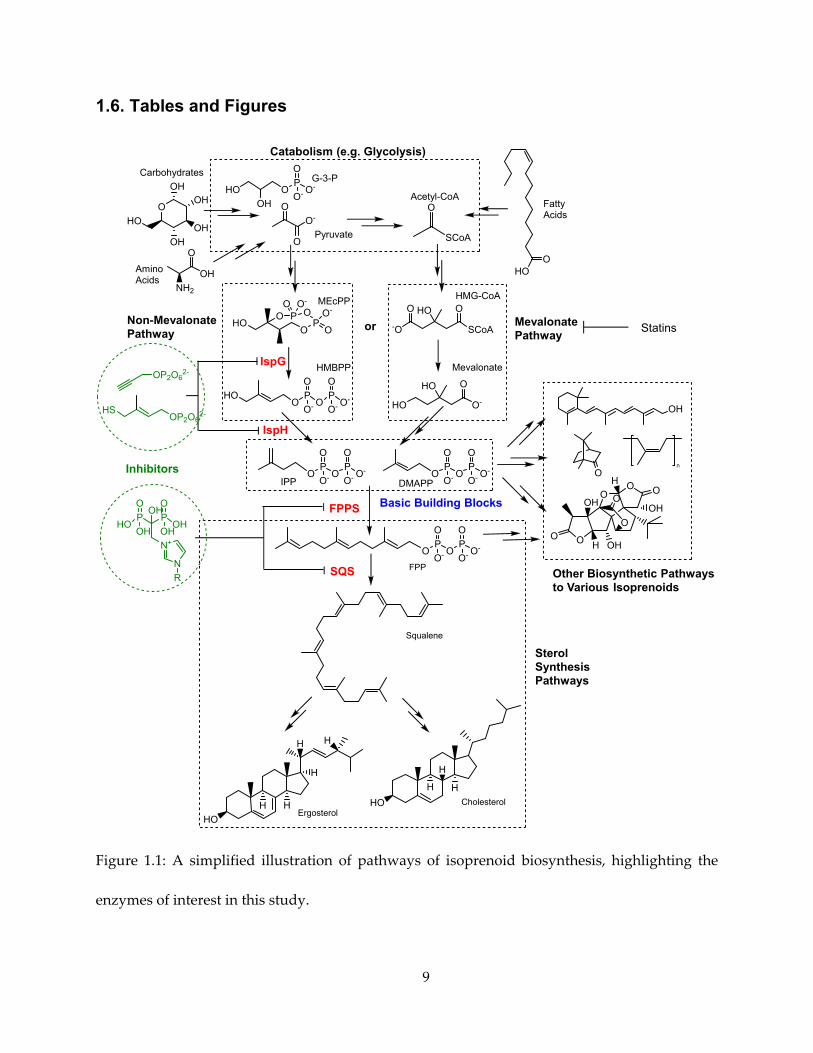
target. As shown in Figure 1.2, the MEP pathway utilizes G-3-P (glyceraldehyde-3-phosphate)
and pyruvate as starting material to make IPP and DMAPP. The enzyme catalyzing the second
(and the first committed) step of the pathway, 1-deoxy-D-xylulose-5-phosphate reductoisomerase
(Dxr or IspC), has been under active development (5) as a target against E. coli, Plasmodium spp.,
and Mycobacterium tuberculosis.
In addition to their potential as drug targets, the last two enzymes of the MEP pathway,
IspG and IspH, are also of interest to bioinorganic chemists for the mysteries in their mechanisms
of action involving cubane type 4Fe-4S clusters. Unlike ferredoxin type of 4Fe-4S clusters, where
each of the four Fe atoms attaches to the protein via coordination to a deprotonated cysteine thiol
4
group, their 4Fe-4S clusters have only three cysteine-liganded Fe atoms while the fourth unique
Fe binds to a different ligand.
Both IspG and IspH catalyze 2e-/2H+ reductions of their respective substrates, 2-C-methyl-
D-erythritol 2,4-cyclopyrophosphate (MEcPP) and HMBPP. For each enzyme, researchers have
proposed multiple mechanisms of action: at least 4 for IspG (6–9) and 6 for IspH (10–15), with
carbocation, carbanion, epoxide, radical and ferraoxetane intermediates.
Therefore, the structures and ligand binding modes of IspG and IspH in complexes with
substrates, reaction intermediates or products are of great mechanistic interest, as will be
discussed in Chapters 2 and 3 of this dissertation, respectively.
Mechanistic insights have also facilitated ideas of inhibitor design of IspG and IspH. Many
chemical species have been screened and potent inhibitors found. In Chapter 4 it will be shown
that several of these inhibitors exhibit diverse binding modes, with the focus on IspH inhibitors.
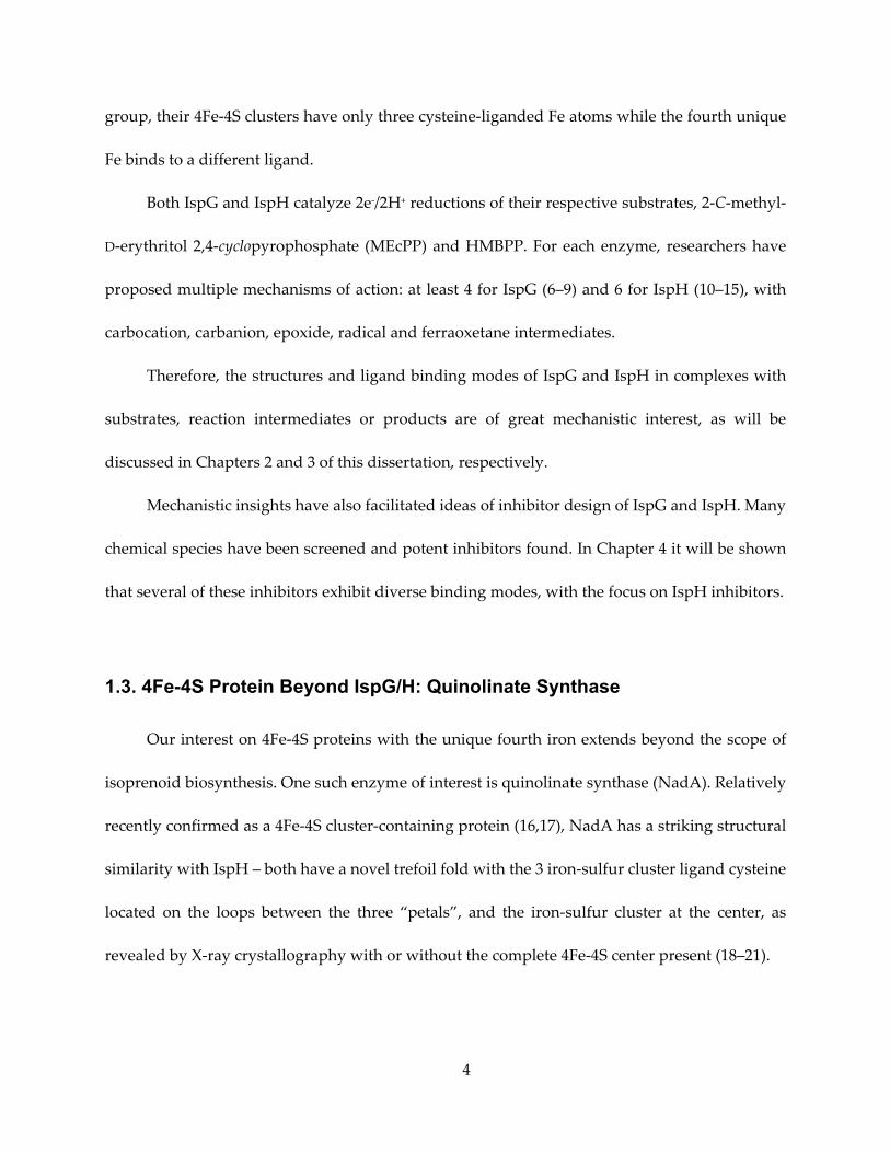
1.3. 4Fe-4S Protein Beyond IspG/H: Quinolinate Synthase
Our interest on 4Fe-4S proteins with the unique fourth iron extends beyond the scope of
isoprenoid biosynthesis. One such enzyme of interest is quinolinate synthase (NadA). Relatively
recently confirmed as a 4Fe-4S cluster-containing protein (16,17), NadA has a striking structural
similarity with IspH – both have a novel trefoil fold with the 3 iron-sulfur cluster ligand cysteine
located on the loops between the three “petals”, and the iron-sulfur cluster at the center, as
revealed by X-ray crystallography with or without the complete 4Fe-4S center present (18–21).
5
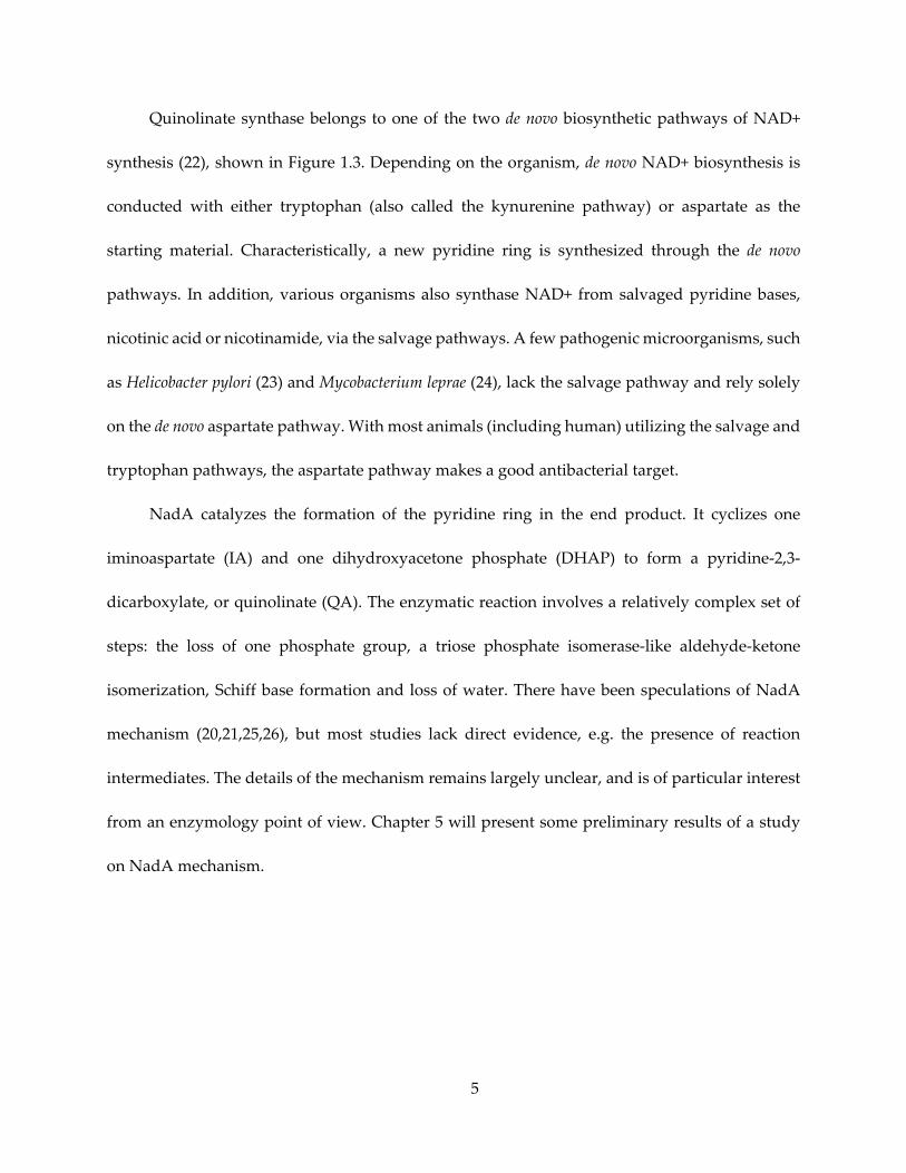
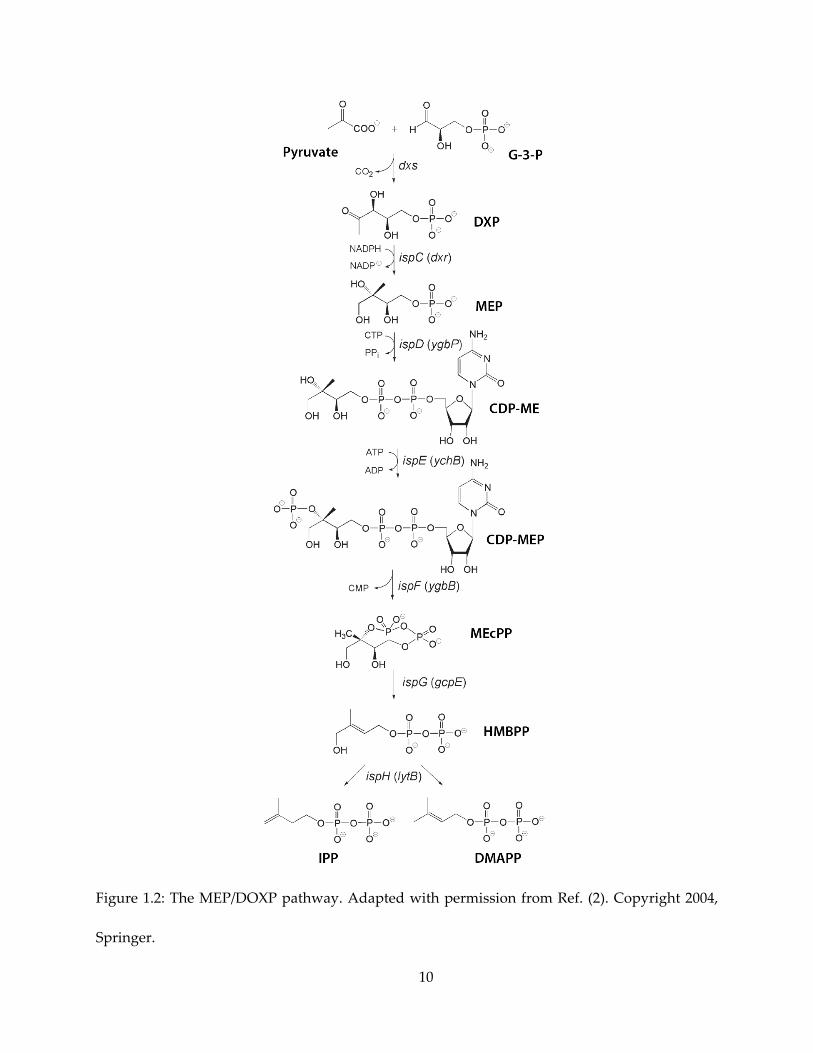
Quinolinate synthase belongs to one of the two de novo biosynthetic pathways of NAD+
synthesis (22), shown in Figure 1.3. Depending on the organism, de novo NAD+ biosynthesis is
conducted with either tryptophan (also called the kynurenine pathway) or aspartate as the
starting material. Characteristically, a new pyridine ring is synthesized through the de novo
pathways. In addition, various organisms also synthase NAD+ from salvaged pyridine bases,
nicotinic acid or nicotinamide, via the salvage pathways. A few pathogenic microorganisms, such
as Helicobacter pylori (23) and Mycobacterium leprae (24), lack the salvage pathway and rely solely
on the de novo aspartate pathway. With most animals (including human) utilizing the salvage and
tryptophan pathways, the aspartate pathway makes a good antibacterial target.
NadA catalyzes the formation of the pyridine ring in the end product. It cyclizes one
iminoaspartate (IA) and one dihydroxyacetone phosphate (DHAP) to form a pyridine-2,3-
dicarboxylate, or quinolinate (QA). The enzymatic reaction involves a relatively complex set of
steps: the loss of one phosphate group, a triose phosphate isomerase-like aldehyde-ketone
isomerization, Schiff base formation and loss of water. There have been speculations of NadA
mechanism (20,21,25,26), but most studies lack direct evidence, e.g. the presence of reaction
intermediates. The details of the mechanism remains largely unclear, and is of particular interest
from an enzymology point of view. Chapter 5 will present some preliminary results of a study
on NadA mechanism.
6
1.4. Prenyl-Transferases FPPS and SQS
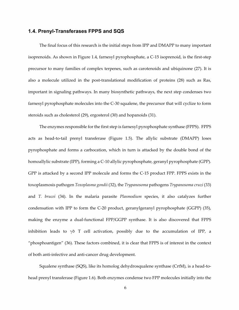
The final focus of this research is the initial steps from IPP and DMAPP to many important
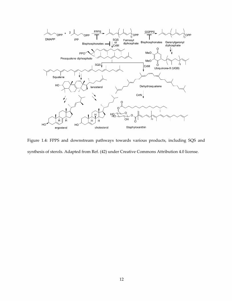
isoprenoids. As shown in Figure 1.4, farnesyl pyrophosphate, a C-15 isoprenoid, is the first-step
precursor to many families of complex terpenes, such as carotenoids and ubiquinone (27). It is
also a molecule utilized in the post-translational modification of proteins (28) such as Ras,
important in signaling pathways. In many biosynthetic pathways, the next step condenses two
farnesyl pyrophosphate molecules into the C-30 squalene, the precursor that will cyclize to form
steroids such as cholesterol (29), ergosterol (30) and hopanoids (31).
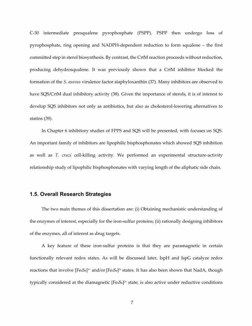
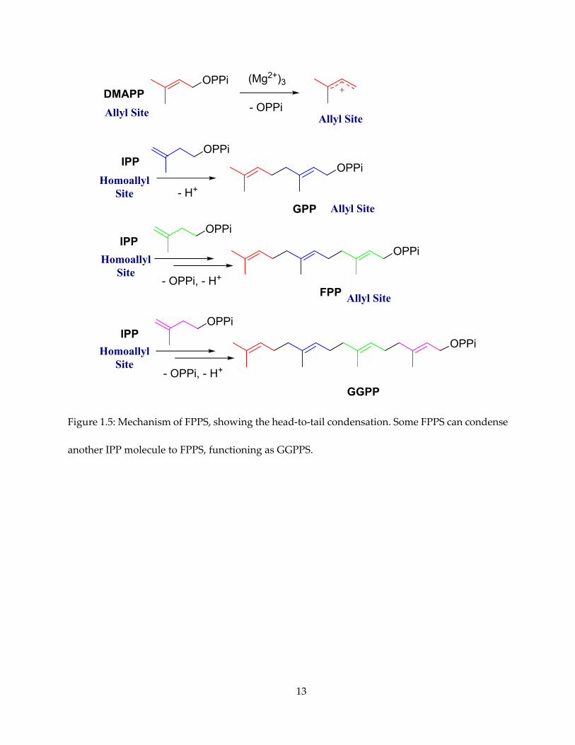
The enzymes responsible for the first step is farnesyl pyrophosphate synthase (FPPS). FPPS
acts as head-to-tail prenyl transferase (Figure 1.5). The allylic substrate (DMAPP) loses
pyrophosphate and forms a carbocation, which in turn is attacked by the double bond of the
homoallylic substrate (IPP), forming a C-10 allylic pyrophosphate, geranyl pyrophosphate (GPP).
GPP is attacked by a second IPP molecule and forms the C-15 product FPP. FPPS exists in the
toxoplasmosis pathogen Toxoplasma gondii (32), the Trypanosoma pathogens Trypanosoma cruzi (33)
and T. brucei (34). In the malaria parasite Plasmodium species, it also catalyzes further
condensation with IPP to form the C-20 product, geranylgeranyl pyrophosphate (GGPP) (35),
making the enzyme a dual-functional FPP/GGPP synthase. It is also discovered that FPPS
inhibition leads to γδ T cell activation, possibly due to the accumulation of IPP, a
“phosphoantigen” (36). These factors combined, it is clear that FPPS is of interest in the context
of both anti-infective and anti-cancer drug development.
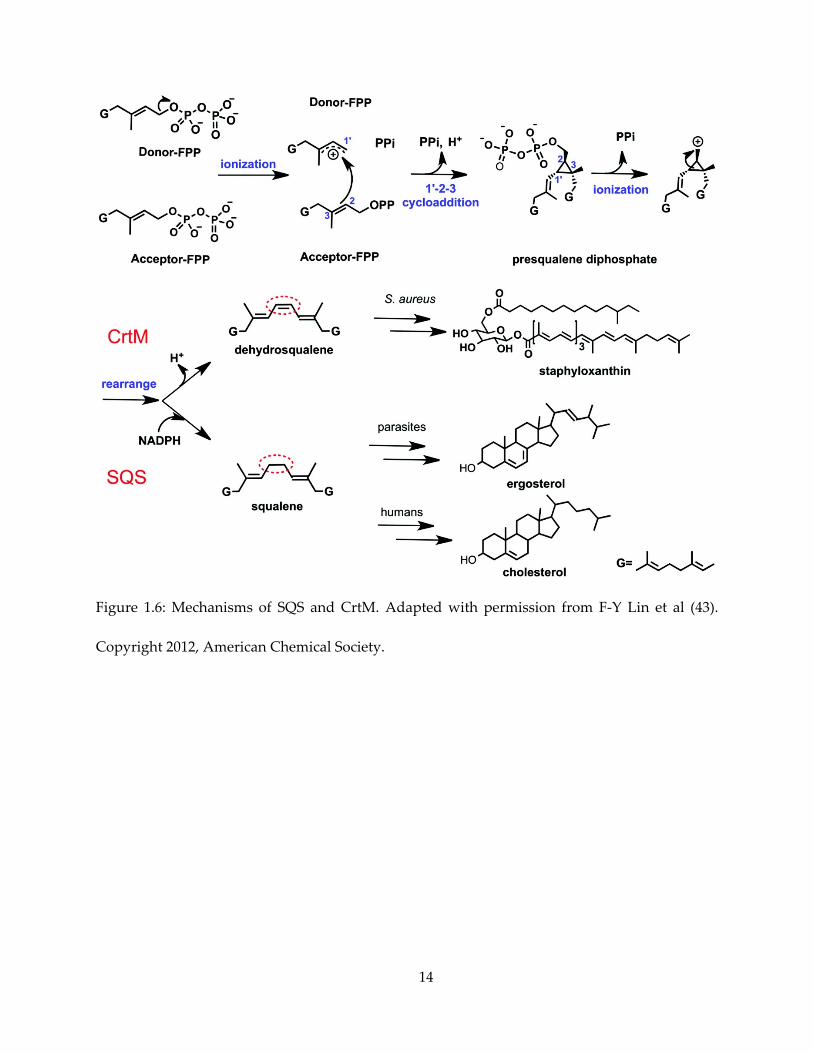
Squalene synthase (SQS), like its homolog dehydrosqualene synthase (CrtM), is a head-to-
head prenyl transferase (Figure 1.6). Both enzymes condense two FPP molecules initially into the
7
C-30 intermediate presqualene pyrophosphate (PSPP). PSPP then undergo loss of
pyrophosphate, ring opening and NADPH-dependent reduction to form squalene – the first
committed step in sterol biosynthesis. By contrast, the CrtM reaction proceeds without reduction,
producing dehydrosqualene. It was previously shown that a CrtM inhibitor blocked the
formation of the S. aureus virulence factor staphyloxanthin (37). Many inhibitors are observed to
have SQS/CrtM dual inhibitory activity (38). Given the importance of sterols, it is of interest to
develop SQS inhibitors not only as antibiotics, but also as cholesterol-lowering alternatives to
statins (39).
In Chapter 6 inhibitory studies of FPPS and SQS will be presented, with focuses on SQS.
An important family of inhibitors are lipophilic bisphosphonates which showed SQS inhibition
as well as T. cruzi cell-killing activity. We performed an experimental structure-activity
relationship study of lipophilic bisphosphonates with varying length of the aliphatic side chain.
1.5. Overall Research Strategies
The two main themes of this dissertation are: (i) Obtaining mechanistic understanding of
the enzymes of interest, especially for the iron-sulfur proteins; (ii) rationally designing inhibitors
of the enzymes, all of interest as drug targets.
A key feature of these iron-sulfur proteins is that they are paramagnetic in certain
functionally relevant redox states. As will be discussed later, IspH and IspG catalyze redox
reactions that involve [Fe4S4]1+ and/or [Fe4S4]3+ states. It has also been shown that NadA, though
typically considered at the diamagnetic [Fe4S4]2+ state, is also active under reductive conditions
8
(26). Given that intact 4Fe-4S clusters are essential for the function of the enzymes, EPR
spectroscopy (40), which probes the electronic structure around of paramagnetic species, would
provide highly informative data about the mechanism, especially when we can freeze-trap
reaction intermediates along the reaction coordinate.
However, continuous-wave EPR is limited in resolution. With pulsed EPR techniques (41),
such as electron nuclear double resonance (ENDOR), electron spin echo envelope modulation
(ESEEM) and two-dimensional hyperfine sublevel correlation (HYSCORE), superior resolution
can be attained. Particularly, when ligands with non-zero spin nucleus are present in the vicinity
of the paramagnetic iron-sulfur cluster, hyperfine and quadrupole (when the nucleus has spin I
≥ 1/2) couplings can be determined through simulation of spectrum. In this light, ligands with
non-zero spin nuclei, including natural abundance 1H, and 14N, as well as enriched 2H, 13C, 15N,
and 17O-labeled proteins and/or ligands, will provide a valuable mechanistic probe.
Molecular biology methods, for example site-directed mutagenesis, also provide important
tools for gaining mechanistic insight. Bioinformatic tools, such as conservation scoring, thus
provide guidance for mutagenesis experiments.
For inhibitors and drug discovery, kinetic assays are the most important routines on the lab
bench. Various analytical methods, including spectrophotometry, scintillation counting and
HPLC, are utilized.
Finally, X-ray crystallography provides most direct evidence of structural features of
enzyme-ligand complexes.
9
1.6. Tables and Figures
HO
H
H
H
HO
H H
H H
H
P
O
OO-O- OP
O
O-HO
PO
HOO P O O-
O
O O-
O
O
O-
HO
OH
P
O
OO- O-
O
OHOH
OH
OH
HOO
SCoA
P
O
OO-O- OP
O
O-P
O
OO-O- OP
O
O-
P
O
OO-O- OP
O
O-
O
-O
O
SCoA
HO
HO
HO O
O-
Non-MevalonatePathway
MevalonatePathway
G-3-P
Pyruvate
Acetyl-CoA
MEcPP
HMBPP
HMG-CoA
Mevalonate
IPP DMAPP
SterolSynthesisPathways
FPP
Squalene
ErgosterolCholesterol
IspG
SQS
FPPS
IspH
N+
N
PP
O
OHOH
O
HOOH
R
OH
OP2O62-
HSOP2O6
2-
or
Inhibitors
Statins
OHO
Carbohydrates
FattyAcids
Catabolism (e.g. Glycolysis)
O
OH
NH2
AminoAcids
Other Biosynthetic Pathwaysto Various Isoprenoids
OH
Basic Building Blocks
On
O
OO
OH
HO
O
OH
H
OH
O
O
Figure 1.1: A simplified illustration of pathways of isoprenoid biosynthesis, highlighting the
enzymes of interest in this study.
10
Figure 1.2: The MEP/DOXP pathway. Adapted with permission from Ref. (2). Copyright 2004,
Springer.
11
Figure 1.3: Two different de novo pathways of NAD+ biosynthesis. (A) The aspartate pathway,
genes in E. coli naming; (B) The tryptophan (kynurenine) pathway, genes in S. cerevisiae naming.
Adapted with permission from Ref. (22). Copyright 2009, American Society for Microbiology.
12
Figure 1.4: FPPS and downstream pathways towards various products, including SQS and
synthesis of sterols. Adapted from Ref. (42) under Creative Commons Attribution 4.0 license.
13
OPPi
- OPPi
OPPi
- H+
OPPi
OPPi
- OPPi, - H+
OPPi
- OPPi, - H+
OPPi
OPPi
DMAPP
IPP
IPP
IPP
GPP
FPP
GGPP
Allyl Site
Allyl Site
HomoallylSite
HomoallylSite
HomoallylSite
Allyl Site
(Mg2+)3
Allyl Site
Figure 1.5: Mechanism of FPPS, showing the head-to-tail condensation. Some FPPS can condense
another IPP molecule to FPPS, functioning as GGPPS.
14
Figure 1.6: Mechanisms of SQS and CrtM. Adapted with permission from F-Y Lin et al (43).
Copyright 2012, American Chemical Society.
15
Chapter 2. Mechanism of Action of 4Fe-4S Protein IspG
2.1. Notes and Acknowledgements
This chapter is partly adapted, with permission, from the following publications: Wang, W.
et al., 2010. Organometallic mechanism of action and inhibition of the 4Fe-4S isoprenoid
biosynthesis protein GcpE (IspG). Proceedings of the National Academy of Sciences, 107(25),
pp.11189–11193, Copyright 2010, National Academy of Sciences; Wang, W. et al., 2011. An
ENDOR and HYSCORE Investigation of a Reaction Intermediate in IspG (GcpE) Catalysis. Journal
of the American Chemical Society, 133(22), pp.8400–8403, Copyright 2011, American Chemical
Society.
Dr. Weixue Wang performed a large part of the CW/Pulsed EPR spectroscopy. Dr. Ke Wang
synthesized most of the isotope-labeled substrates. Dr. Yi-Liang Liu performed mutagenesis,
Prof. Tatyana I. Smirnova provided guidance and instrumentation for part of the EPR
spectroscopy, and Prof. Yong Zhang and his group performed quantum chemical calculations.
This work was supported by NIH grant (GM065307) to Eric Oldfield, NIH grant
(GM085774) to Yong Zhang, NSF grant MCB-0843632 to Tatyana I. Smirnova and equipment
grants from NIH (S10RR023614), NSF (CHE-0840501), and the North Carolina Biotechnology
Center (NCBC 2009-IDG-1015). I was supported by a pre-doctoral fellowship from the American
Heart Association, Midwest Affiliate (11PRE7500042) from July 2011 to June 2013. I also thank
Drs. Hassan Jomaa and Jochen Wiesner for providing the T. thermophiles GcpE expression
systems, Dennis Dean for providing the isc plasmids for iron-sulfur cluster generation, and Dr.
Mark J. Nilges for assistance with the EPR spectroscopy.
16
2.2. Introduction
As discussed in Section 1.2, in the early days of IspG research, the mechanism of IspG had
been elusive, with a number of proposals without enough direct evidence of observation of the
proposed intermediate species. In 2007, it was observed that while it was difficult to fully reduce
Thermus thermophilus IspG cluster with dithionite, in a steady state condition with both dithionite
and the substrate MEcPP, a species with intense EPR signal similar to a HiPIP (high-potential iron
protein) 4Fe-4S cluster become observable and then disappeared after a duration of minutes (44).
This observation suggested that a paramagnetic reaction intermediate, hereafter dubbed “X”, is
present. Effort was therefore made to elucidate the structure of “X”, with isotope-labeled
substrates and pulsed EPR spectroscopy.
2.3. Results and Discussion
2.3.1. CW-EPR observation of reaction intermediate “X”
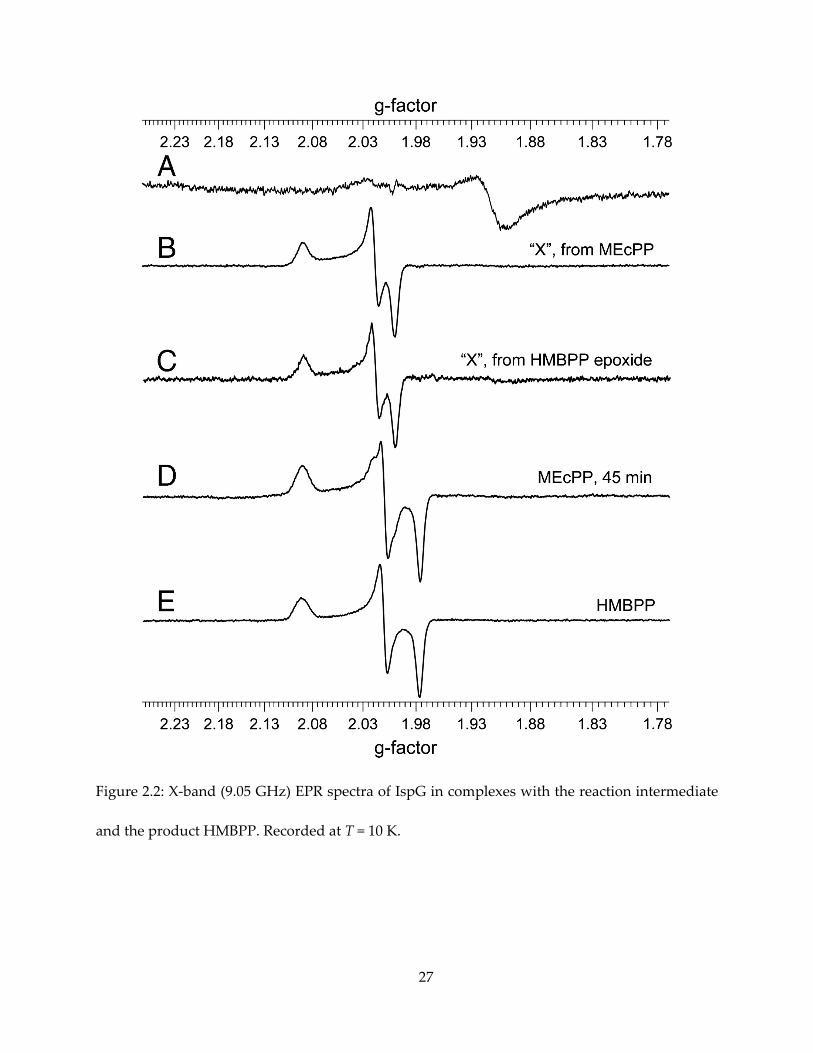
The IspG reaction substrate MEcPP and product HMBPP, shown in Figure 2.1, form
paramagnetic complexes in the presence of dithionite. The intermediate “X” is clearly observable
by continuous-wave (CW) EPR upon MEcPP binding and reduction by dithionite (Figure 2.2B),
with g tensor gi = [2.092, 2.018, 1.999]. It converts to the HMBPP-IspG complex after 45 minutes of
incubation (Figure 2.2D, E), with gi = [2.092, 2.010, 1,976]. Since Nyland et al (45) reported that
HMPPP-epoxide also converts to HMBPP catalyzed by IspG, an attempt to produce the
17
intermediate “X” with HMBPP-epoxide was also made, and an essentially identical EPR
spectrum was recorded (Figure 2.2C).
2.3.2. Fermentation and chemical synthesis of MEcPP
Because of the difficulties of chemical synthesis of MEcPP, “fermentation engineering”
experiments to produce MEcPP biosynthetically were performed, according to modified
previously described protocols (46) using Corynebacterium ammoniagenes cells. With this method,
production of several isotope-labeled MEcPP was also possible. For [u-2H]-MEcPP, C.
ammoniagenes cells were acclimatized with increasing D2O percentage in growth media, and
uniformly-deuterated MEcPP is produced in 100% D2O growth media with [u-2H7]-D-glucose as
carbon source. For [u-13C5]-MEcPP, cells are fed with [u-13C6]-D-glucose. For specifically 13C-
labeled MEcPP, various 13C-labeled glucose was fed to the bacteria to produce MEcPP labeled
according to knowledge of glycolysis and MEP pathways (2), but enrichment was reduced. In the
end only [2-13C]-D-glucose produced MEcPP with 30% enriched at both C-2 and C-3 positions
(numbering according to Figure 2.1). In addition, [1,3,4-13C3]-MEcPP was gifted to us by authors
who reported methods of producing labeled-MEcPP with purified enzymes (47).
For 17O and 2H at specific positions, HMBPP-epoxides were chemically synthesized by Dr.
Ke Wang.
2.3.3. ENDOR and HYSCORE spectra and assignment
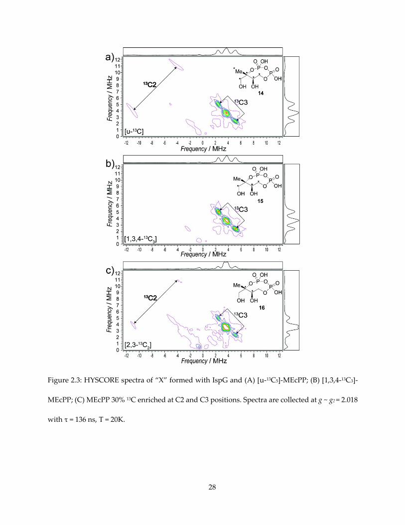
The first important clue of the structure of “X” comes from the hyperfine interaction of 13C-
labeled MEcPPs with the iron-sulfur cluster, as shown in the HYSCORE spectra of them in Figure
18
2.3. In the HYSCORE (48) spectra of an I = ½ nucleus, a pair of peaks appear on the spectrum,
centered at the Larmor frequency νL of the nucleus, and separated by the hyperfine coupling A.
With “X” formed from [U-13C]-MEcPP, we can observe one pair of peaks in the (-, +)
quadrant representing a large hyperfine coupling, which can be simulated with Aii = [14.5, 12.0,
26.5] MHz, Aiso = 17.7 MHz. Next, there is a pair of peaks representing smaller hyperfine coupling,
with simulation Aii = [1.8, 2.0, 5.1] MHz, Aiso = 3.0 MHz. There might be additional unresolved
small hyperfine peaks around the 13C Larmor frequency (~ 3.6 MHz), overlapping with double
quantum transition signal from 14N in the protein backbone.
The Aiso = 17.7 MHz peak is present with [U-13C5] and [1,3,4-13C3] substrates, but not present
in [2,3-13C2] substrate, and therefore can be unequivocally assigned to the quaternary carbon, C2,
of MEcPP. The Aiso = 3.0 MHz signal, present in all three 13C labeled samples, can then be assigned
to C3.
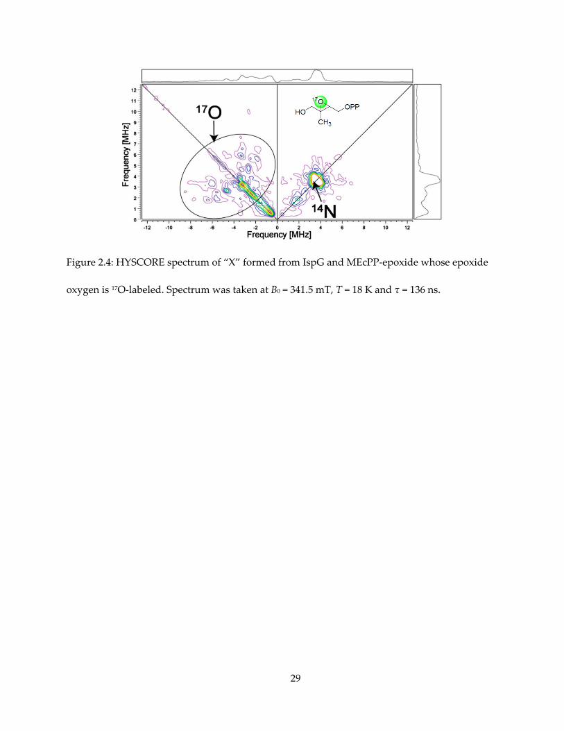
The HYSCORE spectrum from 17O labeled HMBPP-epoxide also exhibits signal at the (-, +)
quadrant, with A ~ 8 MHz, as shown in Figure 2.4. Due to low isotopic enrichment and complex
broadening schemes of an I = 5/2 nucleus, it is difficult to quantitatively simulate the 17O
HYSCORE spectrum. But qualitatively, this large hyperfine interaction indicates high electron
spin density on the 17O atom.
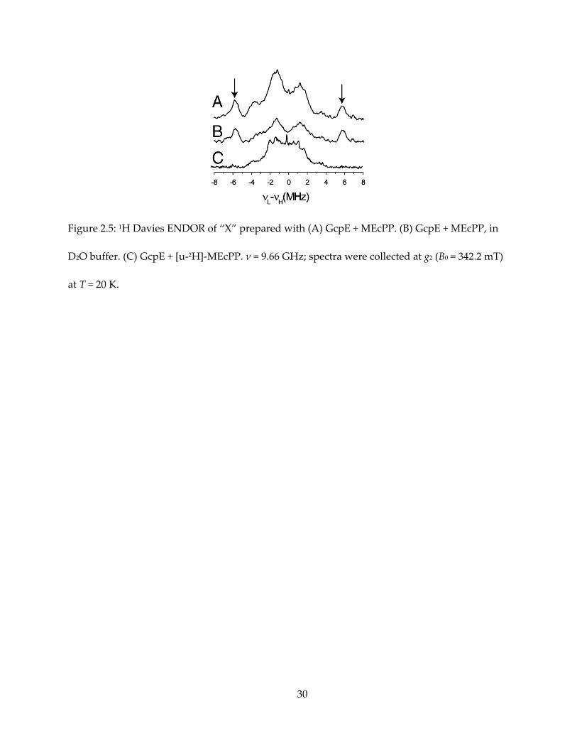
Next, we prepared “X” from deuterated MEcPP or HMBPP-epoxide, and utilized pulsed
ENDOR technique (49) to explore 1H and 2H hyperfine coupling at high resolution. With natural
abundance MEcPP, ENDOR of “X” shows a 1H signal with Aii = [14, 11, 11] MHz, Aiso ~ 12 MHz
(Figure 2.5A). This proton does not exchange with deuterated solvent (Figure 2.5B), while its
signal disappears when “X” is prepared with uniformly deuterated MEcPP (Figure 2.5C). To
19
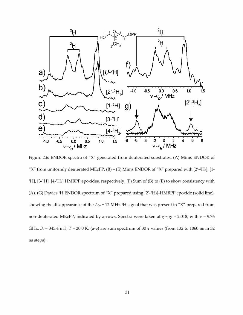
specifically assign this signal, deuterated HMBPP-epoxide were utilized to prepare “X” for
ENDOR spectroscopy (Figure 2.6). Given that the gyromagnetic ratio of 1H is about 7 times that
of 2H, one can expect this signal to appear in 2H spectrum with A ~ 1.7 MHz. As a result, this
signal corresponds to HMBPP-epoxide deuterated at the 2’-methyl group, while samples
deuterated at other positions showed weaker hyperfine coupling, overlapping with each other
and could explain the central envelope of the [u-1H]-MEcPP and [u-2H]-MEcPP spectra.
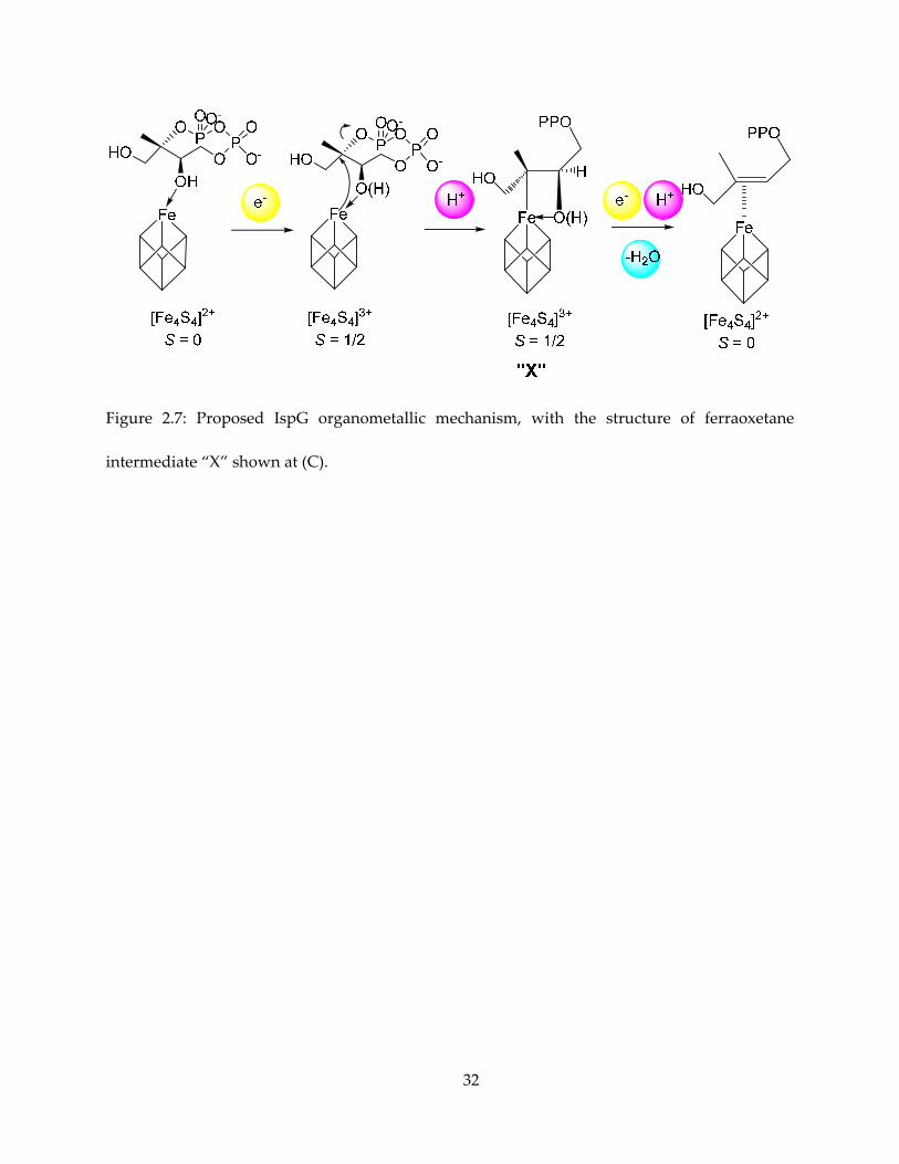
2.3.4. Structure of “X” and the ferraoxetane mechanism
Taken the above spectroscopic results together, we conclude that the most plausible
structure of the reaction intermediate is a ferraoxetane, the possible pathway to it is shown in
Figure 2.7, and the ferraoxetane structure shown in Figure 2.7C. This structure of “X” is favored
for the following reasons: (i) The large 13C hyperfine coupling from C2 has both large isotropic
and anisotropic terms, Aii = [14.5, 12.0, 26.5], indicating both large Fermi contact interaction
(isotropic term) and spatial proximity (anisotropic term), hence the possibility of a direct Fe-C
bond. The observed 13C hyperfine coupling in “X” is also close to that seen for 13CO directly
bonded to one of the irons in the H cluster in the Hox-CO state of an [FeFe] hydrogenase, for which
Aii(13CO) = [19.2, 16.6, 15.6] MHz (50). (ii) The methyl signal, with Aii(1H) = [14, 11, 11] MHz, has a
relatively large isotropic term indicating some significant Fermi contact interaction, but the small
anisotropy indicates long spatial distance. With the structure of “X” in Figure 2.7C, the methyl
group is spatially far away from the paramagnetic center, while the proton is 3 bonds away from
the unique fourth iron, and hyperconjugation could presumably enhance the Fermi contact
interaction. The large spin density on one of the methyl hydrogens has also been corroborated by
20
a DFT calculation (51). (iii) The large 17O hyperfine coupling, A ~ 8 MHz indicates direct Fe-O
interaction, similar to that previously obtained with aconitase (52,53), A ~ 8 - 15 MHz.
2.4. Conclusions
In a nutshell, we have been able to freeze-trap the paramagnetic reaction intermediates and
elucidate the structural features with CW and pulsed EPR spectroscopies. With the help of
biosynthesized and chemically synthesized isotope-labeled substrates, we were able to assign the
spectroscopic features to atoms in the reaction intermediates. The results strongly supported an
organometallic ferraoxetane species with direct Fe-C bonding – an unusual phenomenon of great
general interest – as an enzymatic reaction intermediate. With these results, we were able to
establish a preliminary picture of mechanism of action for IspG involving a ferraoxetane, while
other chemical steps before and after this intermediate might be understood by further
spectroscopy, mutagenesis, inhibition and crystallography studies.
2.5. Materials and Methods
2.5.1. T. thermophilus IspG protein expression and purification
Plasmids with T. themophilus gene construct was transformed into BL-21(DE3) cells
(Invitrogen). Cells were grown in LB media supplemented with 150 mg/L ampicillin at 37 °C with
225 rpm shaking. When OD600 reached ~ 0.3, medium was supplied with 0.1 g/L L-cysteine and
0.1 mM FeCl3. When the OD600 reached ~ 0.7 – 0.8. Cells were then induced with 1 mM IPTG.
Incubation then continued at 25 °C for 8 – 16 h with the incubator shaking at a reduced speed of
21
80 rpm. Cells were then harvested by centrifugation (9000 rpm, 10 min, 4 °C), flash frozen at -80
°C, and kept frozen until further use.
Purification was then carried out in a Coy anaerobic chamber in a cold room set at 4 ∘C.
Care was taken to ensure all actions were performed when the oxygen content is less than 2 ppm
in the anaerobic chamber. All the buffers used are also degassed on a Schlenk line before being
transferred into the anaerobic chamber for use. Cell pellets were re-suspended in 15 - 30 mL buffer
containing 50 mM Tris ̇⋅HCl, pH 8.0 and 150 mM NaCl. 2 μL of benzonase nuclease solution (EMD
chemicals), ~ 2 mg of phenylmethanesulfonyl floride (PMSF, Aldrich), and 5 mM TCEP were
added to the mixture before cells were lysed by sonication (Fisher Scientific Sonic Dismembrator
Model 500) with 5 second pulse totaling 1 – 2 minute at 50% power. The suspension is then
carefully sealed with 3M vinyl tape in a 50 mL centrifugation tube and centrifuged at 4 °C for 15
min (outside the anaerobic chamber). The supernatant was applied to a HisPur Ni-NTA spin
column (Pierce) equilibrated with a pH 8.0 buffer containing 50 mM Tris · HCl, 10 mM imidazole
and 150 mM NaCl. After washing with 8 mL of the same loading buffer 3 times (by adding the
buffer onto the column and centrifuging at 600×g for 2 minutes), protein was eluted with the same
buffer plus 250 mM imidazole. Dark-brown fractions were collected, pooled, concentrated with
a VivaSpin 20 centrifugal concentrator (GE Healthcare) and then desalted with a PD-10 column
(GE Healthcare), to remove imidazole. The protein is stored at 4∘C until use.
2.5.2. Biosynthesis for MEcPP
MEcPP was produced by growing of Corynebacterium ammoniagenes cells on glucose under
oxidative stress conditions. C. ammoniagenes (ATCC 6872) was cultured aerobically in a medium
containing peptone (10 g/L), yeast extract (3 g/L), NaCl (5 g/L). Cells are incubated at 30 °C with
22
shaking at 225 rpm. When the cells reached late log phase of growth (OD600 ∼ 1.4), the medium
was supplemented with benzyl viologen to a final concentration of 50 mg/L, and 0.5 – 4.0 g/L
glucose was added. Growth was allowed to continue for 18 h, and the cells were then harvested
by centrifugation.
The cell pellets (from 1 L growth media) were extracted with 7:3 (v/v) ethanol:water three
times, by stirring in a flask at 4 ∘C with 40 mL of solvent each time. After rotary evaporation at
60∘C to remove the solvent, the crude extract was dissolved in 30 mL H2O, loaded onto a 25 mL
QAE-Sephadex A-25 (GE Healthcare) anion exchange column pre-equilibrated with 0.05 M
ammonium acetate buffer (pH ~ 7). The column was washed with approximately 100 mL of the
same buffer, then eluted with a linear gradient of 0.3–1.5 M NH4OAc, with a total volume of 100
mL of eluents. The desired product eluted at approximately 0.8 M NH4OAc. The fractions were
pooled and lyophilized, and MEcPP detected by using a GcpE activity assay and 31P NMR
spectroscopy. The MEcPP-containing fractions were further purified by cellulose flash
chromatography, with acetonitrile: isopropyl alcohol: 1% NH4HCO3 (in H2O) of ratios of 4:2:1
and later 1:2:1 as eluents. The 1:2:1 solvent fractions were pooled and lyophilized.
2.5.3. Biosynthesis of isotope-labeled MEcPP
[u-13C5]-MEcPP is obtained by using the method above while feeding the cells with 1.0 g/L
[u-13C6] D-glucose before adding benzyl viologen. Isotope incorporation was close to 100%, as
determined by 13C NMR. 13C NMR (126 MHz, D2O) δ: 16.63, 16.30 (d); 65.72, 65.77, 66.06, 66.11
(doublet of doublets); 66.98, 67.14, 67.31 (t); 68.25, 68.44, 68.60, 68.76, 68.95 (q); 83.55, 83.87, 84.22,
84.55 (q). 31P NMR (202 MHz, D2O) δ: −13.89, −14.00 (d); −9.95, −9.85 (d).
23
MEcPP with ∼30% overall 13C labeling at C2 and/or C3 was prepared using the same
protocol. 0.5 g/L D-glucose were added. After another 18 hours of incubation, MEcPP was
extracted and purified as described above. The overall 13C-enrichment was ∼30% (13C0, 65.4%;
13C1, 29.4%; 13C2, 5.2%), as determined by mass spectrometry. The sample is therefore
essentially a mixture of [2-13C] and [3-13C]-MEcPP, but for simplicity is referred to in as [2,3-13C]-
MEcPP in this chapter.
For the biosynthesis of [u-2H]-MEcPP, C. ammoniagenes was first “acclimatized” to growth
in D2O. 5 mL aliquots of peptone/yeast extract/NaCl media were made, with varying H2O/D2O
contents. Cells were first cultured in 100% H2O media, then when the OD600 reached ∼0.8, 50 μL
of the suspension was transferred to a medium with 25%:75% D2O:H2O. Cells were then cultured
sequentially in 50%, 75% and 100% D2O-containing media. For [u-2H]-MEcPP production, we
used 1 L of medium with 100% D2O, cells were grown to an OD600 ∼ 1.4, 50 mg/L benzyl viologen
and 1 mg/L [u-2H7]-D-glucose were added, and after 18 hours incubation, cells were harvested
and MEcPP extracted as described above.
2.5.4. EPR sample preparation
In general, the protein sample was usually exchanged to 20%-40% glycerol containing
buffers before being used for making EPR samples. Samples are usually prepared in a Coy
anaerobic chamber at room temperature, with oxygen < 2 ppm during operation. For most
samples, the protein solution was reduced with 5-20 mM sodium dithionite before any ligand(s)
was added. The sample was injected into an 4.0 mm outer diameter EPR tube (3.8 mm for X-band
ENDOR experiments and 1.6 mm for Q-band experiments, from Wilmad LabGlass) with a
24
syringe and a 18 cm stainless steel needle, incubated for the required amount of time, and frozen
in liquid nitrogen before spectroscopy experiments.
To prepare one-electron reduced AaIspH EPR samples, 0.5 – 1 mL reconstituted protein
solution (2.5 mM) was first incubated with 20 – 25 mM sodium dithionite for 30 minutes in the
anaerobic chamber at 4∘C. After reduction, the solution was desalted with a PD-10 column to
remove excess sodium dithionite. The desalted eluent was mixed (1:1 volume) with a degassed
40% glycerol solution, and then concentrated with an Amicon centrifugal filter (10,000 molecular
weight cutoff) so that the final volume approximated the initial volume. The protein solution was
aliquoted according to the sample volume required by EPR spectroscopy (typically 100 – 200 μL
for X-band, 10 – 15 μL for Q-band). Ligand solution was then added to an aliquot of the protein
solution to a typical final concentration of 10 – 15 mM, swiftly mixed and injected into the X-band
and/or Q-band EPR tubes which were then frozen in liquid nitrogen. Typically the time from
ligand addition to sample freezing should take less than 30 seconds.
2.5.5. CW-EPR spectroscopy
All CW-EPR experiments were performed on a Varian E-line 122 X-band spectrometer with
an Air Products helium cryostat. Typical data acquisition parameters were: microwave frequency
9.04 - 9.25 GHz; field center 3250 gauss or 2500 gauss, with field sweep 1600 gauss or 5000 gauss;
modulation frequency 100 kHz; modulation amplitude 5 gauss; time constant 32 milliseconds;
gain 1000 to 20000; temperature 8 - 20 K. Usually each field scan is set to take 60 seconds, with 10
seconds between scans.
25
2.5.6. Pulsed EPR spectroscopy
Pulsed spectra were obtained on a Bruker ElexSys E-580 FT-EPR spectrometer equipped
with an Oxford Instruments CF935 cryostat.
HYSCORE used the four-pulse sequence: (π/2)mw – τ – (π/2)mw – t1 – πmw – t2 – (π/2)mw –
τ – echo. Typically parameters for X-band HYSCORE were: microwave pulse width (π/2)mw = 16
ns, microwave frequency = 9.62 - 9.71 GHz, magnetic field = 3200 – 3500 G, τ = 128 – 300 ns. The
time t1 and t2 are incremented in 16, 20 or 24 ns increments for 128 or 256 steps, starting from 80
ns. Recorded time-domain data undergo baseline correction with a 3rd order polynomial. The
time-series is then multiplied by a Hamming window function, followed by zero-filling, 2D-
Fourier transformation and symmetrization with the minimum value.
For ENDOR experiments, a Bruker RF amplifier was used. The following pulse sequences
were used:
Mims ENDOR: (π/2)mw – τ – (π/2)mw – πrf – (π/2)mw – τ – echo.
Davies ENDOR: πmw – πrf – (π/2)mw – τ – πmw – τ – echo.
With (π/2)mw = 16 ns, πrf = 20 μs for Mims, and (π/2)mw = 48 ns, πrf = 10 μs for Davies ENDOR
experiments. For Mims experiment, usually multiple τ values are used and spectra added
together because of the τ suppression effect for which signal at sin(2πντ) = 0 is suppressed.
26
2.6. Tables and Figures
Figure 2.1: Reaction catalyzed by IspG, showing the numbering convention of IspG and IspH
substrates in this dissertation.
27
Figure 2.2: X-band (9.05 GHz) EPR spectra of IspG in complexes with the reaction intermediate
and the product HMBPP. Recorded at T = 10 K.
28
Figure 2.3: HYSCORE spectra of “X” formed with IspG and (A) [u-13C5]-MEcPP; (B) [1,3,4-13C3]-
MEcPP; (C) MEcPP 30% 13C enriched at C2 and C3 positions. Spectra are collected at g ~ g2 = 2.018
with τ = 136 ns, T = 20K.
29
Figure 2.4: HYSCORE spectrum of “X” formed from IspG and MEcPP-epoxide whose epoxide
oxygen is 17O-labeled. Spectrum was taken at B0 = 341.5 mT, T = 18 K and τ = 136 ns.
30
Figure 2.5: 1H Davies ENDOR of “X” prepared with (A) GcpE + MEcPP. (B) GcpE + MEcPP, in
D2O buffer. (C) GcpE + [u-2H]-MEcPP. ν = 9.66 GHz; spectra were collected at g2 (B0 = 342.2 mT)
at T = 20 K.
31
Figure 2.6: ENDOR spectra of “X” generated from deuterated substrates. (A) Mims ENDOR of
“X” from uniformly deuterated MEcPP; (B) – (E) Mims ENDOR of “X” prepared with [2’-2H3], [1-
2H], [3-2H], [4-2H2] HMBPP epoxides, respectively. (F) Sum of (B) to (E) to show consistency with
(A). (G) Davies 1H ENDOR spectrum of “X” prepared using [2’-2H3]-HMBPP epoxide (solid line),
showing the disappearance of the Aiso = 12 MHz 1H signal that was present in “X” prepared from
non-deuterated MEcPP, indicated by arrows. Spectra were taken at g ~ g2 = 2.018, with ν = 9.76
GHz; B0 = 345.4 mT; T = 20.0 K. (a-e) are sum spectrum of 30 τ values (from 132 to 1060 ns in 32
ns steps).
32
Figure 2.7: Proposed IspG organometallic mechanism, with the structure of ferraoxetane
intermediate “X” shown at (C).
33
Chapter 3. Mechanism of Action of 4Fe-4S Protein IspH
3.1. Notes and Acknowledgements
This chapter is partly adapted, with permission, from the following publications: Wang, W.
et al., 2010. Bioorganometallic mechanism of action, and inhibition, of IspH. Proceedings of the
National Academy of Sciences, 107(10), pp.4522–4527, Copyright 2010, National Academy of
Sciences; and Li, J. et al., 2013. Isoprenoid Biosynthesis: Ferraoxetane or Allyl Anion Mechanism
for IspH Catalysis? Angewandte Chemie, 125(25), pp.6650–6653, Copyright 2013 WILEY-VCH
Verlag.
Dr. Weixue Wang performed part of the CW/Pulsed EPR spectroscopy. Dr. Ke Wang
synthesized most of the isotope-labeled substrates. Prof. Tatyana I. Smirnova provided guidance
and instrumentation for part of the EPR spectroscopy, and Prof. Yong Zhang and his group
performed quantum chemical calculations.
This work was supported by NIH grant (GM065307) to Eric Oldfield, NIH grant
(GM085774) to Yong Zhang, NSF grant MCB-0843632 to Tatyana I. Smirnova and equipment
grants from NIH (S10RR023614), NSF (CHE-0840501), and the North Carolina Biotechnology
Center (NCBC 2009-IDG-1015). I was supported by a pre-doctoral fellowship from the American
Heart Association, Midwest Affiliate (11PRE7500042) from July 2011 to June 2013. I also thank
Drs. Hassan Jomaa and Jochen Wiesner for providing the A. aeolicus IspH expression systems,
and Dr. Mark J. Nilges for assistance with the EPR spectroscopy.
34
3.2. Introduction
Various methods have been attempted to trap reaction intermediates of IspH. An inactive
E126A (Aeolicus aquifex numbering) mutant facilitates the trapping of a species with g1 ~ 2.12 (54).
However, under steady-state conditions, a weak signal for another species was observed (55).
This species also shows a HiPIP-like EPR spectrum, with its g1 ~ 2.17 significantly higher than the
g1 ~ 2.12 species trapped with E126A mutant. Later, Xu et al (56) reported producing a species by
using one-electron reduced IspH and removing excessive dithionite reducing reagent. In this way
a species with an intense EPR signal was trapped with identical g values as the steady-state
intermediate observed previously. Therefore, efforts are again made to elucidate the structure of
this reaction intermediate, with the help of isotope-labeled HMBPP and pulsed EPR experiments.
3.3. Results and Discussion
3.3.1. Proposals of plausible reaction intermediate structure
There have been a considerable number of proposals of IspH mechanism of action, with
cationic, anionic, radical, butadiene and organometallic species proposed as intermediates (13–
17, 48, 18). Most of these proposals have been without direct spectroscopic or structural evidence.
Previous research in the Oldfield Group accumulated considerable evidence in support of the
organometallic hypothesis (55), in which the intermediate is stabilized by a π interaction between
the alkene group of HMBPP and the cluster, as shown in Figure 3.1A. Alternatively, a second
organometallic mechanism was proposed (15) in which a ferraoxetane, similar to previously
35
described for IspG, was the intermediate, as shown in Figure 3.1B, based on CW-EPR and 1H, 2H
and 31P ENDOR spectroscopy of a newly reported reaction intermediate with g1 ~ 2.17.
I thus carried out a series of investigation of the chemical nature of this species, with 2H, 13C
and 17O-labeled substrates and one-electron reduced A. aeolicus IspH. This species is prepared by
reducing IspH with dithionite, yielding an [Fe4S4]+ cluster, and then removing excess reductant.
The one-electron reduced IspH was then mixed with HMBPP substrate, and frozen quickly,
yielding an intense EPR spectrum. The CW-EPR spectrum of this species is shown in Figure 3.2,
with the HiPIP-like g tensor, gi = [2.17, 2.01, 1.99]. The CW (continuous wave) EPR spectrum was
essentially the same as the one we reported previously for a sample prepared from wild-type E.
coli IspH in the presence of excess dithionite and HMBPP under steady-state conditions.
3.3.2. The absence of 17O hyperfine signal
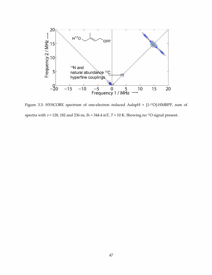
I first obtained HYSCORE spectra one-electron reduced IspH (AaIspH) with [1-17O]-
HMBPP (that is, labeled at the 1, CH2OH position in Figure 3.1), since in the IspH ferraoxetane
model this group is directly bonded to the unique fourth iron in the 4Fe-4S cluster. The HYSCORE
spectrum obtained using this one-electron reduced sample is shown in Figure 3.3 and, as with
the E. coli protein under turnover conditions, there is no evidence for any 17O HYSCORE signal.
Previous work found evidence for a weak (Aiso ~ 1 MHz) 17O hyperfine coupling from the same
batch of [1-17O]-HMBPP bound to an E. coli IspH E126Q mutant, proposed to be a weak complex
(55), but in systems where Fe-O bonds are found, Aiso values are an order of magnitude larger
(52,53,57). These results, therefore, do not provide support for the IspH ferraoxetane hypothesis
but are consistent with formation of an allyl anion complex, in which the 17O has been lost.

36
3.3.3. The 13C HYSCORE spectra, and signal assignment
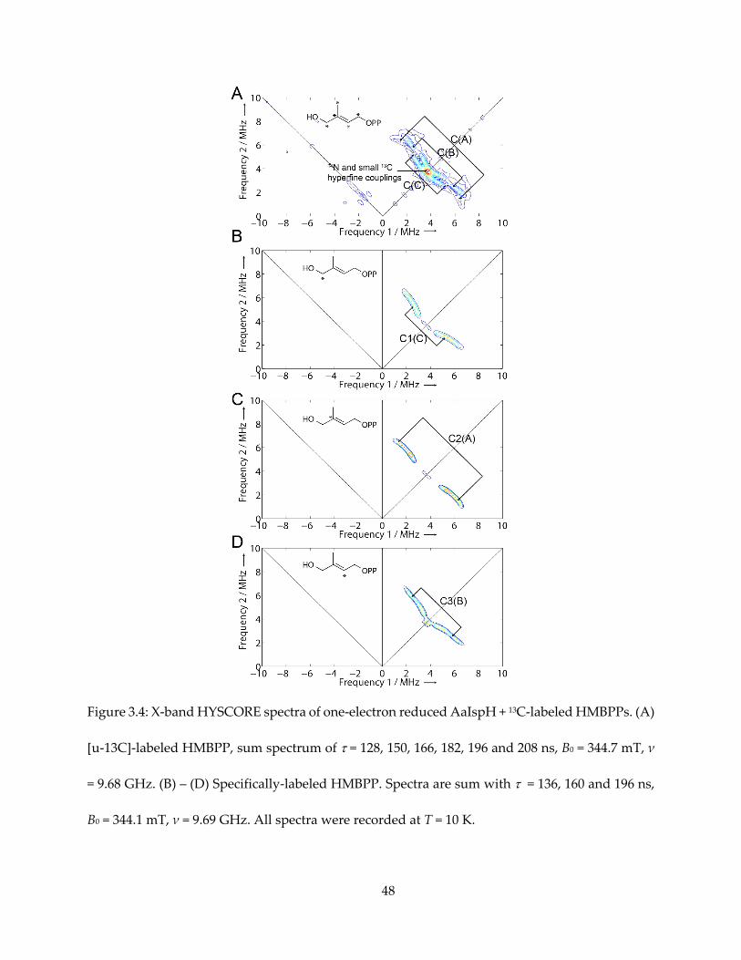
Next, I used a uniformly 13C-labeled HMBPP (converted from [u-13C]-MEcPP with GcpE) to
produce the g1 = 2.17 intermediate species, again with one-electron reduced AaIspH, shown in
Figure 3.4A. There was no evidence for a large (as large as ~ 17 MHz for the GcpE ferraoxetane
complex) 13C hyperfine coupling. There were, however, three sets of intermediate-sized hyperfine
couplings, denoted C(A), C(B) and C(C) in Figure 3.4A, along with possible smaller 13C hyperfine
signal overlapping with 14N double quantum transition signal. The isotropic hyperfine couplings
are much smaller than the Aiso (2-13C) ~ 17 MHz reported for the intermediate “X” in IspG.
To assign these three resonances, Dr. Ke Wang synthesized [1-13C]-, [2-13C]- and [3-13C]-
HMBPPs, and I obtained HYSCORE spectra of the g1 = 2.17 intermediates prepared with them.
The resultant spectra are shown in Figure 3.4B–D. The sets of signal could unequivocally be
assigned: C1 to signal C(C), C2 to signal C(A) and C3 to signal C(B).
3.3.4. Measurement of 13C hyperfine tensors
The principle values and Euler angles of the hyperfine tensors could be obtained with
EasySpin simulations (58). To better determine the tensors, HYSCORE spectra of [u-13C]-HMBPP
+ IspH were acquired at three different g values (or magnetic fields) with two different τ, as
shown in Figure 3.5. For each specifically-labeled species, simulation was also carried out for two
different τ values, as shown in Figure 3.6.
The resultant hyperfine tensors are:
C1: Aii = [7.3, 0.9, 0.9] MHz, Aiso = 3.0 MHz, α = 15, β = 10, γ = 30.
C2: Aii = [2.0, 0.5, 6.7] MHz, Aiso = 3.1 MHz, α = 30, β = 50, γ = 80.
C3: Aii = [-1.1, -0.8, 7.1] MHz, Aiso = 1.8 MHz, α = 0, β = 80, γ = 60.
37
As can be seen, these tensors, although very anisotropic, are quite small compared to the
tensor obtained with the C-2 of ferraoxetane species we proposed for IspG.
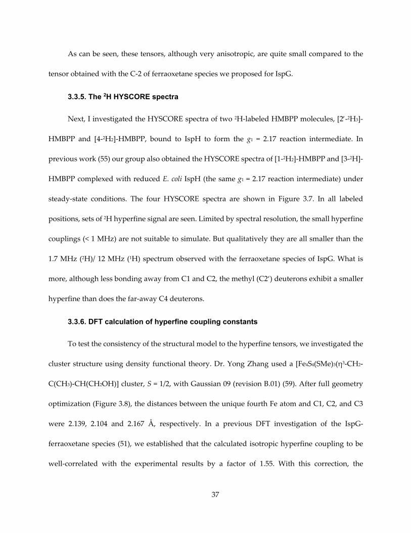
3.3.5. The 2H HYSCORE spectra
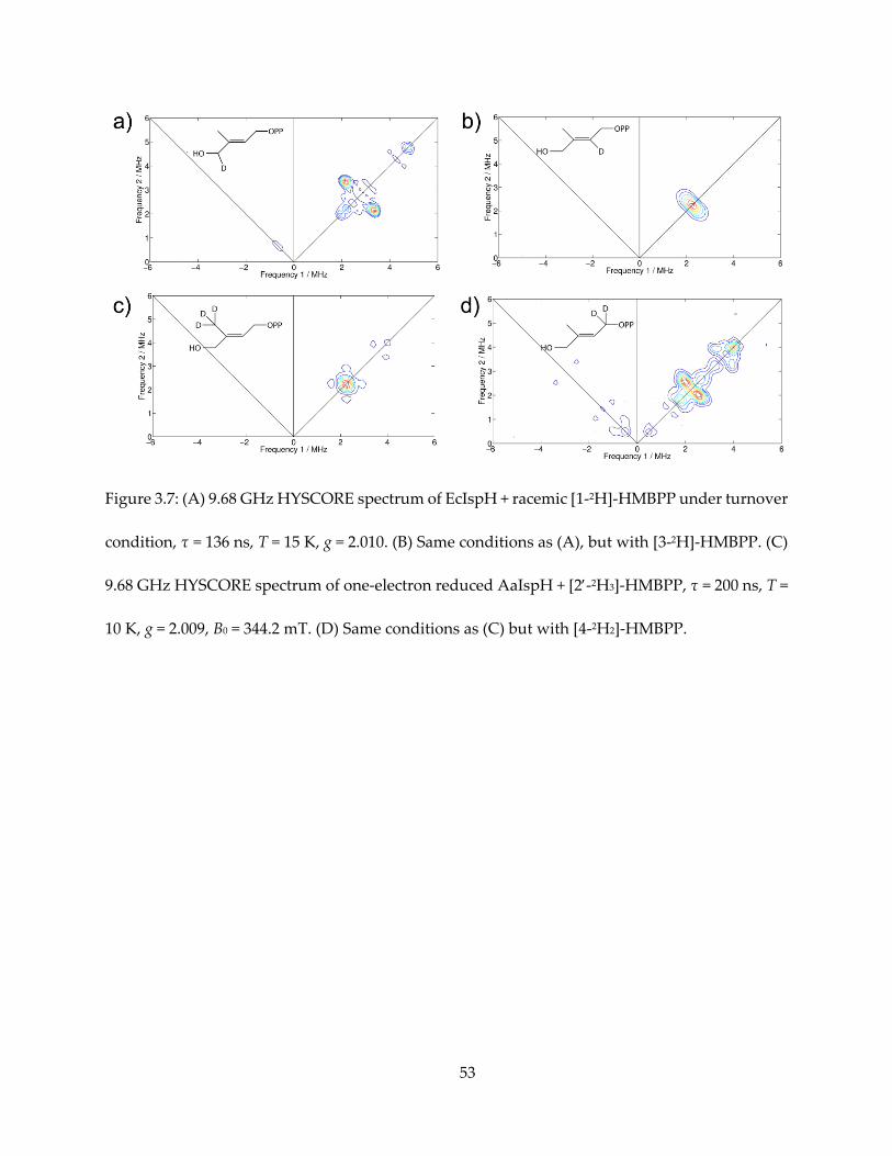
Next, I investigated the HYSCORE spectra of two 2H-labeled HMBPP molecules, [2-2H3]-
HMBPP and [4-2H2]-HMBPP, bound to IspH to form the g1 = 2.17 reaction intermediate. In
previous work (55) our group also obtained the HYSCORE spectra of [1-2H2]-HMBPP and [3-2H]-
HMBPP complexed with reduced E. coli IspH (the same g1 = 2.17 reaction intermediate) under
steady-state conditions. The four HYSCORE spectra are shown in Figure 3.7. In all labeled
positions, sets of 2H hyperfine signal are seen. Limited by spectral resolution, the small hyperfine
couplings (< 1 MHz) are not suitable to simulate. But qualitatively they are all smaller than the
1.7 MHz (2H)/ 12 MHz (1H) spectrum observed with the ferraoxetane species of IspG. What is
more, although less bonding away from C1 and C2, the methyl (C2‘) deuterons exhibit a smaller
hyperfine than does the far-away C4 deuterons.
3.3.6. DFT calculation of hyperfine coupling constants
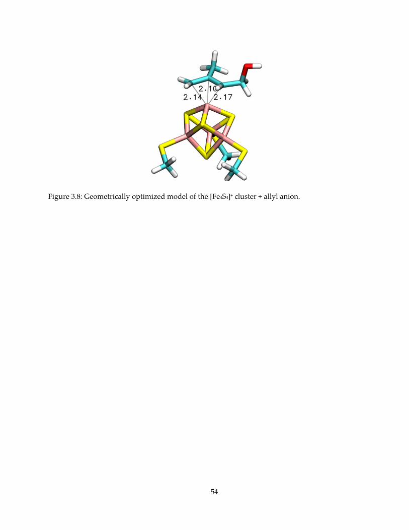
To test the consistency of the structural model to the hyperfine tensors, we investigated the
cluster structure using density functional theory. Dr. Yong Zhang used a [Fe4S4(SMe)3(η3-CH2-
C(CH3)-CH(CH2OH)] cluster, S = 1/2, with Gaussian 09 (revision B.01) (59). After full geometry
optimization (Figure 3.8), the distances between the unique fourth Fe atom and C1, C2, and C3
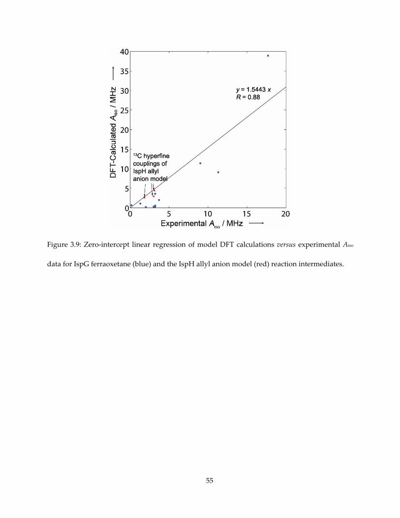
were 2.139, 2.104 and 2.167 Å, respectively. In a previous DFT investigation of the IspG-
ferraoxetane species (51), we established that the calculated isotropic hyperfine coupling to be
well-correlated with the experimental results by a factor of 1.55. With this correction, the
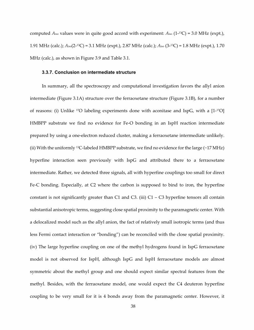
38
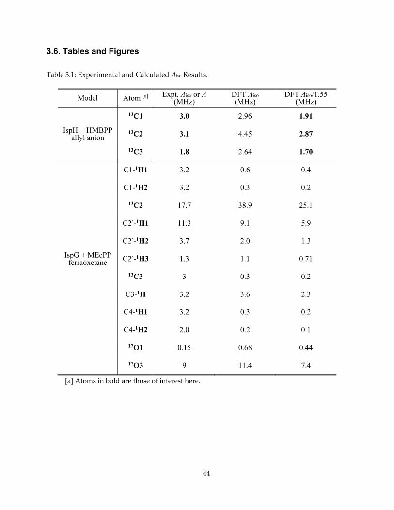
computed Aiso values were in quite good accord with experiment: Aiso (1-13C) = 3.0 MHz (expt.),
1.91 MHz (calc.); Aiso(2-13C) = 3.1 MHz (expt.), 2.87 MHz (calc.); Aiso (3-13C) = 1.8 MHz (expt.), 1.70
MHz (calc.), as shown in Figure 3.9 and Table 3.1.
3.3.7. Conclusion on intermediate structure
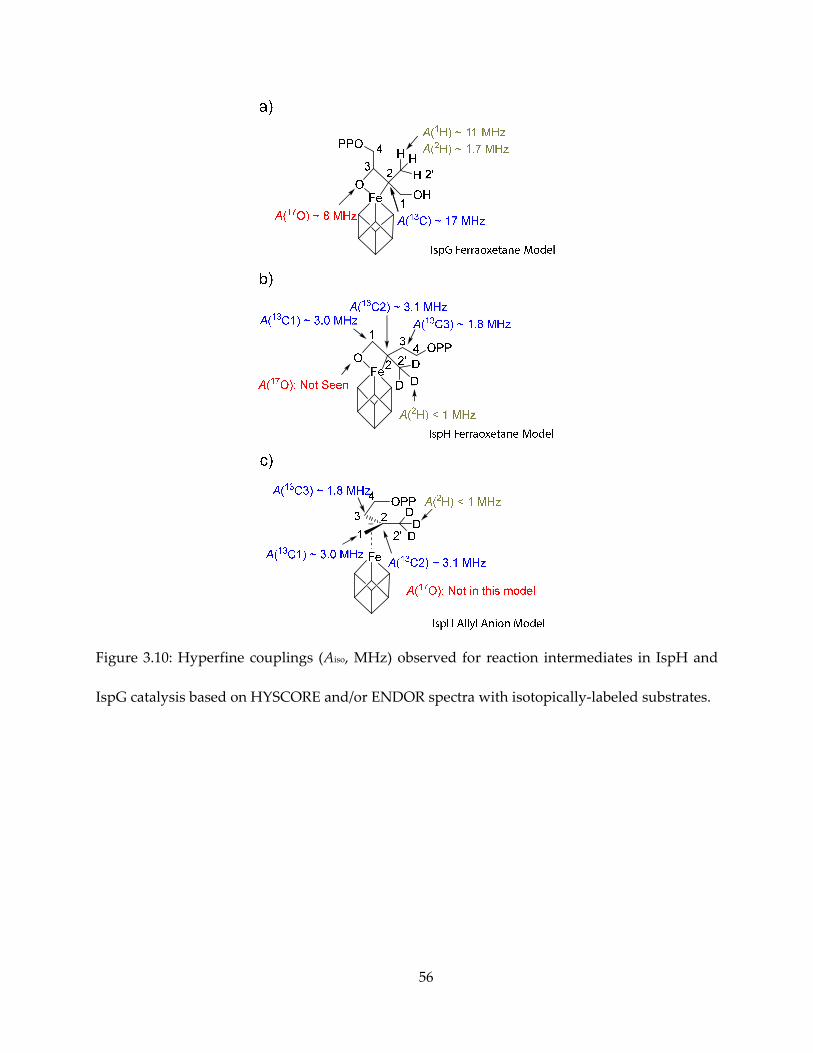
In summary, all the spectroscopy and computational investigation favors the allyl anion
intermediate (Figure 3.1A) structure over the ferraoxetane structure (Figure 3.1B), for a number
of reasons: (i) Unlike 17O labeling experiments done with aconitase and IspG, with a [1-17O]
HMBPP substrate we find no evidence for Fe-O bonding in an IspH reaction intermediate
prepared by using a one-electron reduced cluster, making a ferraoxetane intermediate unlikely.
(ii) With the uniformly 13C-labeled HMBPP substrate, we find no evidence for the large (~17 MHz)
hyperfine interaction seen previously with IspG and attributed there to a ferraoxetane
intermediate. Rather, we detected three signals, all with hyperfine couplings too small for direct
Fe-C bonding. Especially, at C2 where the carbon is supposed to bind to iron, the hyperfine
constant is not significantly greater than C1 and C3. (iii) C1 – C3 hyperfine tensors all contain
substantial anisotropic terms, suggesting close spatial proximity to the paramagnetic center. With
a delocalized model such as the allyl anion, the fact of relatively small isotropic terms (and thus
less Fermi contact interaction or “bonding”) can be reconciled with the close spatial proximity.
(iv) The large hyperfine coupling on one of the methyl hydrogens found in IspG ferraoxetane
model is not observed for IspH, although IspG and IspH ferraoxetane models are almost
symmetric about the methyl group and one should expect similar spectral features from the
methyl. Besides, with the ferraoxetane model, one would expect the C4 deuteron hyperfine
coupling to be very small for it is 4 bonds away from the paramagnetic center. However, it
39
showed a hyperfine constant comparable to that of deuterons on C1. (v) The allyl anion model is
consistent with the formation of both products IPP and DMAPP, because it could be protonated
at C1 or C3 to form the 1,2- or 2,3- double bond., while for the ferraoxetane model it is difficult to
account for the formation of DMAPP. For a clearer picture, the spectroscopic features are drawn
and compared in Figure 3.10.
3.3.8. Comparison to X-ray crystallographic results
Spectroscopic data aside, X-ray crystallography studies provide direct and valuable
evidence for mechanisms of reaction. However, caveats remains for interpreting crystal
structures because crystalizing conditions may capture the enzyme-ligand complex in a different
redox state or different position on the reaction coordinate. Our collaborator Prof. M. Groll’s
group have crystalized and solved structures of multiple wild-type/mutant IspH-ligand
complexes (60,61). The question then arises as to whether the g1=2.17 species corresponds to the
intermediate observed crystallographically.
While crystal structures give bond lengths and atomic distances directly, hyperfine
anisotropy provides an estimate of spatial distance between the paramagnetic center and the I ≥
½ nucleus. A convenient approximation is to crudely treat the unpaired electron and the
paramagnetic nuclei both as point magnets, and thus the average distance between them can be
calculated from the anisotropy part of the hyperfine tensor. In addition, Walsby et al (62)
improved this method by taking the non-unity spin density at the iron centers into account using
a spin projection coefficient. Using the spin projection coefficient calculated from aconitases 57Fe
hyperfine measurements (63), I estimated that the C-Fe distances range from 2.0 ± 0.2 to 2.5 ± 0.2
40
Å for C1-C3. This distance estimate is consistent with the optimized DFT model geometry, but
shorter than the 2.8 ± 0.3 Å observed in crystal structures (60).
It was observed by crystallography that the HMBPP alkoxide can bind to the unique fourth
Fe (PDB 3KE8), but some crystals undergo X-ray radiation for diffraction quality examination
before data acquirement at synchrotron lost the oxygen density, leaving a planar IPP-like species
in place (PDB 3KE9). In addition, a structure was obtained with the IspH E126Q mutant with
HMBPP hydroxyl rotated away from the cluster (although the fourth Fe was lost) and pointed
towards the mutated E126Q. Therefore it would be conceivable that the C1-C2 bond rotation
could have taken place along the reaction coordinate.
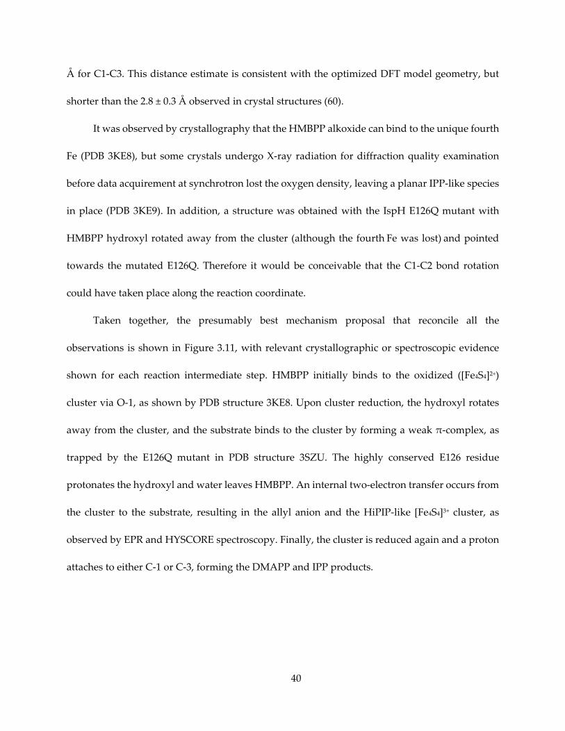
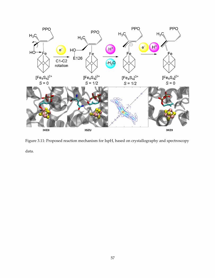
Taken together, the presumably best mechanism proposal that reconcile all the
observations is shown in Figure 3.11, with relevant crystallographic or spectroscopic evidence
shown for each reaction intermediate step. HMBPP initially binds to the oxidized ([Fe4S4]2+)
cluster via O-1, as shown by PDB structure 3KE8. Upon cluster reduction, the hydroxyl rotates
away from the cluster, and the substrate binds to the cluster by forming a weak π-complex, as
trapped by the E126Q mutant in PDB structure 3SZU. The highly conserved E126 residue
protonates the hydroxyl and water leaves HMBPP. An internal two-electron transfer occurs from
the cluster to the substrate, resulting in the allyl anion and the HiPIP-like [Fe4S4]3+ cluster, as
observed by EPR and HYSCORE spectroscopy. Finally, the cluster is reduced again and a proton
attaches to either C-1 or C-3, forming the DMAPP and IPP products.
41
3.4. Conclusions
With one-electron reduced IspH and fast freeze-trapping, we are able to capture the
paramagnetic intermediate and elucidate its chemical nature. Thanks to Dr. Ke Wang’s isotope-
labeled substrates, assignment of signal was straightforward. The results strongly supports, with
the corroboration of crystallography data, a route of reaction via a weak π-complex converted to
a stronger allyl-anion-cluster complex. The result here also lends insight to inhibitor design, as
will be discussed in Chapter 4.
3.5. Materials and Methods
3.5.1. Expression, purification and reconstitution of Aquifex aeolicus IspH
A. aeolicus IspH was transformed into BL-21(DE3) E. coli cells with a pQE-60 plasmid. The
cells are grown in LB media with 200 mg/L ampicillin at 37 °C until the A600 reached ~ 0.6. Cells
were then induced with 200 μg/L anhydrotetracycline and grown at 20 °C for 16 h and harvested
by centrifugation and frozen at -80 °C.
The purification process is similar to that of T. thermophilus IspG. However, because AaIspH
does not incorporate iron-sulfur clusters particularly well, even when purified in a glove box,
cluster reconstitution was performed. Protein was transferred to a room-temperature Coy vinyl
anaerobic chamber, diluted to ~ 1 mM, and incubated with 20 mM DTT for 1 hour. Then, 0.5 mM
FeCl3 and 0.5 mM Na2S were slowly added to the protein solution with gentle stirring (~ 60 rpm),
followed by incubation for 6 – 8 hours. These additions and incubations of FeCl3 and Na2S were
repeated five times. Care was taken that iron sulfide and protein precipitation were kept to a
42
minimum. The protein solution was then centrifuged at 11,000 rpm for 20 minutes, the
supernatant concentrated, desalted again (to remove excess metal or sulfide ions), and the
purified, reconstituted protein was stored in the anaerobic chamber at 4°C.
3.5.2. Biosynthesis of [u-13C]-HMBPP
[u-13C]-HMBPP was prepared from [u-13C]-MEcPP. In a Coy anaerobic chamber under
room temperature, approximately 0.5 mM T. thermophilus GcpE, along with 5 mM sodium
dithionite and MEcPP was added into a Wilmad screw-cap NMR tube, in a 100 mM ammonium
bicarbonate buffer (pH ~ 7.8) with 50% D2O, and incubated overnight. The progress of the
reaction is monitored by 31P NMR spectroscopy. When no significant amount of MEcPP was left,
the enzyme is removed with a centrifugal concentrator (collecting the filter flow-through instead
of the retained protein solution). The HMBPP-containing solution is lyophilized, then cation-
exchanged using a DOWEX 50WX8 column pre-equilibrated with NH4+, and then lyophilized
again before further use.
3.5.3. EPR spectra simulation
EPR spectra are usually plotted and analyzed with Matlab (R2011b, The MathWorks, Inc.,
Natick, Massachusetts, United States), and simulate with the EasySpin package. CW-EPR
spectrum was simulated with the “pepper” function of EasySpin to obtain the principle values of
the g tensor, which is in turn used to define the system for simulation of pulsed spectrum with
the “saffron” function.
Because the parameter space of hyperfine and quadrupole tensors are high-dimensional, it
is unrealistic to make an exhaustive search for the best tensors. Instead, usually 2 – 3 parameters
43
are varied systematically together, with scripts parallelizing EasySpin threads, and generate
hundreds to thousands of simulated spectra. The spectra are subjectively compared to the
experimental spectrum. Parameters are varied interactively and calibrated against multiple
experimental spectra to get the best tensor fit to experimental data.
3.5.4. Distance estimate from anisotropic hyperfine coupling
To obtain an estimate of the actual distances involved, we employed the point dipole
approximation. The through-space dipolar coupling energy between the unpaired electron and a
nucleus with a magnetic dipole, and the average distance between them can be estimated by:
< >= 4 1ℎ ℎ ℎ ℎ /
where h is the Planck constant, γe/h and γN/h are the gyromagnetic ratios (in MHz/Tesla) of the
electron and the nucleus in question, and <r> is the average distance to be estimated. We assumed
the largest component of the experimental anisotropic hyperfine coupling T (= A – Aiso) (in
frequency units) tensor to be 2T (in an ideal situation, T will be in the form of [2T, -T, -T], with the
interaction purely between two point dipoles). K is the spin-projection coefficient used to account
for non-unity spin density on the relevant iron, which has been reported to range from 0.78 to
1.86 in the 4Fe-4S cluster protein aconitase, resulting in the lower and upper limits of distance
estimates.
44
3.6. Tables and Figures
Table 3.1: Experimental and Calculated Aiso Results.
Model Atom [a] Expt. Aiso or A (MHz)
DFT Aiso (MHz)
DFT Aiso/1.55 (MHz)
13C1 3.0 2.96 1.91
IspH + HMBPP allyl anion
13C2 3.1 4.45 2.87
13C3 1.8 2.64 1.70
C1-1H1 3.2 0.6 0.4
C1-1H2 3.2 0.3 0.2
13C2 17.7 38.9 25.1
C2-1H1 11.3 9.1 5.9
C2-1H2 3.7 2.0 1.3
IspG + MEcPP ferraoxetane C2-1H3 1.3 1.1 0.71
13C3 3 0.3 0.2
C3-1H 3.2 3.6 2.3
C4-1H1 3.2 0.3 0.2
C4-1H2 2.0 0.2 0.1
17O1 0.15 0.68 0.44
17O3 9 11.4 7.4
[a] Atoms in bold are those of interest here.
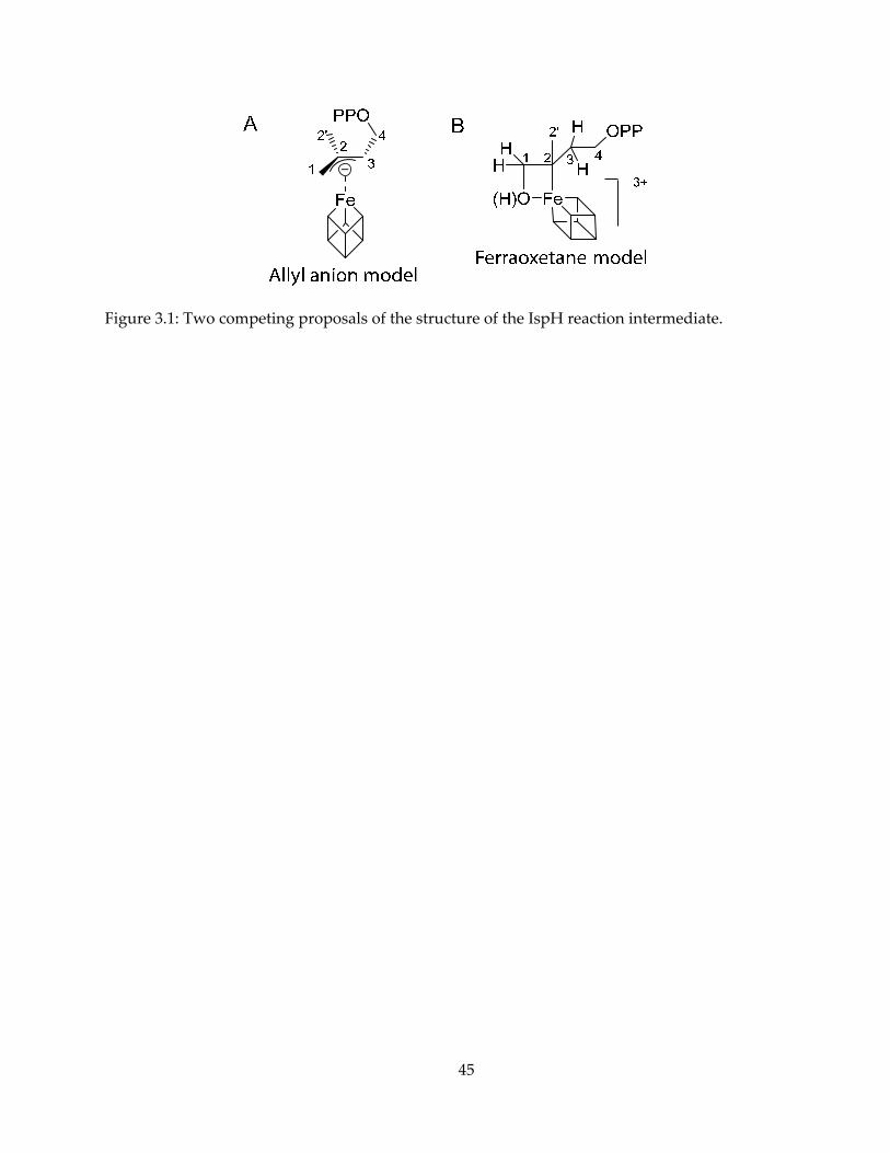
45
Figure 3.1: Two competing proposals of the structure of the IspH reaction intermediate.
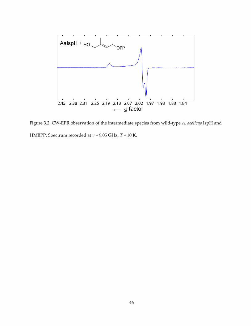
46
Figure 3.2: CW-EPR observation of the intermediate species from wild-type A. aeolicus IspH and
HMBPP. Spectrum recorded at ν = 9.05 GHz, T = 10 K.
47
Figure 3.3: HYSCORE spectrum of one-electron reduced AaIspH + [1-17O]-HMBPP, sum of
spectra with = 128, 182 and 236 ns, B0 = 344.4 mT, T = 10 K. Showing no 17O signal present.
48
Figure 3.4: X-band HYSCORE spectra of one-electron reduced AaIspH + 13C-labeled HMBPPs. (A)
[u-13C]-labeled HMBPP, sum spectrum of = 128, 150, 166, 182, 196 and 208 ns, B0 = 344.7 mT, ν
= 9.68 GHz. (B) – (D) Specifically-labeled HMBPP. Spectra are sum with = 136, 160 and 196 ns,
B0 = 344.1 mT, ν = 9.69 GHz. All spectra were recorded at T = 10 K.
49
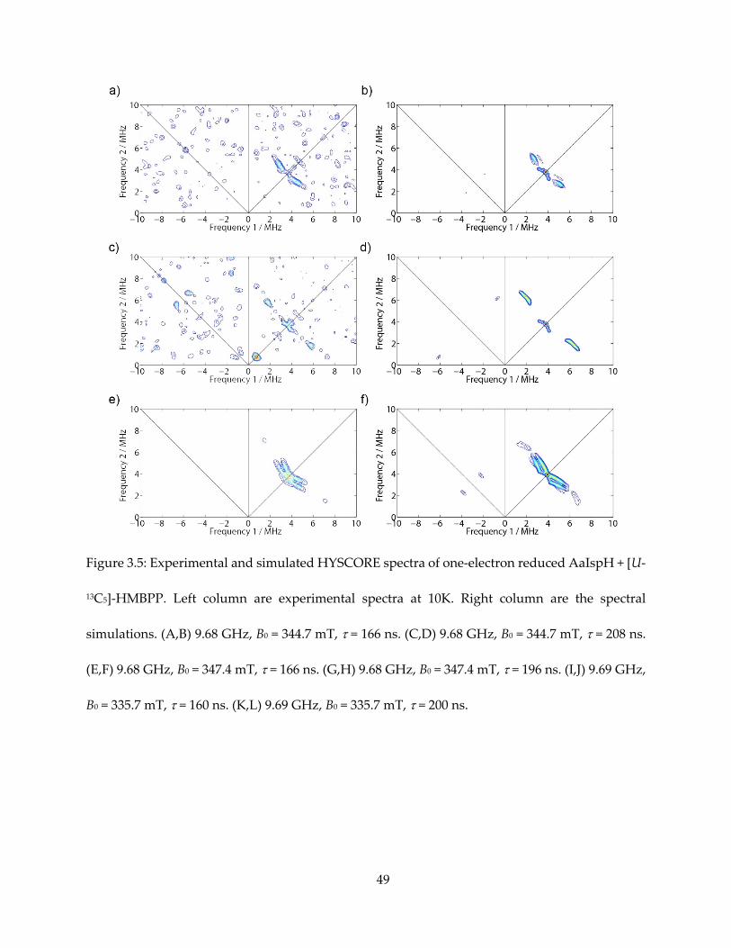
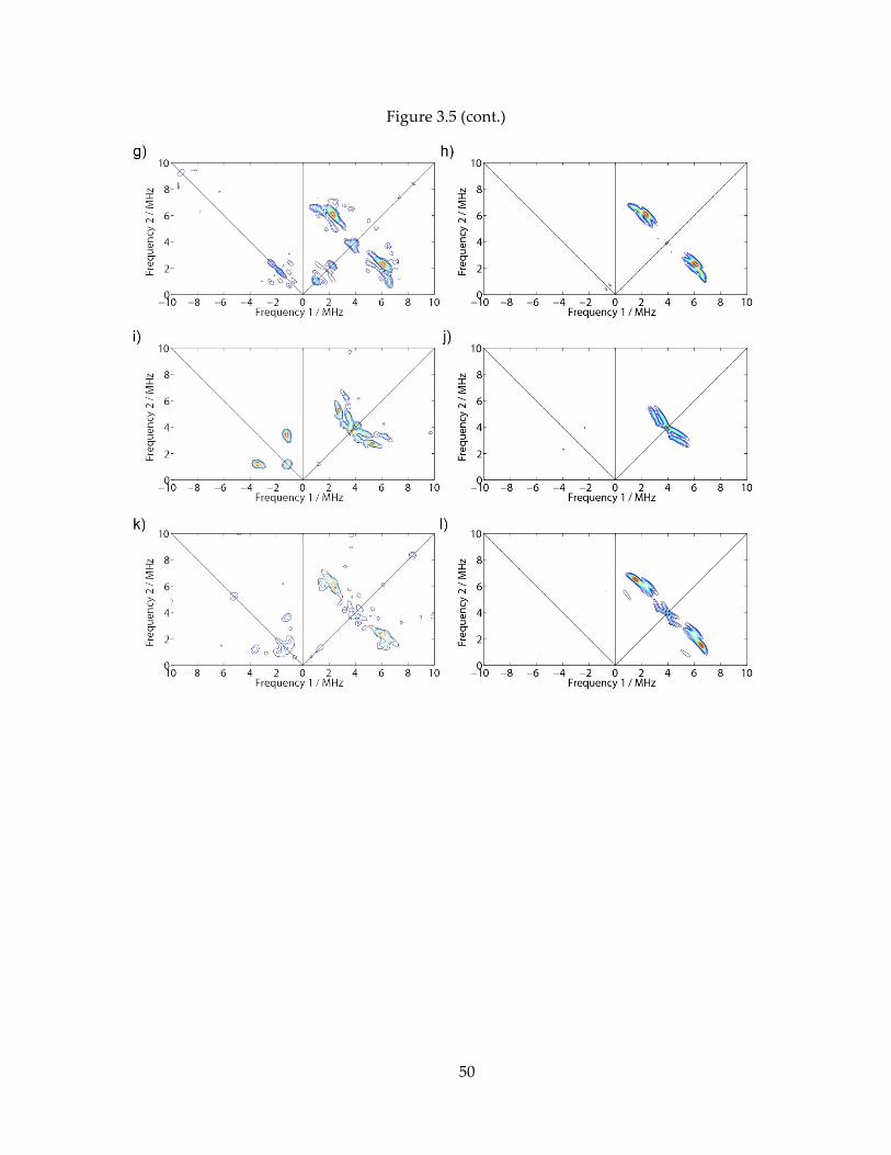
Figure 3.5: Experimental and simulated HYSCORE spectra of one-electron reduced AaIspH + [U-
13C5]-HMBPP. Left column are experimental spectra at 10K. Right column are the spectral
simulations. (A,B) 9.68 GHz, B0 = 344.7 mT, = 166 ns. (C,D) 9.68 GHz, B0 = 344.7 mT, = 208 ns.
(E,F) 9.68 GHz, B0 = 347.4 mT, = 166 ns. (G,H) 9.68 GHz, B0 = 347.4 mT, = 196 ns. (I,J) 9.69 GHz,
B0 = 335.7 mT, = 160 ns. (K,L) 9.69 GHz, B0 = 335.7 mT, = 200 ns.
50
Figure 3.5 (cont.)
51
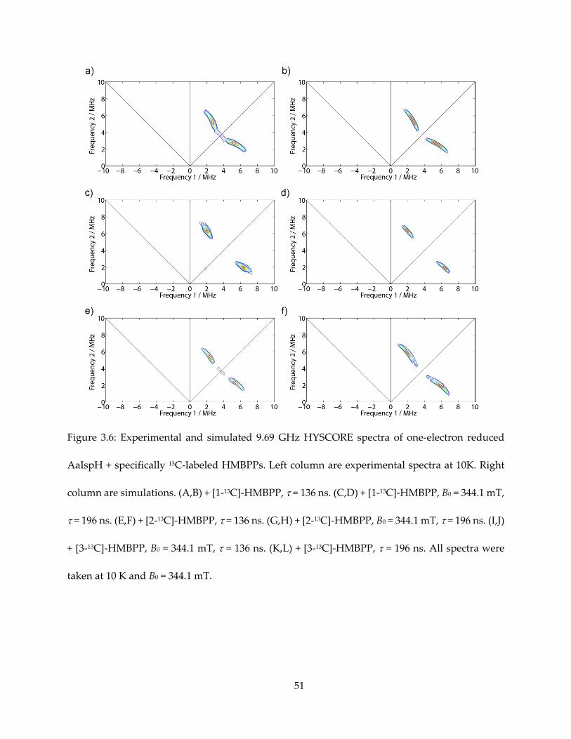
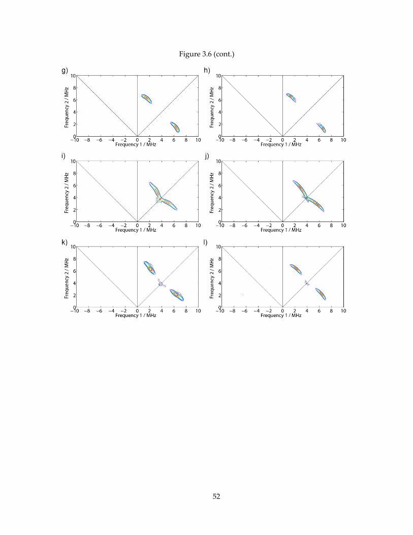
Figure 3.6: Experimental and simulated 9.69 GHz HYSCORE spectra of one-electron reduced
AaIspH + specifically 13C-labeled HMBPPs. Left column are experimental spectra at 10K. Right
column are simulations. (A,B) + [1-13C]-HMBPP, = 136 ns. (C,D) + [1-13C]-HMBPP, B0 = 344.1 mT,
= 196 ns. (E,F) + [2-13C]-HMBPP, = 136 ns. (G,H) + [2-13C]-HMBPP, B0 = 344.1 mT, = 196 ns. (I,J)
+ [3-13C]-HMBPP, B0 = 344.1 mT, = 136 ns. (K,L) + [3-13C]-HMBPP, = 196 ns. All spectra were
taken at 10 K and B0 = 344.1 mT.
52
Figure 3.6 (cont.)
53
Figure 3.7: (A) 9.68 GHz HYSCORE spectrum of EcIspH + racemic [1-2H]-HMBPP under turnover
condition, τ = 136 ns, T = 15 K, g = 2.010. (B) Same conditions as (A), but with [3-2H]-HMBPP. (C)
9.68 GHz HYSCORE spectrum of one-electron reduced AaIspH + [2-2H3]-HMBPP, τ = 200 ns, T =
10 K, g = 2.009, B0 = 344.2 mT. (D) Same conditions as (C) but with [4-2H2]-HMBPP.
54
Figure 3.8: Geometrically optimized model of the [Fe4S4]+ cluster + allyl anion.
55
Figure 3.9: Zero-intercept linear regression of model DFT calculations versus experimental Aiso
data for IspG ferraoxetane (blue) and the IspH allyl anion model (red) reaction intermediates.
56
Figure 3.10: Hyperfine couplings (Aiso, MHz) observed for reaction intermediates in IspH and
IspG catalysis based on HYSCORE and/or ENDOR spectra with isotopically-labeled substrates.
57
Figure 3.11: Proposed reaction mechanism for IspH, based on crystallography and spectroscopy
data.
58
Chapter 4. Diverse Inhibitor Binding Modes of IspH
4.1. Notes and Acknowledgements
This chapter is partly adapted, with permission, from the following publications: Wang, W.,
Li, J., Wang, K., Smirnova, T.I., Oldfield, E., 2011. Pyridine Inhibitor Binding to the 4Fe-4S Protein
A. aeolicus IspH (LytB): A HYSCORE Investigation. J. Am. Chem. Soc. 133, 6525–6528. Copyright
2011, American Chemical Society; and Guerra, F., Wang, K., Li, J., Wang, W., Liu, Y.-L., Amin, S.,
Oldfield, E., 2014. Inhibition of the 4Fe–4S proteins IspG and IspH: an EPR, ENDOR and
HYSCORE investigation. Chem. Sci. 5, 1642–1649. Copyright 2014, Royal Society of Chemistry.
Dr. Weixue Wang and Francisco Guerra performed a large part of the CW/Pulsed EPR
spectroscopy. Dr. Ke Wang synthesized most of the inhibitors. Prof. Michael Groll and Dr. Ingrid
Span performed crystallography.
This work was supported by NIH grant (GM065307) to Eric Oldfield, I was supported by a
pre-doctoral fellowship from the American Heart Association, Midwest Affiliate (11PRE7500042)
from July 2011 to June 2013. I also thank Drs. Hassan Jomaa and Jochen Wiesner for providing
the A. aeolicus IspH expression system, Dr. Deb Berthold (Rienstra Lab) for guidance on 15N-
labeled protein expression, and Dr. Mark J. Nilges for assistance with the EPR spectroscopy.
NCSA/Terigrid provided computational resources for the pyridine inhibitor study.
4.2. Introduction
The MEP pathway is present in most bacteria as well as in the protozoan parasite,
Plasmodium falciparum, the causative agent of the most common and serious form of malaria (3,64).
59
This pathway is essential for survival for many pathogens, while not present in humans. In
addition, the MEP pathway is present in plants, making inhibitors of interest as potential
herbicides. The discovery of organometallic species as reaction intermediates, as discussed in the
previous chapters, facilitates the design of new inhibitors. Many moderately potent inhibitors (~
μM level) of IspH have been discovered, with some of them potent against IspG as well. In
general, three classes of inhibitors have been reported: alkyne pyrophosphates, pyridine
pyrophosphates and HMBPP analogs. Spectroscopy, crystallography and computational
chemistry all facilitated the understanding of the binding of these inhibitors to IspH. In this
chapter, I will focus the discussion on the binding of pyridine and HMBPP analogs to IspH.
4.3. Results and Discussion
4.3.1. π/σ-complexes in IspH mechanism and inhibition
Synthetic 4Fe-4S clusters have been shown to form complexes with alkynes and even
aromatic species (65–67). The formation of η2- or η3- alkenyl species, or π/σ metallacycle by IspG
and IspH has been noticed in the context of both reaction mechanism and inhibition (68,54).
HMBPP in complex with the IspH E126 mutant is characterized by weak π-interaction based on
the HYSCORE/ENDOR observations of small 17O hyperfine coupling (55) and moderate (A ~ 1-2
MHz) 13C hyperfine coupling (Figure 4.1).
Knowledge from the weak and strong π/σ-complexes discussed in the IspH reaction
mechanism gave inspiration for rational design for inhibitors that form such complexes. It has
been confirmed that alkyne species, such as propiolic acid and propargyl pyrophosphate (69),
60
also bind to IspH, causing significant spectroscopic change as evidenced by EPR (Figure 4.2).
Propargyl pyrophosphate has been found as an IspH inhibitor, with Ki = 0.97 μM. Upon binding,
it is typical that the alkyne species cause the EPR spectrum to become HiPIP-like, similar as the
binding of the HMBPP substrate. Propargyl pyrophosphate is also found to be a potent IspG
inhibitor, with Ki ~ 330 nM.
The discovery of alkyne inhibitors has been useful for the design and understanding of
other species as IspH inhibitors.
4.3.2. Pyridine inhibitors: spectroscopic observations
With a screening of a group of compounds previously available in the Oldfield Lab,
pyridine bisphosphonates are found to be mild inhibitors of IspH activity. Considering that the
branched nature of the bisphosphonate group might be a hindrance for binding, the synthetic
chemists in our lab prepared analogs containing pyrophosphate groups (shown in Figure 4.3).
Better inhibitors were discovered, with the most potent two having IC50s of 38 μM and 9.1 μM,
labeled 1 and 2 in Figure 4.3.
Both inhibitors are pyridines substituted at the meta-position. Adding one CH2 spacer to
the simplest molecule 1 gave the better inhibition activity of compound 2. However, further
addition to the length of side chain, or making the substitution at different positions of the
pyridine ring, caused diminishing inhibition activity. Chlorination on the pyridine ring also gave
much weaker inhibition.
We thus sought to understand the binding modes of the pyridine inhibitors to the IspH
protein. Initially, it was conceived that given the essentiality of E126 residue in IspH catalysis, 1
and 2, while anchored by the pyrophosphate group, could be protonated and the pyridinium
61
forming a salt bridge to E126 carboxylate (Figure 4.4C), simply blocking the substrate from access
to the iron-sulfur cluster instead of having a direct interaction with it, based on computational
docking results. However, is this binding mode true, and can direct evidence from spectroscopy
support it? We next conducted a series of experiments and computational studies to answer the
question.
We first investigated a series of pyridine ligands binding to wild-type IspH from A. aeolicus.
The results are shown in Figures 4.5 (for 1) and 4.6 (for others). It appeared that intense 14N
HYSCORE signal is positively correlated with inhibition. On addition of the inhibitor 1, the EPR
spectrum changes (Figure 4.6A) and new signals attributable to 14N single- and double-quantum
transitions appear in the (+, −) quadrant of the HYSCORE spectrum (Figure 4.5A). By contrast,
there was no evidence for any sizable pyridine-14N hyperfine interaction in the HYSCORE
spectrum (Figure 4.6B), indicating only very weak binding affinity to IspH. The same results are
obtained with the more basic (pKa = 6.8 vs 5.2) species 2-aminopyridine (Figure 4.6C).
The ortho- and para-pyridyl analogues of 1 (IC50 = 1.2 mM and 149 μM) show no evidence
of any sizable pyridine-14N hyperfine interaction in their HYSCORE spectra (Figure 4.6D, E), due
presumably to their inability to bind to the fourth Fe, for “steric” reasons. Moreover, chlorine
substitution of 1 results in loss of all activity (IC50 > 3 mM), due presumably to loss in donor ability
of the pyridine nitrogen (the computed pKa values of the pyridine fragments in 1 and 6-chloro-1
are 4.7 and 0.7, respectively), consistent with the absence of a pyridine-14N HYSCORE signal
(Figure 4.6F). Addition of one CH2 group to the side chain of 1 results in a better inhibitor (2, IC50
= 9.1 μM), although there is no significant difference between the HYSCORE spectra of 1 (Figure
4.5A) and 2 (Figure 4.6G), indicating that differences in enzyme inhibition are due to differences
62
in the alkyl diphosphate fragment binding in the active site, rather than differences in Fe-pyridine
interactions.
The 14N hyperfine interaction is quite large, with the hyperfine coupling constant being 8
MHz. Simulation of the HYSCORE spectrum yielded Aiso(14N) = 7.4 MHz, Aii(14N) = [6.2, 7.6, 8.4]
MHz for the hyperfine interaction, and e2qQ/h = 3.0 MHz for the nuclear quadrupole coupling
constant.
These results do not, however, definitively prove that the 14N HYSCORE signals in the (+,
−) quadrant arise directly from the inhibitors 1 and 2 because, in principle, inhibitor binding might
result in a protein conformational change and binding of a protein ligand to Fe, e.g., the nearby
His 42 or 124, which form part of the active site (18). To investigate this possibility, we prepared
a sample using uniformly 15N-labeled IspH and inhibitor 1. As can be seen in Figure 4.5B, the 14N
signals centered at 3.6 MHz seen in Figure 4.5A are no longer present and are replaced by a
signal centered at 1.5 MHz, the 15N Larmor frequency. Moreover, the 14N signals in the (+, −)
quadrant are essentially identical to those seen in samples prepared using unlabeled IspH. This
strongly suggests that the signals centered at 3.6 MHz arise from protein 14N near the 4Fe-4S
cluster, while the 14N signals in the (+, −) quadrant arise from the bound inhibitor 1, rather than
from any protein residues.
4.3.3. Pyridine inhibitors: computational investigation for binding modes
This large Aiso(14N) is similar to, or even larger than, those of a number of systems in which
nitrogens directly bind to Fe centers, for example, in met-myoglobin the porphyrin nitrogens (70),
in the Rieske-type 2Fe-2S proteins (71), and in the case of the 4Fe-4S enzyme MoaA (a SAM-
dependent enzyme which also has an unique fourth iron) (72). On average, these previous results
63
gave an Aiso(14N) ~ 6 MHz for systems containing Fe−N bonds, suggesting that the IspH + 1
complex also contains an Fe−N bond.
The large 14N hyperfine interaction seen in the IspH + 7 complex might also, at least in
principle, indicate that the pyridine fragment is just close to the reduced 4Fe-4S cluster, without
directly bonding to the fourth iron. For example, the pyridine group might be protonated and
interact with the E126 carboxylate group that is close to the cluster, or it could be close by but
deprotonated. Fortunately, determination of the 14N nuclear quadrupole coupling constant
(e2qQ/h) enables an answer to this question, since protonated, neutral, and metal-coordinated
pyridine ligands have very different e2qQ/h values (73).
For pyridine itself, e2qQ/h = 4.6 MHz, but in species in which there is a formal 1+ charge on
N, such as the pyridinium ion, pyridine-N-oxide, and N-methylpyridinium, e2qQ/h values of
approximately 1 MHz are observed experimentally. For pyridine complexes with N-metal bonds,
in the case pyridine bonded to Fe in Fe(CO)4(pyr), e2qQ/h is in between these extreme values
(e2qQ/h ≈ 2.4 MHz), and for Mo(pyr)2(CO)4 and Cr(CO)4(2,2′-bipyridyl), e2qQ/h ≈ 3.1 MHz. So,
when pyridine N is bonded to Cr, Mo, or Fe, the e2qQ/h decreases from the 4.6 MHz seen in free
pyridine to 2.4 − 3.1 MHz, due to metal-ligand bonding, close to the 3.0 MHz value we find from
the 14N HYSCORE results.
To see to what extent these e2qQ/h values might be reproduced computationally, we used
the Gaussian 09 program to calculate the 14N nuclear quadrupole coupling constant (NQCC)
values for a series of pyridine, imidazole or triazole complexes, as shown in Figure 4.7. Results
are shown in Table 3.1, with 4 different functional/basis sets combinations attempted. All four
model chemistries gave reasonably well correlation between theory and experiment (Figure 4.8).
64
We also performed the same set of NQCC calculation for a model of IspH + 1, a simple
[Fe4S4(SMe)3(η1-pyr)] compound (Figure 4.9). A full geometrical optimization yielded the head-
on η1 complex with N-Fe distance of 1.95 Å. NQCC calculation finds e2qQ/h = 2.3 for the pyridine
14N, in quite good accord with experiment.
This large decrease in e2qQ/h, from the 4.6 MHz value found for free pyridine to the 2.4 - 3.1
MHz values observed in model systems and the IspH + 1 complex, is also seen in proteins in
which imidazole (histidine) ligands bind to Fe. For example, for imidazole, the N3 (deprotonated)
e2qQ/h is 4.032 MHz, in good accord with the 3.894 MHz computed using DFT. The e2qQ/h values
for solid imidazole and solid histidine are both smaller and essentially identical (3.27 MHz for
imidazole; 3.36 MHz for histidine), due presumably to very strong hydrogen bonding in the solid
state. But when imidazole and histidine are bound to Fe in metalloproteins, e2qQ/h decreases
considerably from the 4 MHz gas-phase value (for imidazole).
These results all support the idea that the IspH pyridine inhibitors bind to IspH via a Lewis
acid/base (Fe4S4 cluster/ligand) mechanism, with the donor orbital occupancy (σ) decreasing from
2 (pyridine) to 1.73 (73), and that the hyperfine coupling seen experimentally is due to this η1-
bonding, rather than being due to a neutral pyridine or pyridinium ligand just being close to the
4Fe-4S cluster. This, in turn, suggests that stronger Lewis bases (such as imidazole-containing
ligands) may be more potent IspH inhibitors. These results also support the idea that other
inhibitors, such as alkynes, as well as reaction intermediates (η3-allyls), also act as Lewis bases
when interacting with the 4Fe-4S cluster in IspH.
65
4.3.4. Amine and thiol analogs of HMBPP
More recently, the amine (3) and thiol (4) analogs of HMBPP have been reported (74) to be
even more potent IspH inhibitors (IC50 ~ 0.2 μM) and it was shown via Mössbauer spectroscopy
and quantum chemical calculations that both bound to the oxidized ([Fe4S4]2+) form of IspH,
forming Fe-N and Fe-S bonds, respectively, with the fourth Fe. We have found that, like the
acetylene inhibitors, they are also IspG inhibitors, with IC50 values ranging from 0.8 to 2.5 μM
(75). The structures of 3 and 4 are shown in Figure 4.10. Moreover, we also performed EPR and
HYSCORE investigations into the nature of the inhibition of both IspH by 3 and 4, in the presence
of reductant.
We began by investigating binding of the amine ligand 3 to A. aeolicus, Plasmodium
falciparum, and E. coli IspHs. The 9.05 GHz EPR spectrum at 10K of dithionite-reduced,
unliganded AaIspH was reported previously to be a mixture of an S = 1/2 and S = 3/2 species(15),
with the S = 1/2 species having gi = [2.041, 1.919, 1.849], Figure 4.11A. In the presence of 3, intense
and sharp signals with gi = [2.138, 2.005, 1.978] (for AaIspH) are seen (Figure 4.11A-D), suggesting
that the ligand binds at or close to the [Fe4S4]+ cluster in all three IspHs.
Two main possibility of 3 binding to IspH exists: (i) 3 binds via its NH2 group to the [Fe4S4]+
cluster in basically the same manner as proposed for binding to the oxidized cluster, forming a
Fe-N bond to the fourth Fe. (ii) 3 might bind to IspH with its aminomethyl group “rotated-out”,
away from the cluster, interacting with E126, as discussed earlier for the CH2OH group in the
HMBPP substrate, as seen crystallographically and as confirmed by 17O-HYSCORE spectroscopy
results in the previous chapter.
66
To help distinguish between these two possibilities, we acquired the HYSCORE spectra of
3 bound to AaIspH, Figure 4.12A, as well as to EcIspH, Figure 4.13. To distinguish any 14N
hyperfine interaction with the protein backbone from that with the ligand 3, we also obtained the
HYSCORE spectrum of 3 bound to 15N-labeled AaIspH, Figure 4.12B.
If there were a Fe-N bond present in the complex, we would expect a sizeable 14N hyperfine
coupling since there would be a strong metal-ligand orbital overlap facilitating spin density
transfer from the metal to the ligand. For example, with the pyridine inhibitor 1 and 2 bound to
IspH, the 14N hyperfine interaction Aiso (14N) is 7.4 MHz, and on average ~ 6 MHz for species
containing Fe-N bonds. In addition, the gi = [2.138, 2.005, 1.978] tensor principle values for EcIspH
+ 3 are essentially very close to those we find with HMBPP bound to EcIspH E126A mutants, gi =
[2.132, 2.003, 1.972]. We thus conclude that there is no direct Fe-N bond in the reduced IspH + 3
complex when the cluster is reduced, rather, 3 most likely binds to reduced IspH in basically the
same manner as does HMBPP in the “rotated-out” HMBPP X-ray structure (60). A model is built
with HMBPP ligand’s OH group is replaced by an NH3+ group, shown in Figure 4.13. As can be
seen in this (X-ray based) structural model, the ligand’s CH2NH3+ group can interact with the
E126 carboxylate, providing strong Coulombic interactions that may help account for its potent
IspH inhibition under reducing conditions.
Interestingly, there is a small feature centered close to (-1.5, 1.5) MHz in both AaIspH + 3,
and EcIspH + 3 spectra that has not been seen previously with any other ligands. It did not change
when 15N-labeled AaIspH protein was used instead of natural abundance AaIspH (Figure 4.12B),
so it does not arise from a 14N hyperfine coupling with the protein backbone. The feature can be
simulated with a 14N hyperfine interaction having Ai = [1.85, 1.25, 3.70] MHz, Aiso = 2.33 MHz,
67
e2qQ/h = 0.8 MHz, η = 0.2 (Figure 4.14). The quadrupole coupling constant is consistent with that
expected for an alkyl ammonium group, e2qQ/h ~ 0 – 1 MHz (76), and the hyperfine coupling
anisotropy suggests close proximity to the paramagnetic center, consistent again with the
“rotated-out” model.
We next investigated binding of the thiol ligand 4, to EcIspH. It is reported that the IC50
values for 3 and 4 in inhibiting EcIspH are very similar and both bind to the oxidized protein,
forming Fe-N and Fe-S bonds respectively with the fourth Fe in the cluster (74). However, the
electronic structure of the complex with 4 is spin-delocalized while that with 3 is spin-localized,
suggesting that there might be significant differences in electronic structure between 3 and 4
when bound to reduced IspH.
The EPR spectrum of AaIspH in the presence of sodium dithionite is shown in Figure 4.15A
(blue) and is characteristically broad, as is the case without the substrate. However, essentially
no signal is seen in the g ~ 2 region (or at lower field, Figure 4.15A inset) for a sample incubated
with dithionite in the presence of the thiol ligand 4 (Figure 4.15A, red), unlike the situation with
3. This suggested to us the possibility that in the presence of the thiol ligand, the cluster might
not be reduced, remaining in the S = 0 state.
This appears to be the case, as corroborated by the UV-Vis spectra shown in Figure 4.15B.
The spectrum of oxidized ([Fe4S4]2+) AaIspH (blue) shows a characteristic peak at ~ 420 nm, which
disappears on dithionite reduction (green). The spectrum of oxidized IspH + 4 (red) is similar to
that of the oxidized protein in the absence of 4; however, addition of dithionite minimally changes
the spectrum; the shoulder at ~ 420 nm is still seen with IspH + 4 + dithionite. We also find that
addition of 4 to dithionite-reduced IspH regenerates the 420 nm shoulder (superimposed on the
68
large dithionite background peak), suggesting that reduced IspH + 4 is relatively unstable,
consistent with the lack of any EPR signal for this system.
4.4. Conclusions
Overall, the results presented here exhibit a surprising variety of binding behavior of IspH
inhibitors. Acetylenes, like HMBPP, bind to the cluster side-on forming a π/σ-complex. Pyridine
derivatives 1 and 2, by contrast, form a strong Fe-N bond with a large hyperfine coupling
constant. The amide HMBPP analog, 3, while forming a Fe-N bond with the unique fourth Fe of
the [Fe4S4]2+ cluster, seems to rotate the NH2/NH3+ group away and bind to E126 via Coulombic
interactions, evidenced by the absence of large 14N hyperfine interaction. Most unexpectedly, the
binding of the thiol HMBPP analog, 4, appears to alter the redox properties of the clusters, and is
not reduced by dithionite. This diversity in spectroscopic behavior, and hence binding modes are
of obvious interest for drug design targeting isoprenoid biosynthesis.
Very recently, a crystallographic study of the IspH-pyridine inhibitor complex revealed
that the fourth iron binds to the pyridine via a N and a C atoms in a side-on η2 manner (77). With
quantum chemical calculations on model systems, the 14N isotropic hyperfine and quadrupole
coupling constants predicted are consistent with that obtained from spectroscopy. Again the
binding mode mimics the π-complex seen with the substrate/intermediate binding to IspH.
69
4.5. Materials and Methods
4.5.1. 15N-labeled A. aeolicus IspH expression and purification.
To express 15N-labeled protein, the Chapter 3 protocol for AaIspH was modified. Aside
from LB medium, M9 minimal medium is also prepared, without the 14N nitrogen source, but
supplemented with 4 g/L glucose, 1 g/L [15N2]-(NH4)2SO4 (Cambridge Isotope Labs), 1% (v/v)
MEM vitamin solution (Sigma-Aldrich), 0.5% (v/v) 15N-labeled BioExpress cell growth medium
(Cambridge Isotope Labs), 0.2 mM trisodium citrate, and 0.1 mM FeCl3. In addition, 2 – 20 μM
trace element supplements including CaCl2, MnCl2, ZnSO4, CoCl2, CuCl2, NiCl2, Na2MoO4,
Na2SeO3 and H3BO3 were added through a .22μM filter, according to the protocol provided in Ref
(78). Usually iron-sulfur proteins are also expressed in medium supplemented with L-cysteine,
but for effective incorporation of 15N into amino acids, this step is omitted.
BL-21(DE3) cells were grown at 37∘C aerobically in 1 L of LB medium first, until OD600
reached ~ 1.4. The cells are spun down with gentle centrifugation at 20 ∘C, 5000 rpm for 10
minutes. The cells are re-suspended in PBS, and transferred to M9 + supplement medium
described above, and protein expression was induced at 25 °C with anhydrotetracycline. Cells
were harvested 20 hours after. 15N-labeled AaIspH protein was then purified and reconstituted as
described in Chapter 3.
4.5.2. IspH inhibition assay
The inhibition assay was performed in a room-temperature anaerobic chamber with O2
level < 1 ppm, and all buffers and solvents were degassed with a vacuum manifold and stirred in
the anaerobic chamber for > 12 hours.
70
The assay mixture (200 μL on a 96-well plate) contains 40 nM IspH, 34 μM HMBPP, 2 mM
methylviologen and 0.3 mM sodium dithionite in a 50 mM Tris/HCl, 100 mM NaCl, pH 8.0 buffer.
The IspH enzyme was incubated with or without inhibitor. Reaction were started by addition of
the HMBPP substrates, and monitored by decrease in OD600.
4.5.3. IC50 determination
The IC50, half maximal inhibitory concentration, is defined as the concentration of inhibitor
needed for inhibiting half of the enzyme activity. For different inhibitor concentration [I], the
reaction rate for a competitive inhibitor is
= [ ] 1 + [ ] + 1
where Vmax is the reaction rate at infinite inhibitor concentration without any inhibitors, Km is the
Michaelis-Menten constant, and Ki is the association constant of the inhibitor to the enzyme. Only
the latter of these constants are related to the inhibitor, while all others are intrinsic kinetic
properties of the enzyme. Therefore, given a fixed substrate [S] in an assay, the IC50 can be
expressed as = (1 + [ ]/ )
which is proportional to Ki, which in turn is the constant that characterizes the inhibitor’s binding
efficiency to the enzyme.
For each inhibitor of interest, different concentrations (usually a serial 2x – 4x dilution is
made) of the inhibitor (from mM to nM, depending on the efficiency of the inhibitor) was
incubated with the enzyme, and the rates were plotted against inhibitor concentrations. The curve
is fitted in Origin Pro to the following dose-response curve.
71
= + −1 + exp (− (log[ ] − log ) )
Where r is determined from linear regression of first few minutes of the time trace of OD600
obtained from a spectrophotometer. Pairs of (r, log[I]) are the data points and V0, V1, p and IC50
are the parameters to be estimated with non-linear curve fitting.
4.5.4. Quantum chemical calculations of 14N NQCC
All QM calculations were performed with Gaussian 09 software. For all systems, geometry
optimization was carried out using the BPW91 functional, a Wachters+f (79,80) basis set for first-
row transition metals, 6-311+G* (81) for other atoms. The optimized structures were then used
to calculate the 14N nuclear quadrupole coupling constants using BPW91 (82,83) or B3LYP (84,85)
functionals, and the aforementioned Wachters/6-311+G* or TZVP (86) basis sets. There was small
difference between the calculated NQCC results.
The quadrupole coupling of 14N nucleus is just the product of its quadrupole and the electric
field gradient at the position of the nucleus (the nucleus is very small compared to the atom, so it
is valid to consider it as a point). The QM calculations gave electric field gradient (eq) in atomic
units; a conversion factor eQ/h = 4.8027 MHz/a.u (with the nuclear quadrupole Q = 0.02044 barn,
1 barn = 10-28 m2) for 14N was used to calculate the energy e2Qq/h value in MHz to fit
spectroscopists’ convention.
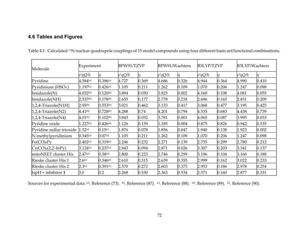
72
4.6 Tables and Figures
Table 4.1: Calculated 14N nuclear quadrupole couplings of 15 model compounds using four different basis set/functional combinations.
Molecule Experiment BPW91/TZVP BPW91/Wachters B3LYP/TZVP B3LYP/Wachters
e2qQ/h η e2qQ/h η e2qQ/h η e2qQ/h η e2qQ/h η Pyridine 4.584[a] 0.396[a] 4.727 0.369 4.686 0.326 4.944 0.364 4.990 0.410 Pyridinium (HSO4-) 1.197[a] 0.426[a] 1.105 0.211 1.262 0.109 1.070 0.206 1.247 0.098 Imidazole(N) 4.032[b] 0.120[b] 3.894 0.050 3.825 0.002 4.160 0.108 4.081 0.055 Imidazole(NH) 2.537[b] 0.178[b] 2.655 0.177 2.778 0.218 2.696 0.165 2.831 0.209 1,2,4-Triazole(N1H) 2.95[b] 0.553[b] 3.021 0.462 3.133 0.417 3.068 0.477 3.195 0.425 1,2,4-Triazole(N2) 4.41[b] 0.728[b] 4.288 0.74 4.201 0.794 4.535 0.683 4.438 0.739 1,2,4-Triazole(N4) 4.01[b] 0.102[b] 3.843 0.052 3.781 0.001 4.065 0.087 3.995 0.033 Pyridine oxide 1.227[a] 0.426[a] 1.126 0.159 1.185 0.004 0.875 0.826 0.942 0.535 Pyridine sulfur trioxide 1.52[c] 0.15[c] 1.876 0.078 1.856 0.047 1.940 0.138 1.923 0.002 N-methylpyridinium 0.545[a] 0.07[a] 1.105 0.211 1.262 0.109 1.070 0.206 1.247 0.098 Fe(CO)4Py 2.402[a] 0.319[a] 2.246 0.232 2.271 0.139 2.755 0.299 2.780 0.212 Cr(CO)4(2,2'-biPy) 3.124[a] 0.237[a] 2.843 0.094 2.871 0.026 3.307 0.203 3.341 0.137 mitoNEET cluster His 2.47[d] 0.38[d] 2.800 0.223 2.746 0.299 3.196 0.104 3.160 0.188 Rieske cluster His 1 2.6[e] 0.346[e] 2.610 0.315 2.639 0.355 2.999 0.162 3.022 0.233 Rieske cluster His 2 2.3[e] 0.391[e] 2.570 0.272 2.603 0.375 2.953 0.186 2.978 0.254 IspH + inhibitor 1 3.0 0.2 2.268 0.530 2.363 0.534 2.571 0.160 2.877 0.331
Sources for experimental data: [a]. Reference (73). [b]. Reference (87). [c]. Reference (88). [d]. Reference (89). [e]. Reference (90).
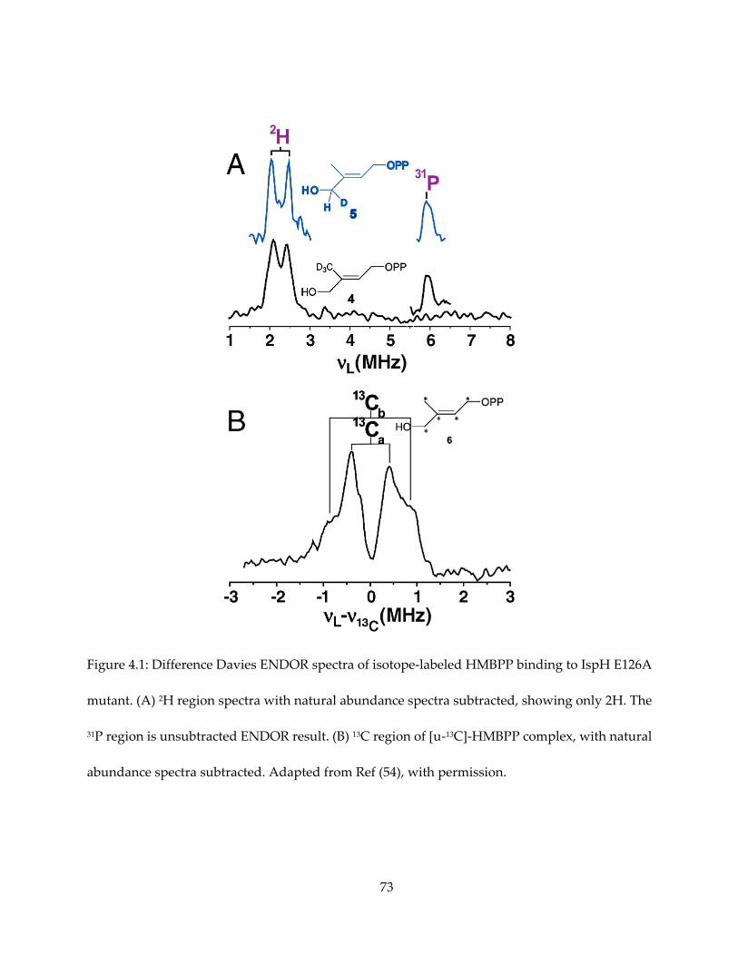
73
Figure 4.1: Difference Davies ENDOR spectra of isotope-labeled HMBPP binding to IspH E126A
mutant. (A) 2H region spectra with natural abundance spectra subtracted, showing only 2H. The
31P region is unsubtracted ENDOR result. (B) 13C region of [u-13C]-HMBPP complex, with natural
abundance spectra subtracted. Adapted from Ref (54), with permission.
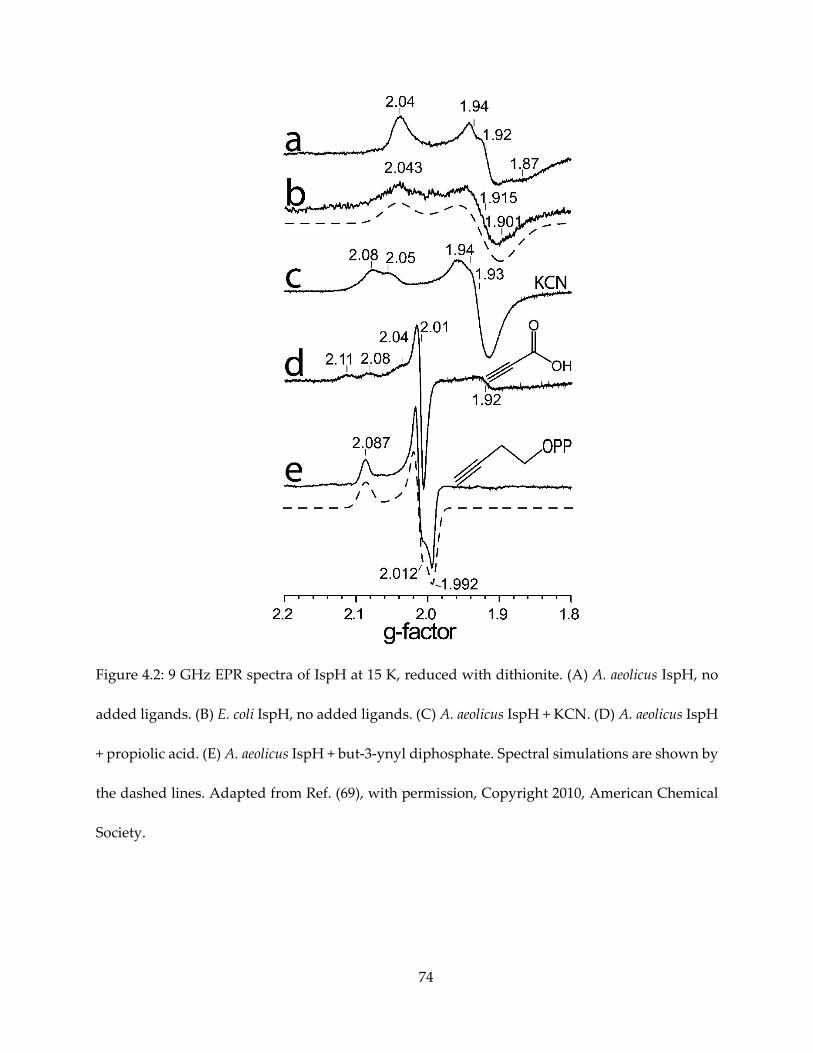
74
Figure 4.2: 9 GHz EPR spectra of IspH at 15 K, reduced with dithionite. (A) A. aeolicus IspH, no
added ligands. (B) E. coli IspH, no added ligands. (C) A. aeolicus IspH + KCN. (D) A. aeolicus IspH
+ propiolic acid. (E) A. aeolicus IspH + but-3-ynyl diphosphate. Spectral simulations are shown by
the dashed lines. Adapted from Ref. (69), with permission, Copyright 2010, American Chemical
Society.
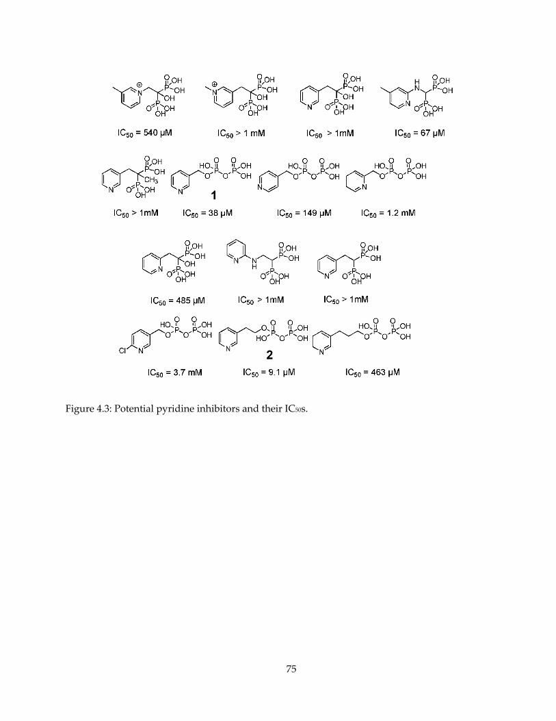
75
Figure 4.3: Potential pyridine inhibitors and their IC50s.
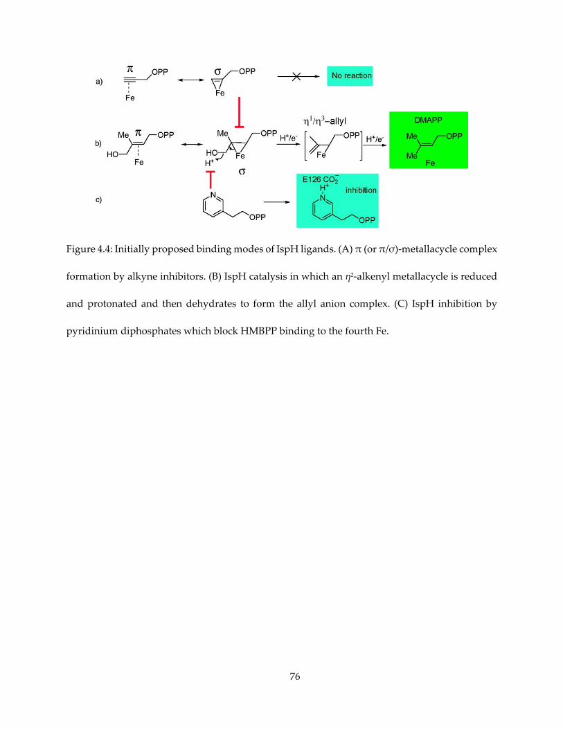
76
Figure 4.4: Initially proposed binding modes of IspH ligands. (A) π (or π/σ)-metallacycle complex
formation by alkyne inhibitors. (B) IspH catalysis in which an η2-alkenyl metallacycle is reduced
and protonated and then dehydrates to form the allyl anion complex. (C) IspH inhibition by
pyridinium diphosphates which block HMBPP binding to the fourth Fe.
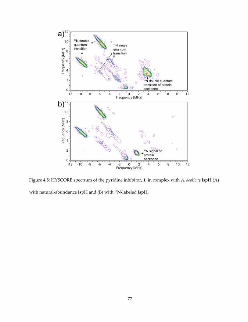
77
Figure 4.5: HYSCORE spectrum of the pyridine inhibitor, 1, in complex with A. aeolicus IspH (A)
with natural-abundance IspH and (B) with 15N-labeled IspH.
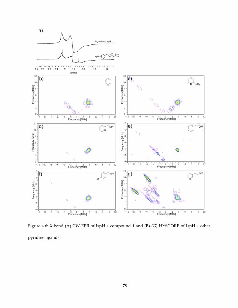
78
Figure 4.6: X-band (A) CW-EPR of IspH + compound 1 and (B)-(G) HYSCORE of IspH + other
pyridine ligands.
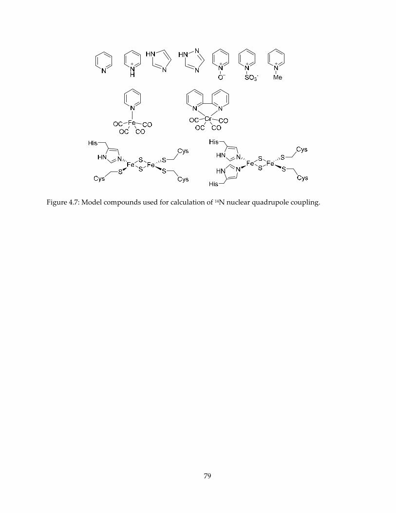
79
Figure 4.7: Model compounds used for calculation of 14N nuclear quadrupole coupling.
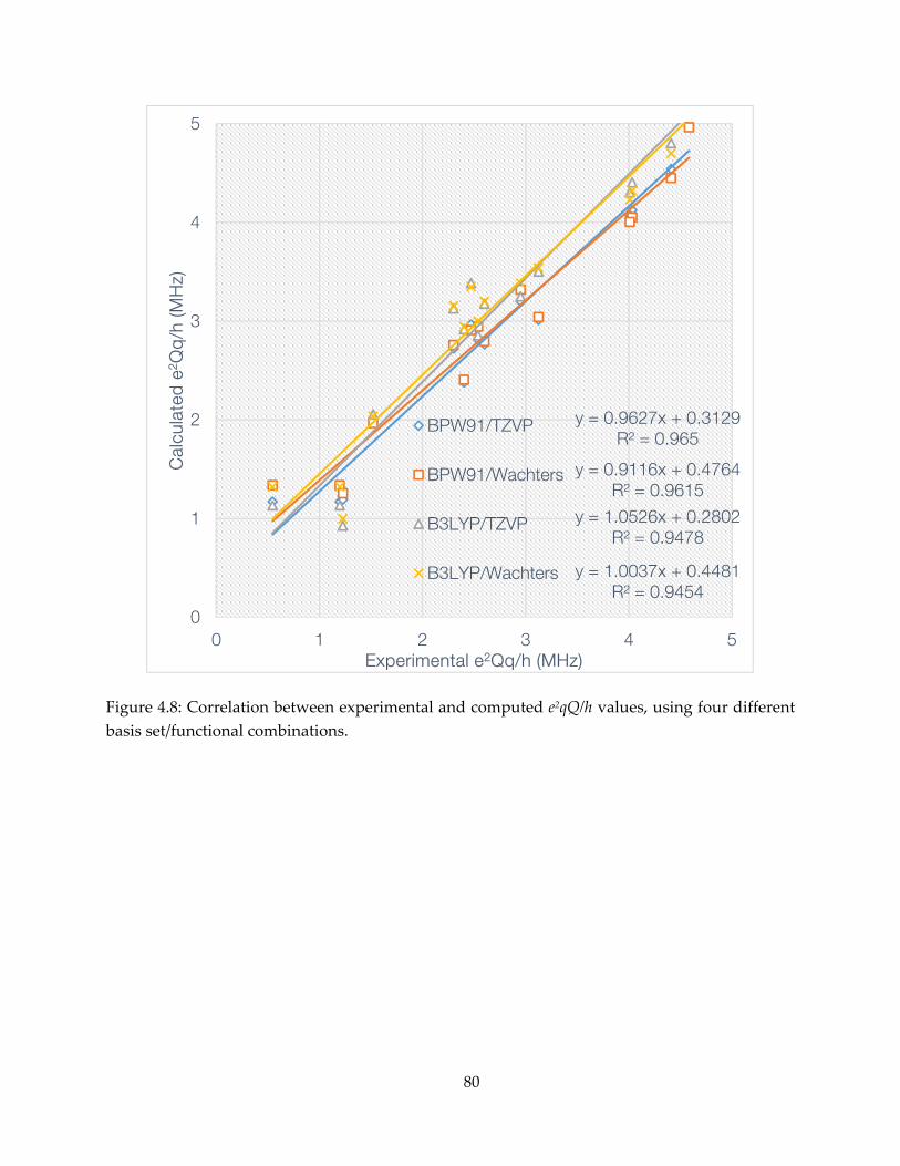
80
Figure 4.8: Correlation between experimental and computed e2qQ/h values, using four different basis set/functional combinations.
y = 0.9627x + 0.3129R² = 0.965
y = 0.9116x + 0.4764R² = 0.9615
y = 1.0526x + 0.2802R² = 0.9478
y = 1.0037x + 0.4481R² = 0.9454
0
1
2
3
4
5
0 1 2 3 4 5
Cal
cula
ted
e2 Q
q/h
(MH
z)
Experimental e2Qq/h (MHz)
BPW91/TZVP
BPW91/Wachters
B3LYP/TZVP
B3LYP/Wachters
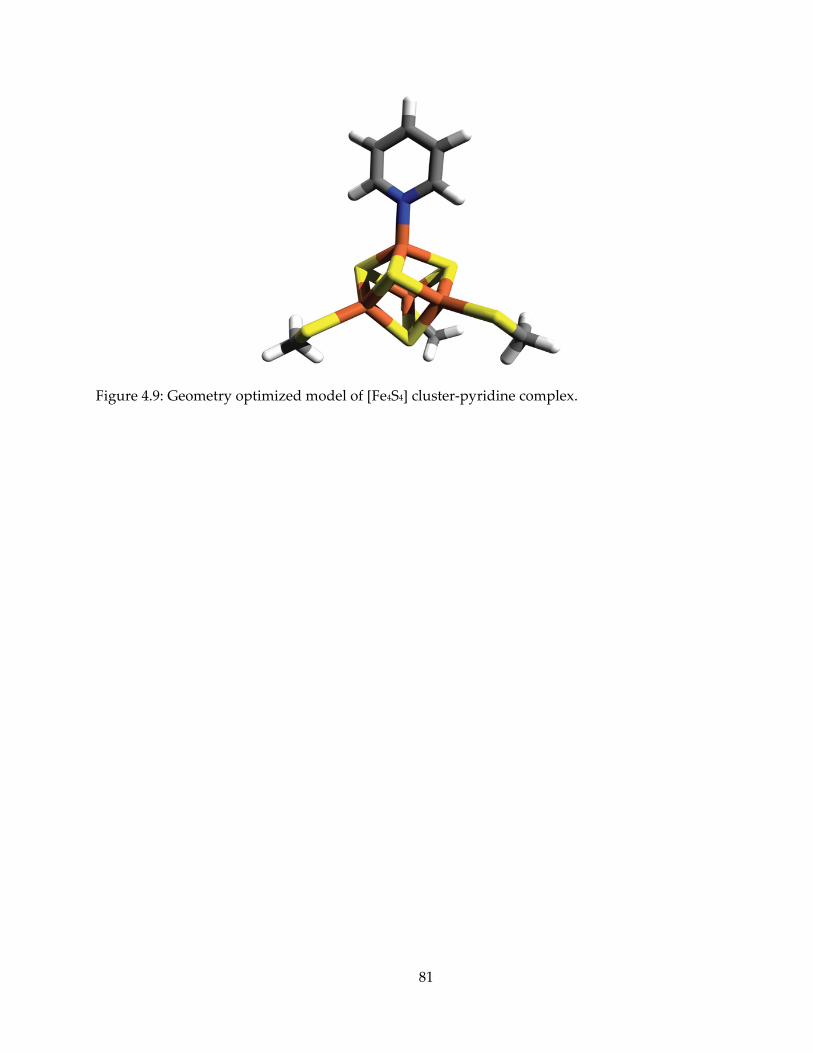
81
Figure 4.9: Geometry optimized model of [Fe4S4] cluster-pyridine complex.
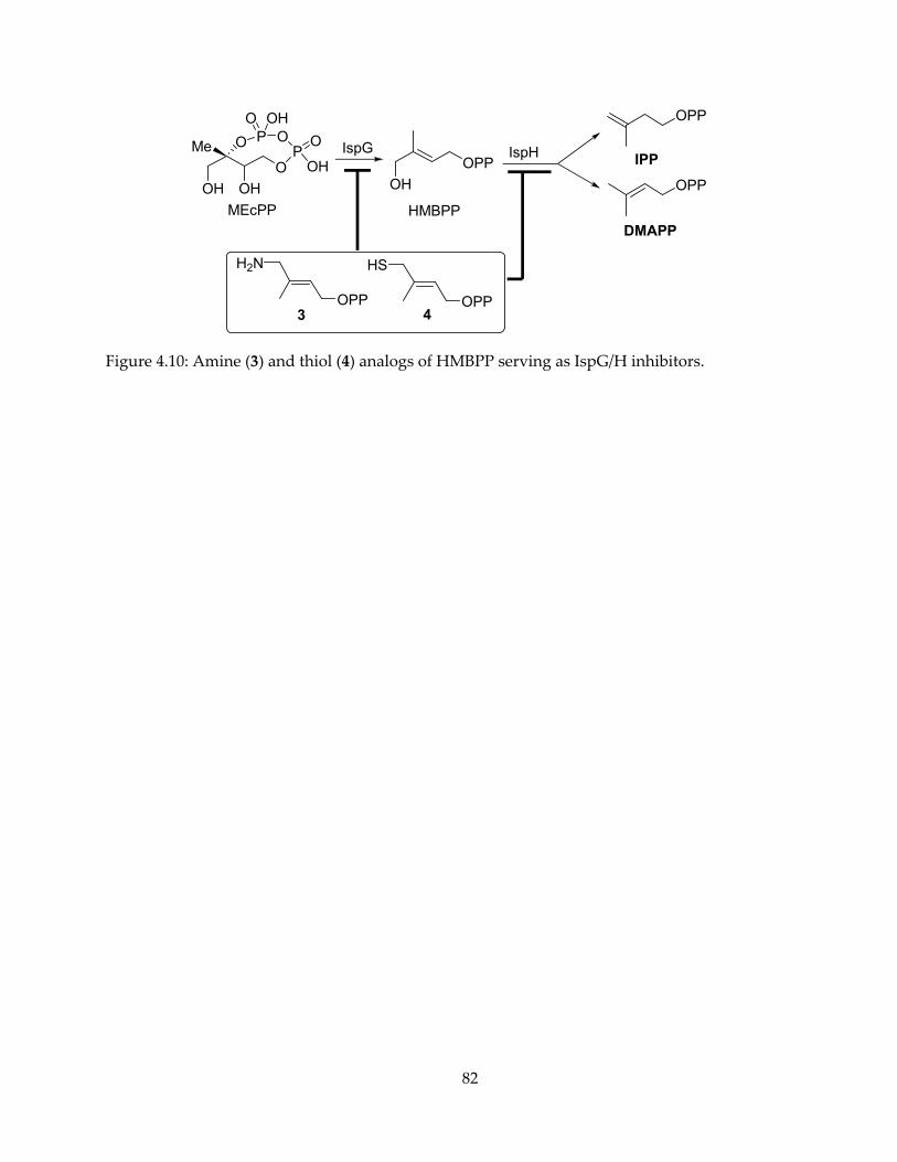
82
O
O P OP
O
OH
OH
Me
OH
O
MEcPP
OH
IspGOPP
OH
HMBPP
IspH
OPP
OPP
DMAPP
IPP
OPP
H2N
3OPP
HS
4
Figure 4.10: Amine (3) and thiol (4) analogs of HMBPP serving as IspG/H inhibitors.
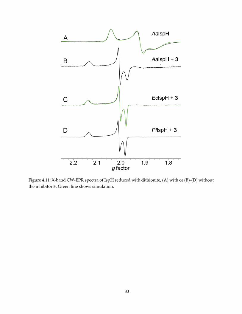
83
Figure 4.11: X-band CW-EPR spectra of IspH reduced with dithionite, (A) with or (B)-(D) without the inhibitor 3. Green line shows simulation.
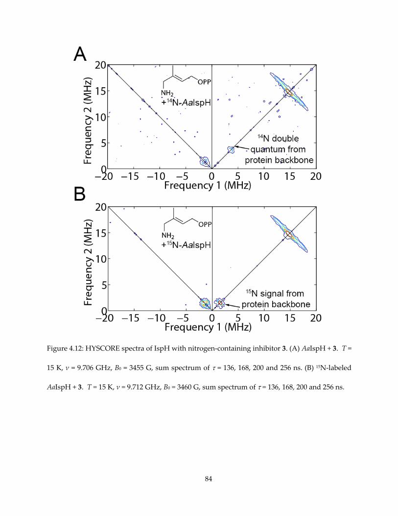
84
Figure 4.12: HYSCORE spectra of IspH with nitrogen-containing inhibitor 3. (A) AaIspH + 3. T =
15 K, ν = 9.706 GHz, B0 = 3455 G, sum spectrum of = 136, 168, 200 and 256 ns. (B) 15N-labeled
AaIspH + 3. T = 15 K, ν = 9.712 GHz, B0 = 3460 G, sum spectrum of = 136, 168, 200 and 256 ns.
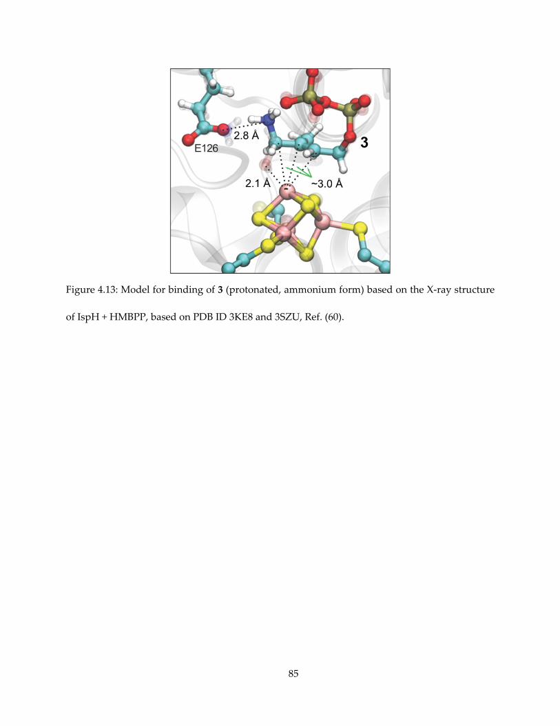
85
Figure 4.13: Model for binding of 3 (protonated, ammonium form) based on the X-ray structure
of IspH + HMBPP, based on PDB ID 3KE8 and 3SZU, Ref. (60).
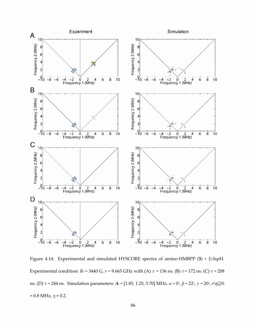
86
Figure 4.14: Experimental and simulated HYSCORE spectra of amino-HMBPP (3) + EcIspH.
Experimental condition: B0 = 3440 G, ν = 9.665 GHz with (A) = 136 ns. (B) = 172 ns. (C) τ = 208
ns. (D) τ = 244 ns. Simulation parameters: Ai = [1.85, 1.25, 3.70] MHz, α = 0∘, β = 22∘, γ = 20∘, e2qQ/h
= 0.8 MHz, η = 0.2.
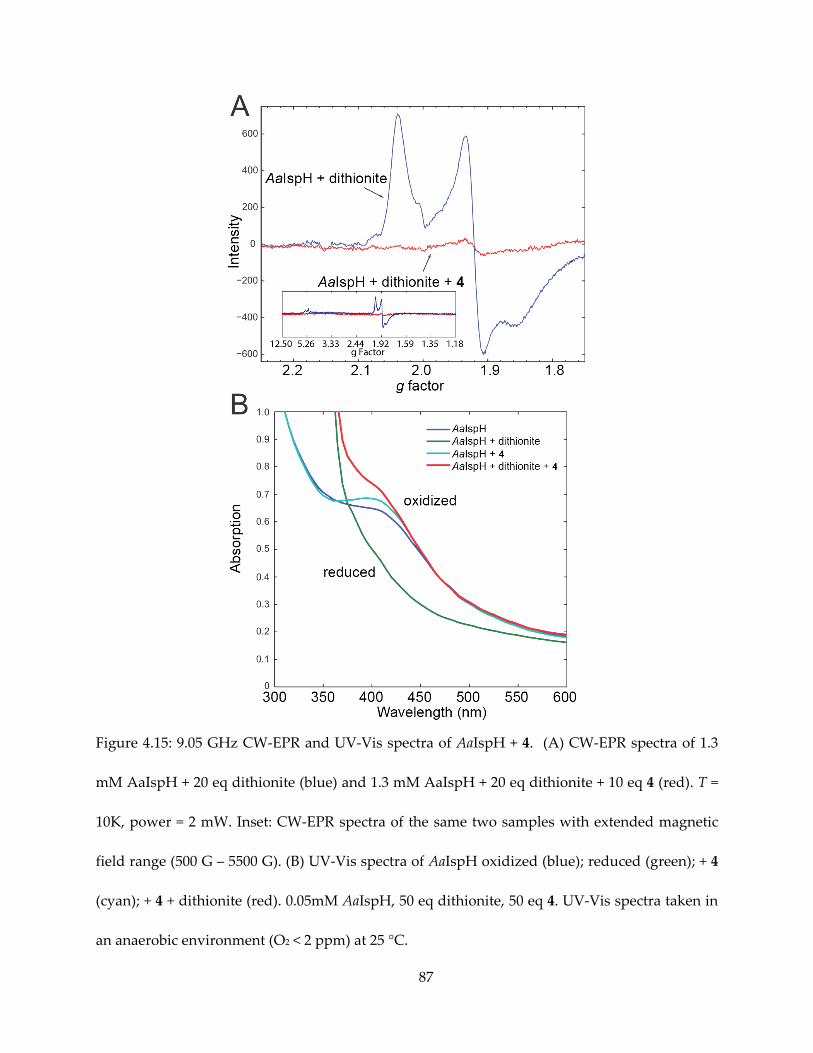
87
Figure 4.15: 9.05 GHz CW-EPR and UV-Vis spectra of AaIspH + 4. (A) CW-EPR spectra of 1.3
mM AaIspH + 20 eq dithionite (blue) and 1.3 mM AaIspH + 20 eq dithionite + 10 eq 4 (red). T =
10K, power = 2 mW. Inset: CW-EPR spectra of the same two samples with extended magnetic
field range (500 G – 5500 G). (B) UV-Vis spectra of AaIspH oxidized (blue); reduced (green); + 4
(cyan); + 4 + dithionite (red). 0.05mM AaIspH, 50 eq dithionite, 50 eq 4. UV-Vis spectra taken in
an anaerobic environment (O2 < 2 ppm) at 25 °C.
88
Chapter 5. Preliminary Study of Mechanism of 4Fe-4S Protein
Quinolinate Synthase
5.1. Notes and Acknowledgements
This work was supported by NIH grant (GM065307) to Eric Oldfield, I was supported by a
pre-doctoral fellowship from the American Heart Association, Midwest Affiliate (11PRE7500042)
from July 2011 to June 2013. I thank Prof. James Imlay for the NadB plasmid, Prof. Dennis Dean
for providing the isc plasmids for iron-sulfur cluster generation, and Dr. Mark J. Nilges for
assistance with the EPR spectroscopy.
5.2. Introduction
This chapter is a foray into the unfamiliar territory, beyond the context of isoprenoid
biosynthesis. NadA catalyzes the key step of the formation of a pyridine ring, and like the other
pyridine ring formation enzyme 3-hydroxyanthranilate 3,4-dioxygenase (91) of the tryptophan
pathway, it requires iron centers to perform its function. It is a 4Fe-4S cluster-containing protein,
with a unique fourth iron and a trefoil overall structure also seen with other 4Fe-4S proteins such
as IspH and the radical SAM protein Dph2 (92).
The mechanism of NadA has remained elusive, partly because the many reaction steps are
relatively independent and the sequence of events along the reaction coordinate is difficult to
determine (Figure 5.1), as is the role of the 4Fe-4S cluster. Here, I present our preliminary results
on research into the mechanism of NadA, with bioinformatics and spectroscopic methods.
89
5.3. Results and Discussion
5.3.1. Homology modeling and conserved residues
The crystal structure of NadA containing an intact 4Fe-4S cluster was reported only very
recently (21). Before it became available, we used the Phyre 2 server (93) to build a homology
model for the E. coli NadA protein we worked with, based on previously reported structures
without the cluster (20), shown in Figure 5.2.
I used the Jpred server (94) to generate a multi-sequence alignment of NadA homologs, and
with the SCORECONS server (95), residues in this alignment were scored and ranked. Apart from
aliphatic amino acids and cysteines, the top 7 conserved residues are listed in Table 5.1. When
located on the homology model as well as the crystal structure, they naturally cluster into two
“green” and “red” domains, and all of them are close to the iron-sulfur cluster in the crystal
structure. The next highest-ranked residue, K147, appeared to form a salt bridge with E93, both
highly conserved but far away from the center where the cluster resides.
5.3.2. Site-directed mutagenesis
Next, I performed site-direct mutagenesis for the top 7 conserved residues and attempted
expression and purification of 9 mutants, as listed in Table 5.2. For these mutants, 4 of them did
not show the dark brown color typically associated with iron-sulfur proteins, and all these 4
precipitated soon after elution from the Ni-NTA column. The other 5 seemed to harbor the iron-
sulfur cluster, but unsurprisingly for top conserved residues, their mutants are all inactive, as
judged by an NMR activity assay.
90
5.3.3. HYSCORE investigation of ligand binding
It was determined by Mössbauer spectroscopy that anaerobically purified E. coli NadA
cluster is in the [Fe4S4]2+ redox state (17). It has also been shown that NadA is also active under
reductive conditions (26) by HPLC, and our own NMR experiment confirm this activity in the
presence of dithionite. Therefore, it is interesting to see how the cluster, when reduced, binds to
substrates, product, and other ligands, through CW and pulsed EPR spectroscopy.
We thus prepared EPR samples of E. coli NadA with quinolinate (product), DHAP +
iminoaspartate (substrates), and a known inhibitor, phosphoglycolohydroxamate (PGH). The
HYSCORE spectra are shown in Figure 5.3. However, even though the ligands contained nitrogen
and phosphate atoms, we could not observe any large 14N or 31P hyperfine coupling. This indicates
that, when the cluster is reduced, the N atoms on iminoaspartate, quinolinate and PGH are far
away from the cluster, as are the P atoms in DHAP and PGH.
5.3.4. HYSCORE investigation of a purported reaction intermediate
The recently published crystal structure of T. maritama NadA does not contain a ligand
other than water (21). However, the authors proposed a “last-step” intermediate, 5-hydroxyl-4,5-
dihydroquinolinate (Figure 5.4A), with the 5-OH group binding to the unique fourth Fe, based
on computational docking. I thus set out to investigate whether this intermediate can be observed
with the reduced protein by HYSCORE.
As found with NMR, if the NadA reaction is carried out in D2O, 5-H of the final product
will be deuterated. Therefore, if the intermediate binds to the reduced cluster, in D2O, one will
likely observe a large D hyperfine for 5-D, because it is only two bonds away from the iron and
presumably also very close spatially. In addition, the authors also proposed E228 serves as the
91
final relay of proton abstraction from the intermediate. Presumably, an E228 mutant would arrest
the reaction at this stage and raise the concentration of this intermediate. Therefore, we prepared
3 samples in 85% D2O for HYSCORE investigation: WT protein without the substrates, WT
protein plus substrates, and E228D mutant protein plus substrates.
The HYSCORE results are shown in Figure 5.4B-D. Rather disappointingly, all spectra only
showed one intense 2H hyperfine signal at the deuteron Larmor frequency with very small Aiso,
most likely from deuteron exchanged into the protein.
5.4. Conclusions
The HYSCORE results reported here so far have been mostly negative results rather than
positive evidence for iron-sulfur cluster-ligand binding. There could be several possible reasons:
(i) The protein is not stoichiometrically reduced by dithionite, and the [Fe4S4]2+ species remaining
unreduced is active rather than the reduced [Fe4S4]+-IspH, which can bind the ligands without
involvement of the cluster. (ii) The reduced cluster binds to the ligands via the carboxylate
groups, like observed in aconitase (96), rather than the 5-OH. (iii) The reaction goes through
another mechanism for which the E228D mutant fails to trap the intermediate. Taken together,
with these preliminary results, the reaction mechanism or structure of intermediate-protein
complexes still remains elusive, and further research should be warranted.

92
5.5. Materials and Methods
5.5.1. E. coli NadA expression and purification
E. coli NadA gene was cloned into a pET46 Ek/Lic vector. The plasmid bearing the gene was
co- transformed with a pDB1282 plasmid into BL-21(DE3) E. coli cells. Cells were grown in LB
medium with 100 mg/L ampicillin and 50 mg/L kanamycin at 37 ∘C. 1 g/L L-arabinose was added
to induce the isc gene expression when OD600 reached 0.3, along with 0.2 mM FeCl3 and 0.1 g L-
cysteine. 0.5 mM IPTG was added when OD600 reached 0.8. The incubation was continued for 16
hours at 20 ∘C, and then the cells were harvested by centrifugation. Cell pellets were re-
suspended in buffer A (50 mM HEPES/KOH and 100 mM KCl, 10 mM imidazole, pH 7.5), lysed
by sonication and then centrifuged at 24,000 rpm at 4 °C for 20 min. The supernatant was loaded
onto a HisPur Ni-NTA spin column equilibrated with a buffer A. After washing with 2x column
volume buffer A for three times, protein was eluted with 2x column volume of buffer B (same as
buffer A, but containing 250 mM imidazole) three times. Dark-brown colored fractions
(confirmed to contain AaIspH by SDS-PAGE) were collected, pooled, concentrated and then
desalted on a PD-10 column with an imidazole-free buffer (50 mM HEPES/KOH and 100 mM
KCl, pH 7.5) to remove imidazole. Furthermore, the buffer was exchanged with a 40% glycerol
buffer with the same pH and salts with an Amicon filter, and the protein was stored at -80 ∘C
before use. Because NadA is not very stable at room temperature and incorporate Fe-S clusters
reasonably well, no further reconstitution was performed. For D2O EPR samples, protein are
exchanged into a D2O + 100 mM NH4HCO3 buffer and 20% pure glycerol was added.
93
5.5.2. Site-directed mutagenesis
Site-directed mutagenesis was carried out with the QuikChange II Site-Directed
Mutagenesis Kit (Agilent). The primer used are synthesized by Integrated DNA Technologies:
Y139A: 5'-atcgtactgtcgtcgtcgccgccaacacttctgctg-3’
5'-cagcagaagtgttggcggcgacgacgacagtacgat-3'
N141A: 5'-gtcgtcgtctacgccgccacttctgctgcggt-3'
5'-accgcagcagaagtggcggcgtagacgacgac-3'
T155A: 5'-acggcaatgcttgaagctaccacccaatctgcg-3'
5'-cgcagattgggtggtagcttcaagcattgccgt-3'
D178E: 5'-gccccagatgtttctcgggtgcccaga-3'
5'-tctgggcacccgagaaacatctggggc-3'
D178N: 5'-gccccagatgtttgttgggtgcccagatgat-3'
5'-atcatctgggcacccaacaaacatctggggc-3'
H203A: 5'-ctgagtcttaaattcatcagccacaatacaggcaccctgc-3'
5'-gcagggtgcctgtattgtggctgatgaatttaagactcag-3'
H226A: 5'-gcttgtggtgattctggagccaccagtatggcagcatc-3'
5'-gatgctgccatactggtggctccagaatcaccacaagc-3'
E228D: 5'-gcttgtggtgaatctggatgcaccagtatggca-3'
5'-tgccatactggtgcatccagattcaccacaagc-3'
E228Q: 5'-caatagcttgtggtgactgtggatgcaccagtatggcagc-3'
5'-gctgccatactggtgcatccacagtcaccacaagctattg-3'
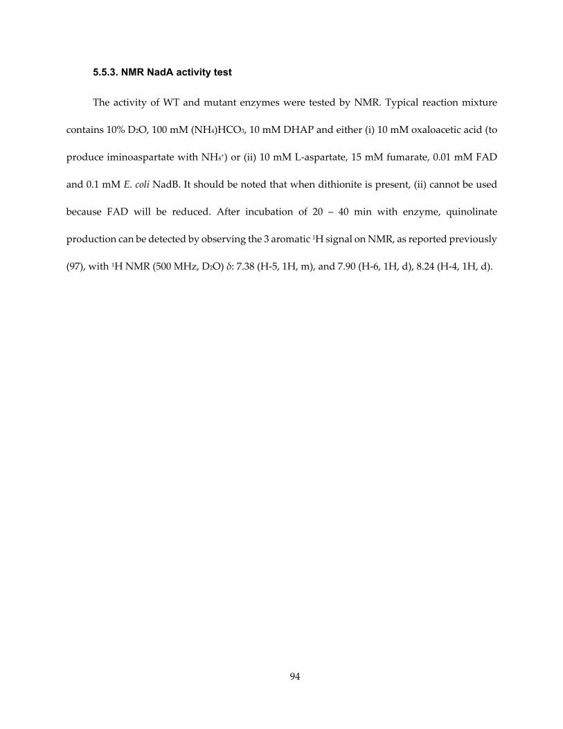
94
5.5.3. NMR NadA activity test
The activity of WT and mutant enzymes were tested by NMR. Typical reaction mixture
contains 10% D2O, 100 mM (NH4)HCO3, 10 mM DHAP and either (i) 10 mM oxaloacetic acid (to
produce iminoaspartate with NH4+) or (ii) 10 mM L-aspartate, 15 mM fumarate, 0.01 mM FAD
and 0.1 mM E. coli NadB. It should be noted that when dithionite is present, (ii) cannot be used
because FAD will be reduced. After incubation of 20 – 40 min with enzyme, quinolinate
production can be detected by observing the 3 aromatic 1H signal on NMR, as reported previously
(97), with 1H NMR (500 MHz, D2O) δ: 7.38 (H-5, 1H, m), and 7.90 (H-6, 1H, d), 8.24 (H-4, 1H, d).
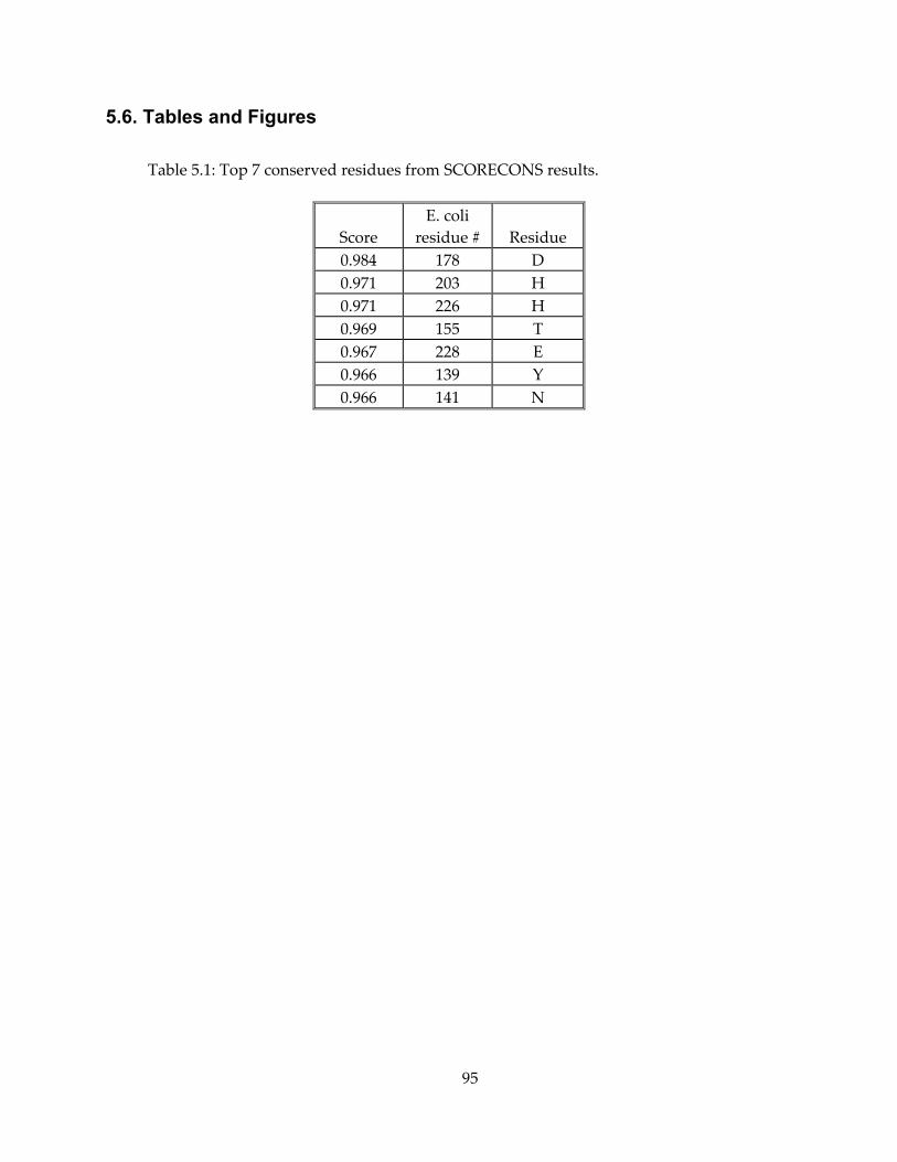
95
5.6. Tables and Figures
Table 5.1: Top 7 conserved residues from SCORECONS results.
Score E. coli
residue # Residue 0.984 178 D 0.971 203 H 0.971 226 H 0.969 155 T 0.967 228 E 0.966 139 Y 0.966 141 N
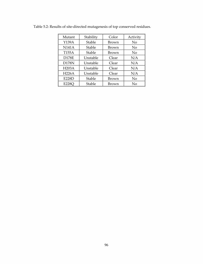
96
Table 5.2: Results of site-directed mutagenesis of top conserved residues.
Mutant Stability Color Activity Y139A Stable Brown No N141A Stable Brown No T155A Stable Brown No D178E Unstable Clear N/A D178N Unstable Clear N/A H203A Unstable Clear N/A H226A Unstable Clear N/A E228D Stable Brown No E228Q Stable Brown No
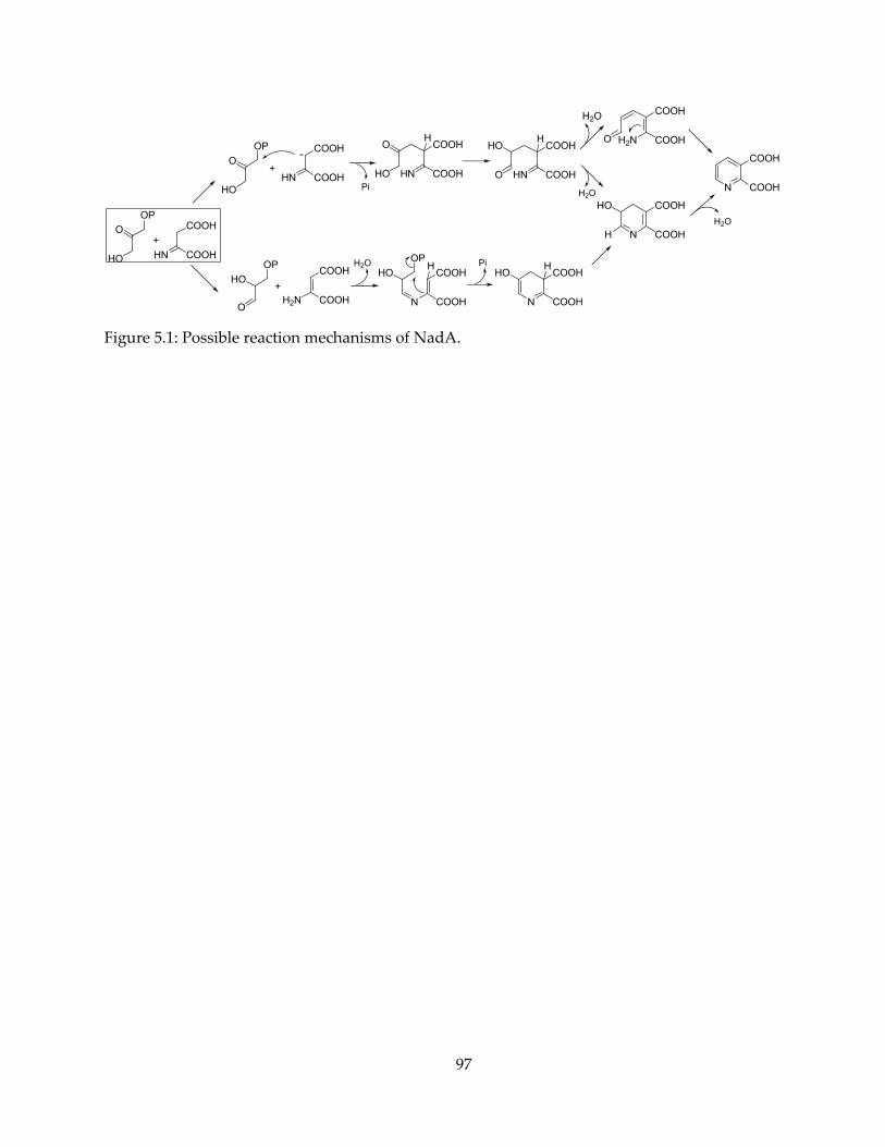
97
N
COOH
COOH
N
COOH
COOH
HO
H
N
COOH
COOH
HO
HN
COOH
COOH
HO
O
H
H
HN
COOH
COOH
O
HO
H
N
COOH
COOH
HOH
COOHHN
COOH
COOHH2N
COOH
HO
O
OP
+
O
HO
OP
+
COOHHN
COOH
HO
O
OP
+
Pi
H2O OP
H2O
Pi
H2O
H2N
COOH
COOHO
H2O
-
Figure 5.1: Possible reaction mechanisms of NadA.
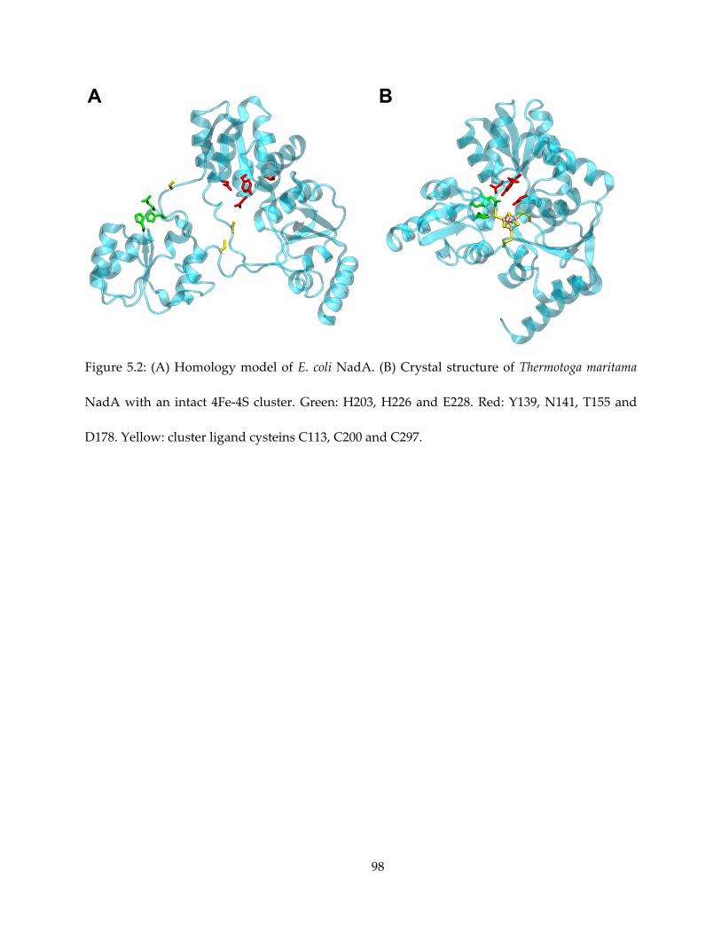
98
Figure 5.2: (A) Homology model of E. coli NadA. (B) Crystal structure of Thermotoga maritama
NadA with an intact 4Fe-4S cluster. Green: H203, H226 and E228. Red: Y139, N141, T155 and
D178. Yellow: cluster ligand cysteins C113, C200 and C297.
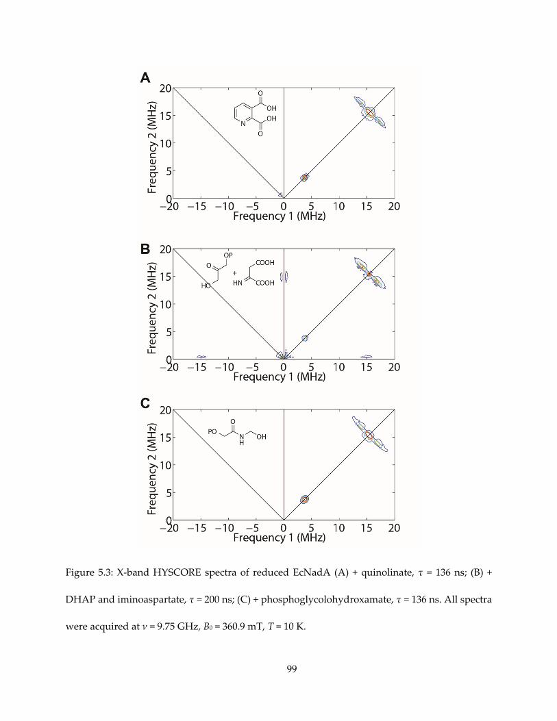
99
Figure 5.3: X-band HYSCORE spectra of reduced EcNadA (A) + quinolinate, τ = 136 ns; (B) +
DHAP and iminoaspartate, τ = 200 ns; (C) + phosphoglycolohydroxamate, τ = 136 ns. All spectra
were acquired at ν = 9.75 GHz, B0 = 360.9 mT, T = 10 K.
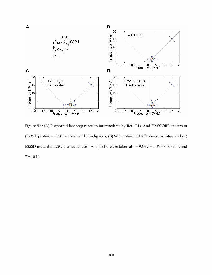
100
Figure 5.4: (A) Purported last-step reaction intermediate by Ref. (21). And HYSCORE spectra of
(B) WT protein in D2O without addition ligands; (B) WT protein in D2O plus substrates; and (C)
E228D mutant in D2O plus substrates. All spectra were taken at ν = 9.66 GHz, B0 = 357.6 mT, and
T = 10 K.
101
Chapter 6. Lipophilic Bisphosphonates Targeting Squalene
Synthase and Farnesyl Pyrophosphate Synthase
6.1. Notes and Acknowledgements
This chapter is partly adapted, under Creative Commons Attribution 4.0 license, from the
following publication: Shang, N. et al., 2014. Squalene Synthase as a Target for Chagas Disease
Therapeutics. PLoS Pathog, 10(5), p.e1004114.
Dr. Yonghui Zhang, Dr. Kai Li and Dr. Ke Wang synthesized most of the inhibitors. Dr. Wei
Zhu provided human FPPS inhibition results. Dr. Kai Li performed ubiquinone extraction
experiments. Dr. Rey-Ting Guo’s group at Tianjin Institute of Industrial Biology performed X-ray
crystallography of SQS-inhibitor complexes. Prof. Roberto Docampo’s group at University of
Georgia performed T. cruzi cell growth inhibition assays.
This work was supported by the National High Technology Research and Development
Program of China (grant 2012AA022200), the National Basic Research Program of China (grants
2011CB710800 and 2011CBA00805), and the United States Public Health Service (National
Institutes of Health grants CA158191 and GM065307 to Eric Oldfield). I was supported by a Pre-
doctoral Fellowship from the American Heart Association, Midwest Affiliate (11PRE7500042).
We thank the National Synchrotron Radiation Research Center of Taiwan for beam-time
allocation and data-collection assistance. The GC/MS analyses were carried out in the
Metabolomics Center, Roy J. Carver Biotechnology Center at University of Illinois at Urbana-
Champaign.
102
6.2. Introduction
The study described in this chapter is conducted in the context of neglected tropical diseases
research, against trypanosomatid parasites. For example, Chagas disease, caused by the
protozoan parasite Trypanosoma cruzi, affects eight million individuals (98), primarily in Latin
America. Ergosterol is the essential membrane sterol in many trypanosomatid parasites like T.
cruzi and plays the same structural role as cholesterol does in humans. Therefore, there is
currently considerable interest in inhibiting endogenous sterol biosynthesis as a route to the
development of novel anti-infective drugs.
A prominent example of such inhibitors is bisphosphonates (Figure 6.1), originally
established as treatment to human bone resorption diseases (99) with human farnesyl
pyrophosphate synthase (FPPS) as the target. They were found to inhibit the enzyme FPPS and
active against T. cruzi (100,101). Nitrogen containing bisphosphonates (Figure 6.1A) are thought
to mimic the cationic transition state or reaction intermediate of FPPS with the positively charged
N group, while the bisphosphonate group mimics the diphosphate group of natural
substrates/products (102,103).
More recently, lipophilic bisphosphonates (Figure 6.1B), analogs of zoledronate and
risedronate, has been shown to target other enzymes such as geranylgeranyl pyrophosphate
synthase (GGPPS) (104) in addition to FPPS, and the FPPS/GGPPS dual-function enzyme in
malarial parasites (105,106). Curiously, in these studies the best activity against FPPS/GGPPS
were found with short-to-medium size side chains, while the activity against malaria parasite
almost monotonically increase with larger side chains.
103
In this chapter, I report that besides FPPS, squalene synthase (SQS) is also targeted by these
lipophilic bisphosphonates. We explored the relationship between the side chain size and its
inhibition activity against FPPS, SQS, as well as in cells. In addition, our discovery of lipophilic
bisphosphonates as SQS inhibitors also facilitated new crystallographic endeavors that have
revealed interesting binding phenomenon.
6.3. Results and Discussion
6.3.1. Two series of lipophilic bisphosphonates, derivative of zoledronate
Dr. Yonghui Zhang and Dr. Ke Wang prepared two series of analogs of one small
bisphosphonate, zoledronate. One series is synthesized by varying the group attached to the N3
of the imidazole ring, with an n-alkyl group, from H up to C15H31.
The other series has the same substitution, and additionally the 1-hydroxyl group from the
zoledronate “base molecule” was replaced with a hydrogen. The rationale for the removal of -
OH group is that, as reported previously, bisphosphonates bind to bone mineral, which involves
the interaction of the phosphonate groups, the 1-OH group, as well as of an unhindered cationic
side-chain with the bone surface (107).
For a shorthand, each compound was assigned an internal number in the lab, and are
abbreviated as Cn-OH and Cn-H series, n ranging from 0 to 15.
6.3.2. Lipophilic bisphosphonates are indeed potent SQS inhibitors
Suspecting SQS might be a target of lipophilic bisphosphonates in T. cruzi, we initially
attempted to accelerate the measurement of IC50s of the two series of lipophilic bisphosphonates
104
against T. cruzi SQS with an assay continuously monitoring the release of pyrophosphate (108).
As a result, such an assay yielded results showing very modest inhibition, Figure 6.3. The best
inhibitor has an IC50 of 550 nM.
However, the pyrophosphate release assay is an indirect measurement, not monitoring the
formation of squalene but that of a side product that is released in both steps of the reaction, and
its low sensitivity requires a high concentration (450 μM) of substrates, nearly 90 times of the
reported Km (5.2 μM) of the enzyme (109). To avoid these complications, we used a more
established end-point assay, which directly measures the amount of [3H]-labeled FPP substrate
incorporated into the final product, squalene, by scintillation counting (109). Both series of
bisphosphonates were tested against T. cruzi and human SQS (Figure 6.4).
In addition, T. cruzi FPPS inhibition were also tested (Figure 6.5), and plotted along with
previously obtained HsSQS inhibition results (106). All assay results are also tabulated in Table
6.1.
As seen in Figure 6.4, short side-chain bisphosphonates exhibit very little SQS inhibition.
For medium to long side-chains, there is an increase in potency with increasing chain length up
to n = 10 or 11, but then potency decreases on further chain elongation. There is no obvious effect
of the 1-OH/1-H group on enzyme inhibition.
By contrast, the increase in activity against FPPS (both species) with chain length is less
pronounced, and maximum activity is found with a C6 – C8 substituent. When the side chain
elongates beyond C10, the activity drastically decreases (Figure 6.5), unlike the more moderate
decrease found with SQS.
105
6.3.3. Parasite growth inhibition, rescue and ergosterol synthesis inhibition
Our collaborator Roberto Docampo also tested a subset of these bisphosphonates for their
ability to inhibit the proliferation of the clinically relevant amastigote form of T. cruzi, with results
shown in Figure 6.6.Activity in T. cruzi cell growth inhibition increases almost monotonically with
increasing chain length up to the longest C14 – C15 side-chain species investigated (Figure 6.6).
This could be presumably attributed to enhanced cell permeability, similar to the case of
FPPS/GGPPS bisphosphonate inhibitors in Plasmodium (106).
Rescue experiments were performed in the presence of the C10-H species, no rescue was
afforded by farnesol, and only ~ 20% rescue by geranylgeranyl. Sterol biosynthesis analysis was
also performed in the presence of the C10-H species at 4 μM, with ergosterol components
analyzed by GC/MS, LC/MS and LC/UV. The content of exogeneously-acquired cholesterol in T.
cruzi epimastigotes increased from 51% to 71% of total sterols, while that of endogenous
ergosterol decreased from 36% to 15% of total sterols.
6.3.4. Crystal structures of lipophilic bisophosphonates bound to SQS
Previous crystallography efforts of SQS/CrtM have identified two substrate binding sites
(38), the allylic binding site S1, where FPP or the substrate analog FSPP bind to a cluster of highly
conserved aspartate residues via Mg2+, and a homoallylic binding site S2 which binds to the
FPP(FSPP) diphosphate group via basic residues.
We next sent the C10/C15 species, with or without the hydroxyl, to Dr. Rey-Ting Guo’s lab
for crystal structure determination. Two T. cruzi SQS structures (with C10-OH and C15-H) and
four human SQS structures were successfully determined. With the FSPP structures
superimposed to serve as a guide to the S1 and S2 sites, the newly solved structures are shown in
106
Figure 6.7. Basically, the S2 farnesyl-binding site was occupied by the n-alkyl side chains. For the
shorter, C10 inhibitor species, the site S2 is occupied. For the C15 species, the 8 distal side-chain
carbons occupy the S2 site, but the side-chain then bends and occupies the S1 site, before bending
back with the bisphosphonate head group returning to the S2 site.
6.4. Conclusions
Summarizing the enzyme inhibition, parasite growth inhibition/rescue and sterol
biosynthesis inhibition results, we can conclude that, for the most effective long side-chain
lipophilic bisphosphonates, FPPS was not likely a major target, although inhibition of ergosterol
biosynthesis is indeed targeted. Instead, SQS was highly likely the target. The most T. cruzi
amastigote growth inhibition was achieved with the long side-chain (C14-15) inhibitors, which,
although not the best SQS inhibitor, are still highly-potent and with its effects balanced by
enhanced hydrophobicity and better cell penetration. In addition, our inhibition studies
facilitated determination of new crystal structures showing interesting binding modes, which will
also be of great interest in future development of SQS inhibitors as anti-infective drug leads.
6.5. Materials and Methods
6.5.1. SQS expression and purification
Both TcSQS and HsSQS were expressed according to previously established protocols
(109,110). Briefly, pET-28a plasmids bearing truncated but functional and His-tagged TcSQS and
HsSQS were transformed into BL-21 (DE3) cells. Cells are grown at 37 ∘C in LB medium with 50
107
mg/L kanamycin. Incubation was continued until OD600 reached 0.6-0.8. Then 1 mM IPTG was
added and incubation continued for 4 hours. Cells were harvested by centrifugation (9000 rpm,
15 minutes), and frozen at -80 ∘C before use.
The proteins were purified using HisPur Ni-NTA spin columns. Cell pellets were
suspended in 15 mL buffer A (50 mM HEPES/KOH, pH 7.5, and 100 mM NaCl), and sonicated
for 5 minutes in 5 second pulses. The suspension was centrifuged (24000 rpm, 20 minutes), and
the supernatant was loaded onto the column pre-equilibrated with 96% buffer A + 4% buffer B
(same as buffer A, but with 250 mM imidazole) by gravity flow. The spin column was washed
three times with 2x column volume of 96% buffer A + 4% buffer B, and then His-tagged protein
was eluted three times with 100% buffer B. Both proteins were desalted with buffer A on PD-10
columns, concentrated, and frozen at -80 ∘C in buffer A + 20% glycerol before use.
6.5.2. T. cruzi FPPS expression and purification
T. cruzi FPPS was basically purified with previously reported protocols (111). DNA coding
for an N-terminal tag and a thrombin cleavage site was attached to the TcFPPS gene and
cloned in to a pET28a vector, transformed BL-21(DE3) E. coli cells. Cells are grown and harvested
the same way as SQS.
To purify the protein, the cells were re-suspended in 20 mL (per 1 L of cell growth
medium) of buffer A (50 mM phosphate buffer, 500 mM NaCl, 10 mM imidazole, 2 mM tris(2-
carboxyethyl)phosphine (TCEP), final pH 8.0), placed on ice, and broken with sonication.
The sonicated suspension was centrifuged (24000 rpm for 20 minutes), and the supernatant was
loaded onto a Ni-NTA column pre-equilibrated with buffer A on an AKTA Prime FPLC system.
The column was washed with buffer A until no more protein was washed down the column
108
(judged by O D280 of the flow-through < 0.06). The protein was then eluted with a gradient
of 0 – 100% of buffer B (same as buffer A, but with 250 mM imidazole). The 42 kDa FPPS
containing fractions (judged by OD280 and SDS-PAGE) were pooled and concentrated with an
Amicon filter. 10 units of thrombin was added to the mixture and incubated at 4 ∘C overnight
to cut off the His-tag. The completion of the cleavage was confirmed by native PAGE. The
mixture was then l oaded onto a benzamidine column to remove the thrombin, and a Ni-NTA
column to remove His-tag peptide. The flow-through containing TcFPPS was pooled and
dialyzed 3 times against 1 liter of buffer C (20 mM Tris/HCl, 50 mM NaCl and 2 mM TCEP) and
concentrated. The purified protein (judged by SDS-PAGE and native-PAGE) were stored in -80
∘C until use.
6.5.3. SQS inhibition assay
Initially, SQS enzyme inhibition assays were carried out, in duplicate, in 96-well plates,
with a total of 200 μL of reaction mixture in each well. Reactions were monitored by using a
continuous spectrophotometric assay for phosphate releasing enzymes, in a buffer contained 50
mM Tris-HCl pH 7.4, 1 mM MgCl2, 250 μM NADPH, and 450 μM FPP. The compounds
investigated were pre-incubated with 2 μg of SQS for 30 min at 20 °C.
After the initial assay we used a radioactivity assay with 3H-labelled FPP substrate. The
reaction mixture (50 μL) had 2.5 nM T. cruzi or H. sapiens SQS, 0.5 μM [1-3H]-FPP, and 250 μM
NADPH, in a detergent-containing buffer (50 mM HEPES-KOH, pH 7.5, 100 mM NaCl, 4 mM
MgCl2, 0.5% Tween 80, 2 mM CHAPS, and 4 mM DTT). The enzymes were incubated with
inhibitor for 10 minutes before addition of the substrates. The reaction was allowed to continue
at 37 ∘C for 15 minutes and then terminated with 50 μL of 0.1 M NaOH/methanol. The product
109
was extracted with 500 μL of hexanes and 300 μL of the organic layer was measured by
scintillation counting.
6.5.4. FPPS inhibition assay
For FPPS inhibition, the condensation of geranyl diphosphate (100 μM) and IPP (100 μM)
was monitored at room temperature by using a continuous spectrophotometric assay for
phosphate. The reaction buffer contained 50 mM Tris-HCl, pH 7.4, 1 mM MgCl2, and 0.01 % Triton
X100. The compounds investigated were pre-incubated with enzyme for 30 min at room
temperature.
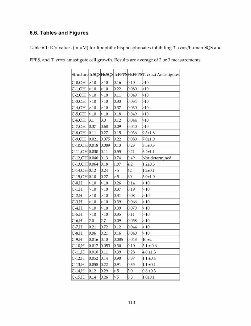
110
6.6. Tables and Figures
Table 6.1: IC50 values (in μM) for lipophilic bisphosphonates inhibiting T. cruzi/human SQS and
FPPS, and T. cruzi amastigote cell growth. Results are average of 2 or 3 measurements.
Structure TcSQS HsSQSTcFPPSHsFPPST. cruzi Amastigotes
C-0,OH > 10 > 10 0.16 0.10 >10 C-1,OH > 10 > 10 0.22 0.080 >10 C-2,OH > 10 > 10 0.11 0.049 >10 C-3,OH > 10 > 10 0.33 0.034 >10 C-4,OH > 10 > 10 0.37 0.030 >10 C-5,OH > 10 > 10 0.18 0.049 >10 C-6,OH 3.1 3.0 0.12 0.044 >10 C-7,OH 0.37 0.68 0.09 0.040 >10 C-8,OH 0.11 0.27 0.15 0.036 9.3±1.8 C-9,OH 0.021 0.075 0.22 0.080 7.0±1.0 C-10,OH 0.018 0.089 0.13 0.23 3.5±0.3 C-11,OH 0.030 0.11 0.55 0.21 6.4±1.1 C-12,OH 0.046 0.13 0.74 0.49 Not determined C-13,OH 0.064 0.18 1.07 4.2 1.2±0.3 C-14,OH 0.12 0.24 > 5 42 1.2±0.1 C-15,OH 0.10 0.27 > 5 60 3.0±1.0 C-0,H > 10 > 10 0.26 0.14 > 10 C-1,H > 10 > 10 0.37 0.19 > 10 C-2,H > 10 > 10 0.31 0.08 > 10 C-3,H > 10 > 10 0.39 0.066 > 10 C-4,H > 10 > 10 0.39 0.079 > 10 C-5,H > 10 > 10 0.35 0.11 > 10 C-6,H 2.0 2.7 0.09 0.058 > 10 C-7,H 0.21 0.72 0.12 0.044 > 10 C-8,H 0.06 0.21 0.16 0.040 > 10 C-9,H 0.016 0.10 0.085 0.043 10 ±2 C-10,H 0.017 0.053 0.30 0.10 3.1 ± 0.6 C-11,H 0.010 0.11 0.39 0.28 4.0 ±1.3 C-12,H 0.052 0.14 0.90 0.37 1.1 ±0.4 C-13,H 0.058 0.22 0.91 0.35 1.1 ±0.1 C-14,H 0.12 0.29 > 5 3.0 0.8 ±0.3 C-15,H 0.14 0.26 > 5 8.3 1.0±0.1
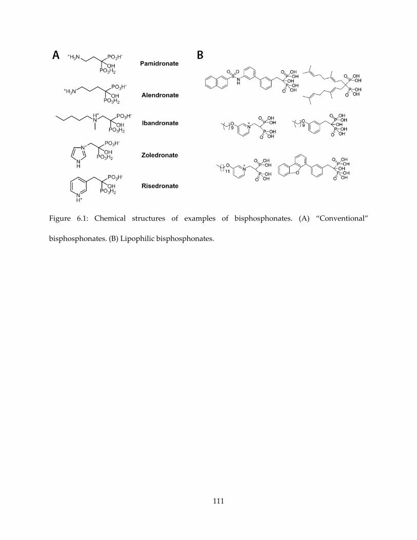
111
Figure 6.1: Chemical structures of examples of bisphosphonates. (A) “Conventional”
bisphosphonates. (B) Lipophilic bisphosphonates.
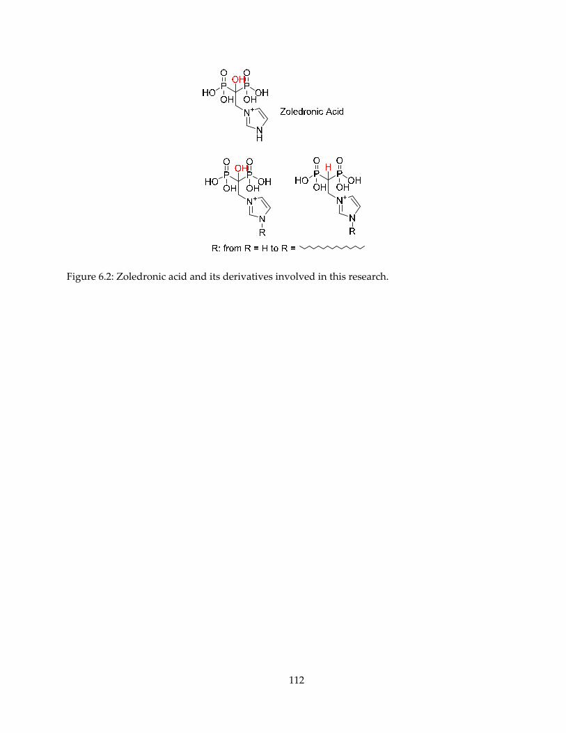
112
Figure 6.2: Zoledronic acid and its derivatives involved in this research.
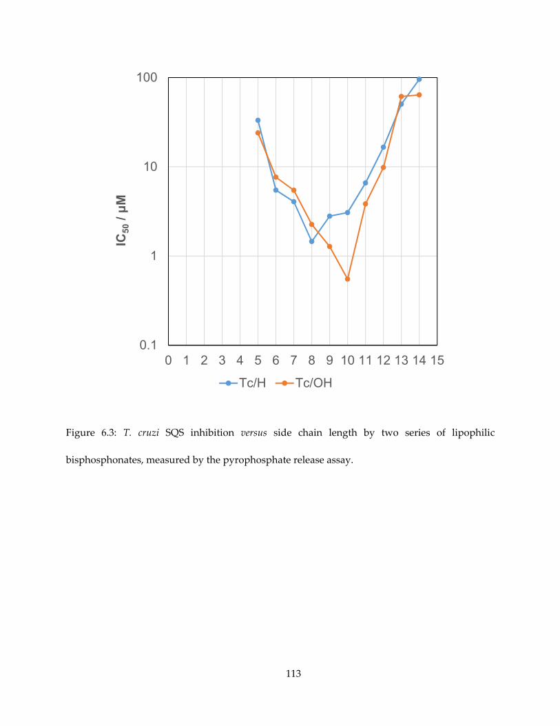
113
Figure 6.3: T. cruzi SQS inhibition versus side chain length by two series of lipophilic
bisphosphonates, measured by the pyrophosphate release assay.
0.1
1
10
100
0 1 2 3 4 5 6 7 8 9 10 11 12 13 14 15
IC50
/ μM
Tc/H Tc/OH
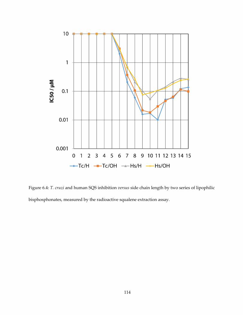
114
Figure 6.4: T. cruzi and human SQS inhibition versus side chain length by two series of lipophilic
bisphosphonates, measured by the radioactive squalene extraction assay.
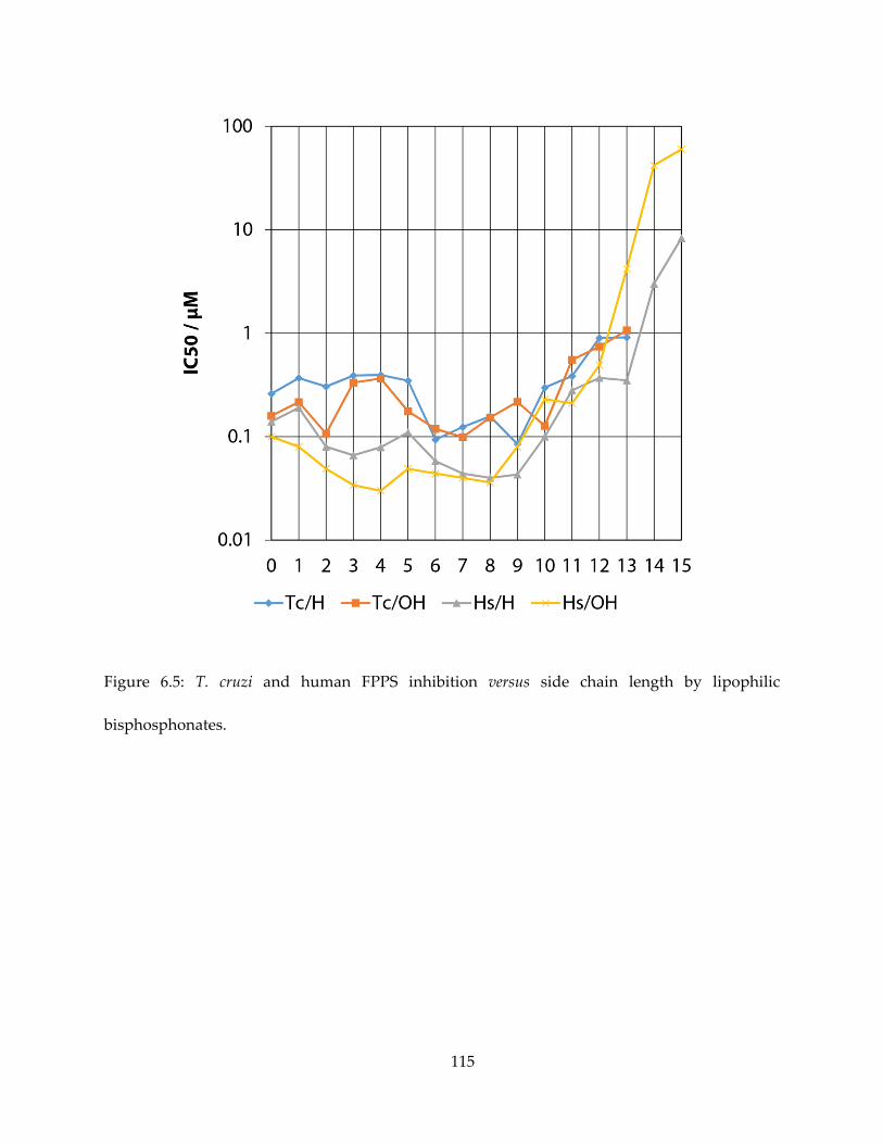
115
Figure 6.5: T. cruzi and human FPPS inhibition versus side chain length by lipophilic
bisphosphonates.
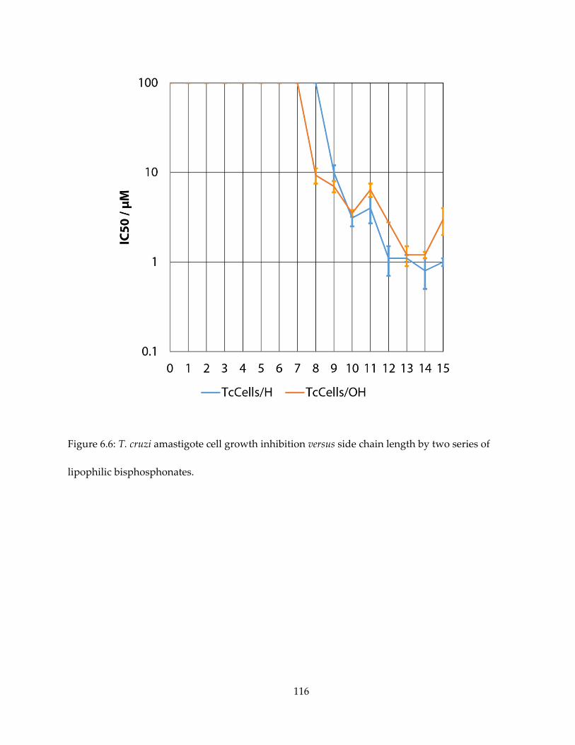
116
Figure 6.6: T. cruzi amastigote cell growth inhibition versus side chain length by two series of
lipophilic bisphosphonates.
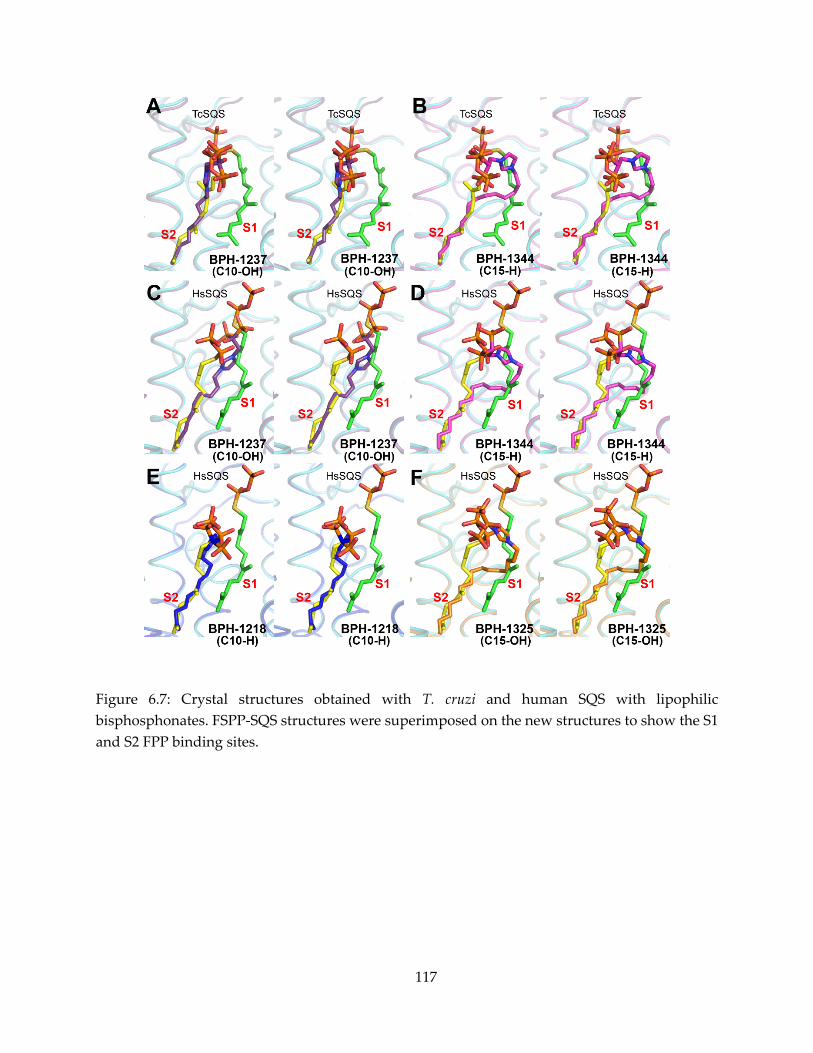
117
Figure 6.7: Crystal structures obtained with T. cruzi and human SQS with lipophilic bisphosphonates. FSPP-SQS structures were superimposed on the new structures to show the S1 and S2 FPP binding sites.
118
Bibliography
1. John Buckingham. Dictionary of Natural Products on DVD - CRC Press Book. Boca Raton, FL; 2011.
2. Eisenreich W, Bacher A, Arigoni D, Rohdich F. Biosynthesis of isoprenoids via the non-mevalonate pathway. Cell Mol Life Sci CMLS. 2004 Jun 1;61(12):1401–26.
3. Heuston S, Begley M, Gahan CGM, Hill C. Isoprenoid biosynthesis in bacterial pathogens. Microbiology. 2012 Mar 30;158(Pt_6):1389–401.
4. Lombard J, Moreira D. Origins and Early Evolution of the Mevalonate Pathway of Isoprenoid Biosynthesis in the Three Domains of Life. Mol Biol Evol. 2011 Jan 1;28(1):87–99.
5. Jackson ER, Dowd CS. Inhibition of 1-deoxy-D-xylulose-5-phosphate reductoisomerase (Dxr): a review of the synthesis and biological evaluation of recent inhibitors. Curr Top Med Chem. 2012;12(7):706–28.
6. Kollas A-K, Duin EC, Eberl M, Altincicek B, Hintz M, Reichenberg A, et al. Functional characterization of GcpE, an essential enzyme of the non-mevalonate pathway of isoprenoid biosynthesis. FEBS Lett. 2002 Dec 18;532(3):432–6.
7. Seemann M, Bui BTS, Wolff M, Tritsch D, Campos N, Boronat A, et al. Isoprenoid Biosynthesis through the Methylerythritol Phosphate Pathway: The (E)-4-Hydroxy-3-methylbut-2-enyl Diphosphate Synthase (GcpE) is a [4Fe–4S] Protein. Angew Chem. 2002 Nov 15;114(22):4513–5.
8. Brandt W, Dessoy MA, Fulhorst M, Gao W, Zenk MH, Wessjohann LA. A Proposed Mechanism for the Reductive Ring Opening of the Cyclodiphosphate MEcPP, a Crucial Transformation in the New DXP/MEP Pathway to Isoprenoids Based on Modeling Studies and Feeding Experiments. ChemBioChem. 2004 Mar 5;5(3):311–23.
9. Rohdich F, Zepeck F, Adam P, Hecht S, Kaiser J, Laupitz R, et al. The deoxyxylulose phosphate pathway of isoprenoid biosynthesis: Studies on the mechanisms of the reactions catalyzed by IspG and IspH protein. Proc Natl Acad Sci. 2003 Feb 18;100(4):1586–91.
10. Wolff M, Seemann M, Tse Sum Bui B, Frapart Y, Tritsch D, Garcia Estrabot A, et al. Isoprenoid biosynthesis via the methylerythritol phosphate pathway: the (E)-4-hydroxy-3-methylbut-2-enyl diphosphate reductase (LytB/IspH) from Escherichia coli is a [4Fe-4S] protein. FEBS Lett. 2003 Apr 24;541(1-3):115–20.
119
11. Altincicek B, Duin EC, Reichenberg A, Hedderich R, Kollas A-K, Hintz M, et al. LytB protein catalyzes the terminal step of the 2-C-methyl-D-erythritol-4-phosphate pathway of isoprenoid biosynthesis. FEBS Lett. 2002 Dec 18;532(3):437–40.
12. Rohdich F, Zepeck F, Adam P, Hecht S, Kaiser J, Laupitz R, et al. The deoxyxylulose phosphate pathway of isoprenoid biosynthesis: studies on the mechanisms of the reactions catalyzed by IspG and IspH protein. Proc Natl Acad Sci U S A. 2003 Feb 18;100(4):1586–91.
13. Xiao Y, Zhao ZK, Liu P. Mechanistic studies of IspH in the deoxyxylulose phosphate pathway: heterolytic C-O bond cleavage at C4 position. J Am Chem Soc. 2008 Feb 20;130(7):2164–5.
14. Gräwert T, Rohdich F, Span I, Bacher A, Eisenreich W, Eppinger J, et al. Structure of active IspH enzyme from Escherichia coli provides mechanistic insights into substrate reduction. Angew Chem Int Ed Engl. 2009;48(31):5756–9.
15. Xu W, Lees NS, Hall D, Welideniya D, Hoffman BM, Duin EC. A Closer Look at the Spectroscopic Properties of Possible Reaction Intermediates in Wild-Type and Mutant (E)-4-Hydroxy-3-methylbut-2-enyl Diphosphate Reductase. Biochemistry (Mosc). 2012 Jun 19;51(24):4835–49.
16. Cicchillo RM, Tu L, Stromberg JA, Hoffart LM, Krebs C, Booker SJ. Escherichia coli Quinolinate Synthetase Does Indeed Harbor a [4Fe-4S] Cluster. J Am Chem Soc. 2005 May 1;127(20):7310–1.
17. Ollagnier-de Choudens S, Loiseau L, Sanakis Y, Barras F, Fontecave M. Quinolinate synthetase, an iron–sulfur enzyme in NAD biosynthesis. FEBS Lett. 2005 Jul 4;579(17):3737–43.
18. Rekittke I, Wiesner J, Röhrich R, Demmer U, Warkentin E, Xu W, et al. Structure of (E)-4-Hydroxy-3-methyl-but-2-enyl Diphosphate Reductase, the Terminal Enzyme of the Non-Mevalonate Pathway. J Am Chem Soc. 2008 Dec 24;130(51):17206–7.
19. Span I, Wang K, Wang W, Zhang Y, Bacher A, Eisenreich W, et al. Discovery of acetylene hydratase activity of the iron–sulphur protein IspH. Nat Commun. 2012 Sep 4;3:1042.
20. Sakuraba H, Tsuge H, Yoneda K, Katunuma N, Ohshima T. Crystal Structure of the NAD Biosynthetic Enzyme Quinolinate Synthase. J Biol Chem. 2005 Jul 22;280(29):26645 –26648.
21. Cherrier MV, Chan A, Darnault C, Reichmann D, Amara P, Ollagnier de Choudens S, et al. The Crystal Structure of Fe4S4 Quinolinate Synthase Unravels an Enzymatic Dehydration Mechanism That Uses Tyrosine and a Hydrolase-Type Triad. J Am Chem Soc. 2014 Apr 9;136(14):5253–6.
120
22. Gazzaniga F, Stebbins R, Chang SZ, McPeek MA, Brenner C. Microbial NAD Metabolism: Lessons from Comparative Genomics. Microbiol Mol Biol Rev MMBR. 2009 Sep;73(3):529–41.
23. Gerdes SY, Scholle MD, D’Souza M, Bernal A, Baev MV, Farrell M, et al. From Genetic Footprinting to Antimicrobial Drug Targets: Examples in Cofactor Biosynthetic Pathways. J Bacteriol. 2002 Aug 15;184(16):4555–72.
24. Boshoff HIM, Xu X, Tahlan K, Dowd CS, Pethe K, Camacho LR, et al. Biosynthesis and Recycling of Nicotinamide Cofactors in Mycobacterium tuberculosis AN ESSENTIAL ROLE FOR NAD IN NONREPLICATING BACILLI. J Biol Chem. 2008 Jul 11;283(28):19329–41.
25. Begley TP, Kinsland C, Mehl RA, Osterman A, Dorrestein P. The biosynthesis of nicotinamide adenine dinucleotides in bacteria. Cofactor Biosynthesis [Internet]. Academic Press; 2001. p. 103–19. Available from: http://www.sciencedirect.com/science/article/pii/S0083672901610033
26. Soriano EV, Zhang Y, Colabroy KL, Sanders JM, Settembre EC, Dorrestein PC, et al. Active-site models for complexes of quinolinate synthase with substrates and intermediates. Acta Crystallogr D Biol Crystallogr. 2013 Sep 1;69(9):1685–96.
27. Oldfield E, Lin F-Y. Terpene biosynthesis: modularity rules. Angew Chem Int Ed Engl. 2012 Jan 27;51(5):1124–37.
28. Resh MD. Targeting protein lipidation in disease. Trends Mol Med. 2012 Apr;18(4):206–14.
29. Nes WD. Biosynthesis of Cholesterol and Other Sterols. Chem Rev. 2011 Oct 12;111(10):6423–51.
30. Volkman J. Sterols in microorganisms. Appl Microbiol Biotechnol. 2003 Jan 1;60(5):495–506.
31. Rohmer M. From Molecular Fossils of Bacterial Hopanoids to the Formation of Isoprene Units: Discovery and Elucidation of the Methylerythritol Phosphate Pathway. Lipids. 2008 Dec 1;43(12):1095–107.
32. Ling Y, Li Z-H, Miranda K, Oldfield E, Moreno SNJ. The Farnesyl-diphosphate/Geranylgeranyl-diphosphate Synthase of Toxoplasma gondii Is a Bifunctional Enzyme and a Molecular Target of Bisphosphonates. J Biol Chem. 2007 Oct 19;282(42):30804–16.
121
33. Gabelli SB, McLellan JS, Montalvetti A, Oldfield E, Docampo R, Amzel LM. Structure and mechanism of the farnesyl diphosphate synthase from Trypanosoma cruzi: Implications for drug design. Proteins Struct Funct Bioinforma. 2006 Jan 1;62(1):80–8.
34. Montalvetti A, Fernandez A, Sanders JM, Ghosh S, Van Brussel E, Oldfield E, et al. Farnesyl pyrophosphate synthase is an essential enzyme in Trypanosoma brucei. In vitro RNA interference and in vivo inhibition studies. J Biol Chem. 2003 May 9;278(19):17075–83.
35. Artz JD, Wernimont AK, Dunford JE, Schapira M, Dong A, Zhao Y, et al. Molecular characterization of a novel geranylgeranyl pyrophosphate synthase from Plasmodium parasites. J Biol Chem. 2011 Feb 4;286(5):3315–22.
36. Zhang Y, Cao R, Yin F, Lin F-Y, Wang H, Krysiak K, et al. Lipophilic Pyridinium Bisphosphonates: Potent γδ T Cell Stimulators. Angew Chem Int Ed. 2010 Feb 1;49(6):1136–8.
37. Liu C-I, Liu GY, Song Y, Yin F, Hensler ME, Jeng W-Y, et al. A Cholesterol Biosynthesis Inhibitor Blocks Staphylococcus aureus Virulence. Science. 2008 Mar 7;319(5868):1391–4.
38. Lin F-Y, Liu C-I, Liu Y-L, Zhang Y, Wang K, Jeng W-Y, et al. Mechanism of action and inhibition of dehydrosqualene synthase. Proc Natl Acad Sci. 2010 Dec 14;107(50):21337–42.
39. Do R, Kiss R, Gaudet D, Engert J. Squalene synthase: a critical enzyme in the cholesterol biosynthesis pathway. Clin Genet. 2009 Jan 1;75(1):19–29.
40. Weil JA, Bolton JR. Electron Paramagnetic Resonance: Elementary Theory and Practical Applications. 2nd edition. Hoboken, N.J: Wiley-Interscience; 2007. 664 p.
41. Schweiger A, Jeschke G. Principles of Pulse Electron Paramagnetic Resonance. 1st edition. Oxford, UK�; New York: Oxford University Press; 2001. 608 p.
42. Shang N, Li Q, Ko T-P, Chan H-C, Li J, Zheng Y, et al. Squalene Synthase As a Target for Chagas Disease Therapeutics. PLoS Pathog. 2014 May 1;10(5):e1004114.
43. Lin F-Y, Liu Y-L, Li K, Cao R, Zhu W, Axelson J, et al. Head-to-Head Prenyl Tranferases: Anti-Infective Drug Targets. J Med Chem. 2012 May 10;55(9):4367–72.
44. Adedeji D, Hernandez H, Wiesner J, Köhler U, Jomaa H, Duin EC. Possible direct involvement of the active-site [4Fe–4S] cluster of the GcpE enzyme from Thermus thermophilus in the conversion of MEcPP. FEBS Lett. 2007 Jan 23;581(2):279–83.
122
45. Nyland II RL, Xiao Y, Liu P, Freel Meyers CL. IspG Converts an Epoxide Substrate Analogue to (E)-4-Hydroxy-3-methylbut-2-enyl Diphosphate: Implications for IspG Catalysis in Isoprenoid Biosynthesis. J Am Chem Soc. 2009 Dec 16;131(49):17734–5.
46. Santos H, Fareleira P, Pedregal C, LeGall J, Xavier AV. In vivo 31P-NMR studies of Desulfovibrio species. Detection of a novel phosphorus-containing compound. Eur J Biochem FEBS. 1991 Oct 1;201(1):283–7.
47. Schuhr CA, Hecht S, Kis K, Eisenreich W, Wungsintaweekul J, Bacher A, et al. Studies on the Non-Mevalonate Pathway − Preparation and Properties of Isotope-Labeled 2C-Methyl-D-erythritol 2,4-Cyclodiphosphate. Eur J Org Chem. 2001 Sep 1;2001(17):3221–6.
48. Höfer P, Grupp A, Nebenführ H, Mehring M. Hyperfine sublevel correlation (hyscore) spectroscopy: a 2D ESR investigation of the squaric acid radical. Chem Phys Lett. 1986 Dec 12;132(3):279–82.
49. Davies ER. A new pulse endor technique. Phys Lett A. 1974 Feb 25;47(1):1–2.
50. Silakov A, Wenk B, Reijerse E, Albracht SPJ, Lubitz W. Spin distribution of the H-cluster in the H(ox)-CO state of the [FeFe] hydrogenase from Desulfovibrio desulfuricans: HYSCORE and ENDOR study of (14)N and (13)C nuclear interactions. J Biol Inorg Chem JBIC Publ Soc Biol Inorg Chem. 2009 Feb;14(2):301–13.
51. Liu Y-L, Guerra F, Wang K, Wang W, Li J, Huang C, et al. Structure, function and inhibition of the two- and three-domain 4Fe-4S IspG proteins. Proc Natl Acad Sci. 2012 May 29;109(22):8558–63.
52. Telser J, Emptage MH, Merkle H, Kennedy MC, Beinert H, Hoffman BM. 17O electron nuclear double resonance characterization of substrate binding to the [4Fe-4S]1+ cluster of reduced active aconitase. J Biol Chem. 1986 Apr 15;261(11):4840–6.
53. Kennedy MC, Werst M, Telser J, Emptage MH, Beinert H, Hoffman BM. Mode of substrate carboxyl binding to the [4Fe-4S]+ cluster of reduced aconitase as studied by 17O and 13C electron-nuclear double resonance spectroscopy. Proc Natl Acad Sci. 1987 Dec 1;84(24):8854–8.
54. Wang W, Wang K, Liu Y-L, No J-H, Li J, Nilges MJ, et al. Bioorganometallic mechanism of action, and inhibition, of IspH. Proc Natl Acad Sci. 2010 Mar 9;107(10):4522–7.
55. Wang W, Wang K, Span I, Jauch J, Bacher A, Groll M, et al. Are Free Radicals Involved in IspH Catalysis? An EPR and Crystallographic Investigation. J Am Chem Soc. 2012 Jul 11;134(27):11225–34.
56. Xu W, Lees NS, Hall D, Welideniya D, Hoffman BM, Duin EC. A Closer Look at the Spectroscopic Properties of Possible Reaction Intermediates in Wild-Type and Mutant (E)-
123
4-Hydroxy-3-methylbut-2-enyl Diphosphate Reductase. Biochemistry (Mosc). 2012 Jun 19;51(24):4835–49.
57. Werst MM, Kennedy MC, Beinert H, Hoffman BM. Oxygen-17, proton, and deuterium electron nuclear double resonance characterization of solvent, substrate, and inhibitor binding to the iron-sulfur [4Fe-4S]+ cluster of aconitase. Biochemistry (Mosc). 1990 Nov 1;29(46):10526–32.
58. Stoll S, Schweiger A. EasySpin, a comprehensive software package for spectral simulation and analysis in EPR. J Magn Reson. 2006 Jan;178(1):42–55.
59. Frisch, M. J.; Trucks, G. W.; Schlegel, H. B.; Scuseria, G. E.; Robb, M. A.; Cheeseman, J. R.; Scalmani, G.; Barone, V.; Mennucci, B.; Petersson, G. A.; Nakatsuji, H.; Caricato, M.; Li, X.; Hratchian, H. P.; Izmaylov, A. F.; Bloino, J.; Zheng, G.; Sonnenberg, J. L.; Hada, M.; Ehara, M.; Toyota, K.; Fukuda, R.; Hasegawa, J.; Ishida, M.; Nakajima, T.; Honda, Y.; Kitao, O.; Nakai, H.; Vreven, T.; Montgomery, J. A., Jr.; Peralta, J. E.; Ogliaro, F.; Bearpark, M.; Heyd, J. J.; Brothers, E.; Kudin, K. N.; Staroverov, V. N.; Kobayashi, R.; Normand, J.; Raghavachari, K.; Rendell, A.; Burant, J. C.; Iyengar, S. S.; Tomasi, J.; Cossi, M.; Rega, N.; Millam, N. J.; Klene, M.; Knox, J. E.; Cross, J. B.; Bakken, V.; Adamo, C.; Jaramillo, J.; Gomperts, R.; Stratmann, R. E.; Yazyev, O.; Austin, A. J.; Cammi, R.; Pomelli, C.; Ochterski, J. W.; Martin, R. L.; Morokuma, K.; Zakrzewski, V. G.; Voth, G. A.; Salvador, P.; Dannenberg, J. J.; Dapprich, S.; Daniels, A. D.; Farkas, Ö.; Foresman, J. B.; Ortiz, J. V.; Cioslowski, J.; Fox, D. J. Gaussian 09, Revision B.01, Gaussian, Inc., Wallingford CT, 2010.
60. Span I, Gräwert T, Bacher A, Eisenreich W, Groll M. Crystal structures of mutant IspH proteins reveal a rotation of the substrate’s hydroxymethyl group during catalysis. J Mol Biol. 2012 Feb 10;416(1):1–9.
61. Gräwert T, Span I, Eisenreich W, Rohdich F, Eppinger J, Bacher A, et al. Probing the reaction mechanism of IspH protein by x-ray structure analysis. Proc Natl Acad Sci. 2010 Jan 19;107(3):1077–81.
62. Walsby CJ, Hong W, Broderick WE, Cheek J, Ortillo D, Broderick JB, et al. Electron-Nuclear Double Resonance Spectroscopic Evidence That S-Adenosylmethionine Binds in Contact with the Catalytically Active [4Fe−4S]+ Cluster of Pyruvate Formate-Lyase Activating Enzyme. J Am Chem Soc. 2002 Mar 1;124(12):3143–51.
63. Mouesca J-M, Noodleman L, Case DA, Lamotte B. Spin Densities and Spin Coupling in Iron-Sulfur Clusters: A New Analysis of Hyperfine Coupling Constants. Inorg Chem. 1995 Aug 1;34(17):4347–59.
64. Gräwert T, Groll M, Rohdich F, Bacher A, Eisenreich W. Biochemistry of the non-mevalonate isoprenoid pathway. Cell Mol Life Sci. 2011;68(23):3797–814.
124
65. McMillan RS, Renaud J, Reynolds JG, Holm RH. Biologically related iron-sulfur clusters as reaction centers. Reduction of acetylene to ethylene in systems based on [Fe4S4(SR)4]3-. J Inorg Biochem. 1979 Nov;11(3):213–27.
66. Schunn RA, Fritchie CJ, Prewitt CT. Syntheses of Some Cyclopentadienyl Transition Metal Sulfides and the Crystal Structure of (C5H5FeS)4. Inorg Chem. 1966 May 1;5(5):892–9.
67. Itoh T, Nagano T, Hirobe M. The [Fe4S4(SR)4]2− catalytic reduction of diphenylacetylene to cis-stilbene in the presence of NaBH4. Tetrahedron Lett. 1980;21(14):1343–6.
68. Wang W, Li J, Wang K, Huang C, Zhang Y, Oldfield E. Organometallic mechanism of action and inhibition of the 4Fe-4S isoprenoid biosynthesis protein GcpE (IspG). Proc Natl Acad Sci. 2010 Jun 22;107(25):11189–93.
69. Wang K, Wang W, No J-H, Zhang Y, Zhang Y, Oldfield E. Inhibition of the Fe4S4-Cluster-Containing Protein IspH (LytB): Electron Paramagnetic Resonance, Metallacycles, and Mechanisms. J Am Chem Soc. 2010 May 19;132(19):6719–27.
70. Fittipaldi M, García-Rubio I, Trandafir F, Gromov I, Schweiger A, Bouwen A, et al. A multi-frequency pulse EPR and ENDOR approach to study strongly coupled nuclei in frozen solutions of high-spin ferric heme proteins. J Phys Chem B. 2008 Mar 27;112(12):3859–70.
71. Dikanov SA, Shubin AA, Kounosu A, Iwasaki T, Samoilova RI. A comparative, two-dimensional 14N ESEEM characterization of reduced [2Fe-2S] clusters in hyperthermophilic archaeal high- and low-potential Rieske-type proteins. J Biol Inorg Chem JBIC Publ Soc Biol Inorg Chem. 2004 Sep;9(6):753–67.
72. Lees NS, Hänzelmann P, Hernandez HL, Subramanian S, Schindelin H, Johnson MK, et al. ENDOR spectroscopy shows that guanine N1 binds to [4Fe-4S] cluster II of the S-adenosylmethionine-dependent enzyme MoaA: mechanistic implications. J Am Chem Soc. 2009 Jul 8;131(26):9184–5.
73. Rubenacker GV, Brown TL. Nitrogen-14 nuclear quadrupole resonance spectra of coordinated pyridine. An extended evaluation of the coordinated nitrogen model. Inorg Chem. 1980 Feb 1;19(2):392–8.
74. Ahrens-Botzong A, Janthawornpong K, Wolny JA, Tambou EN, Rohmer M, Krasutsky S, et al. Biosynthesis of Isoprene Units: Mössbauer Spectroscopy of Substrate and Inhibitor Binding to the [4Fe-4S] Cluster of the LytB/IspH Enzyme. Angew Chem Int Ed. 2011 Dec 9;50(50):11976–9.
75. Guerra F, Wang K, Li J, Wang W, Liu Y-L, Amin S, et al. Inhibition of the 4Fe–4S proteins IspG and IspH: an EPR, ENDOR and HYSCORE investigation. Chem Sci. 2014 Mar 4;5(4):1642–9.
125
76. Jakobsen HJ, Hove AR, Hazell RG, Bildsøe H, Skibsted J. Solid-state 14N MAS NMR of ammonium ions as a spy to structural insights for ammonium salts. Magn Reson Chem. 2006;44(3):348–56.
77. Span I, Wang K, Eisenreich W, Bacher A, Zhang Y, Oldfield E, et al. Insights into the Binding of Pyridines to the Iron-Sulfur Enzyme IspH. J Am Chem Soc. 2014 May 22;
78. Cai M, Huang Y, Sakaguchi K, Clore GM, Gronenborn AM, Craigie R. An efficient and cost-effective isotope labeling protocol for proteins expressed in shape Escherichia coli. J Biomol NMR. 1998 Jan 1;11(1):97–102.
79. Wachters AJH. Gaussian Basis Set for Molecular Wavefunctions Containing Third‐Row Atoms. J Chem Phys. 1970 Feb 1;52(3):1033–6.
80. Jr CWB, Langhoff SR, Partridge H, Barnes LA. Theoretical studies of the first‐ and second‐row transition‐metal methyls and their positive ions. J Chem Phys. 1989 Aug 15;91(4):2399–411.
81. Krishnan R, Binkley JS, Seeger R, Pople JA. Self‐consistent molecular orbital methods. XX. A basis set for correlated wave functions. J Chem Phys. 1980 Jan 1;72(1):650–4.
82. Becke AD. Density-functional exchange-energy approximation with correct asymptotic behavior. Phys Rev A. 1988 Sep 15;38(6):3098–100.
83. Perdew JP, Wang Y. Accurate and simple analytic representation of the electron-gas correlation energy. Phys Rev B. 1992 Jun 15;45(23):13244–9.
84. Becke AD. Density‐functional thermochemistry. III. The role of exact exchange. J Chem Phys. 1993 Apr 1;98(7):5648–52.
85. Stephens PJ, Devlin FJ, Chabalowski CF, Frisch MJ. Ab Initio Calculation of Vibrational Absorption and Circular Dichroism Spectra Using Density Functional Force Fields. J Phys Chem. 1994 Nov 1;98(45):11623–7.
86. Schäfer A, Huber C, Ahlrichs R. Fully optimized contracted Gaussian basis sets of triple zeta valence quality for atoms Li to Kr. J Chem Phys. 1994 Apr 15;100(8):5829–35.
87. Palmer MH, Stephenson D, Smith JAS. 14N Quadrupole coupling tensors in solid pyrazole, 1H-1,2,4-triazole, and 1H-tetrazole: Theory and experiment. Chem Phys. 1985 Jul 15;97(1):103–11.
88. Hunt SW, Leopold KR. Molecular and Electronic Structure of C5H5N−SO3: Correlation of Ground State Physical Properties with Orbital Energy Gaps in Partially Bound Lewis Acid−Base Complexes. J Phys Chem A. 2001 Jun 1;105(22):5498–506.
126
89. Dicus MM, Conlan A, Nechushtai R, Jennings PA, Paddock ML, Britt RD, et al. Binding of Histidine in the (Cys)3(His)1-Coordinated [2Fe−2S] Cluster of Human mitoNEET. J Am Chem Soc. 2010 Feb 17;132(6):2037–49.
90. Gurbiel RJ, Batie CJ, Sivaraja M, True AE, Fee JA, Hoffman BM, et al. Electron-nuclear double resonance spectroscopy of nitrogen-15-enriched phthalate dioxygenase from Pseudomonas cepacia proves that two histidines are coordinated to the [2Fe-2S] Rieske-type clusters. Biochemistry (Mosc). 1989 May 1;28(11):4861–71.
91. Zhang Y, Colabroy KL, Begley TP, Ealick SE. Structural Studies on 3-Hydroxyanthranilate-3,4-dioxygenase: The Catalytic Mechanism of a Complex Oxidation Involved in NAD Biosynthesis†,‡. Biochemistry (Mosc). 2005 May 1;44(21):7632–43.
92. Zhang Y, Zhu X, Torelli AT, Lee M, Dzikovski B, Koralewski RM, et al. Diphthamide biosynthesis requires an organic radical generated by an iron–sulphur enzyme. Nature. 2010 Jun 17;465(7300):891–6.
93. Kelley LA, Sternberg MJE. Protein structure prediction on the Web: a case study using the Phyre server. Nat Protoc. 2009;4(3):363–71.
94. Cole C, Barber JD, Barton GJ. The Jpred 3 secondary structure prediction server. Nucleic Acids Res. 2008 Jul 1;36(suppl 2):W197–W201.
95. Valdar WSJ. Scoring residue conservation. Proteins. 2002 Aug 1;48(2):227–41.
96. Lloyd SJ, Lauble H, Prasad GS, Stout CD. The mechanism of aconitase: 1.8 A resolution crystal structure of the S642a:citrate complex. Protein Sci Publ Protein Soc. 1999 Dec;8(12):2655–62.
97. Colabroy KL, Begley TP. The Pyridine Ring of NAD Is Formed by a Nonenzymatic Pericyclic Reaction. J Am Chem Soc. 2005 Jan 1;127(3):840–1.
98. Chagas Disease in the Americas 2011 [Internet]. 2011. Available from: http://www.cdc.gov/parasites/chagas/resources/chagasdiseaseintheamericas.pdf
99. Russell RGG, Xia Z, Dunford JE, Oppermann U, Kwaasi A, Hulley PA, et al. Bisphosphonates. Ann N Y Acad Sci. 2007 Nov 1;1117(1):209–57.
100. Garzoni LR, Waghabi MC, Baptista MM, de Castro SL, Meirelles M de NL, Britto CC, et al. Antiparasitic activity of risedronate in a murine model of acute Chagas’ disease. Int J Antimicrob Agents. 2004 Mar;23(3):286–90.
101. Garzoni LR, Caldera A, Meirelles M de NL, de Castro SL, Docampo R, Meints GA, et al. Selective in vitro effects of the farnesyl pyrophosphate synthase inhibitor risedronate on Trypanosoma cruzi. Int J Antimicrob Agents. 2004 Mar;23(3):273–85.
127
102. Martin MB, Arnold W, Heath HT 3rd, Urbina JA, Oldfield E. Nitrogen-containing bisphosphonates as carbocation transition state analogs for isoprenoid biosynthesis. Biochem Biophys Res Commun. 1999 Oct 5;263(3):754–8.
103. Kavanagh KL, Guo K, Dunford JE, Wu X, Knapp S, Ebetino FH, et al. The molecular mechanism of nitrogen-containing bisphosphonates as antiosteoporosis drugs. Proc Natl Acad Sci. 2006 May 16;103(20):7829–34.
104. Zhang Y, Cao R, Yin F, Hudock MP, Guo R-T, Krysiak K, et al. Lipophilic Bisphosphonates as Dual Farnesyl/Geranylgeranyl Diphosphate Synthase Inhibitors: An X-ray and NMR Investigation. J Am Chem Soc. 2009 Apr 15;131(14):5153–62.
105. No JH, Dossin F de M, Zhang Y, Liu Y-L, Zhu W, Feng X, et al. Lipophilic analogs of zoledronate and risedronate inhibit Plasmodium geranylgeranyl diphosphate synthase (GGPPS) and exhibit potent antimalarial activity. Proc Natl Acad Sci. 2012 Mar 13;109(11):4058–63.
106. Zhang Y, Zhu W, Liu Y-L, Wang H, Wang K, Li K, et al. Chemo-Immunotherapeutic Antimalarials Targeting Isoprenoid Biosynthesis. ACS Med Chem Lett. 2013 Apr 11;4(4):423–7.
107. Mukherjee S, Huang C, Guerra F, Wang K, Oldfield E. Thermodynamics of Bisphosphonates Binding to Human Bone: A Two-Site Model. J Am Chem Soc. 2009 Jun 24;131(24):8374–5.
108. Webb MR. A continuous spectrophotometric assay for inorganic phosphate and for measuring phosphate release kinetics in biological systems. Proc Natl Acad Sci. 1992 Jun 1;89(11):4884–7.
109. Sealey-Cardona M, Cammerer S, Jones S, Ruiz-Pérez LM, Brun R, Gilbert IH, et al. Kinetic Characterization of Squalene Synthase from Trypanosoma cruzi: Selective Inhibition by Quinuclidine Derivatives. Antimicrob Agents Chemother. 2007 Jun 1;51(6):2123–9.
110. Leon A, Liu L, Yang Y, Hudock MP, Hall P, Yin F, et al. Isoprenoid Biosynthesis as a Drug Target: Bisphosphonate Inhibition of Escherichia coli K12 Growth and Synergistic Effects of Fosmidomycin. J Med Chem. 2006 Dec 1;49(25):7331–41.
111. Huang C-H, Gabelli SB, Oldfield E, Amzel LM. Binding of nitrogen-containing bisphosphonates (N-BPs) to the Trypanosoma cruzi farnesyl diphosphate synthase homodimer. Proteins Struct Funct Bioinforma. 2010;78(4):888–99.