mechanical properties derived from phase separation … · mechanical properties derived from phase...
TRANSCRIPT

Available online at www.sciencedirect.com
www.elsevier.com/locate/jmbbm
j o u r n a l o f t h e m e c h a n i c a l b e h a v i o r o f b i o m e d i c a l m a t e r i a l s 5 5 ( 2 0 1 5 ) 2 8 6 – 2 9 4
http://dx.doi.org/10.1751-6161/& 2015 El
Research Paper
Mechanical properties derived from phase separationin co-polymer hydrogels
R.M. Nixona, J.B. ten Hoveb, A. Orozcoc, Z.M. Jenkinsd, P.C. Baenena,M.K. Wiatte, J. Zuluagaa, W.G. Sawyera,f, T.E. Angelinia,g,h
aDepartment of Mechanical and Aerospace Engineering, University of Florida, Gainesville, FL 32611, USAbLaboratory of BioNanoTechnology, PO Box 8038, EK Wageningen, The NetherlandscDepartment of Chemistry, University of Florida, Gainesville, FL 32611, USAdDepartment of Physics, University of Florida, Gainesville, FL 32611, USAeDepartment of Chemical Engineering, University of Florida, Gainesville, FL 32611, USAfDepartment of Materials Science and Engineering, University of Florida, Gainesville, FL 32611, USAgJ. Crayton Pruitt Family Department of Biomedical Engineering, University of Florida, Gainesville, FL 32611, USAhInstitiute for Cell Engineering and Regenerative Medicine, University of Florida, Gainesville, FL 32611, USA
a r t i c l e i n f o
Article history:
Received 8 September 2015
Received in revised form
5 November 2015
Accepted 9 November 2015
Available online 18 November 2015
Keywords:
Hydrogel
Elasticity
Polymer
Phase separation
Failure strain
1016/j.jmbbm.2015.11.003sevier Ltd. All rights rese
a b s t r a c t
Hydrogels can be synthesized with most of the properties needed for biomaterials
applications. Soft, wettable, and highly permeable gels with a practically unlimited breadth
of chemical functionalities are routinely made in the laboratory. However, the ability to
make highly elastic and durable hydrogels remains limited. Here we describe an approach
to generate stretchy, durable hydrogels, employing a high polymer-to-crosslink ratio for
extensibility, combined with an aggregating copolymer phase to provide stability against
swelling. We find that the addition of aggregating co-polymer can produce a highly
extensible gel that fails at 1000% strain, recovers from large strains within a few minutes,
maintains its elasticity over repeated cycles of large amplitude strain, and exhibits
significantly reduced swelling. We find that the gel's enhanced mechanical performance
comes from a kinetically arrested structure that arises from a competition between the
disparate polymerization rates of the two components and the aggregation rate of the
unstable phase. These results represent an alternative strategy to generating the type of
stretchy elastomer-like hydrogels needed for biomedical technologies.
& 2015 Elsevier Ltd. All rights reserved.
rved.
1. Introduction
Soft, wettable, water-permeable materials are needed for biome-dical technologies in which synthetic surfaces make intimatecontact with delicate tissue cells and biopolymer networks(Holloway et al., 2011; Kubinová et al., 2011; Maki andTambyah, 2001; Murakami et al., 1998; Šefca et al., 2002;
Rongen et al., 2014; Spiller et al., 2011). Hydrogels possess these
properties, and are easily synthesized with a wide range of
biochemical functionality, but fall short in their material proper-
ties; hydrogels are notoriously brittle and lacking in durability
(Baumberger et al., 2006; Naficy et al., 2011; Sachlos and
Czernuszka, 2003; Tanaka et al., 2000). Hydrogel coatings of
synovial joint prosthetics, for example, exhibit irrecoverable

j o u r n a l o f t h e m e c h a n i c a l b e h a v i o r o f b i o m e d i c a l m a t e r i a l s 5 5 ( 2 0 1 5 ) 2 8 6 – 2 9 4 287
damage after just one cycle of sliding (Bavaresco et al., 2002).Many approaches have been taken to improve the materialproperties of hydrogels. Inspired by work on hard composites,hydrogels have been impregnated with nanoparticles to arrestcrack propagation, though investigation of this failure mechan-ism in hydrogels continues (Gaharwar et al., 2011; Livne et al.,2005; Wang et al., 2012; Ye et al., 2014). Another approach hasbeen developed in which a stretchy, elastic network interpene-trates a rigid, strong network (Gong et al., 2003; Jaramillo-Boteroet al., 2010; Na et al., 2004; Yin et al., 2013). These gels haveexcellent strength and extensibility on par with many elasto-mers, though new approaches are being developed to increasetheir resilience; pioneering examples of double networks (DN)quickly lose their strength after only a few strain-cycles (Gong,2010; Tanaka, 2007; Webber et al., 2007). Recovery of mechanicalstrength is achieved in very tough DN gels where ionic bonds aredesigned to break and reform, however their partial recoveryrequires added heat and occurs over hours or days (Sun et al.,2012). The development of a soft, elastomer-like hydrogel thatcan withstand large strains and recover within short periods oftime would greatly broaden the applicability of hydrogels tomany critical biomedical technologies.
Here we investigate a class of hydrogels inspired, in part, bythe properties of elastomers. Stretchy elastomers attain theirexceptional elasticity from a composition of long polymer chainsat high concentration with very few crosslinks. In this manuscriptwe use the term extensibility to describe a material's strain atfailure in extensional stress–strain testing. We synthesize poly-acrylamide (pAAm) hydrogels at high concentration and lowcrosslink density, co-polymerizing with (hydroxyethyl)-methacry-late (HEMA). HEMA monomers can covalently bond to bothacrylamide and to a shared crosslinker, bisacrylamide. To preventswelling of the composite gel, we leverage the instability of poly-HEMA (pHEMA), which aggregates at low concentrations. Weexplore the swelling kinetics, extensibility, and resilience ofpAAm–pHEMA composite gels as a function of composition,finding that the addition of pHEMA to pAAmdramatically reducesswelling while maximizing hydrogel extensibility at a criticalcomposition. We explore the physical origins of the compositegel's enhanced extensibility by characterizing the micro-structure,the nano-structure, and the polymerization kinetics of the co-polymer network. We find that small isolated nano-aggregates ofpHEMA reduce network strength by interrupting the pAAmcontinuum, while extensibility is maximized when the polymer-ization rates of pHEMA and pAAm are matched and they form abi-continuous structure. At this composition, the gel is highlyextensible and resilient, recovering fully within four minutes afterrepeated cyclic strains up to about 400%, and failing at approxi-mately 1000% strain. These results represent an alternativestrategy to creating strethcy hydrogels, based on a balancebetween the extensibility of sparsely crosslinked hydrogels andthe aggregating tendency of a phase separating co-polymer.
2. Materials and methods
2.1. Hydrogel synthesis
The gels studied here have a global polymer concentration of27.2% (w/w), with varying relative amounts of (hydroxyethyl)
methacrylate (HEMA) and acrylamide. Solutions of HEMA andacrylamide are gelled using 0.2% (w/w) ammonium persul-fate, 0.2% (w/w) tetramethyl-ethylenediamine (TEMED), and0.03% (w/w) N,N0-methylene-bisacrylamide crosslinker.Before polymerization, the solutions are gently pipette mixed,transferred into a mold made from PTFE tubing, and kept at25 1C in a temperature controlled environment. All samplesare 15 mm in length and 4.3 mm in diameter. Cured gels areremoved from the tubing and stored in a humid environmentat 25 1C. Tests are carried out 18 h after mixing and gels aretested in a humid environment to prevent drying.
2.2. Mechanical testing
We perform tension tests with a Malvern Kinexus rheometer.Cylindrical samples are secured to the tool and bottom platewith cyanoacrylate adhesive and encased in a humid envir-onment while the rheometer extends the sample and recordsforce and gap. This test provides failure strain and is used forresilience studies, where the sample is cycled through pro-gressively larger strain amplitudes. Any failure data asso-ciated with detachment from the plates are discarded.
To characterize the kinetics of polymerization, rheologicaltests of elastic shear modulus are performed. The polymeriz-ing mixtures are pipetted onto the rheometer configured withplate-plate geometry and a 1 mm gap. Immediately afterpipette mixing and depositing the sample between the platesit is enclosed in a humid environment. Oscillatory shearstrain tests are performed at a strain amplitude of 1% andan oscillation frequency of 1 Hz with the sample kept at 25 1C.
2.3. Structural characterization methods
Swelling measurements are performed on cylindrical sam-ples submerged within a glass vial filled with ultrapure water.Changes in gel volume are monitored over the course of 40 husing time-lapse photography. Gel length and diameter aremeasured at each instant in time using Imagej software.Hydrogel microstructure is characterized by using time lapsemicroscopy at 90� magnification on a Nikon inverted micro-scope. The sample is prepared by pipette mixing all of thecomponents and wicking between two microscope coverslipswith a 200 mm spacer. Samples are imaged with bright-fieldmicroscopy in transmission mode and with confocal micro-scopy using reflectance mode with a 488 nm laser.
2.4. Error and statistical analysis
Errorbars on extensional tests are standard deviations fromrepeated experiments on the same samples with the samecomposition. Errorbars on swelling tests are 95% confidenceintervals from curve fits. A main effects analysis of variance(ANOVA) and a generalized linear model (GLM) were per-formed on the tension data to evaluate the significance oftwo factors: testing order, to determine if there was anevolution in the mechanical characteristics over time, andrelative composition. Both the GLM and ANOVA give ap-value of essentially zero for the relative composition whilegiving 0.7 and 0.9, respectively, for the testing order. There-fore, the testing results did not significantly change with

j o u r n a l o f t h e m e c h a n i c a l b e h a v i o r o f b i o m e d i c a l m a t e r i a l s 5 5 ( 2 0 1 5 ) 2 8 6 – 2 9 4288
successive tests but varied significantly with the relative
composition. Additionally, post-hoc testing using Tukey's
honest significant difference (HSD) shows the 66% composi-
tion is statistically different from every other composition
tested.
3. Results
3.1. Composition and properties of hydrogel components
To leverage phase-separated aggregation in the stabilization
of highly-elastic hydrogels with low crosslinking density, we
co-polymerize acrylamide and HEMA at a global polymer
concentration of 27.2% (w/w) and a bisacrylamide crosslinker
concentration of 0.03% (w/w). Pure pAAm at this concentra-
tion and crosslinking density is expected to swell dramati-
cally; a commonly used composition that does not swell after
polymerization is 7.5% pAAm and 0.3% bisacrylamide. Here
we decrease the crosslinker concentration by a factor of 10,
and more than triple the monomer concentration. By con-
trast, pHEMA chains aggregate at 27.2%; optically clear, stable
pHEMA hydrogels are typically formed at 65% pHEMA and
0.3% bisacrylamide. pHEMA gels with polymer concentrations
below 60% are turbid due to chain aggregation (Gulsen and
Chauhan, 2006; Nakamura, 1976). To investigate the balance
of swelling and aggregation, and to study the structural and
mechanical properties of these co-polymer hydrogels, we
vary the relative concentration, ϕ, of pHEMA relative to pAAm
from 0% to 100% (Fig. 1). Additionally, a range of formulations
at different global polymer concentrations were explored (23–
31%). An empirical search found that, within all of these
formulations, a robust gel with great extensibility could be
achieved. However, the whole grid of global polymer concen-
trations at the many different ratios of co-polymer was not
explored, and even better formulations probably exist. The
study presented here demonstrates the details of the over-all
strategy, and leaves the door open for future studies to find
the theoretical optimum for phase-separated gels.
Fig. 1 – (a) A sparsely crosslinked network of concentrated pAAm(b) Excess chain length relative to crosslink density results in higto capture the crosslinks shown in (a)). Scalebar: 15 nm. (c) Thepre-straining the network, reducing extensibility and increasingof pHEMA (red) in pAAm (blue) background network. Unstable Pforming a bi-continuous phase separated structure. Scalebar: 1 l
bonds due to hydrophobic interactions form within the pHEMAreferences to color in this figure legend, the reader is referred to
3.2. Variation in hydrogel mechanics with composition
To test the potential effects of phase separated aggregates onhydrogel extensibility, we perform extensional stress–straintests on cylindrical gel samples, prepared as described above.Samples are secured to the top and bottom plates in arheometer using cyanoacrylate adhesive, and the gel isstretched by raising the upper plate at a rate of 0.1 mm s�1
while measuring the normal force. The extensional strain isgiven by γ¼ΔL/L0, where ΔL is the extension length andL0¼15 mm is the initial sample length. Stress is given byFn/A, where Fn is the measured normal force and A¼14.5 mm2
is the cross sectional area of the samples. The failure strain isthe highest strain before failure, or the strain correspondingto the point at which stress begins to drop rapidly to zero onthe stress–strain curve. Pure pAAm gels fail at very largestrains of around 1400%, and the addition of small amountsof pHEMA precipitously reduces the failure strain to a localminimum of 500% at a pHEMA concentration of 50%. Withfurther increase of pHEMA to 66%, the failure strain risesagain and peaks at 1000% strain. At concentrations above 66%pHEMA, the material weakens again and fails at lowerstrains, reaching a global minimum of 300% strain with the100% pHEMA sample (Fig. 2a).
To relate the failure behavior of the gels to their linear elasticproperties, we measure their moduli at low strains. The elasticmoduli of the samples are determined by fitting lines to the firstfew percent of the extensional stress–strain curves and extract-ing slopes. The modulus of the two-component hydrogel variesnon-monotonically with pAAm–pHEMA ratio, spanning a rangeof 10–120 kPa. This range of elastic modulus is comparable to themoduli of typical concentrated hydrogels and soft elastomers.The addition of pHEMA to pure pAAm reduces themodulus until65–75% pHEMA is reached, and increases the modulus dramati-cally at higher pHEMA concentrations. We find that the softesttwo-component gels are also the most extensible. The non-monotonic dependence of modulus and failure strain on com-position suggests that gels composed mostly of one componentdiffer structurally from gels with comparable amounts of bothpolymers (Fig. 2b). An apparent discontinuity occurs between50% and 66%, also suggesting that a structural phase changeoccurs across this boundary. Below, we explore this potentially
yields an unstable, yet highly extensible gel. Scalebar: 2 nm.h extensibility in unswelled gels (drawn slightly zoomed outse sparsely crosslinked gels swell by about 600%, effectivelybrittleness. Scalebar: 15 nm. (d) Phase separated aggregatesHEMA interpenetrates and links to the pAAm network,m. (e) Close-up of phase boundary. Entanglements and labileaggregates. Scalebar: 10 nm. (For interpretation of thethe web version of this article.)

Fig. 2 – (a) Failure is shown as a function of relative pHEMAconcentration in the 27.2% total polymer gel. Errorbars:standard deviation of repeated experiments. (b) The elasticmodulus is found from the low strain linear region of thestress–strain tests. Errorbars: standard deviation of repeatedexperiments.
Fig. 3 – (a) Increasingly large strains are applied until failureoccurs at over 1000% strain. Stress–strain curves begin toshow fatigue at 408% strain amplitude. Legend: strainamplitude. (b) Relative change in the sample length at thestart of each cycle, plotted vs the strain amplitude, showsthe first sign of fatigue at 408%, where the gel has elongatedby a factor of 0.1. A four minute delay time between cyclesallows for the stretched gel to relax, though longer timesmay provide enhanced recovery.
j o u r n a l o f t h e m e c h a n i c a l b e h a v i o r o f b i o m e d i c a l m a t e r i a l s 5 5 ( 2 0 1 5 ) 2 8 6 – 2 9 4 289
sharp change in structure, which occurs when the pHEMA canform a system spanning structure that interpenetrates with acomplementary system-spanning pAAm network.
To explore whether two-component gels possessing max-imal elasticity are durable, we focus on the 66% pHEMAcomposition. We perform durability tests on this gel bycyclically stretching the gel with progressively larger strainamplitude, spanning 20–1000%. A delay of four minutesbetween tests provides time for the gel to recover to anunstrained state. The sequential stress–strain curves overlayvery well up to a strain amplitude of about 400%, when thegel begins to lose elasticity at low strains. The loss of lowstrain elasticity does not immediately lead to failure; the gelstretches beyond 1000% in the final cycle before failing, whichis the same as the strain achieved in single-pull tests. Theonset of fatigue is observed in the lengthening of the gel; thegel first exhibits non-recovered elongation of about 10% at thebeginning of the 400% strain cycle. Further studies must becarried out to identify the optimal recovery time; modestincreases in recovery time may further reduce lengtheningbetween tests and increase apparent durability (Fig. 3).
3.3. Swelling reduction with the addition of aggregatingpolymer
To investigate the potential effects of pHEMA aggregation on theswelling of pAAm, two-component gels are cast as cylinders oflength L¼15mm and diameter D¼4.3mm. The hydrogel cylin-ders are removed from their molds, submerged in ultrapurewater, and imaged in time-lapse for 40 h. The gels preserve theircylindrical shapes, so changes in length and diameter are used tocompute volume changes over time. Traces in volume changeover time are well fit by a saturating exponential curve,V tð Þ ¼Vf ð1�e� t=τÞ, where V is the volume, Vf is the finalequilibrium volume, t is time, and τ is a characteristic swellingtime. By differentiating this curve, we determine an
instantaneous swelling rate, _V tð Þ ¼Vf τ�1e� t=τ, or equivalently,
_V tð Þ ¼ �τ�1ðV�Vf Þ, which also fits the swelling data (Fig. 4a,b).We find that the pure pAAm gels swell by 600% over 40 h,
consistently swelling at the highest rate with the largest time-constants. By contrast, gels with the highest pHEMA concentra-tions swell by about a factor of two at the slowest rates with theshortest time-constants. These results demonstrate that theaddition of an aggregating co-polymer dramatically reduces bulkswelling. The data show a split in the swelling behavior aroundthe 50% pHEMA composition, resulting in two distinct groups.The separation of swelling data into two groups suggests theexistence of structural phase boundaries, which we explorebelow (Fig. 4c–d). These results, in combination with themechanical tests above, show that the strategy taken here –
enhanced extensibility through reduced crosslinking and swel-ling stabilization through phase-segregation and aggregation –
produces durable, stretchy gels with rapid recovery times.
3.4. Microstructural evolution of phase separatedaggregates
Visual inspection of the pAAm–pHEMA gels reveals a pro-gression of structural heterogeneity across the differentsample compositions. Pure pAAm gels are totally clear, whilepure pHEMA gels are opaque and white in color. Betweenthese two extremes, gels with small amounts of pHEMAbecome slightly turbid and translucent, while gels between50% and 75% pHEMA appear opalescent, somewhat resem-bling the look of colloidal crystals. To investigate spatialheterogeneities within the gels at the microscale, pAAm–
pHEMA mixtures are cast and cured between two microscopecoverslips separated by 200 mm, and imaged with bright-fieldmicroscopy in transmission mode. To capture the temporalevolution of the structure, samples are imaged in time-lapseduring curing.

Fig. 4 – (a) Representative swelling curves showing relative percent volume change vs time. The compositions shown are therelative concentrations, ϕ, of pHEMA in the gel. Errorbars: standard deviations from repeated experiments. (b) The expansionrate is given as a function of time with the corresponding fits in red. (c) The final relative swelling is plotted as a function of therelative pHEMA composition showing a jump in swelling behavior. Errorbars: 95% confidence interval of fits. (d) The timeconstant, τ, is plotted as a function of composition, showing a reduction in the swelling time. Errorbars: 95% confidenceinterval of fits. (For interpretation of the references to color in this figure legend, the reader is referred to the web version ofthis article.)
Fig. 5 – (a) The 66% pHEMA gel shows heterogeneities as small speckles with a 1 lm spacing. All scalebars: 6.5 lm. (b) 77%pHEMA shows a slightly coarser aggregate network (c) 92% pHEMA exhibits a greater characteristic spacing at nearly 4.5 lm.(d) 100% pHEMA has a large pore size, with an average spacing of 6.5 lm (e) Fourier analysis of micrographs gives domainspacing as a function of relative concentrations,ϕ, of pHEMA in the composite gel.
j o u r n a l o f t h e m e c h a n i c a l b e h a v i o r o f b i o m e d i c a l m a t e r i a l s 5 5 ( 2 0 1 5 ) 2 8 6 – 2 9 4290
After curing is complete, 100% pHEMA gels exhibit largeopen pores that appear to be embedded within a continuousmesh, though 3D methods are required to determine theconnectivity of these phases, described later. Fourier analysisof micrographs and direct measurements in real space showthat the average spacing between pores is 6.5 mm. At lowerpHEMA concentration with the addition of pAAm, the appar-ent pore size and spacing drop yet remains homogeneouslydistributed in space. At 66% pHEMA, the heterogeneitiesappear as small speckles with a characteristic spacing of
1 mm. Below 66%, no speckles are observed, though thesamples attenuate and scatter the transmitted light (Fig. 5).The systematic trend in structure that evolves from purepHEMA to 66% pHEMA suggests that at low pAAM concentra-tions the apparent continuous phase is pHEMA, and thepores are interconnected pockets of dilute pAAm gel. Wehave confirmed this with fluorescence imaging, wheredissolved rhodamine exhibits higher intensity in the porespace, reflecting a reduction in solvent volume in thecontinuous phase.

j o u r n a l o f t h e m e c h a n i c a l b e h a v i o r o f b i o m e d i c a l m a t e r i a l s 5 5 ( 2 0 1 5 ) 2 8 6 – 2 9 4 291
Below 66%, no clear structural heterogeneities can beobserved in the microscope with bright field illumination,so confocal reflectance microscopy is performed. Threedimensional stacks are collected in reflectance mode usinga 488 nm laser. The aggregated structures generate a strongreflectance signal, enabling thresholding and cluster analysisto be performed. We filter noise from the images with aGaussian kernel. The filtered stacks are thresholded byanalyzing their voxel intensity histograms. Water or solvatedpolyacrylamide occupies most of the sample volume in all ofthe compositions explored here, so peaks in the histogramsalways occur at the low-end of the intensity range. The low-intensity peak in the histogram is used to threshold byassuming Gaussian-random statistics, choosing one standarddeviation above the peak as the threshold. We set the low-intensity population equal to zero, and the high-intensitypopulation equal to 1. The thresholded stacks are analyzedwith 3D binary labeling code in MATLAB to determine thenumber and size of objects for each of the compositions.
We find small unconnected aggregates dispersed through-out the samples at low pHEMA concentration (26% relativepolymer percentage, 7% absolute by volume). At this compo-sition, the polyacrylamide appears to form a continuoussolvated hydrogel that traps nano-scale pHEMA agglomer-ates. At intermediate pHEMA concentration (66% relative, 18%absolute by volume), the aggregated phase spans the entiresystem, with the two-component gel comprising two inter-penetrating bi-continuous structures. We find that only about2% of the volume is occupied by small-scale isolated aggre-gates. At the highest pHEMA concentrations (92% relative,25% absolute by volume), the aggregated phase once againforms a continuous structure with micron-sized features andpore space as observed previously with bright field micro-scopy. Surprisingly, cluster analysis reveals that at all con-centrations the non-aggregated solvated phase always spansthe entire structure. Additionally at all concentrations, thevolume fraction of aggregate is approximately equal to thetotal pHEMA concentration, indicating that the aggregatedphase is composed of tightly packed HEMA chains (Fig. 6a–c).The same result is found from analysis of bright-field
Fig. 6 – (a) Thin sections through 3D binary maps of aggregatesviewer in Imagej. The 26% pHEMA (7% global) samples show smvolume. (b) At 66% pHEMA (18% global) an interconnected morphthe total volume. (c) 92% pHEMA (25% global) shows a larger sca(d) 100% pHEMA (27.2% global) is imaged with bright-field microphase of 0.4, corresponding to a volume fraction of about 25%.
micrographs of pure pHEMA using bright field microscopy.
The aggregated phase occupies an area fraction of 0.4,
corresponding to an approximate volume fraction of 0.25,
very close to the 27% global pHEMA concentration (Fig. 6d).
3.5. Kinetics of two-component gel formation
We perform curing tests in a rheometer to explore the
potential role of disparate polymerization rates of the two
monomer species in the ultimate structure and mechanical
performance of the hydrogels. Immediately after the initia-
tion of polymerization, the pAAm–pHEMA mixture is pipetted
into a 1 mm gap between parallel plates and oscillated at 1 Hz
with a 1% shear strain amplitude while measuring the elastic
and viscous moduli, G0 and G″, over time. We find that pure
pAAm and pure pHEMA samples evolve rapidly, while pAAm–
pHEMA mixtures take much longer to reach a saturating
modulus value than either of the one-component gels. The
curing curves for many two-component gels exhibit two
shoulders, showing that multiple processes determine the
steady-state hydrogel elasticity (Fig. 7a and c).The mechanical curing tests on one-component gels
reveal that acrylamide polymerizes at about twice the rate
of HEMA. Interestingly, this difference in polymerization rate
indicates that for compositions with less than 67% relative
pHEMA, the pAAM component will complete the polymeriza-
tion process first. Accordingly, G0(t) and G″(t) for the 66%
pHEMA composition has a single-sigmoidal shape suggesting
that both components cure at the same rate; at higher
pHEMA concentrations, two features are observed in the
curve (Fig. 7b and d) Thus, the consistent observation of
physical transitions in gel properties near this boundary most
likely arise from the differing polymerization kinetics of the
two components and the resulting microstructure. The break-
up of the pHEMA aggregates at relative concentrations below
66% suggest that the rapid gelation of pAAM arrests the phase
separation process of pHEMA, trapping small aggregates into
place, preventing pHEMA from forming system-spanning
structures with a macroscopic shear modulus.
are shown as shadowed surfaces, produced using volumeall disconnected aggregates, occupying 9% of the totalology appears with the aggregating phase occupying 18% ofle connected aggregate occupying 27% of the total volume.scopy to reveals an area fraction occupied by the aggregated

Fig. 7 – (a) Elastic shear modulus plotted as a function of timeduring the cure demonstrates disparate polymerizationkinetics of different components and variation withmixtures. Pure pAAm and pure pHEMA cure rapidly.Mixtures of the two polymers have increased equilibrationtimes. Curves are labeled with the relative amount ofpHEMA in the corresponding sample. (b) The shear modulusas a function of time during gelation of the 66% pHEMAevolves as a single-sigmoidal indicating that bothcomponents polymerize at the same rate at thiscomposition. (c) The viscous shear modulus, G″, is muchsmaller in magnitude than G0 throughout the curing process,showing that the response of the system is dominated by itselastic component. (d) The viscous modulus of the 66%pHEMA sample shows a single equilibration time-scale.
j o u r n a l o f t h e m e c h a n i c a l b e h a v i o r o f b i o m e d i c a l m a t e r i a l s 5 5 ( 2 0 1 5 ) 2 8 6 – 2 9 4292
4. Discussion and conclusions
The strategy described here for enhancing hydrogel extensi-
bility and durability leverages a well-known behavior of
polymer networks: decreased crosslinking density increases
extensibility, while increased crosslinking density increases
brittleness. The compromise that comes with reducing cross-
linking density is the tendency of under-crosslinked net-
works to swell. This potential drawback is mitigated by co-
polymerizing an unstable component into the network that
aggregates, driving the network to contract against swelling
forces. We have found that with this approach comes a rich
breadth of phenomena that couple the kinetics of polymer-
ization and aggregation with microscale and nanoscale
structure. The stable component of the copolymer gel, pAAm,
polymerizes much faster than the aggregating component,
pHEMA, rapidly generating a system-spanning network that
inhibits pHEMA aggregation as it slowly polymerizes, con-
trolling the spatial distribution of phase separated structures.
The resulting gel has widely tuneable mechanical properties,
and can be designed to achieve a targeted balance of
elasticity and stability against swelling.The pioneering examples of the double network (DN)
approach to hydrogel synthesis have provided tremendous
enhancement of gel strength and elasticity (Chen et al., 2012).
The more recent efforts to enhance durability build on the DN
concept by building in labile bonds – links that can be broken
and re-formed (Kong et al., 2003; Sun et al., 2012). The early
DN networks and the more recent DN networks with labile
bonds are both based on structures that interpenetrate at the
scale of the network mesh-size – about 10 nm or less.
Illustrations of these networks found in the literature show
the double networks and bond forming polymers crossing
one another over these extremely short lengthscales. The
phase-separation and aggregation approach taken here also
represents a form of labile bond formation. Precipitated
agglomerates of pHEMA chains can be pulled apart since
branching is minimized by maintaining a low crosslinking
density relative to chain length. The network elasticity and
the driving force to re-form these non-specific hydrophobic
bonds is provided by the highly stretchable pAAm network;
gels with very little pAAm break at very low strain levels. In
these gels, the labile bonds are grouped in larger phase-
segregated structures that are most effective when the spatial
scale of separated domains is about 1 μm. We hypothesize
that the rapid recovery of these gels after large strains can be
attributed to this large spatial scale; individual dangling
bonds do not have to find one another and re-connect.
Rather, if the large-scale “bonding network” of aggregated
pHEMA does not break up under stretch, then spatial path-
ways to structural recovery facilitate the rapid relaxation of
strain when applied stress is released (Fig. 8). Future studies
will uncover the structural evolution of phase separated
domains during stretch and recovery, enabling the develop-
ment of more extensible and durable hydrogel materials.For load bearing biomedical implants, the hydrogel cre-
ated here would be limited to applications in which max-
imum stress levels remain below about 10 kPa. Interestingly,
micro-phase separation in biopolymer systems of proteins
and polysaccharides frequently occurs (Capron et al., 2001). In
these systems, living cells could be incorporated into one or
both phases during the gelation process. Thus, our approach
to creating stretchy, robust gels could be used in contexts
where cell encapsulation is desired for therapeutic or bio-
sensing applications.
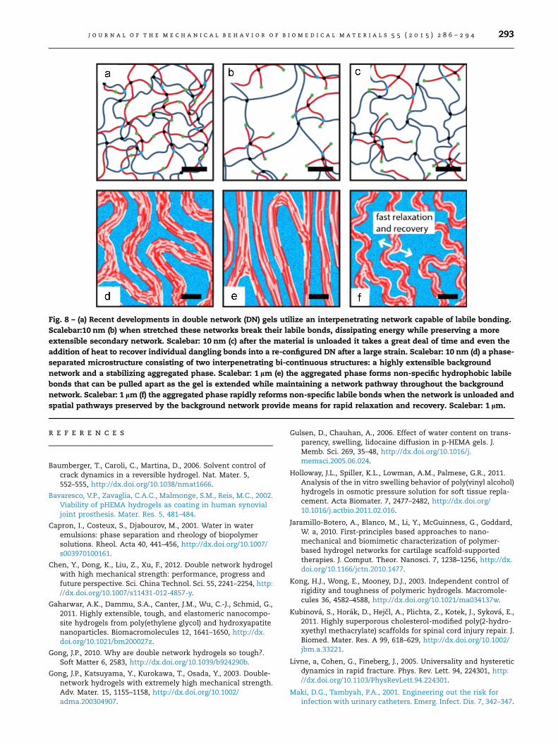
Fig. 8 – (a) Recent developments in double network (DN) gels utilize an interpenetrating network capable of labile bonding.Scalebar:10 nm (b) when stretched these networks break their labile bonds, dissipating energy while preserving a moreextensible secondary network. Scalebar: 10 nm (c) after the material is unloaded it takes a great deal of time and even theaddition of heat to recover individual dangling bonds into a re-configured DN after a large strain. Scalebar: 10 nm (d) a phase-separated microstructure consisting of two interpenetrating bi-continuous structures: a highly extensible backgroundnetwork and a stabilizing aggregated phase. Scalebar: 1 lm (e) the aggregated phase forms non-specific hydrophobic labilebonds that can be pulled apart as the gel is extended while maintaining a network pathway throughout the backgroundnetwork. Scalebar: 1 lm (f) the aggregated phase rapidly reforms non-specific labile bonds when the network is unloaded andspatial pathways preserved by the background network provide means for rapid relaxation and recovery. Scalebar: 1 lm.
j o u r n a l o f t h e m e c h a n i c a l b e h a v i o r o f b i o m e d i c a l m a t e r i a l s 5 5 ( 2 0 1 5 ) 2 8 6 – 2 9 4 293
r e f e r e n c e s
Baumberger, T., Caroli, C., Martina, D., 2006. Solvent control ofcrack dynamics in a reversible hydrogel. Nat. Mater. 5,552–555, http://dx.doi.org/10.1038/nmat1666.
Bavaresco, V.P., Zavaglia, C.A.C., Malmonge, S.M., Reis, M.C., 2002.Viability of pHEMA hydrogels as coating in human synovialjoint prosthesis. Mater. Res. 5, 481–484.
Capron, I., Costeux, S., Djabourov, M., 2001. Water in wateremulsions: phase separation and rheology of biopolymersolutions. Rheol. Acta 40, 441–456, http://dx.doi.org/10.1007/s003970100161.
Chen, Y., Dong, K., Liu, Z., Xu, F., 2012. Double network hydrogelwith high mechanical strength: performance, progress andfuture perspective. Sci. China Technol. Sci. 55, 2241–2254, http://dx.doi.org/10.1007/s11431-012-4857-y.
Gaharwar, A.K., Dammu, S.A., Canter, J.M., Wu, C.-J., Schmid, G.,2011. Highly extensible, tough, and elastomeric nanocompo-site hydrogels from poly(ethylene glycol) and hydroxyapatitenanoparticles. Biomacromolecules 12, 1641–1650, http://dx.doi.org/10.1021/bm200027z.
Gong, J.P., 2010. Why are double network hydrogels so tough?.Soft Matter 6, 2583, http://dx.doi.org/10.1039/b924290b.
Gong, J.P., Katsuyama, Y., Kurokawa, T., Osada, Y., 2003. Double-network hydrogels with extremely high mechanical strength.Adv. Mater. 15, 1155–1158, http://dx.doi.org/10.1002/adma.200304907.
Gulsen, D., Chauhan, A., 2006. Effect of water content on trans-parency, swelling, lidocaine diffusion in p-HEMA gels. J.Memb. Sci. 269, 35–48, http://dx.doi.org/10.1016/j.memsci.2005.06.024.
Holloway, J.L., Spiller, K.L., Lowman, A.M., Palmese, G.R., 2011.Analysis of the in vitro swelling behavior of poly(vinyl alcohol)hydrogels in osmotic pressure solution for soft tissue repla-cement. Acta Biomater. 7, 2477–2482, http://dx.doi.org/10.1016/j.actbio.2011.02.016.
Jaramillo-Botero, A., Blanco, M., Li, Y., McGuinness, G., Goddard,W. a, 2010. First-principles based approaches to nano-mechanical and biomimetic characterization of polymer-based hydrogel networks for cartilage scaffold-supportedtherapies. J. Comput. Theor. Nanosci. 7, 1238–1256, http://dx.doi.org/10.1166/jctn.2010.1477.
Kong, H.J., Wong, E., Mooney, D.J., 2003. Independent control ofrigidity and toughness of polymeric hydrogels. Macromole-cules 36, 4582–4588, http://dx.doi.org/10.1021/ma034137w.
Kubinova, S., Horak, D., Hejcl, A., Plichta, Z., Kotek, J., Sykova, E.,2011. Highly superporous cholesterol-modified poly(2-hydro-xyethyl methacrylate) scaffolds for spinal cord injury repair. J.Biomed. Mater. Res. A 99, 618–629, http://dx.doi.org/10.1002/jbm.a.33221.
Livne, a, Cohen, G., Fineberg, J., 2005. Universality and hystereticdynamics in rapid fracture. Phys. Rev. Lett. 94, 224301, http://dx.doi.org/10.1103/PhysRevLett.94.224301.
Maki, D.G., Tambyah, P.A., 2001. Engineering out the risk forinfection with urinary catheters. Emerg. Infect. Dis. 7, 342–347.

j o u r n a l o f t h e m e c h a n i c a l b e h a v i o r o f b i o m e d i c a l m a t e r i a l s 5 5 ( 2 0 1 5 ) 2 8 6 – 2 9 4294
Murakami, T., Higaki, H., Sawae, Y., Ohtsuki, N., Moriyama, S.,Nakanishi, Y., 1998. Adaptive multimode lubrication in naturalsynovial joints and artificial joints. Proc. Inst. Mech. Eng. PartH J. Eng. Med. 212, 23–35, http://dx.doi.org/10.1243/0954411981533791.
Na, Y.-H., Kurokawa, T., Katsuyama, Y., Tsukeshiba, H., Gong, J.P.,Osada, Y., Okabe, S., Karino, T., Shibayama, M., 2004. Struc-tural characteristics of double network gels with extremelyhigh mechanical strength. Macromolecules 37, 5370–5374,http://dx.doi.org/10.1021/ma049506i.
Naficy, S., Brown, H.R., Razal, J.M., Spinks, G.M., Whitten, P.G.,2011. Progress toward robust polymer hydrogels. Aust. J.Chem. 64, 1007, http://dx.doi.org/10.1071/CH11156.
Nakamura, 1976. Effects of various salts on the mechanicalproperties of homogeneous Poly(2-hydroxyethyl methacry-late) hydrogels. Polym. J.
Rongen, J.J., van Tienen, T.G., van Bochove, B., Grijpma, D.W.,Buma, P., 2014. Biomaterials in search of a meniscus substi-tute. Biomaterials 35, 3527–3540, http://dx.doi.org/10.1016/j.biomaterials.2014.01.017.
Sachlos, E., Czernuszka, J.T., 2003. Making tissue engineeringscaffolds work. Review: the application of solid freeformfabrication technology to the production of tissue engineeringscaffolds. Eur. Cells Mater. 5, 29–39 discussion 39–40.
Sefca, L., Pradny, M., Vacık, J., Michalek, B., Povysil, C., Vıtkova, I.,Halaska, M., Simon, V., 2002. Development of hydrogelimplants for urinary incontinence treatment. Biomaterials 23,3711–3715.
Spiller, K.L., Maher, S. a, Lowman, A.M., 2011. Hydrogels for therepair of articular cartilage defects. Tissue Eng. Part B Rev. 17,281–299, http://dx.doi.org/10.1089/ten.TEB.2011.0077.
Sun, J.-Y., Zhao, X., Illeperuma, W.R.K., Chaudhuri, O., Oh, K.H.,Mooney, D.J., Vlassak, J.J., Suo, Z., 2012. Highly stretchable andtough hydrogels. Nature 489, 133–136, http://dx.doi.org/10.1038/nature11409.
Tanaka, Y., 2007. A local damage model for anomalous hightoughness of double-network gels. Europhys. Lett. 78, 56005,http://dx.doi.org/10.1209/0295-5075/78/56005.
Tanaka, Y., Fukao, K., Miyamoto, Y., 2000. Fracture energy of gels.Eur. Phys. J. E 401, 395–401.
Wang, Q., Hou, R., Cheng, Y., Fu, J., 2012. Super-tough double-network hydrogels reinforced by covalently compositing withsilica-nanoparticles. Soft Matter 8, 6048, http://dx.doi.org/10.1039/c2sm07233e.
Webber, R.E., Creton, C., Polyme, L.D.P., Vauquelin, R., Brown, H.R.,2007. Large strain hysteresis and mullins effect of toughdouble-network hydrogels. Macromolecules 40, 2919–2927.
Ye, L., Tang, Y., Qiu, D., 2014. Enhance the mechanical perfor-mance of polyacrylamide hydrogel by aluminium-modifiedcolloidal silica. Colloids Surf. A Physicochem. Eng. Asp. 447,103–110, http://dx.doi.org/10.1016/j.colsurfa.2014.01.072.
Yin, H., Akasaki, T., Lin Sun, T., Nakajima, T., Kurokawa, T.,Nonoyama, T., Taira, T., Saruwatari, Y., Ping Gong, J., 2013.Double network hydrogels from polyzwitterions: highmechanical strength and excellent anti-biofouling properties.J. Mater. Chem. B 1, 3685, http://dx.doi.org/10.1039/c3tb20324g.