mechanical principles governing pollen tube growth
TRANSCRIPT

Received: 26 April, 2007. Accepted: 15 August, 2007. Invited Review
Functional Plant Science and Biotechnology ©2007 Global Science Books
Mechanical Principles Governing Pollen Tube Growth
Youssef Chebli • Anja Geitmann*
Institut de Recherche en Biologie Végétale, Département de Sciences Biologiques, Université de Montréal, 4101 rue Sherbrooke est, Montréal, Québec H1X 2B2, Canada
Corresponding author: * [email protected]
ABSTRACT Cellular growth and morphogenesis are central aspects of cellular differentiation. In plants, cellular growth is based on the turgor driven expansion of the cell wall and concomitant addition of new cell wall material. In no plant cell does this process occur as rapidly as in the pollen tube, the carrier of the male gametes. This cell is therefore an excellent model system to investigate the processes governing the dynamics of plant cell growth. This review provides a brief overview of the anatomy of the pollen tube focusing on the structural features that are implicated in the growth process – the cell wall and the cytoskeleton as well as spatially focused exocytotic events. The mechanics of the growth process is discussed and various theoretical modeling approaches that explain this process are outlined. In pollen tubes from many plant species the growth process is oscillatory or pulsatory and the ions and signaling molecules that form controlling feedback loops in this growth process are analyzed. A model that explains the oscillatory mechanism based on its mechanical components and that results from converging available data and hypotheses is elaborated. _____________________________________________________________________________________________________________ Keywords: cell mechanics, cellular growth, cell wall, cytoskeleton, feedback mechanism, ions, modeling, oscillation, pollen tube, tip growth, turgor Abbreviations: ATP, adenosine triphosphate; ABP, actin binding protein; ADF, actin depolymerizing factor; ER, endoplasmic reticu-lum; GFP, green fluorescent protein; GTP, guanosine triphosphate; NAD, nicotinamide adenine dinucleotide; PME, pectin methyl-esterase; PtdInsP2, phosphatidylinositol 4,5-bisphosphate; TIRF, total internal reflection fluorescence CONTENTS INTRODUCTION...................................................................................................................................................................................... 232 POLARITY IS REFLECTED IN THE CYTOARCHITECTURE............................................................................................................. 233
Cytoplasm.............................................................................................................................................................................................. 233 Cytoskeleton.......................................................................................................................................................................................... 233 Cell wall ................................................................................................................................................................................................ 234
MECHANICS OF ANISOTROPIC GROWTH......................................................................................................................................... 235 THEORETICAL MODELS FOR UNIDIRECTIONAL GROWTH.......................................................................................................... 235 CONSTRUCTION OF THE APICAL CELL WALL: EXO-/ENDOCYTOSIS......................................................................................... 235 MECHANICS OF OSCILLATORY GROWTH ........................................................................................................................................ 236
i) Fluctuations in cell wall physical properties ...................................................................................................................................... 237 ii) Turgor surges..................................................................................................................................................................................... 238 Vesicle fusion ........................................................................................................................................................................................ 239
OSCILLATORY GROWTH - CONVERGING THE MODELS ............................................................................................................... 239 ION-BASED PARAMETERS INFLUENCING OSCILLATORY GROWTH .......................................................................................... 240
Calcium ions.......................................................................................................................................................................................... 240 Protons................................................................................................................................................................................................... 241 Potential players: Chloride and potassium ions ..................................................................................................................................... 241
OTHER OSCILLATING PARAMETERS................................................................................................................................................. 241 NAD(P)H............................................................................................................................................................................................... 241 Small GTPases and actin ....................................................................................................................................................................... 242 Phospholipase C .................................................................................................................................................................................... 242
POLLEN TUBE GROWTH IN PLANTA - GUIDANCE AND INVASION .............................................................................................. 242 CONCLUSIONS........................................................................................................................................................................................ 242 ACKNOWLEDGEMENTS ....................................................................................................................................................................... 243 REFERENCES........................................................................................................................................................................................... 243 _____________________________________________________________________________________________________________ INTRODUCTION The pollen tube is formed upon contact of a pollen grain with a receptive stigma. The grain swells through water up-take and forms a cellular protrusion that invades the pistil. The purpose of this process is the transport of the male gametes from their carrier vehicle – the pollen grain – to the ovule where fertilization takes place. Depending on the spe-
cies, pollen tube growth can occur extremely rapidly with rates up to tens and even hundreds of micrometers per minute. Very conveniently for the researcher, pollen grains are able to form these tubular protrusions in vitro, albeit usually not with the impressive growth rates observed in planta. However, cellular morphology of the former resem-bles that of the latter, which is why this single cell is an extraordinary system to investigate processes involved in

Functional Plant Science and Biotechnology 1(2), 232-245 ©2007 Global Science Books
plant cell growth. Most types of plant cells grow by expanding large
surface areas simultaneously. In the cylindrical cells com-posing stem tissues, this expansion occurs over the entire cylindrical surface, whereas only the end walls remain almost unaltered in size. Other cell types such as pavement cells in the leaf epidermis grow at more spatially confined sites to generate the typical jigsaw puzzle shape (Mathur 2006). Pollen tubes represent an extreme example of spa-tially confined growth since cellular expansion is limited to a single very small area at the apex of the growing cell. This mode of growth makes sense from an energetic point of view, because pollen tubes need to invade the transmitting tissue of the receptive flower. If the cylindrical wall of the tube expanded during growth, friction would occur against the surrounding tissue. Confining expansion to the apex minimizes the surface area on which friction occurs. This growth strategy is shared by several other cell types, which incidentally also have an invasive way of life. Among them are root hairs and fungal hyphae, and certain parallels can also be drawn to neuronal growth cones (Palanivelu and Preuss 2000). This illustrates that the tip growth strategy is realized in evolutionary very distant organisms.
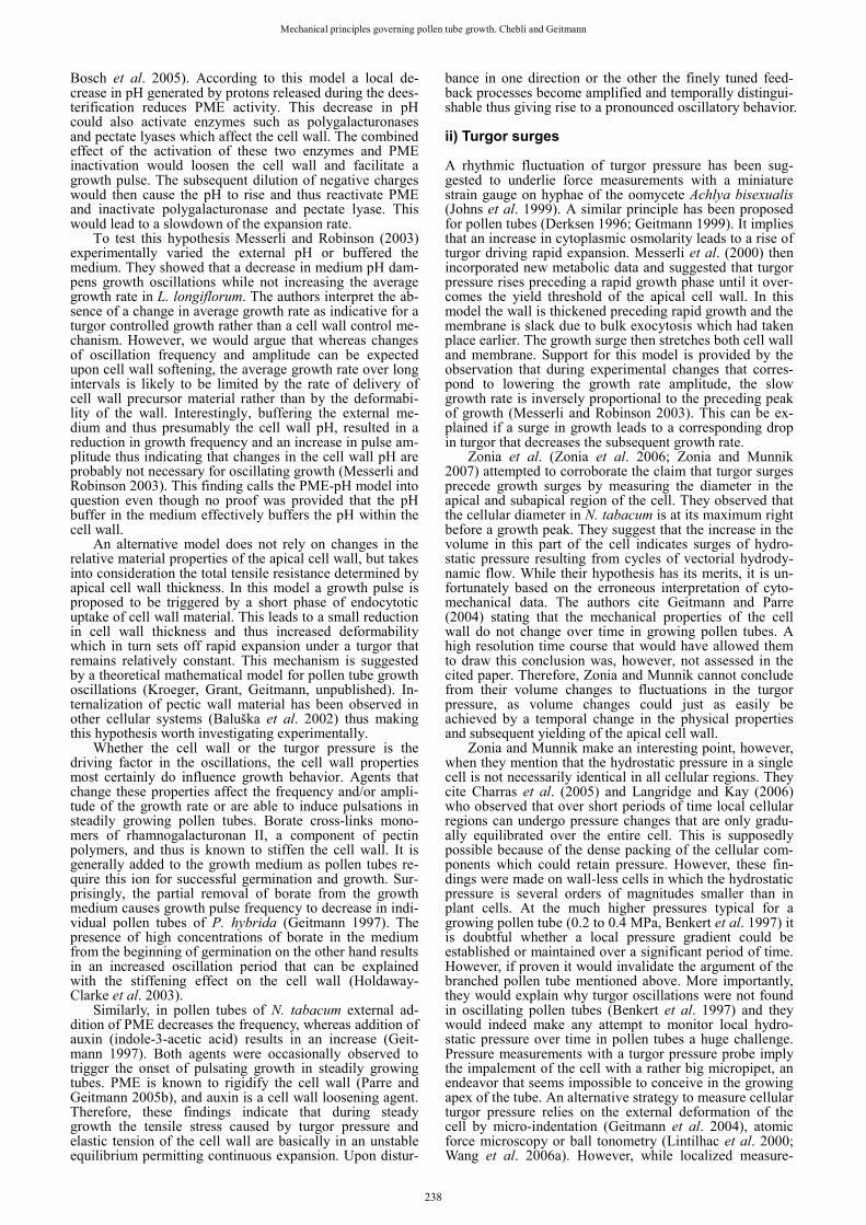
Since pollen tubes are easily cultivated in vitro, and since even under this suboptimal condition their growth rate is impressive, this cell type has become an important model system for the investigation of the processes that govern plant cell growth. Numerous labs investigate different as-pects of pollen tube growth which have been reviewed in countless articles and books (the most recent collection of articles can be found in Malhó (2006)). In the present re-view we will give an overview of the understanding of the mechanics and dynamics of the growth process. We will briefly present the structural features of the pollen tube and how their spatial distribution accounts for the highly aniso-tropic mechanism of growth. We will discuss the molecular feedback mechanisms that govern growth focusing on a particularity of the dynamics of the growth process – the oscillating change of the growth rate. POLARITY IS REFLECTED IN THE CYTOARCHITECTURE Cytoplasm The anatomy of the pollen tube reflects the extreme pola-rization of the cellular processes. Starting from the apex, which is also called the growth zone, several distinct re-gions can be distinguished in the cytoplasm (Fig. 1). The apical dome is filled densely with secretory vesicles. The only other organelles that can occasionally be observed here are mitochondria and cisternae of the endoplasmic reticu-lum. The adjacent subapical and distal regions are densely populated by cellular organelles which move in a direction parallel to the longitudinal axis of the cell – towards or from the apex. This movement is rather rapid with rates of seve-ral micrometers per second. Most of these organelles turn around once they reach the subapical region (Fig. 1A). In larger pollen tubes such as lily, this rearward movement oc-curs mostly in the central area of the cytoplasm, and the flux is therefore characterized as "inverse fountain stream-ing" (Hepler et al. 2001). The shank of the tube contains the male germ unit consisting of the vegetative nucleus and the generative cell or, after its division, the two sperm cells. In longer pollen tubes, the viable part of the cytoplasm is con-centrated in the region close to the apex and it is separated from degenerating distal regions and the pollen grain by callosic plugs.
This non-uniform distribution of cellular organelles is indicative of the compartmentalization of cellular functions (Cheung and Wu 2007), in particular with the growth zone being visibly different from the other regions of the cell. The polarity of the cell is also expressed in the non-uniform distribution of the concentrations of numerous molecules and ions and of the activities of certain enzymes. Among
the principal players is calcium, which is highly concentra-ted in the apex and lower in the shank (Fig. 1C). Equally important are protons whose uneven distribution results in a pH profile that is characterized by a subapical alkaline band (Holdaway-Clarke and Hepler 2003; Fig. 1D). Cytoskeleton The pollen tube cytoskeleton is mainly composed of actin filaments and microtubules. Both are oriented in approxi-mately longitudinal (or slightly helical) direction. Actin is found in the cortical cytoplasm as well as the endoplasm (Geitmann and Emons 2000). The shank of the tube in most species is characterized by the presence of numerous con-spicuous actin bundles. On the other hand, the precise con-figuration of actin in the subapical region and in the apex varies between species as well as with the method of visu-alization. Important information has been gained from im-proved chemical or rapid freeze fixation combined with phalloidin staining, but dynamic aspects could only be studied using transformation with GFP (green fluorescent protein; or its derivatives) coupled to actin binding proteins (Kost et al. 1998; Chen et al. 2002). GFP coupled directly to actin has not been successful for pollen so far. The consensus of these studies is that angiosperm pollen tubes possess a subapical domain in which actin filament bundles are thinner and denser (also called actin fringe), whereas no significant amounts of filamentous actin are present in the very tip of the tube (Fig. 1E). The resolution of fluores-cence images does not allow the precise characterization of the actin configuration in the fringe, not even when fixation is optimized (Lovy-Wheeler et al. 2005). A possible inter-pretation of the images could be that the front of the fringe corresponds to the advancing polymerization front, whereas the rear end represents the advancing actin bundling front (Fig. 1E). Alternatively, the fringe could represent a region of high degree of branching and the rear end might be a re-gion of increased depolymerization. These hypotheses are not mutually exclusive. Depending on the degree of bund-ling either by the pollen tube’s own protein machinery or by agents used to fix and stabilize actin, this fringe could thus appear longer or even funnel shaped as has been observed in several species such as maize, poppy and lily (Gibbon et al. 1999; Geitmann et al. 2000; Foissner et al. 2002). It will be interesting to see whether the corresponding actin bin-ding proteins can be localized specifically to these "front" regions within the fringe that would correspond to high polymerization, depolymerization and/or bundling activities.
Several actin binding proteins (ABPs) act in the pollen cytoplasm to regulate actin polymerization and depolymeri-zation. Villins have been proposed to degrade actin fila-ments in the apical region where the cytoplasmic Ca2+ con-centration is higher thus resulting in a tip region that is rela-tively free of filamentous actin. In the distal region, where the Ca2+ concentration is at its basal levels, villin may act as a cross-linker, thus stabilizing the actin network (Kost et al. 1998; Yokota et al. 1998; Vidali et al. 1999; Yokota et al. 2000; Vidali et al. 2001). Profilin has also been shown to bind G-actin in the apical region, thus preventing actin polymerization (Kovar et al. 2000). Villin and profilin have a role in regulating the polymerization of actin filament bundles in root hairs (Baluška et al. 2000).
Actin depolymerizing factor (ADF) and cofilin are two ABPs that have the ability to segment and to depolymerize actin filaments. The segmentation activity of the ADF and the depolymerization activity of cofilin are both regulated by pH thus pointing to an important regulatory function of the pH profile (Lovy-Wheeler et al. 2006). Other ABPs were described in pollen tubes such as α-actinin (actin fila-ment/cell wall link), formins (polymerization of actin fila-ments) and myosins. Several recent review articles on pol-len tube actin and ABPs are available (Vantard and Blan-choin 2002; Staiger and Blanchoin 2006; Yokota and Shim-men 2006; Ren and Xiang 2007).
Microtubules are longitudinally oriented and sometimes
233

Mechanical principles governing pollen tube growth. Chebli and Geitmann
adopt a slight helical distribution (Geitmann and Emons 2000). They extend from the pollen grain to the subapical region. In contrast to root hairs, microtubules are absent from the apical domain of angiosperm pollen tubes (Lan-celle and Hepler 1992). The role of microtubules in pola-rized growth may be the control of directionality, because taxol (stabilizing agent) and oryzalin (destabilizing drug) have similar effects on the directionality of tube growth (Hepler et al. 2001; Gossot and Geitmann 2007). Recent re-views that summarize the role of microtubules in pollen tube tip growth include Geitmann and Emons (2000), Rau-daskoski et al. (2001), and Cai et al. (2005). Cell wall The pollen tube cell wall is mainly composed of polysac-charides. The main component is pectin which is deposited
at the apex by exocytosis. Cellulose and callose are laid down in more distal regions by plasma membrane localized synthases. In the case of Nicotiana tabacum cellulose depo-sits start at 5 to 15 µm from the tip whereas callose is visi-ble starting at a distance of approximately 30 µm from the tip (Ferguson et al. 1998). The pattern of callose deposition is very universal, whereas there are considerable species dependent differences for cellulose localization. In Arabi-dopsis thaliana calcofluor label has indicated the presence of microfibrils in the apex (Derksen et al. 2002) whereas the apex of other species is devoid of cellulose.
Because of the general absence of a conspicuous "sec-ondary" deposition of cell wall components in the apex, the overall biochemical structure of the cell wall differs consi-derably between the growing apex and the cylindrical shank of the cell (Fig. 1G). This difference is even more pro-nounced due to the gradual change in the configuration of
Fig. 1 Schematic representation of the polar arrange-ment of cellular structures and processes in the api-cal and subapical regions of a growing pollen tube. Because of the radial symmetry of the cell only half of a median section is drawn. Objects are not to scale. (A) Cytoplasmic streaming occurs parallel to the long axis. In larger tubes forward movement takes place in the cor-tex and rearward movement in the center of the tube. Vesicle movements in the apex are more erratic. (B) Secretory vesicles (green) accumulate in the apical region were exocytosis takes place. Rapid endocytosis likely takes place in the entire apex, but clathrin-medi-ated endocytosis (yellow vesicles) occurs predominantly at the base of the apex. (C) The cytosolic calcium con-centration is high at the very apex and drops drastically towards the shank. Calcium influx occurs through the apical plasma membrane. It is unclear which organelles sequester calcium and to what degree calcium release from organelles contributes to the calcium gradient. (D) Proton influx occurs at the tip and efflux in the subapi-cal region. This leads to the formation of an alkaline band that corresponds approximately to the turnaround point of cytoplasmic streaming and to the actin fringe. (E) Filamentous actin is arranged predominantly in lon-gitudinal direction. Long cables characterize the shank region, whereas in the subapex actin seems to be branched or less bundled giving rise to a fringe. (F) According to the laws of thin-walled pressure vessels, the tensile stress in the wall generated by the internal pressure is twice as high in circumferential direction (SH) compared to longitudinal direction (SL) or the approximately hemisphere shaped tip (SS). Shade of orange indicates the degree of cellular stiffness. (G) The biochemical composition of the cell wall changes from the apex to the distal region through decreasing methyl-esterification of pectins and addition of callose and cellulose.
234

Functional Plant Science and Biotechnology 1(2), 232-245 ©2007 Global Science Books
the pectins as they become part of the mature region of the shank. Labeling with monoclonal antibodies JIM5 and JIM7 has revealed that in growing angiosperm pollen tubes pectins have a higher degree of methyl-esterification in the tip than in distal regions (Li et al. 1994). For reviews on pollen tube cell wall structure see Heslop-Harrison (1987), Taylor and Hepler (1997), and Geitmann and Steer (2006). MECHANICS OF ANISOTROPIC GROWTH In general, the process of plant cell growth is driven by the relationship between turgor pressure, controlled water up-take and mechanical cell wall resistance. The controlled yielding of the existing cell wall under the applied pressure leads to an expansion of the cellular surface while simul-taneously new cell wall material is inserted. The combina-tion of these two processes results in a change of cellular shape. The situation is similar in a tip growing cell, with the particularity that the cellular expansion is confined to an extremely small region of the cellular surface. Similarly to diffuse growth, turgor is believed to be the primary motive force behind tip growth, albeit it might not be the rate-controlling parameter as growth rates cannot directly be correlated with the amount of turgor present in Lilium longiflorum Thunb (Benkert et al. 1997).
Given that hydrostatic pressure is a non-vectorial force, the question arises, how it can push a cell to produce a tubular protuberance instead of becoming a ballooning sphere. Green (Green 1969) and others have proposed that tip-localized expansion must be caused by tip-to-base chan-ges in the physical properties of the wall. In other words, the cell wall in the shank of the tube needs to be more resistant to tensile stress than the apical cell wall to assure that the latter yields first thus allowing for tip-confined expansion. However, for geometrical reasons this differ-ence actually has to be bigger than a factor 2. According to the physical laws for thin-walled pressure vessels, tensile wall stress in circumferential direction is twice as high as the tensile stress in longitudinal direction or that in the hemisphere-shaped ends of a cylindrical vessel (Geitmann and Steer 2006; Fig. 1F). The generation of a tubular struc-ture therefore requires a considerable difference in the yield threshold between the shank and the apex. This dif-ference in the physical properties of the pollen tube cell wall has been demonstrated by micro-indentation for Papa-ver rhoeas (Geitmann and Parre 2004) and other species.
Several cell wall components are likely to be responsi-ble for this mechanical gradient in the pollen tube cell wall due to their non-uniform distribution (Heslop-Harrison 1987; Ferguson et al. 1998; Fig. 1G). Since both callose and cellulose are present predominantly in the shank of the tube they are presumed to play a reinforcing role in this part of the cell. Micro-indentation data as well as enzy-matic and pharmacological approaches confirm this (An-derson et al. 2002; Parre and Geitmann 2005a). An even more important role is played by pectins and the spatial distribution of the degree of their methyl-esterification. Concerning the physical properties of the cell wall this is an essential feature as de-esterification allows pectins to become gelated in the presence of calcium ions. In vitro, this gelation process considerably increases the Young’s modulus of this matrix component (Jarvis 1984), thus po-tentially increasing resistance against tensile stress. Micro-indentation revealed that this is also true for in vivo pollen tube cell walls (Parre and Geitmann 2005b). Furthermore, the enzymatic de-esterification and hardening of the usu-ally rather soft apical wall is able to prevent pollen tube from elongating (Bosch et al. 2005; Parre and Geitmann 2005a), thus confirming the crucial role of the physical gradient in the mechanical properties of the cell wall for sustained growth.
THEORETICAL MODELS FOR UNIDIRECTIONAL GROWTH The unidirectional growth typical for pollen tubes and other tip growing cells has inspired many attempts to model this process. Most of these were applied to fungal hyphae (Bartnicki-Garcia 2002), but the similarity between the cell types is such that the models could readily be trans-ferred from one to the other. The early theoretical models for tip growth are based on equations that approximate the shape of these cells very closely. However, most of them are limited to geometric exercises that formulate equations from artificial coordinates and reference points (Reinhardt 1892; da Riva Ricci and Kendrick 1972; Trinci and Saun-ders 1977; Prosser and Trinci 1979; Koch 1982, 1994; Prosser 1994; Denet 1996). Few tried to take into consi-deration the geometrical and physical parameters of subcel-lular structures such as the thickness and chemical compo-sition of the cell wall, the precise location of vesicle fusion or the hydrostatic turgor pressure. The vesicle supply cen-ter model for fungal hyphae by Bartnicki-Garcia et al. (1989) was an attempt to develop a mathematical model based on the intracellular path and secretion of secretory vesicles. The original two-dimensional mathematical for-mulation was based on the concept that tip growth is pro-duced by wall-building vesicles emanating randomly, and in all directions, from a vesicle supply center which advan-ces moving along a straight path. The subsequent three-dimensional derivation of this model included quantitative measurements of the pattern of expansion of the wall (Bartnicki-Garcia et al. 2000; Gierz and Bartnicki-Garcia 2001). Observations of surface expansion are also the basis of models developed by Dumais and co-workers for Medi-cago truncatula root hairs and further versions account for growth fluctuations and changes in morphology (Dumais et al. 2004, 2006). This was achieved by rescaling a given wall extensibility profile over time. The same group found interesting parallels between the tip expansion of lily pollen tubes and rubber balloons swelling under increasing internal pressure (Bernal et al. 2007). While the model by Dumais et al. considers the continuous supply of cell wall material to be provided in bulk form at the very tip of the cell, the vesicle supply center model was further developed by exchanging the theoretical ballistic path of secretory vesicles to the apical membrane for a more realistic dif-fusive vesicle delivery mechanism (Tindemans et al. 2006).
While being able to explain the formation of a cylin-drical tube, many of these models nevertheless fail to in-clude parameters important for the functioning of the living cell such as the turgor pressure and the non-uniform bio-chemical composition of the cell wall. An internal hydro-static pressure was included in the approach by Goriely and Tabor who modeled the cell wall of a tip growing cell as a stretchable and growing elastic membrane with geometry-dependent elastic properties. They used large-deformation elasticity theory and combined the elastic response with surface re-parameterization to simulate wall rebuilding (Goriely and Tabor 2003a, 2003b).
Few of these models are directly based on geometrical and physiological data obtained specifically from pollen tubes. To allow for experimental validation, future model-ing attempts will require precise quantitative data on the pollen tube geometrical features and the mechano-physical properties of its subcellular components. Cytomechanical approaches will therefore certainly gain popularity in this field (Geitmann 2006a, 2006b). CONSTRUCTION OF THE APICAL CELL WALL: EXO-/ENDOCYTOSIS To allow for the construction of the ever elongating tube, new cell wall material as well as membrane bound and secretory proteins need to be transported to the tip in a continuous manner and at a high rate. Exocytosis is there-fore a sine qua non condition for pollen tube growth. In-
235

Mechanical principles governing pollen tube growth. Chebli and Geitmann
hibiting the vesicle supply by adding brefeldin A or mo-nensin arrests pollen tube growth within a few minutes – presumably upon depletion of the apical stock of vesicles (Geitmann et al. 1996). The cell wall material deposited at the tip consists largely of pectin polymers which are trans-ported to the apex within vesicles. This transport occurs via interactions between motor proteins linking the vesicle sur-face to the cytoskeleton. The actin cytoskeleton is thought to play a major role in this context and myosins have been identified in pollen tubes (Yokota and Shimmen 1994; Shimmen et al. 2000). However, dynein- and kinesin-like proteins have also been localized on pollen tube organelles indicating a transport function for microtubules as well (Moscatelli et al. 1998; Romagnoli et al. 2003, 2007). For reviews on motor proteins in pollen see Cai et al. (1996, 1997, 2005).
While the long-distance transport of vesicles along the cytoskeletal rail system in the tube shank is rather well understood, knowledge about the subsequent steps is scant. Once delivered to the apical region, vesicles seem to ac-cumulate and somehow find their way to the apical mem-brane where they dock, fuse, and liberate their contents. Vesicle movement in the vesicle-rich growth zone was ini-tially described as Brownian in Nicotiana tabacum (de Win et al. 1999). However, the situation is probably more com-plex since TIRF (total internal reflection fluorescence) microscopy has revealed that the movement in Picea mey-eri pollen tubes is not random and seems to depend on a functional actin cytoskeleton despite the absence of conspi-cuous F-actin configurations in this area (Wang et al. 2006b). The apical actin cytoskeleton, possibly in form of dynamic individual F-actin arrays which are difficult to visualize, is therefore likely to be involved in guiding the vesicles to the designated fusion sites at the membrane.
Vesicle fusion must be spatially and temporally con-trolled. Spatial control serves to determine the direction of growth (Geitmann and Palanivelu 2007) and temporal con-trol is likely to influence the dynamics of the growth rate. A crucial role in this control mechanism is said to be played by calcium (discussed below) which in turn acts via its activating or de-activating effect on a number of pro-teins. The signaling pathways targeting secretion are also thought to involve the inositide pathway and phosphory-lation cascades (reviewed by Malhó et al. 2000, 2005, 2006; Geitmann and Palanivelu 2007).
Among the proteins that have been proposed to interact directly in vesicle secretion are annexins, SNAREs, and the members of the exocyst complex. Annexins are able to bind to, aggregate, and fuse secretory vesicle membranes in a calcium-dependent manner. They were immunolocalized in the vesicle-rich zone of L. longiflorum pollen tubes (Blackbourn et al. 1992) and are thus likely to be involved in the tip growth process. Annexin action is calcium depen-dent (Trotter et al. 1995) thus suggesting that the cytosolic high calcium concentration plays a role in directing vesicle fusion events.
The exocyst, first described in yeast (Novick et al. 1995) is composed of eight proteins functioning as a tether-ing complex guiding secretory vesicles to their specific plasma membrane site prior to the docking and fusion events mediated by SNARE proteins (Cole et al. 2005). In yeast, the exocyst was found to be associated with the plas-ma membrane at the site of cell-surface expansion (Novick et al. 1995). While orthologs of all exocyst components have been identified in Arabidopsis and rice, their role in tip growth has yet to be elucidated (Cole et al. 2005; Cole and Fowler 2006). However, mutants of SEC8, one of the exocyst components, were found to affect pollen tube growth or germination (Cole et al. 2005) and further in-vestigations are likely to provide more insight.
SNAREs (soluble N-ethyl-maleimide-sensitive fusion protein attachment protein receptors) are integral mem-brane proteins. In plants, fusion of the vesicle with the tar-get membrane depends on activated SNARE molecules in the vesicle (v-SNARE) and target (t-SNARE) membranes
(Sanderfoot and Raikhel 1999; Nebenführ 2002). While SNAREs were found to be implicated in numerous plant functions (Pratelli et al. 2004), they have not been charac-terized or localized in pollen tubes in the context of exo-cytosis. Given the intensive secretory activity in these cells, their involvement is likely, however.
Two types of exocytosis have been proposed to occur in pollen tubes: full fusion and transient fusion. Using TIRF microscopy, Wang et al. (2006b) observed the full fusion type in Picea pollen tubes involving the collapse of the vesicles into the membrane as they release their internal components. Transient exocytosis and the resulting rapid endocytosis were proposed to occur in pollen tubes of vari-ous angiosperm species (Malhó et al. 2005). This exocy-tosis mechanism is a Ca2+-dependent process coupled to endocytosis which requires GTP hydrolysis and dynamin but not clathrin (Monteiro et al. 2005). It is characterized by the formation of a small and short-lived pore which limits the size of particles that can be released or incorpo-rated.
The observation of transient exocytosis points to the initially surprising finding that despite its rapid cell wall production rate and growth sustained by massive exocy-tosis, the pollen tube actually internalizes material at the tip at the same time. An explanation lies in the fact that an excess of membrane material is transported to the tip of the pollen tubes during exocytosis of cell wall material (Picton and Steer 1983b, 1985; Steer 1988; Derksen et al. 1995). Therefore, endocytosis of membrane is a necessity to main-tain the relatively smooth outline of the apical plasma membrane. Next to rapid endocytosis, clathrin-mediated endocytosis is also purported to occur based on the pre-sence of coated pits and vesicles carrying a clathrin-like coat in the tip area of tobacco pollen tubes (Derksen et al. 1995). The highest concentration of coated pits was ob-served in a region 6-15 µm behind the tip in N. tabacum pollen tubes (Fig. 1B). The bulk of endocytosis is therefore purported to occur not at the very apex, but slightly towards the shank. Both, ultrastructural research and, more recently, live cell observations that use fluorescent dyes which are internalized by endocytosis have contributed im-portant information on the dynamics and spatial distribu-tion of these events (reviewed in Malhó et al. 2005). MECHANICS OF OSCILLATORY GROWTH In addition to their rapid elongation, pollen tubes exhibit a dynamic feature which makes the investigation of their growth process extremely attractive: their growth rates fluctuate in regular intervals. This allows the investigation of the feedback mechanisms governing growth as signaling steps and physiological processes can be presumed to show temporal variations in intensity. Cross-correlation analysis of the phase shifts between these events and the growth rate should theoretically allow the identification of cause-effect relationships. A compilation of the parameters that have been assessed in this context is provided in Fig. 2.
While most pollen tubes show a non-continuous growth rate, the frequency and amplitude as well as the shape of the curve representing the growth rate varies con-siderably between species and depends on experimental conditions. Roughly, one can distinguish between oscil-latory growth characterized by a sinusoidal curve and pul-satory growth in which short growth spurts are separated by extended periods of slow growth. In L. longiflorum the oscillations are approximately sinusoidal with a frequency of 15-50 sec (Pierson et al. 1996). In N. tabacum and Petu-nia hybrida pollen tubes show slow growth phases lasting few minutes (typically 3 to 8) interrupted by pulse-like elongations lasting few seconds. Pollen tube elongation during these pulses can reach up to 2 μm in N. tabacum achieving a peak growth rate up to 0.5 μm.sec-1 (Geitmann 1997).
The species that is investigated in most detail is L. longiflorum. The most prominent oscillations are found in
236

Functional Plant Science and Biotechnology 1(2), 232-245 ©2007 Global Science Books
older tubes (>1 mm length). Lily pollen tubes are relatively easy to manipulate and to observe due to their considerable size (16-20 μm diameter vs. 6-10 µm in many other spe-cies). In the in vitro setup typical oscillations in this species have a period of 30-60 sec, and a growth rate oscillating between 0.1 and 0.4 μm.sec-1 (Messerli et al. 2004).
Despite the unidirectional manner of the pollen tube growth, these growth oscillations represent an ideal system to investigate the temporal relationship between mechani-cal events governing plant cell growth in general. The me-chanical principle behind this oscillatory behavior is based on the changing relationship between the turgor pressure and the apical cell wall over short periods of time. The question is, which of the two – turgor or cell wall mecha-nics – is the mechanical oscillator in this system?
From the mechanical point of view, two models have been proposed: i) fluctuations in the mechanical properties of the cell wall allowing its relaxation under an approxi-mately stable turgor, and ii) surges in hydrostatic pressure driving cell wall expansion. The former is based on the widely accepted understanding of plant cell growth as re-viewed by Schopfer (2006). The latter is based on the "Loss-of-stability" principle proposed by Wei and Lintilhac (2006). i) Fluctuations in cell wall physical properties This model is based on the observation that the hydrostatic turgor pressure in growing pollen tubes does not seem to oscillate in L. longiflorum (Benkert et al. 1997). This fin-ding is corroborated by the observation that two growing ends emanating from a single branched cell do not show the same oscillation frequency in Petunia hybrida (Geit-mann 1997). If turgor was the determining oscillator, these branched pollen tube ends should oscillate with the same
frequency, since the cell can be considered as a single vol-ume in which turgor pressure can be assumed to be iden-tical everywhere. It has therefore been suggested that the tensile strength of the apical cell wall and not the internal pressure varies during the oscillation cycles. This alterna-tion between softening and hardening might for example be caused by the secretion of new cell wall material with high plasticity which allows rapid expansion and subsequently hardens either due to strain hardening or enzymatic activity (Geitmann 1999). It is interesting in this context to note that the thickness of the cell wall changes during an oscil-lation cycle. The peak of thickness precedes the most rapid phase of the growth pulse (Holdaway-Clarke and Hepler 2003). This phase relationship is intuitively necessary, since it assures the presence of sufficient cell wall material to allow for the subsequent expansion of the cell wall without the resulting thinning that would otherwise lead to rupture. On the other hand it is surprising, as this would mean that despite being thicker, the tensile stress resistance in the wall would actually have to be lower to allow for rapid expansion to occur. This would require that the newly added material is extremely soft.
Alternatively, the mechanical properties of the apical cell wall material could be controlled by the oscillatory activity of enzymes. The configuration of pectins, the most abundant polymers at the tube tip, is affected by various enzymes. Pectin methyl esterase (PME) acts on the methyl-esterified pectins that are secreted at the growing apex (Bosch et al. 2005). The degree of esterification is essential for cell wall mechanics as unesterified pectins are able to bind calcium ions resulting in the gelation of the polymers. This process rigidifies the wall as revealed by micro-inden-tation (Parre and Geitmann 2005b). It has been proposed that PME activity at the pollen tube tip is subject to a nega-tive feedback mechanism (Holdaway-Clarke et al. 1997;
Fig. 2 Compilation of cellular features and processes that have been observed to undergo changes during oscillatory pollen tube growth. Note that these data have been acquired on pollen tubes from different plant species. Arrows indicate the phase relationships between peaks in concentration or flux rate of a particular parameter and peaks in growth rate. Phase shifts are indicated as degrees with 360° corresponding to a complete oscillation period. In most (but not all) cases this phase relationship has been established by cross-correlation analysis - a process that identifies whether a particular process is leading (negative numbers) or lagging (positive numbers) growth. Two classes of parameters can be distinguished: concentrations (orange frame) and movement rates (orange background). The latter are first derivatives of the former. This distinction is important when identifying cause-effect rela-tionships. This becomes evident for example when looking at the label intensity for secretory vesicles, which is highest at -68°. For this particular para-meter, however, more important than the concentration (the amount of vesicles), is the information that can be derived from the change of label intensity, since it corresponds to exocytosis activity, which in turn is a rate.
237
Mechanical principles governing pollen tube growth. Chebli and Geitmann
Bosch et al. 2005). According to this model a local de-crease in pH generated by protons released during the dees-terification reduces PME activity. This decrease in pH could also activate enzymes such as polygalacturonases and pectate lyases which affect the cell wall. The combined effect of the activation of these two enzymes and PME inactivation would loosen the cell wall and facilitate a growth pulse. The subsequent dilution of negative charges would then cause the pH to rise and thus reactivate PME and inactivate polygalacturonase and pectate lyase. This would lead to a slowdown of the expansion rate.
To test this hypothesis Messerli and Robinson (2003) experimentally varied the external pH or buffered the medium. They showed that a decrease in medium pH dam-pens growth oscillations while not increasing the average growth rate in L. longiflorum. The authors interpret the ab-sence of a change in average growth rate as indicative for a turgor controlled growth rather than a cell wall control me-chanism. However, we would argue that whereas changes of oscillation frequency and amplitude can be expected upon cell wall softening, the average growth rate over long intervals is likely to be limited by the rate of delivery of cell wall precursor material rather than by the deformabi-lity of the wall. Interestingly, buffering the external me-dium and thus presumably the cell wall pH, resulted in a reduction in growth frequency and an increase in pulse am-plitude thus indicating that changes in the cell wall pH are probably not necessary for oscillating growth (Messerli and Robinson 2003). This finding calls the PME-pH model into question even though no proof was provided that the pH buffer in the medium effectively buffers the pH within the cell wall.
An alternative model does not rely on changes in the relative material properties of the apical cell wall, but takes into consideration the total tensile resistance determined by apical cell wall thickness. In this model a growth pulse is proposed to be triggered by a short phase of endocytotic uptake of cell wall material. This leads to a small reduction in cell wall thickness and thus increased deformability which in turn sets off rapid expansion under a turgor that remains relatively constant. This mechanism is suggested by a theoretical mathematical model for pollen tube growth oscillations (Kroeger, Grant, Geitmann, unpublished). In-ternalization of pectic wall material has been observed in other cellular systems (Baluška et al. 2002) thus making this hypothesis worth investigating experimentally.
Whether the cell wall or the turgor pressure is the driving factor in the oscillations, the cell wall properties most certainly do influence growth behavior. Agents that change these properties affect the frequency and/or ampli-tude of the growth rate or are able to induce pulsations in steadily growing pollen tubes. Borate cross-links mono-mers of rhamnogalacturonan II, a component of pectin polymers, and thus is known to stiffen the cell wall. It is generally added to the growth medium as pollen tubes re-quire this ion for successful germination and growth. Sur-prisingly, the partial removal of borate from the growth medium causes growth pulse frequency to decrease in indi-vidual pollen tubes of P. hybrida (Geitmann 1997). The presence of high concentrations of borate in the medium from the beginning of germination on the other hand results in an increased oscillation period that can be explained with the stiffening effect on the cell wall (Holdaway-Clarke et al. 2003).
Similarly, in pollen tubes of N. tabacum external ad-dition of PME decreases the frequency, whereas addition of auxin (indole-3-acetic acid) results in an increase (Geit-mann 1997). Both agents were occasionally observed to trigger the onset of pulsating growth in steadily growing tubes. PME is known to rigidify the cell wall (Parre and Geitmann 2005b), and auxin is a cell wall loosening agent. Therefore, these findings indicate that during steady growth the tensile stress caused by turgor pressure and elastic tension of the cell wall are basically in an unstable equilibrium permitting continuous expansion. Upon distur-
bance in one direction or the other the finely tuned feed-back processes become amplified and temporally distingui-shable thus giving rise to a pronounced oscillatory behavior. ii) Turgor surges A rhythmic fluctuation of turgor pressure has been sug-gested to underlie force measurements with a miniature strain gauge on hyphae of the oomycete Achlya bisexualis (Johns et al. 1999). A similar principle has been proposed for pollen tubes (Derksen 1996; Geitmann 1999). It implies that an increase in cytoplasmic osmolarity leads to a rise of turgor driving rapid expansion. Messerli et al. (2000) then incorporated new metabolic data and suggested that turgor pressure rises preceding a rapid growth phase until it over-comes the yield threshold of the apical cell wall. In this model the wall is thickened preceding rapid growth and the membrane is slack due to bulk exocytosis which had taken place earlier. The growth surge then stretches both cell wall and membrane. Support for this model is provided by the observation that during experimental changes that corres-pond to lowering the growth rate amplitude, the slow growth rate is inversely proportional to the preceding peak of growth (Messerli and Robinson 2003). This can be ex-plained if a surge in growth leads to a corresponding drop in turgor that decreases the subsequent growth rate.
Zonia et al. (Zonia et al. 2006; Zonia and Munnik 2007) attempted to corroborate the claim that turgor surges precede growth surges by measuring the diameter in the apical and subapical region of the cell. They observed that the cellular diameter in N. tabacum is at its maximum right before a growth peak. They suggest that the increase in the volume in this part of the cell indicates surges of hydro-static pressure resulting from cycles of vectorial hydrody-namic flow. While their hypothesis has its merits, it is un-fortunately based on the erroneous interpretation of cyto-mechanical data. The authors cite Geitmann and Parre (2004) stating that the mechanical properties of the cell wall do not change over time in growing pollen tubes. A high resolution time course that would have allowed them to draw this conclusion was, however, not assessed in the cited paper. Therefore, Zonia and Munnik cannot conclude from their volume changes to fluctuations in the turgor pressure, as volume changes could just as easily be achieved by a temporal change in the physical properties and subsequent yielding of the apical cell wall.
Zonia and Munnik make an interesting point, however, when they mention that the hydrostatic pressure in a single cell is not necessarily identical in all cellular regions. They cite Charras et al. (2005) and Langridge and Kay (2006) who observed that over short periods of time local cellular regions can undergo pressure changes that are only gradu-ally equilibrated over the entire cell. This is supposedly possible because of the dense packing of the cellular com-ponents which could retain pressure. However, these fin-dings were made on wall-less cells in which the hydrostatic pressure is several orders of magnitudes smaller than in plant cells. At the much higher pressures typical for a growing pollen tube (0.2 to 0.4 MPa, Benkert et al. 1997) it is doubtful whether a local pressure gradient could be established or maintained over a significant period of time. However, if proven it would invalidate the argument of the branched pollen tube mentioned above. More importantly, they would explain why turgor oscillations were not found in oscillating pollen tubes (Benkert et al. 1997) and they would indeed make any attempt to monitor local hydro-static pressure over time in pollen tubes a huge challenge. Pressure measurements with a turgor pressure probe imply the impalement of the cell with a rather big micropipet, an endeavor that seems impossible to conceive in the growing apex of the tube. An alternative strategy to measure cellular turgor pressure relies on the external deformation of the cell by micro-indentation (Geitmann et al. 2004), atomic force microscopy or ball tonometry (Lintilhac et al. 2000; Wang et al. 2006a). However, while localized measure-
238

Functional Plant Science and Biotechnology 1(2), 232-245 ©2007 Global Science Books
ments at the growing apex are possible with these devices, the experimental results would be influenced by changing cell wall properties rendering conclusions on turgor beha-vior complex. The ideal solution would therefore be the use of a non-invasive intracellular pressure indicator with spa-tial resolution, which to our knowledge has not been deve-loped so far. Vesicle fusion The oscillations in cell wall thickness raise the question of how exocytosis behaves during an oscillation cycle. Is it a continuous process that causes an accumulation of cell wall material at the apex during slow growth, or does the rate of vesicle fusion change over time? The latter is likely, given that the cytosolic calcium concentration undergoes tempo-ral changes (elaborated below) which is known to influence exocytosis rates in other plant and animal cell types (Battey et al. 1999; Beutner et al. 2001). A first indication that vesicle movement might play a role in the oscillations was provided by the result of pollen tube treatment with brefel-din A and monensin, two drugs interfering with vesicle transport. They cause pulsatory growth in N. tabacum to become steady thus abandoning the alterations in growth rate (Geitmann et al. 1996). Parton et al. (2001) then observed that fluorescence intensity of labeled vesicles close to the pollen tube tip oscillates with the same fre-quency as the growth rate. Fluorescence intensity at 3 to 5 µm from the apex peaks about 5-10 seconds before the growth rate (corresponding to an average phase shift of approximately -68°) and declines during the fast phase of growth. If one calculated the rate in the change of fluo-rescence intensity it seems from the graph provided by the authors that peaks in this rate change would pretty much coincide with peaks in growth rate. This indicates that a high rate of vesicle secretion occurs during fast growth. However, no statistical analysis was done to establish which of the two events starts first – secretion or expansion.
Other data that corroborate the hypothesis that massive vesicle fusion precedes or coincides with an increase in growth rate can be derived from observations of the dynamics of other organelle populations. Lovy-Wheeler et al. (2007) observed that the ER localized in the subapical region of growing pollen tubes moves forward in the cortical region preceding the peak of the growth rate by 4 sec (or -51°), followed by a "folding in" that fills the fun-nel-shaped ER accumulation to form a "platform". This oc-curs by 3 sec (or -36°) before the growth peak [The discre-pancy between the numbers in seconds and degrees arises from different sample sizes and average growth periods]. The authors purport that this ER movement is actin-myosin based and thus active. A different interpretation of the data is possible, however. If one assumes that growth peaks are preceded by massive exocytosis, the number of secretory vesicles and thus the cytoplasmic volume they occupy in the apex should be reduced considerably during such epi-sodes. The resulting deficit in cytoplasmic contents has to be filled and ER lying adjacent to the tip could thus be "sucked" into the tip in a passive manner. In this scenario, the forward movement of the ER simply reflects the secre-tory activity thus in turn providing information on the tem-poral relationship between growth rate and exocytotic acti-vity.
Interestingly, Parton et al. (2001) also observed a back-ward movement of vesicles during slow growth in L. longi-florum and speculated that this corresponds to a cycling of excess material that is not used during slow growth phases. Intriguingly, Parton et al. (2003) found later that pollen tubes whose growth had been halted through brefeldin A action still exhibit periodic vesicle cloud movements in the apical cytoplasm. However, being about five times higher these events have a frequency that is significantly different from that of the typical growth rate fluctuations. The au-thors propose that these movements nevertheless provide the underlying periodicity for growth rate fluctuations and
that the reason for the difference in frequency is the lack of a feedback signal in non-growing tubes. OSCILLATORY GROWTH - CONVERGING THE MODELS Because of its relatively rapid oscillatory behavior pollen tube growth has become a model system par excellence for understanding the feedback mechanisms that govern plant cell growth. From the data summarized above it becomes clear that neither turgor pressure nor cell wall is the exclu-sive factor that generates oscillations, but that both influ-ence growth behavior. Due to limitations of space, not all experimental data were summarized here, but we will nevertheless try to propose a converged model that is con-sistent with most of the observations that have been made. More importantly, this model considers the fact that both of the above mentioned models tacitly acknowledge that the "stable" component (cell wall or turgor) does in reality have to undergo some degree of fluctuation. For example, a turgor-driven expansion of the cell will cause the cell wall to thin and/or strain-harden during the process thus altering its physical properties. Alternatively, a growth surge set off by the softening of the cell wall will to some degree alter the hydrostatic pressure in the cell.
We do agree that the cell wall and the turgor pressure are the two mechanical components that determine cellular growth. We suggest that during steady growth, pollen tube expansion is defined by finely tuned feedback mechanisms that react quasi-instantaneously. This situation can be com-pared with a mechanical system in equilibrium, such as a mass attached to a spring, in which the weight of the mass is in equilibrium with the tension of the spring. The visible result in the pollen tube is a steady expansion rate. Upon disturbance, the system would enter an oscillatory rhythm in which the feedback mechanisms are still at work but are now spatially and temporally resolvable. This corresponds to the mechanical mass being displaced and the spring exerting a restoring force on it. This restoring force, how-ever, does not succeed in stabilizing the mass at its original position, since the mass takes up momentum (kinetic ener-gy) and passes this position. This is the principle of a har-monic oscillator. While the movement of the mass-spring setup will dampen after a while and the oscillations will decay as a result, this can be prevented by a continuous energy transfer from the environment. An example would be the phenomenon of flutter in aerodynamics.
The idea of comparing the pollen tube to an oscillator is by no means new (for an excellent analysis refer to Feijó et al. 2001). However, in the past many attempts were done to identify the one parameter that drives the oscillations - a signal generator. If we look closer at the mechanical laws of a simple harmonic oscillator, however, a signal gene-rator is not needed. The only elements required are the fol-lowing:
i) An equilibrium between forces The tensile force on the cell wall generated by the pre-
sence of the turgor pressure, which is counteracted by the elastic tension determined by the deformability of the cell wall.
ii) An initial disturbance A disturbance of the equilibrium could be achieved by
a direct interference with turgor pressure (through changing the osmotic value of the medium) or with cell wall proper-ties (through applying digestive enzymes or cross-linking agents). Alternatively, this effect can be generated indi-rectly by changing the cytoplasmic calcium concentration, calcium fluxes, proton concentrations etc. all of which re-sult ultimately in a change of turgor or in an alteration of the deformability of the cell wall through addition of cell wall material and/or through its rigidification or softening.
iii) A robust feedback mechanism This feedback mechanism that is likely to comprise
many components (see below) amplifies the initial distur-bance into a stable oscillation.
239

Mechanical principles governing pollen tube growth. Chebli and Geitmann
iv) A transfer of energy This energy is supplied by the pollen tube metabolism.
It prevents the two acting forces to balance out and thus sustains the oscillations. In the case of the pollen tube this energy is supplied in form of cell wall material that is deli-vered to and liberated at the apex.
A theoretical model that illustrates that these four para-meters are sufficient to generate oscillatory behavior has been proposed by Kroeger, Grant, Geitmann, unpublished. We can therefore abandon the search for a signal generator. Neither the cell wall nor the turgor generates the oscilla-tions, but both contribute to controlling the outcome by in-fluencing frequency and amplitude.
The great advantage of this model consists in the fact that despite its simplicity, it explains most of the experi-mental data. All the cellular processes that have been stu-died in the context of oscillatory growth ultimately affect either cell wall, turgor pressure or energy supply. For example the effect of cytoskeletal inhibitors can be ex-plained by the fact that they interfere with vesicle delivery. According to the model (point iv) sufficient energy needs to be supplied to sustain oscillations, otherwise they are dampened over time. This is indeed observed in the experi-mental situation as oscillations are attenuated upon the ad-dition of cytochalasin D, an inhibitor of actin polymeri-zation (Geitmann et al. 1996). The model is also consistent with the fact that lily pollen tubes typically start out with steady growth behavior and switch to oscillatory growth at a later stage. It also explains that both cell wall softening and rigidification can induce pulsatory growth in previ-ously steadily growing pollen tubes. ION-BASED PARAMETERS INFLUENCING OSCILLATORY GROWTH To understand the components of the feedback mechanism influencing the growth oscillations in pollen tubes it is helpful to identify reactions, activities and molecular con-centrations that oscillate with the same frequency as the growth rate, but not necessarily in the same phase. Cross-correlation analysis allows then to identify the temporal relationship between these parameters. The first group of molecules for which the temporal behavior was investi-gated were ions, such as Ca2+, K+, H+ and Cl- (for reviews see Feijó et al. 2001; Holdaway-Clarke and Hepler 2003; Hepler et al. 2006). Two approaches were used – the visualization of temporal changes in the local cytoplasmic ion concentrations using fluorescent dyes and the measure-ment of ion fluxes using a vibrating probe. The vibrating probe is an electrode filled with ion exchanger liquid that moves back and forth between two positions measuring the difference in electric potential between them (Jaffe and Nuccitelli 1974). From this information the flux of ions in the adjacent region (in this case between the cell and the surrounding medium) can be extrapolated. Calcium ions The presence of Ca2+ in the growth medium is a necessity for successful germination of most pollen species (Brew-baker and Kwack 1963; Picton and Steer 1983a). Cytosolic Ca2+ concentration has been measured in various species and using different dyes, such as the ratiometric indicator dyes indo-1 (Rathore et al. 1991), and fura-2-dextran (Pier-son et al. 1994, 1996; for a comparison of methods see Camacho et al. 2000), or the Ca2+-sensitive photoproteins aequorin (Messerli et al. 2000) and cameleon (Iwano et al. 2004; Watahiki et al. 2004). All the studies show that there is a striking tip-focused gradient in cytosolic Ca2+ concen-tration. Depending on species and method used the cyto-plasmic region closest to the tip shows a Ca2+ concentration between 3 and 10 μM which drops to tens or hundreds of nM within 20 μm from the apex. This gradient is main-tained by an influx of Ca2+ ions through the apical mem-brane (Feijó et al. 1995a, 1995b; Messerli et al. 1999)
mediated by Ca2+ channels (Dutta and Robinson 2004). While the principal source of Ca2+ seems to be extracellular, Ca2+ release from internal stores, conceivably from vesicles or ER located in the apical region, is likely to contribute (Kost et al. 1999; Hepler et al. 2001). The rapid decrease in Ca2+ concentration in the subapical region might be due to ion uptake into intracellular stores such as mitochondria, or the binding of Ca2+ to secretory vesicles, but no experi-mental evidence exists so far.
The presence of the Ca2+ gradient is closely coupled to growth, since treatment with BAPTA-type buffers (1,2-bis(O-aminophenoxy)ethane-N,N,N′,N′-tetraacetic acid), which dissipate the gradient, also arrest growth (Rathore et al. 1991; Miller et al. 1992; Pierson et al. 1993). Inversely, growth arrest due to other factors is accompanied by a dissipation of the Ca2+ gradient (Franklin-Tong et al. 1997). The cytosolic Ca2+ is thought to be a key player in the intracellular signal transduction and integration. These sig-naling pathways have been shown to also be based on phosphoinositides, phospholipases and Rho GTPases (re-viewed in Franklin-Tong 1999; Malhó et al. 2000, 2006; Geitmann and Palanivelu 2007). The high cytosolic Ca2+ concentration at the very apex has been postulated to pro-vide the spatial information for the localization of exocy-tosis events. This is confirmed by the observation that the artificial displacement of the highest Ca2+ concentration through local photorelease of caged Ca2+ alters the growth direction of the tube (Malhó et al. 1994, 1995; Malhó and Trewavas 1996).
It was therefore not surprising that Ca2+ also seemed to temporally determine growth rate since its concentration oscillates at the same frequency as the growth oscillations (Holdaway-Clarke et al. 1997; Messerli and Robinson 1997). What was surprising, however, was that the peaks in Ca2+ concentration do not precede growth peaks, but are delayed by several seconds in pollen tubes of L. longi-florum (Messerli et al. 2000; Hepler et al. 2006). This cor-responds to a phase lag of 38° (Messerli et al. 2000). The peak in the influx of Ca2+ into the apex is even more delayed; values for this phase lag vary between 123° (Mes-serli et al. 1999) and 149° (Holdaway-Clarke et al. 1997). This phase relationship indicates that Ca2+ is unlikely to be a determining factor in rapid growth events but rather a consequence. However, especially the phase relationship between calcium flux and growth might be skewed by the fact that the cell wall might act as a Ca2+ buffer. Anionic sites on newly secreted pectin polymers can bind Ca2+ ions and therefore the Ca2+ measured with the vibrating probe actually only reflects the Ca2+ flux from the external me-dium into the cell wall. This flux is likely not to correspond to the amount (Holdaway-Clarke and Hepler 2003) nor to the timing of the Ca2+ flux across the plasma membrane into the cytosol.
Another possible explanation for the temporal relation-ship between Ca2+ flux and rise of cytosolic Ca2+ concen-tration is the assumption that the main source of the latter lies in intracellular stores. Messerli et al. (2000) propose that Ca2+ influx through the membrane only raises the cyto-solic Ca2+ concentration to some critical threshold which then triggers the massive release of Ca2+ from internal stores, which is in turn the signal observed in the fluores-cence microscope. Evidence for this hypothesis is provided by the biphasic shape of the curve representing the Ca2+ surge (Messerli and Robinson 1997).
To understand the role of transmembrane Ca2+ flux for oscillating growth, manipulation of external Ca2+ concen-tration as well as pharmacological approaches to inhibit Ca2+ specific ion channels were used. The ten-fold increase of external calcium concentration decreased the amplitude of growth oscillations while increasing the basal growth rate in L. longiflorum (Messerli and Robinson 2003). The former can be explained by the cross-linking and thus stiffening effect of Ca2+ on the cell wall pectins, whereas the latter might be related to an increase in exocytosis rate due to an increase in cytoplasmic Ca2+ concentration. In-
240

Functional Plant Science and Biotechnology 1(2), 232-245 ©2007 Global Science Books
creased calcium concentrations in the medium also in-creased the oscillation period (Holdaway-Clarke et al. 2003). However, drawing conclusions from the data is not straightforward, since externally applied Ca2+ can have at least two effects: modifying the physical properties of the cell wall and changing the intracellular calcium concentra-tion leading to an effect on exocytosis. To eliminate the effect of Ca2+ on the cell wall from the equation, inhibitors of calcium channels have been applied to assess the effect of a reduction in Ca2+ influx on growth. In particular La3+ and Gd3+ successfully reduced pulsation frequencies in N. tabacum (Geitmann and Cresti 1998) indicating that the influx rate of calcium affects the oscillatory behavior. A recently developed theoretical model for the role of Ca2+ in oscillatory growth fits most of these experimental data very well (Kroeger, Grant, Geitmann, unpublished). Protons The medium pH is a very critical condition for in vitro pollen tube growth. In lily pollen tubes, the optimum pH for growth is situated between 5 and 6. When the extracel-lular pH reaches 7, it is unable to support lily pollen tube elongation. In Arabidopsis on the other hand, the optimal pH is around 7, lower values reduce the germination rate. Like Ca2+, H+ concentration in the extracellular medium is higher than its concentration inside the pollen tube. This imbalance in concentration and electric potentials will create a strong force that will cause these ions to enter the cell. Holdaway-Clarke and Hepler (2003) suggested that H+ may enter at the tip of pollen tubes by using the same non-specific cation channel as Ca2+.
Initially, no intracellular pH gradient was thought to be present in the pollen tube cytoplasm. This was due to limi-tations in the methods used (Fricker et al. 1997). The prob-lems were based on the high mobility of H+ ions (compared to Ca2+), the predisposition of dyes to bleach rapidly and the fact that the typical indicator dyes, especially at ele-vated concentrations, dissipate these presumptive gradients (Holdaway-Clarke and Hepler 2003). Low concentrations of BCECF-dextran indicator were eventually used success-fully to visualize a pH gradient in lily pollen tubes. The apex of these cells is characterized by a slightly acidic do-main (pH = 6.8), whereas the base of the clear zone is alka-line (pH = 7.5) (Feijó et al. 1999). Using a vibrating probe, Feijó et al. (1999) showed also that there is a proton influx at the extreme apex of the L. longiflorum pollen tube and an efflux in the region corresponding to the alkaline zone (Fig. 1D). They also demonstrated that the alkaline band correlates with the position of the turnaround point of the reverse fountain streaming at the base of the clear zone. It is thought that this alkaline zone is governed by a plasma membrane H+-ATPase (Hepler et al. 2006). It had been shown previously that the activity of the H+-ATPase seems to regulate the growth rate of pollen tubes. Treatment with an ATPase antagonists such as vanadate inhibits tube growth in lily whereas agonists like fusicoccin, increase pollen tube growth rate (Fricker et al. 1997). When pollen tube growth is inhibited, the apical acidic band disappears while the alkaline zone extends.
H+ influx at the tip oscillates with the same phase as the growth oscillations, but the peak lags behind by 67.5° (Messerli and Robinson 1998) or 103° (Messerli et al. 1999). Correspondingly, cytosolic pH becomes more acidic in the tip with changes up to a full unit during a growth cycle with the cytoplasmic acidification following a rapid growth peak (Messerli and Robinson 1998; Feijó et al. 1999). The authors propose that the rise in cytosolic pH following a growth pulse may lower the affinity of Ca2+ binding proteins for Ca2+ thus shutting off Ca2+-triggered vesicle fusion. While cross-correlation analysis indicated that acidification follows growth, alkalinization was ob-served to actually precede growth peaks by in L. longiflo-rum and Lilium formosanum (Lovy-Wheeler et al. 2006). The alkaline band coincides spatially with the cortical actin
fringe in these species suggesting that the changing pH might affect the actin cytoskeleton, for example via the ac-tion of ADF. Lovy-Wheeler et al. (2006) proposed that the ADF is stimulated by low pH to fragment F-actin in the cortical fringe resulting in the exposure of new plus or barbed ends that in turn enhance new polymerization of actin.
A change in external proton concentration does not affect pollen tube oscillations very dramatically within the range of pH that permits pollen tube elongation (Holda-way-Clarke et al. 2003). However, a lower pH in the exter-nal medium has the tendency to increase the amplitude cha-racterizing the growth oscillations. In addition to affecting the cytoplasmic pH, protons are also cell wall loosening agents; in pollen tube walls they may act through the enzyme PME, and either reduce demethylation or stimulate hydrolysis of pectin. Potential players: Chloride and potassium ions Chloride ions have been shown to play a role in pollen tube growth. The inhibition of presumed chloride channels by inositol-3,4,5,6-tetrakisphosphate (Cl- channel blocker used in animal cells) showed that the pollen tube growth rate diminished accompanied by a swelling of the cell (Zonia et al. 2002). Because of problems with the specificity of the vibrating probe to Cl- channels, reliable data on Cl- flux oscillations are not available yet (Messerli et al. 2004).
The existence of various types of K+ channels in pollen tube plasma membrane and in pollen grains have been evidenced using patch-clamp techniques in Brassica, lily and Arabidopsis (Obermeyer and Kolb 1993; Fan et al. 1999, 2001; Griessner and Obermeyer 2003; Dutta and Robinson 2004). In Arabidopsis, a mutation in the K+ chan-nel reduces ion uptake in pollen tubes and consequently growth (Mouline et al. 2002). Messerli et al. (1999) mea-sured tip-restricted K+ fluxes in lily pollen tubes using the vibrating probe and observed a lag of 100° with respect to the growth rate. The pulses of K+ and H+ are very similar suggesting that K+ is taken up via a K+/H+ co-transporter (Messerli et al. 1999). The authors propose that the K+ in-flux restores either the total ionic concentration and/or os-motic concentration in the cytoplasm after the increase in cell volume resulting from the growth pulse. This would help the cell to rapidly recover turgor pressure for the in-creased volume of the tube. OTHER OSCILLATING PARAMETERS NAD(P)H NAD(P)H are essential coenzymes with a central role in the control of cellular metabolism. Their high energy status and reducing power drive many key biosynthetic reactions and ATP production. Because NAD(P)H but not NAD(P)+ possesses an endogenous fluorescence, the reduced form can be detected in living cells. Cárdenas et al. (2006) showed that the strongest signal for NAD(P)H in lily pollen tubes is observed 20-40 μm behind the apex, where mito-chondria accumulate. This suggests that NADP+ may be coupled to ATP synthesis in mitochondria. The cytosolic concentration of NAD(P)H is observed to change during oscillatory growth. Peaks of NAD(P)H follow growth peaks by 77° to 116° (7 to 11s) whereas troughs anticipate growth maxima by 5 to 10 sec corresponding to -54° to -107°. This oscillation was suggested to be due to a peri-odic change of state of NAD(P)H from reduced to oxidized form thus suggesting that growth peaks might be preceded by increases in NAD(P)+ (Cárdenas et al. 2006). It is there-fore possible that the transformation of NAD(P)H to NAD(P)+ is coupled to ATP synthesis, which is then har-vested to power energy-consuming processes located at the pollen tube apex. Among these could be the proton-pum-ping ATPase responsible for the alkaline band, actin poly-merization (given that G-actin bound to ATP is the prefer-
241

Mechanical principles governing pollen tube growth. Chebli and Geitmann
able form of the monomer), and exo- and endocytosis. Small GTPases and actin Rho-family small GTPases are important signaling swit-ches in all eukaryotes. A single subfamily has been identi-fied in plants (ROPs: rho-like GTPase from plants). These proteins are known to coordinate various pathways regu-lating cellular activities such as the production, targeting and fusion of secretory vesicles, remodeling of cell wall, Ca2+ gradient and other signaling pathways (Hwang and Yang 2006). ROP1 is highly expressed in mature pollen grains and in its active form it forms a tip-high gradient in the extreme of the pollen tube plasma membrane. The tip-localized activity of ROP1 is dynamic as it oscillates with the same frequency as the growth rate (Hwang et al. 2005). The maximum of ROP1 accumulation precedes growth rate by 87°, thus supporting a crucial role of this protein in the control of pollen tube growth. It is not well understood how ROP regulates growth, but it is known that it acts on F-actin dynamics mediated by downstream effectors such as RIC3 and RIC4 (Gu et al. 2005). This points at the impor-tance of visualizing filamentous actin in growing pollen tubes to characterize its dynamics in relationship to the dif-ferent growth phases. Labeling with GFP-talin has pro-vided first indications that the configuration of the actin cytoskeleton varies over time. Fluorescence intensity of the talin marker in the apex precedes growth rate by -70° (Fu et al. 2005; Hwang et al. 2005). However, given that talin is an ABP whose dynamics do not necessarily reflect the dynamics of the actin filaments, more detailed information is elusive at present. To obtain these data a labeling method for actin needs to be developed that allows the satisfactory visualization of the apical actin population in living pollen tubes without interfering with its functioning. To date, the available methods have not been able to combine both of these requirements (Wilsen et al. 2006). Phospholipase C The inositol signaling pathway relies on the activity of phospholipase C, an enzyme that cleaves phosphatidylino-sitol 4,5-bisphosphate (PtdInsP2) into two cellular regula-tors: diacyl glycerol and inositol 1,4,5-trisphosphate. Phos-pholipase C has been localized to the apical plasma mem-brane in pollen tubes of Petunia inflata (Dowd et al. 2006). Pollen tubes from this species do not show sinusoidal oscil-lations, but rather extended phases of slow growth inter-rupted by short growth pulses. It was observed that phos-pholipase C accumulates on the apical plasma membrane during slow growth whereas during a rapid growth phase the enzyme leaves the membrane and accumulates in the cytoplasm. The presence of the enzyme on the plasma membrane during slow growth results in low levels of PtdInsP2, whereas these levels rise during rapid growth. Dowd et al. (2006) propose that this increase in PtdInsP2 sustains growth through regulation of membrane dynamics and/or alterations in cytoskeletal structure. However, the precise phase relationship between accumulation of phos-pholipase C and growth activity was not assessed and cau-sal relationships cannot be drawn directly from these data. POLLEN TUBE GROWTH IN PLANTA - GUIDANCE AND INVASION While pollen tubes are an excellent model system to be stu-died in vitro, there is a lot of interest in the in vivo, or in planta situation. The situation of a pollen tube growing in planta is a very particular situation, since it represents the invasion of one organism (the sporophyte that carries the female gametophyte and future fertilization partner) by an-other (the male gametophyte). The questions that arise con-cern in particular recognition mechanisms occurring during cross- and auto-incompatibility reactions (recently re-viewed by Silva and Goring 2001; Takayama and Isogai
2004) and guidance mechanisms allowing the pollen tube to find its way within a compatible pistil (recently reviewed by Palanivelu and Preuss 2000; Cheung and Wu 2001; Geitmann and Palanivelu 2007). In the context of this re-view we will focus on the mechanical aspect of pollen tube growth through the female tissues of the pistil.
During the invasion of the stigmatic and stylar tissues, in particular in species with solid styles, growing pollen tubes have to invade the apoplast of the stylar transmitting tissue. Even though pollen tubes are able to soften these tissues either by inducing programmed cell death or by enzymatically digesting the cell walls (Hiscock et al. 1994; Greenberg 1996; Wang et al. 1996; Hiratsuka et al. 2002; Suen and Huang 2007), these processes are unlikely to completely liquefy the growth matrix. The advancing apex of the elongating pollen tube, therefore, has to be able to exert sufficient penetration force and to withstand exter-nally applied compressive stress. While most of the growth force is thought to be provided by the hydrostatic turgor pressure in the cell it was observed that the actin cyto-skeleton might be involved in generating an invasive force. Treatment with low concentrations of latrunculin B, an inhibitor of actin polymerization, does not inhibit pollen tube growth but reduces the pollen tube’s ability to invade a stiffened growth matrix in the case of Papaver rhoeas (Gossot and Geitmann 2007). Whether the force generated by the polymerization of filamentous actin actually forms a mechanical contribution to the invasive force is question-able, however, as these forces have been calculated to be very small compared to that generated by the turgor pres-sure (Money 1997). However, it is interesting that actin polymerization inhibitors at low concentrations also inhibit growth oscillations in pollen tubes (Geitmann et al. 1996). It is, therefore, not altogether absurd to speculate that these growth oscillations might facilitate the invasive growth in pollen tubes (Geitmann 1999).
This idea is particularly intriguing if one considers that in many cases the shape of the pollen tube changes upon the initiation of a rapid pulse phase. The rapidly elongating apex has a smaller diameter that broadens during the sub-sequent slow phase (Geitmann 1997; Zonia et al. 2001). While intuition suggests that a smaller apex penetrates easier into a stiff matrix, the physical background can be found in fracture theory. Based on energy conservation principles (Callister 1994) it can be calculated that the smaller the width of a crack in a material, the bigger is the stress on the material that enables the propagation of the crack. This could explain why a pollen tube during rapid elongation would be more successful in penetrating the surrounding matrix if its apex was smaller. CONCLUSIONS In no other cell type can the dynamics of plant cell growth be observed in such a dramatic manner as in pollen tubes. Not only do these cells grow extremely rapidly, but also is the growth behavior oscillatory. This temporal separation of events allows for the investigation of feedback mechanisms that determine the growth process. While numerous pieces of the puzzle have been identified, the completion of the picture will require the acquisition of a lot of additional data and, more importantly, their analysis and interpretation. We proposed a simple mechanical model for oscillatory growth that renders the search for a signal generator superfluous. It is based on three mechanical elements: the turgor pressure, the cell wall deformability and a continuous supply of ener-gy. All other parameters that are known and will be found to influence oscillatory growth ultimately act via their effects on one or several of these key elements. This model might help to understand numerous aspects of the pollen tube growth process and is amenable to refinement.
242
Functional Plant Science and Biotechnology 1(2), 232-245 ©2007 Global Science Books
ACKNOWLEDGEMENTS Research in the Geitmann lab is supported by grants from the Natural Sciences and Engineering Research Council of Canada (NSERC), the Fonds Québécois de la Recherche sur la Nature et les Technologies (FQRNT), the Canadian Foundation for Innova-tion (CFI) and the Human Frontier Science Program (HFSP). REFERENCES Anderson JR, Barnes WS, Bedinger P (2002) 2,6-Dichlorbenzonitrile, a cel-
lulose biosynthesis inhibitor, affects morphology and structural integrity of petunia and lily pollen tubes. Journal of Plant Physiology 159, 61-67
Baluška F, Hlavacka A, Samaj J, Palme K, Robinson DG, Matoh T, McCur-dy DW, Menzel D, Volkmann D (2002) F-actin-dependent endocytosis of cell wall pectins in meristematic root cells. Insights from brefeldin A-induced compartments. Plant Physiology 130, 422-431
Baluška F, Salaj J, Mathur J, Braun M, Jasper F, Šamaj J, Chua NH, Bar-low PW, Volkmann D (2000) Root hair formation: F-actin-dependent tip growth is initiated by local assembly of profilin-supported F-actin mesh-works accumulated within expansin-enriched bulges. Developmental Biology 227, 618-632
Bartnicki-Garcia S (2002) Hyphal tip growth: outstanding questions. In: Osie-wacz HD (Ed) Molecular Biology of Fungal Development, Marcel Dekker, New York, pp 29-58
Bartnicki-Garcia S, Bracker CE, Gierz G, Lopez-Franco R, Lu H (2000) Mapping the growth of fungal hyphae: orthogonal cell wall expansion during tip growth and the role of turgor. Biophysical Journal 79, 2382-2390
Bartnicki-Garcia S, Hergert F, Gierz G (1989) Computer simulation of fun-gal morphogenesis and the mathematical basis for hyphal (tip) growth. Proto-plasma 153, 46-57
Battey NH, James NC, Greenland AJ, Brownlee C (1999) Exocytosis and endocytosis. Plant Cell 11, 643-659
Benkert R, Obermeyer G, Bentrup FW (1997) The turgor pressure of grow-ing lily pollen tubes. Protoplasma 198, 1-8
Bernal R, Rojas ER, Dumais J (2007) The mechanics of tip growth morpho-genesis: What we have learned from rubber balloons. Journal of Mechanics of Materials and Structures 2, 1157-1168
Beutner D, Voets T, Neher E, Moser T (2001) Calcium dependence of exocy-tosis and endocytosis at the cochlear inner hair cell afferent synapse. Neuron 29, 681-690
Blackbourn HD, Barker PJ, Huskisson NS, Battey NH (1992) Properties and partial protein sequence of plant annexins. Plant Physiology 99, 864-871
Bosch M, Cheung AY, Hepler PK (2005) Pectin methylesterase, a regulator of pollen tube growth. Plant Physiology 138, 1334-1346
Brewbaker JL, Kwack BH (1963) The essential role of calcium ion in pollen germination and pollen tube growth. American Journal of Botany 50, 859-865
Cai G, del Casino C, Romagnoli S, Cresti M (2005) Pollen cytoskeleton during germination and pollen tube growth. Current Science 89, 1853-1860
Cai G, Moscatelli A, Cresti M (1997) Cytoskeletal organization and pollen tube growth. Trends in Plant Science 2, 86-91
Cai G, Moscatelli A, del Casino C, Cresti M (1996) Cytoplasmic motors and pollen tube growth. Sexual Plant Reproduction 9, 59-64
Callister WD (1994) Materials Science and Engineering: An Introduction (3rd Edn), John Wiley & Sons Inc., New York, 832 pp
Camacho L, Parton R, Trewavas AJ, Malhó R (2000) Imaging cytosolic free calcium distribution and oscillations in pollen tubes with confocal micro-scopy: a comparison of different dyes and loading methods. Protoplasma 212, 162-173
Cárdenas L, McKenna ST, Kunkel JG, Hepler PK (2006) NAD(P)H oscil-lates in pollen tubes and is correlated with tip growth. Plant Physiology 142, 1460-1468
Charras GT, Yarrow JC, Horton MA, Mahadevan L, Mithcison TJ (2005) Non-equilibration of hydrostatic pressure in blebbing cells. Nature 435: 365-369
Chen CY, Wong EI, Vidali L, Estavillo A, Hepler PK, Wu H, Cheung AY (2002) The regulation of actin organization by actin-depolymerising factor in elongating pollen tubes. The Plant Cell 14, 2175- 2190
Cheung AY, Wu H-M (2007) Structural and functional compartmentalization in pollen tubes. Journal of Experimental Botany 58, 75-82
Cheung AY, Wu H (2001) Pollen tube guidance - Right on target. Science 293, 1441-1442
Cole RA, Fowler JE (2006) Polarized growth: maintaining focus on the tip. Current Opinion in Plant Biology 9, 579-588
Cole RA, Synek L, Zarsky V, Fowler JE (2005) SEC8, a subunit of the puta-tive Arabidopsis exocyst complex, facilitates pollen germination and compe-titive pollen tube growth. Plant Physiology 138, 2005-2018
da Riva Ricci D, Kendrick B (1972) Computer modelling of hyphal tip growth in fungi. Canadian Journal of Botany 50, 2455-2462
de Win AH, Pierson ES, Derksen J (1999) Rational analyses of organelle tra-jectories in tobacco pollen tubes reveal characteristics of the actomyosin
cytoskeleton. Biophysical Journal 76, 1648-1658 Denet B (1996) Numerical simulation of cellular tip growth. Physical Review
53, 986-992 Derksen J (1996) Pollen tubes: a model system for plant cell growth. Botanica
Acta 109, 341-345 Derksen J, Knuiman B, Hoedemaekers K, Guyon A, Bonhomme S, Pierson
ES (2002) Growth and cellular organization of Arabidopsis pollen tubes in vitro. Sexual Plant Reproduction 15, 133-139
Derksen J, Rutten T, Lichtscheidl IK, DeWin AHN, Pierson ES, Rongen G (1995) Quantitative analysis of the distribution of organelles in tobacco pol-len tubes: implications for exocytosis and endocytosis. Protoplasma 188, 267-276
Dowd PE, Coursol S, Skirpan AL, Kao T-H, Gilroy S (2006) Petunia phos-pholipase C1 is involved in pollen tube growth. Plant Cell 18, 1438-1453
Dumais J, Long SR, Shaw SL (2004) The mechanics of surface expansion ani-sotropy in Medicago truncatula root hairs. Plant Physiology 136, 3266-3275
Dumais J, Shaw SL, Steele CR, Long SR, Ray PM (2006) An anisotropic-viscoplastic model of plant cell morphogenesis by tip growth. International Journal of Developmental Biology 50, 209-222
Dutta R, Robinson DG (2004) Identification and characterization of stretch-activated ion channels in pollen protoplasts. Plant Physiology 135, 1398-1406
Fan L-M, Wu W-W, Wang H, Wu W-H (2001) In-vitro Arabidopsis pollen germination and characterization of the inward potassium currents in Arabi-dopsis pollen grain protoplasts. Journal of Experimental Botany 52, 1603-1614
Fan L-M, Wu W-W, Yang H-Y (1999) Identification and characterization of the inward K+ channel in the plasma membrane of Brassica pollen proto-plasts. Plant Cell Physiology 40, 859-865
Feijó JA, Malhó R, Obermeyer G (1995a) Ion dynamics and its possible role during in vitro pollen germination and tube growth. Protoplasma 187: 155-167
Feijó JA, Malhó R, Pais MS (1995b) Electrical currents, ion channels and ion pumps during germination and growth of pollen tubes. Protoplasma 187, 155-167
Feijó JA, Sainhas J, Hackett GR, Kunkel JG, Hepler PK (1999) Growing pollen tubes possess a constitutive alkaline band in the clear zone and a growth-dependent acidic tip. Journal of Cell Biology 144, 483-496
Feijó JA, Sainhas J, Holdaway-Clarke TL, Cordeiro MS, Kunkel JG, Hep-ler PK (2001) Cellular oscillations and the regulation of growth: the pollen tube paradigm. Bioessays 23, 86-94
Ferguson C, Teeri TT, Siika-aho M, Read SM, Bacic A (1998) Location of cellulose and callose in pollen tubes and grains of Nicotiana tabacum. Planta 206, 452-460
Foissner I, Grolig F, Obermeyer G (2002) Reversible protein phosphorylation regulates the dynamic organization of the pollen tube cytoskeleton: Effects of calyculin A and okadaic acid. Protoplasma 220, 1-15
Franklin-Tong VE (1999) Signaling and the modulation of pollen tube growth. Plant Cell 11, 727-738
Franklin-Tong VE, Hackett G, Hepler PK (1997) Ratio-imaging of [Ca2+]i in the self-incompatibility response in pollen tubes of Papaver rhoeas. Plant Journal 12, 1375-1386
Fricker MD, White NS, Obermeyer G (1997) pH gradients are not associated with tip growth in pollen tubes of Lilium longiflorum. Journal of Cell Science 110, 1729-1740
Geitmann A (1997) Growth and formation of the cell wall in pollen tubes of Nicotiana tabacum and Petunia hybrida, Hänsel-Hohenhausen, Egelsbach Frankfurt Washington, 181 pp
Geitmann A (1999) The rheological properties of the pollen tube cell wall. In: Cresti M, Cai G, Moscatelli A (Eds) Fertilization in Higher Plants: Mole-cular and Cytological Aspects, Springer Verlag, pp 283-302
Geitmann A (2006a) Experimental approaches used to quantify physical para-meters at cellular and subcellular levels. American Journal of Botany 93, 1220-1230
Geitmann A (2006b) Plant and fungal cytomechanics: quantifying and model-ing cellular architecture. Canadian Journal of Botany 84, 581-593
Geitmann A, Cresti M (1998) Ca2+ channels control the rapid expansions in pulsating growth of Petunia hybrida pollen tubes. Journal of Plant Physio-logy 152, 439-447
Geitmann A, Emons AMC (2000) The cytoskeleton in plant and fungal cell tip growth. Journal of Microscopy 198, 218-245
Geitmann A, Li YQ, Cresti M (1996) The role of the cytoskeleton and dictyo-some activity in the pulsatory growth of Nicotiana tabacum and Petunia hyb-rida. Botanica Acta 109, 102-109
Geitmann A, McConnaughey W, Lang-Pauluzzi I, Franklin-Tong VE, Emons AMC (2004) Cytomechanical properties of Papaver pollen tubes are altered after self-incompatibility challenge. Biophysical Journal 86, 3314-3323
Geitmann A, Palanivelu R (2007) Fertilization requires communication: Sig-nal generation and perception during pollen tube guidance. Floriculture and Ornamental Biotechnology 1, 77-89
Geitmann A, Parre E (2004) The local cytomechanical properties of growing pollen tubes correspond to the axial distribution of structural cellular ele-
243

Mechanical principles governing pollen tube growth. Chebli and Geitmann
ments. Sexual Plant Reproduction 17, 9-16 Geitmann A, Snowman B, Franklin-Tong VE, Emons AMC (2000) Altera-
tions in the actin cytoskeleton of the pollen tube are induced by the self-in-compatibility reaction in Papaver rhoeas. Plant Cell 12, 1239-1251
Geitmann A, Steer MW (2006) The architecture and properties of the pollen tube cell wall. In: Malhó R (Ed) The Pollen Tube: A Cellular and Molecular Perspective, Plant Cell Monographs, Springer Verlag, Berlin, pp 177-200
Gibbon BC, Kovar DR, Staiger CJ (1999) Latrunculin B has different effects on pollen germination and tube growth. Plant Cell 11, 2349-2363
Gierz G, Bartnicki-Garcia S (2001) A three-dimensional model of fungal mor-phogenesis based on the vesicle supply center concept. Journal of Theoretical Biology 208, 151-164
Goriely A, Tabor M (2003a) Biomechanical models of hyphal growth in acti-nomycetes. Journal of Theoretical Biology 222, 211-218
Goriely A, Tabor M (2003b) Self-similar tip growth in filamentary organisms. Physical Review Letters 90, 1-4
Gossot O, Geitmann A (2007) Pollen tube growth - Coping with mechanical obstacles involves the cytoskeleton. Planta (Berlin) 226, 405-416
Green PB (1969) Cell morphogenesis. Annual Review of Plant Physiology 20, 365-394
Greenberg JT (1996) Programmed cell death: a way of life for plants. Pro-ceedings of the National Academy of Sciences USA 93, 12094-12097
Griessner M, Obermeyer G (2003) Characterization of whole-cell K+ currents across the plasma membrane of pollen grain and tube protoplasts of Lilium longiflorum. Journal of Membrane Biology 193, 99-108
Gu Y, Fu Y, Dowd PE, Li S, Vernoud V, Gilroy S, Yang Z (2005) A Rho family GTPase controls actin dynamics and tip growth via two counteracting downstream pathways in pollen tubes. Journal of Cell Biology 169, 127-138
Hepler PK, Lovy-Wheeler A, McKenna ST, Kunkel JG (2006) Ions and pollen tube growth. In: Malhó R (Ed) The Pollen Tube: A Cellular and Mole-cular Perspective, Plant Cell Monographs, Springer Verlag, Berlin, pp 47-69
Hepler PK, Vidali L, Cheung AY (2001) Polarized cell growth in higher plants. Annual Reviews in Cell and Developmental Biology 17, 159-187
Heslop-Harrison J (1987) Pollen germination and pollen-tube growth. Interna-tional Review of Cytology 107, 1-78
Hiratsuka R, Yamada Y, Terasaka O (2002) Programmed cell death of Pinus nucellus in response to pollen tube penetration. Journal of Plant Research 115, 141-148
Hiscock SJ, Dewey FM, Coleman JOD, Dickinson HG (1994) An active cuti-nase from the pollen of Brassica napus closely resembles fungal cutinases. Planta 193, 377-384
Holdaway-Clarke TL, Feijó JA, Hackett GR, Kunkel JG, Hepler PK (1997) Pollen tube growth and the intracellular cytosolic calcium gradient oscillate in phase while extracellular calcium influx is delayed. Plant Cell 9, 1999-2010
Holdaway-Clarke TL, Hepler PK (2003) Control of pollen tube growth: role of ion gradients and fluxes. New Phytologist 159, 539-563
Holdaway-Clarke TL, Weddle NM, Kim S, Robi A, Parris C, Kunkel JG, Hepler PK (2003) Effect of extracellular calcium, pH and borate on growth oscillations in Lilium formosanum pollen tubes. Journal of Experimental Botany 54, 65-72
Hwang J-U, Gu Y, Li Y-J, Yang Z (2005) Oscillatory ROP GTPase activation leads the oscillatory polarized growth of pollen tubes. Molecular Biology of the Cell 16, 5385-5399
Hwang J-U, Yang Z (2006) Small GTPases and spatiotemporal regulation of pollen tube growth. In: Malhó R (Ed) The Pollen Tube: A Cellular and Mole-cular Perspective, Plant Cell Monographs, Springer-Verlag, Berlin, pp 95-116
Iwano M, Shiba H, Miwa T, Che F-S, Takayama S, Nagai T, Miyawaki A, Isogai A (2004) Ca2+ dynamics in a pollen grain and papilla cell during pol-lination of Arabidopsis. Plant Physiology 136, 3562-3571
Jaffe LF, Nuccitelli R (1974) An ultrasensitive vibrating probe for measuring steady extracellular currents. Journal of Cell Biology 63, 614-628
Jarvis MC (1984) Structure and properties of pectin gels in plant cell walls. Plant Cell and Environment 7, 153-164
Johns S, Davis CM, Money NP (1999) Pulses in turgor pressure and water potential: resolving the mechanics of hyphal growth. Microbial Research 154, 225-231
Koch AL (1982) The shape of the hyphal tips of fungi. Journal of General Microbiology 128, 947-955
Koch AL (1994) The problem of hyphal growth in streoptomycetes and fungi. Fungal Genetics and Biology 21, 173-187
Kost B, Lemichez E, Spielhofer P, Hong Y, Tolias K, Carpenter C, Chua NH (1999) Rac homologues and compartmentalized phosphatidylinositol 4,5-bisphosphate act in a common pathway to regulate polar pollen tube growth. Journal of Cell Biology 145, 317-330
Kost B, Spielhofer P, Chua NH (1998) A GFP-mouse talin fusion protein labels plant actin filaments in vivo and visualizes the actin cytoskeleton in growing pollen tubes. Plant Journal 16, 393-401
Kovar DR, Drøbak BK, Staiger CJ (2000) Maize profilin isoforms are func-tionally distinct. Plant Cell 12, 583-598
Lancelle SA, Hepler PK (1992) Ultrastructure of freeze-substituted pollen tubes of Lilium longiflorum. Protoplasma 167, 215-230
Langridge PD, Kay RR (2006) Blebbing of Dictyostelium cells in response to chemoattractant. Experimental Cell Research 312, 2009-2017
Li Y-Q, Chen F, Linskens HF, Cresti M (1994) Distribution of unesterified and esterified pectins in cell walls of pollen tubes of flowering plants. Sexual Plant Reproduction 7, 145-152
Lintilhac PM, Wei C, Tanguay JJ, Outwater JO (2000) Ball tonometry: a rapid nondestructive method for the measuring cell turgor pressure in thin-walled plant cells. Journal of Plant Growth Regulation 19, 90- 97
Lovy-Wheeler A, Kunkel JG, Allwood EG, Hussey PJ, Hepler PK (2006) Oscillatory increases in alkalinity anticipate growth and may regulate actin dynamics in pollen tubes of lily. Plant Cell 18, 2182-2193
Lovy-Wheeler A, Wilsen KL, Baskin TI, Hepler PK (2005) Enhanced fixa-tion reveals the apical cortical fringe of actin filaments as a consistent feature of the pollen tube. Planta (Berlin) 221, 95-104
Malhó R (2006) The Pollen Tube: A Cellular and Molecular Perspective, Plant Cell Monographs, Springer Verlag, Berlin, 295 pp
Malhó R, Camacho L, Moutinho A (2000) Signaling pathways in pollen tube growth and reorientation. Annals of Botany 85, 59-68
Malhó R, Castanho-Coelho P, Pierson ES, Derksen J (2005) Endocytosis and membrane recycling in pollen tubes. In: Šamaj J, Baluška F, Menzel D (Eds) Plant Endocytosis, Springer-Verlag, Germany, pp 277-292
Malhó R, Liu Q, Monteiro D, Rato C, Camacho L, Dinis A (2006) Signalling pathways in pollen germination and tube growth. Protoplasma 228, 21-30
Malhó R, Read ND, Salomé Pais M, Trewavas AJ (1994) Role of cytosolic free calcium in the reorientation of pollen tube growth. Plant Journal 5, 331-341
Malhó R, Read ND, Trewavas AJ, Salomé Pais M (1995) Calcium channel activity during pollen tube growth and reorientation. Plant Cell 7, 1173-1184
Malhó R, Trewavas AJ (1996) Localized apical increases of cytosolic free calcium control pollen tube orientation. Plant Cell 8, 1935-1949
Mathur J (2006) Local interactions shape plant cells. Current Opinion in Cell Biology 18, 40-46
Messerli M, Robinson DG (1998) Cytoplasmic acidification and current influx follow growth pulses of Lilium longiflorum pollen tubes. Plant Journal 16, 87-91
Messerli M, Robinson KR (1997) Tip localized Ca2+ pulses are coincident with peak pulsatile growth rates in pollen tubes of Lilium longiflorum. Jour-nal of Cell Science 110, 1269-1278
Messerli MA, Creton R, Jaffe LF, Robinson KR (2000) Periodic increases in elongation rate precede increases in cytosolic Ca2+ during pollen tube growth. Developmental Biology 222, 84-98
Messerli MA, Danuser G, Robinson KR (1999) Pulsatile influxes of H+, K+ and Ca2+ lag growth pulses of Lilium longiflorum pollen tubes. Journal of Cell Science 112, 1497-1509
Messerli MA, Robinson KR (2003) Ionic and osmotic disruption of the lily pollen tube oscillator: testing proposed models. Planta 217, 147-157
Messerli MA, Smith PJS, Lewis RC, Robinson KR (2004) Chloride fluxes in lily pollen tubes: a critical reevaluation. Plant Journal 40, 799-812
Miller DD, Callaham DA, Gross DJ, Hepler PK (1992) Free Ca2+ gradient in growing pollen tubes of Lilium. Journal of Cell Science 101, 7-12
Money NP (1997) Wishful thinking of turgor revisited: The mechanics of fun-gal growth. Fungal Genetics and Biology 21, 173- 187
Monteiro D, Coelho PC, Rodrigues C, Camacho L, Quader H, Malhó R (2005) Modulation of endocytosis in pollen tube growth by phosphoinosi-tides and phospholipids. Protoplasma V226, 31-38
Moscatelli A, Cai G, Ciampolini F, Cresti M (1998) Dynein heavy chain-re-lated polypeptides are associated with organelles in pollen tubes of Nicotiana tabacum. Sexual Plant Reproduction 11, 31-40
Mouline K, Very A-A, Gaymard F, Boucherez J, Pilot G, Devic M, Bouchez D, Thibaud J-B, Sentenac H (2002) Pollen tube development and compete-tive ability are impaired by disruption of a Shaker K+ channel in Arabidopsis. Genes and Development 16, 339-350
Nebenführ A (2002) Vesicle traffic in the endomembrane system: a tale of COPs, Rabs and SNAREs. Current Opinion in Plant Biology 5, 507-512
Novick P, Garrett MD, Brennwald P, Lauring A, Finger FP, Collins R, TerBush DR (1995) Control of exocytosis in yeast. Cold Spring Harbor Symposia on Quantitative Biology 60, 171-177
Obermeyer G, Kolb H-A (1993) K+ channels in the plasma membrane of lily pollen protoplasts. Botanica Acta 106, 26-31
Palanivelu R, Preuss D (2000) Pollen tube targetting and axon guidance: Paral-lels in tip growth mechanisms. Trends in Cell Biology 10, 517- 524
Parre E, Geitmann A (2005a) More than a leak sealant – the physical proper-ties of callose in pollen tubes. Plant Physiology 137, 274-286
Parre E, Geitmann A (2005b) Pectin and the role of the physical properties of the cell wall in pollen tube growth of Solanum chacoense. Planta 220, 582-592
Parton RM, Fischer-Parton S, Trewavas AJ, Watahiki MK (2003) Pollen tubes exhibit regular periodic membrane trafficking events in the absence of apical extension. Journal of Cell Science 116, 2707-2719
Parton RM, Fischer-Parton S, Watahiki MK, Trewavas AJ (2001) Dynamics of the apical vesicle accumulation and the rate of growth are related in individual pollen tubes. Journal of Cell Science 114, 2685-2695
Picton JM, Steer JM (1983a) Evidence for the role of Ca2+ ions in tip exten-
244

Functional Plant Science and Biotechnology 1(2), 232-245 ©2007 Global Science Books
sion in pollen tubes. Protoplasma 115, 11-17 Picton JM, Steer MW (1983b) Membrane recycling and the control of secre-
tory activity in pollen tubes. Journal of Cell Science 63, 303-310 Picton JM, Steer MW (1985) The effects of ruthenium red, lanthanum, fluores-
cein isothiocyanate and trifluoperazine on vesicle transport, vesicle fusion and tip extension in pollen tubes. Planta 163, 20-26
Pierson ES, Miller DD, Callaham DA, Shipley AM, Rivers BA, Cresti M, Hepler PK (1994) Pollen tube growth is coupled to the extracellular calcium ion flux and the intracellular calcium gradient: effect of BAPTA-type buffers and hypertonic media. Plant Cell 6, 1815-1828
Pierson ES, Miller DD, Callaham DA, VanAken J, Hackett G, Hepler PK (1996) Tip-localized calcium entry fluctuates during pollen tube growth. Developmental Biology 174, 160-173
Pierson ES, Smith PJS, Shipley AM, Jaffe LF, Cresti M, Hepler PK (1993) Ca2+ fluxes around pollen grains and pollen tubes of lily; Normal develop-ment and effects of thermal shock, BAPTA-type buffer microinjection and depletion of boric acid from the medium. Biological Bulletin 185, 302-303
Pratelli R, Sutter J-U, Blatt MR (2004) A new catch in the SNARE. Trends in Plant Science 9, 187-195
Prosser JI (1994) Mathematical modelling of fungal growth. In: Gow NAR, Gadd GM (Eds) The Growing Fungus, Chapman and Hall, London, pp 319-335
Prosser JI, Trinci APJ (1979) A model for hyphal growth and branching. Jour-nal of General Microbiology 111, 153-164
Rathore KS, Cork RJ, Robinson KR (1991) A cytoplasmic gradient of Ca2+ is correlated with the growth of lily pollen tubes. Developmental Biology 148, 612-619
Raudaskoski M, Åström H, Laitiainen E (2001) Pollen tube cytoskeleton structure and function. Journal of Plant Growth Regulation 20, 113-130
Reinhardt MO (1892) Das Wachstum der Pilzhyphen. Jahrbücher für Wissen-schaftliche Botanik 23, 479-566
Ren H, Xiang Y (2007) The function of actin-binding proteins in pollen tube growth. Protoplasma 230, 171-182
Romagnoli S, Cai G, Cresti M (2003) In vitro assays demonstate that pollen tube organelles use kinesin-related motor proteins to move along micro-tubules. Plant Cell 15, 251-269
Romagnoli S, Cai G, Faleri C, Yokota E, Shimmen T, Cresti M (2007) Microtubule- and actin filament-dependent motors are distributed on pollen tube mitochondria and contribute differently to their movement. Plant Cell Physiology 48, 345-361
Sanderfoot AA, Raikhel NV (1999) The specificity of vesicle trafficking: coat proteins and SNAREs. Plant Cell 11, 629-641
Schopfer P (2006) Biomechanics of plant growth. American Journal of Botany 93, 1415-1425
Shimmen T, Ridge RW, Lambiris I, Plazinski J, Yokota E, Williamson RE (2000) Plant myosins. Protoplasma 214, 1-10
Silva NF, Goring DR (2001) Mechanisms of self-incompatibility in flowering plants. Cellular and Molecular Life Sciences 58, 1988-2007
Staiger CJ, Blanchoin L (2006) Actin dynamics: old friends with new stories. Current Opinion in Plant Biology 9, 554-562
Steer MW (1988) Plasma membrane turnover in plant cells. Journal of Experi-mental Botany 39, 987-996
Suen DF, Huang AH (2007) Maize pollen coat xylanase facilitates pollen tube penetration into silk during sexual reproduction. The Journal of Biological Chemistry 282, 625-636
Takayama S, Isogai A (2004) Self-incompatibility in plants. Annual Review of Plant Biology 56, 467-489
Taylor LP, Hepler PK (1997) Pollen germination and tube growth. Annual
Review of Plant Physiology and Plant Molecular Biology 48, 461-491 Tindemans SH, Kern N, Mulder BM (2006) The diffusive vesicle supply cen-
ter model for tip growth in fungal hyphae. Journal of Theoretical Biology 238, 937-948
Trinci APJ, Saunders PT (1977) Tip growth of fungal hyphae. Journal of General Microbiology 103, 243-248
Trotter PJ, Orchard MA, Walker JH (1995) Ca2+ concentration during bin-ding determines the manner in which annexin V binds to membranes. Bioche-mical Journal 308, 591-598
Vantard M, Blanchoin L (2002) Actin polymerization processes in plant cells. Current Opinion in Plant Biology 5, 502-506
Vidali L, McKenna ST, Hepler PK (2001) Actin polymerization is essential for pollen tube growth. Molecular Biology of the Cell 12, 2534-2545
Vidali L, Yokota E, Cheung AY, Shimmen T, Hepler PK (1999) The 135 kDa actin-bundling protein from Lilium longiflorum pollen is the plant homologue of villin. Protoplasma V209, 283-291
Wang H, Li J, Bostock RM, Gilchrist DG (1996) Apoptosis: a functional paradigm for programmed plant cell death induced by a host-selective phyto-toxin and invoked during development. Plant Cell 8, 375-391
Wang L, Hukin D, Pritchard J, Thomas C (2006a) Comparison of plant cell turgor pressure measurement by pressure probe and micromanipulation. Bio-technology Letters 28, 1147-1150
Wang X, Teng Y, Wang Q, Li X, Sheng X, Zheng M, Šamaj J, Baluška F, Lin J (2006b) Imaging of dynamic secretory vesicles in living pollen tubes of Picea meyeri using evanescent wave microscopy. Plant Physiology 141, 1591-1603
Watahiki MK, Trewavas AJ, Parton RM (2004) Fluctuations in the pollen tube tip-focused calcium gradient are not reflected in nuclear calcium level: a comparative analysis using recombinant yellow cameleon calcium reporter. Sexual Plant Reproduction 17, 125-130
Wei C, Lintilhac LS, Lintilhac PM (2006) Loss of stability, pH, and the aniso-tropic extensibility of Chara cell walls. Planta 223, 1058-1067
Wilsen KL, Lovy-Wheeler A, Voigt B, Menzel D, Kunkel JG, Hepler PK (2006) Imaging the actin cytoskeleton in growing pollen tubes. Sexual Plant Reproduction 19, 51-62
Yokota E, Muto S, Shimmen T (2000) Calcium-calmodulin suppresses the filamentous actin-binding activity of a 135-kilodalton actin-bundling protein isolated from lily pollen tubes. Plant Physiology 123, 645-654
Yokota E, Shimmen T (1994) Isolation and characterization of plant myosin from pollen tubes of lily. Protoplasma 177, 153-162
Yokota E, Shimmen T (2006) The actin cytoskeleton in pollen tubes actin and actin binding proteins. In: Malhó R (Ed) The Pollen Tube: A Cellular and Molecular Perspective, Plant Cell Monographs, Springer Verlag, Berlin, pp 139-155
Yokota E, Takahara K, Shimmen T (1998) Actin-bundling protein isolated from pollen tubes of lily – Biochemical and immunocytochemical charac-terization. Plant Physiology 116, 1421-1429
Zonia L, Cordeira S, Tupý J, Feijó JA (2002) Oscillatory chloride efflux at the pollen tube apex has a role in growth and cell volume regulation and is targeted by inositol 3,4,5,6-tetrakisphosphate. Plant Cell 14, 2233-2249
Zonia L, Cordeiro S, Feijó JA (2001) Ion dynamics and hydrodynamics in the regulation of pollen tube growth. Sexual Plant Reproduction 14, 111-116
Zonia L, Munnik T (2007) Life under pressure: hydrostatic pressure in cell growth and function. Trends in Plant Science 12, 90-97
Zonia LE, Müller M, Munnik T (2006) Hydrodynamics and cell volume oscillations in the pollen tube apical region are integral components of the biomechanics of Nicotiana tabacum pollen tube growth. Cell Biochemistry and Biophysics 46, 209-232
245