measuring and modeling the kinetics of individual …hongwang/publications/... · measuring and...
TRANSCRIPT

WANG ET AL. VOL. 7 ’ NO. 5 ’ 3876–3886 ’ 2013
www.acsnano.org
3876
April 08, 2013
C 2013 American Chemical Society
Measuring and Modeling the Kineticsof Individual DNA�DNA PolymeraseComplexes on a NanoporeHongyun Wang,† Nicholas Hurt,‡ and William B. Dunbar§,*
†Department of Applied Mathematics and Statistics, ‡Department of Chemistry and Biochemistry, and §Department of Computer Engineering, University ofCalifornia, Santa Cruz, California 95064, United States
The DNA polymerases catalyze the in-corporation of deoxyribonucleotidesinto DNA during genome replication
and are the greatest contributors to thehigh accuracy of this process.1 The nucleo-tide incorporation cycle is initiated whenthe enzyme binds to a template that hasbeen primed, thereby forming a binaryprotein/DNA complex. Subsequently, theenzyme preferentially incorporates deoxy-ribonucleoside triphosphates (dNTPs) thatare complementary to the template strand.The sequential progression through a seriesof kinetic checkpoints en route to formingan activated ternary complex that is com-petent for phosphodiester bond formationprevents the incorporation of incorrectlypaired dNTPs.2 To increase fidelity, the poly-merase will sample different conformations,and those most conducive to correct dNTPincorporation are chosen. Crystal structuresof family A polymerases show a conservedpolymerase domain that resembles a righthand, with three principle subdomainstermedpalm, fingers, and thumb.1 The palm
subdomain connects the thumb and fingerssubdomains, with an interior surface thatcontains the enzymatic active site and bearsamino acids essential for catalysis. Thethumb subdomain binds the DNA substrateand facilitates positioning of the primer/template duplex at the active site, whilethe fingers subdomain binds incomingdNTPs. By rotating a portion of the fingersubdomain to form a stable fingers-closedternary complex, an incoming dNTP ispositioned toward the active site, now inposition for phospodiester bond formingchemistry to proceed.This paper examines the assembly kinetics
between the Klenow fragment (exo-) ofEscherichia coli DNA polymerase I (KF) withthe 30 end of the primer/DNA interface. Avariety of ensemble measurement techni-ques have been used to establish and mea-sure KF kinetics and checkpoints, includ-ing rapid quench-flow methods to quantifysteady-state and pre-steady-state kinetics3,4
and stopped-flow fluorescence5 and fluo-rescence resonance energy transfer (FRET)6
* Address correspondence [email protected].
Received for review October 13, 2012and accepted April 5, 2013.
Published online10.1021/nn401180j
ABSTRACT The assembly of a DNA�DNA polymerase binary complex is the precursory step in genome replication, in
which the enzyme binds to the 30 junction created when a primer binds to its complementary substrate. In this study, we use
an active control method for observing the binding interaction between Klenow fragment (exo-) (KF) in the bulk-phase
chamber above an R-hemolysin (R-HL) nanopore and a single DNA molecule tethered noncovalently in the nanopore.
Specifically, the control method regulates the temporal availability of the primer-template DNA to KF binding and unbinding
above the nanopore, on millisecond-to-second time scales. Our nanopore measurements support a model that incorporates
two mutually exclusive binding states of KF to DNA at the primer-template junction site, termed “weakly bound” and “strongly bound” states. The
composite binding affinity constant, the equilibrium constant between the weak and strong states, and the unbound-to-strong association rate are
quantified from the data using derived modeling analysis. The results support that the strong state is in the nucleotide incorporation pathway, consistent
with other nanopore assays. Surprisingly, the measured unbound-to-strong association process does not fit a model that admits binding of only free
(unbound) KF to the tethered DNA but does fit an association rate that is proportional to the total (unbound and DNA-bound) KF concentration in the
chamber above the nanopore. Our method provides a tool for measuring pre-equilibrium kinetics one molecule at a time, serially and for tens of thousands
of single-molecule events, and can be used for other polynucleotide-binding enzymes.
KEYWORDS: active control of DNA . nanopore . R-hemolysin . Klenow fragment . single molecule . modeling kinetics
ARTIC
LE

WANG ET AL. VOL. 7 ’ NO. 5 ’ 3876–3886 ’ 2013
www.acsnano.org
3877
to identify checkpoints and their positions on theselection pathway. The checkpoints for base and sugarrecognition occur after the initial binding of a nucleo-tide to the enzyme, and the binary KF/DNA complexis, preferentially, in a “fingers-open” configuration toreceive the incoming nucleotide. Although the initialexpectation based on crystallographic data was thatthe KF�DNA binary complex would be exclusively inthe open configuration, a recent study using single-molecule FRET (smFRET) showed that the fingers-closed conformation is sampled for a significant sub-population of binary complexes (∼34% in the study).7
In the same study, the transition time scales betweenopen versus closed conformations of individual KFwere estimated to have rapid (e3 ms) transitions forunliganded KF and longer (>10 ms) transitions forbinary complexes. Biological nanopores have beenused to measure millisecond time scale transitionsbetween binary and ternary complexes (in the pre-sence of dNTPs) at the single-molecule level.8�10 Nano-pores, which can only measure KF when complexedwith DNA, have not been able to discriminate betweenopen and closed binary configurations, though it hasbeen shown that both open binary and closed ternaryconfigurations are sampled on the nanopore in thepresence of complementary dNTPs.9 In this paper, weexamine the kinetics of binary complex assembly(absent dNTP substrate) using mathematical modelingand a novel single-molecule measurement techniquethat combines a nanopore with active control. Thecontrol technique is an extension of our previousmethod11 and is the first to permit direct electricalmeasurement of association and unforced dissociationbetween KF and DNA, one molecule at a time.Biological nanopores provide a simple method of
analyzing populations of DNA and enzyme-boundDNA molecules, by serially capturing and measuringone complex at a time.12 The heptameric proteinR-hemolysin (R-HL) is an asymmetric membrane-spanning pore characterized by an expanded vestibuleat one end (cis-chamber side) that tapers to a limiting1.5 nm diameter aperture (lumen), which is justwide enough to accommodate single-stranded DNA(ssDNA).13 Beyond the lumen, the stem extends to thetrans-chamber side. Using a sensitive voltage-clampamplifier, the device monitors ionic current throughthe pore (Figure 1a). Open channel current through theR-HL pore is ∼60 pA at 180 mV applied potential in0.3 M KCl (Figure 1b). The field force exerted on thenegatively charged backbone results in the capture ofindividual DNA molecules into the nanopore from thebulk-phase cis-chamber, with each molecule causing acurrent blockade of finite duration. The change induration and mean amplitude of the current levelscaused by these blockages are used to characterize the“events” in nanopore experiments. With primer-boundtemplate substrate alone in the cis-chamber, capture of
each DNA at the single-stranded end is revealed by asingle-level blockade (22 pA, Figure 1b,i). The durationof these events, known as the “dwell time”, representsthe time required for the applied voltage to dissociatethe duplexed DNA, resulting in the subsequent pas-sage of both strands independently into the trans-chamber; this results in a return to the open channelcurrent.8 When DNA and KF are substrates in the cis-chamber, two event types are observed (Figure 1b,ii):single-level amplitude events comparable to those inthe DNA alone experiments (22 pA), and two-levelevents characterized by an initial 34 pA amplitude thattransitions to an amplitude of 22 pA. One aim of ourstudy is to infer the potential interactions between KFand DNA that cause these two event types.Our objective is to model the pre-equilibrium ki-
netics between the KF and DNA substrate, based oncapture event measurements (Figure 1), and usingaffinity measurements of an individual KF for a singleDNA controlled in the nanopore. This study is the firstto show that KF�DNA complexes captured on the poreare not universally detectable. Specifically, through
Figure 1. Detection of DNA capture events using a nano-pore. (a) A patch-clamp amplifier supplies voltage andmeasures ionic (KCl) current through a single R-hemolysinchannel. DNA and KF molecules are in the cis-chamberabove the pore. (b) Example current recordings at 180 mVin 0.3MKCl, with 1 μMDNA in the cis-chamber, and (i) KF = 0and (ii) KF = 4 μM concentrations in the cis-chamber. Openchannel current is 60 pA. Current reduction events registercapture of individual DNA molecules into the nanopore,with each event characterized by the attenuated amplitudeof finite duration. (i) Absent KF, capture events show asingle-level amplitude (22 pA). (ii) Addition of KF resultsin a new two-level event type, characterized by a 12 pAtransition from a higher (34 pA) to a lower (22 pA) level.Single-level events at 22 pA are also observed.
ARTIC
LE

WANG ET AL. VOL. 7 ’ NO. 5 ’ 3876–3886 ’ 2013
www.acsnano.org
3878
extensive experimentation and logical reasoning, weshow that capture events that register a pattern con-sistent with unbound DNA (with the pattern beingestablished in experiments absent KF, Figure 1b,i) canalso register for binary complexes where the enzyme istoo labile to survive the initial contact force with thenanopore upon capture and is “knocked-off”. Ourresults show that two-level events (Figure 1b,ii) aredue to KF/DNA complex formation that only partiallyimpedes channel conductance when captured, andonly after KF dissociation does full current attenua-tion occur, which is relieved by passing the twoDNA strands independently. Additionally, our studycomplements prior nanopore studies on KF�DNAkinetics9,10 by measuring and quantifying the rates offormation of binary complexes.
RESULTS AND DISCUSSION
We sought to identify what KF/DNA complexes canbe captured on the nanopore by classifying the twoevent types present when performing capture experi-ments involving both KF and DNA in the cis-chamber(Figure 1b,ii). To classify each event type, we estab-lished criteria for events that occur in the absence(type A) or presence (type B) of KF. Due to day-to-dayvariations and imperfections in the nanopore experi-ment, the two event types cannot be perfectly identi-fied. An event is assigned type B if it has two levels withan amplitude difference of at least 3 pA and a first level(pretransition) amplitude above 29 pA; otherwise, it isassigned type A. We computed the average amplitudeand dwell time of each event, based on the pretransi-tion signal for type B events and based on the full eventsignal for type A events. We then plotted these averageamplitude versus dwell times, first in experiments withonly 1 μM DNA in the cis-chamber and without KF(Figure 2a,i). From three experiments repeated ondifferent days, 1004 events were assigned type A andsix were assigned type B, meaning 0.6% of type Aevents being misidentified as type B (false positives) inthe absence of KF. Next, with 1 μM DNA and 1 μM KFadded to the cis-chamber in two separate experiments,298 events were assigned type A with 396 assignedtype B (57.1%, Figure 2a,ii). Histograms of the com-puted event amplitudes show that the events assignedtype A and type B are well-separated in the dimensionof average amplitude (Figure 2b). The choice of 29 pAas the pretransition amplitude for assigning type Bevents provides an acceptable trade-off betweenmini-mizing false positives without appreciably cutting intothe amplitude distribution of type B events. An ampli-tude histogram of the difference between the pre- andpost-transition average in type B events (Figure 2b,inset) also validates the choice of 3 pA as theminimumtransition size for assigning type B, with a negligiblefraction of false negatives (type B events having <3 pAsteps). A statistical analysis that further justifies our
choice of criteria for classifying events as type A or B isprovided in Supporting Information (Figure S2).After establishing criteria to classify the two event
types, we wanted to correlate the nanopore KF/DNAcaptured complex to event type. Since type B eventsare present only with KF and DNA, they correspond toKF bound in some state to the captured DNA. Con-versely, type A events are present with and without KFpresent. Therefore, type A events correspond to eitherunbound DNA or KF bound to DNA in a configurationthat is different from that corresponding to type Bevents. In such a case, KF would be bound to DNA in astate that is too labile to survive the initial contact forcewith the nanopore upon capture, thus registering as anevent that is indistinguishable from unbound DNA. Totest whether KF-bound DNA can cause type A events,we examined the percentage of type B events atsaturating KF concentration. Under these conditions,if the detectable binding state that causes type Bevents is the only KF-binding state, then the fractionof type B events should go to near 100%. In fact, thepercentage of type B events does not approach 100%at saturating KF concentration, but settles at ∼65%(Figure 3a).To test the validity of this result, we considered the
possibility that our experimental method was thereason the observed percentage of type B eventssaturates well below 100%; that is, although 65% aremeasured type B, 100% are type B in the bulk phase.In that case, a candidate explanation is that the un-observed ∼35% is caused by type B events with apretransition signal that is too fast to be observed withthe nanopore. The lifetime of pretransition signals hasa single-exponential distribution (Figure S3), whichsupports the assertion that dissociation of KF fromDNA in this state is dominated by a single kinetictransition. Though our instrument cannot resolve pre-transition signals faster than 0.2 ms, the exponentialdistribution with mean 6.8 ms of the pretransitionduration allows us to calculate the percentage of themissed pretransition signals (ones faster than 0.2 ms).On the basis of that calculation, we determine acorrection factor of η= 1.03, where the true percentageis η times the observed percentage (SupportingInformation). Thus, the unobserved 35% cannot beattributed to type B events too fast for detection.Next, we considered whether the measured type Bpercentage reflects the true fraction of type B com-plexes in the bulk-phase cis-chamber. To test this, thecapture rate of type A events alone (absent KF) wascompared to the capture rate of type A and B eventstogether (at saturating KF). These rates were statisti-cally indistinguishable (Figure S4), suggesting thatthe measured fraction represents the fraction in thebulk-phase cis-chamber. We considered next that theobserved value of only 65% when KF is saturating iscaused by an incomplete hybridization of the primer
ARTIC
LE

WANG ET AL. VOL. 7 ’ NO. 5 ’ 3876–3886 ’ 2013
www.acsnano.org
3879
with template DNA when added to the cis-chamber.To test this, we doubled the primer concentration toensure hybridization goes to 100%. The same satura-tion at ∼65% is observed with 2:1 primer-templateratio (Figure 3a, red data), suggesting that incompleteduplex formation is not the reason for the 35% of typeA events when KF is saturating.Another candidate explanation for saturation below
100% is that a significant subpopulation of the enzymeis “crippled” and does not bind to DNA. As a potentialsource for this, we considered the possibility that theaddition of glycerol, which accompanies each additionof KF to the cis-chamber, causes a substantial fractionof KF to remain inert. When glycerol concentrationat 1.5 μM KF is doubled, the fraction of type B eventswas conserved at 64% (out of 2397 total events),
suggesting that the glycerol does not inhibit KF bind-ing. In any case, even if a significant subpopulation ofthe enzyme was inert, the fraction of type B eventswould still increase or decrease if the glycerol effect isgreater than the effect of additional enzyme, but itwould not plateau. To further show that the KF stockused is competent for binding, we added 40 μMcomplementary dNTP (dGTP) to the cis-chamber, with1 μM DNA and 4 μM KF present. A 30-H-terminatedprimer is used in all results shown, which preventscatalytic turnover. The result was an increase in thefraction of type B events (from 68 to 79%) and adramatic increase in the lifetime of pretransitionsignals of type B events (from 6.8 to 163.3 ms mean)while maintaining the same pretransition amplitude(Figure 3b). This suggests that dGTP stabilizes the
Figure 2. Identifying and classifying event types in experiments with DNA, with and without KF. (a) Event plots show themean amplitude and dwell time for each capture event, using the pretransition signal for type B events (black) and the fullevent signal for type A events (red). (i) With 1 μMDNA in the cis-chamber and without KF, 99.4% of 1006 events are assignedtypeA. (ii) Adding 1μMKF in the cis-chamber, 42.9%of 667 events are assigned typeA. (b) FromexperimentswithDNAandKF(a,ii), the event amplitude histograms of type A events (red, 22.7 ( 5.2 pA) and type B events (gray, 34.0 ( 1.8 pA) show aseparation in the distributions. Inset: Amplitude difference histogram for type B events shows the distribution of transitionstep sizes (12.4 ( 2.1 pA), well above the 3 pA minimum used to assign type B events.
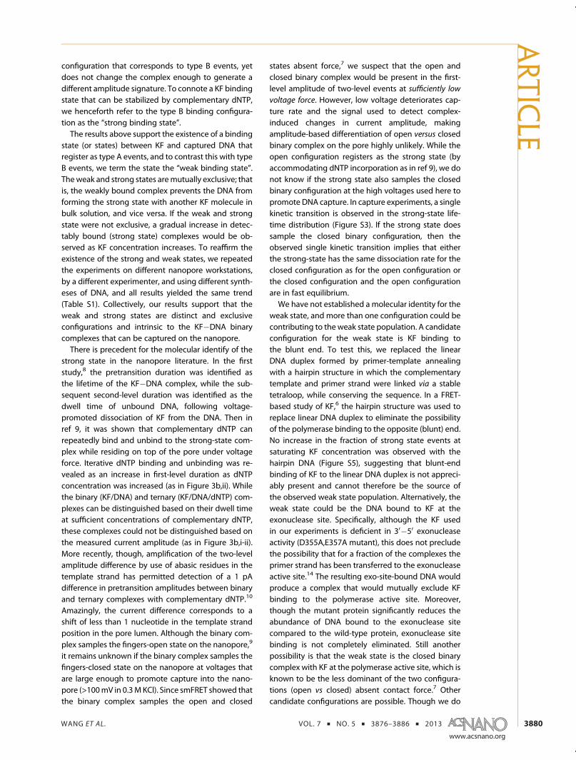
Figure 3. TwoKF�DNAbinding configurations are captured, but only one is detectable. (a)With 1μMDNA in the cis-chamber,the fraction of type B events saturates with increasing KF concentration, suggesting two mutually exclusive binaryconfigurations with one corresponding to type B events. The data points are p = M/N with M type B events out of N totalevents, with standard error (p(1 � p)/N)1/2. Doubling the primer concentration for a 2:1 primer-template ratio (red opencircles) assures that all templates are hybridized and did not affect the fraction at saturation. (b) Event plots show the meanamplitude and dwell time for each capture event (i) without and (ii) with complementary dNTP (40 μMdGTP), with 1 μMDNAand 4μMKF in the cis-chamber. Theplots show that the binary configuration that corresponds to typeB events is stabilizedbydGTP, indicated by an increase in the fraction of type B events ((i) 68% of 1253 events, (ii) 79% of 818 events) and their meanduration ((i) 6.8 ms and (ii) 163.3 ms). Pretransition amplitudes were conserved: (i) 36.3 ( 1.8 pA, and (ii) 36.9 ( 1.2 pA.
ARTIC
LE
WANG ET AL. VOL. 7 ’ NO. 5 ’ 3876–3886 ’ 2013
www.acsnano.org
3880
configuration that corresponds to type B events, yetdoes not change the complex enough to generate adifferent amplitude signature. To connote a KF bindingstate that can be stabilized by complementary dNTP,we henceforth refer to the type B binding configura-tion as the “strong binding state”.The results above support the existence of a binding
state (or states) between KF and captured DNA thatregister as type A events, and to contrast this with typeB events, we term the state the “weak binding state”.Theweak and strong states aremutually exclusive; thatis, the weakly bound complex prevents the DNA fromforming the strong state with another KF molecule inbulk solution, and vice versa. If the weak and strongstate were not exclusive, a gradual increase in detec-tably bound (strong state) complexes would be ob-served as KF concentration increases. To reaffirm theexistence of the strong and weak states, we repeatedthe experiments on different nanopore workstations,by a different experimenter, and using different synth-eses of DNA, and all results yielded the same trend(Table S1). Collectively, our results support that theweak and strong states are distinct and exclusiveconfigurations and intrinsic to the KF�DNA binarycomplexes that can be captured on the nanopore.There is precedent for the molecular identify of the
strong state in the nanopore literature. In the firststudy,8 the pretransition duration was identified asthe lifetime of the KF�DNA complex, while the sub-sequent second-level duration was identified as thedwell time of unbound DNA, following voltage-promoted dissociation of KF from the DNA. Then inref 9, it was shown that complementary dNTP canrepeatedly bind and unbind to the strong-state com-plex while residing on top of the pore under voltageforce. Iterative dNTP binding and unbinding was re-vealed as an increase in first-level duration as dNTPconcentration was increased (as in Figure 3b,ii). Whilethe binary (KF/DNA) and ternary (KF/DNA/dNTP) com-plexes can be distinguished based on their dwell timeat sufficient concentrations of complementary dNTP,these complexes could not be distinguished based onthe measured current amplitude (as in Figure 3b,i-ii).More recently, though, amplification of the two-levelamplitude difference by use of abasic residues in thetemplate strand has permitted detection of a 1 pAdifference in pretransition amplitudes between binaryand ternary complexes with complementary dNTP.10
Amazingly, the current difference corresponds to ashift of less than 1 nucleotide in the template strandposition in the pore lumen. Although the binary com-plex samples the fingers-open state on the nanopore,9
it remains unknown if the binary complex samples thefingers-closed state on the nanopore at voltages thatare large enough to promote capture into the nano-pore (>100mV in 0.3MKCl). Since smFRET showed thatthe binary complex samples the open and closed
states absent force,7 we suspect that the open andclosed binary complex would be present in the first-level amplitude of two-level events at sufficiently low
voltage force. However, low voltage deteriorates cap-ture rate and the signal used to detect complex-induced changes in current amplitude, makingamplitude-based differentiation of open versus closedbinary complex on the pore highly unlikely. While theopen configuration registers as the strong state (byaccommodating dNTP incorporation as in ref 9), we donot know if the strong state also samples the closedbinary configuration at the high voltages used here topromote DNA capture. In capture experiments, a singlekinetic transition is observed in the strong-state life-time distribution (Figure S3). If the strong state doessample the closed binary configuration, then theobserved single kinetic transition implies that eitherthe strong-state has the same dissociation rate for theclosed configuration as for the open configuration orthe closed configuration and the open configurationare in fast equilibrium.We have not established a molecular identity for the
weak state, and more than one configuration could becontributing to the weak state population. A candidateconfiguration for the weak state is KF binding tothe blunt end. To test this, we replaced the linearDNA duplex formed by primer-template annealingwith a hairpin structure in which the complementarytemplate and primer strand were linked via a stabletetraloop, while conserving the sequence. In a FRET-based study of KF,6 the hairpin structure was used toreplace linear DNA duplex to eliminate the possibilityof the polymerase binding to the opposite (blunt) end.No increase in the fraction of strong state events atsaturating KF concentration was observed with thehairpin DNA (Figure S5), suggesting that blunt-endbinding of KF to the linear DNA duplex is not appreci-ably present and cannot therefore be the source ofthe observed weak state population. Alternatively, theweak state could be the DNA bound to KF at theexonuclease site. Specifically, although the KF usedin our experiments is deficient in 30�50 exonucleaseactivity (D355A,E357A mutant), this does not precludethe possibility that for a fraction of the complexes theprimer strand has been transferred to the exonucleaseactive site.14 The resulting exo-site-bound DNA wouldproduce a complex that would mutually exclude KFbinding to the polymerase active site. Moreover,though the mutant protein significantly reduces theabundance of DNA bound to the exonuclease sitecompared to the wild-type protein, exonuclease sitebinding is not completely eliminated. Still anotherpossibility is that the weak state is the closed binarycomplex with KF at the polymerase active site, which isknown to be the less dominant of the two configura-tions (open vs closed) absent contact force.7 Othercandidate configurations are possible. Though we do
ARTIC
LE

WANG ET AL. VOL. 7 ’ NO. 5 ’ 3876–3886 ’ 2013
www.acsnano.org
3881
not here resolve the identity of the weak state(s), theimplicit existence of this population that is mutuallyexclusive from the detectable strong state populationcan be use to model KF�DNA kinetics.The simplest model that is consistent with experi-
mental observations is a three-state model consistingof unbound, weakly bound, and strongly bound states(Figure 4a). Mathematically, notation is introduced toquantify the kinetics of these states:
pu(t): the probability of the unbound state at time t,ps(t): the probability of the strongly bound state attime t,pw(t): the probability of the weakly bound state attime t.The probabilities are governed by the equations
dpu(t)dt
¼ �pu(t)fku f w þ ku f sgþ pw(t)kw f u þ ps(t)ks f u
dpw(t)dt
¼ �pw(t)fkw f u þ kw f sgþ pu(t)ku f w þ ps(t)ks f w
dps(t)dt
¼ �ps(t)fks f u þ ks f wgþ pu(t)ku f s þ pw(t)kw f s
9>>>>>>>=>>>>>>>;
(1)
Our aim is to deduce the six rate constants kifj, andother model-related parameters, from experimentaldata. Capture experiments reveal ps(t) in measure-ments at each KF concentration, but only at the limitingvalue t = ¥ (i.e., only at equilibrium). The novel activecontrol method (described in Figure 5) permitsmeasurement of pre-equilbirium values for ps(t) at anychosen finite t value. While pu(t) and pw(t) cannotbe separated in measurements, we do have access to1� ps(t) = pu(t)þ pw(t). Model parameter identificationis addressed first based onmeasurements of ps(¥) fromcapture experiments at varying KF concentrations.Subsequently, the active control method is used tomeasure ps(t) at finite t values, followed by modelingthe data.
Tomodel capture experiment data, parameters (R,β,Kd)are first defined as
R ¼ [KF]true[KF]
, β ¼ ks f w
kw f s¼ pw(¥)
ps(¥),
Kd ¼ pu(¥)pw(¥)þ ps(¥)
[KF]free
(2)
The parameter R is introduced as a correction factorthat is used to compute the true KF concentration thatis competent to bind DNA in the chamber above thenanopore ([KF]true), given the concentration of KF thatis added to the chamber ([KF]) that is based on theactivity of the enzyme provided by the supplier. Alarger population of KF was anticipated to bind thanthe population that is competent for synthesis, inwhich case R > 1. The parameter β is the equilibriumconstant between the weak and strong states, which isindependent of KF concentration. The parameter Kd isthe effective binding affinity for the composite KFbinding states (weak and strong, together) at equilib-rium, with [KF]free the concentration of free KF mol-ecules in bulk solution.The three parameters (R,β,Kd) can be determined
from capture experiment data {[KF],ps(¥)} as follows.First, we can write pu(¥) and pw(¥) in terms of βand ps(¥), given by pw(¥) = βps(¥) and pu(¥) = 1 �(β þ 1)ps(¥). From these relations and eq 2, [KF]free =Kd(β þ 1)ps(¥)/[1 � (β þ 1)ps(¥)]. Conservation of thetotal amount of KF molecules that are competent tobind dictates that
[KF]true ¼ [DNA]pw(¥)þ [DNA]ps(¥)þ [KF]free w
R[KF] ¼ [DNA](1þ β)ps(¥)þ Kd(βþ 1)ps(¥)
1 � (βþ 1)ps(¥)(3)
In eq 3, the added [DNA] is known, and we vary [KF]while measuring ps(¥). Equation 3 is reformulated and
Figure 4. A three-statemodel is consistent with experimental observations. (a) Diagramof the three states (unbound, weaklybound, strongly bound)with transition rates (kifj). The “S” and “W” labels onKF are a connotation used in subsequent figures.(b) From capture experiment data (Figure 3a), the measured fraction of strongly bound complexes ps(¥) as a function of KFconcentration (points) is well modeled by eq 3 (line). At any [KF], the composite fraction pu(¥) þ pw(¥) is 1 � ps(¥), and atsaturating [KF], pu(¥) ≈ 0 and pw(¥) ≈ 1�ps(¥).
ARTIC
LE

WANG ET AL. VOL. 7 ’ NO. 5 ’ 3876–3886 ’ 2013
www.acsnano.org
3882
maximum likelihood estimation is used to fit (R,β,Kd) tothe {[KF],ps(¥)} data (Supporting Information). Themeasured data and fitted model (3) are plotted to-gether (Figure 4b), corresponding to the parametervaluesR=1.53( 0.11,β= 0.50( 0.03, and Kd = 69.15(38.45 nM. An estimated equilibrium constant betweenthe weak and strong states of β = 50% is consistentwith ps(¥) ≈ 66% and pw(¥) ≈ 33% at saturating [KF].The value R = 1.5 is larger than we expected, suggest-ing a 50% increase in KF competent to bind comparedto the concentration deemed competent for synthesisby the vendor. Finally, the value Kd ≈ 70 nM is withinthe range of 2�200 nM obtained by ensemblemeasurements.4
We next present amethod formeasuring pre-steady-state values for ps(t), at any chosen finite t value, and forindividual DNA�KF molecules. The method combinesthe nanopore with active control, extending our pre-vious method.11 Active control refers to automatedchanges in the voltage across the pore, triggered bytime and/or detected shifts in the measured currentthrough the pore. The method here is fully timetriggered and involves sequentially and repeatedlyexposing the DNA binding site to the bulk-phasecis-chamber for a set “fishing period” tf by a cis-positivevoltage, followed by a cis-negative voltage for 3 ms toprobe the state of the DNA (Figure 5). Switching thevoltage polarity moves the DNA up or down, while theDNA remains tethered in the nanopore by duplex endregions. The 1�3 ms probing period is sufficient todetermine if the DNA is strongly bound or not, basedon the current amplitude pattern (Figure 5iii). The first1 ms of probing is not observable due to capacitancesin the system that are excited when the voltagechanges polarity (see Methods). The probing voltageused is 120mV,which is lower than the capture voltageof 180 mV. The lower voltage is used to extendthe lifetime of strong state complexes to make a largerpercentage of strongly bound complexes observableduring the 1�3 ms probing period.Recorded data sets for each tethered DNA molecule
provide two complementary subsets of data. First, wemeasure ps(tf) t ps(tf|pu(0) = 1) for each fishing time tfchosen by extracting the subset of data that are un-bound at the start of fishing (Figure 5iv, left) andcomputing the fraction of the subset that are subse-quently strongly bound at the beginning of probing(Figure 5ii, right). The complementary subset of datacorresponds to events that are bound at the start offishing (Figure 5iv, right), from which we computethe fraction prb(tf) t ps(tf|ps(0) = 1) that is subse-quently still in the strongly bound state at the start ofprobing. The subscript rb references that the prb(tf)fraction “remains bound” in the sense that the DNA isin the strongly bound state at both the start and theend of each fishing period. It is possible that unbind-ing and rebinding of the same or a different KF
occurred during the fishing period; thus, we are notsuggesting that the same KF remains bound for theduration of the entire fishing period for the frac-tion prb.The results of measuring ps(tf) and prb(tf) in separate
experiments at [KF] = 0.375 μM (below saturating KF)and [KF] = 2.0 μM (saturating KF), with 1 μMDNA also inthe cis-chamber, are reported (Figure 6 and SupportingInformation Tables S2 and S3) combining data fromrepeated experiments at each concentration. Thetrends show ps(tf) increasing and prb(tf) decreasing asa function of tf, converging approximately to commonequilibrium percentages for each KF concentration.At long fishing times, the fraction of strongly boundevents at the start of probing should be at equilibrium,and thus the same fraction should be measured
Figure 5. Active control of duplex-tethered DNA in a nano-pore to measure pre-steady-state KF�DNA kinetics. (i) Fish-ing involves exposing the DNA primer-template bindingsite to the bulk-phase cis-chamber by applying a cis-positive50mVvoltage for a set period tf. During that time, unbound,weak, and strong states can form and interconvert. (ii�iv)Probingbegins at the endof thefishingperiod and applies acis-negative 120 mV voltage for 3 ms. Thus, probing pullsthe DNA toward the trans-chamber, and during 1�3 ms ofprobing, the ionic current displays one of three possiblepatterns as shown in (iii): one pattern consistent with thestrong state complex (right signal), one pattern consistentwith the unbound state (left signal), and one pattern con-sistent with the strong state initially followed by the un-bound state due to KF dissociation (middle signal). After3 ms probing, fishing is restarted from one of two possiblestart states, strong bound or unbound. Automated captureand tethering of each DNA is part of the control logic (seeMethods), and hundreds to tens-of-thousands of fish/probecycles are possible for each tethered DNA (SupportingInformation).
ARTIC
LE

WANG ET AL. VOL. 7 ’ NO. 5 ’ 3876–3886 ’ 2013
www.acsnano.org
3883
regardless of the state of the DNA at the start of fishing.In other words, it should be that prb(tf) ≈ ps(tf) forsufficiently large tf at each KF concentration, providedthe controlled DNA samples KF in the bulk-phasecis-chamber in a way that is not biased by the startingstate of the DNA (unbound or strongly bound), and thisis supported by the data (Figure 6). Also, since the rateof association is proportional to KF concentration,the data show that ps and prb converge to equilibriummore quickly at 2.0 μM than at 0.375 μM.The throughput of our single-molecule-at-a-time
method is unparalleled. During a single 2 h experiment,several DNAwere captured, tethered, and controlled togenerate data for each chosen fishing period. At thefastest fishing period of 1 ms, each fishing/probingcycle lasts 4 ms (3 ms probing and 1 ms fishing). In oneof the experiments at 2.0 μM KF, at total of 14 797cycles were recorded at the 1 ms fishing period, whichcorresponds to just under 60 s of recording time. Foreach fishing time, hundreds to tens-of-thousands ofsingle-molecule events were recorded, one molecularevent at a time. In total, over 200 000 fishing/probingcycles were included in the 2.0 μM KF data plotted inFigure 6; this is 2 orders of magnitude more eventsthan is recorded in a typical nanopore capture experi-ment. We know of no other single-molecule methodthat can generate as much data in as short a timeperiod. By contrast, single-molecule FRET mea-sures individual molecules that are each labeled,but the measurements are images that record allfluorescent signals generated in parallel within theimage frame. Optical trapping and AMF setups canmeasure one molecule at a time, and while eachmeasurement is considerably more informative
(such as tracking amotor for hundreds of steps alongpolynucleotide substrate15), the throughput is con-siderably lower.Finally, based on the data generated in fishing/
probing experiments, we examine the unbound-to-strong association rate (kufs) in the three-state modelillustrated in Figure 4. In each event, the dsDNA�ssD-NA junction is first sent into the bulk-phase cis-cham-ber for a prescribed fishing period of tf, and then thevoltage is reversed to pull the DNA back against thepore to probe if a KF molecule is bound onto the DNAin the strong state. After 3 ms probing, the voltage isreversed again to start the next fishing event. Toestimate kufs, we select only fishing events that startwith the DNA in the unbound state (ps(t)). Mathema-tically, over the ensemble of all events we select, wehave pu(0) = 1, pw(0) = 0, and ps(0) = 0. Substituting intoeq 1, we obtain
dps(t)dt
�����t¼ 0
¼ ku f s
Thus, kufs is the initial rate of change in ps(t). InFigure 7a, we plot ps versus tf measured at various KFconcentrations. The data confirm that ps(tf) is indeedproportional to tf for small tf. At each KF concentration,kufs is calculated as the slope of ps(tf) (Tables S4 andS5). The plot of kufs versus [KF] is shown in Figure 7b.Data points of kufs are fitted using the least-squaresmethod to, respectively, kufs� [KF] and kufs� [KF]free.The plot suggests that the association rate kufs
is approximately proportional to [KF], the total(unbound and DNA-bound) KF concentration addedto the cis-chamber. The least-squares fitting yieldskufs ≈ 10.6 s�1 μM�1[KF] (solid line in Figure 7b)).In contrast, the model kufs � [KF]free (dashed line inFigure 7b)) cannot explain the measured values ofkufs.
CONCLUSIONS
Our work models pre-equilibrium kinetics betweenthe KF and DNA substrate, based on analyzing captureevent measurements from standard nanopore experi-ments and also using our new active control methodfor measuring association and dissociation of indivi-dual KF from a single DNA duplex tethered in thenanopore. Ours is the first nanopore study to examineunforced dissociation between each KF and the con-trolled DNA, a measurement made implicitly availableonly through the control method itself. This study isalso the first to show that KF�DNA complexes cap-tured on the pore are not universally detectable. Thedetectable binding state is shown to sample the openconfiguration and to measurably respond to dNTPincorporation and subsequently sample the closedternary complex, as shown in other nanopore studies.9
Moreover, the undetectable KF�DNA complexes
Figure 6. Fraction of strongly bound probing events as afunction of fishing period tf starting from the unbound (ps)and strongly bound (prb) states. Regardless of the startingstate, the two strongly bound fractions appear to convergeto a common equilibrium for long tf. For short tf, theobserved rate of increase of ps(tf) is proportional to KF,shown at saturating (2.0 μM) and subsaturating (0.375 μM)concentrations added to the cis-chamber and canbeused toidentify the association rate parameter kufs in (1) at each KFconcentration.
ARTIC
LE

WANG ET AL. VOL. 7 ’ NO. 5 ’ 3876–3886 ’ 2013
www.acsnano.org
3884
captured on the nanopore are mutually exclusive fromthe detectable binding state. Though we are not ableto resolve the configuration that corresponds to un-detectable binary events, varying experimental condi-tions support that the state is not an artifact of themeasurement technique but is intrinsic to the KF�DNAcomplexes captured on the nanopore. Using the activecontrol method, our study also complements priornanopore studies on KF�DNA kinetics9,10 by measur-ing and quantifying the rates of formation of binarycomplexes. An unexpected result is that the measuredassociation rate of KF to the controlled DNA cannot beexplained by the model of association rate beingproportional to free KF concentration. Instead, themeasured association rate is approximately propor-tional to the total (unbound and DNA-bound) KFconcentration in the chamber above the pore.The molecular mechanism for this phenomenologi-cal observation is still unknown. One possibilityis that, when a unbound DNA encounters a DNAwith a KF molecule bound, the KF molecule cansomehow transfer from one DNA to the other. Futureexperiments need to be designed to test thishypothesis.The DNA controlmethod presented here is simple to
implement on any nanopore experiment setup, withdedicated hardware that is commercially available. Themethod here is a fully time-triggered two-cycle logic(probe, then fish), whichmakes the logic very simple toprogram and implement. The throughput of this meth-od is unparalleled, recording hundreds-of-thousandsof single-molecule events in a few hours, one molec-ular event at a time. A distinct feature is that themethod allowed us to measure two conditional prob-abilities for the strongly bound complex in repeatedfishing events with the same DNA molecule that is
tethered at the pore: one starting in the unbound state,and the other starting in the strong bound state at theonset of fishing. Another advantage is that, upon thecapture of one DNA, it uses the captured DNA repeat-edly for a large number of fishing events (over 10 000per DNA in some cases). Thus, a slow capture ratecaused by low DNA concentration will not impact thehigh-throughput dramatically. Being able to work withlow DNA concentration will enable us, in particular, todistinguish the effect of DNA-bound KF molecules inbulk from that of free KF molecules in bulk on thetransition rate from unbound to strongly bound. Thedemonstrated control technique is not confined to thestudy of KF with DNA but could be applied to otherDNA or RNA-binding enzymes that are conducive tonanopore experimental conditions. Notably, enzymesthat function in higher salt can be measured at lowerprobing forces (as in ref 10), making it more likely todetect complexes that would not survive the highernanopore contact forces experienced in captureexperiments.The ability to quickly quantitate, even grossly, the
kinetics of protein binding to nucleic acids, whether itbe dsDNA, ssDNA, or RNA, has significant relevance tothe biotechnology industry. For example, it is advanta-geous to re-engineer proteins to bindmethylated DNAwith higher affinity and greater sequence specificityto enable mapping regions of hyper-methylatedDNA, which are common DNA “marks” in cancers andother disease states.16,17 It is also common practice toinvestigate binding kinetics when developing novelDNA binding enzymes used in commercial molecularbiology kits.18 The method discussed in this articleoffers a convenient strategy to quickly screen proteinsand determine whether sequence modifications,either protein or DNA, have increased or decreased
Figure 7. Results of fishing experiments that start with the DNA in the unbound state. (a) Fraction of strongly bound events(ps) over short fishing periods tf at various KF concentrations. For all KF concentrations used in experiments, fraction ofstrongly bound (ps) increases linearly with fishing period for small tf. Each solid line represents the linear fitting at a KFconcentration. The slope of each line gives the unbound-to-strong association rate (the transition rate kufs) at thecorresponding KF concentration. (b) Unbound-to-strong association rate (the slope of ps vs tf in panel a) as a function ofKF concentration. The solid line shows the fitting to c[KF], whereas the dashed line is the fitting to c[KF]free, where [KF]free is thefree KF concentration in solution. It is clear that the association rate is not proportional to [KF]free.
ARTIC
LE

WANG ET AL. VOL. 7 ’ NO. 5 ’ 3876–3886 ’ 2013
www.acsnano.org
3885
the proteins' affinity for its target. Additionally, themethod can be amended to determine the affinity ofbacterial or viral proteins for nucleic acids in thepresence of small molecule drug candidates. For ex-ample, by using a segment of RNA that matches the
Hepatitis C genome and the Hepatitis C RNA bindingreplicase protein in the bulk phase, one could screensmall-molecule inhibitors that prevent enzyme asso-ciationwith the RNA, thereby realizing hits for potentialcompounds for treating Hepatitis C infection.
METHODSMaterials. The D355A,E357A exonuclease-deficient variant
of KF was obtained from New England Biolabs (100 000U mL�1; specific activity 20 000 U mg�1). DNA oligonucleotideswere synthesized by the Stanford University Protein andNucleicAcid Facility and purified by denaturing PAGE. Sequences of theoligonucleotides used in this study are shown below. The abasicresidues in the 79-mer template sequence are indicated by an X.The 30 terminal residue of the 23-mer primer was 20-30 dideoxy-cytidine (ddC), enabling binary and ternary complex formationwithout catalytic turnover. 23-mer primer: 50-GGCTACGACCTG-CATGAGAATGddC-30 . 20-mer tethering oligonucleotide: 50-TGAGTGGAAGGATAGGTGAG-30 . Three abasic (XXX) residue-containing 79-mer template: 50-CTCACCTATCCTTCCACTCATTC-CAATTAATTACCATTCATTXXXTCTCACTATCGCATTCTCATGCA-GGTCGTAGCC-30 .
General Nanopore Methods. Experiments were conducted at23 �C in 10 mM HEPES/KOH, pH 8.00 ( 0.05, 0.3 M KCl, 5 mMMgCl2, which are conditions shown to support KF catalyticfunction.8,9 Single R-HL channels were formed as described.8
A patch-clamp amplifier (AxoPatch 200B, Molecular Devices,Sunnyvale, CA) was used to apply transmembrane voltage andmeasure ionic current, with the 4-pole Bessel filter set at 5 kHzbandwidth. A digitizer (Digidata 1440A, Molecular Devices)stored data sampled at 100 or 200 kHz. Primer/template hybridswere formed by mixing in equimolar concentrations of 50 μMeach in a solution containing 100 mM KCl and 5 mMMgCl2. Thesolution was denatured at 95 �C for 2 min, followed by a slowcool to room temperature to optimize annealing specificity. Thissolution was then added at a concentration of 1 μM into thecis-chamber during nanopore experiments. Tethering primerwas present in the trans-chamber at a concentration of 4 μM.Standardization of cis-chamber volume to 85 μL was performedafter insertion of a single nanopore into the lipid bilayer andafter flushing excess R-HL from the nanopore. Evaporation ofwater from the nanopore setupwas prevented by saturating thehumidity level directly above the nanopore device.
Active Voltage Control Experiments. Active voltage control logicwas implemented as a finite state machine (FSM) on a field-programmable gate array (FPGA, PCI-7831R, National Instru-ments), as described previously.11 The FPGA was connected tothe Axopatch 200B for transmembrane voltage control andionic current measurements at 5.3 μs updates. To improve thesignal-to-noise ratio, the ionic current signal was filtered on theFPGA using a single-pole low-pass filter with 1.58 kHz cutofffrequency. The FSM initialized tethering by detecting capture ofa primer-template DNA molecule at 160 mV and reducing thevoltage to 50mV for 10 s. The holding period permits trans-sideduplex formation (20 bp) between the 20 nucleotides of thetemplate exposed in the trans-chamber (from 50 end) andthe 20-mer tethering oligonucleotide with high likelihood.11
The FSM then began automated cycling between the �20 mVfishing voltage for each chosen fishing period tf specified inresults section with the 120 mV probing voltage for 3 ms. Thelogic looped continuously between fishing and probing volt-ages until the FSM detected an open channel current levelduring probing, which triggered a return to 160 mV capturevoltage until detection of capture of another DNA molecule.Testing for open channel current was at the end of each probingperiod to avoid the effect of the capacitive transient on thesensing signal.
Data Processing. All numerical analysis and data processingwas done using custom code written in Matlab (20011a, TheMathWorks, Natick, MA). Analysis of capture event data was
done with methods established in prior work.8,11 Analysis ofactive control (fishing/probing) data was done with methodscomparable to our prior work in ref 11, with modifications touniquely identify each of the three signal patterns (Figure 5iii)during each 3 ms probing period. In nanopore systems usingpatch-clamp technology, a step change in voltage induces acapacitive transient superimposed on the ionic current mea-surement. To remove the current transient induced by thevoltage change that initiates each 3ms probing period, a simpleexponential subtraction method was used offline (as done inref 10). Briefly, a one-time transient fit was made using oneprobing event for each tethered DNA molecule, and the fitwas subtracted for all probing events for that molecule. In thetransient fit, the first 0.2 ms of the current is saturated andignored in the data, and a three-parameter exponentialfunction f(t) = a0 þ a1 exp(�a3t) was fit in Matlab using abuilt-in nonlinear least-squares to determine parameters a0,a1, a2. Subtraction of the resulting fit a1 exp(�a3t) for eachprobing period yielded events with steady-state amplitudesachieved 1 ms after the start of probing (as shown inFigure 5iii). Each of the three signal patterns (Figure 5iii) weresubsequently identified using the step detection method inref 11. The threshold in step detection was based on theobserved amplitude separation between strongly bound andunbound DNA event amplitudes at 120 mV and was cali-brated for each experiment. Any step signaling the transitionfrom strongly bound to unbound (middle pattern, Figure 5iii)that occurred within the first millisecond of probing wastherefore not detected. To compensate for this, we apply thesame correction factor method used to adjust the stronglybound percentage in capture experiments (SupportingInformation). The correction increases the fraction of stronglybound probing events by a factor of 1.1, with the correctedpercentages plotted in Figure 6 (uncorrected data are re-ported in Tables S2 and S3).
Conflict of Interest: The authors declare no competingfinancial interest.
Acknowledgment. This work was supported by NationalInstitutes of Health Grants HG004035-05 (W.D.) and R21-RR025347-02 (H.W., W.D.). We thank J. Dahl for repeating someof the capture experiments, and K. Lieberman for proposingexperiments and providing feedback on aspects of this work.We also thank T. Morin for helpful editing and suggestingpotential applications for our method.
Supporting Information Available: (1) DNA primer/templatesubstrate (Figure S1). (2) Selection criteria for assigning type A/type B events (Figure S2). (3) Correction factor for the fractionof type B events (Figure S3). (4) Capture rate of DNA withoutand with saturating KF (Figure S4). (5) Fraction of type Bevents under varying experimental conditions (Figure S5,Table S1). (6) Fitting capture data parameters by maximumlikelihood estimation. (7) Fishing/probing data (Tables S2and S3). (8) Correction factor for the fraction of stronglybound probing events in fishing/probing data. (9) Rate ofassociation data from fishing/probing experiments (Tables S4and S5). This material is available free of charge via theInternet at http://pubs.acs.org.
REFERENCES AND NOTES1. Rothwell, P. J.; Waksman, G. Structure and Mechanism of
DNA Polymerases. Adv. Protein Chem. 2005, 71, 401–440.
ARTIC
LE

WANG ET AL. VOL. 7 ’ NO. 5 ’ 3876–3886 ’ 2013
www.acsnano.org
3886
2. Joyce, C. M.; Benkovic, S. J. DNA Polymerase Fidelity:Kinetics, Structure, and Checkpoints. Biochemistry 2004,43, 14317–14324.
3. Astatke, M.; Grindley, N. D.; Joyce, C. M. How E. coli DNAPolymerase I (Klenow Fragment) Distinguishes betweenDeoxy- and Dideoxynucleotides. J. Mol. Biol. 1998, 278,147–165.
4. Kuchta, R. D.; Mizrahi, V.; Benkovic, P. A.; Johnson, K. A.;Benkovic, S. J. Kinetic Mechanism of DNA Polymerase I(Klenow). Biochemistry 1987, 26, 8410–8417.
5. Bermek, O.; Grindley, N. D. F.; Joyce, C. M. Distinct Roles ofthe Active-Site Mg2þ Ligands, Asp882 and Asp705, of DNAPolymerase I (Klenow Fragment) during the PrechemistryConformational Transitions. J. Biol. Chem. 2011, 286, 3755–3766.
6. Joyce, C. M.; Potapova, O.; Delucia, A. M.; Huang, X.; Basu,V. P.; Grindley, N. D. F. Fingers-Closing and Other RapidConformational Changes in DNA Polymerase I (KlenowFragment) and Their Role in Nucleotide Selectivity. Bio-chemistry 2008, 47, 6103–6116.
7. Santoso, Y.; Joyce, C. M.; Potapova, O.; Le Reste, L.;Hohlbein, J.; Torella, J. P.; Grindley, N. D. F.; Kapanidis,A. N. Conformational Transitions in DNA Polymerase IRevealed by Single-Molecule FRET. Proc. Natl. Acad. Sci.U.S.A. 2010, 107, 715–720.
8. Benner, S.; Chen, R. J.; Wilson, N. A.; Abu-Shumays, R.; Hurt,N.; Lieberman, K. R.; Deamer, D. W.; Dunbar, W. B.; Akeson,M. Sequence-Specific Detection of Individual DNA Poly-merase Complexes in Real Time Using a Nanopore. Nat.Nanotechnol. 2007, 2, 718–724.
9. Hurt, N.; Wang, H.; Akeson, M.; Lieberman, K. R. SpecificNucleotide Binding and Rebinding to Individual DNAPolymerase Complexes Captured On a Nanopore. J. Am.Chem. Soc. 2009, 131, 3772–3778.
10. Garalde, D. R.; Simon, C. A.; Dahl, J. M.;Wang, H.; Akeson,M.;Lieberman, K. R. Distinct Complexes of DNA Polymerase I(Klenow Fragment) for Base and Sugar Discriminationduring Nucleotide Substrate Selection. J. Biol. Chem.2011, 286, 14480–14492.
11. Wilson, N. A.; Abu-Shumays, R.; Gyarfas, B.; Wang, H.;Lieberman, K. R.; Akeson, M.; Dunbar, W. B. ElectronicControl of DNA Polymerase Binding and Unbinding toSingle DNA Molecules. ACS Nano 2009, 3, 995–1003.
12. Venkatesan, B.; Bashir, R. Nanopore Sensors for NucleicAcid Analysis. Nat. Nanotechnol. 2011, 6, 615–624.
13. Song, L.; Hobaugh, M. R.; Shustak, C.; Cheley, S.; Bayley, H.;Gouaux, J. E. Structure of Staphylococcal R-Hemolysin, aHeptameric Transmembrane Pore. Science 1996, 274,1859–1866.
14. Guest, C. R.; Hochstrasser, R. A.; Dupuy, C. G.; Allen, D. J.;Benkovic, S. J.; Millar, D. P. Interaction of DNA with theKlenow Fragment of DNA Polymerase I Studied by Time-Resolved Fluorescence Spectroscopy. Biochemistry 1991,30, 8759–8770.
15. Moffitt, J. R.; Chemla, Y. R.; Smith, S.; Bustamante, C. RecentAdvances in Optical Tweezers. Annu. Rev. Biochem. 2008,77, 205–228.
16. Jørgensen, H. F.; Adie, K.; Chaubert, P.; Bird, A. P. Engineer-ing a High-Affinity Methyl-CpG-Binding Protein. NucleicAcids Res. 2006, 34, e96.
17. Shim, J.; Humphreys, G. I.; Venkatesan, B. M.; Munz, J. M.;Zou, X.; Sathe, C.; Schulten, K.; Kosari, F.; Nardulli, A. M.;Vasmatzis, G.; Bashir, R. Detection and Quantification ofMethylation in DNA Using Solid-State Nanopores. Sci. Rep.2013, 3, 1–8.
18. Buchholz, F. Engineering DNA Processing Enzymes for thePostgenomic Era. Curr. Opin. Biotechnol. 2009, 20, 383–389.
ARTIC
LE