measurement and evaluation of body temperature ...21132/fulltext01.pdflinköping university medical...
TRANSCRIPT

Linköping University Medical Dissertations
No 872
Measurement and Evaluation of Body Temperature:
Implications for Clinical Practice
Märtha Sund-Levander
Department of Medicine and Care, Division of Clinical Physiology, Faculty of Health Sciences, Linköping University,
SE-581 85 Linköping, Sweden.
Department of Welfare and Care, Faculty of Health Sciences, Linköping University,
SE-601 74 Norrköping, Sweden
Linköping 2004

© Märtha Sund-Levander 2004
ISBN 91-7373-848-4, ISSN 0345-0082 Printed by Unitryck, Linköping, Sweden 2004

Linköping University Medical Dissertations No 872
Measurement and evaluation of body temperature: Implications for clinical practice
Märtha Sund-Levander
Akademisk avhandling som för avläggande av medicine doktorsexamen kommer att offentligen försvaras i
Berzeliussalen, Hälsouniversitetet i Linköping torsdagen den 2 december 2004 kl 13.00. Fakultetsopponent är professor Karin Axelsson, Luleå Tekniska Högskola.
Abstract
The general aim was to explore factors influencing the normal variation and measurement of body temperature. Additional aims were to study morbidity, mortality and the clinical presentation of pneumonia and predictors for survival in elderly nursing-home residents. Two hundred and thirty seven non-febrile nursing home residents (aged 66-99 years) and 87 healthy adults (aged 19-59 years) were included. In elderly individuals, the morning ear and rectal body temperature was measured at baseline and pneumonia and survival was observed at one- two and three-year. In healthy adults the rectal, ear, oral and axillary temperature were measured simultaneously on one morning and repeated measurements were performed in three subjects.
Overall, the range of normal body temperature was wider then traditionally stated. In elderly nursing-home residents, functional and cognitive impairment and BMI < 20 were related to a lower body temperature and medication with analgesics to a higher. Compared to adults < 60 years elderly persons had a higher average ear and a lower rectal temperature. Men and postmenopausal women < 60 years had lower body temperature than premenopausal women. The repeated measurements showed a wide individual variability irrespective of the site of measurement, and that replicated measurements do not improve accuracy. When comparing the rectal temperature with oral, ear and axillary readings the average difference was > 0.5°C with a wide individual variation.
The yearly incidence of nursing-home acquired pneumonia varied between 6.9% and 13.7%. Functional impairment, chronic obstructive pulmonary disease (COPD) and male sex were related to a higher risk of acquiring pneumonia and presenting non-specific symptoms were common. Age and functional impairment predicted mortality, irrespective of gender, while cerebral vascular insult, a lower body mass index and malnutrition in women and heart disease, COPD, medication with sedatives and mortality rate index in men were gender specific predictors. Surviving women had a higher baseline body temperature than non-surviving, while no such difference was found in men.
When assessing body temperature, it is important to consider the site of measurement, technical design, operator technique, age and gender and, in elderly nursing-home residents, physical and cognitive impairment, body constitution and medication with analgesics. The best approach is to use an unadjusted mode, without adjusting to another site. To prevent a delayed diagnosis of pneumonia, one should be aware of a low baseline body temperature and lack of specific clinical symptoms in elderly nursing-home residents. Preserving and/or improving functional, cognitive, nutritional status and preventing agitation and confusion would improve survival in nursing-home residents.
Keywords: Assessment, body temperature measurement, elderly, evaluation, infection, nursing home, pneumonia, survival.
Department of Medicine and Care, Division of Clinical Physiology,
Faculty of Health Sciences. Linköping University, SE-581 85 Linköping, Sweden
Department of Welfare and Care, Faculty of Health Sciences, Linköping University,
SE-601 74 Norrköping, Sweden
ISBN 91-7373-848-4 ISSN 0345-0082

Would it not be wise to remove the little red arrows from the thermometer?
E F DuBois 1951
To my mother, Maj In memory of my father, Sture
To Lars, Martin and Maria

ORIGINAL PAPERS This doctoral thesis is based on the following papers, which will be referred to by their Roman numerals. [1-4] 1. Sund-Levander, M. and L.K. Wahren, The impact of ADL-status,
dementia and body mass index on normal body temperature in elderly nursing home residents. Archives of Gerontology and Geriatrics, 2002. 35: p. 161-169.
2. Sund-Levander, M., E. Grodzinsky, D. Loyd, and L.K. Wahren, Errors in
body temperature assessment related to individual variation, measuring technique and equipment. International Journal of Nursing Practice, 2004. 10: p. 216-23.
3. Sund-Levander, M., Å. Örtqvist, E. Grodzinsky, Ö. Klefsgård, and L.K.
Wahren, Morbidity, mortality and clinical presentation of nursing home- acquired pneumonia in a Swedish population. Scandinavian Journal of Infectious Diseases, 2003. 35: p. 306-310.
4. Sund-Levander, M., E. Grodzinsky, and L.K. Wahren, Gender differences
in predictors for survival in elderly nursing-home residents. A long-term follow-up. 2004. Manuscript. Submitted.
Reprints were made with kind permission of the publishers.

PageINTRODUCTION
9
AIMS OF THE INVESTIGATION
11
BACKGROUND 12The concept of normality 12Thermoregulation 12Factors influencing “normal” body temperature 13Body temperature measurements 14
Rectal measurement 15Oral measurement 16Ear measurement 16Axillary measurement 16
The febrile response 17Infection and fever in elderly individuals 17Predictors of survival in elderly nursing-home residents
18
MATERIAL AND METHODS 19Subjects 19Procedure 21Measurements 22
Body temperature 22ADL status 23Nutritional status 23Body mass index 24Dementia 24
Statistics 24Ethical considerations
25
RESULTS 26Interindividual variability 26Intraindividual variability 28Adjustment to rectal temperature 28Factors related to a lower baseline body temperature in elderly nursing-home residents 28Pneumonia in elderly nursing-home residents 31Predictors of survival in elderly nursing-home residents 31

PageGENERAL DISCUSSION
32
METHODOLOGICAL CONSIDERATIONS
36
CONCLUSIONS
37
ACKNOWLEDGEMENTS
38
REFERENCES 39 Papers I – IV
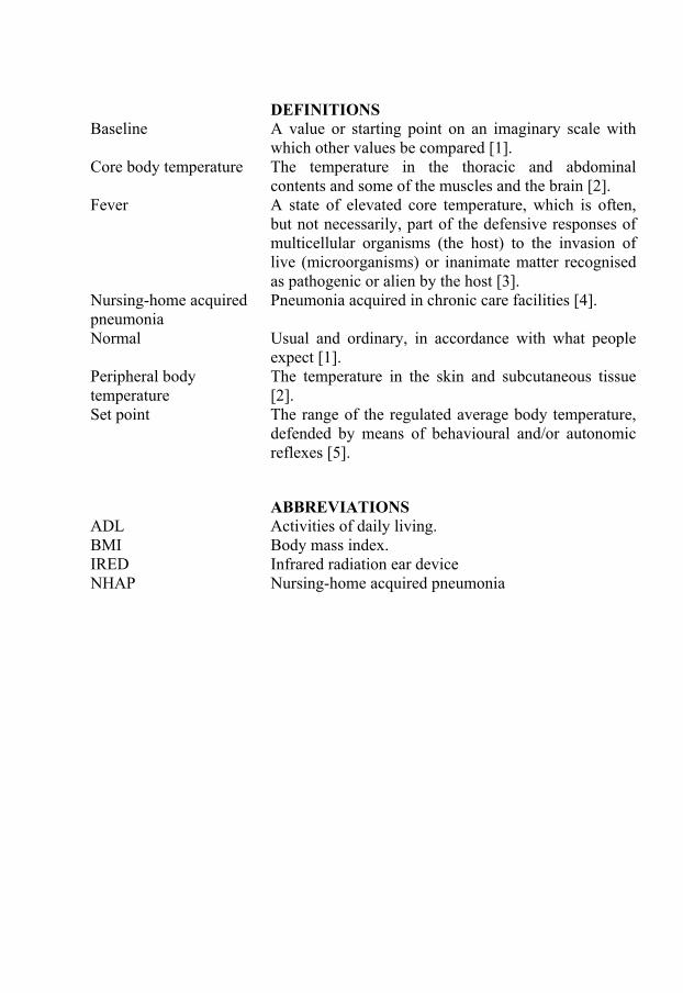
DEFINITIONS Baseline A value or starting point on an imaginary scale with
which other values be compared [1]. Core body temperature The temperature in the thoracic and abdominal
contents and some of the muscles and the brain [2]. Fever A state of elevated core temperature, which is often,
but not necessarily, part of the defensive responses of multicellular organisms (the host) to the invasion of live (microorganisms) or inanimate matter recognised as pathogenic or alien by the host [3].
Nursing-home acquired pneumonia
Pneumonia acquired in chronic care facilities [4].
Normal Usual and ordinary, in accordance with what people expect [1].
Peripheral body temperature
The temperature in the skin and subcutaneous tissue [2].
Set point The range of the regulated average body temperature, defended by means of behavioural and/or autonomic reflexes [5].
ABBREVIATIONS ADL Activities of daily living. BMI Body mass index. IRED Infrared radiation ear device NHAP Nursing-home acquired pneumonia


- 9 -
INTRODUCTION Evaluation of body temperature is one of the oldest known diagnostic
methods [6, 7] and is still an important sign of health and disease, both in everyday life and in medical care. Increased body temperature is associated in the first place with infectious disease and fever [8] and the accompanying feelings of illness. In general, the individual can describe feelings of illness and discomfort, but in conditions where individuals are unable to explain themselves, the nurse has to interpret the clinical signs and rely on objective measurements. This is especially important when assessing elderly individuals [9], as atypical presentation of infection are common in this age group [10].
Before the 17th century, the humoral theory, established by Galen in the second century A.D., dominated medical thinking [7], and the interpretation of symptoms and explanations of disease were closely related to philosophy and religion [11]. Evaluation of body temperature rested on objective and subjective feelings of warmth, i.e. physicians used touch as their principal criterion for evaluating body heat [12]. The discovery of the circulation of the blood emphasised the use of natural sciences in medicine and clinicians started to use the pulse rate to diagnose fever [13]. This remained to be the most important tool even when the first thermometers were available in the late 18th century. A that time, the improved scientific knowledge of human anatomy and physiology, attributed to the introduction of autopsy [14], and new technical knowledge [9] increased the interest in objectively observing the patient [12, 15] and thus the possibility to differentiate between health and disease using biological parameters in terms of what was considered normal and abnormal.
The first comprehensive report on body temperature related to normality was published in 1868 by the German physician Wunderlich [16], who established the use of thermometers in medical practice [17]. Wunderlich measured axillary temperature in patients and declared the normal body temperature to be 37.0°C with a range of 36.2°C to 37.5°C. He defined temperatures above 37.5°C as “the territory of fever” and > 38.0°C as fever [18].
Today there is a general acceptance of normal body temperature as a range rather than a fixed temperature, although there is still widespread confusion in the assessment and evaluation of body temperature in adults [19], especially in elderly individuals [20-22]. A recently performed systematic review of the literature [23] found that the range in normal body temperature was 33.2°C to 38.2°C and that few studies reported average values of body temperature equal to or above 37.0°C, i.e. the rectal temperature in six studies and the ear temperature in one study [20, 24-28]. In addition, calibration of the thermometers, how the measurement was performed (i.e. measuring technique and training of the operators, insertion of the devices), ovulatory

- 10 -
status, time of day and indoor temperatures were not clearly described. Hence, there is a lack of studies performing temperature measurements in a standardised way. Also, studies presenting strong evidence were mainly found in investigations of oral temperature, and in studies reporting body temperature in men [23].
In clinical practice, the nurse retains responsibility for care of the individual [29] including the evaluation of body temperature and notifying the physician of individual changes in the thermal status. The nurse also frequently determines when to start antipyretic therapy and initiate a physical cooling on or without the physician’s order. Hence, nurses have a great impact on both the observation of body temperature as a sign of infection and on implementing nursing care during fever.
Nursing care should be based on sound scientific knowledge and evidence-based experience [30]. The nursing process, i.e. the assessment, diagnosis, planning and implement actions of interventions and evaluating the outcome [29], is a basic framework which ensures that nursing care is performed in a systematic and structured way. In relationship to body temperature, this means that adequate measurement and evaluation are the necessary prerequisites for the nursing process [29].

- 11 -
AIMS OF THE INVESTIGATION The general aim was to explore factors influencing the normal variation and measurement of body temperature in adults in order to improve the assessment of body temperature measurement. The specific objectives were to study
• Factors predicting low body temperature in non-febrile seniors (I).
• Errors introduced by individual variation, repeated measurements, measuring technique and equipment used for rectal, oral, ear and axillary body temperature measurement (II).
• Morbidity, mortality and clinical presentation of pneumonia in a
Swedish group of elderly nursing-home residents (III, IV).
• Modifiable factors and the influence of baseline body temperature on survival with two- and three-year follow-ups in elderly nursing-home residents (IV).
- 12 -
BACKGROUND The concept of normality
Normal is defined as something usual and ordinary, in accordance with what people expect [1]. However, a function per se has no meaning except in relation to something else [14, 31]. Hence, in order to define normality, it is necessary to delineate cut-off limits for what is normal and abnormal. In 1944 Iwy [31] expressed this as the duty of physiology to define in quantitative terms the criteria of normality as the basis for the study and diagnosis of human disease. He defined normality according to the normative view, i.e. only those who manifest the ideal healthy state are normal; the clinical or pathological view, i.e. anyone is normal who is well and not disabled by some disturbance manifested by symptoms: the statistical viewpoint, which varies depending on where it draws the line between the normal and abnormal and the non-arbitrary statistical view, which calculates the extent to which an individual deviates from the mean of the group, but does not sharply distinguish a group or an individual as normal or abnormal. Galen and Gambino [14] defined normality as a probability function; the value most representative of its class; the most commonly encountered in its class; the most suited to survival; carrying no penalty (i.e. does not cause disease); or the most commonly aspired to or most perfect of its class.
Iwy [31] underlined that it is important to understand variability and to be cautious in pronouncing persons normal or abnormal in order to avoid mistakes in the interpretation of the individual condition. Galen and Gambino [14] further stated that the concept of normality is itself inadequate for the proper interpretation of test results if it is not interpreted in relationship to a reference value. They further declared that to use the usual lower and upper limit of normal as a reference value is not enough, as this has varying utility when applied to individual patients. They claimed that to obtain meaningful reference values it is necessary to study and define reference populations. In addition, an individual reference value, i.e. a baseline estimation, defined as a value or starting point on an imaginary scale with which other values are compared [1], may be appropriate when assessing individual changes.
Thermoregulation
Thermosensitive neurons in the preoptic anterior area of the hypothalamus (POAH) integrate information from the surrounding blood and peripheral receptors in order to maintain the body temperature within an individual temperature range, the set point [5, 32]. In addition, there are thermosensitive neurons in the midbrain reticular formation and neurons in the spinal cord that respond to thermal stimulation of the skin [33]. Recently, it was proposed that the vagus nerve is also involved as a signal-transfer pathway from the periphery to the brain [34].
The peripheral receptors in the skin are concentrated in the hands, the feet
- 13 -
and the face and, in the deep tissue, mainly in the spinal cord, the abdominal viscera and the large veins. In a thermally neutral environment, heat loss responses are activated when the POAH is warmed above the set point, and heat production responses when the area is cooled below the set point. In a warm environment, the set point shifts to a lower level to increase heat loss responses and inhibit heat production, and in a cold environment the set point shifts to a higher level to evoke heat production and inhibit heat loss mechanisms [5, 32].
Heat gain and heat loss are effectuated by sympathetic constriction or dilation of cutanous blood vessels, but also with local release of vasodilating nitric oxid [35, 36]. Cold-induced changes in vasomotor tone depend primarily on noradrenaline release and α-adrenoreceptor-mediated vasoconstriction, while the transmitter substance inducing vasodilation is not yet known [32, 37]. In the hands, feet and ears, the blood is also supplied to the venous plexus directly from small arteries by arteriovenous anastomoses, allowing high flow rates directly from arterioles to venules [32].
Piloerection and humoral thermogenesis, i.e. noradrenaline, adrenaline, growth hormone, glucagon, insulin, adrenocoricotropic hormone and thyroxine secretion contribute to the preservation and increase of body temperature, but shivering is the physiological response to a rapid rise in body temperature [33]. The primary motor centre for shivering in the posterior hypothalamus is normally inhibited by the POAH, but is activated when the gradient between the set point and skin or core temperature increases too much [38, 39].
Heat loss is improved by sweating, decreased heat production and the inhibition of mechanisms triggering shivering and chemical thermogenesis. Aside from the autonomous mechanisms, the skin and the subcutaneous tissues effective insulate the body, and behavioural control re-establish comfort through conscious adjustments [40, 41].
Factors influencing “normal” body temperature
Exercise [42], ambient temperature [43], diurnal variation, cellular metabolism [33, 44], hormones [45] and age [33, 46] influence body temperature. The diurnal rhythm is consistent within the individual both in health and disease. Temperatures near the minimum (early morning) vary less compared with maximum temperatures (afternoon–early evening), underscoring the influence of exogenous factors during the daytime [44]. In addition, there is a close relationship between the circadian modulation of sleep, the diurnal rhythm of body temperature, the basal metabolic rate, baseline physical activity, and the amount of skin blood flow [33].
Body temperature has been reported to be higher in women than in men in subjects < 65 years of age [24, 47]. This was explained by a rise in core temperature after the postovalutory progesterone phase [48], and a higher

- 14 -
sweat onset and lower sweating capacity in women compared to men when exposed to heat [40]. In addition, women have a lower basal metabolism rate and it is therefore suggested that they maintain thermal equilibrium by convective and radiant exchange at higher ambient temperatures than men. Furthermore, women generally have a thicker layer of subcutaneous fat, which helps to insulate the core from heat gain during hot conditions [49]. A higher body mass index (BMI) in relation to a lower body temperature has been observed, but only in men [50]. No differences in the average normal body temperature in healthy elderly women and men (> 64 years) have been found [51, 52]. However, Moe et al. [53] reported that healthy elderly women showed a higher peak temperature and an advanced acrophase compared to men, while men showed a greater variability in acrophase than women.
Although data on normal body temperature in older age are sparse [28] and changes in temperature regulation with aging have been studied with conflicting results [51, 54-56], an increased frequency of hypothermia (core temperature < 35°C) [57] and an altered shivering response have been reported [37, 46]. Thermoregulation is suggested to be impaired in older people due to age-related factors and secondary to impairment and disease. Age-related factors include a reduced proportion of heat-producing cells, a decrease in total body water, enhanced baseline vasoconstriction [33], a delayed and reduced vasoconstriction response to body cooling [37, 58], an increased threshold and delayed and reduced cutaneous vasodilatory response [36, 59] and a decreased sweating rate on body warming [58]. The decline in vascular responses is probably due to impaired thermoregulatory control of sympathetic nerve traffic and a decreased release of local nitric oxide [36, 60]. The intake of food and the basal metabolic rate decrease in elderly persons [61], which, in turn is associated with malnutrition and loss of fat-free heat-producing tissue and a decreased level of fitness [33]. Elderly persons also show fewer behavioural adjustments to thermal discomfort [37]. In addition, the sedentary lifestyle of many elderly people lowers heat production [33]. Hence, a subset of aged individuals might have lower normal body temperatures than younger subjects [21].
Body temperature measurements
The core temperature refers to the thoracic and abdominal contents and some of the muscles and the brain, while the peripheral temperature refers to the skin and a relatively small amount of subcutaneous tissue [2]. Generally, a thermal gradient exists between the body surface and the deeper tissues, such that for each 4 mm of depth, there is a rise in temperature of approximately 1°C [62]. The temperature in the pulmonary artery (PA) is generally considered the gold standard of core temperature, as it measures the temperature of mixed venous blood from the upper and the lower parts of the

- 15 -
body as well as the core and the periphery [63, 64]. In clinical practice non-invasive methods, such as rectal, oral, ear and axillary measurement are used to estimate the core temperature [65]. Traditionally, oral and axillary readings are adjusted to the rectal temperature by adding 0.3°C and 0.5°C, respectively.
Two different technical designs of thermometers to measure body temperature non-invasively are used. The digital electronic thermometers have a thermistor or thermocouple sensor that produces electronic signals that change with differences in tissue temperature. The temperature is displayed as an unadjusted or adjusted value either in the steady-state mode after the sensor has reached equilibrium, or the predictive mode. The infrared radiation ear device (IRED) estimates the infrared heat waves from the tympanic membrane [65]. As the probe is placed about 1.5 cm away from the tympanic membrane [66], the thermometer reading is therefore a mix of heat from the tympanic membrane and the aural canal [67]. To compensate for the deviation, there is an offset system included in the instrument [68]. The ear temperature can be measured in the unadjusted (equal) mode, in contrast to the adjusted mode, when the manufacturer has readjusted the value with an offset beforehand in order to equalise the oral, rectal or PA temperatures [69].
Rectal measurement
The rectal site is thought to be an indication of the deep visceral temperature, modified by the temperature of the skin of the buttocks, the iliac artery and the iliac vein [2]. The rectal temperature is higher than at other places in steady state [66, 70-75], which is probably due to the low blood flow and high isolation of the area, giving a low heat loss [76]. As it significantly lags behind changes at other core sites, especially during rapid temperature changes such as warming and cooling during surgery, exercise and fever [66, 77-80], the rectal temperature may be both higher or lower than the core temperature [65]. In cold conditions the difference can be as high as 2.3°C [66] and in head-injured patients, the rectal temperature may underestimate brain temperature by as much as 2.1°C [81]. The reading can also be influenced by hard stool, as this might obstruct adequate placement of the thermometer, by inflammation around the rectum [82] and heat-producing activity of microorganisms in faeces [65, 78]. The insertion by depth may introduce serious variations in rectal temperature [76] due to variation in blood flow from the skin of the buttocks, the upper leg and external genitalia, especially during cooling of the skin [83]. In addition, there is a risk of rupturing the walls of the rectum [65]. As the temperature increase by 0.8°C with each 2.54 cm the device is inserted [66, 80] a standardised depth of 4 cm in adults is recommended [65].
- 16 -
Oral measurement As a branch of the external carotid artery perfuses the area of the posterior
sublingual pockets the oral temperature has been suggested to follow changes in core temperature closely [65], but the sublingual temperature may differ between the posterior pocket and the front area [84], as well as between the posterior pockets [85]. Also, vasomotor activity in the sublingual area affects the temperature [86], e.g. a fall in oral temperature during the onset of fever may occur due to a reduced blood flow [77, 87]. Other factors influencing the oral temperature readings are salivation, open versus closed mouth [65], previous intake of hot or cold food and fluids [27, 65, 88], gum chewing, smoking [88] and rapid breathing [87]. However, smoking [27], the respiratory rate, the presence or absence of teeth, open versus closed mouth [89] and administration of oxygen have been reported not to influence oral measurements [90]. The method is contraindicated in unconsciousness, confused patients and when there is a risk of seizures, and it might be inappropriate in individuals with uncooperative or disturbed behaviour [91]. Ear measurement
The tympanic membrane and hypothalamus share their blood supply from the internal and external carotid arteries [92, 93] and the area is relatively devoid of metabolic activity [80]. The accuracy of IRED for the tympanic membrane temperature [94-97], the repeatability and accuracy for changes in the core temperature during physical exercise and warming [98] and for estimating cranial temperature [99] is reported to be good, The ambient temperature has been suggested to alter IRED readings [69, 73, 96], although the auditory temperature was not affected by a decrease in skin temperature resulting from facial cooling or fanning [95, 96]. The influence of cerumen is inconsistent, with some studies reporting no influence [85, 100, 101], while others observed a higher variability [88] and an underestimation by an average of 0.3°C [102]. The occurrence of otitis media has been associated with 0.1°C higher values [100], while others reported no effect [103]. The value of IRED measurements in clinical practice is not consistent, with some authors in favour of them [47, 73, 78, 80, 94-98, 104-112] and others not [76, 79, 113-117]. Axillary measurement
Historically axillary measurement have been used to estimate core temperature, although the ambient temperature, local blood flow, underarm sweat, inappropriate placing of the probe, inappropriate closure of the axillary cavity, and insufficient duration of the reading strongly affect the accuracy [65]. In addition, temperature differences between the right and left axilla of up to 1.4°C in steady state have been reported [118]. As axillary measurements, even with careful positioning, are slow to register changes in
- 17 -
core temperature, the readings exhibit a wide deviation from other sites [78]. In consequence, during fever, the skin temperature varies dynamically due to vasomotor activity. Since 12 minutes are required to equalise the core temperature [119], monitoring the skin temperature is an insensitive technique for estimating the core temperature [64, 120]. The febrile response
Fever is defined as a state of elevated core temperature, which is often, but not necessarily, part of the defensive responses of multicellular organisms (host) to the invasion of live (microorganisms) or inanimate matter recognised as pathogenic or alien by the host [3]. During fever the set point is shifted to a higher level due to the influence of pyrogenic cytokines [121]. Cytokines are produced from, and act on, both leucocytic and non-leucocytic cells [122] and possess additional biological proprieties apart from their ability to induce fever, e. g. acting as mediators in the acute phase response by increasing the synthesis of hepatic acute-phase proteins. The most prominent pyrogenic cytokines are interleukin-1 (IL-1α and IL-1β), interleukin-6 (Il-6) and tumour necrotic factor (TNF α and TNF β).
Detailed mechanisms of the induction of fever are still incomplete, though the current view is that the chain of reactions starts with the secretion of pyrogenic cytokines, mainly IL-1 in the phagocytic ingestion by macrophages and monocytes. Pyrogenic cytokines convey the message to the POAH neurons, inducing production of prostaglandin E2 (PGE2) in the brain, causing a rise in the set point temperature, which is defended as seriously as the old one [121]. The PGE2 release in the brain is thought to be induced by enzymatic action of microsomal prostaglandin E synthase-1 [123] and the release of cyclic AMP [124].
Data from experimental studies confirm that endogenous substances, such as arginine-vasopressin, α-melanocyte-stimulating hormone, catecholamines, glucocorticoids and adrenocorticotropic hormone, and IL-1, TNF and IL-6 themselves act as antipyretic agents [122, 125-127]. Recently, Van Someren [33] suggested that the observed increased skin blood flow during nocturnal sleep is aimed at enhancing immunological host defence by transporting lymphocytes into the extravascular tissue during sleep. The adaptive value of fever is supported by experimental and clinical studies reporting beneficial effects of fever and the adverse effect of antipyretics on the outcome of infection [128-130]. Infection and fever in elderly individuals
There is an increased risk of infection in elderly persons [131], with the highest incidence in the institutionalised elderly [10, 132]. National data on the incidence of specific infectious diseases in nursing homes are not

- 18 -
available in Sweden, but the overall rate of infection increase from 0.9% in the age group 45-64 years to 2.1% at 75-84 years [133]. Factors contributing to a higher morbidity and mortality in infectious diseases are physiological changes affecting host defence mechanisms, i.e. the skin, mucosa and changes in T cell immunity and altered function of antigen-presenting cells, chronic disease and functional impairment [131, 132, 134] and malnutrition, which impair immune and respiratory function responses in elderly people [135, 136].
Apart from the fact that chronic disease and disability may mimic infectious processes [22], an atypical presentation of infection, including an absence of fever, is common [10, 137], especially in the frail elderly [138]. Infection was estimated to be present in 77% of episodes with a decline in function, defined as new or increasing confusion, incontinence, falling and deteriorating mobility [139]. The mechanisms underlying a blunted fever response are still unclear, but abnormalities in pyrogenic cytokines, decreased sensitivity of the hypothalamus and failure to produce and conserve body heat are suggested explanations [138]. The absence of fever may also be due to a decreased intake of proteins, which may influence the production of cytokines [135, 140], although no relationship between malnutrition and blunted fever has been reported [28, 141]. In addition, it has been suggested that the febrile response may actually be similar in elderly and younger populations, but the degree of fever being lower in the older age groups [142, 143] due to a low normal body temperature [10, 22].
Nursing-home acquired pneumonia (NHAP) is the second most common infection [10, 134, 144], accounting for 15% to 50% [134, 145] and the highest case-fatality rate among all infections occurring in geriatric settings [134, 145, 146]. A change in cognitive or functional physical capacity from baseline and worsening of underlying illness may appear as a single manifestation [145, 147, 148]. The absence of a classic presentation of pneumonia correlates with advanced age and baseline functional and cognitive impairment [147, 149]. Approximately 30% to 57% of nursing-home residents with pneumonia are assessed as being afebrile [137, 150]. The atypical symptoms are associated with a delay in establishing the diagnosis and antibiotic therapy [120] and contribute to a high fatality rate [151].
Predictors of survival in elderly nursing-home residents
Previous research has addressed age, male gender, functional/cognitive decline and chronic disease, such as heart failure and respiratory, neurological, endocrine or metabolic disease and cancer and a poor nutritional status as predictors of mortality in elderly nursing-home residents [152-160]. In a gender perspective, the number of medical diagnoses, sleep disturbance [152], and functional impairment [161] have been observed to be predictors of decreased survival in women and incontinence [152], Chronic
- 19 -
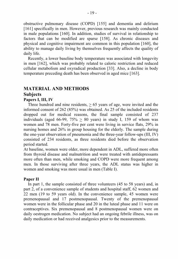
obstructive pulmonary disease (COPD) [155] and dementia and delirium [161] specifically in men. However, previous research was mainly conducted in male populations [160]. In addition, studies of survival in relationship to factors that can be modified are sparse [158]. As chronic diseases and physical and cognitive impairment are common in this population [160], the ability to manage daily living by themselves frequently affects the quality of daily life.
Recently, a lower baseline body temperature was associated with longevity in men [162], which was probably related to caloric restriction and reduced cellular metabolism and oxyradical production [33]. Also, a decline in body temperature preceding death has been observed in aged mice [163].
MATERIAL AND METHODS Subjects Papers I, III, IV
Three hundred and nine residents, > 65 years of age, were invited and the informed consent of 262 (85%) was obtained. As 25 of the included residents dropped out for medical reasons, the final sample consisted of 237 individuals (aged 66-99, 75% > 80 years) in study I, 159 of whom was women and 78 men. Forty-five per cent were living in service flats, 29% in nursing homes and 26% in group housing for the elderly. The sample during the one-year observation of pneumonia and the three-year follow-ups (III, IV) consisted of 234 residents, as three residents died before the observation period started. At baseline, women were older, more dependent in ADL, suffered more often from thyroid disease and malnutrition and were treated with antidepressants more often than men, while smoking and COPD were more frequent among men. In those surviving after three years, the ADL status was higher in women and smoking was more usual in men (Table I). Paper II
In part 1, the sample consisted of three volunteers (45 to 58 years) and, in part 2, of a convenience sample of students and hospital staff, 62 women and 22 men (19 to 59 years old). In the convenience sample, 45 women were premenopausal and 17 postmenopausal. Twenty of the premenopausal women were in the follicular phase and 20 in the luteal phase and 11 were on contraceptives. Six premenopausal and 8 postmenopausal women were on daily oestrogen medication. No subject had an ongoing febrile illness, was on daily medication or had received analgesics prior to the measurements.

- 20 -
Table I. Background data, presented as frequency (%) or m + SD, for elderly nursing-home residents (66 to 99 years old), at baseline and for survivors three year after baseline. The number of individuals is given in brackets.
Variable Baseline Papers I, III
Three-year follow-upPaper IV
Women (159)
Men (78)
Women (70)
Men (32)
Age 85.5 + 6.1 82.6 + 7.4** 83.3 + 5.9 82.0 + 7.6 ADL a 7.0 + 5.1 5.0 + 3.6** 5.5 + 3.6 3.9 + 3.8* BMI b 25.1 + 4.9 26.1 + 4.8 - - S-albumin g/l 37.4 + 3.7 37.7 + 4.1 - - Ear temperature °C 37.0 + 0.6 36.8 + 0.5 - - Rectal temperature °C 36.9 + 0.4 ¶ 36.8 + 0.3 ¶¶ - - Dementia 39.6 30.8 38.6 28.1 Cardiovascular disease 60.4 64.1 75.7 78.1 COPD c 1.3 9.0** 4.3 9.4 Smoking 0.6 11.5*** 1.4 12.5* CVI d 23.3 26.9 32.9 31.3 Diabetes 16.4 12.8 22.9 21.9 Autoimmune disease 3.8 7.7 4.3 6.3 Cancer 7.5 12.8 15.7 31.3 Thyroid disease 11.9 2.6* 14.3 6.3 Cortisone 2.5 6.4 1.4 6.3 Sedatives/tranquillisers 30.2 25.6 32.9 28.1 Antidepressants 37.7 24.4* 32.9 21.9 Paracetamol > 3 g daily 49.7 44.9 65.7 51.6 Malnutrition 22.2 9.1* - - Influenza vaccination 28.3 14.1* - - Pneumococcal vaccination
8.2 5.1 - -
a Activities of daily living b Body mass index. c Chronic obstructive pulmonary disease. d Cerebral vascular insult. ¶ n = 115. ¶¶ n = 46. *p< 0.05.**p < 0.01.***p < 0.001.
- 21 -
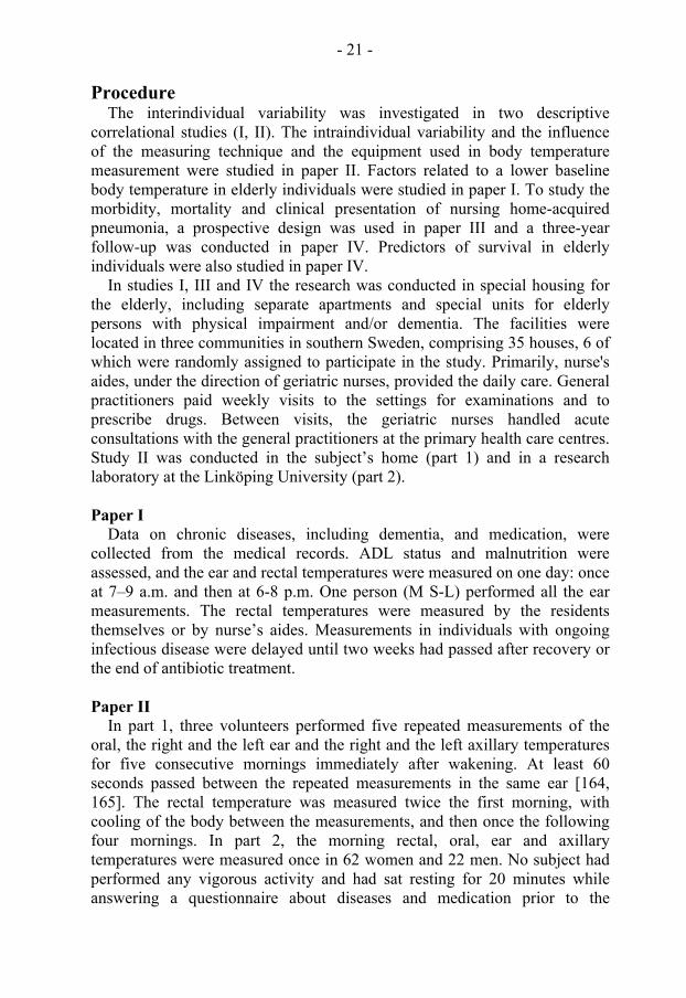
Procedure The interindividual variability was investigated in two descriptive
correlational studies (I, II). The intraindividual variability and the influence of the measuring technique and the equipment used in body temperature measurement were studied in paper II. Factors related to a lower baseline body temperature in elderly individuals were studied in paper I. To study the morbidity, mortality and clinical presentation of nursing home-acquired pneumonia, a prospective design was used in paper III and a three-year follow-up was conducted in paper IV. Predictors of survival in elderly individuals were also studied in paper IV.
In studies I, III and IV the research was conducted in special housing for the elderly, including separate apartments and special units for elderly persons with physical impairment and/or dementia. The facilities were located in three communities in southern Sweden, comprising 35 houses, 6 of which were randomly assigned to participate in the study. Primarily, nurse's aides, under the direction of geriatric nurses, provided the daily care. General practitioners paid weekly visits to the settings for examinations and to prescribe drugs. Between visits, the geriatric nurses handled acute consultations with the general practitioners at the primary health care centres. Study II was conducted in the subject’s home (part 1) and in a research laboratory at the Linköping University (part 2). Paper I
Data on chronic diseases, including dementia, and medication, were collected from the medical records. ADL status and malnutrition were assessed, and the ear and rectal temperatures were measured on one day: once at 7–9 a.m. and then at 6-8 p.m. One person (M S-L) performed all the ear measurements. The rectal temperatures were measured by the residents themselves or by nurse’s aides. Measurements in individuals with ongoing infectious disease were delayed until two weeks had passed after recovery or the end of antibiotic treatment. Paper II
In part 1, three volunteers performed five repeated measurements of the oral, the right and the left ear and the right and the left axillary temperatures for five consecutive mornings immediately after wakening. At least 60 seconds passed between the repeated measurements in the same ear [164, 165]. The rectal temperature was measured twice the first morning, with cooling of the body between the measurements, and then once the following four mornings. In part 2, the morning rectal, oral, ear and axillary temperatures were measured once in 62 women and 22 men. No subject had performed any vigorous activity and had sat resting for 20 minutes while answering a questionnaire about diseases and medication prior to the
- 22 -
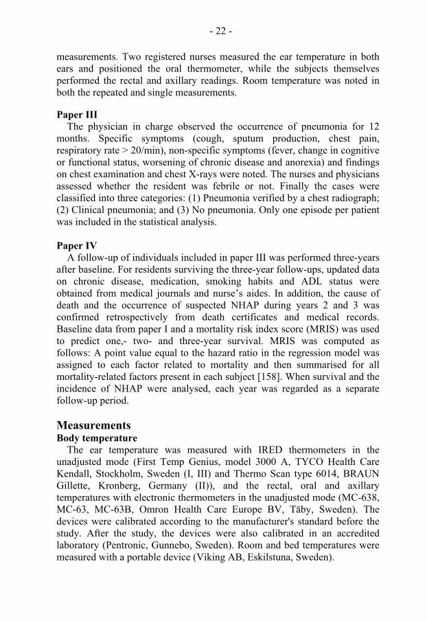
measurements. Two registered nurses measured the ear temperature in both ears and positioned the oral thermometer, while the subjects themselves performed the rectal and axillary readings. Room temperature was noted in both the repeated and single measurements. Paper III
The physician in charge observed the occurrence of pneumonia for 12 months. Specific symptoms (cough, sputum production, chest pain, respiratory rate > 20/min), non-specific symptoms (fever, change in cognitive or functional status, worsening of chronic disease and anorexia) and findings on chest examination and chest X-rays were noted. The nurses and physicians assessed whether the resident was febrile or not. Finally the cases were classified into three categories: (1) Pneumonia verified by a chest radiograph; (2) Clinical pneumonia; and (3) No pneumonia. Only one episode per patient was included in the statistical analysis. Paper IV
A follow-up of individuals included in paper III was performed three-years after baseline. For residents surviving the three-year follow-ups, updated data on chronic disease, medication, smoking habits and ADL status were obtained from medical journals and nurse’s aides. In addition, the cause of death and the occurrence of suspected NHAP during years 2 and 3 was confirmed retrospectively from death certificates and medical records. Baseline data from paper I and a mortality risk index score (MRIS) was used to predict one,- two- and three-year survival. MRIS was computed as follows: A point value equal to the hazard ratio in the regression model was assigned to each factor related to mortality and then summarised for all mortality-related factors present in each subject [158]. When survival and the incidence of NHAP were analysed, each year was regarded as a separate follow-up period. Measurements Body temperature
The ear temperature was measured with IRED thermometers in the unadjusted mode (First Temp Genius, model 3000 A, TYCO Health Care Kendall, Stockholm, Sweden (I, III) and Thermo Scan type 6014, BRAUN Gillette, Kronberg, Germany (II)), and the rectal, oral and axillary temperatures with electronic thermometers in the unadjusted mode (MC-638, MC-63, MC-63B, Omron Health Care Europe BV, Täby, Sweden). The devices were calibrated according to the manufacturer's standard before the study. After the study, the devices were also calibrated in an accredited laboratory (Pentronic, Gunnebo, Sweden). Room and bed temperatures were measured with a portable device (Viking AB, Eskilstuna, Sweden).
- 23 -
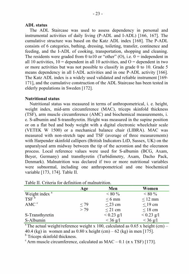
ADL status The ADL Staircase was used to assess dependency in personal and
instrumental activities of daily living (P-ADL and I-ADL) [166, 167]. The cumulative structure was based on the Katz ADL index [168]. The P-ADL consists of 6 categories, bathing, dressing, toileting, transfer, continence and feeding, and the I-ADL of cooking, transportation, shopping and cleaning. The residents were graded from 0 to10 or “other” (O), i.e. 0 = independent in all 10 activities, 10 = dependent in all 10 activities, and O = dependent in two or more activities but was not possible to classify in grade 0 to 10. Grade 5 means dependency in all I-ADL activities and in one P-ADL activity [166]. The Katz ADL index is a widely used validated and reliable instrument [169-171], and the cumulative construction of the ADL Staircase has been tested in elderly populations in Sweden [172].
Nutritional status
Nutritional status was measured in terms of anthropometrical, i. e. height, weight index, mid-arm circumference (MAC), triceps skinfold thickness (TSF), arm muscle circumference (AMC) and biochemical measurements, i. e. S-albumin and S-transthyretin. Height was measured in the supine position or on a flat bed and body weight with a digital electronic wheelchair scale (VETEK W 1500) or a mechanical balance chair (LIBRA). MAC was measured with non-stretch tape and TSF (average of three measurements) with Harpender skinfold callipers (British Indicators LtD, Sussex, UK) on the unparalysed arm midway between the tip of the acromion and the olecranon process. Local reference values were used for S-albumin (BCG, Axam, Beyer, Germany) and transthyretin (Turbidimetry, Axam, Dacho Pack, Denmark). Malnutrition was declared if two or more nutritional variables were subnormal, including one anthropometrical and one biochemical variable [173, 174]. Table II.
Table II. Criteria for definition of malnutrition. Age Men Women Weight index a < 80 % < 80 % TSF b < 6 mm < 12 mm AMC c < 79 < 23 cm < 19 cm > 79 < 21 cm < 18 cm S-Transthyretin < 0.23 g/l < 0.23 g/l S-Albumin < 36 g/l < 36 g/l a The actual weight/reference weight x 100, calculated as 0.65 x height (cm) – 40.4 (kg) in women and as 0.80 x height (cm) – 62 (kg) in men [175]. b Triceps skinfold thickness. c Arm muscle circumference, calculated as MAC – 0.1 (π x TSF) [173].
- 24 -
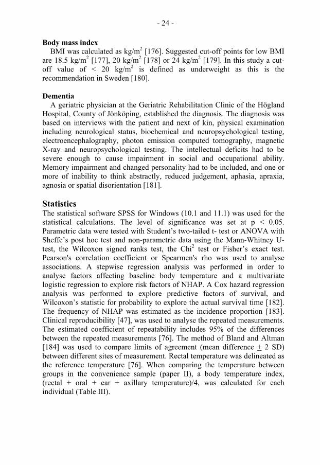
Body mass index BMI was calculated as kg/m2 [176]. Suggested cut-off points for low BMI
are 18.5 kg/m2 [177], 20 kg/m2 [178] or 24 kg/m2 [179]. In this study a cut-off value of < 20 kg/m2 is defined as underweight as this is the recommendation in Sweden [180]. Dementia
A geriatric physician at the Geriatric Rehabilitation Clinic of the Högland Hospital, County of Jönköping, established the diagnosis. The diagnosis was based on interviews with the patient and next of kin, physical examination including neurological status, biochemical and neuropsychological testing, electroencephalography, photon emission computed tomography, magnetic X-ray and neuropsychological testing. The intellectual deficits had to be severe enough to cause impairment in social and occupational ability. Memory impairment and changed personality had to be included, and one or more of inability to think abstractly, reduced judgement, aphasia, apraxia, agnosia or spatial disorientation [181]. Statistics The statistical software SPSS for Windows (10.1 and 11.1) was used for the statistical calculations. The level of significance was set at p < 0.05. Parametric data were tested with Student’s two-tailed t- test or ANOVA with Sheffe’s post hoc test and non-parametric data using the Mann-Whitney U-test, the Wilcoxon signed ranks test, the Chi2 test or Fisher’s exact test. Pearson's correlation coefficient or Spearmen's rho was used to analyse associations. A stepwise regression analysis was performed in order to analyse factors affecting baseline body temperature and a multivariate logistic regression to explore risk factors of NHAP. A Cox hazard regression analysis was performed to explore predictive factors of survival, and Wilcoxon’s statistic for probability to explore the actual survival time [182]. The frequency of NHAP was estimated as the incidence proportion [183]. Clinical reproducibility [47], was used to analyse the repeated measurements. The estimated coefficient of repeatability includes 95% of the differences between the repeated measurements [76]. The method of Bland and Altman [184] was used to compare limits of agreement (mean difference + 2 SD) between different sites of measurement. Rectal temperature was delineated as the reference temperature [76]. When comparing the temperature between groups in the convenience sample (paper II), a body temperature index, (rectal + oral + ear + axillary temperature)/4, was calculated for each individual (Table III).

- 25 -
Table III. Statistical analyses used in the thesis. Statistical methods Paper I II III IV Student’s t-test, two-tailed X X X X Mann-Whitney U-test X ANOVA with Sheffe’s post hoc test X Chi 2 test or Fisher’s exact test X X X Wilcoxon signed ranks test X Pearson’s correlation or Spearmen's rho X X Stepwise regression analysis X Multivariate logistic regression X Coefficient of repeatability X Limits of agreement X Life table with Wilcoxon's statistic for probability
X
Cox hazard regression analysis X Incidence proportion X X Ethical considerations
The studies in the thesis were conducted in accordance with the Declaration of Helsinki and were approved by the local Ethics Committee of the Faculty of Health Sciences, Linköping University (990302, 010508) and the health service directors of community care and geriatric staff nurses. Written and oral information was given to eligible residents or, if the resident was unable to respond, to their next of kin and it was made clear that participation was voluntary and could be interrupted at any time. In the case of cognitively impaired residents, additional information was given before every phase in the examination. The staff nurse decided when to direct the invitation to the resident or the next of kin.
Individuals with a functional and cognitive deficit might experience discomfort and anxiety when an unknown person performs personal care. To avoid threats to their integrity, all rectal measurements were performed by the residents themselves, if possible, or by nurse’s aides. Before data collection, the researcher was present at the unit to familiarise herself with the residents, and the next of kin or a nurse’s aides was invited to be present when the examinations were conducted. If the resident showed in verbal or nonverbal communication that he or she did not want to participate the measurements were stopped. It can be assumed that actions have been taken to minimise threats to personal integrity.
It may be questionable to perform research in aged, frail people, but the advantages is that the results can contribute to a better knowledge in nursing care and improve the quality of life at the end of life.

- 26 -
RESULTS Interindividual variability
In both elderly and younger individuals the range of the rectal temperature was 35.6°C to 38.0°C (I, II). In elderly individuals the range of the ear temperature was 33.8°C to 38.4°C (I) and in younger adults 35.0°C to 37.8°C (II). In younger adults the range of the oral and axillary temperatures was 34.5°C to 37.4°C (II). The average rectal temperature was lower and the ear temperature was higher in elderly compared to younger persons (Figure 1).
Rectal
66-9919-65
Tem
pera
ture
°C
and
99%
CI
37.4
36.9
36.4
35.9
35.4
***
Ear
66-9919-65
37.4
36.9
36.4
35.9
35.4
***
Figure 1. Baseline morning rectal and ear temperatures in 84 adults (aged 19-59 years) and 237 elderly nursing-home residents (aged 66-99 years). Average values are indicated as filled squares and the 99% confidence interval (CI) as horizontal lines. *** p < 0.001.
The ear temperature was higher in elderly individuals irrespective of
gender, and the rectal temperature lower in elderly women than in younger females, while no difference was found in men (Table IV).
Table IV. Baseline morning ear and rectal temperatures (°C) in adult men and women aged 19 to 59 years and elderly nursing-home residents aged 66 to 99 years. The number of subjects is given in brackets. *** p < 0.001. # p < 0.08.
Ear temperature °C Rectal temperature °C 19 to 59 years 66 to 99 years 19 to 59 years 66 to 99 years Men 36.6 + 0.6
(22) 36.8 + 0.5 #
(78) 36.7 + 0.4
(21) 36.8 + 0.3
(46) Women 36.6 + 0.5
(62) 37.0 + 0.6***
(158) 37.2 + 0.4
(62) 36.9 + 0.4***
(115)

- 27 -
No difference was found between elderly men and elderly women, while the average body temperature in younger subjects was higher in women but the range was wider in men, irrespective of the site of measurement (Figure 2).
Rectal
MenWomen
Tem
pera
ture
(°C)
and
99%
CI
37.4
36.9
36.4
35.9
35.4
***
Oral
MenWomen
37.4
36.9
36.4
35.9
35.4
**
Ear
MenWomen
Tem
pera
ture
(°C)
and
99%
CI
37.4
36.9
36.4
35.9
35.4
Axilla
MenWomen
37.4
36.9
36.4
35.9
35.4
*
Figure 2. Baseline morning rectal, oral, ear and axillary body temperature in 62 women and 22 men aged 19-59 years. Average values are indicated as filled squares and the 99% confidence interval (CI) as horizontal lines. * p < 0.05. ** p < 0.01. *** p < 0.001.
In adults aged 19 to 59 years, the body temperature index was 36.6 + 0.3°C in women and 36.3 + 0.6°C in men (p < 0.001) and in premenopausal women in

- 28 -
the follicular phase 36.6 + 0.6°C and in the luteal phase, 36.7 + 0.6°C, which values were significantly higher than in men (p < 0.05 and p < 0.001, respectively). Postmenopausal women (36.4 + 0.6°C) differed from premenopausal women only in the luteal phase (p < 0.05). No difference between postmenopausal women and men was found, nor did the ovulatory phase or contraceptives affect body temperature (II).
Intraindividual variability
The deviation between the lowest and the highest readings when the repeated measurement were performed ranged from 0.1°C to 0.4°C in rectal and oral readings, from 0.2°C to 1.7°C in the ear and from 0.1°C to 0.9°C in the axillary temperatures. The mean difference, including the 95% CI, of the repeated measurements was 0.3°C for oral temperatures, 0.5°C for the right ear, 0.8°C for the left ear, 0.5°C for the right axilla and 0.6°C for the left axilla. The coefficient of repeatability was similar, irrespective of the number of replicated measurements.
In the convenience sample, there was no significant individual difference between simultaneously measured temperatures in the right and left ear, or between the two operators, irrespective of the site of measurement. Room temperature did not affect body temperature, regardless of whether single or repeated measurements were performed, nor did bed temperature influence body temperature (II).
Adjustment to rectal temperature
On comparing the rectal temperature with oral, ear and axillary readings the average difference was > 0.5°C, except for rectal-ear temperatures in men. The limits of agreement were larger for rectal-ear and rectal-axillary temperatures than for rectal-oral temperatures. There was also a trend towards an increase in the deviation at higher rectal temperatures (II) (cf. Figure 2 in paper II). Factors related to a lower baseline body temperature in elderly nursing-home residents
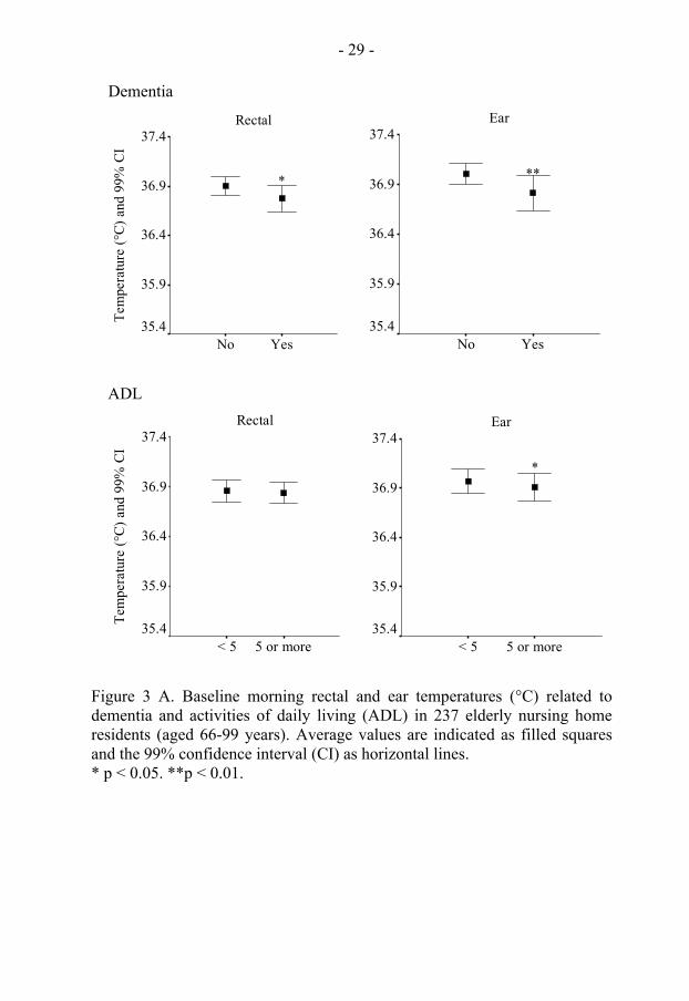
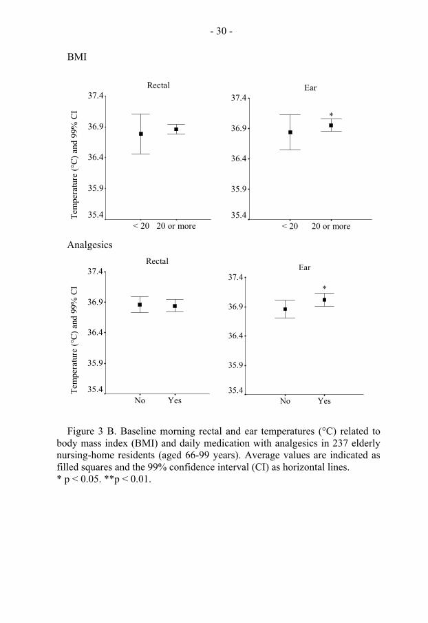
The presence of dementia, dependency in ADL and a lower BMI were related to a lower ear temperature and dementia to a lower rectal temperature, while analgesics were associated with a higher ear temperature (I) (Figures 3 A and B).
- 29 -
Dementia
Rectal
YesNo
Tem
pera
ture
(°C)
and
99%
CI
37.4
36.9
36.4
35.9
35.4
*
Ear
YesNo
37.4
36.9
36.4
35.9
35.4
**
ADL
Rectal
5 or more< 5
Tem
pera
ture
(°C)
and
99%
CI
37.4
36.9
36.4
35.9
35.4
Ear
5 or more< 5
37.4
36.9
36.4
35.9
35.4
*
Figure 3 A. Baseline morning rectal and ear temperatures (°C) related to dementia and activities of daily living (ADL) in 237 elderly nursing home residents (aged 66-99 years). Average values are indicated as filled squares and the 99% confidence interval (CI) as horizontal lines. * p < 0.05. **p < 0.01.
- 30 -
BMI
Rectal
20 or more< 20
Tem
pera
ture
(°C)
and
99%
CI
37.4
36.9
36.4
35.9
35.4
Ear
20 or more< 20
37.4
36.9
36.4
35.9
35.4
*
Analgesics
Rectal
YesNo
Tem
pera
ture
(°C)
and
99%
CI
37.4
36.9
36.4
35.9
35.4
Ear
YesNo
37.4
36.9
36.4
35.9
35.4
*
Figure 3 B. Baseline morning rectal and ear temperatures (°C) related to body mass index (BMI) and daily medication with analgesics in 237 elderly nursing-home residents (aged 66-99 years). Average values are indicated as filled squares and the 99% confidence interval (CI) as horizontal lines. * p < 0.05. **p < 0.01.

- 31 -
Pneumonia in elderly nursing-home residents At one year of follow-up, 15 of the residents with suspected NHAP had X-ray-verified pneumonia and 17 clinical pneumonia, corresponding to a yearly incidence of 13.7%. The 30-day case-fatality rate was 28.2%. COPD (p < 0.01), ADL status > 5 (p < 0.01) and male sex (p <0.05) were related to a higher risk of acquiring pneumonia, and heart disease to a lower one (p < 0.05). Cough and sputum production were the most frequent specific symptoms and, although fever was lacking in 22%, this sign and cognitive decline were the most common non-specific presenting symptoms (III). At the three-year follow-ups, 80 episodes of antibiotic-treated NHAP (range 1 to 5) were found in 68 subjects. The yearly incidence the second year was 9.5 % and the third year 6.9 %, with no difference between genders, and an overall larger incidence in individuals with functional impairment. In women, the yearly mortality rate due to NHAP decreased, while it increased in men. In individuals with suspected NHAP in year 2 or 3, no differences in baseline background factors were found compared to subjects without NHAP (IV).
Predictors for survival in elderly nursing-home residents
The cumulative hazard ratio of survival for the total group was 76.1% for year 1, 57.3% for year 2 and 43.6% at the end of year 3. CVI (25.8%), NHAP (24.2%) and heart failure (23.5%) were the most common causes of death. Death due to dementia and old age was more frequent among women and to CVI and COPD among men. Age and dependency in the ADL status predicted one-year mortality irrespective of gender, while CVI, a lower BMI and malnutrition in women and heart disease, COPD, sedatives and MRIS in men were gender specific predictors. Age predicted two- and three-years mortality and dementia three-year mortality in women, while no factors predicted two- and three-year survival in men.
A lower baseline body temperature at one-and three-year follow-ups was more usual among non-surviving women (p < 0.08 and p < 0.05), while no difference was observed in men.
A lower S-albumin (p < 0.01), malnutrition (p < 0.08) and medication with sedatives (p < 0.08) were more usual in non-surviving men the first year, and a lower BMI (p < 0.08) was more frequent among non-surviving women at three-year follow-ups. No differences between non-surviving and surviving men at two or three-year follow-ups were found (IV).
- 32 -
GENERAL DISCUSSION The paradigm of normal body temperature as 37°C and fever as > 38°C
[18] was formulated at a time when the physiological mechanisms of body temperature regulation, the influence of hormones, cellular metabolism, physical activity and methods for calibration of the thermometers were not known. In addition, the measurements were performed on patients, indicating that a large number might have been febrile. Moreover, the axillary site was used, which gives only an estimate of peripheral temperature [64, 120], and the measurement were performed in a non-standardised way. Mackowiak et al. [185] also showed that the thermometer used at that time measured 1.4°C to 2.2°C higher compared to modern digital devices. Even so, this paradigm from the middle of the 19th century is still the basis for evaluation of body temperature, explaining the confusion in clinical practice [19], and that the measurement and evaluation of body temperature is still based more on imitation, hunches and tradition than a theoretical framework and scientific knowledge [186, 187].
The present results underline the lack of evidence for normal body temperature of 37°C and demonstrates that the variation, even with standardised measurements, is wider then traditionally stated [18] due to inter- and intraindividual variability. As normal body temperature shows individual variations, it is reasonable that the same should hold true for the febrile range [125]. However, the use of cut-off values depending on the normal range at different sites [28, 138], is confusing and not significant. This thesis explores in accordance with others [14, 31] that body temperature should be evaluated in relation to the normal range characteristic of a specific reference group and individual variability, i.e. a baseline value.
The results confirm that fertile women have a higher average body temperature than men [24, 47], but also that this difference disappears on comparing men and postmenopausal women. Contradicting Cagnacci et. al., [48] the postovalutory temperature rise in fertile women was not significant, but there was a difference between postmenopausal and fertile women in the luteal phase. Other factors which can explain dissimilarities between genders are that women have a thicker layer of subcutaneous fat, a lower baseline metabolic rate and a delayed sweat onset and a lower sweating capacity than men [40, 49].
Age-related alterations in thermoregulation, metabolic rate and vascular response [33, 36, 37, 58, 59, 61, 188, 189] may explain a higher ear temperature in elderly nursing-home residents compared to younger adults, i.e. the set point is at a higher level in order to improve the thermoregulatory responses. The difference in rectal temperature between younger and elderly persons can be understood in terms of the higher temperature in fertile females (cf Table I in paper II).
In line with the findings of others [51, 52], no difference between elderly

- 33 -
men and women was observed. More importantly, the results confirm that subgroups of elderly individuals have a lower baseline body temperature [28, 190] due to functional and cognitive impairment and loss of isolating tissue. However, earlier suggestions [189, 191] of a relationship between body temperature and malnutrition were not confirmed. Daily medication with paracetamol was related to an increased baseline body temperature, reflecting the possibility that pain, which is common in aged individuals [154], is related to chronic low-grade inflammation with increased circulating levels of IL-6 and consequent fever [192]. Together, the results indicate that body temperature is stable in adult men < 60 years of age, while pre- or postmenopausal status influences body temperature in females < 60 years of age and that body temperature in aged persons is affected by age-related changes and fragility.
Other studies have focused on the degree of closeness between different sites of measurement in order to define the best choice for estimating the core temperature non-invasively [23], but our results show that no factor does exists which allows accurate conversion of temperatures recorded at one site to estimate the temperature at another site [89]. The lack of agreement between measurements reflects the fact that it is not possible to interchange different temperatures [193] or to claim that there is one range of core body temperature [194]. To define an acceptable accuracy as + 0.5°C [73] or + 0.6°C [79] between sites or to use the term equivalence only contributes to misunderstandings and confusion [69]. The probably reason for adjustments is to make the readings more familiar to clinicians. However, temperatures at the rectal and oral sites are themselves estimates of the core temperature with their own variability [119]. In the case of IREDs, there are considerable variations in how manufactures have calculated the mathematical algorithm for adjusting to the rectal, oral or pulmonary artery site [74, 94, 107, 195, 196]. Therefore, the best approach is to use an unadjusted mode and become accustomed to typical values [119], irrespective of the site of measurement.
Our results confirmed the stability of rectal temperature, probably explaining why this site has long been regarded as the most accurate method for routine estimates of body temperature [64]. Since the number of repeated measurements did not improve the reproducibility, thereby contradicting earlier suggestions for duplicate or triplicate ear temperature measurements [47, 111, 114] the results indicate that one measurement is sufficient also when the ear, oral or axillary site is used.
The repeated ear readings showed the highest variability, which may be attributed in part to the technical design of the IREDs [95]. All methods require careful handling and experienced users [194, 195], but the results indicate that training in operator technique ensures consistency in ear temperature measurement in particular [108]. Childs et al. [111] recommended measurements at the same ear, but we suggest the right ear,
- 34 -
and also the right axilla, due to less variability. However, the temperatures measured in either the left [197] or the right ear [198] have been observed to be lower and more variable, indicating the possible impact of operator hand dominance on the temperature readings [197]. While Terndrup [195] recommended an ear tug technique, Erickson [119] pointed out that when not using this technique, the free hand of the operator can support the opposite side of the patient’s head and thereby reduce the patient’s tendency to move away from the probe. In this study, the use of an ear tug technique was not controlled for, implying that this does not affect the reading, as no deviation was found between the two operators in study II.
As the calibration of the ear devices showed different deviations from the target temperature, the results also indicate that it is preferable to use the same thermometer in one individual (data presented in paper II).
Use of the rectal temperature as the reference may result in the ear reading as an inaccurate and insensitive method for detecting fever when the reading instead reflects the fact that deferversced has already occurred, thus resulting in the recording of a lower ear temperature and a still high rectal temperature [47]. In addition, as medication with analgesics is common in elderly individuals and antipyretics are frequently used in febrile patients [130], the low reading may be due to antipyretics affecting the ear temperature while the rectal temperature is still increased. It is striking that medication with analgesics and anti-inflammatory agents with an antipyretic effect [199] is rarely discussed in research involving assessment of body temperature [73].
We found that NHAP was as frequent as CVI and heart failure as a cause of death in elderly nursing-home residents. Consistent with previous research [138, 139, 200], atypical symptoms were common in infected individuals. Non-specific symptoms are also present in a lot of other physical conditions [138], indicating that "something is going on". On the other hand, fever is a cardinal sign of infection, underscoring the importance of observation and evaluation of body temperature in the nursing home setting. In contrast to earlier observations [148, 201, 202], the presence of fever was associated with increased fatality. One explanation may be that a low baseline temperature, atypical symptoms and the fact that fever was not reported until the temperature was > 38°C contributed to a delayed diagnosis and antibiotic therapy [120] and, thereby, mortality [151]. In addition, high dependency in ADL was strongly associated with dementia, a low S-albumin and malnutrition, factors that correlate with morbidity and mortality in NHAP [203, 204]. Hence, as a lower baseline body temperature may be related to a lower febrile response [142, 143], there is an increased risk of both acquiring infection and not being assessed as febrile if stipulated thresholds are used. It would be wise to establish the individual baseline body temperature in the morning on admission to the nursing home facility as a basis for assessing fever. In order to manage infections early on in elderly persons [120], further

- 35 -
research is needed to clarify special characteristics of early signs of infection in the geriatric population.
Overall, fragility is related to mortality [152, 155, 160], but the present results explore important differences in predictors of survival in men and women. The decreased survival due to COPD, heart disease and MRIS in men reflects smoking habits [156, 158, 159], and the fact that men in this generation have done harder physical work than women. The negative effect of medication with sedatives on survival in men may indicate confusion in individuals suffering from dementia [161], but also disturbed behaviour associated with frustration, unmet needs and feelings of helplessness [152]. Non-pharmacological interventions, such as bright light during the daytime, physical activity and tactile stimulation have been found to prevent disruptive behaviour and improve the sleep-wake rhythm as effectively as pharmacological treatment in physically impaired and demented persons [205]. Also, psychosocial activities have positive effects on cognitive behaviour by satisfying psychosocial needs for communication, self-esteem, safety and security, personal identity and cognitive understanding [206]. Nocturnal sleep may also enhance the immunological host defence [33].
In women, nutritional status was an important predictor of survival, but, contradicting Lapani et al. [161], malnutrition was related to death irrespective of the cognitive status. Along with the fact that medication with antidepressants was common in women, this may reflect depression and loss of appetite when the ability to perform daily activities decrease. The tendency for malnutrition to influence survival in men can be attributed to loss of appetite due to serious disability, while no correlation between ADL and nutritional status was observed (data shown in paper IV).
Together, the present results indicate that preserving or improving capabilities in daily functioning and preventing malnutrition, agitation and confusion are modifiable factors that would improve survival in nursing-home residents.
Although body temperature was not a predictor of survival, surviving women had a higher baseline body temperature than non-surviving women. No such difference was found in men, thereby contradicting the findings of Roth et al. [162], who observed a relationship between a lower normal body temperature and longevity in men. As a lower baseline body temperature was related to impaired functional and cognitive status, a decline of body temperature might precede death also in humans, as has been reported in animal studies [163]. This interpretation should be considered with caution as body temperature was measured only once, and the deviation was small. Nevertheless, the finding indicates that it would be of interest to further study body temperature as an indicator of survival in aged individuals.
- 36 -
Methodological considerations A larger, random sample and an interreliability test and documentation of
the operator as right or left-handed [47, 111, 197] would have improved the validity and reliability of the measurements in paper II.
Although the sample was not randomly selected, the nursing homes were randomly chosen and the sample studied correspond to nursing home populations in Sweden [207] as well as in other countries [22].
Other infections are of interest, but NHAP was chosen as this is the most serious infection in long-term care facilities [134, 145, 146] and national data on morbidity and mortality from NHAP are lacking. Since a chest radiograph and a blood culture or sputum examination is difficult to perform in the nursing-home setting, a diagnosis based on clinical symptoms is a reasonable approach [149].
One limitation is that the occurrence of pneumonia was partly noted retrospectively and the cause of death was abstracted from death certificates completed by different physicians, with no available data on their validity.
Use of a proxy to assess ADL status is a reliable method for persons showing cognitive decline [208, 209]. Less than 5% (9/237) were assessed as O (other), which shows good reliability [210].
The influence of analgesics on lowering temperature in fever was avoided, as the residents had not received their morning medication when the baseline temperatures were measured. As the presence of infection or trauma was controlled for, the bias with decreased S-albumin because of synthesis of acute-phase proteins was avoid [211].
- 37 -
CONCLUSIONS The range of body temperatures in healthy adults and non-febrile
institutionalised elderly individuals is wider than traditionally asserted. The inter- and intraindividual baseline body temperature varies with age, gender, site of measurement and operator technique.
In elderly nursing-home residents, the baseline temperature is altered due
to functional and cognitive impairment, loss of isolating tissue and chronic pain condition.
Body temperature should be evaluated in relation to the normal range
characteristic of a specific reference group. When measuring body temperature, the best approach is to use the
unadjusted mode irrespective of the site of measurement, without adjusting to another site. Repeated measurements do not improve the accuracy of ear, oral or axillary temperature readings.
To prevent a delayed diagnosis of pneumonia, one should be aware of a low baseline body temperature and lack of typical symptoms in elderly nursing-home residents.
Preserving and/or improving the functional, cognitive and nutritional status and actions preventing agitation and confusion would improve survival in elderly nursing-home residents.
- 38 -
ACKNOWLEDGEMENTS I wish to express my gratitude to all of those who have supported me
during my graduate studies and made this work possible. Especially, I would like to thank the following persons:
Nursing-home residents, their relatives and caregivers and volunteers involved in the studies, without whom this work would not have been accomplished.
Lis Karin Wahren, my supervisor, and Ewa Grodzinsky, my co-tutor, for their invaluable knowledge, advice, structured criticism, inspiring discussions, and for their never-ending enthusiasm and support throughout this sometimes confusing time.
Bengt Wranne, Head of the Department of Physiology, for accepting me as a PhD student and for supporting the projects.
My co-authors, Christina Forsberg, Örjan Klefsgård, Dan Loyd and Åke Örtqvist, for their knowledge, support and valuable discussions.
My office-mates, Ragnhild Raak and Agneta Karlsson, for their interest and questions from different angles worth considering.
Former and present PhD students and lecture at the Division of Nursing Science and the Division of Welfare and Care, Linköping University and the “revolution seminar group”, Falu University, for interesting discussions and useful comments.
Former and present colleagues at the administration department at the Högland Hospital, for their enthusiastic support.
Anders Wikby, for shearing his knowledge about the immune defence and supporting me at the beginning of my graduate studies.
Ulla Edell-Gustavsson, for spending early mornings with me, measures body temperature.
Christina Andersson and Anita Ljungblad, for all practical help. Isac Austin and Inger Sund-Battista, for skilful language revision of my
manuscripts. Mats Fredriksson and Ann-Britt Wiréhn, for expert statistical help. Kerstin Eknert and Pia Tingström, for pleasant company, always listen and
constructive comments. My sisters Inger, Kerstin and Birgitta, for engaged discussions during
superb dinners. My father, Sture, and my mother, Maj, for always promoting curiosity and
given me the courage to ask questions. Lars, my husband, and our children, Martin and Maria, for patience, love
and support during this febrile time.
- 39 -
REFERENCES 1. Sinclair, J., Collins Cobuild English Language Dictionary. Collins
Publishers, ed. J. Sinclair, Haaks, P, Fox, G, Moon, R, Stock, P. 1987, Glasgow: William Collins Sons & Co Ltd.
2. DuBois, E.F., The many different temperatures of the human body and its parts. Western Journal of Surgery, 1951. 59: p. 476-90.
3. IUPS, T.C., Glossary of Terms to Thermal Physiology. Pflugers Archives, 1987. 410: p. 567-87.
4. Cunha, B.A., Pneumonia in the elderly. Clinical Microbiology and Infection, 2001. 7(11): p. 581-8.
5. Boulant, J., Thermoregulation, in Fever. Basic Mechanisms and Management, P.A. Mackowiak, Editor. 1997, Lippincott Raven: Philadelphia, New York. p. 35-58.
6. Atkins, E., Fever- the old and the new. Journal of Infectious Diseases, 1984. 149(3): p. 339- 48.
7. Atkins, E., Fever: Its history, cause, and function. The Yale Journal of Biology and Medicine, 1982. 55: p. 283-9.
8. van Laar, P. and J. Cohen, A prospective study of fever in the accident and emergency department. Clinical Microbiology and Infection, 2003. 9(8): p. 878-80.
9. Mackowiak, P.A., History of clinical thermometry, in Fever. Basic Mechanisms and management, P.A. Mackowiak, Editor. 1997, Lippincott Raven: Philadelphia, New York. p. 1-10.
10. Cristofaro, P., Infections and fever in the elderly. Journal of Podiatric Medical Association, 2004. 94(2): p. 126-34.
11. Johansson, I. and N. Lyneö, Medicin och Filosofi en Introduktion (Medicine and Philosophy an Introduction). In Swedish. 1997, Uddevalla: MediaPrint Uddevalla AB.
12. Estes, J.W., Quantitative observations of fever and its treatment before the advent of short clinical thermometers. Medical History, 1991. 35: p. 189-216.
13. Sigal, A.L., Fever theory in the seventeenth century: Building toward a comprehensive physiology. The Yale Journal of Biology and Medicine., 1978. 51: p. 571-82.
14. Galen, R. and S. Gambino, Beyond Normality: The Predictive Value and Efficiency of Medical Diagnosis, ed. M.D. C. 1975, New York: Colombia University College of Physicians and Surgeons John Willey & Sons. 237.
15. Wilson, L.G., Fevers and science in the early nineteenth century medicine. Journal of the History of Medicine and Allied Sciences., 1978. 33 (3): p. 386-407.
16. Ivy, A.C., Comment: What is normal body temperature? Gastroenterology, 1945. 5: p. 326-9.
- 40 -
17. Musher, D.M., E. Dominguez, and A. Bar-Sela, Edouard Segun and the social power of thermometry. The New England Journal of Medicine, 1987. 316(2): p. 115-7.
18. Wunderlich, C.A. and J.C. Reeve, The course of the temperature in diseases: a guide to clinical thermometry. American Journal of Medical Science, 1869. 57: p. 423-47.
19. Mackowiak, P.A. and S.S. Wassermann, Physicians perceptions regarding body temperature in health and disease. Southern Medical Journal, 1995. 88(9): p. 934-8.
20. Darowski, A., J. Weinberg, and A. Guz, Normal rectal, auditory canal, sublingual and axillary temperatures in elderly afebrile patients in a warm environment. Age and Ageing, 1991. 20: p. 113-9.
21. Bender, S.B. and P.J. Scarpace, Fever in the elderly, in Fever. Basic Mechanisms and Management, P.A. Mackowiak, Editor. 1997, Lippincott Raven: Philadelphia, New York. p. 363-73.
22. Bentley, D.W., S.F. Bradley, K. High, S. Schoenbaum, G. Taler, and T.T. Yoshinkawa, Practice guideline for evaluation of fever in infection in long-term care facilities. Journal of the American Geriatric Society, 2001. 49(2): p. 210-22.
23. Sund-Levander, M., C. Forsberg, and L.K. Wahren, Normal oral, rectal, tympanic and axillary body temperature in adult men and women: A systematic literature review. Scandinavian Journal of Caring Sciences, 2002. 16(2): p. 122-128.
24. Horwath, S.M., H. Menduke, and G.M. Pierson, Oral and rectal temperatures of man. Journal of the American Medical Association, 1950. 144: p. 1562-5.
25. Tanner, J.M., The relationship between the frequency of heart, oral temperature and rectal temperature in man at rest. Journal of Physiology, 1951. 151: p. 391-09.
26. Fox, R.H. and A.J. Fry, Diagnosis of accidental hypothermia of the elderly. Lancet, 1971. 1 (7696): p. 424-427.
27. Terndrup, T., J. Allegra, and J. Kealy, A comparison of oral, rectal, and tympanic membrane-derived temperature changes after ingestion of liquids and smoking. American Journal of Emergency Medicine, 1989. 7: p. 150-4.
28. Castle, S.C., M. Yeh, S. Toledo, T.T. Yoshinkawa, and D.C. Norman, Lowering the temperature criterion improves detection of infections in nursing home residents. Aging: Immunology and Infectious Diseases, 1993. 4: p. 67-76.
29. Bellack, J.P. and B.J. Edlund, A framework for nursing assessment and diagnosis, in Nursing Assessment and Diagnosis. 1992, Jones and Bartlett Publishers: Boston. p. 2-26.
- 41 -
30. Socialstyrelsen, Socialstyrelsen föreskrifter och allmänna råd om omvårdnad inom hälso- och sjukvård. In Swedish. 1993, National Board of Health and Welfare: Stockholm.
31. Ivy, A.C., What is normal or normally? Q Bull NW Univ Med Sci, 1944. 18: p. 22-32.
32. Charkouidan, N., Skin blood flow in adult human thermoregulation: how it works, when it does not, and why. Mayo Clinic Proceedings, 2003. 12(108): p. 729-35.
33. Van Someren, E., R. Raymann, E. Scherder, H. Daanen, and D. Swaab, Circadian and age-related modulation of thermoreception and temperature regulation: mechanisms and functional implications. Ageing Research Reviews, 2002. 1(4): p. 721-78.
34. Nagashima, K., S. Nakai, M. Tanaka, and K. Kanosue, Neuronal circuitries involved in thermoregulation. Autonomic Neuroscience: Basic & Clinical, 2000. 85: p. 18-25.
35. Iwase, S., J. Cul, B. Wallin, A. Kamyia, and T. Maso, Effects of increased ambient temperature on skin sympathetic nerve activity and core temperature in humans. 327, 2002. 1(37-40).
36. Minson, C., L. Holowatz, and B. Wong, Decreased nitrit oxide- and axon reflex-mediated cutaneous vasodilation with age during local heating. Journal of Applied Physiology, 2002. 93: p. 1644-9.
37. Frank, S., S. Raja, C. Bulcao, and D. Goldstein, Age-related thermoregulatory differences during core cooling in humans. American Journal of Physiology. Regulatory, Integrative and Comparative Physiology, 2000. 279: p. 349-54.
38. Jansky, L., Shivering, in Physiology and Pathophysiology of Temperature Regulation, C.M. Blatteis, Editor. 1998, World Scientific Publishing Co. Ptc. Ltd.: Singapore. p. 48-58.
39. Bulcao, C., S. Frank, S. Raja, K. Tran, and D. Goldstein, Relative contribution of core and skin temperature to thermal comfort in humans. Journal of Thermoregulatory Biology, 2000. 25: p. 147-50.
40. Cabanac, M., Thermiatrics and behaviour, in Physiology and Pathophysiology of Temperature Regulation, C.M. Blatties, Editor. 1998, World Scientific Publishing Co. Pte. Ltd: Singapore. p. 108-25.
41. Tochihara, Y. Thermal comfort and blood pressure changes in the elderly. in Proceedings of the IX Conference on Environmental Ergonomics. 2000. Dortmund.
42. Nielsen, B. and H. Kaciuba-Uscilko, Temperature regulation in exercise, in Physiology and pathophysiology of temperature regulation, C. Blatteis, Editor. 1998, World Scientific Publishing Co. Pte. Ltd: Singapore. p. 128-43.
43. Werner, J., Biophysics of heat exchange between body and environment, in Physiology and Pathophysiology of Temperature

- 42 -
Regulation, C. Blatteis, Editor. 1998, World Scientific Publishing Co. Pte. Ltd: Singapore. p. 25-45.
44. Stephenson, L.A., Circadian timekeeping, in Fever. Basic Mechanisms and Management, P.A. Mackowiak, Editor. 1997, Lippincott Raven: Philadelphia, New York. p. 59-77.
45. Baker, F., D. Mitchell, and H. Driver, Oral contraceptives alter sleep and raise body temperature in young women. European Journal of Physiology, 2001. 424: p. 729-737.
46. Collins, K.J., T.A. Abdel-Rahman, J. Goodwin, and L. McTiffin, Circadian body temperatures and the effects of a cold stress in elderly and young subjects. Age and Ageing, 1995. 24: p. 485-9.
47. Chamberlain, J.M., T.E. Torndrup, D.T. Alexander, F.A. Silverstone, G. Wolf-Klein, R. O'Donell, and J. Grandner, Determination of normal ear temperature with an infrared emission detection thermometer. Annals of Emergency Medicine, 1995. 25: p. 15-20.
48. Cagnacci, A., R. Soldani, G. Laughlin, and S. Yen, Modification of circadian body temperature rhythm during the luteal menstrual phase: role of melatonin. Journal of Applied Physiology, 1996. 80(1): p. 26-9.
49. Fox, R.H., B.E. Lofstedt, P.M. Woodward, E. Eriksson, and B. Werkström, Comparison of thermoregulatory function in men and women. Journal of Applied Physiology, 1969. 26(4): p. 444-53.
50. Kim, H., C. Richarsdon, J. Roberts, L. Gren, and J. Lyon, Cold hands, warm heart. Lancet. Letter, 1998. 351(9114): p. 1492.
51. Thatcher, R.M., 98.6 °F, what is normal? Gerontological Nursing, 1983. 9: p. 22-7.
52. McGann, K.P., G.S. Marion, D. Lawrence, and J.G. Spangler, The influence of gender and race of mean body temperature in a population of healthy older adults. Arch Family Medicine, 1993. Dec. 2: p. 1265-7.
53. Moe, K.E., P.N. Printz, M.V. Vitiello, A.L. Marks, and L. Larsen, Healthy elderly women and men have different entrained circadian temperature rhythm. Journal of the Americam Geriatric Society, 1991. 39: p. 383-7.
54. Vitiello, M.V., R.G. Smallwood, D.H. Avery, R.A. Pascually, M.D. C, and P.N. Printz, Circadian temperature rhythms in young adults and aged men. Neurobiology of Aging, 1986. 7: p. 97-100.
55. Weitzman, E.D., M.L. Moline, C.A. Czeisler, and J.C. Zimmerman, Chronobiology of aging: temperature, sleep-wake rhythms and entrainment. Neurobiology and Aging, 1982. 3: p. 299-09.
56. Collins, K.J., C. Dore, A.N. Exton-Smith, R.H. Fox, I.C. Mac Donald, and P.M. Woodward, Accidental hypothermia and impaired temperature homeostasis in the elderly. British Medical Journal, 1977. 1: p. 353-6.
- 43 -
57. Mercer, J., Hypothermia and cold injuries in man, in Physiology and Pathophysiology of Temperature Regulation, C. Blatties, Editor. 1998, World Scientific Publishing Co. Pte. LtD: Singapore. p. 246-57.
58. Kenney, W. and T. Munce, Invited review: Aging and human temperature regulation. Journal of Applied Physiology, 2003. 95(6): p. 2598-603.
59. Pierzga, J., A. Frymoyer, and W. Kenney, Delayed distribution of active vasodilation and altered vascular conductance in aged skin. Journal of Applied Physiology, 2003. 94: p. 1045-53.
60. Grassi, G., G. Seravalle, C. Turri, G. Bertinieri, R. Dell Oro, and M. G, Impairment of thermoregulatory control of sympathetic nerve traffic in the elderly. Circulation, 2003. 108: p. 729-35.
61. Elia, M., P. Ritz, and R. Stubbs, Total energy expenditure in the elderly. European Journal of Clinical Nutrition, 2000. 54(Suppl 3): p. S92-103.
62. Hasday, J.D., The influence of temperature on host defences, in Fever. Basic Mechanisms and Management, P.A. Mackowiak, Editor. 1997, Lippincott Raven: Philadelphia New York. p. 177-96.
63. Lattawo, K., J. Britt, and M. Dobal, Agreement between measures of pulmonary artery and tympanic temperatures. Research in Nursing & Health, 1995. 18: p. 365-70.
64. Mackowiak, P.A., Clinical thermometric measurements, in Fever. Basic Mechanisms and Management, P.A. Mackowiak, Editor. 1997, Lippincott Raven: Philadelphia, New York,. p. 27-33.
65. Blatties, C., Methods of temperature measurement, in Physiology and Pathophysiology of Temperature Regulation, C. Blatties, Editor. 1998, World Scientific Publishing Co. Pte. Ltd: Singapore. p. 273-9.
66. Togawa, T., Body temperature measurement. Clinical Physiological Measurement, 1985. 6(2): p. 83-102.
67. Fraden, J., The development of Thermoscan instant thermometer. Clinical Pediatrics, 1991. Suppl 1991: p. 11-2.
68. Lefrant, J.-Y., et al., Temperature measurement in intensive care patients: comparison of urinary bladder, oesophageal, rectal, axillary, and inguinal methods versus pulmonary artery core method. Intensive Care Medicine, 2003.
69. Betta, V., F. Cascetta, and D. Sepe, An assessment of infrared tympanic thermometers for body temperature measurement. Physiological Measurement, 1997. 18: p. 215-25.
70. Eicha, L.W., A.R. Berger, B. Rader, and W. Becker, Comparison of intracardiac and intravascular temperatures with rectal temperatures in man. Clinical Investigator, 1951. 30: p. 353-9.
- 44 -
71. Ilsley, A.H., A.J. Ruten, and W.B. Runciman, An evaluation of body temperature measurement. Anaesthetic Intensive Care, 1983. 11: p. 31-9.
72. Milewski, A., K.L. Ferguson, and T.E. Terndrup, Comparison of pulmonary artery, rectal and tympanic membrane temperatures in adult intensive care unit patients. Clinical Pediatrics, 1991. 4(Suppl): p. 13-6.
73. Zehner, W.J. and T.E. Terndrup, The impact of moderate ambient temperature variance on the relationship between oral, rectal, and tympanic membrane temperatures. Clinical Pediatrics, 1991. 4: p. 61-4.
74. Brennan, D., J. Falk, S. Rothbrock, and R. Kerr, Reliability of infrared tympanic thermometry in the detection of rectal fever in children. Annals of Emergency Medicine, 1995. 25(1): p. 21-30.
75. Smitz, S., T. Giagoultsis, W. Dewe', and A. Albert, Comparison of rectal and infrared ear temperatures in older hospital inpatients. Journal of the American Geriatric Society, 2000. 48: p. 63-6.
76. Petersen, M. and H. Hauge, Can training improve the results with infrared tympanic thermometers? Acta Anaesthesiologica Scandinavica, 1997. 41: p. 1066-70.
77. Gerbrandy, J., E.S. Snell, and W.I. Cranston, Oral, rectal and oesophageal temperatures in relation to central temperature control in man. Clinical Science, 1954. 13: p. 615-24.
78. Robinson, J., J. Charlton, R. Seal, D. Spady, and M. Joffres, Oesophageal, rectal, axillary, tympanic and pulmonary artery temperatures during cardiac surgery. Canadian Journal of Anaesthesia, 1998. 45(4): p. 317-23.
79. Giuliano, K., S. Scott, S. Elliot, and A. Giuliano, Temperature measurement in critically ill orally intubated adults: A comparison of pulmonary artery core, tympanic, and oral methods. Critical Care Medicine, 1999. 27(10): p. 2188-93.
80. Rotello, L., L. Crawford, and T. Terndrup, Comparison of infrared ear thermometer derived and equilibrated rectal temperatures in estimating pulmonary artery temperatures. Critical Care Medicine, 1996. 24(9): p. 1501-6.
81. Rumana, C.S., S.P. Gopinath, M. Uzura, A.B. Valadka, and C.S. Robertson, Brain temperature exceeds systemic temperature in head-injured patients. Critical Care Medicine, 1998. 26(3): p. 562-7.
82. Varney, S., D. Manthey, V. Culpepper, and J. Creedon, A comparison of oral, tympanic, and rectal temperature measurement in the elderly. The Journal of Emergency Medicine, 2001. 22(2): p. 153-7.

- 45 -
83. Mead, J. and C.L. Bonmarito, Reliability of rectal temperatures as an index of internal body temperature. Journal of Applied Physiology, 1949. 2: p. 97-109.
84. Erickson, R., Oral temperature differences in relation to thermometer and technique. Nursing research, 1980. 29: p. 157-64.
85. Modell, J., C. Katholi, S. Kumaramangalam, E. Hudson, and D. Graham, Unreliability of the infrared tympanic thermometer in clinical practice: A comparative study with oral mercury and oral electronic thermometers. Southern Medical Journal, 1998. 91(7): p. 649-54.
86. Cooper, K., W. Cranston, and E. Snell, Temperature in the external auditory meatus as an index of central temperature changes. Journal of Applied Physiology, 1964. 19: p. 1032-5.
87. Cranston, W.I., J. Gerbrandy, and E.S. Snell, Oral, rectal and oesophageal temperatures and some factors affecting them in man. Journal of Physiology, 1954. 126: p. 347-58.
88. Rabinowitz, R.P., S.Y. Cookson, S.S. Wasserman, and et al, Effects of anatomic site, oral stimulation, and body position on estimates of body temperature. Archives of Internal Medicine, 1996. Apr(6): p. 777-80.
89. Konopad, E., J.R. Kerr, T. Noseworthy, and M.G. Peng, A comparison of oral, axillary, rectal and tympanic-membrane temperatures of intensive care patients with and without an oral endotracheal tube. Journal of Advanced Nursing, 1994. 20: p. 77-84.
90. Fallis, W., Oral measurement of temperature in orally intubated critical care patients: State-of-the-science review. American Journal of Critical Care, 2000. 9(5): p. 334-43.
91. Norman, D.C., Fever in the elderly. Clinical Infectious Diseases, 2000. 31: p. 145-51.
92. Benzinger, M., Tympanic thermometry in anaesthesia and surgery. The Journal of the American Medical Association, 1969. 209: p. 1207-11.
93. Giuliano, K., A. Giuliano, S. Scott, E. MacLachlan, E. Pysznik, S. Elliot, and D. Woytowicz, Temperature measurement in critically ill adults: A comparison of tympanic and oral methods. American Journal of Critical Care, 2000. 9(4): p. 254-61.
94. Shinozaki, T., R. Deane, and F.M. Perkins, Infrared tympanic thermometer. Evaluation of a new clinical thermometer. Critical Care Medicine, 1988. 16: p. 148-50.
95. Shibasaki, M., N. Kondo, H. Tominaga, K. Aoki, E. Hasegawa, Y. Idota, and T. Moriwaki, Continuous measurement of tympanic temperature with a new infrared method using an optical fiber. Journal of Applied Physiology, 1998. 85(3): p. 921-6.
- 46 -
96. Sato, K., N. Kane, G. Soos, C. Gisolfi, N. Kondo, and S. K, Reexamination of tympanic membrane temperature as a core temperature. Journal of Applied Physiology, 1996. 80: p. 1233-9.
97. Matsukawa, T., M. Ozaki, K. Hanagata, H. Iwashita, T. Miyaji, and t. Kumazawa, A comparison of four infrared tympanic thermometers with tympanic membrane temperatures measured by thermocouples. Canadian Journal of Anaesthesia, 1996. 43(12): p. 124-8.
98. Jakobsson, J., A. Nilsson, and L. Carlsson, Core temperature measured in the auricular canal: comparison between four different tympanic thermometers. Acta Anaesthesiologica Scandinavica, 1992. 36: p. 819-24.
99. Mariak, Z., M. White, T. Lyson, and J. Lewko, Tympanic temperature reflects intracranial temperature changes in humans. Pflugers Archives, 2003. 446(2): p. 279-84.
100. Chamberlain, J.M., J. Grandmer, J. Rubinoff, L, B.L. Klein, Y. Waisman, and M. Huey, Comparison of a tympanic thermometer to rectal thermometer and oral thermometers in a pediatric emergency department. Clinical Pediatrics, 1991. 4(Suppl): p. 124-9.
101. Castle, S.C., S. Toledo, S. Daskel, and D. Norman, The equivalence of infrared tympanic membrane thermometry with standard thermometer in nursing home residents. Journal of American Geriatric Society, 1992. 40: p. 1212-6.
102. Doezema, D., M. Lunt, and D. Tandberg, Cerumen occlusion lowers infrared tympanic membrane temperature measurement. Academic Emergency Medicine, 1993. 2(1): p. 17-9.
103. Robb, P. and R. Shahab, Infrared transtympanic temperature measurement and otitis media with effusion. International Journal of Otorhinolaryngology, 2001. 59: p. 195-200.
104. Christensen, H. and G. Boysen, Acceptable agreement between tympanic and rectal temperature in acute stroke patients. International journal of clinical practice, 2002. 56(2): p. 82-4.
105. Talo, H., M.L. Macknin, and S. VanderBrug Medendorp, Tympanic membrane temperatures compared to rectal and oral temperatures. Clinical Pediatrics, 1991. 4: p. 30-3.
106. Smith, D. and P. Fehling, Reliability of infra-red tympanic thermometry (ITT). Aviation, Space and Environmental Medicine, 1996. 67(3): p. 272-4.
107. Erickson, R.S. and S.K. Kirklin, Comparison of ear-based, bladder, oral and axillary methods for core temperature measurement. Critical Care Medicine, 1993. 21(10): p. 1528-34.
108. Klein, D.G., C. Mitchell, A. Petrinec, M.K. Monroe, M. Oblak, B. Ross, and J.M. Youngblut, A comparison of pulmonary artery, rectal,

- 47 -
and tympanic membrane temperature measurement in the ICU. Heart & Lung, 1993. 22(5): p. 435-41.
109. Nierman, D.M., Core temperature measurement in the intensive care unit. Critical Care Medicine, 1991. 19(6): p. 818-23.
110. Terndrup, T., Estimation of contact tympanic membrane temperature with a noncontact infrared thermometer. Annals of Emergency Medicine, 1997. 30(2): p. 171-5.
111. Childs, C., R. Harrison, and C. Hodkinson, Tympanic membrane temperature as a measure of core temperature. Archives of disease in Childhood., 1999. 80: p. 262-6.
112. Harasawa, K., O. Kemmotsu, T. Mayumi, and Y. Kawano, Comparison of tympanic, oesophageal, and blood temperatures during mild hypothermic cardiopulmonary bypass: A study using an infrared emission detection tympanic thermometer. Journal of Clinical Monitoring, 1997. 13: p. 19-24.
113. Freed, G.L. and J.K. Fraley, Lack of agreement of tympanic membrane temperature assessments with conventional methods in a private practise setting. Pediatrics, 1992. 89 (3): p. 384-6.
114. Stavem, K., H. Saxholm, and N. Smith-Erichsen, Accuracy of infrared ear thermometry in adult patients. Intensive Care Medicine, 1997. 23: p. 100-5.
115. Hoffman, C., M. Boyd, B. Briere, F. Loos, and P. Norton, Evaluation of three brand of tympanic thermometers. Canadian Journal of Nursing Research, 1999. Jun;31(1): p. 117-30.
116. Valle, P., O. Kildahl-Andersen, and K. Steinvoll, A comparative study of infrared tympanic thermometry and rectal mercury thermometry. Scandinavian Journal of Infectious Disease, 1999. 31: p. 105-6.
117. Craig, J., G. Lancaster, S. Taylor, P. Williiamsson, and R. Smyth, Infrared ear thermometry compared with rectal thermometry in children: a systematic review. The Lancet, 2002. 360: p. 603-9.
118. Howell, T.H., Axillary temperatures in aged women. Age and Ageing, 1972. 1: p. 250-4.
119. Erickson, R.S. and L. Meyer, Accuracy of infrared ear thermometer and other temperature methods in adults. American Journal of Critical Care, 1994. 3(1): p. 40-54.
120. Metlay, J., R. Schulz, Y. Li, S. DE, T. Marrie, C. Coley, D. Houg Obrosky, W. Kapoor, and M. Fine, Influence of age on symptoms at presentation in patients with community-acquired pneumonia. Archives of Internal Medicine, 1997. 157(13): p. 1453-9.
121. Blatties, C., The afferent signalling of fever. The Journal of Physiology, 2000. 526(3): p. 470.
122. Dinarello, C.A., Proinflammatory cytokines. Chest, 2000. 118: p. 503-8.

- 48 -
123. Engblom, D., Saha S, Engström L, Westman M, Audoly L P, Jakobsson P-E, and B. a, Microsomal prostaglandin E synthase-1 is the central switch during immune-induced pyresis. Nature Neuroscience, 2003. 6(11): p. 1137-8.
124. Blatties, C., E. Sehic, and S. Li, Pyrogen sensing and signalling: Old views and new concepts. Clinical Infectious Diseases, 2000. 31: p. S168-77.
125. Mackowiak, P.A., Fever's upper limit, in Fever. Basic Mechanisms and Management, P.A. Mackowiak, Editor. 1997, Lippincott Raven: Philadelphia, New York. p. 147-63.
126. Kozak, W., M.J. Kluger, J. Tesfaigzi, A. Kozak, K.Y.P. Mayfield, M. Wachulec, and K. Dokladny, Molecular mechanisms of fever and endogenous antipyresis, in Annals of the New York Academy of Sciences, A. Conti, Maestroni, G J M., McCann, S M M., Sternberg, E M., Lipton, J M., Smith, C C, Editor. 2000, The New York Academy of Sciences: New York. p. 121-34.
127. Zeisberger, R., S. Vybiral, and L. Jansky, Endogenous antipyretics; neuropeptides and glucocorticoids. Frontiers in Bioscience, 2004. 1(9): p. 816-26.
128. Kluger, M., The adaptive value of fever, in Fever. Basic Mechanisms and Management, P.A. Mackowiak, Editor. 1997, Lippincott Raven: Philadelphia, New York. p. 255-66.
129. Mackowiak, P.A., Benefits versus risks of the febrile response, in Fever. Basic Mechanisms and Management, P.A. Mackowiak, Editor. 1997, Lippincott Raven: Philadelphia, New Yok. p. 279-86.
130. Mackowiak, P.A., Physiological rationale for suppression of fever. Clinical Infectious Diseases, 2000. 31 (Suppl. 5): p. 185-9.
131. Rafi, A., S. Castle, K. Uyemyra, and T. Makinodan, Immune dysfunction in the elderly and its reversal by antihistamines. Biomedicine & Pharmacotherapy, 2003. 57: p. 246-50.
132. Uyemura, K., S. Castle, and T. Makinodan, The frail elderly: role of dendritic cells in the susceptibility of infection. Mechanisms of Ageing and Development, 2002. 123(8): p. 955-62.
133. Statistics, Yearbook of Health and Medical Care. 2000, Stockholm: The National Board of Health and Welfare.
134. Rothan-Tondeur, M., S. Meaume, L. Girard, S. Weill-Engerer, E. Lancien, S. Abdelmalak, P. Rufat, and A. Le Blanche, Risk factors for nosocomial pneumonia in a geriatric hospital: a control-case one-centre study. Journal of American Geriatric Society, 2003. 51(7): p. 997-1001.
135. Chandra, R.J., McCollum award lecture. Nutrition and immunity: lessons from the past and new insights into the future 1- 4. American Journal of Clinical Nutrition, 1991. 53: p. 1087-101.
- 49 -
136. Potter, J., K. Klipstein, J.J. Reilly, and M. Roberts, The nutritional status and clinical course of acute admissions to a geriatric unit. Age and Ageing, 1995. 24: p. 131-6.
137. Loeb, M., Pneumonia in the elderly. Current Opinion in Infectious Diseases, 2004. 17(2): p. 127-30.
138. Norman, D.C. and T.T. Yoshikawa, Fever in the elderly. Infectious Disease Clinics of North America, 1996. 10(1): p. 93-9.
139. Berman, P., D. Hogan, and R. Fox, The atypical presentation of infection in old age. Age and Ageing, 1984. 16: p. 201-7.
140. Kauffman, C.A., P.G. Jones, and M.J. Kluger, Fever and malnutrition: Endogenous pyrogen/Interleukin-1 in malnourished patients. American Journal of Clinical Nutrition, 1986. 44: p. 449-52.
141. Riquelme, R., A. Torres, M. El-Ebiary, J. Mensa, R. Estruch, M. Ruiz, J. Angrill, and N. Soler, Community-acquired pneumonia in the elderly. Clinical and nutritional aspects. American Journal of Respiratory Critical Care Medicine, 1997. 156: p. 1908-14.
142. Marrie, T.J., Community-acquired pneumonia requiring hospitalization. A five-year prospective study. Review of Infectious Disease, 1999. 11: p. 586-99.
143. Roghmann, M.-C., J. Warner, and P. Mackowiak, The relationship between age and fever magnitude. The American Journal of the Medical Sciences, 2001. 322(2): p. 68-70.
144. Golliot, F., P. Astagneau, B. Cassou, N. Okra, M. Rothan-Tondeur, and G. Brucker, Nosocomial infections in long-term-care and rehabilitation facilities: exploration in the development of a risk index for epidemiological surveillance. Infection Control and Hospital Epidemiology, 2001. 22(12): p. 746-53.
145. Feldman, C., Pneumonia in the elderly. The Medical Clinics of North America, 2001. 85(6): p. 1441-59.
146. Naughton, B.J., J.M. Mylotte, and A. Tayara, Outcome of nursing home-acquired pneumonia: Derivation and application of a practical model to predict 30 days mortality. Journal of the American Geriatric Society, 2000. 48: p. 1292-9.
147. Marrie, T.J., Community-acquired pneumonia in the elderly. Clinical Infectious Diseases, 2000. 31: p. 1066-78.
148. Torres, A., M. El-Biary, R. Riquelme, M. Ruiz, and R. Cells, Community-acquired pneumonia in the elderly. Seminar in Respiratory Infection, 1999. 14(2): p. 173-83.
149. Mandell, L.A., T.J. Marrie, R.F. Grossman, A.W. Chow, and R.H. Hyland, Summary of Canadian guidelines for the initial management of community-acquired pneumonia: An evidence-based update by the Canadian Infectious Disease Society and the Canadian Thoracic Society. Canadian Respiratory Journal, 2000. 7(5): p. 371-82.
- 50 -
150. Loeb, M., Pneumonia in older persons. Clinical Infectious Diseases, 2003. 37(10): p. 1335-9.
151. Beck-Sague, C., S. Banerjee, and W.R. Jarvis, Infectious diseases and mortality among US nursing home residents. American Journal of Public Health, 1993. 83(12): p. 1739-42.
152. Cohen-Mansfield, J., M.S. Marx, S. Lipson, and P. Werner, Predictors of mortality in nursing home residents. Journal of Clinical Epidemiology, 1999. 52: p. 273-80.
153. Engle, V.F. and M. Graney, Predicting outcomes of nursing home residents: death and discharge home. Journal of Gerontology, 1993. 48(5): p. 269-75.
154. Axelsson, K., K. Ahrel, A. Friström, I. Hallgren, and I. Nydevik, Pain among persons living in a nursing home. Vård i Norden, 2000. 20(2): p. 20-3.
155. Baldereschi, M., A. Di Carlo, S. Maggi, F. Grigoletto, G. Scarlato, L. Amaducci, and D. Inzitari, Dementia is a major predictor of death among the Italian elderly. Neurology, 1999. 52: p. 709-713.
156. Breuer, B., S. Wallenstein, C. Feinberg, M.J. Camargo, and B.S. Libow; L, Assessing life expectancies of older nursing home residents. Journal of the American Geriatric Society, 1998. 46: p. 954-62.
157. Dale, M.C., A. Burns, L. Panter, and J. Morris, Factors affecting survival of elderly nursing home residents. International Journal of Geriatric Psychiatry, 2001. 16: p. 70-6.
158. Flacker, J. and D. Kiely, Mortality-related factors and 1- year survival in nursing home residents. Journal of American Geriatric Society, 2003. 51: p. 213-21.
159. Corti, M.C., M. Guralnik, M.E. Salive, and J. Sorkin, Serum albumin level and physical disability as predictors of mortality in older people. Journal of American Medical Association, 1994. 272: p. 1036-42.
160. Fried, L.P., R.A. Kronmal, A.B. Newman, D.E. Bild, M.B. Mittelmark, J.F. Polak, J.A. Robbins, and J.M. Gardin, Risk factors for 5-year mortality in older adults. Journal of American Geriatric Society, 1998. 279: p. 585-92.
161. Lapani, K.L., G. Gambassi, F. Landi, A. Sgadari, V. Mor, and R. Bernabei, Gender differences in predictors of mortality in nursing home residents with AD. Neurology, 2001. 56: p. 650-4.
162. Roth, G.S., M.A. Lane, D.K. Ingram, J.A. Mattison, D. Elahi, J.D. Tobin, D. Muller, and E.J. Metter, Biomarkers of restriction may predict longevity in humans. Science, 2002. 297(2 August): p. 811.
163. Reynolds, M.A., D.K. Ingram, and M. Talan, Relationship of body temperature stability to mortality in aging mice. Mechanisms of Ageing and Development, 1985. 30: p. 143-52.
- 51 -
164. Fritz, U., M. Rohrberg, C. Lange, W. Weyland, A. Bräuer, and U. Braun, Infrared temperature measurement in the ear canal with the DIATEK 9000 Instatemp and the DIATEK 9000 Thermoguide. Comparison with methods of temperature measurement in either body parts. Anaesthesist, 1996. 45: p. 1059-66.
165. Rohrberg, M., U. Fritz, W. Weyland, and U. Braun, Temperature measurement in the ear canal: comparison of an infrared thermometer with conventional temperature probes and evaluation of clinical factors in infrared measurement. Anasthesiologie Intensivmedizin Notfallmedizin Schmerztherapie, 1997. 32: p. 409-13.
166. Hulter Åsberg, K., ADL- trappan (The ADL Staircase). In Swedish. 1990, Lund: Studentlitteratur. 143.
167. Hulter Åsberg, K. and U. Sonn, The cumulative structure of personal and instrumental ADL. Scandinavian Journal of Rehabilitation Medicine, 1988. 21: p. 171-7.
168. Katz, S., A.B. Ford, W. Moskiwitz, and M.W. Jaffe, Studies of illness in the aged. The index of ADL: A standardized measure of biological and psychosocial function. Journal of the American Medical Association, 1963. 185: p. 914-9.
169. Katz, S. and C. Akbom, A measure of primary sociobiological functions. International Journal of Health Services, 1976. 6(3): p. 493-508.
170. Brorsson, B. and K. Hulter Åsberg, Katz index of independence in ADL. Reliability and validity in short-term care. Scandinavian Journal of Rehabilitation Medicine, 1984. 16: p. 125-32.
171. Hulter Åsberg, K., Assessment of ADL in home-care for the elderly. Scandinavian Journal of Social Medicine, 1986. 14: p. 105-11.
172. Ivarsson, S. and A. Isacsson, On scaling methodology and environmental influences in disability assessments: The cumulative structure of personal and instrumental ADL among older adults in a Swedish rural district. Canadian Journal of Occupational Therapy, 1997. 64(5): p. 240-51.
173. Symreng, T., Arm anthropometry in a large reference population and in surgical patients. Clinical Nutrition, 1982. 1: p. 211-9.
174. Christensson, L., M. Unosson, and A.-C. Ek, Malnutrition in elderly people newly admitted to a community resident home. Journal of Nutrition Health & Aging, 1999. 3: p. 133-9.
175. Warnold, I. and K. Lundholm, Clinical significance of preoperative nutritional status in 215 noncancer patients. Annals of Surgery, 1984. 199: p. 299-305.
176. Whitney, E.N., C.B. Cataldo, and S.R. Rolfes, Energy balance and body composition, in Understanding Normal and Clinical Nutrition.
- 52 -
1998, Wodsworth Publishing Company: Belmont, CA. p. 256-86 Appendix E.
177. WHO, Energy and protein requirements; report of a FAO/WHO/UNU expert consultation. 1985, WHO Technical Report Series 724.
178. Detsky, A., P. Smalley, and J. Chang, Is the patient malnourished? Journal of the American Medical Association, 1994. 271(1): p. 54-8.
179. Beck, A. and L. Ovesen, At which body mass index and degree of weight loss should hospitalized elderly be considered at nutritional risk? Clinical Nutrition, 1998. 17(5): p. 195-8.
180. Sandström, B.M., A. Aro, W. Becker, N. Lyhne, J.I. Pedersen, and I. Dorsdottir, Nordiska Näringsrekommendationer. In Swedish. 1996, Nordiska Ministerrådet: Copenhagen.
181. DSM, Diagnostic and Statistical Manual of Mental Disorders. 4 ed. DSM-IV. 1994, Washington, DC: American Psychiatric Association.
182. Armitage, P. and G. Berry, Statistical Methods in Medical Research. 3 ed. 1994, London: Blackwell Science Ltd.
183. Norell, S.E., Workbook of Epidemiology. 1995, NewYork: Oxford University Press.
184. Bland, J. and D. Altman, Measuring agreement in method comparison studies. Statistical Methods in Medical Research, 1999. 8: p. 135-60.
185. Mackowiak, P.A. and G. Worden, Carl Reinhold August Wunderlich and the evolution of clinical thermometry. Clinical Infectious Diseases., 1994. 18: p. 458-67.
186. Emmoth, U. and M. Edwinsson-Månsson, Omvårdnad vid feber (Nursing care in fever). In Swedish. Vård i Norden, 1997. 17(4): p. 4-8.
187. Sund-Levander, M., L.K. Wahren, and E. Hamrin, Nursing care in fever: Assessment and implementation. Vård i Norden, 1998. 18(2): p. 27-30.
188. Khan, F., V.A. Spence, and J.J.F. Belch, Cutaneous vascular responses and thermoregulation in relation to age. Clinical Science, 1992. 82: p. 521-8.
189. Cooper, K., Thermoregulation in the elderly, in Physiology and Pathophysiology of Temperature Regulation, C. Blatties, Editor. 1998, World Scientific Publishing: Singapore. p. 162-71.
190. Fox, R.H., P.M. Woodward, A.N. Exton-Smith, M.F. Green, D.V. Donnison, and M.H. Wicks, Body temperature in the elderly: A national study of physiological, social and environmental conditions. British Medical Journal, 1973. 1: p. 200-6.
191. Collins, K.J., Effects of cold on old people. British Journal of Hospital Medicine, 1987: p. 506-14.
192. Bruunsgaard, H., M. Pedersen, and B.K. Klarlund Pedersen, Aging and proinflammatory cytokines. Current Opinion in Haematology, 2001. 8: p. 131-6.

- 53 -
193. Erickson, R.T. and T.M. Woo, Accuracy of infrared ear thermometry and traditional temperature methods in young children. Heart & Lung, 1994. 23(3): p. 182-95.
194. Holtzclaw, B.J., New trends in thermometry for the patient in the ICU. Critical Care Nursing Quarterly, 1998. 21(13): p. 12-25.
195. Terndrup, T.E., An appraisal of temperature assessment by infrared emission detection tympanic thermometry. Annals of Emergency Medicine, 1992. 21(12): p. 1483-92.
196. Schmitz, B., N. Bair, M. Falk, and C. Levine, A comparison of five methods of temperature measurement in febrile intensive care patients. American Journal of Critical Care, 1995. 4(4): p. 286-92.
197. Lee, V., N. McKenzie, and M. Cathcart, Ear and oral temperatures under usual practice conditions. Research for Nursing Practice, 1999. 1(1): p. 8.
198. White, N., S. Baird, and D.L. Anderson, A comparison of tympanic thermometer readings to pulmonary artery catheter core temperature recordings. Applied Nursing Research, 1994. 7(4): p. 165-9.
199. Mackowiak, P., Antipyretic therapy future. Clinical Infectious Diseases, 2000. 31: p. 242-3.
200. Smith, M.A. and W.M. Duke, A retrospective review of nosocomial infections in an acute rehabilitative and chronic population at a large skilled nursing facility. Journal of the American Geriatric Society, 1994. 42: p. 45-9.
201. Riquelme, R., A. Torres, M. El-Ebiary, J.P. de la Bellacasa, R. Estruch, J. Mensa, J. Fernandez-Sola, C. Hernandez, and R. Rodriguez-Roisin, Community-acquired pneumonia in the elderly: A multivariate analysis of risk and prognostic factors. American Journal of Respiratory Critical Care Medicine, 1996. 154: p. 1450-5.
202. Janssens, J.-P., L. Gauthey, F. Herrmann, L. Tkatch, and J.P. Michel, Community-acquired pneumonia in older patients. Journal of American Geriatric Society, 1996. 44(5): p. 539-44.
203. Marrie, T.J., Pneumonia in the elderly. Current Opinion in Pulmonary Medicine, 1996. 2: p. 192-7.
204. Meehan, T.P., J.M. Chua-Reyes, K.M. Prestwood, J.D. Scinto, M.K. Petrillo, and M.l. Metersky, Process of care performance, patient characteristics, and outcome in elderly patients hospitalized with community-acquired or nursing home-acquired pneumonia. Chest, 2000. 117: p. 1378-85.
205. Luijpen, M., E. Scherder, E. Van Someren, D. Swaab, and J. Sergeant, Non-pharmacological interventions in cognetively imaired and demented persons - a comparison with cholinesterase inhibitors. Reviews in Neurosciences, 2003. 14: p. 343-368.
- 54 -
206. Beck, C., T. Vogelpohl, J. Rasin, J. Topps Uriri, P. O'Sullivan, R. Walls, R. Philips, and B. Baldwin, Effects of behavioral interventions on disruptive behavior and affect in demented nursing home residents. Nursing Research, 2002. 51(4): p. 219-228.
207. Statistics, Social service, care and welfare in Sweden 1996. 1997, National Board of Health and Welfare: Stockholm.
208. Östbye, T., S. Tyas, I. McDowell, and J. Koval, Reported activities of daily living: Agreement between elderly subjects with and without dementia and their caregivers. Age and Ageing, 1997. 26: p. 99-106.
209. Magaziner, J., Use of proxies to measure health and functional outcomes in effectiveness research in persons with Alzheimer disease and related disorders. Alzheimer Disease and Associated Disorders, 1997. 11(6): p. 168-74.
210. Hulter Åsberg, K., The common language of Katz's index of ADL in six studies of aged and disabled patients. Scandinavian Journal of Caring Sciences, 1989. 2(4): p. 171-8.
211. Ferguson, R.P., P. O'Connor, and B. Crabtree, Serum albumin and prealbumin as predictors of clinical outcome of hospitalized elderly nursing home residents. Journal of the American Geriatric Society, 1993. 41: p. 545-9.