mdct evaluation of pe in children - pedrad.org · •variable clinical presentation ... bundle...
TRANSCRIPT

MDCT Evaluation of PE in Children
What is New? Edward Y. Lee, MD, MPH
Chief, Division of Thoracic Imaging
Director, Magnetic Resonance Imaging
Associate Professor of Radiology
Boston Children’s Hospital and Harvard Medical School

Introduction
• Pulmonary embolism (PE) is a potentially
life-threatening condition – Tapson VF. Acute PE. NEMJ. 2008
• Historically, the incidence (0.73 – 4.2%) of PE
has been believed to be low – Buck JR, et al. PE in Children. J Pediatr Surg. 1981
• Recent studies showed much higher incidence
(14 – 15%) of PE in children – Kritsaneepaiboon S, Lee EY, et al. CTPA Evaluation of PE in Children. AJR. 2009
– Victoria T, et al. Evaluation of PE in Children. Pediatr Radiol. 2009

Objectives
• Discuss up-to-date information on PE in children – Lee EY, et al. PE in Children: Survey of CTPA Practices. Acad Radiol. 2010
• Review currently available imaging modalities & techniques – MDCT parameters
– Post-processing techniques
• 2D imaging
• 3D imaging (MIP)
• Provide practical recommendations on imaging
management of PE in children
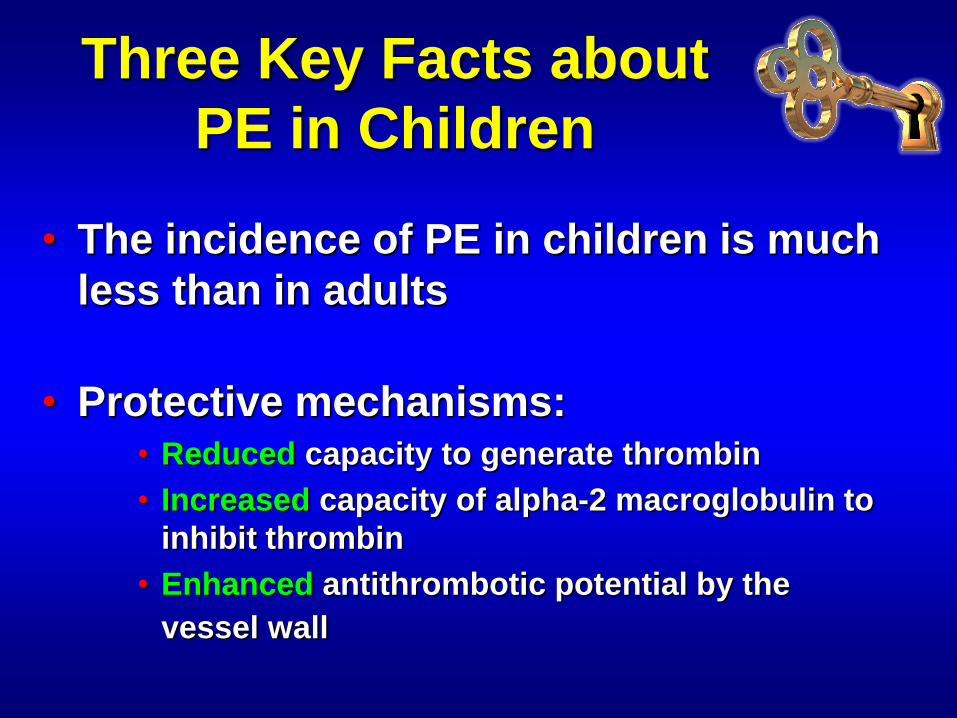
Three Key Facts about
PE in Children
• The incidence of PE in children is much
less than in adults
• Protective mechanisms: • Reduced capacity to generate thrombin
• Increased capacity of alpha-2 macroglobulin to
inhibit thrombin
• Enhanced antithrombotic potential by the
vessel wall

Three Key Facts about
PE in Children
• Neonates & infants are at greatest risk of
childhood thrombosis
• Risk factors:
– Peripartum asphyxia
– Dehydration
– Septicemia
– Trauma / surgery
– Central venous lines
Schmidt B, Andrew M. Neonatal Thrombosis. Pediatrics. 1995

Three Key Facts about
PE in Children
• Idiopathic PE occurs uncommonly in
children
– ~4% in children vs. ~30% in adults • Andrew M, et al. VTE in Children. Blood. 1994
• Most PE occurs in association with
underlying risk factors
– Interrelated & multiple risk factors
Lee EY, et al. Unsuspected PE in Pediatric Oncology Patients: Detection with MDCT. AJR 2010

Next: Ingredients for a Proper
DX of PE
Clinical
Findings
Laboratory
Tests
Imaging
Evaluation
Kritsaneepaiboon S, Lee EY, et al. CTPA Evaluation of PE in Children. AJR. 2009
Victoria T, et al. Evaluation of PE in Children. Pediatr Radiol. 2009

Clinical Evaluation for PE
• Variable Clinical Presentation
– Chest pain (70%)
– Tachypnea (70%)
– Cough (40%)
– Tachycardia (33%)
– SOB (25%)
– Pulmonary HTN (5%)
Kritsaneepaiboon S, Lee EY, et al. CTPA Evaluation of PE in Children. AJR. 2009
Victoria T, et al. Evaluation of PE in Children. Pediatr Radiol. 2009

EKG & Laboratory Tests
• EKG Findings
– Sinus tachycardia
– ST-T segment changes
– Right axis deviation &
bundle branch block
• Blood Findings
– Hypocapnia
– Hypoxemia with a-ACO2 gradient

Evaluation of PE in Children: Imaging Evaluation
• No published studies documenting the
sensitivity & specificity
– Clinical evaluation
– Diagnostic imaging tests
• Imaging protocols have been usually
extrapolated from adult studies
– Little justification
– Lack of applicability

Currently Available Imaging Studies
CXR V/Q Scan CTPA Angio
Advantages
Easy to perform
Relatively cheap
Fast
Disadvantages
Not sensitive
Not specific
Advantages
Gold Standard
Embolectomy
Disadvantages
Invasive
+ Radiation
Long
Advantages
High Sen / Spec
Fast
R/O other Dx
Disadvantages
Radiation
IV contrast
Advantages
Low radiation
No IV contrast
High spec/prob
Disadvantages
Lung Dz
Interm. prob
Long (~45 min)

What Are We Currently Doing for
PE Evaluation in Children?
• Survey of PE Evaluation in Children
– To determine the current policies &
practices of SPR members
– Survey sent electronically to the 1575
members (416 institutions) of the SPR

Pulmonary Embolism in Children:
Survey Items
• Existence of written policies
• Imaging study of choice
• Currently used CTPA techniques
• Modifications of protocols for radiation
dose reduction

Results
• 28% (118/416) response rate on an institutional
basis
• Written policy only in 25% institutions
• CXR performed before CTPA (64%)
• CTPA = imaging modality of choice (89%)

Results: Radiation Dose Reduction
Lee EY, et al. Pulmonary Embolism in Pediatric Patients: Survey of CTPA Practices & Policies
Acad Rad. 2010
• 60 Respondents (58%) modify CTPA imaging protocols

MDCT Imaging Techniques:
CTPA
• Sedation / Intubation
– Usually < 5 years old
– Conscious sedation
– Sedative medications
• Oral chloral hydrate
• IV Pentobarbital sodium
– Adequate cardiorespiratory support
Kritsaneepaiboon S, Lee EY, et al. MDCT CTPA Evaluation of PE in Children. AJR. 2009

Imaging Techniques:
CTPA Parameters • 64 MDCT
• Detector thickness = 0.6 mm
• kVp
– 80 kVp for infants (up to 1 year of age)
– 100 kVp for children (up to 4 years of age / 40 kg)
– 120 kVp for older children
• mAs (using dose modulated tube current)
• Rotation time = 0.33 sec. (0.5 sec for 2nd phase)
• Pitch = 0.2 (0.55 for 2nd phase)
• Slice thickness = 1 mm (1.5 mm for 2nd phase)
• Scan in caudal to cranial direction
Kritsaneepaiboon S, Lee EY, et al. MDCT CTPA Evaluation of PE in Children. AJR. 2009

MDCT Techniques:
Optimizing Contrast Opacification
• Contrast dose = 1.5 cc/kg (Isovue 370)
• Contrast injection rate depends on size & stability of IV catheter
• Suggested injection rates
Catheter Size (gauge) Injection Rate (mL/s)
24 1 – 1.5 (by hand)
22 2.0 – 2.5
20 3.0 – 4.0
18 4.0 – 5.0

MDCT Technique: Scan Analysis & Post-processing
• MPR (Multiplanar)
– Coronal & sagittal reformats
• 3D Volume Rendered
Reconstructions
– Maximum Intensity Projection
(MIP) images
• Are these post-processed
CT images helpful?

CTPA: Value of MPR Reformatted
Images in Detecting PE in Children
Lee EY, et al. CTPA: Value of Multiplanar Reformatted Images in Detecting PE in Children, AJR, 2011

Results

Results
Lee EY, et al. CTPA: Value of Multiplanar Reformatted Images in Detecting PE in Children, AJR, 2011

Case 1
16-year-old boy with sudden onset of SOB and right chest pain
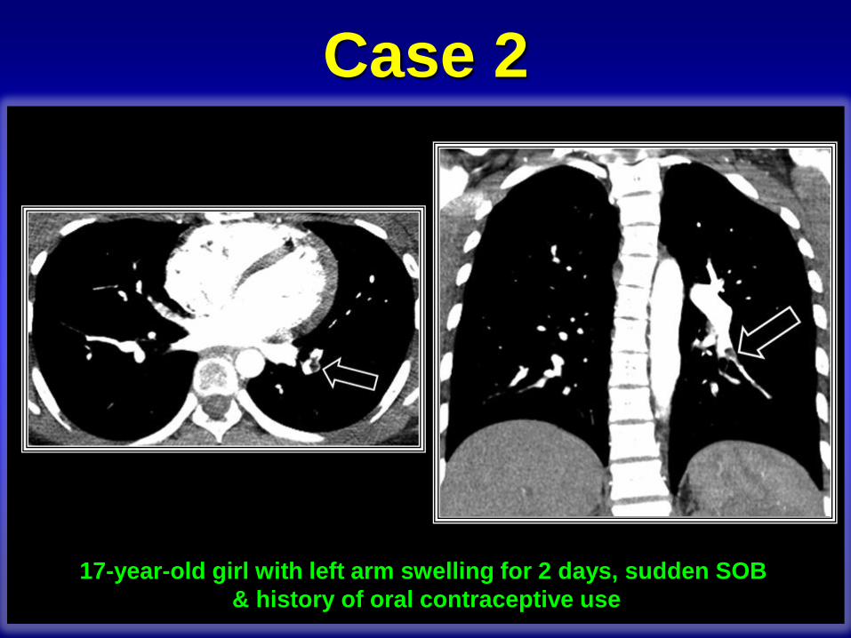
Case 2
17-year-old girl with left arm swelling for 2 days, sudden SOB
& history of oral contraceptive use

Next: CT Imaging Findings in
Children with PE

Imaging Findings: Location of PE
Figure 2
Lobar Distribution of Pulmonary Emboli
RLL LLL RUL LUL RML LML
Nu
mb
er
of
Pu
lmo
na
ry E
mb
oli
0
1
2
3
4
5
6
7
8
9
10
11
12
13
37%
24%
15%
12% 12%
0%
Total number of emboli = 33
Figure 1
Pulmonary Artery Location of Pulmonary Emboli
LPA SPA SSPA MCPAN
um
ber
of
Pu
lmo
nary
Em
bo
li0
1
2
3
4
5
6
7
8
9
10
11
12
13
39%
35%
16%
10%
Total number of emboli = 31
Kritsaneepaiboon S, Lee EY, et al. MDCT CTPA Evaluation of PE in Children. AJR. 2009

Imaging Findings:
Parechymal Findings
Lee EY, et al. Parenchymal & Pleural Abnormalities in Children at CTPA. Pediatr Radiol. 2009

Parenchymal Findings in PE: Wedge-shaped Peripheral Opacity

Parenchymal Findings in PE: Wedge-shaped Peripheral Opacity

Beyond the Pulmonary Arteries: Alternative Diagnoses
Lee EY, et al. Alternative Diagnoses in Children with CTPA Negative for PE. AJR. 2009
Normal
CTPA
(N=39)
32%
Alternative
Diagnosis
(N=57)
48%
Non-
Diagnostic
CTPA
(N=6)
PE
(N=21)
17%
CTPA in children with
clinically suspected PE
(N = 123)
CTPA in children with
clinically suspected PE
(N = 123)
CTPA in children with
clinically suspected PE
(N = 123)

Pneumonia
15-year-old girl with SOB and left sided pleuritic chest pain

Atelectasis
3-year-old girl with SOB / increased oxygen requirement status
post left hemispherectomy for refractory seizure disorder

Lung CA
14-year-old girl with SOB and de-saturation

Right Atrial Thrombosis
18-year-old girl with factor V Leiden mutation with
de-saturation and chest pain

Overutilization of CTPA in
Children?
• Recent studies showed increasing use of CTPA in children suspected of having PE
• But, the rate of positive studies is relatively low, suggesting overutilization of this test
• Thromboembolic risk factor assessment was shown to be useful for directing when to perform CTPA in a recent study in adult patients
Lee EY, et al. PE in Pediatric Patients: Survey of CTPA Practices & Policies. Acad Radiol 2010
Mamlouk MD, et al. PE at CTPA: Implications for Appropriateness, Cost & Radiation Exposure. Radiology 2010

Thromboembolic Risk Factors &
Implications for Appropriate Use
• To evaluate thromboembolic risk factors for
PE detected by using CTPA in children
• To determine whether such information may
guide more appropriate use of CTPA
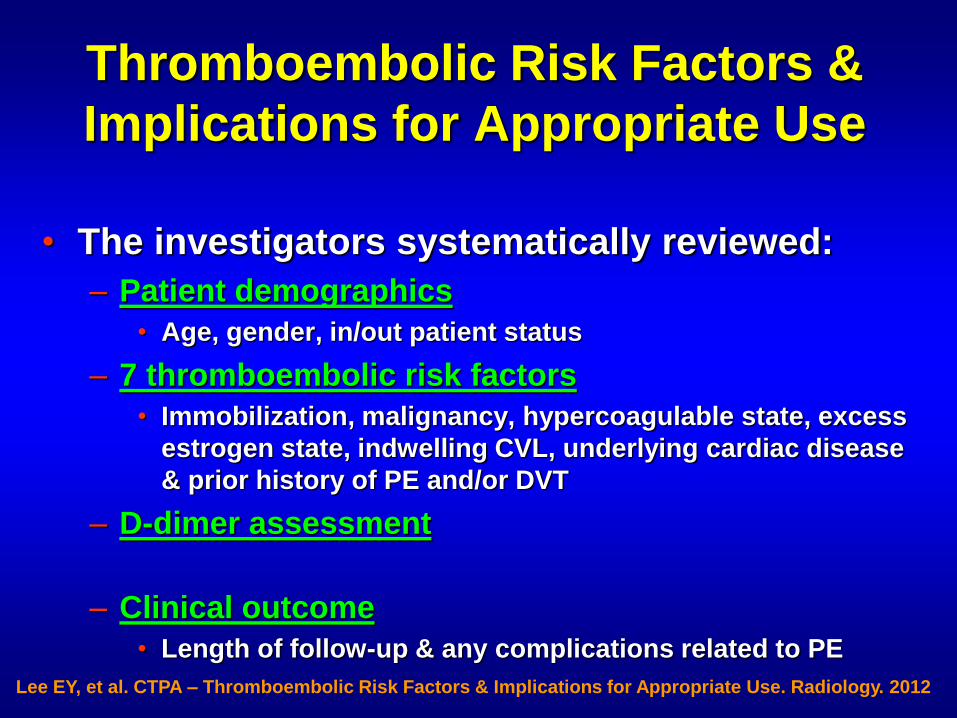
• The investigators systematically reviewed:
– Patient demographics
• Age, gender, in/out patient status
– 7 thromboembolic risk factors
• Immobilization, malignancy, hypercoagulable state, excess
estrogen state, indwelling CVL, underlying cardiac disease
& prior history of PE and/or DVT
– D-dimer assessment
– Clinical outcome
• Length of follow-up & any complications related to PE
Thromboembolic Risk Factors &
Implications for Appropriate Use
Lee EY, et al. CTPA – Thromboembolic Risk Factors & Implications for Appropriate Use. Radiology. 2012

Comparison in Pts with & without PE

Comparison of Five Statistically Significant
Risk Factors b/n Pts with & without PE

Number of Risk Factors in Patients
with & without PE

ROC Curve for Differentiating Pts with
PE from Those without PE
Sensitivity = 89%
Specificity = 94%
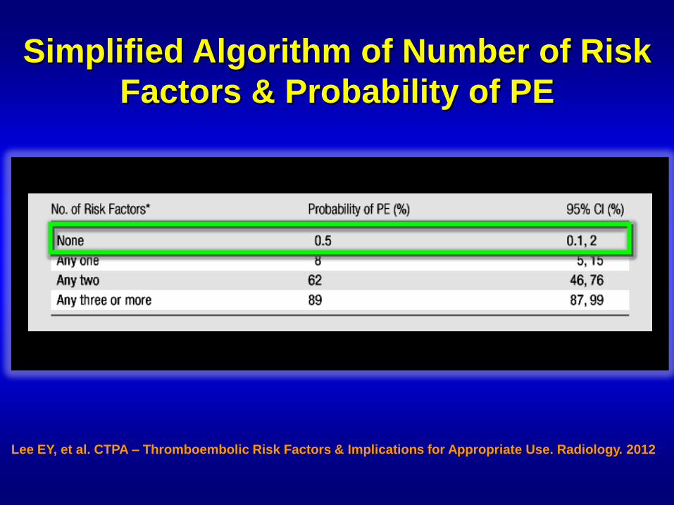
Simplified Algorithm of Number of Risk
Factors & Probability of PE
Lee EY, et al. CTPA – Thromboembolic Risk Factors & Implications for Appropriate Use. Radiology. 2012

Risk Factor Assessment for PE:
Older Children & Young Adult
Lee EY, et al. PE Detected by CTPA in Older Children & Young Adults: Risk Factor Assessment. AJR. 2012
Take Home Points
• Diagnosis of PE is not easily done clinically, even with the help of biochemical tests
• Failure to be diagnosed in the greatest threat to patients with PE
• Utilization of CTPA are helpful in diagnosis – Presence of PE
– Associated lung findings
– Alternative diagnoses

Take Home Points
• Important to know proper CTPA techniques – Contrast optimization
– Radiation dose reduction techniques
• Use of MPR MDCT images significantly increases confidence level & interobserver agreement among radiologists
• Use of risk factor assessment as a first-line triage tool has the potential to guide more appropriate use of CTPA in children – Reductions in radiation exposure & costs