mcst - medicine2017.wikispaces.com…لخص+بيديا.pdf · signs of malnutrition: ↓muscle...
TRANSCRIPT
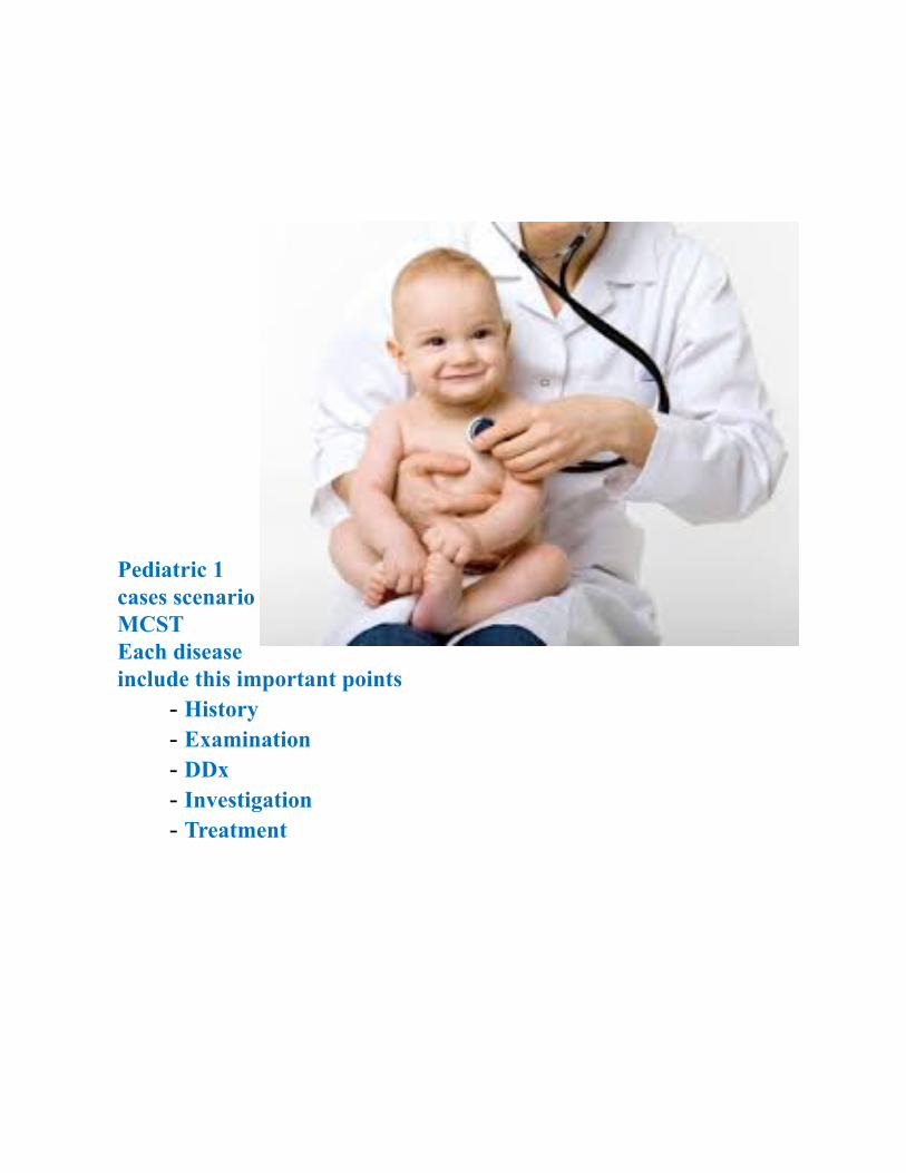
Pediatric 1cases scenarioMCSTEach diseaseinclude this important points
- History- Examination- DDx- Investigation- Treatment
بسم اهلل الرحمن الرحيم
الحمد هلل والصالة والسالم على رسوله االمني وعلى اله وصحبه اجمعني
شكرا لكل من ساهم في اتمام هذا العمل من جمع للمعلومات وتنسيقها
إن أصبنا فبتوفيق من اهلل ،، وان أخطأنا فمن أنفسنا ومن الشيطانأسأل اهلل التوفيق للجميع
فريق العمل :
بشر موسى باشامحمد كلبون
خالد الحساميحازم الشيخ
أحمد الرفاعييوسف شققينواف العليانعامر الخليف
Congenital heart disorder , RDS , viral infection are not included in this work , please go*to slides and review it there J
UPPER RESPIRATORY TRACT INFECTION***Cause: Viruses cause >90% of URTIs.
.Coryza: Rhinovirus, coronavirus, RSVPharyngitis: Adenovirus, enterovirus, rhinovirus, group A b-hemolyticstreptococcus in older children.
.Tonsillitis: EBV (infectious mononucleosis), group A b-haemolytic streptococcusOtitis media: Influenza, parainfluenza, enteroviruses and adenovirus, Streptococcuspneumoniae, non-typeable Haemophilus influenzaeHISTORYGeneral: Lethargy, poor feeding.Coryza: Sneezing, sore throat, fever is variable.Pharyngitis/tonsillitis: Fever, sore throat, cough, abdominal pain; mesentericadenitis is often preceded by a URTI with subsequent enlargement of the mesentericlymph nodes.Infectious mononucleosis: Prolonged lethargy, malaise, sore throat. Otitis media:Ear pain; infant may scream and pull at ear, conductive hearing loss in chronicsecretory otitis media.EXAMINATIONGeneral: Pyrexia, tachycardia, cervical lymphadenopathy.Coryza: Nasal discharge.Pharyngitis: The pharynx, soft palate and tonsillar fauces are inflamed and swollen.Tonsillitis: Red, swollen tonsils with or without white exudates.Follicular tonsillitis with white exudates may be due to adenovirus, EBV or group Ab-hemolytic streptococcus.Otitis media: Tympanic membranes bright redand bulging on otoscopy with lossofnormal light reflex. May see pus in the middle ear.
INVESTIGATIONS
1- Throat swab: May grow group A b-haemolytic streptococcus2- Bloods: ASOT, monospot test (EBV).
DDxEach disease in URTI could be deffrential for the other Manegament :
1- Antipyretic as: paracetamon or ibibrufin2- If it is bacterial : Oral antibiotics such aspenicillin
orerythromycin(ifpenicillin allergic) for 10 days to prevent rheumaticfever are indicated if group A b-haemolytic streptococcus grows on throatswab.
3- If it is viral : supportive manegament + rest + good fluid
4- Surgical intervention: Tonsillectomy is rarely indicated, only whenrecurrent tonsillitis is causing significant loss of schooling or upperairways obstruction and sleep apnoea.
Bronchiolitis*** Cause: RSV in 75%HISTORYCough, breathlessness and expiratory wheeze. In more severe cases infants maybecome too breathless to feed, have apnoeic spells or become lethargic. EXAMINATIONGeneral: Mild pyrexia, tachycardia, irritability. Respiratory distress: Tachypnea,
subcostal/intercostal recession, nasal flaring, grunting, widespread expiratorywheeze +/- fine crepitations. Severe disease: Cyanosis, lethargy.DDxAsthma - pneumonia - cystic fibrosis INVESTIGATION
• CBC,blood gas analysis •
• chest X-ray : hyperinflation due to small airways obstruction and Collapseclassically of the right upper lobe
MANAGEMENT
1- Criteria for admission: Feeding difficulty (<50% usual amount), grunting,severe recession and/or tachypnoea, episodes of apnoea, saturations <95%,lethargy.
2- Supplemental oxygen if saturation <92% to keep it more than 94%

3- Maintenance 5%D +NS .Bronchodilator : Ventolin, if not effective, should be discontinued -4
5- If improved and saturation become >94% >>> discharge6- If not continue the admission
Pneumonia*** Cause :• Neonate: groub B hemolytic streptococcus – E-coli – chlamydia
trachomatis• Infant-preschool children:Viral : parainflunza – influenza – adenovirus – RSVBacterial : streptococcas pneumonia 90% - staph aureus• Older children: as above + atypical organism such chlamydia pneumonia –
mycoplasma pneumoniaHistoryGeneral: Fever, tachycardia, tachypnea, cough, sputum (yellow, green or rustyin Strep. pneumoniae), vomiting particularly post-coughing, poor feeding,diarrhoea, preceding URTI (especially viral infections).ExaminationSigns of consolidation: decrease Breath sounds, dullness to percussion,bronchial breathing, coarse crepitations.DDxBronchiolitis - Asthma - Lung abcess - Atelectasis - RDSINVESTIGATION
• CXR: Focal consolidation suggests a bacterial cause; diffuse consolidationbronchopneumonia suggests a viral cause.
• Bloods: increase WCC, increase ESR/CRP, U&Es (SIADH), mycoplasmaserology.
• Urine: Pneumococcal antigen.• Blood film : RBC agglutination by MycoplasmaManegamentSupportive treatment: Maintain oxygen saturations>92%, IV resuscitationindehydration or shock.Antibiotics: Determined by presentation, i.e. viral/bacterial aetiology, severityand CXR appearance; normally oral amoxicillin or erythromycin. If severe,IV cefuroxime +/- erythromycin, metronidazole for aspiration pneumonia
CYSTIC FIBROSIS***
Case:
An 8 month old child presents with a history of poor growth and a chronic cough. He was theproduct of a 21 year old Gravida 2 Para 1, Ab 0 mother and was born at 41 weeks of
gestational age. Soon after birth, he developed respiratory distress and was admitted to theneonatal intensive care unit. He was initially breast-fed, but due to frequent vomiting and
loose bowel movements, he was changed to formula feeding. Despite trials of different typesof formulas, his clinical course was remarkable for bloating, diarrhea and failure to thrive. He
developed a daily cough and some respiratory difficulty. At the age of 5 months he washospitalized for respiratory distress and was diagnosed as having asthma. He continued to
have loose, large, greasy, foul-smelling stools and failure to thrive. An iontophoresis of.pilocarpine sweat test is now being obtained
:CausesObstructive azoospermiaChanged mutated gene
:Sings & SymptomsFrom history:
- Neonatal: 15% present with meconium ileus (bowel obstruction by thickmeconium).
Infancy: Recurrent chest infections, steatorrhoea, failure to thrive, developmental -.delay
Older children: Asthma, allergic bronchopulmonary aspergillosis (episodic -.wheeze, low-grade fever, brown sputum), recurrent sinusitis
:On examination.Signs of malnutrition: ↓Muscle mass, protuberant abdomen -
.Respiratory: Hyperinflation (air trapping), coarse crepitations, expiratory rhonchi -.Others: Jaundice, early digital clubbing, nasal polyps, rectal prolapse -
Differential Diagnosis:Acute SinusitisBronchiolitis
AsthmaPneumothorax
Investigations:Sweat test (gold standard): Sweat Cl of >50 mmol/L and Na2 of >60 mmol/L by pilocarpineiontophoresis, weight of sweat >100 mg on 2 occasions.Lung function: Obstructive picture with air trapping and hyperinflation (↓FEV1, ↑TLC).
Guthrie’s test: ↑Serum immunoreactive trypsinAntenatal tests: First trimester CVS (95% sensitivity). Second trimester ↓intestinal ALP in
.(amniotic fluid (90% sensitivity
:Management
Respiratory:Flucloxacillin prophylaxis for Staphylococcus aureus .1
If infected by pseudomonas treated with nebulised antibiotics (colomycin .2(gentamicin/tobramycin/
itraconazole/ Allergic bronchopulmonary aspergillosis: steroids .3inhaled / Airway responsiveness presents with asthma-like symptoms: bronchodilators↑ .4
steroids if responsive.Nebulised recombinant human DNAse acts as a mucolytic .5
.Nutritional: ↑Calorie, ↑protein diet for ↑energy requirements and malabsorption.pneumococcal, influenza + Immunisations: Usual schedule
.Physiotherapy: >2 × daily to be continued even when well; swimming is ideal exercise.Heart-lung transplant: If lung function deteriorates to <30% predicted best
.Gene therapy: Viral vectors/liposomes to deliver normal copies of the CF gene
Rheumatic fever***Cause: group A beta streptococcal infectionHISTORY AND EXAMINATIONRheumatic fever occurs around 20 days after streptococcal throat infection.Diagnosed by modified Duckett Jones criteria (2 major or 1 major and 2minor):Major: Carditis - Migratory polyarthritis - Erythema marginatum(serpiginous, flat, non-scarring, painless rash) - SC nodules - Sydenhamchorea (rapid unco-ordinated jerky movements primarily of hands, feet andface ) .Minor :fever – arthralgia – previous RF or carditis – posistve ESR/CPR –leukocytosis – prolonged PR intervalTypically beginning with a polyarthritis 2–6 weeks after streptococcalpharyngitis, and usually characterised by pyrexia and toxicityDDx
Rheumatoid Arthritis - Septic Arthritis - Sickle Cell Anemia - Juvenile Idiopathic Arthritis
INVESTIGATIONBY THE CRITERIA
In ECG : PR pronlogationIn ECHO : pericarditis – myocarditisIn Blood : increase the ESR/CPRManagementEradicate streptococcus: Penicillin or macrolide (if penicillin allergic).Arthritis: AnalgesicsCarditis: NSAIDs to suppress inflammation. In severe carditis with heartfailure, corticosteroids (prednisolone) may be started.Antistreptococcal prophylaxis: Penicillin V orally for 25 years to preventrecurrence
Kwasaki diseaseDEFINITION A childhood acute febrile illness with small and medium vesselvasculitis.Casue: could be infectious or geneticHISTORY AND EXAMINATION:Classic features: High fever for 5 days or more and the presence of 4 of:1. Erythema/oedema of hands and feet followed by desquamation2. Diffuse maculopapular rash (usually within 5 days)3. Bilateral, non-exudative conjunctivitis4. Erythema of lips and oral mucosa, strawberry tongue5. Cervical lymphadenopathy (1.5cm or more ), usually unilateral.Revised guidelines: Fever persisting 4 days or more if other clinical features arepresentDDx
• Mononucleosis• Parvovirus B19 infection• Scarlet fever
INVESTIGATIONSBloods: increase WCC, increase CRP/ESR, increase plateletsMicrobiology: Blood culture, throat, nose and rectal swab 2D ECHO/coronaryangiography:KDmaybediagnosedifcoronaryarteryaneurysm,fever and <4 classicfeatures.MANAGEMENTAntibiotics: Until bacterial infection has been excluded.IVIG:Decreasestheriskofcardiaccomplications.AsprinSteroid: only considered if 2 effusion of IVIG is not effectiveVSD***Cause: most common congenital heart disease. History:Asymptomatic - Rapid breathing - Excessive Sweating - Poor weight
gain - Congestive Heart Failure, usually within 6 to 8 weeks of life if defect islarge ( dysnea , palpitation , failer to thrive )
Examination :
- Loud harsh pansystolic heart murmur.. Palpable thrill -
.(Parasternal heave (RVH - - Signs of congestive heart failure: tachycardia , tachypnea, respiratory
distress (retractions), grunting, difficulty with feeding , diaphoresis,displaced apex beat and hepatomegaly.
DDx• Pulmonary Stenosis• Patent Ductus Arteriosus (PDA) INVESTIGATION
CXR >> RVH & RAH
ECG >> fisrt degree heart blook , right axis deviation
Doppler + echo **diagnostic
MANAGEMNET
• Lasix, Digoxin and Captopril (ACE inhibitors).
Surgery is patching the defect by pericardium or Dacron (open heart surgery •
.(with cardiopulmonary bypass
Pulmonary artery banding to reduce blood flow to lungs if not stable for •
.surgery
. Percutaneous Device closure •
Scarlet feverScarlet fever is an upper respiratory tract infection which iscaused by an infection with pyrogenic exotoxin(erythrogenic toxin)–producing Group A β-hemolyticstreptococcus.
: Main clinical manifestations are 3 Acute fever •
Pharyngitis with sore throat and strawberry tongue •Diffuse and red exanthematous rash followed by •
desquamation and hyperpigmentationfirst sign ofdisease
: 3weeks later~2Rheumatic fever •
Glomerulonephritis •Arthritis •
INVESTIGATION :History of close contact with a well-documented case ofGAS pharyngitisClinical data: Fever, Pharyngitis, Rash
Throat swab culture on a sheep blood agar plate has asensitivity of 90-95% for detecting the presence of GAS in
.the pharynx. Is the gold standard testRapid antigen detection tests : these rapid tests are
more expensive than the blood-agar culture, the advantagethey offer over the traditional procedure is the speed with
which they can provide results, often less than 10-15
.minutesNucleic acid amplification tests including isothermal loop
.amplification are also available to detect GAS pharyngitisDDx
• MEASLESRUBELLA •
MENINGOCOCCEMIA • MANEGMENTPenicillin isthe drug of choice.A single intramuscular injection of Benzathinepenicillin G is the most efficacious and often the mostpractical method of treatment.Erytromycin for 10 days is the drug of choice forpatients allergic to penicillin. Jaundice HistoryMay be asymptomatic in physiological jaundice or unwell (vomiting,
lethargy, poor feeding, behavioral changes, tachypnoea, instability of temperature,palestools and dark urine).Age of onset:<24 h (pathological),>24h (probably physiological, butbeware sepsis and galactosaemia)>2/52(investigate). EXAMINATIONClinically jaundiced at bilirubin of 80–120mmol/L. Sclera is best place todetectjaundice as there are variations in skin colour.Examine also for pallor, presence ofhepatosplenomegaly, signs of sepsis andpetechiae INVESTIGATIONSEarly jaundice (<24 h):FBC, blood film, maternal and infant ABO and Rhesus typing, directCoombs(antiglobulin) test, infection screen (blood cultures, TORCH screen).Jaundice at>24 h:If normal history and examination, monitor only.
If normal history and examination, monitor only.Persistent jaundice (>2 weeks):Total serum bilirubin and conjugated fraction should beobtained. TFTs, LFTs, urinefor reducing agents in G6PD, direct antiglobulin test.Conjugated hyperbilirubinaemia :Requires urgent investigation; USS biliary tree and liver biopsy isotope scanningHIDA/DISIDA and referral to a paediatric liver centre. MANAGEMENTGeneral : Treat the cause if present.Treatment of jaundice: Independent of disease process if bilirubin levels are high orareincreasing rapidly to prevent bilirubin encephalopathy (kernicterus).
- Intensive phototherapy- Exchange transfusion:If intensive phototherapy fails to lower bilirubin level, or
inconjunction with phototherapy with extremely elevated bilirubin levels inall age groups.
COMPLICATIONSBilirubin neurotoxic effects: seizures, athetoid cerebral palsysensorineural deafnessand learning difficulties.Rickets***Rani, a four year old girl is brought to the primary health care for not being to walkproperly. On examination, there are skeletal deformities of both upper and lower limbswith marked bowing General examination shows pallor, otherwise there are no other
signs. The abdomen is distended, otherwise systemic examination is normal Sign & symptoms
• Knock knee deformity (genu valgum)
(Bowleg deformity (genu varum •Wrist enlargement •
(Rib beading (rachitic rosary •Wrist enlargement •
(Rib beading (rachitic rosary •Frontal bossing •
Scoliosis • DDx :
• Chronic renal failureHypophosphatasia •Blount's syndrome •
INVESTIGATION:1- Calcium.
.Phosphorus -2Alkaline phosphatase -3Parathyroidhormone -4
hydroxy vitamin D-25 -5dihydroxyvitamin D-1,25 -6
7- Radiology: wrist X-ray; cupping and fraying of metaphyseal surfaces andwidened epiphyseal plate in rickets.
: MANAGEMNET
• Exposure to sunlight.Alkaline phosphatase •
vitamin D fat-soluble vitamin IM or orally •Phosphorus •
Febrile seizure2-year-old boy was in his normal state of good health untilthis morning, when he complained of a headache and thenfell to the floor. While waiting for the ED physician to cometo the phone, you review the patient’s chart and find that hehas had normal development. His family history issignificant for a single seizure of unknown etiology that hisfather had at age 4 years. According to the ED physician,the boy’s mother saw jerking of both arms and legs. Whenthe ambulance arrived 5 minutes later, the child hadstopped jerking but was not arousable; his heart rate was108 bpm, respiratory rate 16 breaths/min, blood pressure90/60 mm Hg, and temperature 104°F (40°C). His bloodsugar level was 135 mg/dL. By the time the child arrived tothe ED, he was awake and he recognized his parents. Hisphysical examination in the ED is normal. Sign & symptoms
• In a child between 5 months to 5 y.
.Fever •Convulsion , Confusion & amnesia •
.After immunization •.eyes and head turn away from focus •
.loss of consciousness •.tonic–clonic movements involving face •
• salivation, arrested speechDDX :
CNS infection : Meningitis - encephalitis , Epilepsy , Drug intoxication ,Head trauma.
INVESTIGATIONMSU (midstream urine sample)
CXR , CT & MRI(LP (Lumber puncture
Blood: Glucose , U&E , CRP , WBC MANAGEMENT
Termination of seizure: Rectal diazepam/buccal midazolam if seizurepersists longer than 3–4 min.Reassurance and education:
1. Antipyretics ,2. Advise parents how to assess for dehydration and signs of more
serious disease .3. Prevent accidental injury from fall during seizure but do not restrain. 3.
Inform of excellent prognosis with very minimal risk of epilepsy.Criteria for admission:1. First febrile convulsion2. Diagnostic uncertainty about the underlying cause of the seizur3. Complex febrile seizure4. <18months of age5. Pretreatment with antibiotics (masked meningitis)6. ‘Unwell child’7. Social circumstances.Seizure prophylaxis(Children at risk of/with history of prolonged/multipleseizures): Rectal diazepam at onset of febrile illness.
Congenital hypothyroidism
• DEFINITION: Deficiency of thyroid hormone present at birth which ifuntreated leads tosevere neurodevelopmental delay
• Most common type :Thyroid gland dysgenesis (85%HistoryNeonate :Majority asymptomatic , May present with poor feeding , constipation,jaundice, thickened skin, hypothermia with poor perfusion, peripheral ,cyanosis,bradycardiasInfant:First sign is often prolonged neonatal jaundice lethargy, slow feeding,
respiratory distress with feeds, excessive sleeping, little vocalization andconstipation.EXAMINATIONCoarse dry hair, flat nasal bridge, protruding tongue due to macroglossia,hypotonia, slowly relaxing reflexes, umbilical hernia, slow pulse andcardiomegaly , Developmental delay, delayed puberty, short stature, slowrelaxation of tendon reflexes,sensorineural hearing loss.INVESTIGATIONFT4 : low - TSH : high in primary , and low in secondaryBone age : delayed ( even at birth ) TREATMENT
- Thyroxin replacement
- Thyroxin replacementIn newborn : 10-15 ug /kg
In childhood : 3 ug/kg Congenital Adrenal Hyperplasia
DEFINITIONInherited disorder of adrenal steroidogenesisCause Enzyme deficiency: 21-hydroxylase (90%)History & Examination
• Male classic: Salt-losing crisis :occurs with severe 21-hydroxylasedeficiency.Males present <1/12 old as genitalia are normal therefore diagnosis delayedSymptoms :failure to thrive, recurrent vomiting,sweating,dehydration,hyponatraemia,hyperkalemia, hypotension and comarapidly followed by death
• Male non-classic: Early development of 2 characteristics (pubic hair andphallic enlargement) and accelerated growth and increase skeletal maturation
• Female classic: Ambiguous genitalia; clitoromegaly, fused labia at birth.• Female non-classic: Virilisation: acne, hirsutism, accelerated growth, increase
skeletal maturationINVESTIATION• Bloods: 17-hydroxyprogesterone (increase in 21-hydroxylase deficiency and
11b-hydroxylase deficiency), testosterone, increase basal ACTH, LH, FSH,U&E.
ACTH stimulation test: Inappropriately elevated 17-hydroxyprogesterone •.levels after IM ACTH
Pelvic ultrasound: Presence of uterus/polycystic ovary syndrome/renal •.anomaly
TREATMENT
Acute salt-losing crisis: IV 0.9% NaCl (20ml/kg) over first hour and repeated asnecessary, dextrose and hydrocortisone, monitor for hypoglycaemia.Medical:Glucocorticoid and mineralocorticoid replacement. NaCl supplementation Surgical:Virilized females usually undergo surgery between 4-12 mo of age
***CELIAC DISEASE:Case
A 15 year old girl presents to the physician's office with a three year history of intermittentdiarrhea. Further history reveals a past history of anemia, anorexia, and minor abdominal pain.Her weight has been the same for 3 years now. Her mother has attributed this to her having a"rough time in school". She has not yet reached menarche. A diet history suggests a normaldiet with adequate iron intake. Her family history is negative for malabsorption andinflammatory bowel disease.Causes:First degree relativesGluten-induced enteropathy
Sings & Symptoms:From history:
Malabsorption (diarrhoea, steatorrhoea, failure to thrive or weight loss) Most patientshave milder symptoms of fatigue, irritability, abdominal pain, bloating, indigestion or
.no symptoms at all:On examination
.Many children will have no abnormal findings on examinationClassic severe presentation: Miserable, pale, aphthous stomatitis, digital clubbing,
.abdominal distension, ‘pot-belly’ appearance, buttock wasting, delayed pubertyDermatitis herpetiformis: Itchy blisters on elbows, knees, face and buttocks.Differential Diagnosis:
Cystic FibrosisInflammatory Bowel Disease
Irritable Bowel SyndromeProtein Intolerance
Investigations:Serology: Check IgA level
.(Antibody directed against tissue transglutaminase (tTGA .1.Antiendomysial antibodies if the result of the tTGA test is equivocal .2
.albumin↓ ,Bloods: ↓Hb, ↓MCV in iron deficiency, ↑MCV in B12/folate deficiency, ↓Ca2Jejunal biopsy: Gold standard for diagnosis. Classic criterion on jejunal biopsy is flattened
.smooth mucosa with subtotal villous atrophy
Management:Prevention: Continued breastfeeding during weaning onto wheat has been postulated aspotentially protective.Nutritional advice: Strict lifelong gluten-free diet (no wheat, barley or rye products) withdietetic input. Pure uncontaminated oats are compatible with a gluten-free diet. Vitamin,calcium and iron supplements.
***(GASTROESOPHAGEAL REFLUX DISEASE (GERD
:CaseA one month old male is brought to your office by his first time parents with a complaint ofconstant irritability and spitting up. The 2.8 kg product of an uneventful full term pregnancyand delivery. He always seems to be hungry, and since his mother is certain that she is notproducing enough milk, she has been following the breast feedings with formula for the last 2weeks. He currently will feed at the breast for 10 minutes, then consume another 4 ounces bybottle. When left with his grandparents, he will finish an entire 8 ounce bottle in 5-10 minutesand they report he will cry if they try to cut him off at the recommended 4-5 ounces. Thevomiting generally occurs immediately after feedings. It is not forceful, nor is it blood or bile-tinged. He fills 10 diapers with urine daily, and lately he has been having watery stools, whichhave further worried his grandparents. Despite all this, he weighed 3.5 kg at the two weekcheckup and he now weighs 4.3 kg.Causes:Cow.s milk protein intolerance
Esophageal atresiaHiatus hernia
Sings & Symptoms:General: Feeding avoidance (associating feeding with discomfort) or constant eating/drinking (milk is alkali), irritability (discomfort of acid indigestion), failure to thrive, toothenamel decay, may present with ALTEs.
Gastrointestinal: Difficulty/pain on swallowing, frequent spitting up or vomiting hours after.feed, haematemesis, gastric/abdominal/retrosternal pain
Respiratory: Apnoea, intermittent stridor, recurrent chest infections.Differential Diagnosis:
Irritable bowel syndromePeptic ulcer disease
Tracheoesophageal fistula
Investigations:24-hour pH monitoring of the esophagus: Calculated refluxindex (time at which the lower esophagus is pH < 4).
Impedance testing: More sensitive technique for testing smallchanges in pH levels by measuring resistance to electrical
.currents within esophagusContrast studies: Upper GI tract to exclude anatomical
.abnormalitiesEndoscopy: Confirms esophagitis, biopsies of the loweresophagus, fundus and the duodenum. Management:
Conservative: Time and reassurance unless child is exhibitingfailure to thrive. Thickened feeds, ↓volume, ↑frequency offeeds, position infant upright for 30 minutes after feeding.Pharmacological: H2-antagonists with prokinetic (ranitidine
and domperidone) used with symptomatic infants or confirmed.GORD with 24-h pH study/contrast/OGD
Surgical: Only with children who have failed
conservative/medical management Fundoplication andgastrostomy by laparoscopic .
GASTROENTERITIS***
Case:
An 18 month old male is brought to the emergency departmentwith a chief complaint of diarrhea and vomiting for 2 days. Hismother describes stools as liquid and foul smelling, with nomucous, slime or blood. Vomiting after every feeding, evenwater. He has about 6 episodes of diarrhea and 4 episodes ofvomiting per day. His mother reports that he is not feedingwell and his activity level is decreased. He seems weak andtired. He has a decreased number of wet diapers. His lastweight at his 15 month check up was 25 pounds (11.4 kg).
:CausesPovertyMalnutrition
Bottle feedingAntibiotic use and immunocompromise
Sings & Symptoms:
From history:Pyrexia, anorexia, vomiting, abdominal pain and diarrhea
:On examinationMild (<4%): no clinical signs
Moderate (>5%): dry mucous membranes, ↓skin turgor,cool peripheries, ↓CRT
Severe (10%): skin laxity, sunken eyes and fontanelle,impaired peripheral circulation, acidotic breathing,
restlessness, lethargy and oliguria.Extreme (10–15%): anuric, shock or coma
Differential Diagnosis:Inflammatory bowel diseaseAppendicitis
Food allergyCrohn Disease
:Investigations
General: Weight and temperature monitoring.Bloods: FBC, U&Es, LFTs.
.Stool: MC&S
:ManagementGeneral: Mild–moderate dehydration may be managed athome with ORT.Oral rehydration therapy (ORT): rapid rehydration over 3–4hours.Mild dehydration: Short-term substitution of normal feeds
with maintenance type of oral glucose-electrolyte solution.“Dioralyte”, until ↓symptoms
Moderate dehydration: 6-h trial of oral rehydration (PO/NG)with 100 ml/kg. If no improvement >6 h, intravenous
.rehydrationSevere dehydration: IV rehydration. Treat shocked patientswith plasma expanders. Fluid deficit replacement as well as
maintenance fluid requirement with allowances for futurelosses. Potassium supplementation should commence once the
.patient is passing urine

Meningitis***Cause: viral >> enteroviruse 90% Bacterial >>neonate : group B beta hemolytic strep , E-coli children : Hib , Neisseria meningitis , Group BStreptococcuSign & symptoms :High fever, headache. Infants < 2 years of age may appear slow or inactive,vomit, or feeding poorly.
- - nausea, vomiting, - discomfort looking into bright lights,(photophobia) - loud noises (phonophobia) - confusion, and sleepiness. - Seizures may occur as illness progresses. - Small children often do not exhibit the above symptoms,andmay only be irritable and look unwell.
- - If a rash is present, it may indicate a particular cause ofmeningitis; for instance, meningitis caused by meningococcalbacteria may be accompanied by a characteristic rash. Stiff neckare common in anyone over the age of 2 years
Investigation:Complete blood count , C-reactive protein , Blood cultures
CSF analysis by LP (high wbc , low glucose in bacterial) Meningococcal Meningitis***Cause : Neisseria meningitisSign & symptomsFever, Intense headache, Nausea and often vomiting , Bulging fontanelle (softspot) in infants Stiff neck, Stiff back in older children ,Pinpointrash Treatmentin generalResuscitation: Stabilise airway, breathing and circulation.Start empirical parenteral antibiotic treatment before the results of investigationsas meningococcal septicaemia is often rapidly fatal.Bacterial meningitis: Third-generation cephalosporin: ceftriaxone or cefotaximeIV. In infants <3 months, ampicillin should be started empirically for L.monocytogenesCulture and sensitivity will indicate subsequent antibiotic therapy.Corticosteroids: Studies of patients with Hib meningitis have shown someimprovement in morbidity (deafness or neurological deficit) with corticosteroidtreatment alongside antibiotics.
treatment alongside antibiotics.Supportive care: Analgesics, antipyretics
Contact prophylaxis: 2/7 of rifampicin for meningococcal infection. Encephalitis**Causes: Viral: The most common causes of acute viral encephalitis are:rabies virus, Herpes simplex, poliovirus, measles virus, and JC virus. Bacterial and others: bacterial meningitis, spreading directly to the brain(primary encephalitis), or may be a complication of a current infectious diseasesyphilis (secondary encephalitis). Diagnosis:
- drowsy or confused patient.- Stiff neck, due to the irritation of the meninges covering the brain. - CSF ( increase in protein and wbc , normal of glucose )
Treatment:Treatment is usually symptomatic.Reliably tested specific antiviral agents are few in number (e.g. acyclovir forherpes simplex virus)
Corticosteroids (e.g., methylprednisolone) are used to reduce brain swelling and.inflammation
IRON DEFICIENCY anemia*** A 16-year-old girl was referred by her paediatrician microcytic anemia. Two yearspreviously, complaining of fatigue and weakness. At that point, her hemoglobinlevel Hb > 11 g/d , with a low mean volume and decreased iron and ferritinlevels. She had no evidence of gastrointestinal bleeding.. She received a bloodtransfusion and was started on iron supplements, to which she had a good responseas her hemoglobin. Her clinical symptoms also resolved and iron supplementationwas discontinued one year later. A hemoglobin electrophoresis was normal Signs and symptoms :
• Pallor (pale palm, nail bed, and conjunctiva): The mostimportant sign of iron deficiency. • Irritability, headache • Anorexia • Lethargy, extreme fatigue • Poor appetite or feeding . • Sore tongue • Brittle nails االظافر هشة • Dizziness. • Pagophagia: A desire to eat ice • Impairment in intellectual functioning, behavioral problems • impaired progress at school.
EXAMINATION General: Signs of anemia (pallor of skin and mucous membranes,tachycardia) and systolic flow murmurs Investigation : Blood film: Microcytic, hypochromic (central pallor), anisocytosis (variablesizes), poikilocytosis (variable shapes). CBC :decrease all Hb ,serum ferritin , serum Fe2 þ ,haematocrit and MCV Hb electrophoresis: to Exclude b-thalassemia trait or Sickle cell disease. Bone marrow (only in complicated cases): decrease bone marrow Fe2 þ DDx:
DDx: Anemia of chronic infection Thalassemia Lead poisoning
Management Preterm: breast milk with Fe2 þ( ferrous ) . Use Fe2 þ -fortified milk formula. Infants: " Highly absorbable haem iron sources (meat, fish) and sources of non-haem iron (such as grains) in vegetarian families. Enhance non-haem ironabsorption by eating vitamin C-rich foods at the same meal. Iron tablets (ferrous sulphate) : Maximum rise of Hb 0.25–0.4 g/dl/day. Blood transfusion: Indicated with severe anemia . Treatment of the underlyingcause such as gastritis or bleeding ulcer
Cause on iron deficiency anemia : 1-Nutritional (inadequate Fe2 þ supply) 2-Increase Fe2 þ demand for example Growth. 3-Hemorrhage . 4- Decrease ferritin stores at birth ( premature baby )
Megaloblastic Anemia Most commonly result from lack of folic acid or vitamin B12
signs and symptoms : Pallor, failure to grow, irritability , apathy, fatigability, restlessness and lethargy. sore red tongue, atrophy of papillae, angular stomatitis, glossitis and diarrhea Hyperpigmentation of knuckles neurological abnormalities : defective cerebral function.
Investigation: CBC
1. MCH –increased 2. MCHC – NORMAL 3. Reticulocytes- decreased
Peripheral Smear: Red Blood Cells : macrocytosis is the earliest sign in VitB
12 deficiency
(megaloblast) may be seen. Bone Marrow Examination .
DDx: Chronic Anemia Hemolytic Anemia
Management : folic acid may be administered orally or parenterally at 0.5-1.0 mg/day. Vitamin B12 . blood Transfusions only when the anemia is severe
causes: Decreased intake: commonly seen with vegetarian diets Chronic intestinal diseases like Crohn’s disease Drugs like phenytoin Increased demand during rapid growth
SICKLE CELL ANAEMIA*** Red Flags in any history in exam The diagnosis of sickle cell disease is suspected in infants or youngchildren with painful swelling of the hands and feet ("hand-footsyndrome"), pallor, Sickle Cell Disease jaundice, pneumococcal sepsis ormeningitis, severe anemia with an enlarged spleen, or acute chestsyndrome هي العبارة مأخوذة من موقع امريكي معروف مشان أي كيس سكلر Signs and symptoms: 1-acute and chronic pain in any body part: The most common clinicalmanifestation is vaso-occlusive crisis; pain crises are the most distinguishingclinical feature of SCD. 2- Bone pain: The long bones of the extremities are often involved, oftendue to bone marrow infarction 3- Aplastic crisis: Serious complication due to infection Splenic sequestration: Characterized by the onset of life-threatening anemiawith rapid enlargement of the spleen and high reticulocyte count 4- Infection: Organisms that pose the greatest danger include encapsulatedrespiratory bacteria, particularly Streptococcus pneumonia; 5- cute chest syndrome: Young children present with chest pain, fever, cough,tachypnea, 6-Avascular necrosis of the femoral or humeral head 7-Leg ulcers 8- Clubbing of fingers & toes 9-Decreased physical growth & delayed puberty. 10-Enlarged heart and hemic murmur. 11-Hepatomegaly 12-Splenomegaly 13-Hand-foot syndrome INVESTIGATIONS : CBC: decrease Hb, increase reticulocytes in haemolytic crisis, decreasereticulocytes in aplastic crisis. U&Es.
Blood film: sickle shaped red cells. Haemoglobin electrophoresis: Hb S, absence of Hb A (in Hb SS) and "dlevels of Hb F.
DDx:Acute Anemia Hemolytic anemia Septic Arthritis MANAGEMENT Acute crisis: O2, IV fluids with fluid resuscitation, opiate analgesia, antibiotics. Infection prophylaxis: Penicillin and Hib vaccination Folic acid: For cell turnover. RBC transfusion: Maintain Hb S level Exchange transfusion: before surgery Bone marrow transplantation( dependent on )Surgery: Limited to disease complications treatment (AVN-joint replacements, skingraft for chronic leg ulcers). Causes: Genetic :mutation of beta-globin gene
THALASSAEMIA***
2years old girls came to ER complain from easy fatigability, ,
Abdominal distension. Bone pain and joint pain ( osteopenia,osteoporosis, AVN head of femur), Change infacial her hx of frequency of transfusion , swelling legs,palpitation h/o jaundice, right hypochondriac pain ( liver ironoverload)
This is key point very important in history of thalassemia and weshould ask ( you can ask theses Q for any type of hemolytic anemia )
• Consanguinity • Community • Was admitted for receiving blood transfusion • Onset of noticing pallor —- months of age • Investigated and found to have an abnormal blood disorder • Time of 1sttransfusion • Frequency of transfusion • Any increase in frequency now • How many bags of blood at present… so calculate his trans requirement in ml/kg/year • Where receives trans?… charitable blood bank? Hospital? General ward bed ( to highlight financial constraints • h/o rash or fever during trans • when was chelation started • what drug… how is it taken, how much dose, how many times a week • any other treatment (ca, folic acid, antifailure drugs, hormonal supplements etc) • registered with thal society? • Advised for inv every 3 months • any special vaccines( if taken mention here or mention in immunization history) • if advised surgery ( splenectomy), mention here
singe and symptom Variable presentation but may include severe pallor, slight to moderately severejaundice, marked hepatosplenomegaly, growthretardation, bonyabnormalities (frontal bossing, prominent facial bones and dentalmalocclusion) . Diabetes and thyroid or adrenal disorders. INVESTIGATIONS CBC : decrease of Hb, MCV and MCH, increase of WBCs, , normal platelets,increase of serum Fe2 þ /ferritin level. Peripheral blood film: Marked hypochromasia and microcytosis,hypochromic macrocytes and nucleated RBCs, Hb electrophoresis: decrease of Hb-F +/- Hb-H/Hb-Barts. Imaging: Bone surveys Bone marrow aspiration is needed in certain patients at the time of the initial DDx:
• Acute Anemia • Chronic Anemia • Pyruvate Kinase Deficiency
MANAGEMENT : Medical: Blood transfusion life-long . Iron Chelation Therapy Deferasirox (PO chelation agent). Parenteral desferrioxamine (DFO) may also be used in combination with POtherapy Bone marrow transplant: IF HLA- matched siblings Assess Iron Overload: via S. Ferritin Surgical: Splenectomy may be necessary if massive splenomegaly present. Causes: Genetic : are inherited disorders caused by various gene mutations.
G6PD Deficiency Hemolytic Anemia 3 years old girls come to ER complain from severe hemolysis and jaundice And hx of allergic to fava beans and some drug for example sulfapreparation and Antimalarial: e.g., Primaquine. Sings and symptom : neonatal jaundice, abdominal back pain, dizziness, headache, irregularbreathing, and palpitations. Investigation: CBC and reticulocytic count. Peripheral smear: bite cells, heinz bodies Screening: Qualitative assessment of G6PD enzymatic activity (UV-based test) Confirmatory test: Quantitative measurement of G6PD enzymatic activity Molecular test: Detection of G6PD gene mutation Management: The best treatment now to prevent favism and hemolytic anemia In the acute phase of hemolysis,blood transfusions might be necessary, orevendialysis inacute kidney failure Cause: Genetic gen mutation.
Hereditary Spherocytosis It is rare to come in the exam but reed it Sign and symptoms: Jaundice splenomegaly aplastic crisis gallstones Investigation CBC : Hb mildly decreased or normal, normal MCV, high MCH content .Marked reticulocytosis Peripheral smear: hyperchromatic spherocytes The most common diagnostic screening test used is osmotic fragility test DDX 1-ABO incompatibility 2- burns Management • Lifelong folate supplementation • Prevention of iron deficiency • Blood transfusion may be required on a severe case or during aplastic crisis • Regular follow-up is recommended to monitor growth, spleen size, Hb trend, and occurrence of gall stones Causes: Genetic:Red Cell Membrane Defects
PEDIATRIC NEPHROTIC SYNDROME*** DEFENITION of NS
• presence of nephrotic-range proteinuria (protein excretion of more than40 mg/m2/h or a first-morning urine protein/creatinine of 2-3mg/mg creatinine or greater). • edema • Hypoalbuminemia. • Hyperlipidemia HISTORY AND EXAMINATION
General: Anorexia, lethargy, oliguria, hypertension.GI: Diarrhoea, poor feeding, abdominal pain.Oedema: Swelling of face, ascites, oedema of legs/scrotum.Symptomsof complications: Infections, renal vein thrombosis, loin pain, haematuria.
INVESTIGATIONS Bloods: U&E, low albumin, high ESR/CRP, lipid profile (due to hyperlipidaemia).Post-infectious nephropathy: Plasmodium falciparum (thick and thin blood films), ASOT,HBV/EBV/HIV serology, HIV PCR.Urine dipstick: 3/4 + protein, microscopic haematuria.24-h urine collection: Creatinine clearance and 24-h protein excretion.Renal biopsy: Reserved for older children with haematuria, "BP, renal impairment, steroid-resistant patients.
DDx
• • Chronic Glomerulonephritis• Diabetic NephropathyTreatment
Symptomatic treatment: Limit oedema with low-sodium diet and diuretics.Monitor: BP, U&E, Ca2þ, weight, fluid balance.Treatment of initial presentation:Longer duration (6 months) of initialprednisolone treatment is associated with fewer relapses and lower totalprednisolone dose over the first 2 years.Treatment of relapse: Prednisolone daily until in remission, then a slowgradual reduction of dosage. Treatment of steroid-resistant patients: Alternate-day prednisolone withlong-term cyclosporin or cyclophosphamide. Steroid-sensitive patients (85–90% cases) respond after 4 weeks, steroid-resistant (10–15% cases) haveno remission after 4 weeks.Treatment of hypertension: ACE inhibitors are the drug of choice.Prevention of complications: Penicillin prophylaxis to prevent pneumococcalperitonitis and septicaemia, mobilisation/TED stockings to preventthrombosis Acute post-streptococcal glomerulonephritis
• follows either a pharyngeal or skin infection with ‘nephritogenic’ by group Abeta-haemolytic streptococcus (GAS).
• school-aged children between 5 and 15 years of age . Presenting features of acute nephritic syndrome
• Acute flluid overload o Peripheral oedema o Pulmonary oedema o congestive cardiac failure
• Hypertension • Haematuria • Proteinuria • Renal functional impairment (Oliguria, Elevated plasma creatinine)
Initial tests of acute nephritic syndrome Hematology:
• full blood count; • peripheral blood film*
Renal function: • creatinine, urea, and electrolytes • acid-base status • plasma proteins • calcium and phosphate
Urinalysis:
Urinalysis: • RBCs • qauntification of proteinuria (early morning protein; creatinine ratio)
Bacteriology (throat swab) Immunology:
• C3 and C4 • anti-glomerular basement membrane (GBM) antibodies
indications for renal biopsy :
• diagnosis other than APSGN (family history of glomerular disease,under 4 years or over 15 years, chronic renal disease) • GFR <50% of normal for age • Nephrotic proportion proteinuria • C3 depressed for greater than 3 months
Indications for in-patient • Hypertension • Oedema • Oliguria • Elevated plasma creatinine • Electrolyte abnormalities
When to consult pediatrics nephrology (All indications for renal biopsy): • Hypertension • Oliguria/oedema • electrolyte abnormalities • Impaired renal function • Nephrotic proportion proteinuria
Most of the clinical symptoms of APSGN ARE: hematuria, oliguriaand oedema. Also C3 levels is low and usually normalize within 8-12 weeks.
Downs Syndrome Trisomy 21 HISTORY AND EXAMINATIONMost cases of Down syndrome are now diagnosed antenatally (see Investigations).General: Neonatal hypotonia, short stature.Developmental: Mild–moderate learning disability (IQ 25–70, with social skillsexceeding other intellectual functions)Craniofacial: Microcephaly, brachycephaly (shortness of skull), round face,epicanthic folds, upward sloping palpebral fissures, protruding tongue, flat nasalbridge, small ears, excess skin at back of neck, atlantoaxial instability.Eyes: Strabismus, nystagmus, Brushfield spots in iris, cataracts.Limbs: Fifth finger clinodactyly, single palmar crease, wide gap between first andsecond toes, hyperflexible joints in infants.CVS: Murmurs dependent on congenital heart disease, arrhythmias, signs of heartfailure.
GI: ConstipationINVESTIGATIONSAntenatal screening: Maternal age combined with the triple test at 19/40 onmaternal serum: AFP (#), unconjugated oestriol (#) and b-hCG (")Confirmation of diagnosis: Prenatal examination of fetal cells from amniocentesisor chorionic villus sampling, postnatal chromosomal analysis.Screening for complications: Echocardiography, TFTs, hearing and vision tests.MANAGEMENTMultidisciplinary approach: Parental education and support, genetic counselling,IQ testing with appropriate educational input.Medical: Antibiotics in recurrent respiratory infections, thyroid hormone forhypothyroidism.Surgical: Congenital heart defects, oesophageal/duodenal atresia.
Klinefelter Syndrome 47,XXYHISTORY AND EXAMINATIONPrenatal: Detection with chromosomal analysis (amniocentesis).Childhood: Possible speech, language and reading problems, but most XXYindividuals have normal intelligence.Puberty: Patients may lack 2 sexual characteristics due to decrease androgenproduction; sparse facial/body/sexual hair, high-pitched voice, female fatdistribution and decrease muscle mass. Small and firm testesAdulthood: Usually present with infertility or gynaecomastia. Also suffer fromerectile dysfunction and low libido. Usually tall, with disproportionately long armsand legs.INVESTIGATIONSBloods: increase LH and FSH, decease testosterone.Chromosomal studies: Karyotype analysis confirms diagnosis (>20 cell analysis todetect mosaics).Semen analysis: Assess whether conception is possible using intracytoplasmicsperm injection (ICSI). MANAGEMENTTestosterone replacement: Regular IM injections or skin patches, commenced aspatients enter puberty. This helps in developing 2 sexual characteristics but does not" fertility. Dose increase until normal levels of oestrogen/testosterone/FSH/LH.Surgical intervention: Persistent gynaecomastia treatment by mastectomy (#dbreast carcinoma).Regular follow-up: Bone mass density (DEXA scan), treatment monitoring.Psychosocial therapy: Speech, language, physical and occupational combined withpsychological support.Infertility: Artificial reproductive technologies combined with genetic counselling;microsurgical testicular sperm extraction with in vitro fertilisation.
Turner Syndrome (45,X)Clinical features :
Only females , Edema of dorsum of hand & feet , Webbing of neckBroad chest & wide spaced nipple , secondary sexual characters
CHD >> coarctation of aorta ,Bicuspid aortic valveHorseshoe kidney , Recurrent Otitis Media
Horseshoe kidney , Recurrent Otitis Media.(Sensorineural hearing loss , ovarian dysgenesis (95%
INVESTIGATIONSAntenatal:Amniocentesis or chorionic villious sampling and karyotype analysisif diagnosis suspected (nuchal cystic hygroma/horseshoe kidney/left-sided cardiacanomalies, non-immune fetal hydrops).Chromosomal studies: Karotype confirmation, confirmation of presence ofpossible Y chromosomal material (Y-centromeric probe).Bloods: decrease LH and FSH confirm ovarian failure.MANAGEMENTSurgical treatment: decrease risk of keloid formation.Specific surgical interventions: congenital heart defect correction, grommets(significant secretory otitis media), plastic surgery (neck webbing), remove ofgonads if present (50% malignant change).Hormonal treatment: Growth hormone therapy from mid-childhood "s finalheight. Oestrogen replacement therapy (12–15 years) promotes the development of2 sexual characteristic
Edward Syndrome Trisomy 18Features80 % are females.
Micrognathia •Clenched fists •
Rocker-bottom feet •Microcephaly •
Investigation• Chromosome analysis
TTT• If any complication treat it
Physiotherapy •
Patau Syndrome Trisomy 13
Features• Cleft lip
Polydactyly •Small Head •Small eyes •
Hernia •( Heart disease (ASD •
Investigation• chromosome testing• If any complication treat it
Physiotherapy • Marfan Syndrome mutations in the fibrillin geneFeatures
• superior lens dislocationhigh palate •
Mitral valve prolapse •Dural ectasia •
Tall stature •Thumb sign •
Wrist sign •Joint hypermobility •
INVESTIGATION
• EchocardiographyGenetic Testing •
CT scan •MRI •
TTT
• Complication treatment
