maturation of the neuronal metabolic response to vibrissa stimulation in the developing...
TRANSCRIPT

Det,elopmental Brain Research, 77 (1994) 227-250 227 © 1994 Elsevier Science B.V. All rights reserved 0165-3806/94/$07.00
BRES 51746
Maturation of the neuronal metabolic response to vibrissa stimulation in the developing whisker-to-barrel pathway of the mouse
Peter Melzer **, Egbert Welker *, Josef D6rfl, Hendrik Van der Loos
Institute of Anatomy, Unit,ersity of Lausanne, Rue du Bugnon 9, 1005 Lausanne, Switzerland
(Accepted 24 August 1993)
Key words: 2-Deoxyglucose autoradiography; Cytochrome oxidase; Mouse somatosensory system; Whisker stimulation; Barrel cortex; Somatotopy; Development of brain function
We examined functional maturat ion in the mouse whisker-to-barrel pathway from P2 (P0 is the day of birth) to adulthood using the autoradiographic deoxyglucose (DG) method. After intraperitoneal DG injection, left whiskers C1-3 and E1 were stimulated. Sections were cut transversely through the brainstem, and coronally or tangentially through the parietal cortex. After autoradiography, the sections were stained for Nissl or for cytochrome oxidase (CO) activity. In subnuclei caudalis and interpolaris of the spinal trigeminal nucleus ipsilateral to stimulation, DG uptake evoked by the deflection of whiskers C1-3 was present at P2; in subnucleus oralis, nucleus principalis and the contralateral nucleus ventrobasalis of the thalamus, at P4; and in the contralateral barrel cortex, at P7. The first s t imulus-dependent DG uptake appeared a few days after the appearance of whisker-related patterns seen in the CO- or Nissl-stained sections. In subnuclei caudalis and interpolaris, areas of s t imulus-dependent DG uptake were initially larger than the CO segments representing the stimulated whiskers. Later, areas of st imulus-depen- dent DG uptake and CO segments matched well. DG uptake evoked by the stimulation of whisker E1 appeared 2-3 days later than that evoked by stimulation of whiskers C1-3. In nucleus principalis, one large area of s t imulus-dependent DG uptake covered the representat ions of the caudal whiskers of all five rows - an observation made at all ages studied. In thalamus, s t imulus-dependent DG uptake was found laterally in nucleus ventrobasalis. In barrel cortex, at P7, s t imulus-dependent DG uptake was restricted to layers III and IV, but covered more barrels than whiskers stimulated. At P9, a second spot of high DG uptake was seen in deep layer V in register with that in layers Ill and IV. From P10 onwards, s t imulus-dependent DG uptake stretched from layer II to layer VI, and in layer IV, in which it was highest, it was restricted to the barrels C I - 3 and El . In all stations, s t imulus-dependent DG uptake decreased in magni tude after P10. While the onset of s t imulus-dependent DG uptake is the result of the establ ishment of functional projections up to that station, the subsequent changes in size of the responding areas may well be due to the partial elimination of terminals, the maturat ion of local inhibitory circuits, a n d / o r the development of cortical projections to the nuclei of termination and to the thalamic relay.
I N T R O D U C T I O N
We here report on functional maturation in the mouse whisker-to-barrel pathway during postnatal de- velopment, studied with the autoradiographic de- oxyglucose (DG) method 62. Earlier, this technique has been used to study the functional development of two other sensory systems, also in rodents.
In the rat olfactory bulb, Astic and Saucier 3 ob- served that areas of increased DG uptake, evoked by exposure to odors, rose in number but shrank in size between the day of birth (P0) and P20 (notations of age based on different conventions have been converted to fit those we have adopted; see Material and Methods).
The increase in the number of such areas coincided with the growth of axon terminals of first order olfac- tory neurons and with an increase in the number of glomeruli. The authors proposed that the shrinkage in size of areas was associated with the development of interglomerular inhibitory circuits a n d / o r with the se- lective stabilization of synaptic connections between first and second order olfactory neurons.
In a study of the postnatal development of the gerbil auditory pathway, Ryan et al. 54, reported that DG uptake could be increased by stimulation with wide band noise in the cochlear nuclei at P l l . At P13, stimulus-dependent DG uptake appeared, in addition, in the superior olivary complex and in the ventral
* Corresponding author. Fax: (41) (21) 3132925. ** Present address: Laboratory of Cerebral Metabolism, National Insti tutes of Mental Health, Bldg. 36, Room 1A05. Bethesda, MD 20892,
USA.
SSDI 01 65 -3 806( 93)E()1 5 0 - J

228
nucleus of the lateral lemniscus. At P15, it was seen in the dorsal nucleus of the lateral lemniscus, in the inferior colliculus and in the medial geniculate body, and finally, at P17, in the auditory cortex. Hence, in this pathway functional maturation proceeds from lower to higher stations.
In the trigeminal sensory system of mice and rats, the various stations possess whisker representations arranged in patterns homeomorphic with the pattern of the mystacial whiskers. In all stations but one, the subnucleus oralis of the spinal trigeminal nucleus, these representations are visible as segments of high suc- cinyldehydrogenase (SDH) activity 5'2°'34 and high cy- tochrome oxidase (CO) activity 4"37'3~'76's°. In two sta-
tions, nucleus ventrobasalis of thalamus (VB) and bar- rel cortex (BC), the representations can also be seen in Nissl-stained sections, respectively, as barreloids 64 and barrels 77. Whisker representations have been described to be visible in Nissl-stained sections through Sc, Si and Np of the mouse 39'4~ (but see Discussion). In
subnuclei caudalis (Sc) and interpolaris (Si) of the spinal trigeminal nucleus, CO and SDH segments de- velop shortly before birth, while subnucleus oralis (So) never develops such segmentation a. In nucleus princi- palis (Np), the segmentation develops during the first three postnatal days 2j, in VB on P2 5'2°'8°, and in BC on p420.34,5:~.
With the DG method, the postnatal maturation of responses to whisker deflection has been investigated only in BC of the rat by Kossut and Hand 36, who reported that the first response to the stimulation of one whisker occurs at P4. A distinct patch of stimulus- dependent DG uptake appeared in the cortical plate. At P8, a second focus of stimulus-dependent DG up- take appeared below the first one in deep layer V. Subsequently, the stimulus-dependent DG uptake filled the gap between the two loci, to finally extend towards pia and white matter. This 'metabolic column' was fully deve loped at P21 and r ema ined t h rou g h o u t adulthood 36. The authors did not document how they related the areas of increased DG uptake to the barrel whose whisker was stimulated.
Using the 'Lausanne whisker stimulator' (essentially a magnetic coil controlled by an electronic timer), a selected set of whiskers can be deflected at given repetition rates and amplitudes (but not in given direc- tions) over any period of time. This manner of stimula- tion allowed us to demonstrate that stimulus-depen- dent DG uptake in barrel cortex was restricted to the barrel (or barrels) whose whisker (or whiskers) were deflected 43. Moreover, we observed that stimulation of three adjacent whiskers in one row led to greater accumulation of DG in each individual whisker repre-
sentation, than deflection of only one whisker. This held not only for the cortex but also for the nuclei of termination. Using the same stimulation paradigm we here report on a study aimed at answering the follow- ing questions: (i) when during postnatal development does glucose metabolism in the mouse somatosensory pathway start to reflect peripheral sensory stimulation?; (ii) how do the areas of stimulus-dependent DG uptake relate to the morphologically defined central represen- tations of the stimulated periphery?; (iii) does the magnitude of the stimulus-dependent DG uptake change during postnatal development?
Preliminary reports of this work have appeared in abstract f o r m 44'45'47.
MATERIALS AND METHODS
Animals Nearly "~ terme ' pregnant albino mice of different lines bred
from ICR-stock 65, were checked every day at 08.00 h. The 24 h period following the detection of a litter was called postnatal day 0 (P0). As a consequence, a given animal may have been as much as 24 h older than its assigned age. Whisker patterns of the newborn animals were recorded and the litters reduced to about eight ani- mals. We only studied animals with whisker patterns that were close to standard; notably, animals with supernumerary follicles between rows 13 and C were not used 65. Infant mice from both sexes were used, their age ranging from P2 to PI8. Mice older than 6 weeks were taken as adults.
Preparation of mice and stimulation of whisker follicles Mice from P2 to P13 were separated from their mothers about 15
h prior to stimulation to reduce their plasma glucose concentration; they were kept in a cage at room temperature without access to food and water, neither before nor during stimulation. About 30 min before stimulation, they were immobilized with gauze and adhesive tape. Without anesthesia, pieces of mu-metal wire, 1.0 (animals at P2) or 1.5 mm long (P4-P13) and 0.2 mm in diameter, were glued onto left whiskers C1, C2, C3, and E1 about 2 mm above the skin; all other whiskers on both sides were clipped.
Mice at PI8 and adult animals were anesthetized with Nembutal (sodium pentobarbital, i.p. 60 m g / k g b.wt.) at least 15 h prior to stimulation. Pieces of mu-metal wire, 1.5 mm long and 0.2 mm in diameter, were glued onto left whiskers C1 -3 and E1 about 2 mm (for P18) or 5 mm (for adults) above the skin and all other whiskers on both sides were clipped. The mice were restrained on polystyrene foam casts with adhesive tape. This procedure did not seem to upset the animals; it rendered them incapable of removing the metal pieces. Total time that animals were under anesthesia was about 45 rain. Until stimulation, the animals were deprived of food but not of water, and during stimulation they were deprived of water as well.
The mice were injected intraperitoneally with [l-J4C]2-deoxy-D- glucose (DG) in saline (New England Nuclear, DuPont de Nemours and Co., Wilmington, DE, USA; 16.5/J, Ci /100 g b.wt.; 100/zCi /100 tzl) and immediately afterwards exposed to magnet ic field bursts for 45 min. The design of the magnetic st imulator is such that the magnetic field strength is the same in any horizontal plane, while it changes little in the vertical plane near the center of the coil. Since animals were restrained head movements were limited, and there- fore did not cause the impact of the magnetic field on the metal pieces to differ much between animals and, in one animal, between rows of whiskers. For further details of the stimulation procedure see ref. 43. Table I summarizes the experiments. Stimulus parameters are detailed in Table II. At all ages, we applied a ' routine ' stimula- tion consisting of magnetic field bursts with a repetition rate of 7.4
229
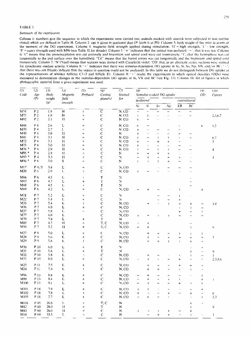
T A B L E I
Summary of the experiments
C o l u m n 1: n u m b e r s give the s e q u e n c e in wh ich the e x p e r i m e n t s w e r e c a r r i e d out ; a n i m a l s m a r k e d wi th as te r i sk we re sub j ec t ed to n o n - r o u t i n e s t imul i w h i c h a re d e f i n e d in T a b l e II. C o l u m n 2: age is given in p o s t n a t a l days (P: b i r th is at P0). C o l u m n 3: body we igh t of the mice in g r a m s at the m o m e n t o f the D G e x p e r i m e n t . C o l u m n 4: m a g n e t i c f ie ld s t r e n g t h a p p l i e d d u r i n g s t imula t ion ; ' H ' = high s t r eng th , ' L ' = low s t r eng th . 'S ' = s u p e r s t r e n g t h u sed wi th M 9 6 (see T a b l e II fo r deta i ls ) . C o l u m n 5: ' + ' i nd ica t e s t ha t the a n i m a l was p e r f u s e d : ' ', t ha t it was not . C o l u m n 6: 'C" m e a n s t ha t the p r o s e n c e p h a l o n was cut c o r o n a l l y a n d b r a i n s t e m a n d sp ina l co rd we re cut t r ansverse ly : 'T', t ha t the h e m i s p h e r e was cut t a n g e n t i a l l y to the pia l su r f ace over the ba r re l f i e ld . ' T , C ' m e a n s t ha t the b a r r e l co r t ex was cu t t angen t i a l l y a n d the b r a i n s t e m a n d sp ina l co rd t r ansverse ly . C o l u m n 7: ' N ' (Nissl) m e a n s t h a t s ec t ions w e r e s t a i ned wi th Cresy l ech t violet: ' C O " tha t , in an a l t e r n a t e ser ies , sec t ions w e r e s t a ined for c y t o c h r o m e ox idase activity. C o l u m n 8: ' + ' i nd ica t e s t ha t t h e r e was s t i m u l u s - d e p e n d e n t D G u p t a k e in Sc, Si, So, Np, VB, a n d / o r BC; ' - " , t ha t t h e r e was no t (b l anks i nd i ca t e t h a t the r eg ion in q u e s t i o n cou ld no t be ana lyzed) . In this tab le we d o no t d i s t ingu i sh b e t w e e n D G u p t a k e at t he r e p r e s e n t a t i o n s o f w h i s k e r foll icles C 1 - 3 a n d follicle E l . C o l u m n 9: ' + ' m a r k s the e x p e r i m e n t s in wh ich op t i ca l dens i t i es ( O D s ) were m e a s u r e d to d e m o n s t r a t e c h a n g e s in the s t i m u l u s - d e p e n d e n t D G u p t a k e in Si, V B a n d B C (see Fig. 11). C o l u m n It): list of f igures in which p h o t o g r a p h i c m a t e r i a l f r om a given e x p e r i m e n t w a s used .
(1) (2) (3) (4) (5) (6) (7) (8) (9) (10) Code Age Body Magnetic Perfused Cutting Stained Stimulus-et'oked DG uptake OD Figures
(P) weight field plane(s) for ipsilateral contralateral (g) strength
Sc Si So Np VB BC
M74 P 2 1.9 H + C N, C O . . . . . . M75 P 2 1.9 H + C N, C O + + - - - 2,3,6,7 M92 P 2 2.1 H + C N, C O + . . . .
M48 P 4 2.6 L + C N, C O + + - + + 1,2 M 7 0 P 4 2,7 L + C N, C O + - - M40 P 4 3,0 H + C N + + - - + -
M41 P 4 3,1 H + C N, C O + + + + + - 6,7 M72 P 4 3.1 H + C N, C O + + + + + 3 M73 P 4 3.0 H + C N, C O + + - - M 7 6 * P 4 2.9 H + C N, C O + + + + + - 4 M95 * P 4 2.8 H + C N + + + + - - M97 * P 4 3.3 H + C N + + + + + - 4 M 9 6 * P 4 3.0 S + C N + + + + + -
M17 P 4 / 5 3.4 L + C N, C O . . . . M 2 0 P 5 2.9 L + C N, C O + + - - + -
M 6 6 P 6 4.5 L + T N M 6 7 P 6 4.2 L + T N M 6 8 P 6 4.5 L + T N M69 P 6 4.2 L + C N, C O + + - + +
M21 P 7 5.2 L + C N - + - M 2 2 P 7 5.4 L + C N - - +
M35 P 7 5.4 L + C N, C O + + + + + + 1,8 M36 P 7 6.0 L + C N, C O + + + M 3 7 P 7 5.8 L - C N, C O + + + M 7 7 P 7 6.0 L - C N, C O + + + + + + M78 P 7 5.8 L + T N M89 P 7 4.7 H + T, C N, C O + + M90 P 7 3.2 H + T, C N, C O + + + 9
M27 P 9 5.0 L + C N, C O + + + + + + M28 P 9 5.6 L + C N, C O + + + + + + 7 M29 P 9 5.6 L + C N, C O + + + + + +
M 3 0 P 10 6.0 L + T N + + M31 P 10 5.6 L + T N + + 9 M32 P 10 5.8 L - C N, C O + - + + + 1 M71 P 10 6.0 L + C N, C O + + + + + + + 2,3,5,6
M23 P 11 7.5 L + C N, C O + + + + + + M24 P 11 7.4 L + C N, C O + + + + + +
M98 P 13 8.8 L + C N, C O + + - + - + + M 9 9 P 13 9.4 L + C N, C O + + + + + + 1 M 1 0 0 P 13 9 . l L + C N, C O + + + + + + +
M101 P 18 7.9 L + C N, C O + + - + - + + M102 P 18 7.8 L + C N, C O + + - + - + + 6 M103 P 18 7.7 L + C N, C O + + - + - + + 2.3
M141 P 45 26.6 L + T, C N + + M42 P 60 28.0 H + T N + 9 M43 P 60 26.0 H + C N + + + + - + M18 P 69 33.5 L + C N + + + - + + 1

230
s ~. The bursts contained the 50 Hz alternating current of the mains. As part of routine stimulation, a low and, in several experiments at P2, P4, P7 and P60, a high magnetic field strength were used (see Table II). In one experiment at P4 (M76), the magnetic field bursts as well as the interburst pauses were lengthened and the period of stimulation was increased, while in three other experiments with P4 mice (M95, 96, 97), bursts were delivered in trains (Table II)
Tissue processing and histology Immediately after the 45 rain of stimulation, animals were anes-
thetized with Nembutal and perfused through the heart with 3.3% formalin in 0.! M S6rensen-buffer (pH 7.4) for 10 min m. Upon perfusion, the brains, including the two upper cervical segments of the spinal cord, were dissected out. Three mice were not perfused (Table 1); their brains were removed immediately and processed as the others.
For coronal sections, the brains were placed on Teflon trays so that the cerebral hemispheres and the brainstem were in one longitu- dinal axis. Thus, transverse sections through the brainstems were in the same plane as the coronal sections through the hemispheres. In order to obtain sections cut tangential to the pial surface above BC, the brain was divided in the mid-sagittal plane and the hemispheres were further t r immed to obtain the appropriate angle for tangential cutting using a miniature guillotine s2, and placed on a plastic holder. Following this procedure it was impossible to collect sections through VB from these hemispheres. In three experiments, the brainstem had been separated before the hemispheres were divided. These brainstems were cut transversely and the hemispheres cut tangen- tially (cases 'TC' in column 6, Table I).
Subsequently, all specimens were frozen in dry-ice-chilled isopen- tane, and mounted on an object holder with cooled M-I embedding matrix (Lipshaw, Detroit, MI 48210, USA). Sections were cut at 20 /zm in a cryostat at - 16°C (Frigocut 2700, Reicher t - Jung , Nussloch, Germany). The sections were collected on chrome alum-coated slides. From most of the coronally cut brains, two alternate series of sections were collected. One series was dried on a hot plate at 60°C, whereas the other series was thaw-mounted and freeze-dried overnight at - 18°C.
Both series of sections were exposed to Cronex M R F 31 film (Du Pont de Nemours and Co., Wilmington, DE, USA) with the vacuum-contact-method 35 at 5°C for 2 weeks. The sections were co-exposed with calibrated radioactivity sources. These sources were prepared t5 and calibrated for equivalent radioactivity content in brain tissue (nCi /g ) using a series of poly[14C]metamethacrylate reference sources (set no. C 791, Amersham, Amersham, UK). After development of the film 43, the hot plate-dried sections were stained with Cresylecht violet, whereas the freeze-dried sections were stained for CO activity 43'7s.
Qualitatit e assessment of the autoradiograms The autoradiograms were inspected for localized stimulus-depen-
dent DG uptake (Table I). We have analyzed DG uptake in Sc. Si. So, Np, VB and BC. Uptake was considered s t imulus-dependent when the opposite side was available for comparison and showed no such uptake. Referring to the amount of local DG accumulation we use the term 'magni tude ' .
From each mouse with s t imulus-dependent DG uptake in one or more stations of the pathway, some sections were selected to study the correspondence between the focus, or foci, of DG uptake and the representat ions of the stimulated whiskers as seen in the CO- or Nissl-stained preparations.
Using a light microscope fitted with a drawing tube, the perime- ters of the sections as well as those of barrels were drawn from Nissl-stained tangential sections through BC. Outlines of CO seg- ments were drawn from coronal sections through BC and VB. and from transverse sections through Np, Si and Sc. The drawings were used as anatomical reference for areas of DG uptake by superimpos- ing them on photographic enlargements of the corresponding autora- diograms. For photographic prints we routinely used relatively hard paper (numbers 3 and 4). In order to obtain prints of adequate contrast, it was found to be advantageous to slightly underexpose. For the topological correspondence between CO segments and whisker follicles, see Discussion.
Densitometry of autoradiograms In order to compare quantitatively s t imulus-dependent DG up-
take among mice, we selected animals which had been exposed to routine stimulation of low strength (Table I, column 9). Transmit- tance of light in and near areas of s t imulus-dependent DG uptake was measured on digitized images of autoradiograms with a TV camera-based image analysis system (ASBA, Wild and Leitz, Ziirich, Switzerland). These areas were outlined by the observer on the system's video display. The system calculates the mean transmittance of light from all pixels in the outlined area (one such area contained from about 15 to about 400 pixels). From these means, mean optical densities (mODs) were determined, roOD was defined as the loga- rithm of the ratio between the mean transmit tance of light in an area of 'bare ' film near the autoradiogram and the mean transmittance of light determined in the area of interest in the autoradiogram.
roODs were determined for: (i) the areas with a st imulus-depen- dent increase in DG uptake in a given station and (ii) an immediately neighboring area in that station up to the station's approximate boundaries, representing 'non-activated' tissue. We took the ratio (R) of the mODs of (i) and (ii) as an index for the magni tude of the s t imulus-dependent DG uptake. In VB, we tested whether the ratio of activation would change, when roODs from the homotopic area on the 'non-st imulated ' side were taken as denominator. The ratios
TABLE 1I
Summary of routine and of unconventional parameters of stimulation applied to follicles of left whiskers C1- 3 and E1
Upper two rows: low and high field strength routine stimulation as used in the majority of experiments (see Table I). In the lower rows, the st imulus parameters of four P4 animals are presented (column 1 gives their codes; same animals as those marked with an asterisk in Table I, column 1). In columns 3 and 4, ' - ' signifies that bursts with a continuous repetition rate were used instead of trains of bursts. Column 8 gives the sum of all burst durations during the time of stimulation ('Ton'). In column 9 the power 'P', and in column 10 the energy ( 'PXTon') to which the mice were exposed are given.
(l) (2) (3) (4) (5) (6) (6) (7) (8) (9) (10) Strength of Duration Duration of Burst Pause Repiti- Period of To, ̀ Power Energy magnetic of burst intertrain duration duration ion rate stimula- (min) P P × Ton field trains pauses (ms) (ms) (s - :) tion (W) (104 Ws) (103 A / m , rms) (ms) (ms) (min)
Routine, low Routine, high M76 M95 M97 M96
6.1- 7.2 - - 46 90 7.4 45 15.3 16.9 1.6 9.2-10.2 - - 46 90 7.4 45 15.3 36.2 3.3
10.1 - - 150 300 2.2 90 30.0 36.2 6.5 10.1 490 510 46 24 7.0 45 14.5 36.2 3.1 10.1 1400 1400 46 24 7.1 45 14.8 36.2 3.2 14.3 700 1400 46 54 3.3 45 6.8 74.6 3.1

were not significantly different from those calculated using roODs of non-activated tissue adjacent to activated area. We opted for the latter approach, if only to avoid the consequences of occasional deviations from the coronal plane of section.
Quantification was performed in Si, VB and BC. In Si, activated and non-activated areas were measured ipsilateral to stimulation; in VB and BC, contralateral to stimulation. In Si and VB, autoradio- grams of transverse ( = coronal) sections were used. In BC, activated and non-activated areas were measured in layer IV; measurements were made from autoradiograms of coronal and tangential sections, and the data were pooled. If in Si, and in tangentially cut BC, areas of DG uptake evoked by stimulation of whiskers C1-3 and El could be distinguished, they were measured separately. Per animal, read- ings were collected from as many autoradiograms from a given station as possible. No measurements were made when the areas of interest showed even small destruction of the tissue.
Statistical analysis of densitometry Using a statistical analysis package 56, the results were submitted
to the following procedures: (i) linear regressions were calculated between the mOD of all measured areas as dependent variable, and radioactivity content as the independent variable; (ii) since for an individual mouse the distribution of Rs from a given station was never normal, the median of the ratios was taken to represent the Rs of Si, VB and BC. These medians were subjected to a non-paramet- ric analysis of variance using the Wilcoxon/Kruskal-Wallis test. In this analysis, the medians of R for a given area were compared among ages. For those sets of data for which the Wilcoxon/ Kruskal-Wallis tests detected significance at P _< 0.05, a Tukey's studentized range test was performed on ranks so as to determine which groups were significantly (P _< 0.05) different. The non-para- metric analysis does not allow multiple statistical comparison be- tween observations made in the same subject. This holds in our case for the comparison at a given age between values from C1-3 and E1 within a station, and between values from different stations.
RESULTS
Qualitatil~e description
The overal l D G up t ake in the b ra ins of mice at P7
and younge r var ied cons iderab ly and i n d e p e n d e n t l y
f rom age. Seven mice had a low, un i fo rm t race r distr i-
but ion , pa t t e rn l e s s to the po in t that ne i t he r s t imulus-
d e p e n d e n t D G u p t a k e nor the b o u n d a r i e s b e t w e e n
gray and whi te m a t t e r could be d i scerned . T h e s e mice
were exc luded f rom fu r the r analysis. A p p a r e n t l y , a
th resho ld a m o u n t of D G must accumula t e in b ra in
t issue to reveal me tabo l i c p a t t e r n s in genera l , and
s t i m u l u s - d e p e n d e n t changes in neura l me t abo l i sm in
par t icu la r . T h e whisker s t imula t ion as used in this
s tudy could not be ca r r i ed out on mice younge r than
P2, s ince the whiskers of these mice were too weak to
suppor t the p ieces of meta l .
The mice ana lyzed are l is ted in Tab le I, where we
r e c o r d e d the p re sence or absence of s t imu lus -depen-
den t D G up t ake in the nuclei of t e rmina t i on (Sc, Si, So
and Np) ips i la te ra l to s t imula t ion as well as in VB and
BC, bo th con t r a l a t e r a l to s t imula t ion . Fig. 1 provides
an overview of the a p p e a r a n c e of s t i m u l u s - d e p e n d e n t
D G up take in b ra ins tem, t ha l amus and cor tex in five
se lec ted ages. The size of the a reas of s t imu lus -depen -
231
den t D G up take is typical for each age. It s eemed
i n d e p e n d e n t f rom field s t rength of the st imulus, as
shown by expe r imen t s with mice at P4 (where th ree
f ield s t reng ths were used) and at P7 and with adul ts
(two field s trengths) . In the desc r ip t ion below, e m p h a -
sis is given to cases in which s t i m u l u s - d e p e n d e n t D G
up t ake is p resen t , and to ages at which new aspects of
s t i m u l u s - d e p e n d e n t D G up take a p p e a r in deve lop-
ment .
The brainstem nuclei (Figs. I to 6) At P2, th ree mice were s t imula ted with high s t rength
rou t ine s t imula t ion (Table II). A whisker map of C O
segments was visible in Sc, Si and Np. In two mice,
s t i m u l u s - d e p e n d e n t D G up take in Sc was found in a
single a rea at its med ia l boundary . The a rea covered
C O segments of row C and sp read into ne ighbor ing
rows (Fig. 2). In one of these mice, a single a rea of
s t i m u l u s - d e p e n d e n t D G up take s t r e t ched over a num-
b e r of cen t ra l CO segments in Si, including those of
row C, at the la te ra l b o u n d a r y of the subnuc leus (Fig.
3). In So and Np (Fig. 6), s t i m u l u s - d e p e n d e n t D G
up take was not observed.
A t P4, a total of 10 animals was analyzed. Six had
been sub jec ted to rou t ine s t imula t ion with e i the r high
or low s t rength (Table II). The i r Sc showed an a rea of
s t i m u l u s - d e p e n d e n t D G up take cover ing the media l
C O segments of row C and ne ighbor ing rows (Fig. 2).
In Si, in five of the six mice, one a rea of s t imulus-de-
p e n d e n t D G up take was found at the l a t e ra lmos t seg-
men t s of row C and, again, it sp r ead into ne ighbor ing
rows (Fig. 3). Wi th low and high s t rength rou t ine
s t imula t ion , the n u m b e r of C O segmen t s with
s t i m u l u s - d e p e n d e n t D G up take was about equal . St im-
u la t ion of the four mice with st imuli o the r than rout ine
(Table II) r esu l ted in a reas of s t i m u l u s - d e p e n d e n t D G
up t ake in Sc and Si c o m p a r a b l e to those desc r ibed
above (M97 in Fig. 4). In one of these four mice (M76),
an add i t i ona l small a r ea of inc reased D G up take was
obse rved in row E, media l in Sc and la te ra l in Si (Fig.
4). The magn i tude of D G up take of this a r ea was
smal le r than tha t of the D G up take r e l a t ed to the
s t imula t ion of whiskers C 1 - 3 . This an imal was s t imu-
la ted for 90 min at a r epe t i t ion ra te cons ide rab ly lower
than that of rou t ine s t imula t ion . Of the 10 mice, 6 had
s t i m u l u s - d e p e n d e n t D G up take in So and Np. In So,
one large a rea of s t i m u l u s - d e p e n d e n t D G up take could
be d i sce rned in the cen te r of the nucleus close to its
l a t e ra l boundary . In Np, one large a rea s p a n n e d the
en t i re vent ra l b o r d e r (Fig. 6).
A t P7, with low and high s t rength s t imula t ion , in Sc
and Si one large a rea of s t i m u l u s - d e p e n d e n t D G up-
t ake was c e n t e r e d over, but was la rger than, C O seg-

P4
P7
P 1-
PI
3
Fig.
1.
Aut
orad
iogr
ams
of 2
0 ~t
m t
hick
sec
tion
s th
roug
h su
bnuc
leus
int
erpo
lari
s (t
op r
ow o
f pa
nels
), n
ucle
us v
entr
obas
alis
of
the
thal
amus
(ce
nter
row
) an
d ba
rrel
cor
tex
(bot
tom
row
) of
mic
e at
po
stna
tal
days
(P
) 4,
7,
10,
13 a
nd 6
9 (a
dult
. a)
. S
ecti
ons
wer
e cu
t tr
ansv
erse
ly (
= co
rona
lly)
thr
ough
bra
inst
em a
nd b
rain
. In
eac
h co
lum
n, a
utor
adio
gram
s of
the
sam
e an
imal
are
dis
play
ed.
Fol
licl
es
of l
eft
whi
sker
s C
1-3
an
d E
1 w
ere
stim
ulat
ed u
sing
low
str
engt
h ro
utin
e st
imul
atio
n (T
able
II
). A
t al
l ag
es,
in t
he l
eft
subn
ucle
us i
nter
pola
ris
ther
e is
one
lar
ge a
rea
of h
igh
DG
up
take
(so
lid
tria
ngle
s) r
elat
ed t
o th
e st
imul
atio
n of
the
thr
ee w
hisk
ers
in r
ow C
. A
n ad
diti
onal
sm
all
area
of
less
inc
reas
ed D
G u
ptak
e (o
pen
tria
ngle
s),
rela
ted
to t
he s
tim
ulat
ion
of w
hisk
er E
l, i
s pr
esen
t fr
om
P7 t
o P1
3. F
rom
P4
to P
13,
ther
e is
an
area
of
stim
ulus
-dep
ende
nt D
G u
ptak
e in
the
eon
tral
ater
al n
ucle
us v
entr
obas
alis
of
the
thal
amus
(ar
row
s).
In t
he r
ight
bar
rel
cort
ex,
stim
ulus
-dep
ende
nt D
G
upta
ke b
ecom
es v
isib
le a
t P
7 (a
rrow
head
). W
ith
prog
ress
ing
age,
neu
ral
acti
vity
is
tran
smit
ted
from
bra
inst
em t
o co
rtex
and
rad
iate
s fr
om l
ayer
IV
(w
here
its
mag
nitu
de r
emai
ns h
ighe
st)
into
lay
er~
11 a
nd V
I. O
rien
tati
on:
dors
al i
s up
; th
e an
imal
's l
eft
is t
o th
e ri
ght.
Bar
at
low
er r
ight
rep
rese
nts
1 m
m a
nd h
olds
for
all
pan
els,

m e n t s in row C. A s e c o n d , s m a l l e r a r e a o f s t i m u l u s - d e -
p e n d e n t D G u p t a k e was l o c a t e d m o r e dorsa l ly in t h e s e
s u b n u c l e i (as s h o w n for Si in Fig. 1, t o p row). T h i s h e l d
fo r all a n i m a l s in w h i c h t h e s e s u b n u c l e i c o u l d be s tud-
ied. S t i m u l a t i o n at h igh s t r e n g t h d id n o t r e su l t in
p a t t e r n s o f D G u p t a k e d i f f e r e n t f r o m t h o s e e v o k e d by
s t i m u l a t i o n at low s t r eng th . In f o u r an ima l s , So and N p
len t t h e m s e l v e s to analysis ; in two of t h e m a s ingle
l a rge a r e a o f s t i m u l u s - d e p e n d e n t D G u p t a k e was p re -
sent in t h e m i d d l e o f t h e nuc le i c lose to t h e i r r e spec -
t ive l a t e r a l a n d v e n t r a l b o r d e r s .
A t P10, in Sc a l a rge a r e a o f s t i m u l u s - d e p e n d e n t
D G u p t a k e was p r e s e n t n e a r its m e d i a l b o r d e r in t h e
o n e a n i m a l in w h i c h this s u b n u c l e u s was ava i l ab le for
s tudy. T h e h ighes t D G u p t a k e was c e n t e r e d o v e r t h e
233
C O s e g m e n t s o f row C, wi th s p r e a d o v e r s e g m e n t s of
a d j a c e n t rows (Fig. 2). In Si o f t he two a n i m a l s ana-
lyzed, two s e p a r a t e a r ea s o f s t i m u l u s - d e p e n d e n t D G
u p t a k e w e r e p r e s e n t n e a r its l a t e ra l b o r d e r . O n e a r e a
was in t h e m i d d l e o f t he s u b n u c l e u s ; a n o t h e r was m o r e
do r sa l (Fig. 3). In o n e m o u s e we f o u n d a s ingle a r e a o f
s t i m u l u s - d e p e n d e n t D G u p t a k e c o v e r i n g a l a rge par t of
So (Fig. 5) and o f N p (Fig. 6).
A t P13, in t he t h r e e an ima l s s tud ied , in Sc o n e la rge
a r e a o f s t i m u l u s - d e p e n d e n t D G u p t a k e was l imi ted to
t h r e e C O s e g m e n t s in row C at t he m e d i a l b o r d e r o f
t he subnuc l eus . A s e c o n d a r e a o f s t i m u l u s - d e p e n d e n t
D G u p t a k e was c lear ly s e p a r a t e d f r o m it and was
r e s t r i c t e d to o n e m e d i a l C O s e g m e n t in row E. In Si,
aga in in all t h r e e m i c e ana lyzed , s t i m u l u s - d e p e n d e n t
ili , m l , +) ' "~+"+:+ , - : . + ~ . + .~'~,.1.,~: +- ~t,
+ : +,,.•.++++ , , + + , , . . . . . . .
,+r-:',,++:.'~;+~ +,r:~',.m~-,+'+,;'. ~ ~ ~ ,~ "S>+.,~.',~ S':' ;"+ ....~.;+ ,¢4~, +?+¢l<~i,..+.+~.~i ' -+.~ .t+ . . . . , +;+> ~ +. • .+. : ~ , : : ~ ¢ + ~ ,',',,+. : ,
t + ' , ~ . + ' + - , ~ + " : '+,~,*~.;.+.,¢,<.'.#'..•,; +++.~+++:.'. - + . ; . ' - :
:+ ~+'+?'; . +" : ' + + ' , - ' . L ~ " ~ . % : : ~+i'~"'<+:+~"+ ;,,t~ ~,.,,',;,j ,.:+ '-+',: . . . . "+...,,~,~+.' +~ . : ' .,>"
. . . . .' ~j,.'x+<, +.. ,~+ : ¢ , ~ ~. !., ,+.~: P~%~,',d~: ~i.'+," .:"~< : : " ~ - ' + . . . . ~ . t , ] . , i ! + + " . ; ~ i + + . . ¢ . . , , . - . ~ . . . . . . ~
P 2 +.~..-:::~ +.e<-, ,~- v,,:,++. - :,, p.4 ++z+,~.e,+ , , : . ; L' "
. + . . ~ . , ~ , : . . : . - . ; .
L 3 " +,,-'+ • + . :. o • : , c . ~ ,+ ' ; , , .
r P t"" ' '. " : . : " ' ' 7, • ".. ",/.;
. . . . . . . ~ . ~ , . : . . . . . . . , . , . : . , , . . ' . . , . , , . , , +,".., .,+- ,-. .,.', "
0 ~ 2, '
L y:~'<"%*'X" ,i) i
S U B N U C L E U S C A U D A L I S
e , + ; , , " ] ' + t : + ,
,.. + ,<>..,~++~'-,,':'>+- I"U,I~ . • , , . ~ * +.++ ; ,+~, ,~ ~ ; ; ~ + + t + + •
. p ' ~+ : . , . .%.~= ~ ¢ " j,l+ .++
' ; ' > .+ . ~ + ~ ~ : + ++' ?% "
~+;:'+~ ++ ++ +!~).+ ,_,
+". :,+ ~+";+ "-¢%+ ',<. ,"<';0"~.
~"+,,~, ~ . ~ t ' . ~ . ¢
:C' .~+<,V
It+ m
;,,+;,%~ +, ,.~ .+: <.,++<:,;: ,..-,++
:¢ :Te ; . i ; , e , ', : - ?i(,
+3; ~+.
,+ % , ¢ ,
" ~ . ~ ,~."~':,-+-..~:.~-_: :7; I ,
: ~ + i i i i l l P t V l ~ ' . ;)
~N , - ,iL++~'
.+% ~ ~ , + . ~ x ~,~h ~ , v + ~ - ' : + +
~.', ,, 1'3 ,-+ ,~,7. • ~:,<:~4;t ~-c.'+.'#
? '~.., + : •5,: 7• + +,':.i -"
' ? ' + " z , - ' . , " - ? " - " - K "
Fig. 2. Left subnucleus caudalis: photomicrographs of autoradiograms of 20 /+m thick transverse sections through the brainstems of mice at postnatal days (P) 2, 4, 10, and 18. The photographs in the top and in the bottom row of panels are identical, hut in the bottom row, the outlines of CO segments representing the tall, caudal whiskers in rows A to E (from ventral to dorsal) are superimposed on the autoradiograms. These outlines were based on camera-lucida drawings of the CO-segments as seen in the microscope; see Fig. 3 for correspondence between drawings and the photomicrograph of the section stained for CO-reactivity. Follicles of left whiskers C l -3 and E1 were stimulated. The insets in the top row serve to identify the part of the autoradiograms from which the photomicrographs had been taken• At P2 high strength routine stimulation was applied; at P4, PI0 and P18, low strength routine stimulation. At all ages shown, there is one area of high DG uptake, located at the medial end of row C (long arrows). At P10 and P18, there is an additional, smaller area, located at the medial end of row E (short arrows)• The areas of stimulus-dependent DG uptake extend beyond the CO segments whose whiskers were stimulated. For all illustrations dorsal is up• The bar at
lower left is 100/~m and holds for all panels at the larger magnification.

234
D G uptake was conf ined to two sites (Fig. 1): a large
area covering three segments in row C and a small area
covering one segment in row E, both at the lateral ends
of the rows. In So (only in two mice) and Np (three
mice), one area of s t imulus -dependen t D G uptake
covered a large part of the whisker representa t ion .
At P18, in the three animals studied, the two areas
of s t imu lus -dependen t D G uptake, medial in Sc (Fig.
2) and lateral in Si (Fig. 3), were restricted to only a
few CO segments in rows C and E. Precisely which
whiskers these segments per ta ined to could not be
de te rmined , owing to f ragmenta t ion of the individual
segments at that age. In the same three mice So did
not, but Np did, conta in one large area of s t imulus-de-
p e n d e n t D G uptake (Fig. 6).
In adults, only two animals allowed inspection of the
brainstem. In Sc and Si (Fig. 1), using low and high
s t rength s t imulat ion, we again found the large and the
small area of increased D G uptake in row C and in row
E, respectively. Due to f ragmenta t ion of the CO seg-
ments, common at this age, it was impossible to at-
t r ibute areas of s t imulus -dependen t D G uptake to the
individual segments. Both in So and Np, one focus of
high D G uptake was located at the lateral boundary of
these nuclei.
Two points meri t emphasis: (i) in all exper iments in
which s t imulus -dependen t D G uptake was observed,
the large area at row C and the small area at row E
formed cont inuous rods of variable length. These rods
were s i tuated at different posit ions along the rostro-
SUBNUCLEUS INTERPOLARIS
Fig. 3. Left subnucleus interpolaris: photomicrographs of autoradiograms (columns A and B), and of cytochrome oxidase- (CO; column C) and Cresylecht violet- (column D) stained 20/xm thick transverse sections through the brainstems of mice at postnatal days (P) 2, 4, 10, and 18. For each brainstem, the sections shown lie within 80/.tm from one another. The photographs in columns A and B are the same, but in column B, the outlines of CO segments representing the tall, caudal whiskers in rows A to E are superimposed on the autoradiograms. The outlines in column B were drawn with a camera-lucida from the sections of which photomicrographs are shown in the corresponding panels of column C. The insets in column A serve to identify the part of the autoradiograms from which the photomicrographs had been taken. The CO segments corresponding to whiskers A1 and B1 are indicated at P4. Follicles of left whiskers C1-3 and E1 were stimulated. At P2 and P4 high strength routine stimulation was applied; at P10 and P18, low strength routine stimulation. At the four ages, there is one area of high DG uptake, located at the lateral end of row C (long arrows in panels of column A), while at P10 and P18, there is an additional, smaller area, located at the lateral end of row E (short arrows). The areas extended beyond the CO segments whose whiskers were stimulated. This mismatch was still present at P/0, whereas at P18 the two areas of stimulus-dependent DG uptake are dearly separated. For all illustrations, dorsal is up; the two bars to the right
are 100/~m and hold for all panels.

S U B N U C L E U S I N T E R P O L A R I S
Fig. 3 (continued).
235
caudal extent of Si and Sc. Both forms of variation occurred at all ages. At the transition from Si to Sc, the rods were never continuous, and their position changed from lateral in Si to medial in Sc. More caudally in Sc, the stimulus-dependent DG uptake was observed to gradually shift in a lateral direction; (ii) from P4 to P10, with low strength routine stimulation, the magni- tude of the stimulus-dependent DG uptake in the sensory trigeminal brainstem nuclei increased continu- ously (for Np, see Fig. 6; for Si, Fig. 1, top row; see also quantitative analysis). It dropped at P13 and later. In Sc and Si of adults, stimulus-dependent DG uptake in the representation of whisker E1 was almost at the level of adjacent non-activated tissue, while in the representations of C1-3 it was weak (for Si, see Fig. 1).
The nucleus L'entrobasalis of the thalamus (Figs. 1 and 7) At P2, we could not find stimulus-dependent DG
uptake (Fig. 7), whereas CO segmentation was present. At P4, a band of stimulus-dependent DG uptake
could be observed curving from dorsolateral to ventro-
medial along the ventral boundary of VB contralateral to stimulation (Fig. 7). The band was found in seven of the ten mice analyzed, which had been exposed to routine and non-routine stimulation.
At P6, stimulus-dependent DG uptake was re- stricted to the dorsolateral edge of the nucleus. At this age, and at P7, this focus was found in all animals.
At P9, all mice showed one focus of stimulus-depen- dent DG uptake spanning up to four barreloids per section (Fig. 7). Since the plane of section was coronal, we could not determine which whiskers were repre- sented by these barreloids. We never found a second area of stimulus-dependent DG uptake (which would have corresponded to the representation of El).
From P10 onward, the magnitude of stimulus-de- pendent DG uptake dropped considerably (see Quanti- tative Analysis); at P13, only a small, weak focus was seen (see Fig. 1, center row).
At P18 and in adults, no stimulus-dependent DG uptake could be found, either at low or at high strength routine stimulation. On both sides, overall DG uptake in VB was high.

23 ++1
M76 Fig. 4. Results from stimulation with non-routine parameters of mice M76 and M97 at postnatal day 4 (see 'Fable 11). DG uptake in subnuclei caudalis (top row) and interpolaris (bottom row) as shown in photomicrographs of autoradiograms of 20 / zm thick transverse brainstem sections. Follicles of left whiskers C1-3 and E1 were stimulated. In both animals, DG uptake evoked by the deflection of whiskers C 1 - 3 is clearly visible (long arrows). M76 shows uptake to stimulation of E1 as well (short arrows), which is a unique observation for this age group. Dorsal is up; the
left side of the brainstem is to the right. The bar at lower right represents 1 mm and holds for the four illustrations.
Fig. 5. Left subnucleus oralis: photomicrographs of an autoradiogram (A), a cytochrome oxidase-stained section (B) and a Cresylecht violet-stained section (C) of a postnatal day 10 mouse, the same as shown in Figs. 2, 3, and 7. The transverse sections, cut at 20 # m , lie within 80 /zm from one another. Follicles of left whiskers C 1 -3 and E1 were subjected to low strength routine stimulation. The cytochrome oxidase-stained section (B) does not reveal any segmentation. We find one area of s t imulus-dependent DG uptake (arrows in A). Orientation: dorsal i su p . The
bar at lower right of C represents 100 # m and applies to the three panels.

The barrel cortex (Figs. I, 8 and 9) At P2, we did not detect stimulus-dependent DG
uptake. At P4, where CO segmentation first appeared, we
found high DG uptake in a thin subpial zone in the parietal cortex. It appeared of equal magnitude on both sides (Fig. 1, column 'P4'). It was not confined to
237
the incipient BC but extended occipitally, and there- fore, we do not relate this DG uptake to stimulation.
The same observation was made at P5 and P6. At P7, in three of six mice that had been stimulated
with low strength routine stimulation, and whose brains were cut coronally, we found the earliest stimulus-de- pendent DG uptake in BC. The autoradiograms showed
NUCLEUS PRINCIPALIS
Fig. 6. Left nucleus principalis: photomicrographs of autoradiograms (column A), and of cytochrome oxidase- (CO: column B) and Cresylccht violet- (column C) stained 20/~m thick transverse sections through the brainstems of mice at postnatal days (P) 2, 4. Ill. and 18. In each row. the sections - of which the same region are depicted - lie within 80 g m from one another. Follicles of left whiskers CI 3 and El were stimulated. At P2 and P4, high strength routine stimulation was applied; at PI0 and P18, low strength routine stimulation. At P2, there is no stimulus-dependent DG uptake. At P4 and PI0, there is o n e diffuse area of DG uptake ventral in the nucleus (arrows in column A). At P4, it covers the 5 rows of CO segments representing whisker rows A to E (as indicated in photomicrographs of the Pl(/-animal in column 13). Wc cannot determine whether this DG uptake is due to stimulation of whiskers C1-3 only, or to that of E1 as well. At PI(), an area of marked DG uptake is located at the ventromedial border of the nucleus. The magnitude of the stimulus-dependent DG uptake is higher in PI0 than in P4: at P18, the uptake has nearly vanished (arrows in column A). All illustrations are oriented so that dorsal is up, and shown ~t the same
magnification; bars at the right represent 100/xm.

238
NUCLEUS PRINCIPALIS
Fig. 6 (continued).
a small, well-defined region of increased DG uptake in BC, contralateral to stimulation (Fig. 1, bottom row). It comprised layer IV and lower layer II1 (Fig. 8). Of the three mice whose hemispheres were cut tangentially, only one, stimulated with high strength routine stimu- lation, showed an area of stimulus-dependent DG up- take. This area comprised barrels c~, /3, 3', 3, A1, A2, B1-3, C1-4, D1-5 and E l - 4 (Fig. 9).
At P9, in all three experiments the hemispheres were cut coronally. In BC contralateral to stimulation, layer IV contained the highest stimulus-dependent DG uptake. The coronal sections did not allow us to deter- mine whether the uptake was restricted to barrels C1-3 and El. In deep layer V, there was another focus
of stimulus-dependent DG uptake, lower in magnitude than that in layer IV. Still less elevated stimulus-de- pendent DG uptake stretched radially between these two foci and extended into layers II and VI, thus forming a radial 'metabolic column' flanked medially and laterally by bands of DG uptake lower than that in BC beyond these bands.
At P10, the four animals studied showed stimulus- dependent DG uptake in BC. On autoradiograms from tangential sections, two areas of stimulus-dependent DG uptake were limited to contralateral barrels C1-3 and E1 (Fig. 9). The two areas covered the barrels in question together with the adjacent septa. DG uptake in barrels CI-3 was higher than that in barrel E1 (see

239
Quantitative Analysis). Activated barrels C1-3 were surrounded by a band of depressed DG uptake about one barrel wide; barrel E1 appeared to be surrounded by a similar, though less pronounced, band. DG uptake in the barrels of row D was lower than that in the barrels of row B (Fig. 9, P10). The zones of diminished DG uptake extended into the supragranular layers. Autoradiograms from coronally sectioned hemispheres displayed a radial metabolic column, including foci of high DG uptake in layer IV and deep layer V (Fig. 1), not unlike the situation at P9. However, two distinct DG columns that may represent whiskers C1-3 and E1 separately were not observed.
At P13 the magnitude of stimulus-dependent DG uptake in layer IV decreased (see Quantitative Analy- sis). In autoradiograms from coronal sections, the metabolic column became more accentuated, due to an
increase in DG uptake in supra-and infragranular lay- ers (Fig. 1, P13, bottom panel).
In adults, in the coronal plane, the columnar pattern of stimulus-dependent DG uptake remained, although the focus in layer V had become less pronounced. In the tangential plane, stimulus-dependent DG uptake remained restricted to the barrels whose whiskers were stimulated. The surrounding band of depressed DG uptake, while present, was never as clear as at P10.
Fig. 10 summarizes when during postnatal develop- ment the first signs of stimulus-dependent DG uptake in each station were observed and how they are related to the appearance of the whisker representations as observed in CO- and Nissl-stained sections. In each station the onset of stimulus-dependent DG uptake lagged behind the first appearance of the morphologi- cal whisker maps. The delay was 2 to 3 days shorter for
; . . . . " ' - , 7 - " , " "; ' " "
Fig. 7. Nucleus ventrobasalis of the right thalamus: photomicrographs of autoradiograms (left column), and of Cresylecht violet-stained 21) /zm thick coronal sections (right column) through the brains of mice at postnatal days (P) 4 and 9. In each row of panels, the sections lie within 80 # m from one another. In the left column, the low magnification insets show those parts of the autoradiograms from which the photomicrographs were taken (arrows point at areas of s t imulus-dependent DG uptake). Follicles of left whiskers C1-3 and El were stimulated. At P4, high strength routine stimulation was applied; at P9, low strength routine stimulation. At P4, the autoradiogram shows one diffuse area of s t imulus-dependent DG uptake. At P9, the crisp, dark area covers only 3 to 4 barreloids a few of which can been seen at arrows in the right
column. Illustrations are oriented so that dorsal is up and lateral to the left. Bar bottom right - holds for all panels and represents l(10 ~m.

241)
DG uptake evoked by the stimulation of whiskers C1-3 than that evoked by the stimulation of whisker El. For the possible effect of modification of the stimulus parameters upon the appearance of stimulus-depen- dent DG uptake, see Discussion.
Quantitative analysis
?iiiiii! i i ! i i ~ ̧ ~ i ~!ii! ¸?i¸ ! iiii!iil '¸ ! ¸i¸i¸i
Fig. 8. Barrel cortex at postnatal day 7; top: autoradiogram (same as in Fig. 1 at 'PT, bottom row), taken from a 20 /xm thick coronal section of the parietal cortex; center: detail of autoradiogram shown above; bottom: photomicrograph of the corresponding detail from the Cresylecht violet-stained section. The arrowheads in the top panel correspond to the arrows and arrowheads in the center and the bottom panel and delineate the area of s t imulus-dependent DG uptake. Follicles of left whiskers C1-3 and El were st imulated according to the low strength routine paradigm. Comparison of the autoradiogram and the Nissl-stained section shows that increased DG uptake covers two adjacent barrels (the arrows in lower panel point to the walls surrounding them). Radially, s t imulus-dependent DG uptake spans layer IV plus part of layer III. Bars: 1 mm (in top panel), and 200 /zm (in bottom panel; this bar holds for center and bottom panel). Note thai photomicrographs in center and bottom
panel are slightly rotated clockwise with respect to top panel.
Transmittance of light, measured at activated and at the neighboring non-activated sites, ranged from 0.36 to 0.93. Mean optical densities (mODs) lay between 0.07 and 1.02, and radioactivity content in brain tissue between 49 and 716 nCi/g. In this range, mOD was linearly correlated with radioactivity content (r = 0.964, P < 0.0001). Therefore, in our case, the measurements used to calculate R lay in a range in which OD was directly proportional to radioactivity in brain tissue, which in turn is directly proportional to DG uptake.
In Fig. 11, for each age, either the median R of one animal, or, where several mice could be analyzed, the median of median Rs of the mice in that age group, were plotted. The stations analyzed were Si, VB and BC. The values obtained for the areas corresponding to stimulated whiskers C1-3 and E1 were kept sepa- rate.
Subnucleus interpolaris. DG uptake evoked by stimu- lation of whiskers C1-3 peaked at P7. Activation to stimulation of whisker E1 was highest at P7 and P10. None of the differences between ages reached signifi- cance in the Wilcoxon/Kruskal-Wallis test. The re- sponse to stimulation of whiskers C1-3 is on average 20% higher than that to stimulation of whisker El.
Nucleus ventrobasalis of the thalamus. Here, stimu- lus-dependent DG uptake at P7, P9 and PI0 was significantly higher than that at P4, P l l and P13. In this nucleus, DG uptake evoked by stimulation of whiskers C1-3 and E1 could not be measured sepa- rately.
The barrel cortex. DG uptake in barrels C1-3 was highest at P10 and lowest at P7 and P18; only differ- ences between P10 and P7, and between P10 and P18 were significant. DG uptake in barrel E1 could be measured at P10 and in adults. In both groups, activa- tion in barrels C1-3 was higher than that in barrel El.
Comparison between stations. In mice between P7 and P18, DG uptake evoked by stimulation of whiskers C1-3 was always higher in Si than that in BC. The highest stimulus-dependent DG uptake in Si (at P7), was 1.3 times higher than that in VB (at P10) and in BC (at P10). In adults, this relationship reversed and activation in BC surpassed that in Si. In VB, activation

241
at P7 and P9 was higher than that in BC; this inversed
as of P10. In summary, during postnatal development stim-
ulus-dependent DG uptake reaches peaks in Si, VB and BC. In the three stations, the peaks were followed by a decline, which was most outspoken in Si. In Si and BC, the magnitude of DG uptake evoked by stimula- tion of whiskers C1-3 was always higher than that evoked by stimulation of whisker El. This difference could not be tested for statistical significance (see Materials and Methods).
DISCUSSION
Four major observations of this study will be dis- cussed:
(I) The onset of stimulus-dependent DG uptake in the whisker-to-barrel pathway follows a time sequence from brainstem to cortex.
(II) At the earliest signs of stimulus-dependent DG uptake, the pattern of uptake reveals a rough somato- topic representation of the whisker follicles in brain- stem and cortex.
! |
r'lrl
PIO
a m
Fig. 9. Right barrel cortex: autoradiograms of 20 p,m thick tangential sections from the parietal cortex of mice at postnatal days (P) 7. l(I and 60 (adult, a). In each row of panels a pair of identical autoradiograms is shown, but on those in the right column camera lucida-drawings of the barrels, reconstructed from the corresponding series of Cresylecht violet-stained sections, are superimposed. The caudalmost barrel of each row is labeled A to E from lateral to medial; caudal to, and between, the rows are the barrels of the straddlers a - ~ . Each autoradiogram was taken from the middle section of the series from which the barrelfield was reconstructed. Follicles of left whiskers C l - 3 and E1 were stimulated. At P7 as well as in the adult, high strength routine stimulation was applied; at PI0, low strength routine stimulation. At P7, s t imulus-dependent DG uptake is diffuse (arrow in left panel); in this section it spans barrels a, /3, ~,, A1, A2, B1 3, C1-4, and DI. Adjacent sections (not shown), have high DG uptake in barrels y, D2-5 and E l - 4 . At P10, high DG uptake is limited to barrels C1 -3 (arrow in left panel), and less increased DG uptake to barrel El (arrowhead in left panel); note the barrel-wide rim of depressed tracer uptake surrounding barrels C1 3. In the adult, the high DG uptake remains restricted to barrels C1-3 (white arrow in left panel) and El (not on the autoradiogram shown), i.e. the barrels whose whiskers were stimulated; the magni tude of the DG uptake in barrel E1 is less than that in barrels C I - 3 . Compass: a, anterior: m, medial. Bar
represents 1 mm. Compass and bar hold for the six panels.

242
BC
VB
Np
Si
Sc
m •
• . . . . . . . . . . . . . . • l a
f l g - - O . . . . . . . . . •
~ - - 0 . . . . . . . . . •
I I I I i l I i i i i I I
0 1 2 3 4 5 6 7 8 9 10 11 12
IX~ tna la l days
I
m
13
Fig. 10. Diagram summarizing some salient events in the postnatal development of five stations in the whisker-to-barrel pathway (Sc, subnucleus caudalis; Si, subnucleus interpolaris; Np, nucleus princi- palls; VB, nucleus ventrobasalis; BC, barrel cortex). Time in postna- tal days (P; P0 = day of birth) is plotted on the abscissa. Empty segmented icons indicate the first appearance of CO segments repre- senting the whisker map in a given station; those pertaining to the brainstem nuclei are derived from the work of Ma 4°. Dots indicate the first appearance of stimulus-dependent DG uptake, which may stem from both stimulation of whiskers C1-3 and stimulation of whisker El and from high or low strength routine stimulation. Triangles indicate the appearance of a small area of high DG uptake related to the stimulation of whisker El, separate from the large area of high DG uptake related to the stimulation of whiskers C1-3. The triangles at P4 reflect data obtained from other than routine- stimulation (Table II). Stippled segmented icons indicate the first day on which areas of stimulus-evoked DG uptake and the represen- tations of the stimulated whiskers could, in the present study, be shown to match. The horizontal line segments emphasize that CO segmentation develops before the onset of stimulus-dependent DG uptake; the dashed line segments, that barrel El, as a distinct entity,
responds later than barrels C1-3.
(II1) Early in postnatal development, the areas of stimulus-dependent DG uptake in subnuclei caudalis and interpolaris as well as in barrel cortex were larger than the corresponding morphological whisker repre- sentations, whereas later a good match was established.
(IV) In all stations a maximum of stimulus-depen- dent DG uptake was observed at about P10, followed by a decrease.
1. The time sequence of the onset of stimulus-dependent DG uptake in the whisker-to-barrel pathway
The occurrence of stimulus-dependent DG uptake in the stations along the pathway within an individual varied considerably between animals. Possible factors here are: while all mice used are from ICR-origin, they did not form a genetically homogeneous group; more- over, as mentioned in Material and Methods, animals used may have been 24 h older than the age assigned to them; a third factor resides in the stimulus applied, where head movement could have induced variability between mice. However, notwithstanding this interindi- vidual variability, one tendency is clear: in develop- ment the onset of DG uptake evoked by whisker stimu-
$i
V B
RATIO
2 t,
Z 4
Z
d 0
I '_,2
Z 6
4
1 2
Z 8
9 8
•I
T
1,2 ~r"2
T
12;, !21
] J
f
] r
f
& l 1
1/-----¢/~
4 S 6 7 8 9 10 Ii 12 13 i:5' ADULT
I ~
i 4
i Z
i 0
92 i 2
I 1
- iF ..... CA -
4 S ~ 7 :E: i i i C L J : : L ~ " 9 A D U L T
l d
1 F,
BC i 4
I 2
i 0
- , . 2 ÷2 A 1
I '3
4 E ~ 7 P~ 9 t 0 11 1. : 1 : [E A D U L T
AGE c l - 3 • E~A
Fig. ] 1. Graphs showing the age dependence of the relative magni- tude of stimulus-evoked DG uptake in subnucleus interpolaris (Si), nucleus ventrobasalis of the thalamus (VB) and barrel cortex (BC). Only results of experiments using low strength routine stimulation are plotted. The magnitude (ordinate) is expressed as the ratio between the mean optical density taken from the area of stimulus- evoked DG uptake and the mean optical density taken from an immediately neighboring area within the same station. In BC, mea- surements were taken from layer IV. In the mice whose hemispheres were sectioned coronally, the site of barrels C1-3 and barrel E1 could not be identified with certainty. Here, the large area of increased DG uptake was taken to represent the activation in barrels C1-3. At P10 and in adults some hemispheres were cut tangentially. In these experiments, metabolic responses to stimulation of whiskers CI-3 and E1 could be distinguished and separate readings were taken. DG uptake related to the stimulation of whiskers C1-3 (dots) and to stimulation of whisker El (triangles) are plotted separately. This convention was also used in the display of data related to Si. For VB the data were considered to represent stimulus-dependent DG uptake in barreloids of row C. The age (abscissa) is expressed in postnatal days (P) (adult is P45 and P69). Note that stimulus-depen- dent DG uptake drops in all stations after P10. Data points pertain to the median ratio of each age group; bars indicate ranges. Arrows indicate data points with ranges smaller than the symbol. Numbers within the graphs indicate the number of mice analyzed in any one group. At ages represented by one mouse, the data point without range bar is the median result of that individual. At ages at which the ranges for Cl -3 and E1 overlap, data points are spaced horizon-
tally.

243
lation follows a sequence from brainstem, via thalamus,
to cortex. Once established in a station, the metabolic
response remains present, except in thalamus where it may be present but masked by the high uptake in all of VB. Although the onset of st imulus-dependent DG uptake in a station may advance by a few days with
changing stimulus parameters , the sequence of onset along the pathway could not be modified by changing them. Interestingly, in our study there was no variabil-
ity in the presence of CO-segmentat ion between the animals of any given age.
Does absence of a st imulus-dependent DG uptake
to whisker deflection during development signify the absence of functional activity due to immaturity of that part of the pathway? Or does it signify the use of a
substrate of neuronal energy metabolism other than glucose? With respect to the latter possibility, ketone bodies may well be one such alternative source of
energy~': in the early postnatal rat it has been shown that their contribution to the total brain energy metabolism can be as high as 30% L~. Thus, in our
experiments the preferential use of ketone bodies might have prevented DG uptake evoked by the stimulation of whiskers in thalamus (at P2) and cortex (at P2-P6). This attractive notion does not receive support from a
pilot study in which we found that the stimulation of whiskers of P4 mice injected with a radioactively la- beled ketone body, [1-~4C]l>/3-hydroxybutyrate, did not
demonstrate a st imulus-dependent uptake in VB and BC. However, there was such uptake in Sc and Si4S;
regions that also showed stimulus-dependent DG up- take as demonstrated in the present study.
In adults, the magnitude of D G uptake in barrel E1 was shown to be independent from the number of simultaneously stimulated whiskers in row C 43. In the
design and the discussion of the present study we have
assumed that during maturation DG uptake evoked by the stimulation of whisker E1 was independent from
that evoked by the stimulation of whiskers C1-3 (and vice versa).
The earliest st imulus-dependent DG uptake in the stations of the whisker-to-barrel pathway was obtained by stimulating whiskers C1-3. In all stations DG up- take to stimulation of whisker E1 lagged behind by two
to three days. Where there was DG uptake in the representation of El , it was lower than that in the representation of C I - 3 . The difference is preserved to adulthood, where it persists even under increasing
stimulus strength 4~'. The time lag and the difference in
magnitude of DG uptake indicate that in infant mice,
as in adults, there is a mutually reinforcing effect on
metabolic responses by the deflection of adjacent whiskers in one row. This observation may well be explained by the model proposed by McCasland et al. 42. It would be interesting to study whether stimula-
tion of a number of adjacent whiskers across rows
leads to a similar effect.
Using routine stimulation, st imulus-dependent DG uptake in the representation of El appears at PT.
However, by reducing the burst rate to a third of
routine and doubling the total duration of the stimula- tion period from 45 to 90 rain (see Table 11), we obtained DG uptake in Sc and Si at P4 upon stimula-
tion of a single whisker follicle (M76 in Fig. 3), but could not influence cortical metabolism *. With single-
and multi-unit recordings in SI of anesthetized rats. a
higher degree of habituation of cortical units upon
peripheral stimulation was shown at P5 and P6 than in adults ~. The lower stimulus rate a n d / o r longer inter- burst pauses in experiment M76 may have prevented
such habituation in the immature pathway, here stud- ied while the animal was unanesthetized. This mode of stimulation may have permitted to drive brainstem nuclei more effectively, which may explain the appear-
ance of the functional representation of whisker El in M76. This observation might suggest that, indeed, for all ages investigated alternative stimulation paradigms could have been applied - obviously an impossible
undertaking. In comparison with the low strength rou- tine stimulation, the high strength stimulation gave
more consistent results at P4, probably because the latter stimulus drove cells more effectively a n d / o r increased the number of driven cells.
When confronted with the results of other studies,
there is a problem in counting postnatal days and in the determination of the moment of birth. The discrep- ancy of one day between laboratories which define the birthday of a litter as postnatal day l 1"211"4°'~'7"~'s, and
laboratories, including our own, which define the birth- day as postnatal day ()4.>,34..~,,s0 can be accounted for.
As to the second point, only few authors define the precision with which the moment of detection of a
litter was determined, and whether postnatal days were counted in calendar days or, as done in this study, in sets of 24 h, each set beginning at the moment of detection. Together, this may cause a discrepancy of up
* Our finding that stimulus-dependent DG uptake is present 90 min after injection is in harmony with the finding that adult guinea pigs, exposed to pure tones, show tone-specific DG uptake in the inferior colliculus 90 min after injection 72. Both findings support the reports on a slow hydrolysis of DG-6-phosphate by phosphatase 5¢~'6t.62 and contrast with those claiming rapid hydrolysis of DG-6~phosphate 1~,22,25,5~.

244
to two days for a given postnatal day. Throughout this
report we tried to match the observations by other laboratories to our age scheme. Another factor con- tributing to the uncertainty incurred when trying to
match dates, is that some observations on the matura- tion of the pathway in question derive from rats, and
others from mice. All these factors may have con- tributed to the reported variations in the onset of
events during postnatal development.
In our CO-stained preparations, segmentation was already present in Sc, Si, Np and VB at P2, while in BC it was first seen at P4. Ma 4° observed segmentation
in Sc, Si and Np to arise in CO-stained preparat ions of mice brainstem first at P1. These observations are in
harmony with the finding that, in rat, segments of high
succinyl dehydrogenase (SDH) activity in Np develop between birth and P3 2j. In VB of mice, the first signs
of segmentation as seen in CO-stained preparat ions are observed at P2 sc~, whereas in SDH-stained prepara-
tions of rat and mouse, segmentation was first reported at P4 s. We cannot explain this discrepancy (but see
previous paragraph). In BC, SDH-stained sections from rats show that segmentation develops between P3 and 6 -~4. This is in accord with the first appearance of barrel hollows in mice between late P3 and P4, ob- served in Nissl-stained sections 53 and with the pres-
ence of densely CO-stained hollows at P4 in our prepa- rations. Comparing the onset of st imulus-dependent
D G uptake with the appearance of visible segmenta- tion in the stations of the whisker-to-barrel pathway, we note that the functional maturation in Np, VB and
BC lagged behind the establishment of CO segmenta- tion by several days. In Sc and Si, such a comparison could not be made for, as mentioned earlier, we did not analyze animals younger than P2. This time lag between segmentation and onset of function may indi- cate that the appearance of functional synapses follows pattern formation in the whisker-to-barrel pathway.
We have applied CO- and Niss-staining to visualize the central whisker representations, since both tech- niques could be combined with the autoradiographic DG method in the same animal. In cortex and in thalamus the results obtained with the CO- and Nissl staining agree; in brainstem we relied on CO-material only: in Nissl-material we could not discern whisker representations in consecutive sections. An occasional clustering of cell bodies, more or less in register with the CO-patches, was observed, but virtually never found back in adjacent section. We were not able to confirm the observations by Ma and Woolsey 41 and by Ma ~'4°
who obtained, in transverse Nissl-stained brainstem sections, structures Ma 39 termed barrelettes, and inter- preted as whisker representations. The difference be-
tween those observations and ours may be explained by
differences in section thickness and histological procc- dure; and by differences between the strains of mice used. Other methods have allowed the visualization of the whisker representation in the barrel cortcx at ear-
lier stages of postnatal development than described here 7"112'~'31"51"7~j'~1. These earlier patterns increase the
time lapse between the appearance of morphological signs of segmentation and the onset of stimulus-depen- dent DG uptake.
The time lag between the onset of DG uptake evoked by the stimulation of three whiskers and the
onset of that evoked by the stimulation of a single whisker in brainstem and cortex points to the possibil-
ity that the onset of st imulus-dependent DG uptake observed in the present study may be specific for the
type of stimulation applied. A case in point is given by our analysis on P4: while routine stimulation did not result in activation of the EI representation in the
sensory trigeminal brainstem nuclei, non-conventional stimulation did (Table I). Alternatively, we cannot rule out that the mechanical stimulation of all whiskers
would have resulted in neural responsivity in thalamus and cortex at ages earlier than we observed, perhaps even at a time that the morphological whisker maps are not yet laid down. Therefore, the possibility that
neural activity emanating from the stimulation of all whiskers plays a role in the development of morpholog- ical whisker representations is an interesting point.
Studies that address this issue have shown that block- ing neuronal activity postnatally does not prevent the
formation of the whisker pattern in the central stations of the trigeminal pathway ~'2~.
Another question concerns the role of experience- dependent neural activity in neonatal plasticity. Up to P6, pe rmanen t damage to whisker follicles in mice 2°'2s'66`7s and rats 34 causes morphological changes
in the barrel cortex: the lesion of follicles in row C led to an enlargement of the adjacent rows (i.e. rows B and D) and to the absence of barrels of row C. In the present study, the first st imulus-dependent neural ac- tivity in BC occurred after the critical period for these structural modifications was over. It has been proposed that st imulus-dependent DG uptake in a particular station is related to spiking activity in the presynaptic element in that station 32,~~'57. Opting for this hypothe-
sis, the absence of st imulus-dependent DG uptake in one station, together with the presence of such re- sponse in the preceding station may be due to a sub- threshold number of spikes traveling from the lower station to the next one up. Interestingly, the onset of st imulus-dependent DG uptake may occur long after the arrival of thalamocortical axons in the immature

cortex 3LSs and after the end of the 'critical period' of
that station.
P7 was the earliest age at which st imulus-dependent DG uptake was seen in BC. In coronal sections, the highest D G uptake was in layer IV, inside the barrels. Layer III showed DG uptake, too, in register with that in the barrels, but of smaller magnitude. This pattern of st imulus-dependent DG uptake is similar to that found in rats at P6 3~'. Our study agrees with these
findings, in that subsequently a second focus of stimu- lus-dependent DG uptake appears deep in layer V. While in the rat this occurs at P8, we detected the second focus at P9. Finally, both in rats and mice, a further expansion of st imulus-dependent D G uptake
into supra- and infragranular layers was seen to occur later in development, forming a 'metabolic column'. In
the rat the onset of st imulus-dependent DG uptake in the cortical plate was as early as about P4, i.e. 3 days earlier than in mice. Rats appear to run ahead of mice
in another aspect of cortical maturation: the onset of evoked potentials in the somatosensory cortex elicited by electrical stimulation of the trigeminal nerve or of the thalamus is at P3 in rats ~'v and at P7 in mice ~.
The appearance of high st imulus-dependent DG uptake in deep layer V at P9 in mice agrees with the finding in adults that, whereas the majority of the thalamic afferents terminate in lower layer III and layer IV, they do have branches terminating in deep layer V and upper layer Vl 6'30'74. Therefore, these two
foci of st imulus-dependent DG uptake may reflect
spiking activity in the terminals of thalamocortical pro- jections. The endings of these deeper collaterals may
have grown out, or may have become functional, two days later than the prime terminals in layers II1 and IV. The appearance of a column of stimulus-depen- dent activity reaching from layer II to layer VI, first
seen at PI0, could be due to a further spread of thalamocortical terminals into layers II, superficial V,
and VI, But only very few of these endings have been found in adults TM. Thus, activity of thalamocortical ter- minals alone is not likely to be the basis for the continuous 'metabolic column' in adults. The establish- ment of such a column would rather point to the functional maturation of intracortical connections, thus implying activation of cortical interneurons. Indeed, such interneurons have been postulated to play a role in limiting the size of receptive fields of neurons in layer IV of the barrel cortex of rats ~°.
H. The somatotopic organization of the stations of the pathway
As pointed out in the Introduction, an important property of all but one of the stations of the whisker-
245
to-barrel pathway is that representations of individual
whisker follicles are visible in histological sections. We
observed st imulus-dependent DG uptake in each sta- tion, and for Sc, Si and BC we confirmed that somato- topically organized functional maps exist. In brainstem
relays, CO segments representing single whisker folli- cles were observed in mice of up to PI3. From PI8
onwards we were unable to delineate the representa- tions of individual whisker follicles owing to the frag-
mentation of CO segments. However, the identification of rows remained possible.
Belford and Killackey s were the first to propose an orderly representation of whisker follicles in Sc and Si
of rats and mice. They based themselves on the pattern of SDH segments. The arrangement of the large seg-
ments in 5 rows and an irregular cluster of small segments located dorsomedially to these rows, led the
authors to the conclusion that the 5 rows of large segments represent, from ventral to dorsal, rows A to
E of the large caudal whiskers, while the clustered
small segments represent the follicles of the small sinus hairs near and in the mouth. In his study in rats, using
transganglionic transport of H R P administered to the proximal stumps of transected follicular nerves, Arvids- son 2, observed that the orientation of the follicle rep-
resentation within rows is reversed in Sc with respect to that in Si. Recordings from individual afferents in the descending tract of the trigeminal nerve to Sc :7
and Si 2(' of the adult rat, followed by the tracing of the
morphologically identified fibers and their terminals, demonstrated that in each subnucleus the follicles are represented in cylinders of afferent terminals resem- bling the cylinders of SDH and CO activity. In agree-
ment with Arvidsson's description, the cylinders of terminals representing the caudalmost whiskers in each row were located at the lateral boundary of Si and at
the medial boundary in the rostral part of Sc; moving
caudad in Sc, the representations of caudal whiskers progressively shifted laterally.
In Si and Sc, the large area of high DG uptake corresponding to the representation of whiskers C I - 3 (where present), covered part of the center row of large CO segments; the small area (where present), corresponding to whisker El , covered part of the dor-
salmost row. These findings confirm the ventral-to-dor- sal representation of rows A to E in both subnuclei. The regions of st imulus-dependent DG uptake com- monly do not cover the entire rostro-caudal extent of Si and Sc, which may be due to the influence of other inputs to the subnuclei in question. We are currently investigating this interesting notion by studying the role of the projection from BC on patterns of stimulus-de- pendent DG uptake in Sc and Si ~'-.

246
In Si, the areas of st imulus-dependent DG uptake were located at the lateral boundary of the subnucleus; rostral in Sc, at the medial boundary. More caudal in Sc, we noticed a medial-to-lateral shift of the loci of
high DG uptake progressing from rostra[ to caudal. These observations are in harmony with the descrip- tion of the organization of the whisker representation in Si and Sc 2"26"27.
In So, only one area of st imulus-dependent DG uptake was present at the middle of the nucleus close to its lateral boundary. While moving rostrad we ob- served its position to shift from the lateral to the
ventral border of the nucleus. In the absence of CO segments, the somatotopic representat ion of the
whiskerpad could not be ascertained. However, the
spatially restricted zone of increased DG uptake points to a discrete representat ion of whisker follicles in this
subnucleus as well, even in the absence of a visible map. Our observation is supported by the report of Bates and KiIlackey 4, who saw, in horizontal sections
of the rat brainstem, ' rostrocaudal columns of dense
SDH staining in the ventral portion of the nucleus'. It remains unclear, why neither these authors nor Ma 4° and ourselves saw such columns in transverse sections
through So. In Np, one area of st imulus-dependent DG uptake
was found. It spanned CO segments across all five rows at the ventral and lateral boundary of the nucleus. The location of the area in the nucleus agrees with the morphological whisker map: the more peripheral CO segments that represent the more caudal whiskers, are situated near the ventrolateral limit of the nucleus 2°.
The lack of precision of D G uptake of individual whisker representations in Np is probably due to limi- tations of the method, such as the traveling range of the beta rays (reported to be 20 /zm) and the diffusion
of the tracer. In VB, the pat tern of CO segments observed in our
material matched the one in SDH-stained sections of the rat and mouse thalamus as documented by Belford and Killackey 5. Comparing CO- with adjacent Nissl-
stained sections, we agree with the conclusion of others s'z°37,s°, that the segments of high enzymatic activity coincide with the hollows of barreloids 64. In adjacent coronal sections through VB, we found in mice from P7 to P l l one area of st imulus-dependent D G uptake covering up to five neighboring barreloids. The area of st imulus-dependent D G uptake was lo- cated at the lateral boundary of VB. This observation agrees with the functional 69 and morphological 64 maps
reported earlier. The coronal sections were of no use to allocate individual barreloids to particular whiskers. The fact that we found more barreloids with high D G
uptake than whiskers stimulated, may be related to the
widespread distribution of neural activation in Np. In BC, reconstructions of the barrelfield were based
on Nissl-stained tangential sections. The superposition
of these reconstructions on autoradiograms showed that, already at P10, stimulus-dependent DG uptake was restricted to the barrels whose whiskers were stim-
ulated. In summary, in Sc, Si, Np and BC, whisker follicles
are represented in metabolic maps which are in har- mony with the topography of follicle representations
shown by histological methods. In VB, of which we had only coronal sections at our disposal, we could not
establish such a relationship. Only brains cut in a near-horizontal plane ~4 would allow comparison of
metabolic and morphological maps.
III. Relationship between areas of increased DG uptake and areas of representations of whisker follicles
In brainstem as well as in cortex, we observed that,
at the onset of stimulus-dependent DG uptake, the area of uptake covered more CO segments or barrels than the number of deflected whiskers. This may have been due to stimulation of follicles neighboring the
deflected follicles as a consequence of closer mutual proximity of the whiskers and softness of the muzzle tissue in the young animals. If true, one would expect that using higher stimulus strength would lead to an increased area of stimulus-dependent D G uptake. However, the size of activated areas was found not to be related to stimulus strength as observed in mice
stimulated at P4 or P7. Other possible mechanisms explaining the initial mismatch are discussed below.
a. Subcortical stations In Sc and Si of animals of up to P l l , the large and
the small area of st imulus-dependent D G uptake were larger than the CO segments representing whisker follicles C1-3 and El , respectively. The greatest mis- match occurred at P 7 - P l l . At P13, the areas were almost confined to the appropriate segments.
The initial mismatch may be partially due to a dislocation of the tracer during the processing of the tissue. In the small and more watery brainstems of young mice, such a dislocation may have more serious consequences than in those of adults; moreover, the risk of melting the frozen tissue when blocking the brain is higher in small than in large specimens. On the other hand, the comparison between areas of increased DG uptake and the CO segments was made at an increased magnification at which CO boundaries could be drawn. For this we reproduced autoradiograms to

247
scale using hard paper, which may have led to an
underest imation of the size of areas of stimulus-depen-
dent DG uptake. The greatest relative enlargement of the areas of
st imulus-dependent DG uptake coincides with the
highest magnitude of uptake. This may suggest a tran- sient exuberant projection of fibers spreading into neighboring representations accompanied by hyperin- nervation of the representations themselves. This is in harmony with the observation that trigeminal afferents in the rat have widespread arborizations in the nuclei
of termination before P7 M. Unfortunately, so far there is no report on the development of the ascending
projections to VB. As an alternative or complementary process we propose the establishment of inhibitory
corticofugal circuits (see Section IV).
b. Barrel cortex
At the onset of st imulus-dependent D G uptake in barrel cortex (P7), D G uptake was also increased in
barrels whose whisker follicles had not been stimu- lated. The tangential extent of the area of stimulus-de-
pendent D G uptake was independent from the strength of stimulation. This contrasts with our findings at P10 where, at low strength routine stimulation, stimulus-de-
pendent DG uptake formed columns reaching from layer II to white matter, while in layer IV the uptake was confined to the barrels corresponding to the de- flected whisker(s). DG uptake in the columns of bar- rels C1-3 was highlighted by a rim of depressed DG uptake which surrounds the excited barrels, and which
was more accentuated in the D than in the B barrels. Such a rim was also observed after stimulation in adult mice of whisker B2 (see Fig. 5 in ref. 43 column a, bottom) or of whisker C3 (see Fig. 3D in ref. 9).
The restriction of the area of st imulus-dependent DG uptake to the barrels whose whiskers were stimu- lated, shown to occur between P7 and P10, may be
explained by the development of two mechanisms op- erating in the adult forebrain.
The development of one circuit is related to in- hibitory interneurons within the barrel cortex. These neurons may be responsible for the suppression of neuronal activity in the zone surrounding the activated
cortical column; see below. Alternatively, the restriction may result from the
maturat ion of the connections from the barrel cortex to the reticular nucleus of the thalamus 24, a n d / o r be- tween that nucleus and VB. As a result of the develop- ment of this pathway, pyramidal cells in layer VI of the stimulated cortical column may activate neurons of the reticular nucleus, known to be GABAergic ~7. This acti-
vation, in turn, would result in the suppression of
neuronal activity of neurons in VB that surround the
stimulated barreloids. The restriction of the area of
st imulus-dependent DG uptake to the barrels whose whiskers were stimulated, was accompanied by the appearance of a rim of decreased DG uptake sur- rounding the stimulated area. We interpret this de-
crease to be a sign of inhibition, in analogy to the observations of Webster et al. v~ in the cat auditory
pathway, where it was shown that the electrophysiolog- ically identified 'inhibitory side band' in the inferior
colliculus was characterized by a level of DG uptake
lower than neighboring unstimulated zones. The observed shrinkage of areas of stimulus-depen-
dent DG uptake during cortical maturation is in har-
mony with electrophysiological data from units in layer IV of the rat somatosensory cortext. At P5 or 6, units were found which could be driven by 6 to 12 separately
deflected whiskers. In the adult, and under identical
conditions, a mean of two whiskers was found. In the same study, units were recorded in SI which responded
to indentation of skin of the hind limb. In young rats,
these neurons responded purely with excitation, whereas in adults the area of skin that elicited an
excitatory response was surrounded by a field from which the cortical unit was inhibited. Mountcastle 4'~
had been the first to demonstrate what he termed 'surround inhibition' in somatosensory cortex of the adult cat by recording single- and multi-unit responses to skin indentation. Armstrong-James L, in analogy to his observations on the hind limb representation, as- sumed that in the developing barrel cortex the shrink-
age of receptive fields of neurons responding to whisker deflection, was based on an increase of surround inhi- bition. In our experiments an increase of surround inhibition during development may well have been the
cause for the restriction of st imulus-dependent DG uptake to the barrels whose whiskers were stimulated. The zones of st imulus-dependent inhibition, centered around the stimulated barrels C1-3 and El , are super-
imposed in the barrels of row D. This may have caused DG-uptake in the D barrels to be lower than in the barrels of row B.
Interestingly, the restriction of the area of stimulus- dependent D G uptake so as to match the morphologi- cal representations of the stimulated follicles first oc- curs in barrel cortex and manifests itself only three days later in the brainstem. This sequence of events suggests a role of the cortex in the refinement of the topography of the metabolic whisker representations in the brainstem. In adult mice projections originating from one cortical barrel column terminate in the corre- sponding whisker representation in the brainstem 73.

248
However, the deve lopment of these project ions is not
known.
IV. The increase and the decline o f stimulus-dependent
D G uptake
In the exper iments with mice at P4 and older, stimu-
lu s -dependen t D G uptake in the subcortical relays of
the whisker- to-barrel pathway increased to a maximum
be tween P7 and P10, then d ropped considerably. This
decl ine in neura l activity may be due to inhibi t ion
media ted by corticofugal connect ions . It would be in-
terest ing to confirm whether , indeed, corticofugal pro-
jections are laid down and become funct ional dur ing
this period. Single- and mul t i -un i t recordings in adult
cats showed that cortical inputs to the sensory trigemi-
nal bra ins tem nuclei 59 can facilitate or inhibit the
spiking activity of ascending project ion neu rons 19. Con-
cern ing the adult whisker- to-barrel pathway, a corti-
cally media ted inhibi t ion in the b ra ins tem nuclei may
well domina te a cortically media ted excitation. Corti-
cally media ted inhibi t ion in this pathway was repor ted
to exist in the rat 79. In cat, au toradiography of antero-
gradely t ranspor ted proteins after intracort ical injec-
t ion of t r i t ia ted amino acids showed that cort icotrigem-
inal axons invade the t r igeminal sensory nuclei towards
the end of the first postnatal week 63. A n an te rograde
t racer study a t t empt ing to elucidate the deve lopment
of the cort icotr igeminal project ion in mice would be
most interest ing, as would be a D G study of the
sensory t r igeminal nuclei in adult mice whose barrel
cortex is inact ivated by ei ther an acute lesion or cool-
ing, in order to test whether or not the corticofugal
connec t ions play the inhibi tory role we postulate here
and their role in the reduct ion in area of D G uptake as
proposed in section III of the Discussion.
In conclusion, the present study showed that, early
in pos tnata l deve lopment of the mouse, funct ional
whisker maps in the whisker- to-barrel pathway not only
are somatotopical ly organized, but also are laid down
sequent ia l ly from bra ins tem to cortex. Fu tu re work will
be directed to the quest ion whether the delivery of
per iphera l stimuli dur ing deve lopment may inf luence
temporal , s t ructural a n d / o r funct ional aspects of the
deve lopment of the organiza t ion of a sensory system.
Acknowledgements. We thank S. Daldoss, E. Bernardi, D. Stucki and E. Brun for photography; R. Kraftsik and N. Jeanpr~tre, for assistance with statistical problems; G.M. Innocenti and members of the Lausanne Barrel Club for helpful comments; the family Fritschy for hospitality during one of the critical phases in the preparation of the final manuscript. Support: Swiss National Science Foundation Grants 3.238, 3.158 and 31-30932 to H.V.d.L., and fellowships by the 'Studienstiftung des deutschen Volkes' and the 'Deutsche Forschungsgemeinschaft' to P.M., who submitted this study to the
Johann Wolfgang Goethe Universit~it, Frankfurt a.M.. Germany, as partial fulfillment of the requirements for the Ph.D.-degree.
REFERENCES
1 Armstrong-James, M., The functional status and columnar orga- nization of single cells responding to cutaneous stimulation in neonatal rat somatosensory cortex S1, J. PhysioL. 256 (1975) 501-538.
2 Arvidsson, J., Somatotopic organization of vibrissae afferents in the trigeminal sensory nuclei of the rat studied by transganglionic transport of HRP, J. Comp. NeuroL, 211 (1982)84-92.
3 Astic, L. and Saucier, D., Ontogenesis of the functional activity of rat olfactory bulb: autoradiographic study with the 2-deoxyglu- cose method, Dec. Brain Res., 2 (1982) 243-256.
4 Bates, C.A. and Killackey, H.P., The organization of the neonatal rat's brainstem trigeminal complex and its role in the formation of central trigeminal patterns, .L Comp. Neurol., 240 (1985) 265- 287.
5 Belford, G.R. and Killackey, H.P, Vibrissae representation in subcortical trigeminal centers of the neonatal rat, J. Comp. NeuroL, 183 (1979) 305-322.
6 Bernardo, K.L. and Woolsey, T.A., Axonal trajectories between mouse somatosensory thalamus and cortex, Z Comp. NeuroL, 258 (1987) 542-564.
7 Blue, M.E. and Molliver, M.E., Serotonin influences barrel for- mation in developing somatosensory cortex of the rat, Soc. Neu- rosci. Abstr., 15 (1989) 419 (abstract).
8 Chiaia, N.L., Fish, S.E., Bauer, W.R., Bennett-Clarke, C.A., Rhoades, R.W., Postnatal blockade of cortical activity by tetrodotoxin does not disrupt the formation of vibrissa-related patterns in the rat's somatosensory cortex, Det,. Brain Res., 66 (1992) 244-250.
9 Chmielowska, J., Kossut, M. and Chmielowski, M., Single vib- rissal cortical column in the mouse labeled with 2-deoxyglucose, Exp. Brain Res., 63 (1986) 607-619.
10 Collins, R.C., Use of cortical circuits during focal penicillin seizures: an autoradiographic study with [14C]deoxyglucose, Brain Res., 150 (1978) 487-501.
11 Cooper, N.G.F. and Steindler, D.A., Lectins demarcate the bar- rel subfield in the somatosensory cortex of the early postnatal mouse, J. Comp. NeuroL, 249 (1986) 157-169.
12 Corth4sy, M.-E., Rao, S.B., Welker, E., D6rfl, J., Van der Loos, H. and Hornung, J.-P., Plasticity occurs in the spinal trigeminal nucleus of the adult mouse upon partial peripheral deprivation, Soc. Neurosci. Abstr., 17 (1991) 291 (abstract).
13 Cremer, J.E., Substrate utilization and brain development. J. Cereb. Blood Flow MetaboL, 2 (1982) 394-407.
14 Crissman, R.S., Parsons, L.L., Chiaia, N.L. and Rhoades, R.W., The primary afferent innervation of the trigeminal brainstem complex of fetal rats as demonstrated by anterograde transport of DI-I, Soc. Neurosci. Abstr., 15 (1989) 873 (abstract).
15 Curtis, C.G., Cross, S.A.M., McCulloch, R.J. and Powell, G.M., Whole-body autoradiography, Academic Press, London, 1981, pp. 38-39.
16 Dahlquist, G. and Persson, B., The rate of cerebral utilization of glucose, ketone bodies, and oxygen: a comparative in vivo study of infant and adult rats, Pediat. Res., 10 (1976) 910-917.
17 De Biasi, S., Frassoni, C. and Spreafico, R., lmmunoreactivity in the thalamic reticular nucleus of the rat, Brain Res., 399 (1986) 143-147.
18 Deuel, R.K., Yue, G.M., Sherman, W.R., Schickner, D.J. and Ackerman, J.J.H., Monitoring the time course of cerebral de- oxyglucose metabolism by 3ip nuclear magnetic resonance spec- troscopy, Science, 228 (1985) 1329-1331.
19 Dubner, R. and Sessle, B.J., Presynaptic modification of corti- cofugal fibers participating in a feedback loop between trigeminal brainstem nuclei and sensorimotor cortex. In R. Dubner and Y. Kawamura (Eds.), Oral-Facial Sensory Motor Mechanisms, Apple- ton-Century-Crofts Educational Division, Meredith Cor., New York, 1971, pp. 299-313.

20 Durham, D. and Woolsey, T.A., Effects of neonatal whisker lesions on mouse central trigeminal pathway, J. Comp. Neurol., 223 (1984) 424-447.
21 Erzurumlu, R.S. and Killackey, H.P., Development of order in the rat trigeminal system, J. Comp. Neurol., 213 (1983) 365-380.
22 Hawkins, R.A. and Miller, A.L., Loss of radioactive 2-deoxy-D- glucose-6-phosphate from brains of conscious rats: implications for quantitative autoradiographic determination of regional glu- cose utilization, Neuroscience, 3 (1978) 251-258.
23 Henderson, T.A., Woolsey, T.A. and Jacquin, M.F., Infraorbital nerve blockade from birth does not disrupt central trigeminal pattern formation in the rat, Dec. Brain Res., 66 (1992) 146 152.
24 Hoogland, P.V., Welker, E. and Van der Loos, H., Organization of the projections from barrel cortex to thalamus in mice studied with Phaseolus t.ulgaris-leucoagglutinin and HRP, Exp. Brain Res., 68 (1987) 73-87.
25 Huang, M. and Veech, R.L., The quantitative determination of the in vivo dephosphorylation of glucose-6-phosphate in rat brain, J. Biol. Chem., 257 (1982) 11358-11363.
26 Jacquin, M.F., Renehan, W.E., Mooney, R.D. and Rhoades, R.W., Structure-function relationships in rat medullary and cervi- cal dorsal horns. I. Trigeminal afferents, J. NeurophysioL, 55 (1986) 1153-1186.
27 Jacquin, M.F., Woerner, D., Szczepanik, A.M., Riecker, V.. Mooney, R.D. and Rhoades, R.W., Structure-function relation- ships in rat brainstem subnucleus interpolaris. I. Vibrissae pri- mary afferents, J. Comp. NeuroL, 243 (1986) 266-279.
28 Jeanmonod, D., Rice, F.L. and Van der Loos, H., Mouse so- matosensory cortex: alterations in the barrelfield following recep- tor injury at different early postnatal ages, Neuroscience, 6 (1981) 1503-1535.
29 Jensen, K.F., The development of the vibrissae related neocorti- cal afferents to somatosensory cortex of the rat, Soc. Neurosci. Abstr., 13 (19871 77 (abstract).
30 Jensen, K.F. and Killackey, H.P., Terminal arbors projecting to the somatosensory cortex of the adult rat. I. The normal mor- phology of specific thalamocortical afferents, J. Neurosci., 7 (1987) 3529-3543.
31 Jhaveri, S., Erzurumlu, R.S. and Crossin, K., Barrel construction in rodent neocortex: role of thalamic afferents versus extracellu- lar matrix molecules, Proc. Natl. Acad. Sci. USA, 88 (19911 4489-4493.
32 Kadekaro, M., Crane, A.M. and Sokoloff, L., Differential effects of electrical stimulation of sciatic nerve on metabolic activity in spinal cord and dorsal root ganglion in the rat, Proc. Natl. Acad. Sci. USA, 82 (1985) 6010-6013.
33 Kadekaro, M., Vance, W.H., Terrell, M.L., Gary Jr., H., Eisen- berg, H.M. and Sokoloff, L., Effects of antidromic stimulation of the ventral root on glucose utilization in the ventral horn of the spinal cord in the rat, Proc. Natl. Acad. Sci. USA, 84 (1987) 5492-5495.
34 Killackey, H.P. and Belford, G.R., The formation of afferent patterns in the somatosensory cortex of the neonatal rat, J~ Comp. Neurol., 183 (1979) 285 304.
35 Kloss, G., Kellner, H.-M. and K6tter, Chr., Vakuum-Kontakt- Methode bei der Makroautoradiographie, Z. Naturforsch., 28 (1973) 468.
36 Kossut, M. and Hand, P., The development of the vibrissal cortical column: a 2-deoxyglucose study in the rat, Neurosci. Lett., 46 (19841 1-6.
37 Land, P.W. and Simons, D.J., Metabolic and structural correlates of the vibrissae representation in the thalamus of the adult rat, Neurosci. Lett., 60 (1985) 319-324.
38 Land, P.W. and Simons, D.J., Cytochrome oxidase staining in the rat Sml barrel cortex, J. Comp. NeuroL, 238 (1985) 225-235.
39 Ma, P.M., The barrelettes architectonic vibrissal representa- tions in the brainstem trigeminal complex of the mouse. I. Nor- mal structural organization, J. Comp. Neurol., 309 (1991) 161-199.
40 Ma, P.M., Barrelettes - architectonic vibrissal representations in the brainstem trigeminal complex of the mouse. II. Normal post-natal development, J. Comp. Neurol., 327 (1993) 376-397.
249
41 Ma, P.M. and Woolsey, T.A., Cytoarchitectonic correlates of the vibrissae in the medullary trigeminal complex of the mouse, Brain Res., 306 (1984) 374-379.
42 McCasland, J.S., Carvell, G.E., Simons, D.J. and Woolsey, T.A., Functional asymmetries in the rodent barrel cortex, Somatosens. Motor Res., 8 (19911 111-116.
43 Melzer, P., Van der Loos, H., D6rfl, J.. Welker, E., Robert, P., Emery, D. and Berini, J.-Ch., A magnetic device to stimulate selected whiskers of freely moving or restrained small rodents: its application in a deoxyglucose study, Brain Res., 348 (19851 220- 240.
44 Melzer, P., Welker, E., D6rfl, J. and Van der Loos, Ft., The onset of response to whisker stimulation in mouse barrelfield (BF), a deoxyglucose (DG) and cytochrome oxidase (CO) study, Experi- entia, 40 (19841 631 (abstract).
45 Melzer, P., Welker, E., DOrfl, J. and Van der Loos, It., Metabolic signs of early cortical response to whisker stimulation in mice, a deoxyglucose (DG) and cytochrome oxidase (CO) study, Neurosci. Lett., Suppl. 18 (1984) $299 (abstract).
46 Melzer, P., Welker, E., D6rfl, J. and Van der Loos, 1t., The dependence of stimulus-dependent responses in the barrelfield (BF) of the mouse on the intensity of vibrissa stimulation: a deoxyglucose (DG) study, Neurosci. Lett., Suppl. 22 (10851 $424 (abstract).
47 Melzer, P., Welker, E., D6rfl, J. and Van der Loos, H., Develop- ment of order in the whisker-to-barrel pathway of the mouse: neuronal metabolic responses to whisker stimulation, Soc. Neu- rosci. Abstr., 12 (1986) 953 (abstract).
48 Melzer, P., Welker, E., D6rfl, J. and Van der Loos, H., Whisker stimulation increases ketone body (KB) metabolism in infant mouse trigeminal subnuclei caudalis (SC) and interpolaris (SI), Experientia, 43 (1987) 671 (abstract).
49 Mountcastle, V.B., Modality and topographical properties of sin- gle neurons of cat's somatic sensory cortex. J. Neurophysiol., 20 (1957) 408-434.
50 Nelson, T., Lucignani, G., Atlas, S., Crane, A.M., Diene, G.A. and Sokoloff, L., Reexamination of glueose-6-phosphatase activ- ity in the brain in vivo: no evidence for a futile cycle, Science, 229 (1985) 60-62.
51 Rhoades, R.W., Bennet-Clarke, C.A., Chiaia, N.L., White, F.A., MacDonald, G.J., Haring, G.J. and Jacquin, M.F., Development and lesion induced reorganization of the cortical representation of the rat's body surface as revealed by immunohistochemistry for serotonin, J. Comp. Neurol., 293 (1990) 190-207.
52 Rice, F.L. and Anders, D., A small guillotine to prepare brains for sectioning in defined, odd planes, Neurosci. Lett.. 6 (1977) 157-163.
53 Rice, F.L. and Van der Loos, H., Development of the barrels and the barrel field in the somatosensory cortex of the mouse, J. Comp. Neurol., 171 (1977) 545-560.
54 Ryan, A.F., Woolf, N.K. and Sharp, F.R., Functional ontogeny in the central auditory pathway of the mongolian gerbil, Exp. Brain Res., 47 (1982) 428-436.
55 Sacks, W., Sacks, S. and Fleischer, A.A., A comparison of the cerebral uptake and metabolism of labelled glucose and de- oxyglucose in vivo in rats, Neurochem. Res., 8 (1983) 661-685.
56 SAS Institute Inc., S A S / S T A T User~ guide, SAS Institute Inc., Cary, NC, 1990.
57 Schwartz, W.J., Smith, C.B., Davidson, L., Savaki, H., Sokoloff, L., Mata, M., Fink, D.J and Gainer, H., Metabolic mapping of functional activity in the hypothalamo-neurohypophysial system of the rat, Science, 205 (19791 723-725.
58 Senft, S.S. and Woolsey, T.A., Growth of thalamic afferents into mouse barrel cortex, Cereb. Cortex, 1 (1991)3(18-335.
59 Sessle, B.J. and Dubner, R., Presynaptic depolarization and hy- perpolarization of trigeminal primary and thalamic afferents. In R. Dubner and Y. Kawamura (Eds.), Oral-Facial Sensor3' attd Motor Mechanisms, Appleton-Century-Crofts Educational Divi- sion, Meredith Cor., New York, 1971, pp. 279-297.
60 Simons, D.J., Temporal and spatial integration in the rat SI vibrissa cortex, J. NeurophysioL, 54 (19851 615-635.

250
61 SokolofL L., The I4C deoxyglucose method: four years later, Acta Neurol. Scand., 60 Suppl., 72 (1979) 640-649.
62 Sokoloff, L., Reivich, M., Kennedy, C., Des Rosier, M.H, Patlak, C.S., Pettigrew, K.D., Sakurada, O. and Shinohara, M., The lac deoxyglucose method for the measurement of local cerebral glu- cose utilization: theory, procedure, and normal values in the conscious and anesthetized albino rat, J. Neurochem.. 28 (1977) 897-916.
63 Tolbert, D.L., Dunn Jr., R.C. and Vogler, G.A., The postnatal development of corticotrigeminal projections in the cat, .1. Comp. Neurol., 228 (1984) 478-490.
64 Van der Loos, H., Barreloids in mouse somatosensory thalamus, Neurosci. Lett., 2 (1976) 1-6.
65 Van der Loos, H., D6rfl, J. and Welker, E., Variation in pattern of mystacial vibrissae in mice. A quantitative study of ICR stock and several inbred strains, J. Hered., 75 (1984) 326-336.
66 Van der Loos, H. and Woolsey, T.A., Somatosensory cortex: structural alterations following early injury to sense organs, Sci- ence, 179 (1973) 395-398.
67 Verley, R. and Axelrad, H., Postnatal ontogenesis of potentials elicited in the cerebral cortex by afferent stimulation, Neurosei. Lett., 1 (1975)99-104.
68 Verley, R. and Galliard, P., Postnatal ontogenesis of potentials elicited in the barrelfield of mice by stimulation of the maxillary nerve, Neurosci. Lett., 10 (1978) 121-127.
69 Waite, P.M.E., Somatotopic organization of vibrissal responses in the ventrobasal complex of the rat thalamus, J. Physiol., 228 (1973) 527-540.
70 Waite, P.M.E., Normal nerve fibers in the barrel region of developing and adult mouse cortex, J. Comp. Neurol., 173 (1977) 165-174,
71 Webster, W.R., Servi~re, J., Martin, R. and Brown, M., Un- crossed and crossed inhibition in the inferior colliculus of the cat: a combined 2-deoxyglucose and electrophysiological study, J. Neurosci., 5 (1985) 1820-1832.
72 Webster. W.R., Servi~re, J., Martin, R. and Hartley, E., Tono-
topic bands produced by tones commenced long alter 2-dc- oxyglucose injection, Neurosci. Lett., 40 (1983) 281--28t~.
73 Welker, E., Hoogland, P.V. and Van der 1,oos. tt.. Organization of feedback and feedforward projections ol the barrel cortex: a PHA-L study in the mouse, Exp. Brain Res., 73 (1988) 411 435.
74 White, E.L., Identified neurons in mouse Sml cortex which arc postsynaptic to thalamocortical axon terminals: a combined Golgi-electron microscopic and degeneration study, J. Comp. Neurol., 181 (1978) 627-662.
75 Wong-Riley, M.T.T., Changes in the visual system of monocularly sutured or enucleated cats demonstrable with cytochrome oxidase histochemistry, Brain Res., 171 (1979) 11-28.
76 Wong-Riley, M.T.T. and Welt, C., Histochemical changes in cytochrome oxidase of cortical barrels after vibrissal removal in neonatal and adult mice, Pro(:. Nat. Acad. Sci. USA, 77 (1980) 2333-2337.
77 Woolsey, T.A. and Van der Loos, H., The structural organization of layer 1V in the somatosensory region (SI) of mouse cerebral cortex. The description of a cortical field composed of discrete cytoarchitectonic units, Brain Res., 17 (1970) 205-242.
78 Woolsey, T.A. and Wann, J.R., Areal changes in mouse cortical barrels following vibrissal damage at different postnatal ages, ,L Comp. Neurol., 170 (1976) 53-66.
79 Woolston, D.C., La Londe, J. and Gibson, J.M., Corticofugal influences in the rat on responses of neurons in the trigeminal nucleus interpolaris to mechanical stimulation, Neurosci. Lett., 36 (1983) 43-48.
80 Yamakado, M, Postnatal development of barreloid neuropils in the ventrobasal complex of mouse thalamus: a histochemical study for cytochrome oxidase, Brain Nerve, 37 (1985) 497-506.
81 Yarris, L.M., Chiaia, N.L., Bennett-Clarke, C.A., Jacquin, M.F., Haring, J.H., Macdonald, G.J., and Rhoades, R.W., Serotonin immunoreactive fibers yield a complete map of the body surface in somatosensory cortex of perinatal rats, Soc. NeuroscL Abstr., 15 (1989) 874 (abstract).