matrix metalloproteinases in pulpitis, chronic apical periodontitis and
TRANSCRIPT

Department of Oral and Maxillofacial Diseases, Helsinki University Central Hospital (HUCH), University of Helsinki, Helsinki, Finland
Oral Pathology Unit / Laboratory Diagnostics, Helsinki University Central Hospital (HUCH), Helsinki, Finland, and
Department of Oral Pathology, Institute of Dentistry, University of Helsinki, Helsinki, Finland
Department of Diagnostics and Oral Medicine, Institute of Dentistry, University of Oulu, Oulu University Hospital, Oulu, Finland
Matrix metalloproteinases in pulpitis, chronic apical periodontitis and odontogenic jaw cysts
Jaana Wahlgren
Academic Dissertation
To be presented with the permission of the Faculty of Medicine, University of Helsinki, for public discussion in the main auditorium of the
Institute of Dentistry, Mannerheimintie 172, Helsinki On October 10th, at 12 noon
Helsinki 2003

2
Supervised by: Professor Tuula Salo, DDS, PhD, Department of Diagnostics and Oral Medicine, University of Oulu, Oulu University Hospital, Oulu, Finland Professor Jarkko Hietanen, DDS, MD, PhD, Oral Pathology Unit / Laboratory Diagnostics, Helsinki University Central Hospital (HUCH) and Department of Oral Pathology, Institute of Dentistry, University of Helsinki, Helsinki, Finland Associate Professor Olli Teronen, DDS, PhD Department of Oral and Maxillofacial Diseases, Helsinki University Central Hospital (HUCH), University of Helsinki, Helsinki, Finland Professor Timo Sorsa, DDS, PhD, Dipl Perio Department of Oral and Maxillofacial Diseases, Helsinki University Central Hospital (HUCH), Oral Biology-section, Institute of Dentistry, University of Helsinki, Helsinki, Finland Reviewed By: Associate Professor Tuula Ingman, DDS, PhD, Department of Pedodontics and Orthodontics, Institute of Dentistry, University of Helsinki, Helsinki, Finland Professor Ylermi Soini, MD, PhD, Department of Pathology, University of Oulu, Oulu University Hospital, Oulu, Finland Opponent: Associate Professor Gareth Thomas, DDS, PhD Department of Oral Pathology, Eastman Dental Institute, London,UK

3
to my family

4
Contents List of original publications 6 Abbreviations 7 Abstract 9 1 Introduction 10 2 Review of the Literature 11 2.1 Dental pulp 11 2.1.1 Odontoblasts 11 2.2 Pulpal and periapical pathology and treatment 13 2.2.1 Pulpitis 13 2.2.2 Apical periodontitis 14 2.2.3 Root canal treatment 15 2.3 Odontogenic cysts 17 2.3.1 Radicular cyst 17 2.3.2 Follicular cyst 18 2.3.3 Odontogenic keratocyst 18 2.4 Plasma cells 20 2.4.1 Multiple myeloma and plasmacytoma 20 2.5 Laminin-5 22 2.6 Matrix metalloproteinases 22 2.6.1 Collagenases 23 2.6.2 Gelatinases 26 2.7 Regulation of MMPs 26 2.7.1 Transcriptional regulation of MMP expression 26 2.7.2 MMP activation 27 2.7.3 MMP inhibition 29 3 Aims and outlines of the study 32 3.1 Aims of the study 32 3.2 Outlines of the study 32 4 Materials and methods 33 4.1 Patients and tissue samples 33 4.1.1 Pulp tissue and odontoblasts 33 4.1.2 Patients and diagnosis 33 4.1.3 Tissue specimens 34 4.2 Cells and culture methods 34 4.2.1 Odontoblasts and pulpal cells 34 4.2.1.1 Odontoblast cultures 34 4.2.1.2 Pulp fibroblast cultures 35 4.2.2 Plasma cells 35 4.3 Sample collection from root canals 35 4.4 PCR analysis 36 4.4.1 RNA isolation 36 4.4.2 RT-PCR 36 4.5 Southern blotting 36 4.6 Western immunoblotting and MMP-8 immunofluorometric assay 37 4.6.1 Western immunoblot analysis 37 4.6.2 MMP-8 immunofluorometric assay 37 4.7 Preparation of cRNA probes 37 4.8 In situ hybridization 38
5
4.9 Immunohistochemical staining and evaluation of the staining 38 4.9.1 Immunostaining 38 4.9.2 Evaluation of immunostaining 38 4.10 Statistical analysis 39 5 Results 40 5.1 Pulpal and periapical tissue 40 5.1.1 MMP-8 in odontoblasts and pulp tissue 40 5.1.1.1 Healthy pulp tissue 40 5.1.1.2 Pulp with inflammation 40 5.1.2 MMP-8 in the root canal exudate 40 5.1.3 MMPs in apical periodontitis 41 5.2 Odontogenic cysts 41 5.2.1 Laminin-5 41 5.2.2 Expression of MMP-2 41 5.2.3 Expression of MMP-8 41 5.2.4 Expression of MMP-9 42 5.2.5 Expression of MMP-13 42 5.3 Multiple myeloma and plasmacytoma 42 5.3.1 Plasmacytoma tissue samples 42 5.3.1.1 MMP-8 42 5.3.1.2 MMP-13 42 5.3.2 Multiple myeloma cell cultures 42 5.3.2.1 MMP-8 expression 43 5.3.2.2 MMP-13 expression 43 6 Discussion 44 6.1 The role of MMPs in pulpal and periapical tissue, and root canal exudate 44 6.1.1 MMPs in pulp tissue 44 6.1.2 MMPs in periapical tissue and root canal exudate 44 6.2 MMPs and Ln-5 in odontogenic cysts 45 6.3 MMPs in multiple myeloma 47 7 Conclusions 48 Acknowledgements 49 References 51

6
List of Original Publications
This thesis is based on the following publications, referred to in the text by their Roman numerals:
I. Palosaari H, Wahlgren J, Larmas M, Rönkä H, Sorsa T, Salo T, Tjäderhane L. The
expression of MMP-8 in human odontoblasts and dental pulp cells is down-regulated by TGF- 1. J Dent Res 2000; 79: 77-84.
II. Wahlgren J, Salo T, Teronen O, Luoto H, Sorsa T, Tjäderhane L. Matrix-
metalloproteinase-8 (MMP-8) in pulpal and periapical inflammation and periapical root canal exudates. Int Endod J 2002; 35: 897-904.
III. Wahlgren J, Väänänen A, Teronen O, Sorsa T, Pirilä E, Hietanen J, Maisi P, Tjäderhane
L, Salo T. Laminin-5 gamma 2 chain is colocalized with gelatinase A (MMP-2) and collagenase-3 (MMP-13) in odontogenic keratocysts. J Oral Pathol Med 2003; 32: 100-107.
IV. Wahlgren J, Maisi P, Sorsa T, Sutinen M, Tervahartiala T, Pirilä E, Teronen O, Hietanen
J, Tjäderhane L, Salo T. Expression and induction of collagenases (MMP-8 and MMP-13) in plasma cells associated with bone-destructive lesions. J Pathol 2001; 94: 217-224.
7
Abbreviations
AAP acute apical periodontitis AEC 3-amino-9-ethylcarbazole AP activation protein
1AC 1-antichymotrypsin 1PI 1-proteinase inhibitor 2M 2-macroglobulin
BAG bacterial antigens bp base pair BM basement membrane CAP chronic apical periodontitis, periapical granuloma cDNA complementary DNA CMT chemically modified non-antimicrobial tetracyclins DAB 3,3-diaminobentzidine tetrahydrochloride DEPC diethylpyrocarbonate DIG digoxigenin DMEM Dulbecco’s modified Eagle’s medium ECM extracellular matrix EDTA ethylene diamino tetra acetic acid EGF epidermal growth factor FBS foetal bovine serum FC follicular cyst FGF fibroblasts growth factor FN fibronectin GCF gingival crevicular fluid Gly glycine IFN interferon Ig immunoglobulin IGFBPs insulin-like growth factor binding proteins kDa kilodalton KC keratocyst IL interleukin Ile isoleusine Leu leusine Ln laminin LPS lipopolysaccharide MM multiple myeloma MMP matrix metalloproteinase MMPI matrix metalloproteinase inhibitor mRNA messenger RNA MT-MMP membrane type matrix metalloproteinase Ob osteoblast Oc osteoclast PBS phosphate-buffered saline PCNA proliferating cell nuclear antigen PCR polymerase chain reaction PEA polyoma virus enhancer A binding protein PF pulp fibroblast PGE prostaglandin PISF peri-implant sulcular fluid PLC plasmacytoma Plc plasma cell PMA phorbol myristate acetate PMN polymorphonuclear leukocyte PTCH patched tumour suppressor gene RC radicular cyst RCT root canal treatment

8
SCC squamous cell carcinoma SDS sodium dodecyl sulphate SSC saline-sodium citrate TAT tumour-associated trypsin TGF transforming growth factor TIE TGF inhibitory element TIMP tissue inhibitor of metalloproteinase TNF tumour necrosis factor VN vitronectin

9
Abstract Matrix metalloproteinases (MMPs) form an enzyme family capable of degrading almost all extracellular matrix (ECM) and basement membrane (BM) components. They play an important role in normal tissue remodelling and growth, as well as in many destructive pathological conditions such as inflammation, tumour growth and metastasis. The expression of MMP-8 in cultured human mature pulpal cells and odontoblasts was evaluated with polymerase chain reaction-method (PCR). Mesenchyme-derived cells, pulp tissue cells and odontoblasts expressed MMP-8, which was down-regulated by transforming growth factor (TGF- ) in these cell cultures. Immunohistochemical staining revealed MMP-8 protein in the odontoblasts. These results suggest that MMP-8 may participate in dentin matrix organization during dentin formation. The presence of MMP-8 in inflammatory pulpal and periapical tissues and in root-canal exudate during root-canal treatment was further studied. Using immunohistochemistry, MMP-8 staining was detected in polymorphonuclear leukocytes (PMNs), macrophages and plasma cells in both pulpal and periapical lesions. MMP-8 has evidently a role in pulpal and periapical inflammation. MMP-8 levels in periapical exudate were significantly reduced during root canal treatment. Measuring MMP-8 levels in periapical exudate may be used as a biochemical indicator/ molecular marker to monitor the inflammatory activity and success in root canal treatment. Odontogenic keratocyst (KC) has special characteristics; its epithelium proliferates rapidly and detaches easily from connective tissue stroma, it recurs easily and forms daughter cysts. With this background, the differential expression of MMPs in odontogenic cysts was studied. The results revealed colocalization of MMP-2 and MMP-13 with laminin-5 (Ln-5) 2-chain in the KC basement membrane zone indicating that especially these MMPs may be responsible for the epithelial detachment of KC. In all odontogenic cysts, MMP-8 protein was detected not only in PMNs, but also macrophages and plasma cells by immunohistochemistry. In situ hybridization showed MMP-8 mRNA expression in the plasma cells of follicular cyst (FC). MMP-13 expression was also localized to plasma cells in periapical lesions, KCs and plasmacytoma (PLC) specimens by both immunohistochemical staining and in situ hybridization. Cultured multiple myeloma cells showed that the expression of MMP-8 and MMP-13 was enhanced by phorphol-12-myristate-13-acetate (PMA) and heparin combined with different cytokines like interleukin-6 (IL-6). Overall, these results show that MMPs play an important role in ECM and BM remodelling and destruction during dentin formation, inflammatory processes of pulpitis, apical periodontitis, and enlargement of odontogenic jaw cysts, as well as reflect the special characteristics of them.
10
1. Introduction
MMPs form a group of proteases able to degrade most ECM and BM components (Kähäri and Saarialho-Kere 1999). MMPs are divided according to their substrate specificities and structures to interstitial collagenases, gelatinases, membrane-type MMPs, stromelysins, matrilysins and other MMPs. MMPs are important in physiological growth and tissue remodelling. Their role in tissue destructive pathological conditions is evident but still however not completely clarified. MMP expression is regulated by proinflammatory cytokines and growth factors as well as ECM components. The collagenases include MMP-1 (collagenase-1), MMP-8 (collagenase-2) and MMP-13 (collagenase-3), and the gelatinases (type IV collagenases) include MMP-2 (gelatinase A) and MMP-9 (gelatinase B). Collagenases and gelatinases, being able to break collagens and laminins, are considered to be the key MMPs responsible for ECM and BM destruction in many pathological conditions. (Vu and Werb 2000, Sternlicht and Werb 2001). MMPs have recently been found also in pulpal tissue and odontoblasts, where they play a role in dentin matrix formation and modulation during caries progression and secondary dentin formation (Llano et al 1997, Tjäderhane et al 1998). The role of MMPs in pulpal and periapical inflammation is still unclear. In order to expand odontogenic cysts need to destroy the surrounding bone and other tissues. MMPs take part in these tissue destructive cascades. Gelatinases and collagenases have been found in jaw cyst fluids and tissues (Teronen et al 1995a, b). KCs exert special characteristics (epithelial detachment and high recurrence rate) among the odontogenic jaw cysts. The reasons for these special qualities of the KC are not yet clarified (Neville et al 2002). Solitary bone PLC and multiple myeloma (MM) are malignant destructive pathological conditions of the bone. They are caused by pathologically growing plasma cells of monoclonal origin (Neville et al 2002). MM cells express gelatinases and MMP-1 and their expression is up-regulated during the active phase of MM disease (Vacca et al 1999, Ria et al 2002). In this investigation, the in vivo expression of collagenase MMP-8 in pulpal and periapical tissue was studied. The expression of collagenases, MMP-8 and MMP-13 and gelatinases, MMP-2 and MMP-9, was further studied in odontogenic cysts and plasmacytomas. The in vitro expression of MMP-8 in odontoblasts and MMP-8 and -13 in malignant plasma cell cultures activated by PMA, heparin and different cytokines was also studied. In addition, the expression of MMP-2, -8, -13 and Ln-5 in the epithelium of KCs was investigated. The methods used were cell culture, in situ hybridization, immunohistochemisty, Western blot, Southern blot, immunofluorometric assay and PCR-analysis.

11
2. Review of the Literature 2.1 Dental pulp
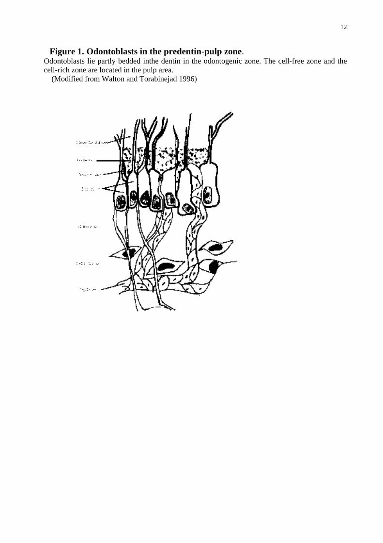
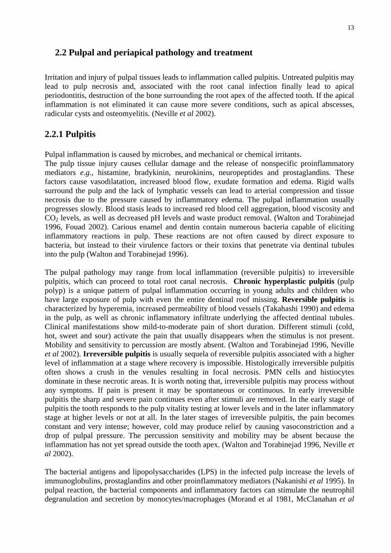
The pulpal tissue is mainly composed of cells, loose connective tissue and ground substance. The main cellular components are fibroblasts, odontoblasts, undifferentiated cells and defence cells. Major extracellular components are mature and immature collagen fibres, mainly collagen I and III, produced by pulpal fibroblasts and odontoblasts. The connective tissue of the pulp lacks elastic fibres. The ground substance in the pulp consists of hyaluronic acid, chondroitin sulphate, glycoproteins, carbohydrates and water. The pulp organ is well vascularised and contains nerve bundles. From its periphery to centre the pulp consists histologically of the odontoblast layer, cell-free zone of Weil, cell-rich zone, and pulp core (Fig. 1). The relations of these different zones vary depending on the age of the pulp. (Okiji 2002).
2.1.1 Odontoblasts Odontoblasts, next to fibroblasts, are the second most prominent cells in the pulp tissue. The odontoblast cell processes extend to dentinal tubules while the odontoblast body is adjacent to the predentin in the pulp (Fig.1). The odontoblast location is called the odontogenic zone of the pulp. In the odontoblastic row, the cells lie close to each other and the adjacent cells are connected by three types of intercellular junctions: impermeable (tight), adhering and communicating (gap) junctions. A capillary network surrounds the odontoblasts. Odontoblasts are highly differentiated mesenchymal cells incapable of proliferating and their main function is to produce dentin. The cell body is responsible of the synthesis of matrix glycoproteins, collagens and ground substance, while the odontoblastic process that extends into the dentinal tubules is the secretory organ (Fig. 1). (Okiji 2002). The main collagen produced by odontoblasts is collagen I, but they also produce type III collagen mRNA (Lukinmaa et al 1992, 1993), and cultured odontoblasts secrete type I and III collagen (Tjäderhane et al 2001). In addition to tissue-building-components odontoblasts express tissue destructive enzymes, MMPs, indicating a role for MMPs in the dentin organic matrix remodelling (Tjäderhane et al 1998, 2001). They secrete at least gelatinase-A (MMP-2), gelatinase-B (MMP-9), collagenase-2 (MMP-8), collagenase-3 (MMP-13), enamelysin (MMP-20) and membrane type matrix metalloproteinase-1 (MT1-MMP) (Tjäderhane et al 2001, Palosaari et al 2002). MMPs can contribute to the dentin matrix modulation in the reparative dentin production and in caries progression (Tjäderhane et al 2001).
12
Figure 1. Odontoblasts in the predentin-pulp zone. Odontoblasts lie partly bedded inthe dentin in the odontogenic zone. The cell-free zone and the cell-rich zone are located in the pulp area.
(Modified from Walton and Torabinejad 1996)
13
2.2 Pulpal and periapical pathology and treatment
Irritation and injury of pulpal tissues leads to inflammation called pulpitis. Untreated pulpitis may lead to pulp necrosis and, associated with the root canal infection finally lead to apical periodontitis, destruction of the bone surrounding the root apex of the affected tooth. If the apical inflammation is not eliminated it can cause more severe conditions, such as apical abscesses, radicular cysts and osteomyelitis. (Neville et al 2002). 2.2.1 Pulpitis
Pulpal inflammation is caused by microbes, and mechanical or chemical irritants. The pulp tissue injury causes cellular damage and the release of nonspecific proinflammatory mediators e.g., histamine, bradykinin, neurokinins, neuropeptides and prostaglandins. These factors cause vasodilatation, increased blood flow, exudate formation and edema. Rigid walls surround the pulp and the lack of lymphatic vessels can lead to arterial compression and tissue necrosis due to the pressure caused by inflammatory edema. The pulpal inflammation usually progresses slowly. Blood stasis leads to increased red blood cell aggregation, blood viscosity and CO2 levels, as well as decreased pH levels and waste product removal. (Walton and Torabinejad 1996, Fouad 2002). Carious enamel and dentin contain numerous bacteria capable of eliciting inflammatory reactions in pulp. These reactions are not often caused by direct exposure to bacteria, but instead to their virulence factors or their toxins that penetrate via dentinal tubules into the pulp (Walton and Torabinejad 1996). The pulpal pathology may range from local inflammation (reversible pulpitis) to irreversible pulpitis, which can proceed to total root canal necrosis. Chronic hyperplastic pulpitis (pulp polyp) is a unique pattern of pulpal inflammation occurring in young adults and children who have large exposure of pulp with even the entire dentinal roof missing. Reversible pulpitis is characterized by hyperemia, increased permeability of blood vessels (Takahashi 1990) and edema in the pulp, as well as chronic inflammatory infiltrate underlying the affected dentinal tubules. Clinical manifestations show mild-to-moderate pain of short duration. Different stimuli (cold, hot, sweet and sour) activate the pain that usually disappears when the stimulus is not present. Mobility and sensitivity to percussion are mostly absent. (Walton and Torabinejad 1996, Neville et al 2002). Irreversible pulpitis is usually sequela of reversible pulpitis associated with a higher level of inflammation at a stage where recovery is impossible. Histologically irreversible pulpitis often shows a crush in the venules resulting in focal necrosis. PMN cells and histiocytes dominate in these necrotic areas. It is worth noting that, irreversible pulpitis may process without any symptoms. If pain is present it may be spontaneous or continuous. In early irreversible pulpitis the sharp and severe pain continues even after stimuli are removed. In the early stage of pulpitis the tooth responds to the pulp vitality testing at lower levels and in the later inflammatory stage at higher levels or not at all. In the later stages of irreversible pulpitis, the pain becomes constant and very intense; however, cold may produce relief by causing vasoconstriction and a drop of pulpal pressure. The percussion sensitivity and mobility may be absent because the inflammation has not yet spread outside the tooth apex. (Walton and Torabinejad 1996, Neville et al 2002). The bacterial antigens and lipopolysaccharides (LPS) in the infected pulp increase the levels of immunoglobulins, prostaglandins and other proinflammatory mediators (Nakanishi et al 1995). In pulpal reaction, the bacterial components and inflammatory factors can stimulate the neutrophil degranulation and secretion by monocytes/macrophages (Morand et al 1981, McClanahan et al
14
1991, Cootauco et al 1993, Panagakos et al 1996, O’Boskey and Panagakos 1998, Chang et al 2001, Lu et al 2002). The released IL-1 and tumour necrosis factor- (TNF- ) are able to induce MMP-1, MMP-2 and the tissue inhibitor of metalloproteinases-1 (TIMP-1) gene expression in pulpal cells (Chang et al 2001, Lin et al 2001). Stimulation by black-pigmented Bacteroides elevates MMP-2 production (Chang et al 2002), and anaerobic bacterial extracts provoke pulp cells to excrete MMP-1 and MMP-2 as well as TIMP-1 (Nakata et al 2000). The levels of MMP-1, -2 and -3 are statistically significantly higher in acute pulpitis than in normal pulp tissue (Shin et al 2002). The treatment of pulpitis varies from the removal of the local irritant, pulp capping to pulpectomy and root canal treatment or even extraction of the tooth depending on the severity of pulpitis.
2.2.2 Apical periodontitis If pulpitis remains untreated and bacteria invade the pulp cavum and pulpal tissue it finally leads to apical periodontitis, which represents a protective and destructive inflammatory reaction also involving the bone surrounding the tooth apex. The infection in root canal is usually a mixed infection with anaerobic domination (Haapasalo 1989). In acute apical periodontitis (AAP), the pulp may be irreversibly inflamed or necrotic and it contains PMNs and monocyte/macrophages-like cells within a localized area at the apex. The bone and root resorption is not yet extensive enough to be detected in radiographs. If not treated AAP develops into chronic apical periodontitis (CAP, periapical granuloma) leading to pulp necrosis and bacterial toxins invading into the apical zone. The host defence responds to these bacterial root canal exudates and a defensive granulation zone is formed. If the bacteria persists in the root canal and, despite the defence responses, invade outside the root apex, the lesion may progress into an abscess with or without fistulation. (Neville et al 2002). Histologically CAP is made up of inflamed granulation tissue surrounded by a fibrous connective tissue wall. CAP is most often infiltrated by lymphocytes, plasma cells, monocytes/macrophages, as well as PMNs, and less frequently, mast cells, eosinophils and foam cells. (Neville et al 2002). A collection of Russell bodies, scattered eosinophilic globules of gammaglobulins, may be seen if numerous plasma cells are present, as well as lightly basophilic particles (pyronine bodies). These plasma cell products may be found in any accumulation of plasma cells and are not specific for CAP. Cholesterol clefts associated with multinucleated foreign body giant cells and red blood cell areas with hemosiderin pigmentation can sometimes be found and small abscess formation with acute inflammation may be present. (Walton and Torabinejad 1996, Neville et al 2002). Most CAPs are asymptomatic with just a little discomfort and CAP is usually discovered in routine radiographic examination: radiolucency is present and the affected tooth lacks apical lamina dura. The tooth does not respond to electric vitality testing because the pulp is necrotic and the percussion sensibility is mild at most. (Walton and Torabinejad 1996, Neville et al 2002). The immune-inflammatory process in apical periodontitis is double-faced. On the one hand, it is a protective host response to prevent bacterial invasion of the tooth apex and surrounding bone tissue, but, on the other the ”protective” mechanism also leads to destruction of the host tissue components (Márton and Kiss 2000). The first barrier to prevent bacterial invasion through apical foramen is provided by PMNs, which, by active phagocytosis and killing, effectively keep the bacterial amount and penetration as low as possible. Monocytes/macrophages also take part in the primary phagocytic protective phase and secrete antigens to act as chemotactic stimuli and to recruit other cells to participate in the inflammatory reaction (Márton and Kiss 2000). The degradation of the mineralised matrix of the alveolar bone surrounding the tooth apex is due to the action of osteoclasts (Bohne 1990, Anan et al 1991). In inflammation the delicate balance between osteoclasts and osteoblasts is disturbed resulting in bone loss. There are many factors
15
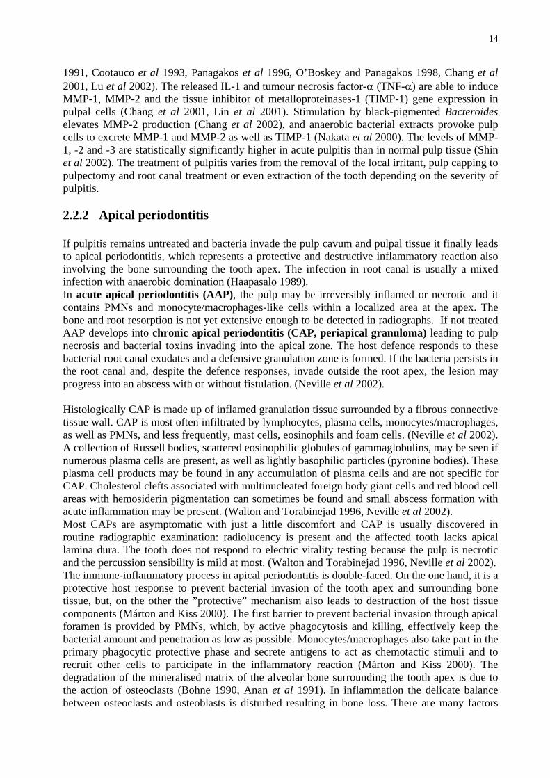
that have a stimulating effect on bone resorption in the apical periodontitis (Fig. 2). These stimulating factors include bacterial components, mainly LPS like endotoxin and short-chain fatty acids, released to the apical area, as well as host-derived substances. The host-defence system stimulates the tissue destructive reactions by releasing arachidonic acid derivates (prostaglandins and leukotrienes) during phagocytosis and other cell-mediated inflammatory factors, such as cytokines. High levels of immunoglobulins, secreted by plasma cells, are also found in periapical lesions (Torres et al 1994, Matsuo et al 1995, Miyauchi et al 1996). Other plasma proteins, such as complement components, bradykinin, kallikrein and thrombin and acute-phase reactants, derived from dilated blood vessels are also present to stimulate bone resorption (Lerner 1994, Márton and Kiss 2000). As a response to stimuli, monocyte/macrophage-like cells, plasma cells, fibroblasts, epithelial and endothelial cells, PMNs, osteoblasts and osteoclasts in periapical periodontitis can produce bone resorptive cytokines IL-1 and IL-1 , TNF- and IL-6 (Artese et al 1991, Piattelli et al 1991, Barkhordar et al 1992, Miller et al 1996, Márton and Kiss 2000). T-cells express IL-2, IL-4, IL-6, IL-10 and interferon- (IFN- ), and B-cells, fibroblasts and monocyte/macrophage-like cells IL-10 (Walker et al 2000). The immune cytokines in CAP can accelerate bone resorption (Stashenko 1990). The tissue destructive process is interactive, and these cytokines can trigger other cells to express proteolytic enzymes, including MMPs, also found to be present in periapical granulation tissue (Barkhordar 1987, Takahashi 1998).(Fig. 2.). 2.2.3 Root canal treatment (RCT) In order to eliminate the pulpal inflammation to avoid root canal necrosis, prevent or heal periapical periodontitis root canal treatment (RCT) has to be performed. The aim of RCT is to remove bacteria and their toxins from the root canal by mechanical and chemical cleaning. The root canal treatment techniques vary according to the person. The treatment needs to be done under aseptic conditions and therefore the tooth is separated from other teeth and the oral cavity by a rubber dam and the working area is sterilized. The first step in RCT is to determine the working length of the root canal instruments, which is often done by radiographs and/or by electrical measuring. The necrotic tissue is removed with special root canal instruments and the canal is mechanically widened using rinsing with antiseptic liquids and usually also with liquids, like ethylene diamino tetra acetic acid (EDTA), to remove the smear layer from the root canal walls. Root filling is done during the same appointment or the next visit. An X-ray is taken to check the root filling. The success of the treatment is usually checked within one year of the basic treatment. It is considered successful if no clinical symptoms remain and the periapical area in a radiological examination seems to be normal without signs of inflammation or resorption. (Walton and Torabinejad 1996). The difficulty during RCT is to determine whether the treatment has been successful enough to eliminate the bacteria in the root canal environment. If not, it is likely that the infection in the apical area persists. Chair-side adjunctive diagnostic tests measuring the inflammatory level or mediators of periapical tissue are suggested as future tools in endodontics (Metzger 2000).

16
Figure 2. The bone destructive mechanism in apical periodontitis. This figure shows the complicated network of tissue reactions against bacterial products in apical periodontitis. (Modified from Márton and Kiss 2000) BAG: bacterial antigens, LPS: Lipopolysaccharides, PMN: polymorphonuclear leukocyte, T: T-lymphocyte, B: B-lymphocyte, Mc: macrophage, Fb: fibroblast, Plc: plasma cell, Ob: osteoblast, Oc: osteoclast, TNF: tumour necrosis factor, IL: interleukin, INF: interferon, PGE: prostaglandin, MMP: matrix metalloproteinase.

17
2.3 Odontogenic cysts
“A cyst is described as a pathological cavity, lined usually totally or partly with epithelium, containing gas, fluid or semi-fluid and not being created by puss accumulation” (Kramer 1974). Cysts are more common in the jawbone than in any other bone. Depending on the epithelial origin, jaw cysts are divided into odontogenic and non-odontogenic cysts. The epithelial lining of odontogenic cysts is derived from the epithelial remnants of tooth- forming organs and these cysts are further divided into developmental and inflammatory cysts. Odontogenic developmental cysts are of unknown origin and inflammatory cysts, like their name suggests, result from inflammation. Developmental cysts include “gingival cysts” of infants (Epstein pearls), odontogenic keratocysts, dentigerous (follicular) cysts, eruption cysts, lateral periodontal cysts, gingival cysts of adults and glandular odontogenic cysts (sialo-odontogenic cysts). Inflammatory cysts include radicular cysts (apical and lateral, and residual cysts) and paradental cysts. Odontogenic cysts comprise 90 % of the jaw cysts. (Kramer et al 1992, Neville et al 2002).
2.3.1. Radicular cyst (RC, Periapical cyst, Apical periodontal cyst) Radicular cysts (RCs) are the most common odontogenic cysts (60-75%). RC originate from the epithelial rests of Malassez entrapped within chronic apical periodontitis. When small they are usually symptomless but may expand the bone while growing. Eventually, the cyst may perforate the cortex but frequently they do not grow to large dimensions. In an X-ray, RC shows a variable size of radiolucency with a loss of lamina dura, but the radiographic examination is not a diagnostic criteria. In RC, non-keratinised stratified squamous epithelium partly or wholly surrounds the chronically inflamed granulation tissue capsule. Epithelial hyperplasia is often present and the epithelium may demonstrate spongiosis. The cyst lumen is filled with fluid and cellular debris. Sometimes the epithelium may include calcifications known as Rushton bodies. Both the cyst lumen and wall may present dystrophic calcifications, cholesterol clefts with multinucleated foreign body giant cells, red blood cells and hemosiderin pigmentation. The fibrous connective tissue wall of the cyst often demonstrates inflammatory infiltrate with lymphocytes, PMN cells, plasma cells, histiocytes, and rarely mast cells and eosinophils. Plasma cells often predominate the infiltration. The cyst wall occasionally contains hyaline bodies – scattered, circumscribed corrugated, condensed collagenous rings containing eosinophilic material - surrounded frequently by multinucleated giant cells and lymphocytes. The eosinophilic material may contain various cells and it may undergo dystrophic calcification. These bodies can be seen in any intraosseous chronic inflammation and they represent inflammatory exudate pools (i.e. extravasated serum) that with time undergo fibrosis and occasionally dystrophic calcification. The multinucleated foreign body giant cells appear at the site to remove the debris. The cyst wall becomes more fibrotic with diminishing vascularity and inflammatory infiltration. (Neville et al 2002). The RCs tend to expand,”balloon-like”, in all directions equally, and the surrounding bone is resorbed, mainly by osteoclast function (Formigli et al 1995). Cyst lining cell cultures have been demonstrated to release osteoclast stimulating bone resorbing factors, prostaglandins, mainly from cyst capsule fibroblasts, and immunoglobulins are secreted by plasma cells (Formigli et al 1995, Meghji et al 1996, Takahashi et al 1996). Interleukins and other cytokines are secreted from monocyte/macrophage-like cells, epithelial cells and other cells to stimulate bone resorption (Bando et al 1993). IL-1 , IL-1 , IL-4, IL-6, IL-10 and IFN- are detected in RC fluid and explant media, IL-1 being characteristics of RCs in comparison to FC and KC (Hoenig et al
18
1991, Formigli et al 1995, Meghji et al 1996, Walker et al 2000). Cytokines are able to trigger cells in RC to produce proteolytic enzymes, such as IL-1 which enhances gelatinase-A and -B (MMP-2 and MMP-9) production in explant culture (Kubota et al 2000). MMP-1 (collagenase –1) protein has been found in subepithelial fibroblasts and epithelial cells of RC (Lin et al 1997). Collagenases (MMP-1 and MMP-8) and gelatinases (MMP-2 and MMP-9) have been detected in cyst fluid and cyst wall extracts (Teronen et al 1995a, b, Kubota et al 2000). Bacterial endotoxins, always present in RC, stimulate keratinocyte proliferation, and bacterial products activate MMP production (Ding et al 1995, 1996, Meghji et al 1996). These proteolytic enzyme cascades are most likely involved in the bone matrix degradation, BM and epithelial cell processing during cyst expansion.
2.3.2. Follicular cyst (FC, Dentigerous cyst) Follicular cysts are next to RCs the most common of odontogenic cysts (10-15%). A FC originates from the reduced enamel epithelium derived from the enamel organ. It encloses partly or totally the crown of an unerupted tooth and it is attached to the tooth at the cementoenamel junction. FCs most frequently surround mandibular third molars, followed by maxillary permanent canines, maxillary third molars and mandibular second premolars. FCs are usually symptomless, but they may grow to a remarkable size expanding the bone in the involved area. Usually they do not show any pain unless secondary inflammation is present. Radiologically, FCs usually show unilocular radiolucency attached to an unerupted tooth. (Neville et al 2002). The cyst epithelium is usually two to five layers thick. It consists of non-keratinised cells and the interface to connective tissue is flat. If inflammation is present, the epithelium may show varying amounts of hyperplasia with development of rete ridges. Focal areas of mucous cells (goblet cells) may be seen in the epithelial lining, and rarely ciliated columnar or sebaceous cells. The connective tissue wall is loosely arranged having sometimes cholesterol clefts and islands of odontogenic epithelium present. When inflammation is present, the collagen amount in the cyst wall increases with variable infiltration of inflammatory cells. The cyst contains a proteinaceous, yellowish fluid frequently with cholesterol crystals. (Neville et al 2002). The developing stimulus of a FC is unknown and the cyst formation mechanism is still unclear. The expansion mechanism of a FC is, anyhow, probably similar to that of a RC but is dependent on bone destruction and osmotic pressure (Neville et al 2002). The triggering mechanism in FCs for the release of bone resorbing factors from the cyst cells may differ from RCs, because inflammation is not necessarily always present. Bone resorption stimulating factors, including growth factors and cytokines, are present in a FC wall and epithelium (Meghji et al 1996, Li et al 1997). These stimuli are able to release MMPs, like gelatinases, from cyst cells (Kubota et al 2000). Collagenases and gelatinases are found in FC wall extracts (Teronen et al 1995a, b) suggesting that these enzymes take part in the FC expansion as well.
2.3.3. Odontogenic keratocyst (KC, Primordial cyst) Odontogenic keratocyst is the third most common of odontogenic cysts (5-10%). It is thought to originate from the cell rests after the dissolution of the dental lamina. Most of the KC cases occur in ages 10 to 40 with a slight male predilection and 70-80 % occur in the mandible, most often in third molar region and ascending ramus. In maxilla it most often involves the region posterior to the first premolar. The expansion mechanism of KCs differs from RCs’ and FCs’ unicentric pattern. An odontogenic keratocyst tends to grow in an antero-posterior direction often not showing bone expansion. KCs may, however, cause swelling and pain. Radiologically, KCs demonstrate a well-defined radiolucent uni- or multilocular area with smooth and often corticated
19
margins. An unerupted tooth is involved in 25-40 % of the cases. The diagnosis of KC is based on histopathology, radiologic findings being only suggestive. The recurrence rate of KCs is high varying from 3 to 60 %. Recurrence takes place more often in mandibular KC, especially in the posterior body and ascending ramus. (Myoung et al 2001, Neville et al 2002). Although the majority of KCs appear as solitary lesions, multiple KCs may be present and they are associated with nevoid basal cell carcinoma (Gorlin-Goltz) syndrome (Myoung et al 2001, Neville et al 2002). The histopathology of a KC often shows a thin friable, folded wall, which is difficult to enucleate in one piece. The cyst wall capsule may contain epithelial rests giving the basis to independent satellite cysts (daughter cysts) around the main lesion. KC lumen may contain liquid material similar to a serum transudate, but often it contains thick cheesy material composed of keratinaceous debris having a low soluble protein level. (Neville et al 2002). The epithelial lining surrounded by the fibrous cyst capsule is usually composed of six to eight cell layers thick squamous epithelium. The uniform thick, often corrugated epithelial surface shows parakeratotic epithelial cells but areas of orthokeratosis may be present. The well-defined basal cell layer consists of palisaded columnar or cuboidal cells, which are often hyperchromatic. When inflammatory changes are present the epithelial lining may lose its keratinization and histological characteristics of its basal cell layer and also rete ridge formation may be seen. The epithelium easily detaches from the cyst stroma, a phenomenon that may increase the recurrence rate of KCs. The orthokeratinized variant (orthokeratinized odontogenic cyst) of the KC is not associated with Gorlin-Goltz syndrome and the recurrence rate is much lower, though it has been suggested that this orthokeratinized type might have a greater risk for malignancy. The orthokeratinized epithelium has a granular layer and the basal layer is poorly organized. Radiographically and clinically 2/3 of the orthokeratinized odontogenic cysts show similarities with the follicular cyst; most often involving an impacted third lower molar. It is suggested that the orthokeratinized odontogenic cyst should be categorized differently from KCs. (Neville et al 2002, Regezi 2002). The growth and enlargement of KCs has often been regarded as evidence of their neoplastic behaviour (Myoung et al 2001, Shear 2002a). Possible inflammation has little effect on the KC growth. The latest point based on the multilocular and loculated outlines of the KC is that the enlargement of a KC appears in a multicentric growth pattern with proliferation of local groups of epithelial cells against the semi-solid cyst contents. The intrinsic growth potential and infolding of the epithelium into the cyst wall during the KCs multiphase growing indicate active epithelial proliferation and support the suggestion that KC may more likely present a benign neoplasm than a cyst (Shear 2002a, c). The finding that KC epithelial cells overexpress p53, a protein usually overexpressed in malignant lesions but not in normal cells, further strengthens the notion of KCs’ potential neoplastic nature (Piattelli et al 2001). A mutation in the p53 gene may be a reason for the increased cell proliferation. The p53+ basal cells are found more often in KCs with epithelial dysplasia and the p53+ epithelium is usually parakeratotic (Piattelli et al 2001). The proliferating cell nuclear antigen (PCNA), indicating active proliferation in p53+ cells, is also found more often in KCs than other odontogenic cysts (Li et al 1994, Piattelli et al 2001, Shear 2002b). It has been shown that the KC epithelium has higher mitotic activity than FC or RC epithelia, and the mitotic activity is often found in basal or suprabasal cells (Matthews et al 1988, Li et al 1994). The dissolution of bone matrix by the cyst wall is mediated by osteoclasts, evidently in concert with the biologically active proteases, including MMPs (Neville et al 2002). Different cytokines and other cell mediators, produced in a KC as well, can stimulate the protease production or activation by KC cells. KC epithelial cells can further express TGF- , IL-1 , IL-6 and proMMP-9 (Meghji et al 1992, 1996, Li et al 1997, Kubota et al 2000). IL-1 is able to enhance MT1-MMP expression in KC fibroblasts and increases both proMMP-2 and proMMP-9 activation (Kubota et
20
al 2000, 2002). Active interstitial collagenases (MMP-1 and MMP-8) and gelatinases (MMP-2 and MMP-9) have been found in the cyst extracts and fluids (Ylipaavalniemi 1978, Teronen et al 1995a, b, Sorsa et al 1988, Kubota et al 2000). The reasons for KCs’ special characteristics, epithelial detachment and growth potential, are not yet completely clarified and require further studies.
2.4 Plasma cells
Plasma cells accumulate in chronic inflammation and by tradition their role is to act in the humoral immune response by secreting antibodies. They derive from B lymphocytes and their differentiation requires multiple stimuli. The primary stimulus is antigen interaction with surface immunoglobulins (IgM and IgG) of B lymphocyte. The activated B lymphocyte ”presents” the antigen to T-helper lymphocytes that in turn secrete distinct growth and differentiation factors. These molecules, particularly IL-4 and IL-5, secreted by T-helper cells, induce B cell differentiation. The final differentiation requires an additional B cell differentiation factor, IL-6 secreted by many lymphoid and nonlymphoid cells, to be present (Nisengard and Newman 1994). Also other cytokines, IL-3 and IL-10, with T cell induction may activate the B cell differentiation into plasma cells (Merville et al 1995, Rousset et al 1995). The main function of plasma cells is to secrete immunoglobulins, antibodies taking part in the host defence system, by binding to antigens. Antibody secretion is the basis of humoral immunity. Antibodies are active against bacteria, viruses and tumours, and they are able to neutralize a number of microbial products, particularly toxins, which can be harmful to the host tissues. Immunoglobulins are glycoproteins containing a basic monomeric structure consisting of two heavy and two light chains. Based on differences in the H chain region, immunoglobulins are divided to five main groups: IgM, IgG, IgA, IgD and IgE, and based on L chain dissimilarities to
and subgroups. Plasma cells produce immunoglobulins and antibodies against foreign antigens, but they are also known to produce and secrete antibodies against self antigens leading to autoimmune diseases (Andrew et al 1991), and also certain cytokines (IL-10, TGF- 1, TNF- ) (Matthes et al 1995, DiGirolamo et al 1997). Plasma cells, specially myeloma cells, and B lymphocytes also secrete certain matrix metalloproteinases (MMP-2, MMP-3 and MMP-9) and their inhibitors (TIMP-1) (Stetler-Stevenson et al 1997, DiGirolamo et al 1998). 2.4.1 Multiple myeloma (MM) and plasmacytoma (PLC) Multiple myeloma is the most common plasma cell malignancy. It generally occurs as a disseminated lesion involving many bones (multiple myeloma) or seldom as a solitary bone/soft tissue lesion (solitary myeloma or plasmacytoma/extramedullary plasmacytoma). Plasmacytoma may lead to multiple myeloma. MM is a progressive and fatal malignancy of plasma cells. It may involve IgA, IgD or IgE but most frequently involving IgG producing monoclonal plasma cells. In MM abnormally high levels of the immunoglobulin or its polypeptide chains are secreted into the serum and urine. Most often MM appears in patients between 50 and 70 years affecting men more often than women. The skull, vertebrae, sternum, ribs and pelvic bones are commonly affected though lesions in jaws are not uncommon (30% of the cases). Jaw lesions appear more commonly in the posterior region of the mandible. The bone lesions present unbalanced bone remodelling: excessive resorption with low bone formation. (Neville et al 2002).

21
The most characteristic of the clinical symptoms of MM is bone pain and some patients may present pathologic fractures caused by bone destruction. Patients may suffer from anaemia and fever may be present as a result of neutropenia. Metastatic calcifications may occur in soft tissues due to hypercalcemia caused by osteolysis. The excess of produced light chains, unattached to heavy chains are excreted to the urine and often precipitate in renal tubules, and may cause renal failure. This light chain protein found in the urine is called Bence-Jones protein. Amyloidosis may appear even as an initial manifestation of the disease. This deposition of extracellular proteinaceous substance, amyloid, often affects the eyelid region, and when present in the tongue may lead to macroglossia. Radiographically MM appears as sharply demarcated radiolucencies or ragged radiolucent areas often well evidenced in skull radiographs. Histologically these lesions show dense cellularity with little or no supporting stroma. The lesions consist of variably differentiated, plasmacytoid cells invading and replacing the normal host tissue. Occasionally homogenous, relatively acellular amyloid material may be present among neoplastic cells. (Neville et al 2002). The criteria to diagnose MM are seen in the Table 1.
Table 1. Diagnostic criteria for plasma cell myeloma (Modified from WHO criteria; edited by Jaffe et al 2001)
The myeloma diagnosis requires a minimum of one major or three minor criteria which must include (1) and (2). These criteria must be manifest in a symptomatic patient with progressive disease.
Major criteria: 1. Marrow plasmacytosis (>30%) 2. Plasmacytoma on biopsy 3. M-component: Serum IgG > 3.5g/dl, IgA >2g/dl Urine >1g/24hr of Bence-Jones (BJ) protein Minor criteria: 1. Marrow plasmacytosis (10-30%) 2. M-component: present but less than above 3. Lytic bone lesions 4. Reduced normal immunoglobulins (<50% normal): IgG <600mg/dl, IgA <100mg/dl, IgM <50mg/dl
In addition to immunoglobulin secretion, the plasma cells in MM excrete many different cytokines and MMPs. IL-6 has been widely studied and its expression by malignant plasma cells is related to MM invasiveness (Pattengale 1997, Sati et al 1998). IL-6, not expressed in normal plasma cells, is essential for the growth of MM, by inducing the differentiation of plasmablasts into mature malignant plasma cells (Lauta 2001), and for the survival of myeloma cells by preventing spontaneous apoptosis (Pattengale 1997). Myeloma cells, in addition to secreting IL-6, also stimulate stromal and bone marrow cells to release large amounts of IL-6 via an IL-1 dependent pathway (Carter et al 1990). Cytokines are able to provoke IL-6 production in bone marrow-derived stromal cells and cytokines provoke MMP production and secretion by many cells, though IL-6 has not been found to interfere with MMP production in MM lesions (Carter et al 1990, Barille et al 1997, Vacca et al 1999). The MM plasma cells interfere with the invasive potential of MMs also by secreting fibroblasts growth factor (FGF-2), a potent angiogenic factor, to increase angiogenesis (Vacca et al 1999). MM cells can produce gelatinases (MMP-2 and MMP-9), and upregulate collagenase-1 (MMP-1) expression and induce MMP-2 activation in bone marrow stromal cells of MM patients (Barille et al 1997, Vacca et al 1999, Ria et al 2002).

22
MMPs (MMP-1, MMP-2, MMP-9) expression of MM cells is known to increase during the active phase of the disease (Barrille et al 1997, Vacca et al 1999). The ECM proteins fibronectin (FN) and vitronectin (VN) are known to enhance MM plasma cell proliferation and gelatinase (MMP-2 and MMP-9) release from MM cells via V 3 integrin pathway (Ria et al 2002).
2.5 Laminin-5 (Ln-5) Laminins are a family of non-collagenous BM proteins that appear in several isoforms each presenting a unique combination of , and chain linked together with disulfide bonds. In addition to acting as structural components, laminins are involved in the cell differentiation, adhesion and migration. (Lohi 2001). Ln-5, previously called kalinin, nicein or epiligrin, functions as an anchoring filament component between epithelial cells and the BM. Keratinocytes synthesize Ln-5 (Amano et al 2001) and it consists of 3: 3: 2 subunits of which the 2 chain is unique for Ln-5 (Kallunki et al 1992). Ln-5 has an ambiguous role both in promoting cell adhesion and migration and its overexpression in tissues is closely related to malignancy (Soini et al 1996, Lohi 2001, Patel et al 2002). The cell adhesive function of intact Ln-5 is mediated through 3 1 and 6 4 integrins being connected to type VII collagen in the BM (Rousselle et al 1997). The 2 chain does not contribute to the adhesive quality of Ln-5, but plays a crucial role in the regulation of cell migration (Salo et al 1999). The specific cleavage of a 2 chain to a 80 kDa form has been reported to cause epithelial cell migration and is connected to cell invasiveness (Giannelli et al 1997, Skyldberg et al 1999, Gilles et al 2001). MMP-2, -3, -13, -14, -20 and MT1-MMP are among the MMPs able to cleave Ln-5 at a special site in order to promote epithelial cell motility (Giannelli et al 1997, 1999, Koshikawa et al 2000, Gilles et al 2001, Pirilä et al 2001b, 2003). These findings suggest that a fragmented laminin 2 chain has a specific role in the regulation of the epithelial cell migration.
2.6 Matrix metalloproteinases (MMPs) Matrix metalloproteinases form a structurally related group of at least 25 (22 human homologues) Ca2+- and Zn2+-dependent endopeptidases collectively capable of degrading practically all ECM and BM components. In addition, MMPs can modulate many other substrates including ILs and other cytokines, serine proteinase inhibitors (serpins), growth factors and chemokines (Uitto et al 2003). MMPs are divided into six subgroups: collagenases, gelatinases, membrane-type MMPs (MT-MMPs), stromelysins, matrilysins and other MMPs (Fig. 3 and Table 2). They have a role in many normal physiological events, like ovulation, embryo implantation, organ development, angiogenesis, wound healing and bone remodelling. Normal embryonic development and tissue remodelling needs a controlled balance between ECM synthesis and degradation, as well as a balance between MMPs and their natural inhibitors TIMPs. Overproduction of MMPs occurs in many tissue destructive pathological conditions and MMPs are known to influence many chronic tissue destructive inflammatory and autoimmune diseases, like rheumatoid arthritis, osteoarthritis, periodontitis, blistering disorders of the skin, lung diseases, eye diseases and chronic ulcerations. They also affect tumour growth, invasion and metastasis. In the inflammatory process, MMPs, being upregulated by cytokines and other proinflammatory mediators, are mainly responsible for the ECM and BM degradation. (Kähäri and Saarialho-Kere 1999, Johansson et al 2000). MMPs are active in physiological and pathological bone remodelling being expressed by osteoblasts and osteoclasts (Rifas et al 1989, Meikle et al 1992, Vaes et al 1992, Reponen et al 1994, Wucherpfennig et al 1994).

23
In addition to tissue remodelling, MMPs participate in many other cellular functions (Vu and Werb 2000). They adapt cellular behaviour, for example, by inducing cell migration in normal growth and tissue remodelling, such as wound healing, angiogenesis, as well as in tumour invasion and metastasis (Curran and Murray 2000, Johansson et al 2000, Vu and Werb 2000, Vihinen and Kähäri 2002). Molecules affecting cellular functions can be modified by MMPs; the precursor of IL-1 can be activated by MMP-2, MMP-3 and MMP-9, although excess or prolonged incubation with MMPs can also result in IL-1 inactivation (Ito et al 1996, Schönbeck et al 1998). Extracellular matrices themselves adjust to the cellular functions, like growth, cell shape changes, migration and differentiation, for example, by controlling cellular adhesion. An ECM influences cell behaviour by maintaining signal molecules, such as growth-factors and growth-factor binding proteins, and acting as a ligand for cellular receptors, including integrins, that can transduce signals to the cell interior. MMPs may cleave the ECM and release bioactive cell surface molecules or regulate the growth-factor availability by cleaving the ECM proteins that can bind to them (Uitto et al 2003). Various MMPs can degrade decorin, a collagen-associated small protein that binds to TGF- , and may as a ”secondary effect” release TGF- to carry out its biological functions. On the other hand, the release of TGF- may have a negative feedback on MMP production. (Sternlicht and Werb 2001, Uitto et al 2003). MMPs modulate the activity of other proteinases by activating latent enzymes or inactivating their inhibitors. MMP-3 can cleave the urokinase type plasminogen activator and MMP-1 and MMP-8 are able to cleave the 1-proteinase inhibitor and 1-antichymotrypsin (Michaelis et al 1990, Vu and Werb 2000). MMPs are also able to activate each other in a complicated cascade network. MMP-2 and MT1-MMP, for example, are able to activate latent MMP-13, and MT1-MMP activates MMP-8, as well as the active form of MMP-2 together with MMP-13 activate MMP-9 (Knäuper et al 1996a, Cowell et al 1998, Saarialho-Kere 2000, Holopainen et al 2003). MMPs’ behaviour in tissues is multivariate and many questions are still unanswered (Uitto et al 2003). MMPs share significant structural similarities; all of them contain the N-terminal signal sequence (”pre”domain) followed by the ”pro”domain that maintains enzyme latency usually until removed, and the zinc-binding catalytic domain. With three exceptions (MMP-7, MMP-23 and MMP-26) the hemopexin/vitronectin domain is connected to the catalytic domain. The hemopexin domain participates in the catalytic activities and it can bind to TIMP and certain substrates (Sternlicht and Werb 2001). MT-MMPs contain a transmembrane domain and a short cytoplasmic C-terminal tail (Fig. 3).
2.6.1 Collagenases ( MMP-1, MMP-8, MMP-13) The human interstitial collagenase subfamily contains three members: MMP-1 (fibroblast type collagenase, collagenase-1), MMP-8 (neutrophil collagenase, collagenase-2) and MMP-13 (collagenase-3). In addition to these human collagenases, a collagenase, MMP-18, has been found and cloned only from Xenopus laevis (Stolow et al 1996). The mammalian analogous has not yet been found. Collagenases cleave the native fibrillar collagen types I-III to generate ¾- and ¼-fragments. The cleavage takes place in a specific site between the glysine-isoleusine (Gly-Ile) of the 1 chain and the glysine-leusine (Gly-Leu) residues of the 2 chain forming triple helical fragments that in body temperature denature into randomly coiled gelatin, being further degraded by other gelatinolytic MMPs and proteinases (Birkedal-Hansen et al 1993, Sternlicht and Werb 2001). The collagenases differ in their substrate specificities and functional roles. MMP-1 preferably

24
Figure 3. MMP structure (Modified from Sternlich and Werb 2001) Pre: Pre domain, Pro: propeptide with ZN-binding thiol group (SH), Fu: furin site, ZN: Zn-binding site, F: fibronectin type II insert, H: Hinge region, TM: transmembrane domain, C: cytoplasmic tail, C/P:cysteine/proline, IL-1R: interleukin-1 receptor.
1) Minimal Domain MMPs
MMP-7: matrilysin, MMP-26:endometase 2) Simple Hemopexin Domain-Containing MMPs
MMP-1: collagenase-1, MMP-8: collagenase-2, MMP-13: collagenase-3, MMP-18: collagenase-4, MMP-3: stromelysin-1, MMP10: stromelysin-2, MMP-27, MMP-12: metalloelastase, MMP-19:RASI-1, MMP-20: enamelysin, MMP-22: CMMP
3) Gelatin-binding MMPs
MMP-2: gelatinase-A, MMP-9: gelatinase-B 4) Furin activated Secreted MMPs
MMP-11: stromelysin -3, MMP-28: epilysin 5) Transmembrane MMPs
MMP-14: MT1-MMP, MMP-15: MT2-MMP, MMP-16: MT3-MMP, MMP-24: MT5-MMP 6) GPI-linked MMPs (glycophosphatidyl 1 inositol-anchoring domain)
MMP-17: MT4-MMP, MMP-25: MT6-MMP 7) Vitronectin-like MMPs MMP-21: XMMP
8) Cysteine/Proline-rich IL-1 Receptor-like Domain MMPs MMP-23

25
degrades collagen III, MMP-8 prefers type I collagen (Birkedal-Hansen et al 1993), and MMP-13 collagen II (Knäuper et al 1996a) (Table 2). Human MMP-1 was first cloned and sequenced from skin fibroblasts (Goldberg et al 1986). Since then it has been found to be produced by keratinocytes, endothelial cells, monocytes and macrophages, chondrocytes, osteoblasts and various tumour cells (Meikle et al 1992, Birkedal-Hansen et al 1993, Giambernardi et al 1998). MMP-1 is produced on demand by transcriptional initiation and the delay after the trigger mechanism can be up to 12 hours (Birkedal-Hansen et al 1993). It is produced as glycosylated proenzymes 52 kDa and 57 kDa in size, and upon activation (i.e. propeptide cleavage or removal) converted to the active size of 42 kDa and 47 kDa respectively. MMP-1 cleaves several ECM components but is ineffective against most BM components. It has been connected to many physiological phenomena, like embryonic development and the migration of keratinocytes during wound healing, as well as to the growth of malignant tumours (Saarialho-Kere et al 1992, 1995, Vaalamo et al 1997, Giambernardi et al 1998, Vu and Werb 2000). MMP-8 is synthesized and stored in PMN cells during their maturation in bone marrow and released upon PMN degranulation induced by various extracellular stimuli, like cytokines or bacterial contact (Ding et al 1995, 1996, 1997). In addition inducible de novo MMP-8 expression has been found to be expressed by chondrocytes, fibroblast-like cells, monocytes, epithelial cells and keratinocytes, as well as melanoma cells and oral cancer cells (Hanemaaijer et al 1997, Giambernardi et al 1998, Bachmeier et al 2000, Tervahartiala et al 2000, Prikk et al 2001, 2002, Moilanen et al 2002). MMP-8 is secreted either as a 55 kDa unglycosylated form or a 75 kDa glycosylated form and after activation its molecular size usually decreases by 10-20 kDa (Hasty et al 1990, Ding et al 1995, Hanemaaijer et al 1997, Moilanen et al 2003). The main substrates for MMP-8 are fibrillar collagens (Birkedal-Hansen et al 1993). MMP-8 is catalytically more effective in cleaving substrates (except collagen III) than MMP-1. Increased MMP-8 levels are detected in many inflammatory diseases like rheumatoid arthritis, chronic lung inflammations and periodontitis (Konttinen et al 1991, Sorsa et al 1994, 1999, Ingman et al 1996, Uitto et al 1998, Tervahartiala et al 2000, Mäntylä et al 2000, 2003, Marchenko et al 2001a, b, Prikk et al 2001, 2002, Lanone et al 2002). MMP-13 has the widest substrate selection among the interstitial collagenases and, in addition to collagens, it is able to cleave various BM components. MMP-13 cleaves type II collagen more efficiently than type I and III and among interstitial collagenases, it is most effective in cleaving gelatin (Mitchell et al 1996, Lindy et al 1997). It is secreted as a 60 kDa glycoprotein and is converted to a 48 kDa form after activation. MMP-13 can be activated by other MMPs including MMP-2, MMP-3, MMP-10, MT1-MMP, MT2-MMP and serine proteases human trypsin-2 and plasmin (Knäuper et al 1996a, b, D’Ortho et al 1997). MMP-13 was first cloned from human breast cancer, indicating a role in malignancy (Freije et al 1994). The physiologic expression of MMP-13 seems to be limited only to developing bone (Ståhle-Bäckdahl et al 1997) and wound healing (Ravanti et al 1999, Pirilä et al 2001b), but it is widely expressed in pathological conditions characterized by excessive ECM degradation including rheumatoid arthritis, osteoarthritis, periodontitis and chronic ulcerations (Lindy et al 1997, Vaalamo et al 1997, Uitto et al 1998, Johansson et al 2000, Tervahartiala et al 2000, Kiili et al 2002). MMP-13 is further expressed in skin and mucosal squamous cell carcinoma (SCC) cells, hypertrophic chondrocytes, osteoblasts, and many carcinoma and melanoma cells (Mitchell et al 1996, Johansson et al 1997a, b, Giambernardi et al 1998, Uria et al 1998, Bachmeier et al 2000, Hofmann et al 2000, Thomas et al 2000). Due to the relative wide substrate specificity, MMP-13 is regarded to be an all-round tool for malignant cells in growth and invasion (Kähäri and Saarialho-Kere 1999).
26
2.6.2 Gelatinases (type IV-collagenases, MMP-2, MMP-9) The gelatinase subgroup contains two enzymes: gelatinase-A (MMP-2) and gelatinase-B (MMP-9). The substrate specificity and primary structure of gelatinases are rather similar (Fig. 3, Table 2). Within their catalytic domain, gelatinases have three head-to-tail cysteine-rich repeats that are required to bind and cleave especially the denatured collagen and elastin. These inserts resemble the collagen binding type II repeats of fibronectin. MMP-2 was first purified from a murine malignant melanoma tumour and found to be a potent BM type IV collagen degrading enzyme (Salo et al 1983). It is expressed in many different cell types including keratinocytes, fibroblasts, chondrocytes, monocytes, osteoblasts, endothelial cells, T-lymphocytes and malignant plasma cells, leukaemia, carcinoma and melanoma cells (Stetler-Stevenson 1990, Barille et al 1997, Giambernardi et al 1998, Sutinen 1998). PMNs do not express or contain MMP-2. The latent MMP-2 is secreted in a 72 kDa form and upon activation is converted to a 59-62 kDa form. Collagens (I, II, IV, V, VII, X, XI), gelatins and BM components are cleaved by MMP-2 (Aimes and Quiqley 1995, Konttinen et al 1998, Sternlicht and Werb 2001). MMP-2 is involved in many processes that require ECM remodelling and its overexpression is closely connected to cell migration and to the invasive and metastatic potential of malignant tumours (Stetler-Stevenson 1990, Autio-Harmainen et al 1992, Tryggvason et al 1993, Soini et al 1994, Mäkelä et al 1999, Pirilä et al 2001b, Vihinen and Kähäri 2002). MMP-9 was originally identified from human macrophages (Vartio et al 1982) but is produced by PMNs, keratinocytes, T-lymphocytes, monocytes, plasma cells and many transformed and malignant cells (Wucherpfennig et al 1994, DiGirolamo et al 1998, Giambernardi et al 1998). The substrate specificity of MMP-9 is similar to MMP-2, though it does not degrade type I-III collagens as widely as MMP-2. MMP-9 plays an essential role in the resorption of collagen during bone remodelling and development (Meikle et al 1992, Vaes et al 1992, Wucherpfennig et al 1994) and its overexpression is also connected to the inflammatory reaction in lung and periodontal diseases (Westerlund et al 1996, Mautino et al 1997, Hoshino et al 1998, Atkinson and Senior 2003). Tumour cells and their metastatic potential, as well as cell invasion, are also linked to MMP-9 (Stetler-Stevenson 1990, Giambernardi et al 1998, Hofmann et al 2000, Thomas et al 2001, Vihinen and Kähäri 2002). The MMP-9 level is elevated in chronic wounds (Mirastschijski et al 2002, Peled et al 2002).
2.7 Regulation of MMPs
2.7.1 Transcriptional regulation of MMP expression MMPs are expressed at a low rate in normal tissues but on demand their production and activation can be promptly induced. MMP-8 and MMP-9 are stored in PMN subcellular granules for a rapid release called selective degranulation, and MMP-7 storage in secretory epithelial cells in exocrine glands is an exception to this inductive de novo production (Johansson et al 2000). MMP expression is regulated at the transcriptional level by many growth factors and cytokines, oncogenes, hormones, ECM components and various cell-to-cell interactions. The stimulation of MMP expression by many growth factors and cytokines usually involves the activation protein-1 (AP-1) pathway. The extracellular stimulus activates the AP-1 transcription factor complexes to bind into the AP-1-binding site in the MMP gene stimulating MMP expression. AP-1 transcription factors regulate gene expression not only involved in development, differentiation

27
and proliferation, but also in stress reactions, inflammation and tumour progression (Angel and Karin 1991). The AP-1 binding site is often accompanied by another sequence called polyoma virus enhancer A binding protein 3 (PEA-3). The PEA binding site is activated in most MMP genes by oncogene-, growth factor- and phorphol ester-responsible elements (Gutman and Wasylyk 1990). Simultaneous activation of MMP-genes via the AP-1 and PEA sites is involved in the MMP regulation during tumour progression (Wernert et al 1992). The TGF- affects the transcription through a special mechanism that involves a TGF- inhibitory element (TIE) found in many MMP genes (Kerr et al 1990). The influence of certain extracellular stimuli is variable depending on the stimulus, cell type and MMP gene in question. Many cytokines, like IL-1 and TNF- , upregulate the inducible MMP production (MMP-1, MMP-3, MMP-7, MMP-8, MMP-9, MMP-13, and MMP-14) in many cell lines, while TGF- , glucocorticoids, IFN- and retinoid acid have mainly suppressive effect. TGF- reduces MMP-9 production in fibroblasts (Overall et al 1991) and also blocks the induction of MMP-1 and MMP-3 while it induces the production of MMP-2, MMP-9 and MMP-13 in keratinocytes and MMP-7 in glioma cells (Salo et al 1991, Nakano et al 1993, Johansson et al 1997b). IL-1 , TGF- and the epidermal growth factor (EGF) induce high-level MMP-3 expression in synovial fibroblasts, but MMP-10 expression is induced in keratinocytes (Birkedal-Hansen et al 1993). Some cell-to-cell adherence proteins, ECM proteins, bacterial cells and products and hormones may also stimulate the MMP production and secretion (Saarialho-Kere et al 1992, 1993, Ding et al 1996, 1997). The production of MMP-2 and MMP-14 is only moderately influenced by transcriptional regulation (Birkedal-Hansen et al 1993, Kähäri and Saarialho-Kere 1999). Overall, it is clear that the transcriptional regulation of MMP production is a very complicated phenomenon including the regulation of the production and degradation of transcription factors and the regulation of their trans-activating activities and via these processes modulate MMP production in cells. 2.7.2 MMP activation MMPs are secreted in biologically latent proenzyme or zymogen forms that require activation to be catalytically competent in the extracellular milieu or at cell surfaces. The activation mechanisms in vivo are not yet completely understood and at least three different pathways have been described. Cleavage by furin-like serine proteases can activate certain MMPs, like MMP-11, MMP-28 and MT-MMPs, intracellularly in the Golgi apparatus before secretion as an active enzymes (Pei and Weiss 1995). The proMMPs latency is maintained by a ”cysteine switch”, the interaction between cysteine residue and zinc ion. During activation the opening of the Cys-Zn2+ bond allows Zn2+ to react with H2O to maintain the stabilized open form of MMP, after which it still needs to pass through several structural changes to become fully active. Several chemical agents, like organomercurials, metal ions (Au2+, Hg2+), thiol reagents, detergents and oxidants can activate or induce the ”cysteine switch” reaction by interacting directly with the Cys-residue. Certain proteinases, such as plasmin, chymotrypsin, cathepsin, trypsins, neutrophil elastase, kallikrein and mast cell tryptase, and also other MMPs, as well as bacterial and fungal proteases, can also induce the activation pathway by a series of successive and/or single cleavages (Sorsa et al 1992, 1997, Birkedal-Hansen et al 1993, Knäuper et al 1996b, Liu et al 2002, Moilanen et al 2003). MMP-9 has recently been detected to be active in binding to a substrate even in its proform, but still the activation needs the disengagement of the propeptide from the enzyme. This activation model is thought to take place via binding to a ligand or substrate (Bannikov et al 2002). In this
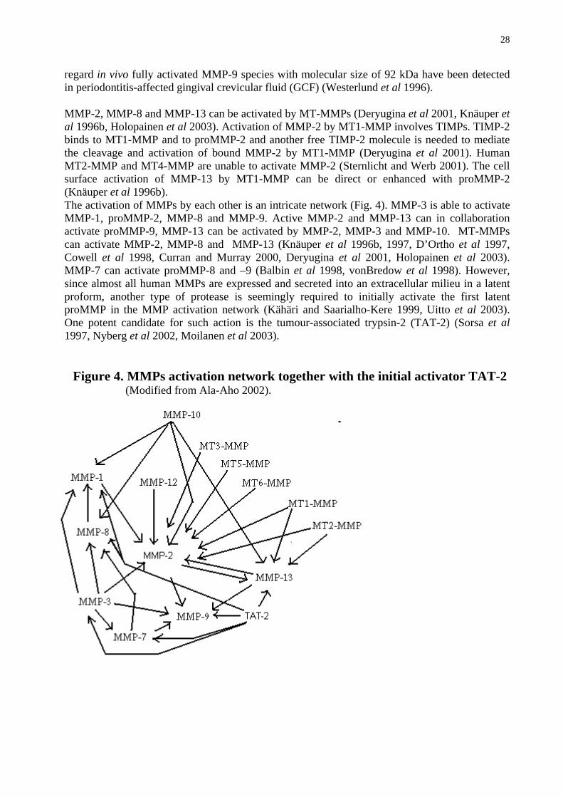
28
regard in vivo fully activated MMP-9 species with molecular size of 92 kDa have been detected in periodontitis-affected gingival crevicular fluid (GCF) (Westerlund et al 1996). MMP-2, MMP-8 and MMP-13 can be activated by MT-MMPs (Deryugina et al 2001, Knäuper et al 1996b, Holopainen et al 2003). Activation of MMP-2 by MT1-MMP involves TIMPs. TIMP-2 binds to MT1-MMP and to proMMP-2 and another free TIMP-2 molecule is needed to mediate the cleavage and activation of bound MMP-2 by MT1-MMP (Deryugina et al 2001). Human MT2-MMP and MT4-MMP are unable to activate MMP-2 (Sternlicht and Werb 2001). The cell surface activation of MMP-13 by MT1-MMP can be direct or enhanced with proMMP-2 (Knäuper et al 1996b). The activation of MMPs by each other is an intricate network (Fig. 4). MMP-3 is able to activate MMP-1, proMMP-2, MMP-8 and MMP-9. Active MMP-2 and MMP-13 can in collaboration activate proMMP-9, MMP-13 can be activated by MMP-2, MMP-3 and MMP-10. MT-MMPs can activate MMP-2, MMP-8 and MMP-13 (Knäuper et al 1996b, 1997, D’Ortho et al 1997, Cowell et al 1998, Curran and Murray 2000, Deryugina et al 2001, Holopainen et al 2003). MMP-7 can activate proMMP-8 and –9 (Balbin et al 1998, vonBredow et al 1998). However, since almost all human MMPs are expressed and secreted into an extracellular milieu in a latent proform, another type of protease is seemingly required to initially activate the first latent proMMP in the MMP activation network (Kähäri and Saarialho-Kere 1999, Uitto et al 2003). One potent candidate for such action is the tumour-associated trypsin-2 (TAT-2) (Sorsa et al 1997, Nyberg et al 2002, Moilanen et al 2003).
Figure 4. MMPs activation network together with the initial activator TAT-2 (Modified from Ala-Aho 2002).
29
2.7.3 MMP inhibition There are different mechanisms to inhibit or down-regulate MMPs. Inhibition may take place by interaction with an active Zn2+-site or by cleaving the active enzyme or binding it to a non-active complex form. To influence the MMP levels it is also possible to down-regulate MMPs at the transcriptional level (Birkedal-Hansen et al 1993). Tissue inhibitors of metalloproteinases (TIMPs) represent a multigene family with four members (Stetler-Stevenson et al 1989, Birkedal-Hansen et al 1993, Apte et al 1994, Leco et al 1994). They inhibit most MMP family members reversibly and the TIMPs are the major endogenous extracellular MMP regulators. Inhibition by TIMPs takes place via developing a MMP-TIMP complex or by inhibiting the activation of proMMPs (Goldberg et al 1989, Sorsa et al 1992, Birkedal-Hansen et al 1993). Different cells express TIMPs and they have been identified not only in tissues, but also in body fluids including gingival crevicular fluid (GCF) (Birkedal-Hansen et al 1993, Leco et al 1994, Sorsa et al 1994, Ingman et al 1996, Lin et al 2001). TIMP inhibition of MMPs is considered to exert a role in healing wounds, bone remodelling and tumour invasion (Edwards et al 1996, Vaalamo et al 1996, Sutinen et al 1998). Nonetheless, TIMP-3 is an exception, since it is mostly bound to ECM (Birkedal-Hansen et al 1993, Leco et al 1994). TIMP-2 has a special role in the regulation of MMP-2 activation by MT1-MMP on the cell membranes (Deryugina et al 2001). TIMPs also have growth-promoting properties (Hayakawa et al 1992, 1994). The delicate balance between TIMPs and MMPs evidently plays a important role in most normal tissue remodelling phenomena and in many tissue destructive inflammatory and malignant pathological conditions (Khokha et al 1989, Poulsom et al 1992, Ingman et al 1996, Stetler-Stevenson et al 1997, Sutinen et al 1998, Soini et al 2001). Serum 2-macroglobulin ( 2M) is a natural MMP inactivator and it entraps and bonds to the MMP after the enzyme has revealed the 2M-bait region. The trapping mechanism is irreversible but does not block the active site totally, so the enzyme is partly active for low molecular weight substrates. The 2M-proteinase combination is cleared from the circulation in the liver through uptake by special receptors (Birkedal-Hansen et al 1993). Chelating agents, such as EDTA and 1,10-phenenthroline, bind to Zn2+ at the active site inhibiting MMPs non-selectively but are regarded as therapeutically useless (Birkedal-Hansen et al 1993). Bisphosphonates are a group of MMP inhibiting and down-regulating pharmacologic compounds which seemingly also act as chelating agents (Teronen et al 1997, 1999, Heikkilä et al 2002, 2003, Vihinen and Kähäri 2002). Doxycycline and chemically modified non-antimicrobial tetracyclines (CMTs) inhibit MMPs by chelating and down-regulating their mRNA and protein expressions (Sorsa et al 1994, Golub et al 1995, Hanemaaijer et al 1997, Pirilä et al 2001ba). CMTs inhibit especially the pathologically elevated MMP-dependent periodontal soft tissue breakdown and bone destruction in vivo and in vitro (Golub et al 1995, 1998) and tumour cell proliferation and invasion (Lukkonen et al 2000, Gu et al 2001, Lokeshwar et al 2002). CMTs have also been tested in animals and shown to inhibit caries progression in tooth and tumour growth and metastasis as well as prevent inflammatory bone resorption (Golub et al 1998, Sulkala et al 2001, Bezerra et al 2002, Lokeshwar et al 2002). When combined with bisphosphonates, CMTs synergistically inhibit bacterial-produced periodontal soft and hard tissue breakdown (Llavaneras et al 1999, 2001). Peptidomimetic matrix metalloproteinase inhibitors (MMPI) mimic the structure of MMP substrates, as well as function as competitive inhibitors (Vihinen and Kähäri 2002). Batimastat is the first synthetic clinically tested MMP inhibitor interacting with Zn2+ in their catalytic site.

30
Animal model studies have shown the inhibition of tumour growth, metastasis, and of angiogenesis (Curran and Murray 2000). Marimastat is the second-generation synthetic wide spectrum MMP inhibitor that has been clinically evaluated as an anticancer agent (Curran and Murray 2000, Vihinen and Kähäri 2002). CTTHWGFTLC-peptide is a selective gelatinase (MMP-2 and MMP-9) inhibitor (Koivunen et al 1999, Pirilä et al 2001b, 2003)

31
Table 2. Matrix metalloproteinases (Modified from Sternlicht and Werb 2001)
MMP Enzyme Substrates_______________________________________ Collagenases MMP-1 collagenase-1 aggrecan, collagens I-III,VII,VIII,X,XI, entactin, FN,gelatin, IGFBPs,
Ln-1, link protein, myelinbasic, tenascin, 1AC, 2M, 1PI, VN, casein, fibrin, fibrinogen, IL1 , IL1 , proTNF
MMP-8 collagenase-2 aggrecan, collagen I-III, laminin-5, fibrinogen, substanceP, 1PI, 2M, MMP-13 collagenase-3 aggrecan, collagens I-IV,VI,IX,X,XIV, fibrillin, FN, gelatin,
Ln-1, -5, osteonectin, casein, factor XII, 2M, fibrinogen Gelatinases MMP-2 gelatinase-A aggrecan, collagens I,III-V,VII,X,XI, decorin, elastin, entactin,
fibrillin, FN, fibulins, gelatin, IGFBPs, Ln-1, -5, link protein, myelin basic,osteonectin, tenascin, VN, fibrin, 1AC, 1PI, fibrinogen, IL1 , proTGF , proTNF , plasminogen, substanceP
MMP-9 gelatinase-B aggrecan, collagens IV,V,XI,XIV, decorin, elastin, fibrillin, gelatin, Ln-1, link protein, myelin basic, osteonectin, tenascin, VN, 2M, 1PI, casein, fibrin, fibrinogen, IL1 , proTGF , proTNF , plasminogen, substanceP
Stromelysins MMP-3 stromelysin-1 aggrecan, collagens III-V,VII,IX,X,XI, decorin, elastin, entactin,
fibrillin, FN, gelatin, IGFBPs, Ln-1, link protein, myelin basic, osteonectin, tenascin, VN, 1AC, 2M, 1PI, casein, fibrin, fibrinogen, IL1 , proTNF- , plasminogen, substanceP, E-cadherin
MMP-10 stromelysin-2 aggrecan, collagens III-V, elastin, FN, gelatin, link protein, casein, fibrinogen
Stromelysin like MMPs MMP-11 stromelysin-3 IGFBPs, 2M, 1PI MMP-12 metalloelastase aggrecan, collagens I,IV, elastin, entactin, fibrillin, FN,gelatin, Ln-1,
myelin basic, vitronectin, 2M, 1PI, factor XII, fibrinogen, proTNF-, plasminogen
Matrilysins MMP-7 matrilysin aggrecan, collagens I, IV, decorin, elastin, entactin, FN, fibulins,
gelatin, Ln-1, link protein, myelin basic,osteonectin, tenascin, VN, 1PI, casein, E-cadherin, fibrinogen, proTNF , plasminogen
MMP-26 endometase, matrilysin-2 collagen IV, FN, gelatin, 1PI, fibrinogen Transmembrane type MMPs MMP-14 MT1-MMP aggrecan, collagens I-III, entactin, fibrillin, FN, gelatin, Ln-1,-5, VN,
2M, 1PI, factor XII, fibrin, fibrinogen, proMMP2, proTNF- MMP-15 MT2-MMP aggrecan, entactin, FN, Ln-1, tenascin, proMMP2
MMP-16 MT3-MMP collagen III, FN, proMMP2 MMP-24 MT5-MMP GPI-type MMPs MMP-17 MT4-MMP fibrillin, FN, gelatin, proTNF MMP-25 MT6-MMP collagen IV, FN, gelatin, fibrin Other MMPs MMP-18 collagenase-4 (Xenopus laevis) collagen I MMP-19 RASI-1 collagens I, IV, FN, gelatin, tenascin, casein MMP-20 enamelysin amelogenin, aggrecan, Ln-5 MMP-23 CA-MMP gelatin MMP-28 epilysin casein MMP-21 XMMP MMP-22 CMMP MMP-27

32
3. Aims and outlines of the study
3.1 Aims of the study
The aims of this study were to gain a better understanding of the role of MMPs in the progression of inflammatory pulpal and periapical diseases, as well as, in the tissue destruction and enlargement of odontogenic cysts and PLC. In this study, I have concentrated on the in vivo presence of collagenases and gelatinases in pulp, periapical tissue, odontogenic cysts and PLC tissue, as well as on their regulation in cell cultures in vitro. I have also studied the changes in the collagenase levels during RTC in order to better understand the role of these enzymes in the healing process of periapical inflammatory changes.
3.2 Outlines of the study 1. To determine the in vitro expression and regulation of collagenase-2 (MMP-8) in pulp cells
and mature odontoblasts.
2. To study the presence, levels and molecular forms of MMP-8 in pulpal and periapical inflammation, and the changes in the concentrations, activation and molecular forms of MMP-8 in root-canal exudates during root-canal treatment.
3. To clarify the differences in the in vivo expression of Ln-5, collagenases (MMP-8 and MMP- 13) and gelatinase (MMP-2) in different odontogenic jaw cysts and their possible role in the enlargement and tissue destruction caused by these cysts.
4. To study the expression of collagenases (MMP-8 and MMP-13) in plasma cells and in plasma cell diseases, such as MM and PLC, both in vivo and in vitro and their regulation by cytokines, such as IL-6 and TNF- .

33
4. Materials and methods
4.1 Patients and tissue samples
4.1.1 Pulp tissue and odontoblasts (I)
All teeth (n=166) used for the experiments were freshly extracted caries-free third molars removed as part of the normal treatment at the Institute of Dentistry, University of Oulu, Oulu, Finland. After sterilizing the tooth and removing the soft tissue and cementum, the root was dissected gently close to the cemento-enamel junction. For culturing, the pulp was removed to a culture medium and the crowns with odontoblasts were placed in a sterile culture medium. For the native mRNA samples, the pulps were gently removed with forceps and placed into Eppendorf tubes. The odontoblasts lining the pulp chamber walls were immediately scraped off with a sterile excavator and stored at -70 C until analysed. For immunohistochemistry, the enamel was cut off to prevent disturbances in sectioning the frozen samples. Immediately the enamel was removed with a thin diamond disc under water cooling, the teeth were immersed in liquid nitrogen and embedded, and serially cut into longitudinal sections on a piece of adhesive tape. The sections were air-dried and fixed in methanol, and air-dried again. Then the sections were demineralized and treated with bovine testicular hyaluronidase to overcome the masking effect of the matrix proteoglycans and then the procedure of immuhistochemical staining was carried out using Vectastain Elite Kit as explained later (Vector laboratories, Burlingame, CA, USA) (Table 3). 4.1.2 Patients and diagnosis (II)
10 patients suffering from CAP were selected for the study. The diagnosis was made by clinical, and mainly, by radiographic examination, showing the disappearance of the lamina dura and clear bone loss in the apical area. All the teeth included into the study were unvital and had only one root canal. No controls were used and the one patient excluded from the study had a root fracture. Root canal treatment was performed under sterile conditions in three visits (during two-week intervals) and the samples from the root canal exudates were collected from the root canals with sterile paper points during these visits. The pulpitis tissue specimens for immunohistochemistry were collected from patients with irreversible pulpitis when starting normal root treatment procedure under sterile conditions. Pulp tissue was gently removed with an excavator to an Eppendorf tube containing formalin. Specimens from the patients were collected at a private dental clinic with the approval of the Ethical Committee of University of Helsinki, Faculty of Medicine, and with permission of the patients.

34
Table 3. Tissue specimens Material Number of fixation publications samples Teeth 166 96%methanol I 3rd molar (received fresh) Pulpitis 10 10 % buffered II formalin Periapical 10 10 % buffered II, IV periodontitis formalin Radicular cysts 11 (10) 10% buffered III (IV) formalin Follicular cysts 11 (10) 10 % buffered III (IV) formalin Odontogenic 16 (7) 10 % buffered III (IV) keratocysts formalin Plasmacytoma 14 10%buffered IV formalin
4.1.3 Tissue specimens (II, III and IV)
Formalin-fixed and paraffin-embedded samples of chronic apical periodontitis, jaw cysts (RCs, FCs and KCs) and PLC, diagnosed histologically according to WHO criteria (Kramer et al 1992, Jaffe et al 2001), were used for immunohistochemistry and in situ hybridization (Table 3). These tissue specimens were randomly selected from the files of the Department of Oral Pathology, Institute of Dentistry, University of Helsinki, Helsinki, Finland, and from the files of the Department of Pathology, University of Oulu, Oulu, Finland.
4.2 Cells and culture methods
4.2.1. Odontoblasts and pulpal cells (I) 4.2.1.1 Odontoblast cultures (I) The odontoblast culture methods have been described in detail by Tjäderhane et al 1998. For odontoblast organ cultures 1.5% (w/v) agarose gel (Agarose NA, Pharmacia Biotech AB, Uppsala, Sweden) was mixed into the culture medium (OPTI-MEM I Reduced serum medium with antimicrobial element) and was used as embedding material for odontoblast cultures. The crowns with odontoblasts lining the pulpal chambers were partially embedded in the agarose gel. The pulp chambers were gently washed with culture medium to remove the pulp tissue fluid, and

35
finally filled with the culture medium. The crowns were covered with glass cover slips to prevent evaporation from the medium. The medium was also placed on top of the surrounding gel to prevent dehydration. To study the effects of TGF- 1 on the MMP-8 mRNA expression, odontoblasts and pulp tissues were cultured for two days with or without human recombinant TGF- 1. The medium was replaced and collected daily. After the culturing procedure the cells were collected and the total RNA was isolated as described below. 4.2.1.2 Pulp fibroblast (PF) cultures (I) The pulp samples for the human pulp fibroblast (PF) isolation were collected from the teeth that were removed as part of the normal treatment of patients. After dissecting the crown with pliers (for odontonblast culture), the pulp was gently removed with forceps and the tissue was cut into pieces and placed into cell culture clusters containing Dulbecco’s modified Eagle’s medium (DMEM) supplemented with foetal bovine serum (FBS) added with an antimicrobial component. The explants were then incubated at 37 C, collecting and replacing the medium twice a week. The cells were examined daily, and when the fibroblast outgrowth was observed the pulp tissue pieces were removed. After reaching the confluence, the fibroblast cell line was established by subsequent trypsinization and culturing through several passages. The cells were always allowed to reach the confluence before being subcultured. To analyse the MMP-8 mRNA production, the PF cells in passage two (n=2) were washed with PBS and the total RNA was isolated as described later. 4.2.2 Plasma cells (IV) Human myeloma cell line RPMI 8226 (ATCC Number: CCL-155; Rockville, MD, USA) was cultured in RPMI 1640 medium (Gibco, Basel, Switzerland), supplemented with newborn calf serum, lactate glutamate and penicillin-streptomycin. Every third day, the cells were washed in phosphate-buffered saline (PBS) and the medium was renewed. After one week the cultured cells were separated to receptacles, 9 x 10¹⁰ cells in each, and cultured in serum-free culture medium for 24 hours. Differently combined cytokines, IL-6, heparin, TNF- , and PMA, were added to the receptacles and after 24 hours the culture medium was centrifuged to separate the cells from the medium. About 10 000 cells were counted for the Western blot analysis described later.
4.3 Sample collection from root canals (II)
The RTC was done under sterile conditions using rubber dam isolation. Primary access was done with low speed dental burs. The instrumentation of the root canal was started with 20 or 25 files and radiograph was taken to measure the working length. A sterile paper point was inserted into the canal for 2 minutes to absorb exudates from the canal. The root canals were widened to the desired width (minimum 40) with rinsing in between files. For the final rinsing with EDTA and NaOCl, the canal was dried before inserting Ca(OH)2 paste for two weeks. After two weeks the access was re-opened and the canal was gently instrumented and rinsed with sterile saline solution before drying. A sterile paper point was again inserted into the canal for 2 minutes for periapical exudate collection. After two weeks, the same procedure was repeated and afterwards the root canal was filled with gutta-percha points and a sealer. After sample collections, the exudate from the absorbent points was diluted into a buffer and stored at -70 ˚C for further handling.
36
4.4 PCR analysis
4.4.1 RNA isolation (I and IV) The total RNA was isolated from odontoblasts (native and cultured), pulp tissue cultures and pulp fibroblasts cultures, as well as from cultured RPMI 8226 myeloma cells using the Trizol® (Gibco BRL, Roskilde, Denmark) reagent isolation method. The RNA yield was measured by absorbance of 260 nm, and the samples were stored at -70˚C until needed. RNA isolated from bone marrow cells (Hanemaaijer et al 1997) served as positive controls. 4.4.2 RT-PCR (I, III and IV)
For cDNA synthesis, a 20 l reverse transcription reaction was carried out on 2 g (plasma cells) and 1.7-11 g (odontoblast and pulp cells) of the total RNA and random hexamer primers with SuperscriptTM II RNaseH-Reverse Transcriptase (Gibco BRL, Roskilde, Denmark) according to the product protocol. Specific primers for MMP-8 (not recognizing MMP-1 or other MMPs) were those designed as described by Hasty et al (1987). MMP-8 sense primer 5´-AAGGCAACCAATACTGGG, and MMP-8 antisense primer 5´-ATTTTCACGGAGGACAGG, with the expected product size of 522 base pair (bp), and MMP-8 nested sense primer was 5'-AGCAGCGTCCAAGCAATTG-3' and MMP-8 nested antisense primer 5'-AATGCATGCTGAACTTCCCTTC-3', with the expected product size of 352 bp. Specific primers for MMP-13 were designed based on the published DNA sequence (Freije et al 1994), MMP-13 sense primer 5'-AGATAAGTGCAGCTGTTCAC and MMP-13 antisense primer 5'-TCATTGACAGACCATGTGTC, with the expected product size of 572 bp. MMP-13 nested sense primer was 5'-AGCATCTGGAGTAACCGT-3' and MMP-13 nested antisense primer 5'-TCAATGTGGTTCCAGCCA-3', with the expected product size of 238 bp. 18S ribosomal RNA was used as a control for the RNA integrity using primers designed based on the published sequence (McCallum et al 1985). PCR amplification reactions were performed with primers and DynazymeTM DNA polymerase (Finnzymes Oy, Espoo, Finland) according to the product protocol and the linear area of the PCR reaction for each reaction was defined. The original amplification was performed as described by Hanemaaijer et al (1997). MMP-8 PCR amplification was done at 30-55 cycles and the nested MMP-8 PCR amplification was performed using 14-18 cycles, and MMP-13 PCR had 35 or 45 cycles and the nested 21-24 cycles. 18S PCR was done using 13 or 14 cycles. After finding the linear portion of each set of primers for the quantitation of the products, they were quantitated with ScionImage program. Values for MMP-8 and -13 cytokine inductions were calculated from the scanned densitometric values and related to the corresponding 18S values.
4.5 Southern blotting (I)
PCR products were fractionated on 1.5 % agarose gels, transferred to a nylon filter (Amersham, Buckinghamshire, UK) and hybridized as described elsewhere (Ausubel et al 1988). The specific cDNA probe for MMP-8 was prepared. The Southern filter was hybridized to a labelled MMP-8 cDNA probe in ExpressHyb hybridization buffer (Clontech, Palo Alto, CA, USA) for 1 hr at 60˚C. The filters were washed in saline-sodium citrate (SSC)/ sodium dodecyl sulphate (SDS) at room temperature and exposed to x-ray film (Kodax X-OMAT).

37
4.6 Western immunoblotting and MMP-8 immunofluorometric assay (IFMA)
4.6.1 Western immunoblot analysis (I, II, and IV)
The molecular forms of MMP-8 and -13 were analysed by the Western blotting method utilizing the specific MMP-8 antibody (Hanemaaijer et al 1997) and specific polyclonal MMP-13 antibody (Freije et al 1994). The dissolved plasma cells, supernatant from the plasma cell culture, pure RPMI L-glutamine media and the root canal exudate samples were parallel treated with Laemmli´s buffer without reducing reagents. To investigate the secretion of MMP-8 into the odontoblast and pulp cell culture medium and the effect of TGF- 1 on the amount of secreted protein, a conditioned serum-free OPTI-MEM medium, with and without TGF- , was used for the Western blot analysis. To investigate the secretion of MMP-8 into the odontoblast culture medium and the effect of TGF- 1 on the amount of secreted protein, a conditioned serum-free OPTI-MEM medium, with and without TGF- , was used for the Western blot analysis. The samples were run on polyacrylamide gels by electrophoresis and electrotransferred onto a nitrocellulose membrane (Schleicher & Schuell, Dassel, Germany and Bio-Rad Laboratories, Richmond, CA, USA) (Hanemaaijer et al 1997). After blocking the non-specific binding sites with gelatin or non-fat dry milk (odontoblast and pulp cell samples), the membrane first reacted with the primary antibody and then with the alkaline phosphatase-conjugated secondary antibody (IV) or peroxidase-conjugated swine anti-rabbit immunoglobulins (I, II). The immunoreactive proteins were visualized by Nitro Blue Tetrazolium and 5-bromo-4-chloro-3-indolyl-phosphate or with diaminobentzidine tetrahydrochloride (DAB) and quantitated by the Imaging Densitometer (Prikk et al 2002). 4.6.2 MMP-8 immunofluorometric assay (IFMA) (I, II)
The concentration of the monoclonal MMP-8 secreted into the odontoblast and pulp tissue culture media, as well as the root canal exudate, was determined by a time-resolved immunofluorescence assay as described previously (Hanemaaijer et al 1997, Sorsa et al 1999). Conditioned culture medium samples, both with and without TGF- 1, were collected from the odontoblast organ and pulp tissue cultures. The monoclonal MMP-8 specific 8708 and 8706 antibodies were used as a catching antibody and the tracer antibody, the tracer antibody being labeled using europium chelate. Samples were diluted and incubated in assay buffer followed by incubation with the tracer antibody. An enhancement solution was added, and fluorescence was measured using a 1234 Delfia Research Fluorometer (Wallac, Turku, Finland). The monoclonal MMP-8 antibody specificity corresponded to that of the polyclonal MMP-8 antibody (Hanemaaijer et al 1997).
4.7 Preparation of RNA probes (III, IV)
For the preparation of RNA probes, an 845 bp PstI-EcoRI fragment of clone L15 of the Ln-5 2 chain was subcloned into the pSP64 and pSP65 vectors in the sense and antisense orientations (Kallunki et al 1992). A 635 bp Sca I-Sac I fragment of human MMP-2 cDNA clone was subcloned into the M13 polylinker site of the pSP64 and pSP65 vectors (Huhtala et al 1990). A 734 bp BamHI-HindIII fragment of human MMP-13 cDNA (Freije et al 1994) was ligated into the pGEM-4Z vector, and a 95 bp HindIII –BamHI fragment of human MMP-8 cDNA (Hasty et al 1990) was ligated into the pGEM-3Z vector. The probes were tested by Northern hybridization for specificity and by sequencing. The vectors were linearized with a suitable restriction enzyme and a riboprobe transcription kit (Boehringen Mannheim, Germany) was used for transcription.
38
The transcripts were labelled with digoxigenin (DIG)-11-UTP for in situ hybridization, and a corresponding sense probe was used as a control for nonspecific hybridization.
4.8 In situ hybridization (III and IV)
For in situ hybridization, all the solutions were treated with 0.1% diethylpyrocarbonate (DEPC). Paraffin sections from different odontogenic jaw cysts were processed prior to prehybridization, and in situ hybridization with DIG-labelled RNA-probes was done as described by Mäkelä et al (1999). The prehybridization buffer was removed and hybridization buffer containing 800 ng/ml DIG-labelled antisense or sense probe was applied to each section and hybridized overnight at 57 C. The posthybridization washes were done under stringent conditions as described by Mäkelä et al (1999). After washing, the sections were blocked and incubated with 1:100 diluted alkaline phosphatase-conjugated anti-DIG Fab-fragments and stained with alkaline-fosphatase substrate Fast Red (Boehringen Mannheim, Germany) and counterstained with Mayer’s haematoxylin (Merck KgaA, Darmstadt, Germany).
4.9 Immunohistochemical staining and evaluation of the staining
4.9.1 Immunostaining (I, II, III and IV) Immunohistochemistry was performed with different antibodies (Table 4) using VECTASTAIN® Elite ABC Kit (Vector Laboratories, Burlingame, CA, USA) according to the protocol. The fresh teeth preparates were fixed in methanol and demineralized in EDTA. Before immunohistochemical staining, they were pre-treated with bovine testicular hyaluronidase (Sigma, St. Louis, MO, USA) and methanol. Formalin-fixed paraffin sections were deparaffinized, hydrated, pre-treated with pepsin and with H2O2 in methanol. Thereafter, all the sections were incubated with normal goat / horse serum, secondly with primary antibody, and then with biotinylated anti-rabbit / anti-mouse immunoglobulin G and with avidin-biotin complex. The sections were stained with 3-amino-9-ethylcarbazole (AEC) with the DAB Kit (Vector Laboratories, Burlingame, CA, USA) and counterstained with the Mayer’s hematoxylin. 4.9.2 Evaluation of immunostaining (III)
The staining positivity and intensity of different MMPs in the basement membrane zone in the different types of odontogenic cysts was analysed using a modification of the method described by Bachmeier et al (2000). Briefly, ten randomly selected areas of two different microscopic slides of each cyst type were inspected with a light microscope using 200 x magnification. The length of the basement membrane zone observed in each area was 0.65 mm. A two step score was used for the positivity of staining (0: no positive staining, 1: positive staining). When staining was observed, the intensity was graded using a three-step scale (1: faint, 2: moderate, 3: strong positive staining). The results of the 20 independently inspected areas were summarized.
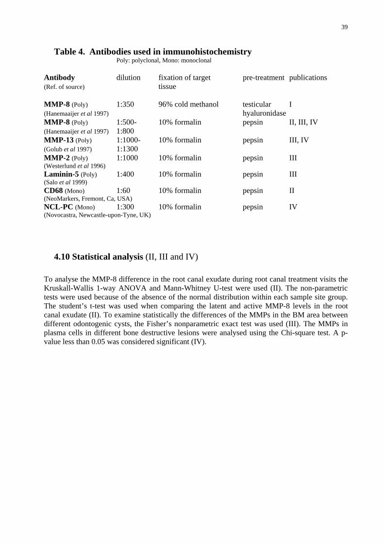
39
Table 4. Antibodies used in immunohistochemistry Poly: polyclonal, Mono: monoclonal Antibody dilution fixation of target pre-treatment publications (Ref. of source) tissue MMP-8 (Poly) 1:350 96% cold methanol testicular I (Hanemaaijer et al 1997) hyaluronidase MMP-8 (Poly) 1:500- 10% formalin pepsin II, III, IV (Hanemaaijer et al 1997) 1:800 MMP-13 (Poly) 1:1000- 10% formalin pepsin III, IV (Golub et al 1997) 1:1300 MMP-2 (Poly) 1:1000 10% formalin pepsin III (Westerlund et al 1996) Laminin-5 (Poly) 1:400 10% formalin pepsin III (Salo et al 1999) CD68 (Mono) 1:60 10% formalin pepsin II (NeoMarkers, Fremont, Ca, USA) NCL-PC (Mono) 1:300 10% formalin pepsin IV (Novocastra, Newcastle-upon-Tyne, UK)
4.10 Statistical analysis (II, III and IV)
To analyse the MMP-8 difference in the root canal exudate during root canal treatment visits the Kruskall-Wallis 1-way ANOVA and Mann-Whitney U-test were used (II). The non-parametric tests were used because of the absence of the normal distribution within each sample site group. The student’s t-test was used when comparing the latent and active MMP-8 levels in the root canal exudate (II). To examine statistically the differences of the MMPs in the BM area between different odontogenic cysts, the Fisher’s nonparametric exact test was used (III). The MMPs in plasma cells in different bone destructive lesions were analysed using the Chi-square test. A p-value less than 0.05 was considered significant (IV).

40
5. Results 5.1 Pulpal and periapical tissue
5.1.1 MMP-8 in odontoblasts and pulp tissue 5.1.1.1 Healthy pulp tissue (I) Expression of MMP-8 mRNA In healthy pulps from extracted third molars, the native and cultured odontoblasts and pulp tissue, as well as the cultured pulp fibroblasts, expressed MMP-8 mRNA by RT-PCR. The transcripts were identical to those from the positive control (bone marrow cells). The specificity of the PCR amplification was confirmed by a Southern blot analysis using a specific MMP-8 cDNA probe. TGF- 1 added to the cell culture down-regulated 10% and 25% the MMP-8 mRNA production in odontoblast and pulp tissue cultures. Expression of MMP-8 protein A Western blot analysis with MMP-8 antibody detected a 65 kDa immunoreactive band in all the native odontoblast, pulp and pulpal fluid samples from sound teeth. A fainter 50 kDa band was also detected in all the pulp and pulpal fluid samples, as well as from the conditioned odontoblast culture medium. A high molecular weight band (>100-112 kDa) of MMP-8 was detected in all the native samples. No characteristic PMN MMP-8 (75-80 kDa) isoform was observed. TGF- 1 had no observable effect on the MMP-8 level in the samples studied by the Western blot analysis. However, in the PCR analysis of secreted MMP-8 from the culture medium of odontoblasts and pulp tissue revealed a detectable down-regulation in the MMP-8 levels when TGF- 1 was added. The medium was collected after 24 and 48 hours of culturing. The MMP-8 levels in the odontoblast and pulp tissue medium were lower in both (24 and 48 h) samples cultured with TGF- 1. Immunohistochemical staining with the same MMP-8 specific antibody revealed MMP-8 positivity in the odontoblast cell layer, localised to the odontoblast body. MMP-8 positivity was also detected in some, but not all, endothelial cells lining the pulp vessels. 5.1.1.2 Pulp with inflammation (II) Expression of MMP-8 protein The Western blot analysis of the pulpitis tissue revealed intensely stained bands of 55-45 kDa, 75-80 kDa and 100-112 kDa. The 75-80 kDa band most likely represents the MMP-8 isoform from the PMNs. By immunohistochemistry, in addition to PMN cells, MMP-8 immunoreactivity was detected in the macrophage-like cells, plasma cells and some endothelial cells. The immunohistochemical analysis revealed that MMP-8 immunoreactivity accumulated around the pulp abscess. 5.1.2 MMP-8 in the root canal exudate (II) RTC clearly decreased the MMP-8 levels in the root canal exudate. However, in order to successfully minimize the MMP-8 concentration in the root canal exudate, the local calcium hydroxide paste medication was required for four weeks. After careful mechanical and chemical root canal preparation with local adjunctive medication for a fortnight, some MMP-8 could still be detected in some cases. There was a statistically significant difference in the MMP-8 levels between the first and last root canal therapy visits (Kruskall-Wallis p=0.02; Mann-Whitney U-test p=0.0107).

41
The high (65-75 kDa) and low (45-55 kDa) molecular size MMP-8 isoforms detected in the root canal exudates were in both latent and active forms. If there was MMP-8 detectable in the root canal exudate during the second visit, the relative amount of the active form was elevated, but not significantly. The only case in which the MMP-8 level in the root canal exudate did not decrease during the root canal treatment, was later diagnosed to have a vertical fracture and the tooth needed to be extracted.
5.1.3 MMPs in apical periodontitis (II) In CAP, MMP-8 was immunohistochemically detected mostly in the PMNs. In addition to the PMNs, MMP-8 protein was present in the subpopulation of macrophages and plasma cells. The histiocytic nature of the macrophages was confirmed by staining with the CD68 macrophage cell marker. NCL-PC positive plasma cells expressed MMP-8 in 2 out of 10 cases. In addition to the plasma cells MMP-13 protein expression was present in macrophages. Half of the samples studied expressed MMP-13 protein in the plasma cells and the MMP-13 mRNA expression was evidenced by in situ hybridization.
5.2 Odontogenic cysts
5.2.1 Laminin-5 (III) The 2 chain of Ln-5 was detectable by immunohistochemistry in the BM zone of all the RC, FC and KC samples studied. In the KC samples, the -2 chain stained the basement membrane zone as a continuous ribbon. In 30% of the cases, a few epithelial cells stained positively. In the FCs, the Ln-5 2 chain staining showed local breaks in the basement membrane zone in 64% of the cases. In the epithelial cells, 2 chain positivity was present in 81% of the cases. In the RCs, the BM zone staining with the Ln-5 2 chain antibody showed local breaks in 18% of the cases and the epithelial cells showed 2 chain positivity in 91 % of the cases. Expression of Ln-5 2 chain mRNA was observed in all cyst types in basal epithelial cells. 5.2.2 Expression of MMP-2 (III) MMP-2 immunoreactivity was detected in the cyst capsule fibroblasts of all the cyst types studied (RC, FC and KC), and mRNA was also detected in some fibroblast-like cells of all the cysts. Only the KCs showed MMP-2 immunoreactivity in all the samples studied. MMP-2 protein expression was detected in the epithelium in all the cyst types studied, but clear expression of MMP-2 in basal cells and the BM zone was only in the detached epithelium of the KCs. There was a statistical difference in the location of the MMP-2 expression in the BM area between the KC and RC (p=0.023). The difference was clear but not statistically significant between the KC and FC (p=0.0698). The quantity and intensity of the MMP-2 staining in the BM area was clearly higher in the KCs than the other cyst types studied.
5.2.3 Expression of MMP-8 (III, IV) MMP-8 immunoreactivity was detected in all the cyst types studied, mainly in the connective tissue cyst capsule. PMNs, plasma cells and FC foam cells (phagocytic macrophages) expressed MMP-8. In the epithelium, MMP-8 protein was observed in the FC goblet cells. Plasma cells in the FCs (6/10) stained more often positively with MMP-8 antibody than those in the RCs (2/10)
42
and KCs (1/7). MMP-8 mRNA was detectable in plasma cells and foam cells, as well as in epithelial goblet cells and some epithelial cells of the FCs. In the FCs both protein and mRNA of MMP-8 could be observed in the same plasma cell accumulation. Interestingly, MMP-8 expression appears to be seen in the FCs more often than in the other (RC and KC) cyst types.
5.2.4 Expression of MMP-9 (unpublished results) MMP-9 was expressed by PMNs in cyst capsules and infiltrating in the epithelium, and cyst capsule plasma cells in KCs and RCs. MMP-9 mRNA was detected in osteoclasts in the bony front of cyst capsule. MMP-9 expression was not detected in the jaw cyst lining epithelial cells. 5.2.4 Expression of MMP-13 (III, IV) MMP-13 protein was observed in all the KCs. MMP-13 positivity could be observed in the plasma cells of theKC cyst capsule close to the lining epithelium and in the epithelial cells, as well as in small satellite cyst epithelial cells in the cyst capsule. In 55% of the KCs, MMP-13 expression was detectable in the BM zone. In the RCs, the number of positive cases was about half that observed in the KCs. MMP-13 mRNA was present in the plasma cells and epithelial cells of the KCs and in some cyst capsule fibroblasts as well. Statistical analysis showed a significant elevation in MMP-13 expression in KC plasma cells compared to the RCs and FCs. When comparing the epithelial expression, a significant difference was detected between the KCs and FCs (p=0.0294), but not between the KCs and RCs (p=0.6372). The quantity and intensity of MMP-13 immunostaining was clearly higher in the KCs than in the FCs and RCs.
5.3 Multiple myeloma and plasmacytoma
5.3.1 Plasmacytoma tissue samples (IV) 5.3.1.1 MMP-8 In the PLC tissue samples, MMP-8 expression was only slightly observable by immunohistochemistry, but in situ hybridization was negative. 5.3.1.2 MMP-13 Clear MMP-13 immunoreactivity was detected in 80% of the PLCs (no expression was detected in one tonsil, as well as the bone marrow and tongue PLC samples). Statistical analysis revealed significantly enhanced MMP-13 expression in the PLC samples in comparison to the RC and FC samples. No significant difference was detected when compared with the KCs. Of the three samples studied by in situ hybridization, MMP-13 mRNA expression was revealed in malignant PLC plasma cells in bone marrow and the upper jaw mucous membrane, but not in the vertebral lesions. 5.3.2 Multiple myeloma cell cultures (IV) We studied the MMP production in vitro in the human myeloma cell line RPMI 8226. The cells were treated with PMA, cytokines (IL-6 and TNF- ) or heparin, alone or in different combinations. The results were analysed by RT-PCR and Western blotting.

43
5.3.2.1 MMP-8 expression MMP-8 mRNA and protein was observed in cultured RPMI plasma cells. By Western blotting clear immunoreactive bands were detected in the cell lysates, but not in the culture media. MMP-8 immunoreactivity was clearly enhanced by IL-6 alone and combined with TNF- . A combination of heparin with cytokines and PMA further induced MMP-8 immunoreactivity. MMP-8 mRNA expression was also increased by PMA, TNF- and IL-6 in cultured RPMI 8226 cells. In 24 hours the MMP-8 mRNA was upregulated by 9.1 (PMA), 4.7 (TNF- ) and 7.2-fold (IL-6). The MMP-8 induction was more clearly presented in the RT-PCR than in the Western blot, probably due to a delay in protein expression. 5.3.2.2 MMP-13 expression The RT-PCR and Western blot showed MMP-13 expression in the RPMI 8226 malignant plasma cell line. Detected by the RT-PCR PMA, TNF- and IL-6 upregulated the MMP-13 mRNA expression by 6.1-, 8.7- and 6.8-fold, and the induction was enhanced when the cytokines were combined with heparin. MMP-13 protein was detected in the plasma cell lysates but not in the culture media. IL-6 alone and combined with TNF- increased the MMP-13 immunoreactivity. Heparin combined with cytokines and PMA also increased the MMP-13 expression slightly. The RT-PCR presented increased MMP-13 expression more clearly than the Western blot.

44
6. Discussion
6.1 The role of MMPs in pulpal and periapical tissue and root canal exudate
6.1.1 MMPs in pulp tissue (I, II) Pulpal cells can express various MMPs, including MMP-1, MT1-MMP, MMP-20 (Llano et al 1997, Lin et al 2001, Palosaari et al 2002), and MMP-2 (Tjäderhane et al 1998, Chang et al 2001) in vitro, and MMP-20 in vivo (Sulkala et al 2002). Bacteria and their virulence factors (toxins) upregulate MMP-2 expression and secretion by pulp cells (Chang et al 2002). Most of the studies done have utilized pulp tissue cells and fibroblasts, but odontoblasts have lacked a good in vitro study model. The new odontoblast culturing method described by Tjäderhane et al (1998) has provided a new way to reveal molecular events and processes associated with the healthy and diseased pulp and its cellular components. As recently shown, odontoblasts can express different MMPs: MMP-2, -9, -20 and MT1-MMP (Llano et al 1997, Tjäderhane et al 1998, 2001, Palosaari et al 2002), and, according to the present results, MMP-8 as well (I). TGF-
can upregulate MMP-9 expression, and down-regulate MMP-2 synthesis (Tjäderhane et al 1998), but has no effect on collagen synthesis in odontoblasts (Palosaari et al 2001). The present study further revealed that TGF- has a reducing effect on MMP-8 synthesis in mature odontoblasts (I). TGF- is a multifunctional growth factor found in dentin, and it is thought to regulate tissue repair in the tooth (Lesot et al 1994). The downregulation of MMP-8 may be a factor leading to a reparative dentin formation because it is regarded as essential for modulating tissues during normal dentin formation. It is evident that MMPs expressed by odontoblasts may have a role in dentin and reparative dentin formation and the growth factors can act as MMP regulators, as well as regulate collagen synthesis. A strong expression of MMP-8 in the inflammatory cells of pulpitis tissue was seen. Studies have shown that bacteria and their products upregulate MMP-1 and MMP-2 in pulp cells, but have no effect on MMP-9 (Nakata et al 2000, Chang et al 2002). The levels of MMP-1, -2, and –3, expressed mainly by monocyte/macrophages and fibroblasts, are significantly higher in acute pulpitis tissue than in healthy pulp tissue (Shin et al 2002). Bacterial products irritate PMNs to release MMP-8 (Ding et al 1996, 1997), and proinflammatory cytokines upregulate MMP-1 and MMP-2 in the pulpal tissue (Tamura et al 1996, O’Boskey et al 1998, Chang et al 2001). The bacteria and their products may act in pulpal inflammation by upregulating cytokine production and via the cytokine pathway increase MMP expression and directly irritate cells to produce MMPs. The MMP-8 in pulpal inflammatory lesions was mainly of PMN origin. PMNs are cells forming pulp abscesses and, thus, activated MMP-8 may well participate in the tissue destruction of pulp necrosis and abscesses. PMNs are migrating and recruiting cells able to penetrate dentinal tubules (Bergenholtz 2000), and they may well need MMP-8 for this. Western blot bands 55-45 and 100-112 kDa probably represent the MMP-8 isoform originating from the non-PMN lineage cells (Hanemaaijer et al 1997). MMP-8 species higher than 90 kDa possibly represented high molecular size MMP-8 complexes formed after binding to inhibitors (TIMPs, 2M) and/or dimerization (Prikk et al 2001, 2002).
6.1.2 MMPs in periapical tissue and root canal exudate (II, IV) Untreated tooth necrosis usually leads to CAP, a destructive inflammatory condition around the tooth apex. It is a complicated process including bacteria and their products, immunoglobulins, proinflammatory cytokines and other inflammatory mediators affecting each other in a complex

45
manner. PMNs, being the first cellular barrier to bacterial invasion, may, in addition to digesting bacteria, destroy surrounding tissue compartments by secreting MMPs, especially MMP-8. PMN cells, induced by bacterial products or toxins, can release proinflammatory cytokines (Barkhordar et al 1992) which autocrinically stimulate PMNs to release MMP. Plasma cells, which enter the inflammatory tissue after the PMNs, secrete immunoglobulins and also express MMP-8 and MMP-13 (IV). Macrophages are the major inflammatory cells in CAP. They take part to PMN and lymphocyte activation and are considered to be the major source of IL-1 , IL-1 and TNF- . MMP-1, -2 and -3 immunoreactivity has been detected in the inflammatory cells (plasma cells, lymphocytes and macrophages) of periapical lesions (Shin et al 2002). Monocytes/macrophages express MMP-8 and MMP-13 (Prikk et al 2001, Kiili et al 2002), and these MMPs may work not only extracellularly taking part in tissue destruction, but also intracellularly in the phagocytic process. The osteoclasts are responsible for gnawing the bone at the periphery of CAP (Bohne 1990, Anan et al 1991). This ”bony digestion” can well be, at least partially, due to MMPs, especially by MMP-9 secreted by osteoclasts (Reponen et al 1994, Wucherphennig et al 1994). Being able to activate proMMP-9, MMP-13 (Knäuper et al 1997) has the potential to act as a proMMP-9 activator in CAP. The MMPs secreted by cells other than osteoclasts in CAP may, thus, be responsible for the degradation of the ECM particles and the waste products appearing after osteoclast bone dissolution during CAP formation. The inflammatory process and tissue destruction in CAP, at least in part, resembles the same phenomenon in periodontitis provoked by putative periodontopathogenic bacteria (Ding et al 1996, 1997). The oral bacteria trigger MMP release and activation by the PMN cells (Ding et al 1997) and in periodontitis the MMPs, and especially their activation, have a pivotal role in periodontal tissue destruction and are considered to be a key biochemical marker of active periodontal disease (Sorsa et al 1994, 1995, 1999, Ingman et al 1996, Tervahartiala et al 2000, Kiili et al 2002, Mäntylä et al 2000, 2003). It is likely that in addition to other bone destructive mechanisms present (Fig. 2), the MMPs exert a destructive role in CAP as well (II, IV). The main focus of RCT in CAP is to eliminate the bacteria, their virulence factors and toxins and associated inflammatory reaction in the apical area. As a result, bacteria and their products no longer continue the inflammatory process and the level of inflammatory cells diminishes. RCT, by eventually reducing the infective bacteria and the apical inflammatory reaction, diminishes the active and latent forms of MMP-8 in the root canal exudates and, thus, the MMP-8 dependent inflammatory tissue destruction in apical area is also decreased (II). Finding of MMP-8 in the root canal exudate even after the root canal was cleaned by chemical and mechanical means shows that the MMP-8 must be of periapical origin and that the inflammatory process in the apical area is diminishing slowly but clearly during three weeks (II). Detection of decreased MMP-8 levels during RCT may lead to adjunctive chair-side or point-of-care diagnostic tools similar to the one developed for periodontitis GCF and peri-implant sulcular fluid (PISF) (Sorsa et al 1999, Kivelä-Rajamäki et al 2003, Mäntyla et al 2003) to evaluate the periapical inflammation process during RCT (II). In this regard, chemical compounds, such as clorhexidine, often used as adjunctive medication in the periapical, as well as in periodontal, treatment in addition to its antimicrobial properties, exert properties directly in vitro and in vivo to inhibit the MMPs and their oxidative activation including MMP-8 (Sorsa et al 1990, Gendron et al 1999, Azmak et al 2002, Mäntyla et al 2003).
6.2 MMPs and Ln-5 in odontogenic cysts (III, IV)
KC is the most aggressive bone destructive odontogenic jaw cyst with a strong tendency to recur. This is considered to be mainly due to the epithelial detachment. KC epithelium also proliferates rapidly compared to other odontogenic cysts. Many MMPs have been detected in odontogenic jaw cyst tissue extracts and cultured cyst cells, but so far only MMP-1 has been located on the
46
cellular level to the cyst epithelium (Lin et al 1997). In addition, KC epithelial cells in culture have been shown to secrete proMMP-9 and proMMP-3 (Kubota et al 2000). Certain MMPs, such as MT1-MMP, MMP-2 and MMP-13 are able to process the Ln-5 2 chain (Giannelli et al 1997, 1999, Koshikawa et al 2000, Pirilä et al 2001b, 2003). Epithelial cell migration and detachment from the BM zone is regulated partially through Ln-5 processing (Giannelli et al 1997, 1999, Koshikawa et al 2000, Gilles et al 2001). Pirilä et al (2003) have recently shown that MMP-3, -8, -12, -14 and –20 can process Ln-5. However, only the processing of the 2 chain by MT1-MMP, MMP-2 and MMP-13, but not MMP-8, induces epithelial cell migration (Giannelli et al 1997, 1999, Pirilä et al 2003). In KCs, the 2 chain of Ln-5 was co-located with MMP-2 and –13 in the BM zone (III). In other cysts (RC, FC) the MMPs studied did not localize to the BM zone (III). These co-localization results in KCs indicate that particularly MMP-2 and –13 and Ln-5 may be involved in the specific molecular processes in the KC epithelium. By splitting the Ln-5 2 chain, they may evoke epithelial proliferation and migration in KCs. MMP-2 and MMP-13 can also cleave collagen IV, the Ln-5 binding site in BM, and MMP-2 also cleaves the anchoring filament type VII collagen (Rousselle et al 1997). MMP-2 expression is particularly strong in the BM of KCs when epithelial detachment occurs. This suggests that MMP-2 has a role in the loosening of the KC epithelium caused by Ln-5 degradation and maybe collagen VII breakdown. MMP-13, together and activated by MMP-2, could contribute to the epithelial migration and growth induced by Ln-5 and collagen IV cleavage. The localization of MMP-8 in the FC cyst epithelium, but not in the BM zone, fits the less aggressive clinical outcome of FCs. The presence of MMP-8 in secreting cells (goblet cells) suggests that goblet cell-derived MMP-8 contributes to the dissolution of the cyst lumen material and eventually affects the osmotic pressure. MMP-9 expression was not detected in epithelial cells either by immunohistochemistry or in situ hybridization, although the epithelial cells of KCs produce pro-MMP-9 in cell culture (Kubota et al 2000). Instead, MMP-9 was detected in plasma cells and PMNs invading in the cyst epithelium probably targeting the cyst lumen. MMP-8 positive PMNs were also detected between the epithelial cells in odontogenic cysts. Earlier studies have shown MMP-1, MMP-2, MMP-8 and MMP-9 in jaw cyst fluids and tissue extracts (Teronen et al 1995a, b, Kubota et al 2000), and MMP-2 together with MT1- MMP has been found in cultured KC fibroblasts (Kubota et al 2002). MMP-1 is also detected in fibroblasts, endothelial cells and osteoblasts in RCs (Lin et al 1997). In addition to the cyst epithelium, the investigation revealed the presence of MMP-2, -8 and -13 in cyst capsule cells, mainly fibroblasts, and in inflammatory cells, PMNs and plasma cells. The expression of different bone resorption stimulative cytokines (IL-1 , TNF- ) and prostaglandins is also found in odontogenic cysts (Meghji et al 1996, Li et al 1997). Thus they may well act as MMP up-regulators during the growth and expansion of cysts. For example, IL-1 adds, collagen I induced, MT1-MMP production and proMMP-2 activation in KC fibroblasts (Kubota et al 2002). Osteoclasts are the front line cells in bone resorptive areas (Formigli et al 1995), and MMP-9 mRNA was present in osteoclasts in the bony front of the cyst capsule. MMP-2 mRNA was mainly detected in the fibroblast-like cells (III). Thus, MMP-2 could mainly be taking part in ECM modulation within the cyst capsule, while MMP-9 might be involved in the osteoclastic bone dissolution. MMP-8 and MMP-9 protein was present in PMNs in the cyst capsule in all cyst types studied. MMP-8 and MMP-9 containing PMNs are able to invade the whole cyst capsule and epithelium possibly enhancing the levels of these MMPs in the cyst lumen fluid, where they have also been previously detected (Teronen et al 1995a, b, Kubota et al 2000). Plasma cells were able to express MMP-8, -9 and -13, although MMP-13 was mainly found in the KC plasma cells. MMP-13 is closely connected to tumours and malignancy (Kähäri and Saarialho-Kere 1999) and KCs are partly regarded as benign neoplasm (Shear 2002a). Thus, it is tempting to speculate that catalytically competent and efficient MMP-13 strengthens the suggested neoplastic nature of KCs (Shear 2002b, c). In fact, MMP-13 might, at least partially,

47
explain the special growth pattern of KCs both by efficiently degrading the epithelium and the BM, as well as dissolving the connective tissue underneath.
6.3 MMPs in multiple myeloma (IV)
MM and PLC are bone destructive lesions where the bone tissue is replaced by malignant monoclonal plasma cells (Neville et al 2002). The expression of both MMP-13 mRNA and protein in the malignant plasma cells of plasmacytoma specimens was evident (IV), and MMP-13 is expressed in other malignancies as well (Airola et al 1997). Thus MMP-13 might participate in the destruction of normal tissue in order to allow the growth of malignant cells (Thomas et al 2000). The growth of MM is affected by various cytokines, of which IL-6 is considered to be the most important (Sati et al 1998, Lauta 2001). Plasma cells in chronic inflammatory diseases can produce other cytokines, such as TNF- (DiGirolamo et al 1997). MM cells express gelatinases, of which MMP-2 is further connected to the increased angiogenic and invasive potential of bone marrow plasma cells (Vacca et al 1999) and MMP-7 being also expressed by MM cells is capable of activating latent MMP-2 (Barille et al 1999). The secretion of MMP-8 and MMP-13 by cultured RPMI 8226 myeloma cells was confirmed and the expression was enhanced by different cytokines including IL-6 and TNF- (IV). In earlier studies, IL-6 has not been detected to affect MMP expression. The presence of MMP-13 protein and mRNA in plasmacytoma tissue specimens was shown by immunohistochemistry and in situ hybridization. The ability of MM cells to express different MMPs, especially the collagenolytic and serpinolytic MMP-8 and MMP-13, might be necessary for the invasive and tissue destructive potential of MM. Thus, MMP inhibitors including bisphosphonates alone or in combination with other MMP selective or broad-spectrum inhibitors or cancer drugs may, in future, be a new putative medication for this lethal disease (Derenne et al 1999, Koivunen et al 1999, Teronen et al 2000, Llavaneras et al 2001, Heikkilä et al 2002, 2003).

48
7. Conclusions
ECM and BM breakdown takes place in many normal and pathological conditions, such as inflammation and tumour growth. Collagenases (MMP-1, -8 and –13) and gelatinases (MMP-2 and –9) are MMPs capable of degrading almost all ECM and BM components. These enzymes are known to be effective in various inflammatory and other tissue destructive diseases. The study shows the presence of MMP-8 in mature human odontoblasts and pulp tissue and TGF-
1 reduced the MMP-8 mRNA expression in odontoblastic cell cultures. The MMP-8 detected by Western blot was the isoform of non-PMN lineage cells. The presence of MMP-8 in healthy odontoblasts and pulp tissue suggests that MMP-8, an efficient inducer of type I collagenolysis, may contribute to the modulation of dentin and secondary dentin formation. In pulpitis, the MMP-8 was expressed in various inflammatory cells, such as PMNs, macrophages, plasma cells and in endothelial cells. In pulpitis, MMP-8 was considered to originate from both PMN and non-PMN lineage cells. The main MMP-8 positivity was accumulated around the pulp abscess suggesting that it also contributes to the abscess formation. In root canal exudates, MMP-8 levels decreased during root canal treatment. Both active and latent MMP-8 were detected. Immunohistochemistry revealed MMP-8 in CAP tissue mostly in PMNs, but occasionally also in macrophages and plasma cells. MMP-13 was seen in some macrophages and plasma cells, a finding also evidenced by in situ hybridization. These results allow one to presume that collagenases are actively taking part in the tissue destruction and granulation tissue formation in CAP. To be able to expand odontogenic cysts need to destroy the bone tissue ahead and their epithelium needs to proliferate. A KC is an odontogenic cyst with special characters due to its growth and epithelial proliferation manners. Ln-5 is a basement membrane protein being connected to both epithelial adhesion and migration. MMP-2, -8 -13 and MT1-MMP cleave Ln-5 though only MMP-2, -13 and MT1-MMP degrade the Ln-5 2 chain into a form able to induce epithelial cell migration. Co-localization of Ln-5, MMP-2 and MMP-13 was present in the KC basement membrane zone but not in other odontogenic cysts studied. Based on this it is suggested that these MMPs, being able to degrade Ln-5, are partially responsible for the epithelial proliferation and detachment of the KC epithelium. MMP-8 was mainly detected in the goblet cells of FC epithelium. In cyst capsules, MMP-2 was detected in fibroblast-like cells, MMP-8 in PMNs, plasma cells and foam cells, mainly in FCs. MMP-9 was detected in osteoclasts in the bony front and MMP-13 was detected in some fibroblasts-like cells and plasma cells mainly in KCs. These findings suggest that both collagenases and gelatinases are active in the cyst enlargement and tissue rebuilding in cyst capsule formation. MMP-13 was found mainly in KCs, speaking for its neoplastic nature.
MM and PLC are tissue destructive monoclonal plasma cell malignancies. In order to expand, they need to destroy the normal tissue. MMP-8 and MMP-13 expression was detected in MM cell cultures and the expression of both was enhanced by cytokines and PMA. In PLC tissue, MMP-13 expression was detected by immunohistochemistry and in situ hybridization. There was a significant difference in the expression of MMP-13 between PLCs and benign cysts and apical periodontitis tissues. This confirms the notion of MMP-13 being present and operating especially in malignant lesions.

49
ACKNOWLEDGEMENTS
Laboratory studies of this work were carried out at the Institute of Dentistry, University of Helsinki, Helsinki, Finland, Biomedicum Research Laboratory, Institute of Dentistry, University of Helsinki, Helsinki, Finland, and the Institute of Dentistry, University of Oulu, Oulu, Finland during 1997-2002. I wish to thank Professor Heikki Murtomaa, the former Dean of Helsinki University, and Professor Jukka H Meurman, the present Dean of Helsinki University, for placing the research facilities at my disposal. The periapical exudates collection took place at a private dental clinic: Ala-Malmin Hammaslääkärit, Helsinki, Finland, and I wish to thank Ari Kalliomäki, DDS, for giving me the opportunity to collect the samples at his Dental office. I warmly thank my supervisors: Professor Tuula Salo, Professor Timo Sorsa, and associate Professor Olli Teronen who opened up the scientific world of matrix metalloproteinases for me, together with Professor Jarkko Hietanen who introduced me to the interesting field of oral pathology. It was a great pleasure to work with such innovative supervisors. I also owe my gratitude to my two “unofficial” supervisors and friends Professor Päivi Maisi (in memoriam) and Professor Leo Tjäderhane with whom I shared many creative discussions. The positive, encouraging attitude of all my wonderful supervisors was needed to push me through this thesis work. Thank YOU!!! I wish to thank my reviewers Associate Professor Tuula Ingman and Professor Ylermi Soini for their constructive criticism and thorough reviewing of my thesis. I would also like to express my gratitude to Roderick Dixon for revising the language of this work and Helene Javen who checked the reference list. My sincere thanks go to all my colleagues and co-workers in the Universities of Helsinki and Oulu. Special thanks goes to Emma Pirilä, MSc, and Taina Tervahartiala, DDS, PhD, who helped me so much in questions dealing with laboratory work. All the other colleagues, including Nina-Li Avellan, DDS, Pia Heikkilä, DDS, Anne Järvensivu, DDS, Marjo Kivelä-Rajamäki, DDS, Kaiu Prikk, MD, PhD, and Mathias Stenman, DDS, as well as other friends from the Universities of Helsinki and Oulu deserve my special thanks too. We have shared so many unforgettable moments together and working without you would have been so dull. You made the work joyable and full of laughter! Thanks!! I also express my gratitude to my other coauthors: Professor Markku Larmas, Hannele Luoto, MSc, Heidi Palosaari, DDS, PhD, Meeri Sutinen , MSc, and Anu Väänänen, MSc. The laboratory staff, including “the boss” Kirsti Kari, MSc, Marjatta Kivekäs, Annikki Siren, and others, also deserve my gratitude. Their work made it possible for me to carry out the laboratory work needed for this thesis. I also thank my cousin Gina Braxenholm for her invaluable help with the statistics and I owe my warmest thanks to Assistant Professor Pirjo-Liisa Lukinmaa for her “mental tutoring”. I am also grateful to Marjo Kostia for her help in questions dealing with book-printing. My warm thanks go to the staff at the private dental clinic, Ala-Malmin Hammaslääkärit, for being so patient with me while I was collecting the root canal samples and during all this time they have been so patient regarding my changing working hours.
50
All my friends and relatives get a big hug from me for being there for me when I needed people to talk to, especially Pirjo, Raija, Saila, the two Sirpas, Sussa, and “Pulmuset”. Thank You for putting up with me for all these years. Do not stop now! And last but not least, I wish to express my gratitude to the most important people in my life: Ari, Ville and Saara. I could not have done this without your love and support. I love You! This thesis study was supported by the Academy of Finland, the HUCH- and OUCH-EVO grants, the Willhelm and Else Stockman Foundation and the Helsinki University Reseach Funds, and my personal grants from the Finnish Female Dentist Association and the Finnish Dental Society, which I gratefully acknowledge.

51
References Aimes RT, Quiqley JP. Matrix metalloproteinase-2 is an interstitial collagenase. Inhibitor free enzyme
catalyzes the cleavage of collagen fibrils and soluble native type I collagen generating the specific 3/4- and 1/4-lenghth fragments. J Biol Chem 1995; 270: 5872-5876.
Airola K, Johansson N, Kariniemi AL, Kähäri VM, Saarialho-Kere UK. Human collagenase-3 is expressed in malignant squamous epithelium of the skin. J Invest Dermatol 1997; 109: 225-231. Ala-Aho R. Human collagenase-3 (MMP-13) in tumor growth and invasion. Dissertation. Turku: University of Turku ; 2002 Amano S, Akutsu N, Matsunaga Y, Kadoya K, Nishiyama T, Champliaud M-F, Burgeson RE, Adachi E. Importance of balance between extracellular matrix synthesis and degradation in basement membrane
formation. Exp Cell Res 2001; 271: 249-262. Andrew EM, Plater-Zyberk C, Brown CM, Williams DG, Maini RN. The potential role of B-lymphocytes in the pathogenesis of rheumatoid arthritis. Br J Rheumatol 1991; 30 (Supl 1): 47-52. Anan H, Akamine A, Hara Y, Maeda K, Hashiguchi I, Aono M. A histochemical study of bone remodeling during experimental apical periodontitis in rats. J Endod 1991; 17: 332-337. Angel P, Karin M. The role of Jun, Fos and the AP-1 complex in cell-proliferation and transformation. Biochim Biophys Acta 1991; 1072: 129-157. Apte SS, Mattei MG, Olsen BR. Cloning of the cDNA encoding human tissue inhibitor of metalloproteinase-3 (TIMP-3) and mapping of the TIMP-3 gene to chromosome 22. Genomics 1994; 19: 86-90. Artese L, Piattelli A, Quaranta M, Colasante A, Musani P. Immunoreactivity for interleukin 1 and tumor necrosis
factor- and ultrastructural features of monocytes/macrophages in periapical granulomas. J Endod 1991; 17: 483-487.
Atkinson JJ, Senior RM. Matrix metallorpoteinase-9 in lung remodeling. Am J Respir Cell Mol Biol 2003; 28: 12- 24.
Autio-Harmainen H, Hurskainen T, Niskasaari K, Höyhtyä M, Tryggvason K. Simultaneous expression of 70 kilodalton type IV collagenase and type IV collagen 1(IV) chain genes by cells early human placenta and gestational endometrium. Lab Invest 1992; 67: 191-200.
Azmak N, Atilla G, Luoto H, Sorsa T. The effect of subgingival controlled-release delivery of clorhexidine chip on clinical parameters and matrix metalloproteinase-8 levels in gingival crevicular fluid. J Periodontol 2002; 73: 608-615.
Bachmeier BE, Nerlich AG, Boukamp P, Lichtinghagen R, Tschesche H, Fritz H, Fink E: Human keratinocyte cell lines differ in the expression of the collagenolytic matrix metalloproteinases –1, -8, and –13 and of TIMP-1. Biol Chem 2000; 381: 509-16. Bando Y, Henderson B, Meghji S, Poole S, Harris M. Immunocytochemical localization of inflammatory cytokines and vascular adhesion receptors in radicular cysts. J Oral Pathol Med 1993; 22: 221-227. Bannikov GA, Karelina TV, Collier IE, Marmer BL, Goldberg GL. Substrate binding of gelatinase B induces its enzymatic activity in the presence of intact propeptide. J Biol Chem 2002; 277: 16022-16027. Balbin M, Fueyo A, Knäuper V, Pendas AM, López JM, Jiménez MG, Murphy G, López-Otín C. Collagenase 2
(MMP-8) expression in murine tissue-remodeling process. Analysis of its potential role in postpartum involution of the uterus. J Biol Chem 1998; 273: 23959-23968.
Barille S, Akhoundi C, Collette M, Mellerin MP, Rapp MJ, Harousseau JL, Bataille R, Amiot M. Metalloproteinases in multiple myeloma: production of matrix metalloproteinase-9 (MMP-9), activation of proMMP-2, and induction of MMP-1 by myeloma cells. Blood 1997; 90: 1649-1655.
Barille S, Bataille R, Rapp M-J,Harousseau J-L, Amiot M. Production of metalloproteinase-7 (matrilysin) by human myeloma cells and its potential involvement in metalloproteinase-2 activation. J Immunol 1999; 163: 5723-5728. Barkhordar RA. Determing the presence and origin of collagenase in human periapical lesions. J Endod 1987; 13: 228-232. Barkhordar RA, Hussain MZ,Hayashi C. Detection of interleukin 1-beta in human periapical lesions. Oral Surg Oral Med Oral Pathol 1992; 73: 334-336. Bergenholtz G. Evidence for bacterial causation of adverse pulpal responses in resin-based dental restorations. Crit Rev Oral Biol Med 2000; 11: 467-480. Bezerra MM, Brito GA, Ribeiro RA, Rocha FA. Low-dose doxycycline prevents inflammatory bone resorption in rats. Braz J Med Biol Res 2002; 35: 613-616. Birkedal-Hansen H, Moore WG, Bodden MK, Windsor LJ, Birkedal-Hansen B, DeCarlo A, Engler JA. Matrix metalloproteinases: a review. Crit Rev Oral Biol Med 1993; 4: 197-250. Bohne W. Light and ultrastructural studies of human chronic periapical lesions. J Oral Pathol Med 1990; 19: 215-220. Bredow DCvon, Cress AE, Howard EW, Bowden GT, Nagle RB. Activation of gelatinase-tissue-inhibitors-

52
of-metalloproteinase complexes by matrilysin. Biochem J 1998; 331: 965-972. Carter A, Merchav S, Silvian-Draxler I, Tatarsky I. The role of interleukin-1 and tumour necrosis-alpha in human multiple myeloma. Br J Haematol 1990; 74: 424-431. Chang YC, Yang SF, Hsieh YS. Regulation of matrix metalloproteinase-2 production by cytokines and pharmacological agents in human pulp cell cultures. J Endod 2001; 27: 679-682. Chang YC, Lai CC, Yang SF, Chan Y, Hsieh YS. Stimulation of matrix metalloproteinases by black-pigmented Bacteroides in human pulp and periodontal ligament cell cultures. J Endod 2002; 28: 90-93. Cootauco CJ, Rauschenberger CR, Nauman RK. Immunocytochemical distribution of human PMN elastase and cathepsin-G in dental pulp. J Dent Res 1993; 72: 1485-1490. Cowell S, Knäuper V, Steward M, d’Ortho MP, Stanton H, Hembry R, López-Otín C, Reynolds JJ, Murphy G. Induction of matrix metalloproteinase activation cascades based on membrane-type 1matrix metalloproteinase: associated activation of gelatinase A; gelatinase B and collagenase 3. Biochem J 1998; 331: 453-458. Curran S, Murray GI. Matrix metalloproteinases: molecular aspects of their roles in tumour invasion and metastasis. Eur J Cancer 2000; 36: 1621-1630. Derenne S, Amiot M, Barilee S, Collette M, Robillard N, Berthaud P, Harousseau JL, BatailleR. Zoledronate is a potent inhibitor of myeloma cell growth and secretion of IL-6 and MMP-1 by the tumoral environment. J Bone Miner Res 1999; 14: 2048-2056. Deryugina EI, Ratnikov B, Monosov E, Postnova TI, DiScipio R, Smith JW, Strongin AY. MT1-MMPinitiates
activation of pro-MMP-2 and integrin v 3 promotes maturation of MMP-2 in breast carcinoma cells. Exp Cell Res 2001; 263: 209-223.
DiGirolamo N, Visvanathan K, Lloyd A, Wakefield D. Expression of TNF-alpha by human plasma cells in chronic inflammation. J Leukoc Biol 1997; 61: 667-678. DiGirolamo N, Tedla N, Lloyd A, Wakefield D. Expression of matrix metalloproteinases by human plasma cells and B lymphocytes. Eur J Immunol 1998; 28: 1773-1784. Ding Y, Uitto V-J, Firth J, Salo T, Haapasalo M, Konttinen YT, Sorsa T. Modulation of host matrix
metallorproteinases by bacterial virulence factors relevant in human periodontal diseases. Oral Dis 1995; 1: 279-286.
Ding Y, Uitto VJ, Haapasalo M, Lounatmaa K, Konttinen YT, Salo T, Grenier D, Sorsa T. Membrane components of Treponema denticola trigger proteinase release from human polymorphonuclear leukocytes. J Dent Res 1996; 75: 1986-1993.
Ding Y, Haapasalo M, Kerosuo E, Lounatmaa K, Kotiranta A, Sorsa T. Release and activation of human neutrophil matrix metallo- and serine proteinases during phagocytosis of Fusobacterium nucleatum,
Porphyromonas gingivalis and Treponema denticola. J Clin Periodontol 1997; 24: 237-248. D’Ortho MP, Will H, Atkinson S, Butler G, Messent A, Gavrilovic J, Smith B, Timpl R, Zardi L, Murphy G. Membrane type matrix metalloproteinases 1 and 2 exhibit broad-spectrum proteolytic capacities comparable to many matrix metalloproteinases. Eur J Biochem 1997; 250: 751-757. Edwards DR, Beaudry PP, Laing TD, Kowal V, Leco KJ, Leco PA, Lim MS. The roles of tissue inhibitors of
metalloproteinases in tissue remodelling and cell growth. Int J Obes Relat MetabDisord 1996; 20 (suppl 3): S9-S15.
Formigli L, Orlandini S, Tonelli P, Giannelli M, Martinin M, Brandi ML, Bergamini M, Orlandini GE. Osteolytic processes in human radicular cysts: morphological a biochemical results. J Oral Pathol Med 1995; 24: 216-220. Fouad AF. Moleclar mediators of Pulpal inflammation. In Hargreaves KM, Goodis HE editors. Seltzer and Bender’s Dental pulp. Quintessence Publishing Co, Inc. 2002; pp 247-279. Freije JM, Díez-Itza I, Balbin M, Sanchez LM, Blasco R, Tolivia J, Lopez-Otin C. Molecular cloning and
expression of collagenase-3, a novel human matrix metalloproteinase produced by breast carcinomas. J Biol Chem 1994; 269: 16766-16773.
Gendron R, Grenier D, Sorsa T, Mayrand D. Inhibition of the activities of matrix metalloproteinases -2, -8 and – 9 by clorhexidine. Clin Diagn Lab Immunol 1999; 6: 437-439.
Giambernardi TA, Grant GM, Taylor GP, Hay RJ, Maher VM, McCormick JJ, Klebe RJ. Overview of matrix metalloproteinases expression in cultured human cells. Matrix Biol 1998; 16: 483-496. Giannelli G, Falk-Marzillier J, Schiraldi O, Stetler-Stevenson WG, Quaranta V: Induction of cell migration by matrix metalloprotease-2 cleavage of laminin-5. Science 1997; 277: 225-228. Giannelli G, Pozzi A, Stetler-Stevenson W, Gardner H, Quaranta V: Expression of Matrix metalloprotease-2 cleaved laminin-5 in breast remodelling stimulated by sex steroids. Am J Pathol 1999; 154: 1193-1201. Gilles C, Polette M, Coraux C, Tournier JM, Meneguzzi G, Munaut C, Volders L, Rousselle P, Birembaut P, Foidart JM. Contribution of MT1-MMP and of human laminin-5 2 chain degradation to
mammary epithelial cell migration. J Cell Sci 2001; 114: 2967-2976.
53
Goetzl EJ, Banda MJ, Leppert D. Matrix metalloproteinases in immunity. J Immunol 1996; 156: 1-4. Goldberg GI, Wilhelm SM, Kronberger A, Bauer EA, Grant GA, Eisen AZ. Human fibroblast collagenase.
Complete primary structure and homology to an oncogene transformation-induced rat protein. J Biol Chem 1986; 261: 6600-6605.
Goldberg GI, Marmer BL, Grant GA, Eisen AZ, Wilhelm S, He CS. Human 72-kilodalton type IV collagenase forms a complex with a tissue inhibitor of metalloproteinases designated TIMP-2. Proc Natl Acad Sci USA 1989; 86: 8207-8211.
Golub LM, Sorsa T, Lee HM, Ciancio S, Sorbi D, Ramamurthy NS, Gruber B, Salo T, Konttinen YT. Doxycycline inhibits neutrophil (PMN)-type matrix metalloproteinases in human adult periodontitis gingiva. J Clin Periodontol 1995; 22: 100-109. Golub LM, Lee HM, Greenwald RA, Ryan ME, Sorsa T, Salo T, Giannobile WV. A matrix metalloproteinase
inhibitor reduces bone-type collagen degradation fragments and specific collagenases in gingival crevicular fluid during adult periodontitis. Inflamm Res 1997; 46: 310-319.
Golub LM, Lee HM, Ryan ME, Giannobile WV, Payne J, Sorsa T. Tetracyclines inhibit connective tissue breakdown by multiple non-antimicrobial mechanisms. Adv Dent Res 1998; 12: 12-26. Gu Y, Lee HM, Roemer EJ, Musacchia L, Golub LM, Simon SR. Inhibition of tumor cell invasiveness by chemically modified tetracyclines. Curr Med Chem 2001; 8: 261-270. Gutman A, Wasylyk B. The collagenase gene promoter contains a TPA and oncogne-responsive unit encompassing the PEA3 and AP-1 binding sites. EMBO J 1990; 9: 2241-2246. Haapasalo M. Bacteroides spp. In dental root canal infections. Endod Dent Traumatol 1989; 5: 1-10. Haapasalo M. Käytännön juurihoito. kerava, Finland: RT-Print; 1998. Hanemaaijer R, Sorsa T, KonttinenYT, Ding Y, Sutinen M, Visser H, van Hinsbergh VW, Helaakoski
T, Kainulainen T, Rönkä H, Tschesche H, Salo T. Matrix metalloproteinase-8 is expressed in rheumatoid synovial fibroblasts and endothelial cells. Regulation by tumor necrosis factor- alpha and doxycycline. J Biol Chem 1997; 272: 31504-31509.
Hasty KA, Jeffrey JJ, Hibbs MS, Welgus HG. The collagen substrate specificity of human neutrophil collagenase. J Biol Chem 1987; 21: 10048-10052. Hasty KA, Pourmotabbed TF, Goldberg GI, Thompson JP, Spinella DG, Stevens RM, Mainardi CL. Human
neutrophil collagenase. A distinct gene product with homology to other matrix metalloproteinases. J Biol Chem 1990; 265: 11421-11424.
Hayakawa T, Yamashita K, Tanzawa K, Uchijima E, Iwata K. Growth- promoting activity of tissue inhibitor of metalloproteinases-1 (TIMP-1) for a wide range of cells. A possible new growth factor in serum. FEBS Lett 1992; 298: 29-32.
Hayakawa T, Yamashita K, Ohuchi E, Shinagawa A. Cell growth-promoting activity of tissue inhibitor of metalloproteinase-2 (TIMP-2). J Cell Sci 1994; 107: 2373-2379. Heikkilä P, Teronen O, Moilanen M, Konttinen YT, Hanemaaijer R, laitinen M, Maisi P, van der Pluijm G,
Bartlett JD, Salo T, Sorsa T. Bisphosphonates inhibit stromelysin-1 (MMP-3), matrix metalloelastase (MMP- 12), collagenase-3 (MMP-13) and enamelysin (MMP-20), but not urokinase-type plasminogen activator, and diminish invasion and migration of human malignant and endothelial cell lines. Anticancer Drugs 2002; 13: 245-254.
Heikkilä P, Teronen O, Hirn MY, Sorsa T, Tervahartiala T, Salo T, Konttinen YT, Halttunen T, Moilanen M, Hanemaaijer R, Laitinen M. Inhibition of matrix metalloproteinase-14 in osteosarcoma cells by clondronate. J Surg Res 2003; 111: 45-52.
Hirshberg A, Sherman S, Buchner A, Dayan D. Collagen fibres in the wall of odontogenic keratocysts: a study with picrosirius red and polarizing microscopy. J Oral Pathol Med 1999: 28: 410-412. Hoenig JF, Rordorf-Adam C, Siegmund C, Erard F. Measurement of interleukin 1 alpha and beta (IL-1alpha and
IL-1 beta) in human cystic lesions of the jaw. Implications for the pathogenesis of radicular cysts. Bull Group Int Rech Sci Stomatol Odontol 1991; 34: 67-72.
Hofmann UB, Westphal JR, van Muijen GN, Ruiter D. Matrix metalloproteinases in human melanoma. J Invest Dermatol 2000; 115: 337-344. Holliday LS, Welgus HG, Fliszart CJ, Veith M, Jeffrey JJ, Gluck SL. Initiation of osteoclasts bone resoption by interstitial collagenase. J Biol Chem 1997; 35: 22053-220589. Holopainen J, Moilanen J, Sorsa T, Kivelä-Rajamäki M, Tervahartiala T, Vesaluoma M, Tervo T. Activation of
matrix metalloproteinase-8 by membrane type 1-MMP and their expression in human tears after photorefractive keratectomy. Invest Ophtalmol Vis Sci 2003; 44: 2550-2556.
Horiba N, Maekawa Y, Abe Y, Ito M, Matsumoto T, Nakamura H. Correlations between endotoxin and clinical symptoms or radiolucent areas in infected root canals. Oral Surg Oral Med Oral Pathol 1991; 71: 492-495. Hoshino M, Nakamura Y, Sim J, Shimojo J, Isogai S. Bronchial subepithelial fibrosis and xpression of matrix metalloproteinase-9 in asthmatic airway inflammation. J Allergy Clin Immunol 1998: 102: 783-788. Huhtala P, Chow LT, Tryggvason K. Structure of the human type IV collagenase gene. J Biol Chem 1990; 265:
54
11077-11082. Ingman T, Tervahartiala T, Ding Y, Tschesche H, Haerian T,Kinane DF, Konttinen YT, Sorsa T. Matrix
metalloproteinases and their inhibitors in gingival crevicular fluid and saliva of periodontitis patients. J Clin Periodontol 1996; 23: 1127-1132.
Ito A, Mukaiyama A, Itoh Y, Nagase H, Thogersen IB, Enghild JJ, SasaguriY, MoriY. Degradation of interleukin 1 IL-1 by matrix metalloproteinases. J Biol Chem 1996; 271: 14657-14660. Jaffe ES, Harris NL, Stein H, Wardi JW. Who classification of tumours, pathology and genetics, tumour of haematopoetic and lymphoid tissues. IARC-Press, Lyon, France. 2001. Johansson N, Airola K, Grenman R, Kariniemi AL, Saariaho-Kere U, Kähäri VM. Expression of collagenase-3
(matrix metalloproteinase-13) in squamous cell carcinomas of the head and neck. Am J Pathol 1997a; 151: 499-508.
Johansson N, Westermarc KJ, Leppä S, Häkkinen L, Koivisto L, Lopez-Otin C, Peltonen J, Heino J, Kähäri VM. Collagenase 3 (Matrix metalloproteinase 13) gene expression by HaCaT keratinocytes is enhanced by tumor necrosis factor alpha and transforming growth factor beta. Cell Growth & Different 1997b; 8: 243-250.
Johansson N, Ahonen M, Kähäri VM. Matrix metalloproteinases in tumor invasion. Cell Mol Life Sci 2000; 57: 5-15.
Kähäri VM, Saarialho-Kere U. Matrix metalloproteinases and their inhibitors in tumour growth and invasion. Ann Med 1999; 31: 34-35. Kallunki P, Sainio K, Eddy R, Byers M, Kallunki T, Sariola H, Beck K, Hirvonen H, Shows TB, Tryggvason K.
A truncated laminin chain homologous to the B2 chain: structure, spatial expression, and chromosomal assignment. J Cell Biol 1992; 119: 679-693.
Kerr LD, Miller DB, Matrisian LM. TGF- 1 inhibition of transin/stromelysin gene expression is mediated through a Fos binding sequence. Cell 1990; 61: 267-278. Khokha R, Waterhouse P, Yagel S, Lala PK, Overall CM, Norton G, Denhardt DT. Antisense RNA-induced reduction in murine TIMP-levels confers oncogenicity on Swiss 3T3 cells. Science 1989; 243: 947-950. Kivelä-Rajamäki M, Teronen O, Maisi P, Husa V, Tervahartiala T, Piriä E, Salo T, Mellanen L, Sorsa T.
Laminin -5 2-chain and collagenase-2 (MMP-8) in human peri-implant sulcular fluid. Clin Oral Impl Res 2003; 14: 158-165.
Knäuper V, López-Otín C, Smith B, Knight G, Murphy G. Biochemical characterization of human collagenase- 3. J Biol Chem 1996a; 271: 1544-1550. Knäuper V, Will H, López-Otín C, Smith B, Atkinson SJ, Stanton H, Hembry RM, Murphy G. Cellular mechanism for human procollagenase-3 activation. Evidence that MT1-MMP (MMP-14) and gelatinase A (MMP-2) are able to generate active enzyme. J Biol Chem 1996b; 271: 17124-17131. Knäuper V, Smith B, López-Otín C, Murphy G. Activation of progelatinase B (proMMP-9) by active collagenase-3 (MMP-13). Eur J Biochem 1997; 248: 369-373. Kiili M, Cox SW, Chen HW, Wahlgren J, Maisi P, Eley BM, Salo T, SorsaT. Collagenase-8 (MMP-8) and collagenase-3 (MMP-13) in adult periodontitis: molecular forms and levels in gingival crevicular fluid and immunolocalization in gingival tissue. J Clin Periodontol 2002; 29: 224-232. Koivunen E, Arap W, Valtanen H, Rainisalo A, Medina O, Heikkilä P, Kantor C, Gahmberg C, Salo T, Konttinen YT, Sorsa T, Ruoslahti E, Pasqualini R. Tumor targeting with a selective gelatinase inhibitor. Nat Biotechnol 1999; 17: 768-774. Konttinen YT, Lindy O, Kemppinen P, Saari H, Suomalainen K, Vauhkonen M, Lindy S, Sorsa T. Collagenase
reserves in polymorphonuclear neutrophil leukocytes from synovial fluid and peripheral blood of patients with rheumatoid arthritis. Matrix 1991; 11: 296-301.
Konttinen YT, Ceponis A, Takagi M, Ainola M, Sutinen M, Sorsa T,Salo T, Ma J, Santavirta S, Seiki M. New collagenolytic enzymes/cascade identified at the pannus-hard tissue junction in rheumatoid arthritis: destruction from above. Matrix Biol 1998; 17: 585-601. Koshikawa N, Giannelli G, Cirulli V, Miyazaki K, Quaranta V. Role of cell surface metalloprotease MT1-MMP in epithelial cell migration over laminin-5. J Cell Biol 2000; 148: 3: 615-624. Kramer IRH. Changing views on oral disease. Proc R Soc Med 1974; 67: 271-276. Kramer, IRH, Pindborg JJ, Shear M. Histological typing of odontogenic tumours, 2nd ed, Berlin: Springer- Verlag, 1992: 35-41. Kubota Y, Ninomiya T, Oka S, Takenoshita Y, Shirasuna K. Interleukin-1alpha-dependent regulation of matrix metalloproteinase-9 (MMP-9) secretion and activation in the epithelial cells of odontogenic jaw cysts. J Dent Res 2000; 79: 1423-1430. Kubota Y, Oka S, Nakagawa S, Shirasuna K. Interleukin 1- enhances type I collagen-induced activation of matrix metalloproteinase-2 in odontogenic keratocyst fibroblasts. J Dent Res 2002; 81: 23-27. Lanone S, Zheng T, Zhu Z, Liu W, Lee CG, Ma B, Chen Q, Homer RJ, Wang J, Rabach LA, Rabach ME, Shipley JM, Shapiro SD, Senior RM, Elias JA. Overlapping and enzyme-specific contributions of matrix
metalloproteinases-9 and –12 in IL-13-induced inflammation and remodelling. J Clin Invest 2002; 110: 463-

55
474. Lauta VM. Interleukin-6 and the network of several cytokines in multiple myeloma: an overview of clinical and experimental data. Cytokine 2001; 16: 79-86. Leco KJ, Khokha R, Pavloff N, Hawkes SP, Edwards DR. Tissue inhibitor of metalloproteinase-3 (TIMP-3) is an extracellular matrix-associated protein with a distinctive pattern of expression in mouse cells and tissue. J Biol Chem 1994; 269: 9352-9360. Lerner UH. Regulation of bone metablism by the kallikrein-kinin system, the coagulation cascade, and the acute- phase reactants. Oral Surg Oral Med Oral Pathol 1994; 78: 481-493. Lesot H, Smith AJ, Tziafas D, Begue-Kirn C, Cassidy N, Ruch JV. Biologically active molecules and dental tissue repair: a comparative review of reactionary and reparative dentinogenesis with the induction of odontoblast differentiation in vitro. Cells Mater 1994; 4: 199-218. Li TJ, Browne RM, Matthews JB. Quantification of PCNA+ cells within odontogenic jaw cyst epithelium. J Oral Pathol Med 1994; 23: 184-189. Li T, Browne RM, Matthes JB. Immunocytochemical expression of growth factors by odontogenic jaw cysts. Mol Pathol 1997; 50: 21-27. Lin SK, Chiang CP, Hong CY, Lin CP, Lan WH, Hsieh CC, Kuo MY. Immunolocalization of interstitial collagenase (MMP-1) and tissue inhibitor of metalloproteinases-1 (TIMP-1) in radicular cysts. J Oral Pathol Med 1997; 26: 458-463. Lin SK, Wang CC, Huang S, Lee JJ, Chiang CP, Lan WH, Hong CY. Induction of Dental pulp fibroblast matrix
metalloproteinases-1 and tissue inhibitor of metalloproteinases-1 gene expression by interleukin-1 and tumor necrosis factor- through a prostaglandin-dependent pathway. J Endod 2001; 27: 185-189. Lindy O, Konttinen YT, Sorsa T, Ding Y, Santavirta S, Ceponis A, López-Otín C. Matrix metalloproteinase 13 (collagenase 3) in human rheumatoid synovium. Arthritis Rheum 1997; 40: 1391-1399. Liu Y, Ryan M, Lee H-M, Sorsa T, Salo T, Tjäderhane L, Walker S, Golub L. A chemically modified tetracycline (CMT-3) inhibits a metalloproteinase of C. Albicans. J Dent Res (Spec Iss A) 2002: 81: A-284. Llano E, Pendas AM, Knäuper V, Sorsa T, Salo T, Salido E, Murphy G, Simmer JP, Bartlett JD, Lopez-Otin C.
Identification and structural and functional characterization of human enamelysin (MMP-20). Biochemistry 1997; 36: 15101-15108.
Llavaneras A, Golub LM, Rifkin BR, Heikkila P, Sorsa T, Teronen O, Salo T, Liu Y, Ryan ME, Ramamurthy NS. CMT-8/clondronate combination therapy synertically inhibits alveolar bone loss in LPS-induced periodontitis. Ann N Y Acad Sci 1999; 878: 671-674. Llavaneras A, Ramamurthy NS, Heikkilä P, Teronen O, Salo T, Rifkin BR, Ryan ME, Golub LM, Sorsa T. A combination of a chemically modified doxycycline and a bisphosphonate synergistically inhibits endotoxin- induced periodontal breakdown in rats. J Periodontol 2001; 72: 1069-1077. Lohi J. Laminin-5 in the progression of carcinomas. Int J Cancer 2001; 94: 763-767. Lohi J, Wilson CL, Roby JD, Parks WC. Epilysin, a novel human matrix metalloproteinase (MMP-28) expressed in testis and keratinocytes and in response to injury. J Biol Chem 2001; 276: 10134-10144. Lokeshwar BL, Selzer MG, Zhu BQ, Block NL, Golub LM. Inhibition of cell proliferation, invasion, tumor
growth and metstasis by an oral non-antimicrobial tetracycline analog (COL-3) in a metastatic prostate cancer model. Int J Cancer 2002; 98: 297-309. Lu HX, Xiao MZ, Niu XY, Guo XM, Zhao SL, Wang HG, Guo HY. Effect of IL-1ra on human dental pulp cells and pulpal inflammation. Int Endod J 2002; 35: 807-811. Lukinmaa PL, Vaahtokari A, Vainio S, Thesleff I. Expression of the type I collagen pro-alfa 2 chain mRNA in adult human permanent teeth as revealed by in situ hybridization. J Dent Res 1992; 71; 36-42. Lukinmaa PL, Vaahtokari A, Vainio S, Sandberg M, Waltimo J, Thesleff I. Transient expression of type III collagen by odontoblasts: developmental changes in the distribution of pro-alpha 1(III) and pro-alpha 1(I) collagen mRNAs in dental tissues. Matrix 1993; 13: 503-515. Lukkonen A, Sorsa T, Salo T, Tervahartiala T, Koivunen E, Golub L, Simon S, Stenman U-H. Down-regulation of trypsinogen-2 expression by chemically modified tetracyclines: association with reduced cancer cell migration. Int J Cancer 2000; 86: 577-581. Mäkelä M, Larjava H, Pirilä E, Maisi P, Salo T, Sorsa T, Uitto VJ. Matrix metalloproteinase-2 (gelatinase-A) is related to migration of keratinocytes. Exp Cell Res 1999; 251: 67-78. Mäntylä P, Kinane D, Luoto S, Tikanoja S, Sorsa T . MMP (matrix metalloproteinase) –8 in monitoring periodontal health and disease from gingival crevicular fluid (GCF). J Clin Periodontol 2000; 27 (suppl 1): 97. Mäntylä P, Stenman M, Kinane D, Tikanoja S, Luoto H, Salo T, Sorsa T. Gingival grevicular fluid collagenase-2 (MMP-8) test stick for chair-side monitoring of periodontitis. J Periodontal Res 2003; 38: 436-439. Marchenko GN, Ratnikov BI, Rozanov DV, Godzik A, Deryugina EI, Strongin AY. Characterization of matrix metalloproteinase-26, a novel metalloproteinase widely expressed in cancer cells of epithelial origin. Biochem J 2001; 356: 705-718. Marchenko GN, Strongin AY. MMP-28, a new human matrix metalloproteinase with an unusual cysteine-

56
switch sequence is widely expressed in tumors. Gene 2001; 265: 87-93. Marton IJ, Kiss C. Protective and destructive immune reactions in apical periodontitis. Oral Microbiol Immunol 2000; 15: 139-150. Matsuo T, Nakanishi T, Ebisu S. Immunoglobulins in periapical exudates of infected root canals: correlations with the clinical findings of the involved teeth. Endod Dent Traumatol 1995; 11: 95-99. Matthes T, Werner-Favre C, Zubler RH. Cytokine expression and regulation of human plasma cells: disappearance of interleukin-10 and persistence of transforming growth factor- 1. Eur J Immunol 1995; 25: 508-512. Matthews JB, Mason GI, Browne RM. Epithelial cell markers and proliferating cells in odontogenic jaw cysts. J Pathol 1988; 156: 283-290.
Mautino G, Oliver N, Chanez P, Bousquet J, Capony F. Increased release of matrix metalloproteinase-9 in bronchoalveolar lavage fluid and by alveolar macrophages of asthmatics. Am J Respir Cell Mol Biol 1997; 17: 583-591.
McClanahan SB, Turner DW, Kaminski EJ, Osetek EM, Heuer M. Natural modifiers of the inflammatory process in the human dental pulp. J Endod 1991; 17: 589-593. Meikle MC, Bord S, Hembry RM, Comston J, Croucher PI, Reynolds JJ. Human osteoblasts in culture
synthesize collagenase and other matrix metalloproteinases in response to osteotropic hormones and cytokines. J Cell Sci 1992; 103: 1093-1099.
Meghji S, Henderson B, Bando Y, Harris M. Interlukin-1: the principal osteolytic cytokine produced by keratocysts. Arch Oral Biol 1992; 37: 935-943. Meghji S, Qureshi W, Henderson B, Harris M. The role of endotoxin and cytokines in the pathogenesis of odontogenic cysts. Arch Oral Biol 1996; 41: 523-31. Merville P, Dechanet J, Grouard G, Durand I, Banchereau J. T cell-induced B cell blasts differentiate into plasma cells when cultured on bone marrow stroma with IL-3 and IL-10. Int Immunol 1995; 7: 635-643. Metzger Z. Macrophages in periapical lesions. Endod Dent Traumatol 2000; 16: 1-8. Michaelis J, Vissers MC, Winterbourn CC. Human neutrophil collagenase cleaves 1-antirypsin. Biochem J 1990; 270: 809-814. Miller GA, DeMayo T, Hutter JW. Production of interleukin-1 by polymorphonuclear leucocytes resident in periradicular tissue. J Endod 1996; 22: 346-351. Mirastschijski U, Impola U, Jahkola T, Karlsmark T, Agren MS, Saarialho-Kere U. Ectopic localization of matrix metalloproteinase-9 in chronic cutaneous wounds. Hum Pathol 2002; 33: 355-364. Mitchell PG, Magna HA, Reeves LM, Lopresti-Morrow LL, Yocum SA, Rosner PJ, Geoghegan KF, Hambor JE. Cloning, expression, and type II collagenolytic activity of matrix metalloproteinase-13 from human osteoarthritic cartilage. J Clin Invest 1996; 97: 761-768. Miyauchi M, Takata T, Ito H, Ogawa I, Kobayashi J, Nikai H, Ijuhin N. Immunohistochemical detection of
prostaglandins E2, F2 , and 6-ketoprostaglandin F1 in experimentally induced periapical inflammatory lesions in rats. J Endod 1996; 22: 635-637.
Moilanen M,Sorsa T, Stenman M, Nyberg P, Lindy O, Vesterinen J, Paju A, Konttinen YT, StenmanU-H, Salo T. Tumor-associated trypsinogen-2 (trypsinogen-2) activates procollagenases (MMP-1, -8,-13) and stromelysin-1 (MMP-3) and degrades type I collagen. Biochemistry 2003; 42: 5414-5420.
Morand MA, Schilder H, Blondin J, Stone PJ, Franzblau C. Collagenolytic and elastinolytic activities from diseased human dental pulps. J Endod 1981; 7: 156-160. Myoung H, Hong S-P, Hong S-D, Lee J-I, Lim C-Y, Choung P-H, Lee J-H, Choi J-Y, Seo B-M, Kim M-J.
Odontogenic keratocyst: Review of 256 cases for recurrence and clinicopathologic parameters. Oral Surg Oral Med Oral Pathol Oral Radiol Endod 2001; 91: 328-333.
Nakanishi T, Matsuo T, Ebisu S. Quantitative analysis of immunoglobulins and inflammatory factors in human pulpal blood from exposed pulps. J Endod 1995; 21: 999-1003. Nakano A, Tani E, Miyazaki K, Furuyama J, Matsumoto T. Expression of matrilysin and stromelysin in human glioma cells. Biochem Biophys Res Commun 1993; 192: 237-244. Nakata K, Yamasaki M, Iwata T, Suzuki K, Nakane A, Nakamura H. Anaerobic bacterial extracts influence production of matrix metallopoteinases and their inhibitors by human dental pulp cells. J Endod 2000; 26: 410- 413. Neville BW, Damm DD, Allen CM, Bouquot JE. Oral & Maxillofacial Pathology. Philadelphia, USA: WB Saunders Co 2002; pp 107-136, 589-601.
Nisengard R, Newman M. Oral microbiology and immunology, second edition. Philadelphia, USA: W.B. Saunders Co 1994; pp 8-10.
Nyberg P, Moilanen M, Paju A, Sarin A, Stenman UH, Sorsa T, Salo T. MMP-9 activation by tumor trypsin-2 enhances in vivo invasion of human tongue carsinoma cells. J Dent Res 2002; 81: 831-835. O’Boskey FJ Jr, Panagakos F. Cytokines stimulate matteix metalloproteinase production by human pulp cells during long-term culture. J Endod 1998; 24: 7-10.

57
Okiji T. Pulp as a connective tissue. In: Hargreaves KM, Goodis HE editors. Seltzer and Bender’s Dental pulp. Quintessence Publishing Co, Inc. 2002; pp 95-122.
Overall CM, Wrana JL, Sodek J. Transcriptional and post-transcriptional regulation of 72-kDa elatinase/type IV collagenase by transforming growth factor- 1 in human fibroblasts. Comparisons with collagenase and tissue inhibitor of matrix metalloproteinase gene expression. J Biol Chem 1991; 266: 14064-14071.
Palosaari H, Tasanen K, Risteli J, Larmas M, Salo T, Tjäderhane L. Baseline expression and effect of TGF-beta1 on type I and III collagen mRNA and protein synthesis in human odontoblast and pulp cells in vitro. Calcif Tissue Int 2001; 68: 122-129. Palosaari H, Ding Y, Larmas M, Sorsa T, Bartlett JD, Salo T, Tjäderhane L. Regulatio and interactions of MT1- MMP and MMP-20 in human odontoblasts and pulp tissue in vitro. J Dent Res 2002; 81: 354-359. Panagakos FS, O'Boskey JF Jr, Rodriguez E. Regulation of pulp cell matrix metalloproteinase production by cytokines and lipopolysaccharides. J Endod 1996; 22: 358-361. Patel V, Aldridge K, Ensley JF, Odell E, Boyd A, Jones J, Gutkind JS, Yeudall WA. Laminin- 2
overexpression in head-and-neck squamous cell carsinoma. Int J Cancer 2002; 99: 583-588. Pattengale PK. Role of interleukin-6 in the pathogenesis of murine plasmacytoma and human multiple myeloma. Am J Pathol 1997; 151: 647-649. Pei D, Weiss SJ. Furin-dependent intracellular activation of the human stromelysin-3 zymogen. Nature 1995;
375: 244-247. Peled ZM, Phelps ED, Updike DL, Chang J,Krummel TM, Howard EW, Longaker MT. Matrix metalloproteinases
and the ntogeny of scarless repair: The other side of the wound healing balance. Plast Reconst Surg 2002; 110: 801-811.
Piattelli A, Artese L, Rosini S, Quaranta M, Musiani P. Immune cells in periapical granuloma: morphological and immunohistochemical characterization. J Endod 1991; 17: 26-29. Piattelli A, Fioroni M, Santinelli A, Rubini C. P53 protein expression in odontogenic cysts. J Endod 2001; 27: 459-461. Pirilä E, Ramamurthy N, Maisi P, WcClain S, Kucine A, Wahlgren J, Golub L, Salo T, Sorsa T. Wound
healing in ovariectomized rats. Effects of chemically modified tetracycline (CMT-8) and estrogen on matrix metalloproteinases –8, -13 and type I collagen expression. Curr Med Chem 2001a; 7: 281-294.
Pirilä E, Maisi P, Salo T, Koivunen E, Sorsa T. In vivo localization of gelatinases (MMP-2 and MMP-9) by in situ zymography with a selective gelatinase inhibitor. Biochem Biophys Res Commun 2001b; 287: 766-774. Pirilä E, Sharabi A, Salo T, Quaranta V, Tu H, Heljasvaara R, Koshikawa N, Sorsa T, Maisi P. Matrix
metalloproteinases process laminin-5 2-chain and regulate epithelial cell migration. Biochem Biophys Res Commun 2003; 303: 1012-1017.
Poulsom R, Pignatelli M, Stetler-Stevenson WG, Liotta LA, Wright PA, Jeffery RE, Longcroft JM, Rogers L, Stamp GW. Stromal expression of 72-kda type IV collagenase (MMP-2) and TIMP-2 mRNAs in colonrectal neoplasia. Am J Pathol 1992; 141: 389-396.
Prikk K, Maisi P, Pirilä E, Sepper R, Salo T, Wahlgren J. Sorsa T. In vivo collagenase-2 (MMP-8) expression by human bronchial epithelial cells and monocytes/macrophages in bronchiectasis. J Pathol 2001; 194: 232-238.
Prikk K, Maisi P, Pirilä E, Reintam MA, Salo T, Sorsa T, Sepper R. Airway obstruction correlates with collagenase-2 (MMP-8) expression and activation in bronchial asthma. Lab Invest 2002; 82:1535-1545. Pulkkinen L, Christiano AM, Airenne T, Haakana H, Tryggvason K, Uitto J. Mutations in the 2 chain gene-
(LAMC2) of kalinin/laminin 5 in the junctional forms of epidermolysis bullosa. Nature Genet 1994; 6: 293-297.
Ramamurthy NS, Rifkin BR, Greenwald RA, Xu JW, Liu Y, Turner G, Golub LM, Vernillo AT. Inhibition of matrix metalloproteinase-mediated periodontal bone loss in rats: a comparison of 6 chemically modifies tetracyclines. J Periodontol 2002; 73: 726-734.
Ravanti L, Häkkinen L, Larjava H, Saarialho-Kere, Foschi M, Han J, KähäriVM. Transforming growth factor- induces collagenase-3 expression by human gingival fibroblasts via p38 mitogen-activated protein kinase. J Biol Chem 1999; 274: 37292-37300.
Regezi JA. Odontogenic cysts, odontogenic tumors, fibroosseus, and giant cell lesions of the jaws. Mod Pathol 2002; 15: 331-341. Reponen P, Sahlberg C, Munaut C, Thessleff I, Tryggvason K.High expression of 92 -kD type IV collagenase (gelatinase B) in the osteoclast lineage during mouse development. J Cell Biol 1994; 124: 1091-1102. Ria R, Vacca A, Ribatti D, Di Raimondo F, Merchionne F, Dammacco F. V 3 integrin engagement enhances
cell invasiveness in human multiple myeloma. Haematologica 2002; 87: 836-845. Rifas L, Halstead LR, Peck WA, Avioli LV, Welgus HG: Human osteoblasts in vitro secrete tissue inhibitor of
metalloproteinases and gelatinase but not interstitial collagenase as major products. J Clin Invest 1989; 84: 692-694.
Ross MH, Reith EJ, Romrell LJ. Histology: a text and atlas, 2nd ed. Baltimore, MD: Williams and Wilkins cop,
58
1989; 309-311. Rousset F, Peyrol S, Garcia E, Vezzio N, Andujar M, Grimaud JA, Banchereau J. Long-term cultured
CD40-activated B lymphocytes differentiate into plasma cells in response to IL-10 but not IL-4. Int Immunol 1995; 7: 1243-1253.
Rousselle P, Keene DR, Ruggiero F,Champliaud MF, Rest M, Burgeson RE. Laminin 5 binds to the NC- 1 domain of type VII collagen. J Cell Biol 1997; 138: 719-728. Saarialho-Kere UK, Chang ES, Welgus HG, Parks WC. Distinct localization of collagenase and tissue inhibitor
of metalloproteinases expression in wound healing associated with ulcerative pyogenic granuloma. J Clin Invest 1992; 90: 1952-1957.
Saarialho-Kere UK, Welgus HG, Parks WC. Distinct mechanisms regulate interstitial collagenase and 92-kDa gelatinase expression in human monocytic-like cells exposed to bacterial endotoxin. J Biol Chem 1993; 268: 17354-17361.
Saarialho-Kere UK, Vaalamo M, Airola K, Niemi KM, Oikarinen AI, Parks WC. Interstitial collagenase is expressed by keratinocytes that are actively involved in reepithelialization in blistering skin diseases. J Invest Dermatol 1995; 104: 982-988.
Salo S, Haakana H, Kontusaari S, Hujanen E, Kallunki T, Tryggvason K. Laminin-5 promotes adhesion and migration of epithelial cells: identification of a migration-related element in the 2 chain gene (LAMC2) with activity in transgenic mice. Matrix Biol 1999; 18: 197-210.
Salo T, Liotta LA, Tryggvason K. Purification and characterization of a murine basement membrane collagen- degrading enzyme secreted by metastatic tumor cells. J Biol Chem 1983; 258: 3058-63. Salo T, Lyons JG, Rahemtulla F, Birkedal-Hansen H, Larjava H. Transforming growth factor- 1 up-regulates type IV collagenase expression in cultured human keratinocytes. J Biol Chem 1991; 266: 11436-11441. Sati HI, Apperley JF, Greaves M, Lawry J,Gooding R, Russell RG, Croucher PI. Interleukin-6 is expressed by plasma cells from patients with multiple myeloma and monoclonal gammopathy of undetermined significance. Br J Haematol 1998; 101: 287-295. Schönbeck U, Mach F, Libby P. Generation of biologically active IL-1 beta by matrix metalloproteinases: a novel caspase-1-independent pathway of IL-1 beta processing. J Immunol 1998; 161: 3340-3346. Shear M. The aggressive nature of the odontogenic keratocyst: is it a bening cystic neoplasm? Part1. Clinical and early experimental evidence of aggressive behaviour. Oral Oncol 2002a; 38: 219-226. Shear M. The aggressive nature of the odontogenic keratocyst: is it a benign cystic neoplasm? Part 2. Proliferation and genetic studies. Oral Oncol 2002b; 38: 323-331. Shear M. The aggressive nature of the odontogenic keratocyst: is it a benign cystic neoplasm? Part 3. Immunocytochemistry of cytokeratin and other epithelial cell markers. Oral Oncol 2002c; 38: 407-415. Shin SJ, Lee JI, Baek SH, Lim SS. Tissue levels of matrix metalloproteinases in pulps and periapical lesions. J Endod 2002; 28: 313-315. Skyldberg B, Salo S, Eriksson E, Aspenblad U, Moberger B, Tryggvason K, Auer G. Laminin-5 as a marker of invasiveness in cervical lesions. J Natl Cancer Inst 1999; 91: 1882-1887. Soini Y, Hurskainen T, Höyhtyä M, Oikarinen A, Autio-Harmainen H. 72 KD and 92 KD type IV collagenase,
type IV collagen, and laminin mRNAs in breast cancer: a study by in situ hybridization. J Histochem Cytochem 1994; 42: 945-951.
Soini Y, Määttä M, Salo S, Tryggvason K, Autio-Harmainen H. Expression of the laminin 2 chain in pancreatic adenocarsinoma. J Pathol 1996; 180: 290-294. Soini Y, Satta J, Määttä M, Autio-Harmainen H. Expression of MMP2, MMP9, MT1-MMP, TIMP1, and TIMP2 mRNA in valvular lesions of the heart. J Pathol 2001; 194: 225-231.
Sorsa T, Ylipaavalniemi P, Suomalainen K, Vauhkonen M, Lindy S. Type-specific degradation of interstitial collagens by human keratocyst wall collagenase. Med Sci Res 1988; 16: 1189-1190. Sorsa T, Suomalainen K, Helenius J, Lindy S, Saari H, Konttinen YT, Uitto VJ. Periodontal disease. (A letter). New Engl J Med 1990; 323: 133-135. Sorsa T, Ingman T, Suomalainen K, Haapasalo M, Konttinen YT, Lindy O, Saari H, Uitto VJ. Identification of
proteases fom periodontopathogenic bacteria as activators of latent human neutrophil and fibroblast-type interstitial collagenases. Infect immun 1992; 60: 4491-4495.
Sorsa T, Ding Y, Salo T, Lauhio A, Teronen O, Ingman T, Ohtani H, Andoh N, Takeha S, KonttinenYT. Effects of tetracyclines on neutrophil, gingival, and salivary collagenases. A functional and western-blot assessment with special reference to their cellular sources in periodontal diseases. Ann N Y Acad Sci 1994; 732: 112-131.
Sorsa T, Ding Y, Ingman T, Salo T, Westerlund U, Haapasalo M, Tschesche H, Konttinen YT. Cellular source, activation and inhibition of dental plaque collagenase. J Clin Periodontol 1995; 22: 709-717. Sorsa T, Salo T, Koivunen E, Tyynelä J, Konttinen YT, Bergmann U, Tuuttila A, Niemi E, Teronen O, Heikkilä P, Tschesche H, Leinonen J, Osman S, Stenman UH. Activation of type IV procollagenases by human tumor-associated trypsin-2. J Biol Chem 1997; 272: 21067-21074. Sorsa T, Ramamurthy NS, Vernillo AT, Zhang X, Konttinen YT, Rifkin BR, Golub LM. Functional sites of
59
chemically modified tetracyclines: inhibition of the oxidative activation of human neutrophil and chicken osteoclast pro-matrix metalloproteinases. J Rheumatol 1998; 15: 975-982. Sorsa T, Mäntylä P, Rönkä H, Kallio P, Kallis GB, Lundqvist C, Kinane DF, Salo T, Golub LM,Teronen O, Tikanoja S. Scientific basis of a matrix metalloproteinase-8 specific chair-side test for monitoring periodontal and peri-implant health and disease. Ann N Y Acad Sci 1999; 878: 130-140. Stashenko P. The role of immune cytokines in the pathogenesis of periapical lesions. Endod Dent Traumatol 1990; 6: 89-96. Sternlicht MD, Werb Z. How matrix metalloproteinases regulate cell behaviour. Annu Rev Cell Dev Biol 2001; 17: 463-516. Stetler-Stevenson WG, Kruntzsch HC, Liotta LA. Tissue inhibitor of metalloproteinase (TIMP-2). A new member of the metalloproteinase inhibitor family. J Biol Chem 1989; 264: 17374-17378. Stetler-Stevenson WG. Type IV collagenases in tumor invasion and metastasis. Cancer Metastasis Rev 1990; 9: 289-303. Stetler-Stevenson M, Mansoor A, Lim M, Fukushima P, Kehrl J, Marti G, Ptaszynski K, Wang J, Stetler-
Stevenson WG. Expression of matrix metalloproteinases and tissue inhibitors of metalloproteinases in reactive and neoplastic lymphoid cells. Blood 1997; 89: 1708-1715.
Stolow MA, Bauzon DD, Li J, Sedgwick T, Liang VC, Sang QA, Shi YB. Identification and characterization of a novel collagenase in Xenopus laevis: possible roles during frog development. Mol Biol Cell 1996; 7: 1471-1483.
Ståhle-Bäckdahl M, Sandstedt B, Bruce K, Lindahl A, Jiménez MG, Vega JA, Lopéz-Otín C. Collagenase-3 (MMP-13) is expressed during human fetal ossification and re-expressed in postnatal bone remodeling and in rheumatoid arthritis. Lab Invest 1997; 76: 717-728.
Sulkala M, Wahlgren J, Larmas M, Sorsa T, Teronen O, Salo T, Tjaderhane L. The effect of MMP activity and caries progression in rats. J Dent Res 2001; 80: 1545-1549. Sulkala M, Larmas M, Sorsa T, Salo T, Tjäderhane L. The localization of matrix metalloproteinase-20 (MMP- 20, enamelysin) in mature hyman teeth. J Dent Res 2002; 81: 603-607. Sutinen M. Expression of MMP-2 and MMP-9 in mucocutaneous and bone lesions.Acta Universitas Dissertation. Oulu: University of Oulu, Finland; 1998; pp 64-66. Sutinen M, Kainulainen T, Hurskainen T, Vesterlund E, Alexander JP, Overall CM, Sorsa T, Salo T.
Expression of matrix metalloproteinases (MMP-1 and -2) and their inhibitors (TIMP-1, -2and -3) in oral lichen planus, dysplasia, squamous cell carsinoma and lymph node metastasis. Br J Cancer 1998; 77: 2239-2245.
Takahashi K. Changes in the pulpal vasculature during inflammation. J Endod 1990; 16: 92-97. Takahashi K, MacDonald DG, Kinane DF. Analysis of immunoglobulin-synthetizing cells in human dental periapical lesions by in situ hybridization and immunohistochemistry. J Oral Pathol Med 1996; 25: 331-335. Takahashi K. Microbiological, pathological, inflammatory, immunological and molecular biological aspects of periradicular disease. Int Endod J 1998; 31: 311-325. Tamura M, Nagaoka S, Kawagoe M. Interleukin-1 alpha stimulates interstitial collagenase gene expression in human dental pulp fibroblast. J Endod 1996; 22: 240-243. Teronen O, Salo T, Konttinen YT, Rifkin B, Vernillo A, Ramamurthy NS, Kjeldsen L, Borregaard N,
Hietanen J, Sorsa T. Identification and characterization of gelatinases/type IV collagenases in jaw cysts. J Oral Pathol Med 1995a; 24: 78-84.
Teronen O, Salo T, Laitinen J, Tornwall J, Ylipaavalniemi P, Konttinen YT, Hietanen J, Sorsa T. Characterization of interstitial collagenases in jaw cyst wall. Eur J Oral Sci 1995b; 103: 141-147. Teronen O, Konttinen YT, Lindqvist C, Salo T, Ingman T, Lauhio A, Ding Y, Santavirta S, Valleala H, Sorsa T.
Inhibition of matrix metalloproteinase-1 by dichloromethylene bisphosphonate (clodronate). Calcif Tissue Int 1997; 61: 59-61.
Teronen O, Heikkilä P, Konttinen YT, Laitinen M, Salo T, Hanemaaijer R, Teronen A, Maisi P, Sorsa T. MMP inhibition and downregulation by bisphosphonates. Ann N Y Acad Sci 1999; 30: 453-465. Teronen O, Laitinen M, Salo T, Hanemaaijer R, Heikkilä P, Konttinen YT, Sorsa T. Inhibition of matrix
metalloproteinases by bisphosphonates may in part explain their effects in the treatment of multiple myeloma. Blood 2000; 96: 4006-4007.
Tervahartiala T, Pirilä E, Ceponis A, Maisi P, Salo T, Tuter G, Kallio P, Törnwall J, Srinivas R,Konttinen YT, Sorsa T. The in vivo expression of the collagenolytic matrix metalloproteinases (MMP-2, -8, -13, and -14) and matrilysin (MMP-7) in adult and localized juvenile periodontitis. J Dent Res 2000; 79: 1969-1977.
Thomas GJ, Lewis MP, Hart IR, Marshall JF, Speight PM. V 6integrin promotes invasion of squamous carcinoma cells through up-regulation of matrix metalloproteinase-9. Int J Cancer 2001; 92: 641-650. Thomas P, Khokha R, Shepherd FA, Feld R, Tsao MS. Differential expression of matrix metalloproteinase and their inhibitors in non-small cell lung cancer. J Pathol 2000; 190: 150-156. Tjäderhane L, Salo T, Larjava H, Larmas M, Overall CM. A novel organ culture method to study the function of
60
human odontoblasts in vitro: gelatinase expression by odontoblasts is differentially regulated by TGF-beta1. J Dent Res 1998; 77: 1486-1496.
Tjäderhane L, Sulkala M, Sorsa T, Teronen O, Larmas M, Salo T. The effect of MMP inhibitor Metastat on fissure caries progression in rats. Ann N Y Acad Sci 1999; 878: 686-688. Tjäderhane L, Palosaari H, Wahlgren J, Larmas M, Sorsa T, Salo T. Human odontoblast culture method: the expression of collagen and matrix metalloproteinases (MMPs). Adv Dent Res 2001; 15: 55-58. Torres JO, Torabinejad M, Matiz RA, Mantilla EG. Presence of secretory IgA in human periapical lesions. J Endod 1994; 20: 87-89. Tryggvason K. The laminin family. Curr Opin Cell Biol 1993; 5: 877-882. Tryggvason K, Höyhtyä M, Pyke C. Type IV collagenases in invasive tumors. Breast Cancer Res Treat 1993; 24: 209-218. Uitto VJ, Airola K, Vaalamo M, Johansson N, Putnins EE, Firth JD, Salonen J, Lopez-Otin C, Saariaho-Kere U,
Kähäri VM. Collagenase-3 (Matrix metalloproteinase-13) expression is induced in oral mucosal epithelium during chronic inflammation. Am J Pathol 1998; 152: 1489-1499.
Uitto V-J, Overall C, McCulloch C. Proteolytic host cell enzymes in gingival crevice fluid. Periodontol 2000. 2003; 31: 77-104. Uria JA, Balbin M, López JM, Alvarez J, Vizoso F, Takigawa M, López-Otín C. Collagenase-3 (MMP-13)
expression in chondrosarcoma cells and its regulation by basic fibroblast growth factor. Am J Pathol 1998; 153: 91-101.
Vaalamo M, Weckroth M, Puolakkainen P, Kere J, Saarinen P, Lauharanta J, Saarialho-Kere UK. Patterns of matrix metalloproteinase and TIMP-1 expression in chronic and normally healing human cutaneous wounds. Br J Dermatol 1996; 135: 52-59. Vaalamo M, Mattila L, Johansson N, Kariniemi AL,Karjalainen-Lindsberg ML, Kahäri VM, Saarialho-Kere U.
Distinct populations of stromal cells express collagenase-3 (MMP-13) and collagenase-1 (MMP-1) in chronic ulcers but not in normally healing wounds. J Invest Dermatol 1997; 109: 96-101.
Väänänen A, Srinivas R, Parikka M, Palosaari H, Bartlett JD, Iwata K, Grenman R, Stenman UH, SorsaT, Salo T. Expression and regulation of MMP-20 in human tonque carcinoma cells. J Dent Res 2001; 80: 1884-1889.
Vacca A, Ribatti D, Presta M, Minischetti M, Iurlaro M, Ria R, Albini A, Bussolino F, Dammacco F. Bone marrow neovascularization, plasma cell angiogenic potential, and matrix metalloproteinase-2 secretion parallel progression of human multiple myeloma. Blood 1999; 93: 3064-3073.
Vaes G, Delaissé JM, Eeckhout Y. Relative roles of collagenase and lysosomal cysteine-proteinases in bone resorption. Matrix 1992; 1 (suppl): 383-388. Vartio T, Vaheri A: A gelatin-binding 70,000-dalton glycoprotein synthetized distinctly from fibronectin by normal and malignant adherent cells. J Biol Chem 1981; 256: 13085-13090. Vartio T, Hovi T, Vaheri A. Human macrophages synthesize and secrete a major 95,000-dalton gelatin-binding protein distinct from fibronectin. J Biol Chem 1982; 257: 8862-8866. Vihinen P and Kähäri VM. Matrix metallorpoteinases in cancer: prognostic markers and therapeutic targets. Int J Cancer 2002; 99: 157-166. Vu TH, Werb Z. Matrix metalloproteinases: effectors of development and normal physiology. Genes & Development 2000; 14: 2123-2133. Walker KE, Lappin DF, Takahashi K, Hope J, Macdonald DG,Kinane DF. Cytokine expression in periapical granulation tissue as assessed by immunohistochemistry. Eur J Oral Sci 2000; 108: 195-201. Walton RE, Torabinejad M. Principles and practice in endodontics. WB Saunders, Philadelphia, USA . 1996. Wernert N, Raes MB, Lassalle P, Dehouck MP, Gosselin B, Vandenbunder B, Stehelin D. C-ets 1 protooncogene is
a transcription factor expressed in endothelial cells during tumor vascularization and other forms of angiogenesis in humans. Am J Pathol 1992; 140: 547-557.
Westerlund U, Ingman T, Lukinmaa P-L, Salo T, Kjeldsen L, Borregaard N, Tjäderhane L, Konttinen YT, Sorsa T. Human Neutrophil gelatinase an associated lipocalin in adult and localized juvenile periodontitis. J Dent Res 1996; 75: 1553-1563.
Wucherpfennig AL, Li YP, Stetler-Stevenson WG, Rosenberg AE, Stashenko P. Expression of 92kD type IV collagenase/gelatinase B in human osteoclasts. J Bone Miner Res 1994; 9: 549-55 Ylipaavalniemi P. Proteins in jaw cysts. Dissertation. Helsinki: University of Helsinki. 1978.