matrices and scaffolds for drug delivery in dental, oral ... review 2007.pdffig. 1. drug delivery in...
TRANSCRIPT
ews 59 (2007) 308–324www.elsevier.com/locate/addr
Advanced Drug Delivery Revi
Matrices and scaffolds for drug delivery in dental, oraland craniofacial tissue engineering☆
Eduardo K. Moioli a, Paul A. Clark b, Xuejun Xin a, Shan Lal a, Jeremy J. Mao a,⁎
a Columbia University, Tissue Engineering and Regenerative Medicine Laboratory (TERML), College of Dental Medicine, Fu Foundation School of Engineeringand Applied Sciences, Department of Biomedical Engineering, 630 W. 168 St. — PH7 East, New York, NY 10032, USAb University of Wisconsin — Madison, UW-Hospitals and Clinics, Department of Neurological Surgery, CSC K4/879,
600 Highland Ave., Madison, WI 53792, USA
Received 15 January 2007; accepted 28 March 2007Available online 18 April 2007
Abstract
Current treatments for diseases and trauma of dental, oral and craniofacial (DOC) structures rely on durable materials such as amalgam andsynthetic materials, or autologous tissue grafts. A paradigm shift has taken place to utilize tissue engineering and drug delivery approachestowards the regeneration of these structures. Several prototypes of DOC structures have been regenerated such as temporomandibular joint (TMJ)condyle, cranial sutures, tooth structures and periodontium components. However, many challenges remain when taking in consideration the highdemand for esthetics of DOC structures, the complex environment and yet minimal scar formation in the oral cavity, and the need foraccommodating multiple tissue phenotypes. This review highlights recent advances in the regeneration of DOC structures, including the tooth,periodontium, TMJ, cranial sutures and implant dentistry, with specific emphasis on controlled release of signaling cues for stem cells, biomaterialmatrices and scaffolds, and integrated tissue engineering approaches.© 2007 Elsevier B.V. All rights reserved.
Keywords: Teeth; Dental pulp; Periodontium; Cranial sutures; Temporomandibular joint; Implant dentistry
Contents
1. Introduction . . . . . . . . . . . . . . . . . . . . . . . . . . . . . . . . . . . . . . . . . . . . . . . . . . . . . . . . . . . . . . 3092. Teeth and periodontium . . . . . . . . . . . . . . . . . . . . . . . . . . . . . . . . . . . . . . . . . . . . . . . . . . . . . . . . 309
2.1. Molecular fingerprints during tooth development and regeneration . . . . . . . . . . . . . . . . . . . . . . . . . . . . . . 3102.2. Dental repair by delivery of matrices and growth factors . . . . . . . . . . . . . . . . . . . . . . . . . . . . . . . . . . . 3112.3. Advanced delivery systems — Multiple factor and dose dependence . . . . . . . . . . . . . . . . . . . . . . . . . . . . . 3122.4. Cell–scaffold interactions . . . . . . . . . . . . . . . . . . . . . . . . . . . . . . . . . . . . . . . . . . . . . . . . . . . 3132.5. Periodontal regeneration . . . . . . . . . . . . . . . . . . . . . . . . . . . . . . . . . . . . . . . . . . . . . . . . . . . . 314
3. Cranial sutures. . . . . . . . . . . . . . . . . . . . . . . . . . . . . . . . . . . . . . . . . . . . . . . . . . . . . . . . . . . . . 3154. Temporomandibular joints (TMJ) . . . . . . . . . . . . . . . . . . . . . . . . . . . . . . . . . . . . . . . . . . . . . . . . . . . 3165. Drug delivery in implant dentistry. . . . . . . . . . . . . . . . . . . . . . . . . . . . . . . . . . . . . . . . . . . . . . . . . . . 3176. Conclusions . . . . . . . . . . . . . . . . . . . . . . . . . . . . . . . . . . . . . . . . . . . . . . . . . . . . . . . . . . . . . . 319Acknowledgements . . . . . . . . . . . . . . . . . . . . . . . . . . . . . . . . . . . . . . . . . . . . . . . . . . . . . . . . . . . . . 319References . . . . . . . . . . . . . . . . . . . . . . . . . . . . . . . . . . . . . . . . . . . . . . . . . . . . . . . . . . . . . . . . . 319
☆ This review is part of the Advanced Drug Delivery Reviews theme issue on “Matrices and Scaffolds for Drug Delivery in Tissue Engineering".⁎ Corresponding author. Columbia University Medical Center, 630 W. 168 St.— PH7E, New York, NY 10032, USA. Tel.: +1 212 305 4475; fax: +1 212 342 0199.E-mail address: [email protected] (J.J. Mao).
0169-409X/$ - see front matter © 2007 Elsevier B.V. All rights reserved.doi:10.1016/j.addr.2007.03.019

309E.K. Moioli et al. / Advanced Drug Delivery Reviews 59 (2007) 308–324
1. Introduction
The complexity of dental, oral and craniofacial (DOC)structures presents special challenges for the regeneration ofcartilage, bone, muscle, tendons, cranial sutures, temporoman-dibular joints (TMJ), salivary glands, periodontium and teeth(Fig. 1). Awide range of drug delivery matrices and scaffolds hasbeen applied to accommodate the needs of regenerating DOCtissues. Cells that are responsible for tissue regeneration may beeither delivered via biomaterial carriers or recruited in vivo bysignalingmolecules. Drug delivery by controlled release providesseveral critical advantages because bioactive factors are trans-ported to the desirable milieu in biocompatible carriers, releasedin a controlled manner and may locally regulate multipleprocesses of cell chemotaxis, attachment, proliferation, differen-tiation, and morphogenesis. In the absence of drug delivery andcontrolled release, bioactive factors may undergo rapid diffusionand denature shortly after in vivo delivery, and often fail to inducethe intended effects on target cells and tissues. Although control-released drugs or bioactive factors may also diffuse and denature,the continuous dosing provides sustained effects where subse-quent doses act on cells that have already been sensitized byprevious growth factor exposure.
The auspice of regenerative medicine has rested on thedelivery of stem cells towards the healing of diseased,traumatized or ageing tissues and organs. At this time, webelieve that drug delivery is a highly effective adjunctiveapproach for cell based therapies. There is also the potential thatcontrolled delivery of bioactive factors may circumvent theneeds for cell delivery in certain defects, and may induce thehoming of stem cells, progenitor cells and/or tissue-formingcells in traumatized or diseased tissues. The advantage of cellhoming over ex vivo cell manipulation and delivery is theminimized laboratory and graft preparation as well as acommercially attractive potential for “off-the-shelf” products.
The nature of DOC structures determines that the regener-ation process involves complex and composite tissues [1,2]. Forexample, the regeneration of the arthritic temporomandibularjoint (TMJ) condyle involves at least two tissue phenotypes:cartilage and bone. Biological repair of dental caries frequentlyinvolves both enamel and dentin. When carious lesions infectthe tooth pulp, healing involves the repair and regeneration of
Fig. 1. Drug delivery in dental, oral and craniofacial tissue engineering. Growth factomatrices or pre-shaped implants for the regeneration of teeth, periodontium, cranial
multiple cell types, such as neurons, odontoblasts, vascular cellsand fibroblasts. Periodontal disease involves the periodontalligament, cementum and alveolar bone. Salivary gland consistsof ductal and accini cells interfacing with connective tissue cellsin a vascularized environment. Accordingly, DOC regenerationfrequently requires the manipulation of multiple tissue pheno-types [1,2]. Some of the features of DOC structures with regardto tissue engineering and tissue regeneration are as follows:
• High demand for esthetics• Many tissues are highly vascularized• Often infectious environment of the oral cavity• Minimal scar formation in the oral cavity• Many DOC tissues are potential sources for stem cells• Multiple tissue phenotypes integrated or nearby• Demand for composite tissue engineering• Need to accommodate multiple tissue functions• Most tissues are superficial or readily exposed• Practiced by multiple medical and dental specialties
A number of tissue engineering projects geared towards DOCstructuresmay serve as prototypes for tissue engineered structureselsewhere in the body. For example, the temporomandibular joint(TMJ) represents a prototype for the regeneration of othersynovial joints, given its relatively smaller size in comparisonwith the hip or knee joints [2–4]. Success in regenerating afunctional TMJ may be then scaled up to larger joints of theappendicular skeletal system. Similarly, many of the current tissueengineering techniques that target other regeneration systemsmaybe applicable to DOC regeneration (Fig. 1). However, the scale orthe depth of previous and existing work in the areas of dentaltissue engineering is far inferior to what is needed for theregeneration of these structures. This review addresses thestructural and functional requirements for DOC regenerationwith special emphasis on matrices, scaffolding, drug delivery andbiomaterial approaches.
2. Teeth and periodontium
Common conditions requiring dental treatment includecaries, pulp and odontogenic infections, periodontal disease,tooth impactions, and malocclusion. Current clinical therapies
rs and other bioactive molecules may be delivered via in situ forming injectablesutures, salivary glands, temporomandibular joints, and calvarial bone.

310 E.K. Moioli et al. / Advanced Drug Delivery Reviews 59 (2007) 308–324
for dental diseases are metallic or synthetic materials. Many ofthe dental diseases are unexplored fields in terms of biologicallyderived regeneration. For example, tooth decay, the secondmost prevalent disease in the United States, affects over 90% ofthe population. Tooth decay is initiated by acidogenic bacterialinfection in plaque and can range from superficial enameldefects to widespread carious destruction. Partial or completeloss of tooth structure, may require restorations, toothextraction, surgical titanium implants or tooth replacementusing denture-based prostheses. Dental material in use broadlyincludes metal alloys, cements, acid-based polymer products,and synthetic materials such as composites resins. Also in useare known carcinogens such as mercury in amalgam andformocresol used in pulp therapy. Most restorative dentalmaterials have a limited lifespan and may need to be repaired orreplaced periodically. Tissue engineering may provide a viablealternative for the repair of tooth structures including theenamel, dentin, cementum and the tooth pulp. Fortunately,several groups have begun to explore approaches toward theultimate regeneration of tooth as an organ [5–9]. Although theregeneration of an entire tooth as an organ is an admirable goal,it is also a long-term goal with numerous hurdles, not the leastof which is that the eruption of native teeth is poorlyunderstood. Even when a tooth is engineered in the alveolarbone, the eruption mechanism is likely a road block towardsregenerating a functional and occluding tooth. On the otherhand, if a tooth is regenerated ex vivo and ready forimplantation, reconnection of apical vessels and nerves wouldbe a critical challenge. Although it is expected that a great dealof biology is learned towards whole tooth engineering, it ispredictable that the regeneration of separate tooth structureswould be more fruitful at this time.
The anatomy of DOC tissues demonstrates distinct yetintegrated structures. Externally, enamel coats the tooth,providing a compact and hard surface. During tooth develop-ment, enamel matrix is produced by the ameloblasts that derivefrom neural crest cells that migrate to form the oral ectoderm.Following enamel deposition, ameloblasts cease to exist in post-natal tissues. This is in contrast to the existence of adult orsomatic stem cells in post-natal mammals such as mesenchymalstem cells [4,10–12]. To date, there is little experimentalevidence that ameoloblasts can be isolated from one of the adultstem cell lineages. On the engineering side, enamel is agenerously designed structure and is the hardest mineralizedtissue in the body. The elastic modulus of enamel isapproximately 12.2×10⁶ psi with some spatial variabilitybetween apical and lateral aspects [13]. Without surprise, strongglass-, ceramic-or metal-based substitutes have been used asenamel replacements such as amalgam and composites.
A second dental cell type is the odontoblast that isresponsible for the formation of dentin that underlies theenamel and is the bulk of the tooth. Odontoblasts derive fromneural crest derived mesenchymal cells. Similar to the enamel,the mechanical properties of dentin are strenuous and requireadequate substitutes and fixation [14]. In the context of tissueengineering, scaffolds and matrices for enamel and dentin mustwithstand the minimal forces necessary during mastication and
occlusion shortly after reconstruction. Cementum coats thedentin in the root portion of the tooth and is formed bycementoblasts that also derive from neural crest derivedmesenchymal cells. Cementum is considered a part of theperiodontium and attaches to the alveolar bone via collagenfibers. Cementum is typically thinner, softer and morepermeable than enamel, leading to greater susceptibility toabrasion upon root exposure to the oral environment. In contrastto the overlaying hard tissues of dentin, enamel and cementum,the dental pulp is soft tissue that resides in the central core ofthe tooth. The pulp, also partially originating from neuralcrest derived mesenchymal cells, has a rich vascular and nervesupply.
Erupted teeth are supported in bony sockets of the alveolarbone via fibers of the periodontal ligament (PDL). The PDL,similar in function to ligaments and tendons in the musculo-skeletal system such as ACL, transmits mechanical forces andacts as a cushion during mastication, although the PDL hasdifferent structural attributes. Each of the hard tissues of thetooth including the enamel, dentin and cementum is formed byapparently a homogeneous cell type. For example, the dentin isformed apparently by odontoblasts alone. In contrast, the PDLand the dental pulp each is formed by a number of diversecell populations. The PDL is formed by the interactions ofcementoblasts, fibroblasts, osteoblasts in addition to bloodvessel borne cells, whereas the dental pulp is formed byodontoblasts, fibroblasts, vascular and lymphatic system cells,as well as neurons. Accordingly, the selection of matrices andscaffolds in dental tissue regeneration must reflect these specificconditions and cell lineages, in addition to functional andmechanical requirements.
The multifaceted structure of teeth requires multidirectionalengineering perspectives which likely include compositematerials that satisfy the numerous requirements for function-ality. These perspectives must then converge to the formation ofregenerative tissues suitable for rapid established functionalitypost-intervention. Accordingly, previous efforts in DOC tissueengineering have encompassed a wide range of biomaterials,including biopolymers, ceramics, and metals. Additionally, thestimulatory, scaffolding, or structural functions of biomaterialscan be improved by the synchronous activity of bioactivemolecules. Drug delivery technology has been describedextensively in tissue engineering procedures and is directlyapplicable in dental regeneration. Controlled delivery of growthfactors have been demonstrated to promote alveolar boneregeneration, periodontal ligament formation, as well as pulpcell activation as described in the following paragraphs.
2.1. Molecular fingerprints during tooth development andregeneration
Similarly to hair and glands, teeth develop from ectoderm invertebrate embryos with critical roles played by interactionsbetween the ectoderm and the underlying mesenchyme [15].Each step during tooth morphogenesis, ectoderm thickening,followed by the bud, cap, bell and late bell stages, hostssequential and reciprocal molecular events that regulate the

Table 1Summary of delivery systems for growth factors involved in dentin, pulp,periodontium, cranial sutures and temporomandibular joint regeneration
Tissue type Growthfactor
Delivery vehicle References
Dentin and pulp TGF-β1,2and 3
Agarose beads [57]
BMP-7 Agarose beads [58]BMP-2/BMP-4
Collagen sponge [36]
TGF-β1 Alginate gels [60]Periodontium FGF-2 Gelatinous carrier [105]
FGF-2 Sandwich collagenMembrane
[180]
IGF-1 Dextran-co-gelatin hydrogelmicrospheres
[108]
BMP-12 Collagen sponge [111]BMP-2 Slow dissolving collagen
membranes[116,117]
PDGF-BB βTCP [53]Cranial sutures TGF-β3 Collagen gel [181]
TGF-β3 PLGA microspheres [182]Temporomandibularjoint
BMP-2 Collagen carrier [135]BMP-2 PLA/PGA copolymer-gelatin
sponge complex[143]
TGF-β3 Thermo-reversible hydrogel/hyaluronic acid
[146]
TGF-β1 Oligo(poly(ethylene glycol)fumarate) (OPF)
[148]
311E.K. Moioli et al. / Advanced Drug Delivery Reviews 59 (2007) 308–324
initiation, morphogenesis, differentiation and mineralization, aswell as root formation and eruption of the tooth. Most of thesignaling molecules regulating ectoderm and mesenchymeinteractions during tooth development belong to the transform-ing growth factor-β (TGF-β), fibroblast growth factor (FGF),Hedgehog, and Wnt families [15].
The sequential morphogenesis steps are characterized by theformation of signaling centers in the epithelium. Initially, thedental placodes are found during budding, and are character-ized by the expression of activin, FGF and BMP-4 (bonemorphogenetic protein-4). Placode development is furtherregulated by Wnts and ectodysplasin. Budding begins alongwith condensation of mesenchymal stem cells. At this point, thetranscription factors Runx2 and Fgf3 regulate epithelialmorphogenesis during the bud to cap transition. BMP-4 isalso involved in enamel knot formation at the tip of the bud, byinducing expression of p21, resulting in knot cells leaving thecell cycle. The enamel knot expresses many other signalingmolecules such as Shh, Bmp-2, Bmp-4, and Bmp-7, Fgf-3, Fgf-4, Fgf-9 and Fgf-20, and Wnt-3, Wnt 10a, and Wnt-10b [15].SHH signaling is also crucial for the development of epithelialcervical loops flanking the enamel knots. Patterning of the toothcrown is regulated by the secondary enamel knots, whichconserve the molecular expression signature and determine thesites for epithelial sheet folds and cusp development. The exactrecapitulation of the molecular fingerprints during toothformation may not be necessary in the process of engineeringtooth structures. It is prudent however, to consider the spatialand temporal activity of cytokines and morphogens in order toestablish optimal signaling cascades using drug deliverytechnologies. The delivery of commonly used growth factorssuch as FGF, BMP and TGF-β are discussed below in thecontext of regeneration of the dental pulp, as well as dentin andperiodontal structures. A summary of drug delivery approachesfor DOC regeneration is highlighted in Table 1.
2.2. Dental repair by delivery of matrices and growth factors
The dental pulp is a well vascularized and innervatedconnective tissue that shows some potential for healing inresponse to various stimuli and injury [16]. Typically, followingpulpal exposure, pulp capping is a common procedure thatinduces dentine bridge formation and has proven to be anefficient model for repair and wound healing [17–20]. In orderto maintain tooth structural integrity after trauma or correctiveprocedures, calcium hydroxide is sometimes used as a directcapping agent for the pulp [21]. Calcium hydroxide maycontribute to reactionary dentin formation as a cavity liner aswell as contributing to repair of pulp exposures [22]. However,endodontic treatment post-calcium hydroxide capping maybecome difficult when formation of necrotic and acute orchronic inflammatory zones are observed [23–26]. The highalkaline pH that calcium hydroxide promotes at the surface ofthe exposed pulp causes a controlled burn and subsequent scar,below which reparative cells are recruited in the central part ofthe pulp [21]. Given the replication potential of such reparativecells, it is unlikely that these cells are mature odontoblasts, since
these typically are terminally differentiated. Consequently, thereis the possibility for the existence of “dental pulp stem cells”,which have high proliferation and differentiation potential.These cells aggregate and proliferate adjacent to the woundedarea and initiate repair forming osteodentin [21]. Furthercharacterization of pulp stem cells indicate some similaritiesto bone marrow derived progenitor cells, such as extended self-renewal capabilities as well as multiple lineage differentiation[16,27–30]. Previous studies have demonstrated the differen-tiation potential of pulp derived cells into many lineage specificcells including dentin and cementum producing cells such asodontoblasts and cementoblasts [31–35]. TGFβs such as BMPsinduce differentiation of pulp cells into odontoblasts [36–38].Human dental pulp cells (hDPCs) may be cultured in varioussurfaces in order to regulate their differentiation. For instance,hDPCs cultured on dentin surfaces assume odontoblast-likemorphologies with cytoplasmic processes that extend thedentinal tubule [33]. The high expandability potential ofDPCs was demonstrated in comparable levels to immortal celllines (NIH 3T3). Additionally, DPCs may be differentiated intomineral nodule forming cells with exposure to dexamethasoneand 1,25-dihydroxyvitamin D3.
Alternatively, hyaluronic acid (HA) has been proposed as asubstitute to calcium hydroxide, based on previous datademonstrating the stimulatory effects of high molecular weightHA on early healing of rat long bones in addition tocircumventing the necrotic and inflammatory shortcomings ofcalcium hydroxide [39]. Results indicate that capping pulpexposures of rat molars with HA promotes the formation ofreparative dentine that extended throughout the pulp chamber
312 E.K. Moioli et al. / Advanced Drug Delivery Reviews 59 (2007) 308–324
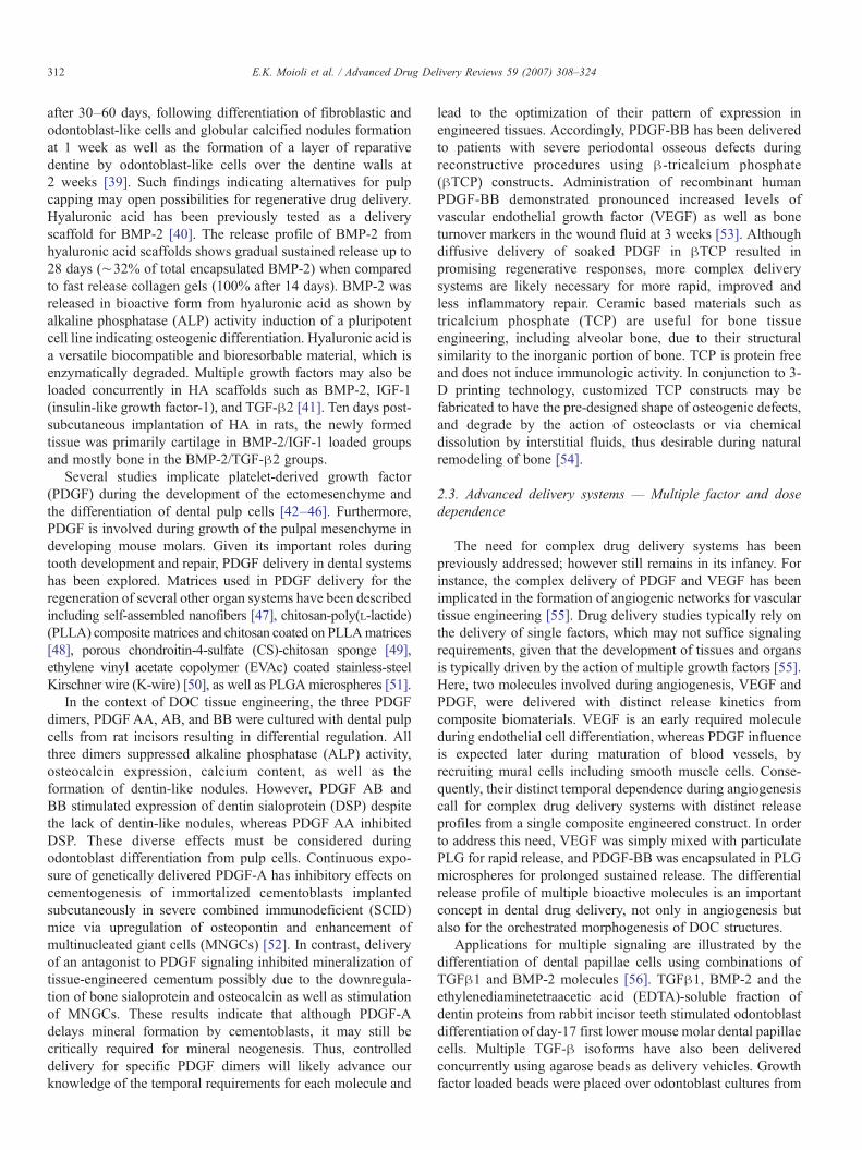
after 30–60 days, following differentiation of fibroblastic andodontoblast-like cells and globular calcified nodules formationat 1 week as well as the formation of a layer of reparativedentine by odontoblast-like cells over the dentine walls at2 weeks [39]. Such findings indicating alternatives for pulpcapping may open possibilities for regenerative drug delivery.Hyaluronic acid has been previously tested as a deliveryscaffold for BMP-2 [40]. The release profile of BMP-2 fromhyaluronic acid scaffolds shows gradual sustained release up to28 days (∼32% of total encapsulated BMP-2) when comparedto fast release collagen gels (100% after 14 days). BMP-2 wasreleased in bioactive form from hyaluronic acid as shown byalkaline phosphatase (ALP) activity induction of a pluripotentcell line indicating osteogenic differentiation. Hyaluronic acid isa versatile biocompatible and bioresorbable material, which isenzymatically degraded. Multiple growth factors may also beloaded concurrently in HA scaffolds such as BMP-2, IGF-1(insulin-like growth factor-1), and TGF-β2 [41]. Ten days post-subcutaneous implantation of HA in rats, the newly formedtissue was primarily cartilage in BMP-2/IGF-1 loaded groupsand mostly bone in the BMP-2/TGF-β2 groups.
Several studies implicate platelet-derived growth factor(PDGF) during the development of the ectomesenchyme andthe differentiation of dental pulp cells [42–46]. Furthermore,PDGF is involved during growth of the pulpal mesenchyme indeveloping mouse molars. Given its important roles duringtooth development and repair, PDGF delivery in dental systemshas been explored. Matrices used in PDGF delivery for theregeneration of several other organ systems have been describedincluding self-assembled nanofibers [47], chitosan-poly(L-lactide)(PLLA) compositematrices and chitosan coated on PLLAmatrices[48], porous chondroitin-4-sulfate (CS)-chitosan sponge [49],ethylene vinyl acetate copolymer (EVAc) coated stainless-steelKirschner wire (K-wire) [50], as well as PLGAmicrospheres [51].
In the context of DOC tissue engineering, the three PDGFdimers, PDGFAA, AB, and BB were cultured with dental pulpcells from rat incisors resulting in differential regulation. Allthree dimers suppressed alkaline phosphatase (ALP) activity,osteocalcin expression, calcium content, as well as theformation of dentin-like nodules. However, PDGF AB andBB stimulated expression of dentin sialoprotein (DSP) despitethe lack of dentin-like nodules, whereas PDGF AA inhibitedDSP. These diverse effects must be considered duringodontoblast differentiation from pulp cells. Continuous expo-sure of genetically delivered PDGF-A has inhibitory effects oncementogenesis of immortalized cementoblasts implantedsubcutaneously in severe combined immunodeficient (SCID)mice via upregulation of osteopontin and enhancement ofmultinucleated giant cells (MNGCs) [52]. In contrast, deliveryof an antagonist to PDGF signaling inhibited mineralization oftissue-engineered cementum possibly due to the downregula-tion of bone sialoprotein and osteocalcin as well as stimulationof MNGCs. These results indicate that although PDGF-Adelays mineral formation by cementoblasts, it may still becritically required for mineral neogenesis. Thus, controlleddelivery for specific PDGF dimers will likely advance ourknowledge of the temporal requirements for each molecule and
lead to the optimization of their pattern of expression inengineered tissues. Accordingly, PDGF-BB has been deliveredto patients with severe periodontal osseous defects duringreconstructive procedures using β-tricalcium phosphate(βTCP) constructs. Administration of recombinant humanPDGF-BB demonstrated pronounced increased levels ofvascular endothelial growth factor (VEGF) as well as boneturnover markers in the wound fluid at 3 weeks [53]. Althoughdiffusive delivery of soaked PDGF in βTCP resulted inpromising regenerative responses, more complex deliverysystems are likely necessary for more rapid, improved andless inflammatory repair. Ceramic based materials such astricalcium phosphate (TCP) are useful for bone tissueengineering, including alveolar bone, due to their structuralsimilarity to the inorganic portion of bone. TCP is protein freeand does not induce immunologic activity. In conjunction to 3-D printing technology, customized TCP constructs may befabricated to have the pre-designed shape of osteogenic defects,and degrade by the action of osteoclasts or via chemicaldissolution by interstitial fluids, thus desirable during naturalremodeling of bone [54].
2.3. Advanced delivery systems — Multiple factor and dosedependence
The need for complex drug delivery systems has beenpreviously addressed; however still remains in its infancy. Forinstance, the complex delivery of PDGF and VEGF has beenimplicated in the formation of angiogenic networks for vasculartissue engineering [55]. Drug delivery studies typically rely onthe delivery of single factors, which may not suffice signalingrequirements, given that the development of tissues and organsis typically driven by the action of multiple growth factors [55].Here, two molecules involved during angiogenesis, VEGF andPDGF, were delivered with distinct release kinetics fromcomposite biomaterials. VEGF is an early required moleculeduring endothelial cell differentiation, whereas PDGF influenceis expected later during maturation of blood vessels, byrecruiting mural cells including smooth muscle cells. Conse-quently, their distinct temporal dependence during angiogenesiscall for complex drug delivery systems with distinct releaseprofiles from a single composite engineered construct. In orderto address this need, VEGF was simply mixed with particulatePLG for rapid release, and PDGF-BB was encapsulated in PLGmicrospheres for prolonged sustained release. The differentialrelease profile of multiple bioactive molecules is an importantconcept in dental drug delivery, not only in angiogenesis butalso for the orchestrated morphogenesis of DOC structures.
Applications for multiple signaling are illustrated by thedifferentiation of dental papillae cells using combinations ofTGFβ1 and BMP-2 molecules [56]. TGFβ1, BMP-2 and theethylenediaminetetraacetic acid (EDTA)-soluble fraction ofdentin proteins from rabbit incisor teeth stimulated odontoblastdifferentiation of day-17 first lower mouse molar dental papillaecells. Multiple TGF-β isoforms have also been deliveredconcurrently using agarose beads as delivery vehicles. Growthfactor loaded beads were placed over odontoblast cultures from

313E.K. Moioli et al. / Advanced Drug Delivery Reviews 59 (2007) 308–324
rat incisor teeth and stimulated predentine secretion [57,58].Expression of the TGFβ isoforms TGFβ1, TGFβ2 and TGFβ3,result in the sequestration of odontoblasts in the dentin matrix.Although TGFβ2 showed minimal effects, mitogenic effects onthe cells of the subodontoblast layer were observed in proximityto the sites of TGFβ3 application. Additionally, tertiary dentineformation typically follows administration of BMP-7 whenapplied to dentine slices from monkey teeth [58].
In addition to complex delivery systems, growth factorrelease must account for the probable dose dependence ofvarious cell types. For instance, 2 μg of BMP-2 delivered incollagen sponges to amputated pulp in dogs show mineralizedosteodentine-like tissue containing embedded osteodentino-cytes 70 days after implantation with only some unmineralizedfibrous tissue and pulp-like loose connective tissue [36].However, when lower doses (660 or 220 ng) of BMP-2 areapplied, only unmineralized fibrous and pulp tissues wereobserved. Furthermore, little pulp tissue proliferation wasobserved with the administration of TGF-β1, suggesting apossible inhibitory effect in pulp regeneration. Nevertheless, itis imperative to also note that when TGF-β1 is implanted shortterm at mechanically exposed pulps of dog molar and canineteeth, differentiation of odontoblast-like cells and stimulation ofprimary odontoblasts is observed [59].
Given the apparent role of TGF-β1 in pulp cell regulation,alginate gels have been used as delivery matrices in the dentin-pulp complex [60]. Alginates provide extracellular matriceswith hydration properties that facilitate cellular wound healingactivities. Alginate gels were fabricated using calcium chlorideTGF-β1 solutions. TGF-β1 loaded gels were cultured on thepulpal cut surface of bisected human tooth slices showing twodifferent forms of repair responses, reactionary dentinogenesisand reparative dentinogenesis. The former was represented byupregulation of matrix secretion by preexisting odontoblasts,and the latter induced de novo dentinogenesis along the cutpulpal surface. Moreover, a delivered TGF-β1 dose responsewas observed revealing the intermediate dose of 50 ng as mostefficient, yielding the largest predentin width. Similarly to othergrowth factors, TGF-β1 has been delivered using manybiocompatible materials, including chitosan/collagen scaffolds[61], chitosan microspheres [62], poly(D,L-lactic-co-glycolicacid) (PLGA) microspheres [63], tri-co-polymer scaffold with1-ethyl-3-(3-dimethylaminopropyl) carbodiimide (EDC) [64],injectable oligo(poly(ethylene glycol) fumarate) (OPF) andgelatin microparticles [65].
2.4. Cell–scaffold interactions
The choice of scaffolding material plays pivotal role indental structure morphogenesis. For instance, dental pulp cellsgrown in collagenous, ceramic or metal (titanium) matrices,may develop differential expression of dental matrix markerssuch as dentin sialophosphoprotein (DSPP) [31]. These cellsinitially express STRO-1, a typical marker for multipotentialcells such as bone marrow derived mesenchymal stem cells.Although many similarities were observed between thebiomaterials used, after implantation, limited mineralization
was found only in the ceramic scaffold. It is postulated that incontrast with other studies, the open porosity of the ceramicHA/TCP sintered construct could have induced a proliferativeresponse resulting in a DSPP positive yet connective tissue-likestructure.
Other extracellular matrix (ECM) components such asfibronectin have been shown to mediate the binding of signalingmolecules and regulate cell attachment, which aid in regulatingthe interactions between cells and ECM resulting in thereorganization of preodontoblasts cytoskeleton during pulpalwound healing [66,67]. Additionally, collagens also regulatepreodontoblast rearrangement and aid in the attachment of newodontoblasts to pulp tissue [68]. Bone sialoprotein when usedfor the restoration of pulp defects appears to stimulate thedifferentiation of cells which secrete a homogeneous dentin-likedeposit and organized extracellular matrix more efficiently thanother capping materials such as calcium hydroxide, inducing thedevelopment of thick reparative dentinal tissue in the pulp [69].Thus, the choice of drug delivery scaffolds and matrices musttake into account the individual contribution of the biomaterialsthemselves during the regeneration process.
Recently, synthetic materials have been fabricated intonanometer scale structures in attempts to simulate the matrixenvironment in which seeded cells can be accommodated toproliferate and differentiate towards desired lineages [70–73].Nanofibers formed by electrospinning have been shown tomimic the ECM environment to various degrees when culturedwith several cell types [74–78]. Poly (D,L-lactide-co-glycolide)(PLGA) has been approved for several biomedical applicationsin humans and is widely used as scaffold materials in tissueengineering [79–81]. The porosity and tensile properties ofelectrospun PLGA nanofibers have been characterized [82].Osteoblast adhesion has been shown to occur when seeded inPLGA nanofibers [83]. Poly(ε-caprolactone) (PLC) nanofibersaccommodated the differentiation of hMSCs into adipogenic,osteogenic and chondrogenic cells [84]. We have recentlydemonstrated that human mesenchymal stem cells can prolif-erate and maintain differentiation characteristics towardsosteoblasts and chondrocytes when seeded in PLGA nanofibersfabricated by electrospinning (Fig. 2C). PLGA of 85:15 ratiowas electrospun into non-woven fibers with an averagediameter of 760±210 nm. The average Young's modulus ofelectrospun PLGA nanofibers was 42±26 kPa, per nanoinden-tation with atomic force microscopy. Human MSCs (hMSCs)were seeded at a density of 2×10⁶ cells/mL in PLGA nanofibersheets. After 2 week culture on PLGA nanofiber scaffold,hMSCs remained as precursors upon immunoblotting withhKL12 antibody. The overwhelming majority of hMSCs wasviable and proliferating in PLGA nanofiber scaffolds up to thetested 14 days, as assayed by live/dead tests, DNA content andBrdU. In a separate experiment, hMSCs seeded in PLGAnanofiber scaffolds were differentiated into chondrogenic(hMSC-Ch) and osteogenic (hMSC-Ob) cells. SEM taken upto 7 days after cell seeding revealed that hMSCs, hMSC-Ob andhMSC-Ch apparently attached to PLGA nanofibers. Histolog-ical assays revealed that hMSCs continuously differentiated intochondrogenic cells and osteogenic cells after 2 week incubation

Fig. 2. Various forms of poly-lactic-co-glycolic acid (PLGA) based matrices and scaffolds for dental, oral and craniofacial tissue engineering. A. Porous PLGA spongefabricated using salt-leaching techniques. B. PLGA microspheres encapsulating growth factors showing smooth spherical surface and wide range of diameters.C. PLGA nanofibers fabricated using electrospining techniques. D. PLGA microspheres in chitosan-based gels for advanced controlled delivery and cell interaction.A,B,C: scanning electron microscopy (SEM). D: phase contrast image.
314 E.K. Moioli et al. / Advanced Drug Delivery Reviews 59 (2007) 308–324
on PLGA nanofibers. Taken together, these data represent anoriginal investigation of continuous differentiation of hMSCsinto chondrogenic and osteogenic cells in PLGA nanofiberscaffolds.
2.5. Periodontal regeneration
The periodontium consists of cementum, periodontalligament (PDL), and alveolar bone and serves to transmitmechanical forces and as supporting structure and anchorage forthe teeth into the mandible and maxilla. Periodontal disease ischaracterized by inflammation of periodontal tissues, eventuallyleading to degeneration of the periodontium [85]. If periodontaldisease is left untreated, tooth loss can occur [86]. Periodontaltissue engineering should address the restoration of the PDLalong with its orientation and fibrous infiltration into exposedtooth roots and surrounding bone, as well as regeneration ofcementum by cementoblasts adjacent to the root, and restorationof the alveolar bone [87]. Previous matrices utilized inregeneration of periodontal structures include bone autografts,allografts, cell occlusive barrier membranes, bioglass, beta-tricalcium phosphate (βTCP), polytetrafluorethylene, polylactic-co-glycolic acid (PLGA), collagen membranes, andenamel matrix derivatives [88–96]. Undoubtedly, the incorpo-ration of bioactive molecules in successful matrix materials mayenhance their varied regenerative capacities. Thus, the effects ofmany growth factors on periodontal tissue cells must be
evaluated for their implication in periodontal tissue engineeringusing drug delivery technologies.
When choosing a scaffold matrix material for periodontalregeneration and drug delivery, careful consideration of thenatural ECM in the periodontal connective tissues mayelucidate some candidates. Collagens, proteoglycans, as wellas non-collagenous glycoproteins are typically found in theperiodontium, and some have been reviewed in the perspectiveof dental regeneration. For instance, powerful morphogens suchas BMP-2 bind to heparin, heparin sulfate, types I and IVcollagens, type II procollagen, fibrillins, proteoglycans, andnoggin, chordin, and DAN decreasing the rate of proteolysis[97,98]. In the context of mineralized tissue regeneration suchas alveolar bone and cementum, hydroxiapatite is a naturalcontender for a delivery vehicle due to its affinity for proteins,and in fact, when compared to collagens, TCP, glass beads,polymethylmethacrylate, and a bone cement, hydroxyapatitewas the most suitable for bone induction along with insolublecollagens.
The initiation of adequate periodontal regeneration may beassisted by an array of growth factors such as platelet-derivedgrowth factor (PDGF), transforming growth factor-βs (TGFβs)and insulin-like growth factor-1 (IGF-1) [86,99,100]. PDGFreceptors α and β are expressed in regenerating periodontal softand hard tissues [101]. PDGF may be used as a mitogen forfibroblasts and progenitors during periodontal regeneration andhas been shown to promote matrix synthesis and cell attachment
315E.K. Moioli et al. / Advanced Drug Delivery Reviews 59 (2007) 308–324
to tooth dentinal surfaces [86,102–104]. Subsequently, fibro-blast growth factor-2 (FGF2) and TGFβ contribute to thefurther proliferation of local cells. Previous reports on theeffects of basic fibroblast growth factor on human gingivalepithelial cells as well as during wound healing and periodontalregeneration of alveolar bone defects, promoted supplementarywork experiments for FGF-2 delivery in periodontal repair.FGF-2 delivered topically with gelatinous carriers in furcationdefects in dogs induced increased new bone formation rate, newtrabecular bone formation rate, as well as new cementumformation rate [105–107]. Additionally, IGF-1 has beenpreviously demonstrated to improve periodontal regenerationusing controlled delivery approaches. IGF-1 was encapsulatedin dextran-co-gelatin hydrogel microspheres by polyioniccomplexation and delivered to Class III furcation defects inmolars of dogs for controlled release. Adequate width ofregenerative PDL, regular Sharpey's fibers, and alveolar bonereconstruction was observed after 4–8 weeks [108].
During alveolar bone regeneration, bone morphogeneticproteins (BMPs) are expressed and may be responsible for theosteogenic differentiation of migrated progenitors [109]. BMPsare typically effective osteogenic molecules that may induceectopic bone formation [110]. Previous studies have demon-strated that BMP-12 loaded in collagen sponges improvedperiodontal ligament regeneration [111]. Although BMP-2 wasmore efficient for bone regeneration, application of BMP-12showed functionally oriented PDL bridging new bone andcementum. Additionally BMP-7, has been shown to inducebone regeneration adjacent to endosseous dental implants, aswell as maxillary sinus floor augmentation [112–114].Interestingly, co-application of BMP-2 and BMP-7 for theregeneration of furcation defects in the mandibular molars ofbaboons, does not enhance alveolar regeneration whencompared to the morphogens administered alone [115]. BMP-2 alone showed greater mineral bone and osteoid formation andremodeling, whereas administration of BMP-7 alone yieldedsignificant cementogenesis. Delivery of BMP-2 from slowdissolving collagen membranes demonstrate improved cemen-togenesis over faster collagen gel delivery [116,117]. Theslowed diffusion rate of the morphogen may play a role inlocalizing and maximizing the effects of released BMP-2,resulting in more adequate cementum formation.
3. Cranial sutures
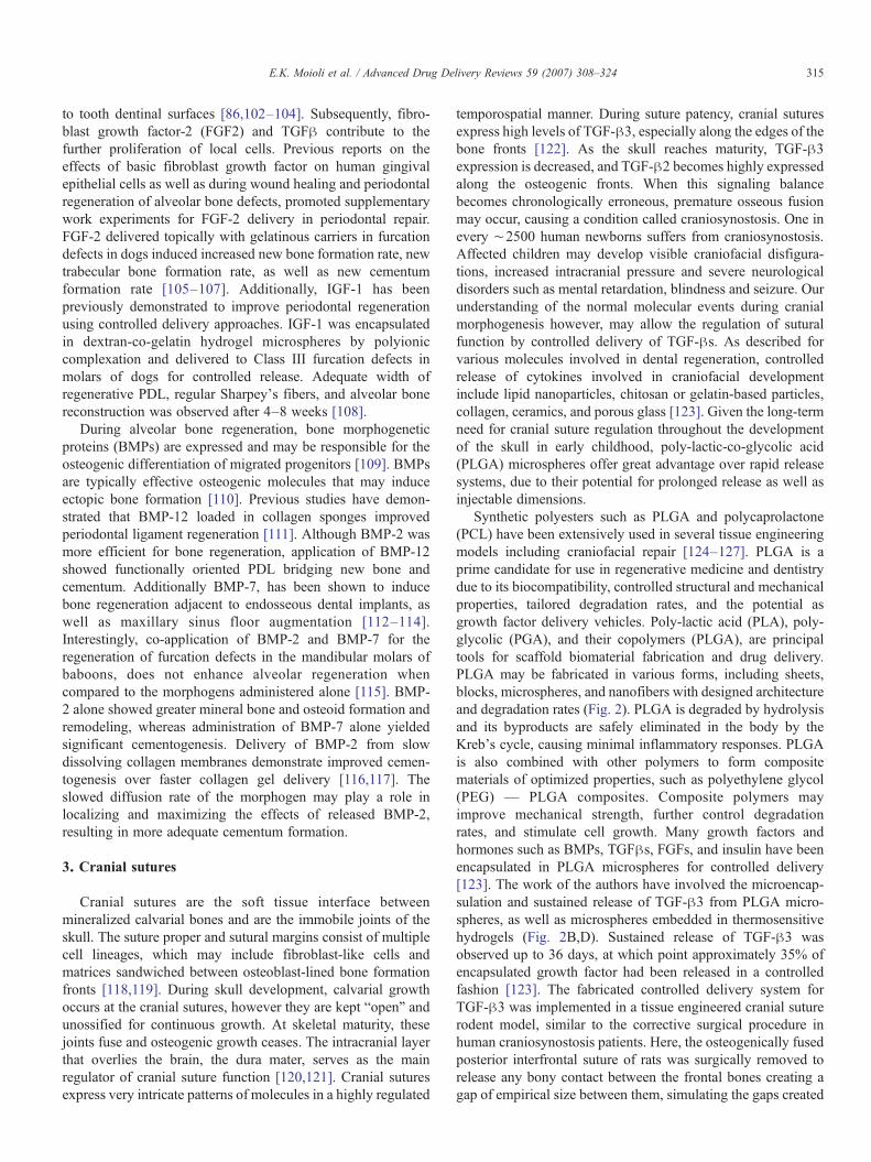
Cranial sutures are the soft tissue interface betweenmineralized calvarial bones and are the immobile joints of theskull. The suture proper and sutural margins consist of multiplecell lineages, which may include fibroblast-like cells andmatrices sandwiched between osteoblast-lined bone formationfronts [118,119]. During skull development, calvarial growthoccurs at the cranial sutures, however they are kept “open” andunossified for continuous growth. At skeletal maturity, thesejoints fuse and osteogenic growth ceases. The intracranial layerthat overlies the brain, the dura mater, serves as the mainregulator of cranial suture function [120,121]. Cranial suturesexpress very intricate patterns of molecules in a highly regulated
temporospatial manner. During suture patency, cranial suturesexpress high levels of TGF-β3, especially along the edges of thebone fronts [122]. As the skull reaches maturity, TGF-β3expression is decreased, and TGF-β2 becomes highly expressedalong the osteogenic fronts. When this signaling balancebecomes chronologically erroneous, premature osseous fusionmay occur, causing a condition called craniosynostosis. One inevery ∼2500 human newborns suffers from craniosynostosis.Affected children may develop visible craniofacial disfigura-tions, increased intracranial pressure and severe neurologicaldisorders such as mental retardation, blindness and seizure. Ourunderstanding of the normal molecular events during cranialmorphogenesis however, may allow the regulation of suturalfunction by controlled delivery of TGF-βs. As described forvarious molecules involved in dental regeneration, controlledrelease of cytokines involved in craniofacial developmentinclude lipid nanoparticles, chitosan or gelatin-based particles,collagen, ceramics, and porous glass [123]. Given the long-termneed for cranial suture regulation throughout the developmentof the skull in early childhood, poly-lactic-co-glycolic acid(PLGA) microspheres offer great advantage over rapid releasesystems, due to their potential for prolonged release as well asinjectable dimensions.
Synthetic polyesters such as PLGA and polycaprolactone(PCL) have been extensively used in several tissue engineeringmodels including craniofacial repair [124–127]. PLGA is aprime candidate for use in regenerative medicine and dentistrydue to its biocompatibility, controlled structural and mechanicalproperties, tailored degradation rates, and the potential asgrowth factor delivery vehicles. Poly-lactic acid (PLA), poly-glycolic (PGA), and their copolymers (PLGA), are principaltools for scaffold biomaterial fabrication and drug delivery.PLGA may be fabricated in various forms, including sheets,blocks, microspheres, and nanofibers with designed architectureand degradation rates (Fig. 2). PLGA is degraded by hydrolysisand its byproducts are safely eliminated in the body by theKreb's cycle, causing minimal inflammatory responses. PLGAis also combined with other polymers to form compositematerials of optimized properties, such as polyethylene glycol(PEG) — PLGA composites. Composite polymers mayimprove mechanical strength, further control degradationrates, and stimulate cell growth. Many growth factors andhormones such as BMPs, TGFβs, FGFs, and insulin have beenencapsulated in PLGA microspheres for controlled delivery[123]. The work of the authors have involved the microencap-sulation and sustained release of TGF-β3 from PLGA micro-spheres, as well as microspheres embedded in thermosensitivehydrogels (Fig. 2B,D). Sustained release of TGF-β3 wasobserved up to 36 days, at which point approximately 35% ofencapsulated growth factor had been released in a controlledfashion [123]. The fabricated controlled delivery system forTGF-β3 was implemented in a tissue engineered cranial suturerodent model, similar to the corrective surgical procedure inhuman craniosynostosis patients. Here, the osteogenically fusedposterior interfrontal suture of rats was surgically removed torelease any bony contact between the frontal bones creating agap of empirical size between them, simulating the gaps created

316 E.K. Moioli et al. / Advanced Drug Delivery Reviews 59 (2007) 308–324
in craniosynostosis procedures. However, in the tissue engi-neering rat model, instead of leaving an empty gap as in presentconventional approaches, an implant designed for preventingre-ossification was locally delivered. The implant consisted ofautologous bone marrow derived stem cells and TGF-β3encapsulated in PLGA microspheres in a pre-sized collagensponge carrier (Fig. 3A). Results indicate a decrease in bonedeposition and the formation of a bone–fibrous tissue–boneinterface analogous to normal cranial sutures after implantationof TGF-β3 loaded constructs (Fig. 3C). Placebo treated cranialdefects resulted in progressive bone healing after 4 weeks(Fig. 3B). Although this approach may need further investiga-tions to achieve more controlled regulation of new tissueformation, it is the first attempt to develop technologies for atissue-engineered implant in craniosynostosis treatments. Thecontinuous release of TGF-β3 may replicate the correctsignaling pathways that occur during normal cranial morpho-
Fig. 3. Engineered cranial suture implants. A. TGF-β3 loaded poly-lactic-co-glycolic acid (PLGA) microspheres (arrows) in gelatin sponge as a pre-sizedengineered implant. B. Osteogenic healing of osteotomized region of the fusedrat cranial suture after implantation of placebo loaded collagen sponge implants(4 weeks). C. Formation of a bone–fibrous tissue–bone interface and regulationof osteogenesis in the osteotomized region of the fused rat cranial suture afterimplantation of TGF-β3 treated engineered cranial suture implants (4 weeks).H&E stain. b: bone, f: fibrous tissue.
genesis offsetting the molecular mishaps of craniosynostosis(e.g. untimely downregulation of TGF-β3 and upregulation ofTGF-β2). These findings demonstrate a regenerated cranialsuture from autologous stem cells, and replacement of asynostosed cranial suture in an in vivo model. A biologicallyderived cranial suture may prove to reduce surgical trauma fromcraniotomy in craniosynostosis patients to localized osteotomy.Additionally, a biologically derived cranial suture mayaccommodate subsequent craniofacial growth, in contrast toclinically observed secondary synostosis after the resection ofsynostosed cranial sutures in some patients.
4. Temporomandibular joints (TMJ)
Disorders of the temporomandibular joint (TMJ) affect over10 million Americans by some estimates (NIH, 2006), withapproximately 3–4% of the population seeking treatment [128].The TMJ connects the mandible to the temporal bone of theskull. As a diarthrotic joint, layers of cartilage are present onboth the condyle of the mandible and the glenoid fossa andarticular eminence of the temporal bone [128]. The TMJ disc isa fibrocartilaginous disc that sits between the two cartilagelayers, connected to the joint capsule around its periphery nearits attachment to the condylar neck [128].
Many skeletal and cartilage factors have been implicatedin the growth of the TMJ cartilage layers and fibrocartilagedisk. Effects on cells isolated from the TMJ disk have beendocumented for PDGF, IGF-1, bFGF, and TGFβ1 [128–133]. Each growth factor displayed distinctive effects oncellular proliferation and extracellular matrix production,suggesting that a combination cocktail of growth factorswould be more beneficial [129]. In mouse explants of themandibular condyle, the growth factors TGFβ1, IGF-1, andgrowth hormone stimulated proteoglycan synthesis incartilage layers, while interleukin-1α (IL-1 α) exerted aninhibitory effect [134]. Using a collagen carrier, BMP-2 wasdemonstrated to increase cartilage regeneration in internallyderanged rabbit TMJ [135].
Efforts are currently underway in tissue engineering thedifferent structures of the TMJ. Toward engineering a TMJ disc,TMJ disc cells were isolated and the growth factors necessary tomaintain the cells in vitro were established. The TMJ disc cellswere seeded onto non-woven poly-glycolic acid (PGA)scaffolds and could be cultured up to 6 weeks [136]. Use ofthe growth factors bFGF, IGF-1, and TGFβ1 either alone [136]or in combination [129] modulated cellular proliferation andmatrix biosynthesis. The use of a rotating bioreactor for TMJdisc culture was also explored, with no clear beneficial results[133]. In these studies, the need for high levels of growth factors(100 ng/mL for IGF-1 and bFGF and 30 ng/mL TGFβ1)suggests that a controlled drug delivery system may potentiallybe advantageous, especially for maintenance of the disc whenimplanted in vivo. Tissue engineering of the condyle portion ofthe TMJ requires the fabrication of an osteochondral constructcontaining both a cartilage layer and underlying subchondralbone. Toward this end, the large amount of work intoosteochondral constructs for appendicular skeletal joints can
317E.K. Moioli et al. / Advanced Drug Delivery Reviews 59 (2007) 308–324
be directly applied to the articular condyle of the TMJ [137–142]. Specifically for the TMJ, polylactic/polyglycolic copol-ymer was used in a gelatin sponge complex (PGS) as a carrierfor BMP-2 and demonstrated increased bone and cartilage-liketissue regeneration in rabbit after condylectomy [143].Interestingly, no significant increase in growth was realizedwith addition of BMP-2, attributed to the osteoinductivecapabilities of the PGS scaffold. Computational design andsolid freeform fabrication allows layer-by-layer fabrication ofcomplex scaffolds with defined architecture, and can even bematched to patient-specific computed tomography (CT) ormagnetic resonance imaging (MRI) scans. This technologyallows production of biphasic osteochondral constructs inwhich the subchondral portion incorporates internal architectureand biological factors ideal for bone ingrowth and the upperlayer incorporates cartilage-specific parameters [144]. Non-invasive injectable systems for drug and/or cell delivery holdpromise for treatment of cartilage defects, and have beenaccomplished using PLGA microspheres [145], thermo-revers-ible hydrogels [146,147], oligo(poly(ethylene glycol) fumarate)(OPF) [65,148], and alginate [149]. Similar approaches to TMJrepair may prove beneficial to regeneration of cartilage withoutsurgery.
The authors have demonstrated the feasibility of tissueengineering a synovial joint condyle in the shape of the TMJusing hydrogel matrix loaded with both cartilaginous andosseous components from a single population of rat bonemarrow-derived mesenchymal stem cells (rMSCs). RatMSCs were isolated from femur and tibia bone marrow ofadult rats and exposed separately to either chondrogenic orosteogenic supplements [3,150]. Poly (ethylene glycol)diacrylate (PEGDA) was dissolved in PBS with a biocom-patible ultraviolet photoinitiator [3,150]. MSC-derivedchondrogenic and osteogenic cells were suspended inPEGDA polymer/photoinitator solution and loaded into anegative mold of a cadaver adult human mandibular condylein two stratified and yet integrated layers. The photopoly-merized osteochondral construct was removed from the moldand implanted in the dorsum of severe combined immuno-deficient (SCID) mice (4–5 week old males). At both 4 and8 weeks in vivo implantation, the tissue-engineered synovialjoints retained the macroscopic shape and dimensions of thecell-hydrogel construct. Importantly, the constructs retainedboth chondrogenic and osseous layers, expressing cartilage-specific glycosaminoglycans (GAGs) in the upper chondro-genic layer and bony mineral nodules revealed by von Kossastaining in the lower osseous layer [3,150]. Using anidentical approach but increasing the cell-seeding density4-fold, further tissue maturation of both the chondrogenicand osseous layers as well as mutual infiltration forming aprimitive osteochondral interface was achieved after 12-week subcutaneous implantation in SCID mice [4]. Improve-ments to the tissue-engineered TMJ condyle are warrantedalong multiple fronts including structural integrity, tissuematuration, mechanical strength, and host integration [151],and the aforementioned approaches of drug delivery fromhydrogels will likely be critical in this process.
5. Drug delivery in implant dentistry
Despite the great advances in completely tissue engineeredoral structures, the current gold standard for load bearingcraniofacial replacements remains titanium implants. Since theinitial studies establishing its use in dentistry [152], titanium'sstrength, durability, and biocompatibility has led to innumerableapplications, including single tooth replacement to completebridges in the mandible and maxilla and even for craniofacialreconstructions. Titanium has high success rates of initialanchorage (over 90%) and has been shown to co-exist with hostbone for the life of the patient [153], but failures still occurduring both initial bone integration and long-term fixation.
Critical for initial and long-term efficacy of titaniumimplants is establishment of adequate bone fixation. Thisprocess resembles natural fracture repair and requires thecooperation of multiple cell and tissue types resulting in boneingrowth into the irregular surfaces of the titanium andeventually direct contact between bone and titanium (osseointe-gration) [152,154]. During the initial inflammatory phase,disrupted bone matrix and inflammatory cells release over 100biologically active substances, including the skeletal growthfactors platelet-derived growth factor (PDGF), transforminggrowth factor β (TGFβ), fibroblast growth factor (FGF), andinsulin-like growth factor (IGF) [155]. This initial phase iscritical to set in motion the complex processes to follow, as theadministration of anti-inflammatory drugs can significantlydecrease bone ingrowth [156,157]. During the repair phase,proliferation and deposition of extracellular matrix by immaturebone progenitors as well as establishment of a new vascular bedvia angiogenesis is induced by growth factors such as TGFβ,bFGF, PDGF, and vascular endothelial growth factor (VEGF).The mineralization and maturation phase is the final phase offracture repair in which differentiation factors such as TGFβand bone morphogenetic protein 2 (BMP2) are elevated andcritical extracellular matrix components such as osteocalcin andosteopontin begin to be expressed. The spatial and temporalprofiles of numerous growth factors have been studied forfracture repair [158,159], and some of these growth factors suchas TGFβ, PDGF, and BMP have been demonstrated in oralimplant sites [160,161].
Because of its similarities to fracture repair, titanium dentalimplant osseointegration could potentially be improved bydelivery of the same growth factors. Although not a definedcocktail, platelet rich plasma contains a multitude of growthfactors expressed during the inflammation phase, and has beenused to increase the volume of peri-implant bone in rat tibiae[162] and increase bone ingrowth when delivered to peri-implant defects via a demineralized freeze dried bone carrier[163]. In applications where there are existing gaps, such ascraniofacial reconstruction or augmentation of bone at implantsites or peri-implant defects, increased regeneration of bone,often at comparable levels to bone grafts, has been accomplishedwith delivery of TGFβ and BMP-2 via various scaffoldsincluding absorbable collagen sponge [164], 25% pluronic gel[165], refined collagen gelatin (Gelfoam) [166,167], titaniummesh [168,169], and beta tricalcium phosphate (β-TCP) [166].
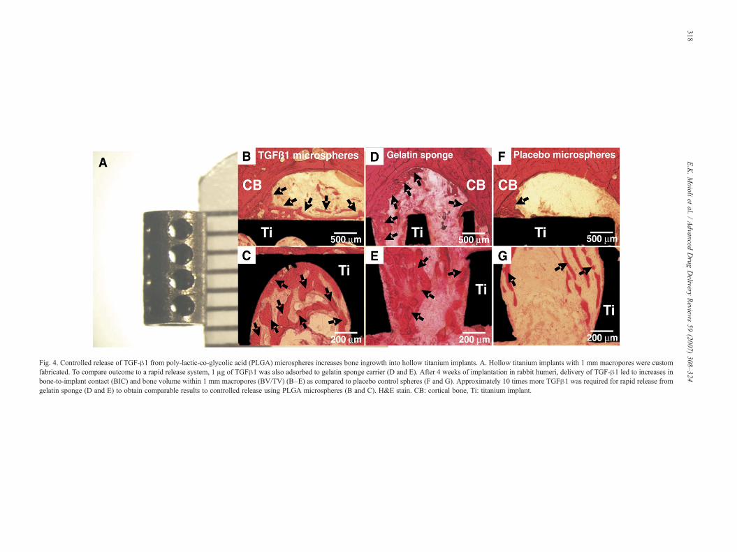
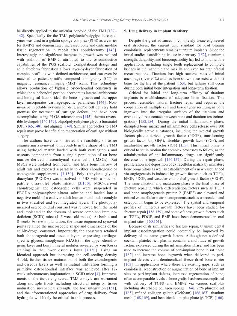
Fig. 4. Controlled release of TGF-β1 from poly-lactic-co-glycolic acid (PLGA) microspheres increases bone ingrowth into hollow titanium implants. A. Hollow titanium implants with 1 mm macropores were customfabricated. To compare outcome to a rapid release system, 1 μg of TGFβ1 was also adsorbed to gelatin sponge carrier (D and E). After 4 weeks of implantation in rabbit humeri, delivery of TGF-β1 led to increases inbone-to-implant contact (BIC) and bone volume within 1 mm macropores (BV/TV) (B–E) as compared to placebo control spheres (F and G). Approximately 10 times more TGFβ1 was required for rapid release fromgelatin sponge (D and E) to obtain comparable results to controlled release using PLGA microspheres (B and C). H&E stain. CB: cortical bone, Ti: titanium implant.
318E.K.Moioli
etal.
/Advanced
Drug
Delivery
Review
s59
(2007)308–324

319E.K. Moioli et al. / Advanced Drug Delivery Reviews 59 (2007) 308–324
However, minimizing gaps between implant and host bone isnecessary, thus the application of bulky scaffolds is usually notpossible and alternative delivery approaches are required.Adsorption of TGFβ and/or BMP to tricalcium phosphate orhydroxyapatite coatings on titanium implants leads to enhance-ment of bone ingrowth in gap models [170–173]. However,adsorbed growth factor in these systems is rapidly exhaustedwhereas levels of TGFβ and BMP-2 remain elevated throughoutthe fracture repair process [158]. More complex methods ofgrowth factor adhesion to surface coatings have been explored,including fabrication of ultrahydrophillic titanium surfaces[174] and incorporation of BMP-2 directly into calciumphosphate coatings [175], showing promising results.
To mimic the temporal profile of growth factors, controlledrelease technology has been explored for titanium implants.Titanium and steel wires were coated with poly(D,L-lactide)incorporating TGFβ or IGF-1 and demonstrated continuousrelease of the growth factors up to 42 days in vitro and in vivo[176]. The authors have fabricated hollow titanium implants with1 mm macropores; these hollow implants maintain the biome-chanical strength required for oral load-bearing applications butprovide space for tissue engineered scaffolds delivering bioactivemolecules or cells (Fig. 4A). A controlled release system forTGFβ1 using PLGA microspheres has also been developed bythe authors, as well as other groups [63], that releases the growthfactor in a controlled fashion up to 4 weeks. With a diameteraveraging 64μm, the PLGAmicrospheres could be injected into agelatin carrier (Gelfoam) and subsequently packed into the hollowcore of the titanium implant. The hollow implants were placedunicortically into the humeri of adult New Zealand white rabbitsand allowed to osseointegrate for 4 weeks. Incorporation ofTGFβ1 in the hollow implant led to significantly increasedosseointegration parameters such as bone-to-implant contact(BIC) and bone volumewithin the 1 mmmacropores (Fig. 4B–E)as compared to placebo controls (Fig. 4F,G). Adsorption ofTGFβ1 to gelatin sponge and controlled release via PLGAmicrospheres resulted in comparable outcomes, but 10× moreTGFβ1 was required when delivered by gelatin sponge. The useof controlled release of growth factors by way of hollow implantsis a novel approach to enhancing osseointegration and may beuseful in implant dentistry, in which hollow implants have been inclinical use for years [177,178]. More advanced biomaterials,such as microporous titanium [179], may also potentially be usedas high strength carriers.
6. Conclusions
Previous work has explored the application of biomaterialmatrices and drug delivery in the regeneration and/or denovo formation of DOC structures. A wide range ofbiomaterials including polymeric hydrogels, porous scaf-folds, nanofibers and microparticles have been utilizedtoward the regeneration of tooth, periodontal ligament,salivary gland, cranial sutures, and the temporomandibularjoint (TMJ). Although the goal of eventually regenerating awhole tooth is admirable, whole tooth regeneration is likelyone of the last technologies to be commercially realized. The
more near-term technologies include the regeneration ofindividual tooth structures, osteochondral grafts for the TMJ,cranial sutures and bone regeneration for periodontal defects.Integrated approaches that capture the fundamental knowl-edge in multiple biology and engineering disciplines are notonly desirable, but necessary for proper regeneration of DOCstructures. Specific structural and mechanical characteristicsas outlined in the Introduction of this review, need to beconsidered in the design of tissue engineering or tissueregeneration approaches. It is predictable that successfulapproaches need to exert not only more control at micro-scale and nano-scale levels, but also rigorously test increas-ingly refined engineering techniques in animal modelstowards the regeneration of DOC structures.
Acknowledgements
We are grateful to the members of the Tissue Engineeringand Regenerative Medicine Laboratory (TERML) for theirdiscussion. We thank Janina Acloque, Maryann Wanner andRichard Abbott for administrative support. Generous supportfrom the National Institutes of Health is gratefully acknowl-edged, through NIH grants DE13964, DE15391 and EB02332to J.J.M., for the effort spent on composing this manuscriptalong with some of the experimental data presented in thismanuscript.
References
[1] M.N. Rahaman, J.J. Mao, Stem cell-based composite tissue constructs forregenerative medicine, Biotechnol. Bioeng. 91 (2005) 261–284.
[2] J.J. Mao,W.V. Giannobile, J.A. Helms, S.J. Hollister, P.H. Krebsbach, M.T.Longaker, S. Shi, Craniofacial tissue engineering by stem cells, J. Dent. Res.85 (2006) 966–979.
[3] A. Alhadlaq, J.J. Mao, Tissue-engineered neogenesis of human-shapedmandibular condyle from rat mesenchymal stem cells, J. Dent. Res. 82(2003) 951–956.
[4] A. Alhadlaq, J.J. Mao, Tissue-engineered osteochondral constructs in theshape of an articular condyle, J. Bone Joint Surg. Am. 87 (2005)936–944.
[5] P.C. Yelick, J.P. Vacanti, Bioengineered teeth from tooth bud cells, Dent.Clin. North Am. 50 (2) (2006).
[6] P.T. Sharpe, C.S. Young, Test-tube teeth, Sci. Am. 293 (2) (2005).[7] C.S. Young, S. Terada, J.P. Vacanti, M. Honda, J.D. Bartlett, P.C. Yelick,
Tissue engineering of complex tooth structures on biodegradable polymerscaffolds, J. Dent. Res. 81 (10) (2002).
[8] W. Sonoyama, Y. Liu, D. Fang, T. Yamaza, B.M. Seo, C. Zhang, H. Liu,S. Gronthos, C.Y. Wang, S. Shi, S. Wang, Mesenchymal stem cell-mediated functional tooth regeneration in Swine, PLoS ONE 1 (2006)1920.
[9] M.J. Honda, S. Tsuchiya, Y. Sumita, H. Sagara, M. Ueda, The sequentialseeding of epithelial and mesenchymal cells for tissue-engineered toothregeneration, Biomaterials 28 (4) (2007).
[10] A.I. Caplan, S.P. Bruder, Mesenchymal stem cells: building blocks formolecular medicine in the 21st century, Trends Mol. Med. 7 (2001)259–264.
[11] A. Alhadlaq, J.J. Mao, Mesenchymal stem cells: isolation andtherapeutics, Stem Cells Dev. 13 (2004) 436–448.
[12] N.W. Marion, J.J. Mao, Mesenchymal stem cells and tissue engineering,Methods Enzymol. 420 (2006) 339–361.
[13] R.G. Craig, F.A. Peyton, D.W. Johnson, Compressive properties ofenamel, dental cements, and gold, J. Dent. Res 40 (5) (1961) 936–945.

320 E.K. Moioli et al. / Advanced Drug Delivery Reviews 59 (2007) 308–324
[14] R.G. Craig, F.A. Peyton, Elastic and mechanical properties of humandentin, J. Dent. Res. 37 (1958) 710–718.
[15] I. Thesleff, Developmental biology and building a tooth, QuintessenceInt. 34 (2003) 613–620.
[16] H. Liu, S. Gronthos, S. Shi, Dental pulp stem cells, Methods Enzymol.419 (2006) 99–113.
[17] S. Kakehashi, H.R. Stanley, R.J. Fitzgerald, The effects of surgicalexposures of dental pulps in germ-free and conventional laboratory rats,Oral Surg. Oral Med. Oral Pathol. 20 (1965) 340–349.
[18] A.H. Rowe, Root canal therapy, Br. Dent. J. 123 (1967) 543.[19] C.F. Cox, G. Bergenholtz, Healing sequence in capped inflamed dental pulps
of Rhesus monkeys (Macaca mulatta), Int. Endod. J. 19 (1986) 113–120.[20] M. Cvek, L. Granath, P. Cleaton-Jones, J. Austin, Hard tissue barrier
formation in pulpotomized monkey teeth capped with cyanoacrylate orcalcium hydroxide for 10 and 60 minutes, J. Dent. Res. 66 (1987)1166–1174.
[21] M. Goldberg, S. Lacerda-Pinheiro, N. Jegat, N. Six, D. Septier, F. Priam,M. Bonnefoix, K. Tompkins, H. Chardin, P. Denbesten, A. Veis, A.Poliard, The impact of bioactive molecules to stimulate tooth repair andregeneration as part of restorative dentistry, Dent. Clin. North Am. 50(2006) 277–298 (x).
[22] G. Bergenholtz, Advances since the paper by Zander and Glass (1949) onthe pursuit of healing methods for pulpal exposures: historical per-spectives, Oral Surg. Oral Med. Oral Pathol. Oral Radiol. Endo. 100(2005) S102–S108.
[23] T.J. Harrop, B. Mackay, Electron microscopic observations on healing indental pulp in the rat, Arch. Oral Biol. 13 (1968) 365–385.
[24] P. Horsted, A.K. El, K. Langeland, Capping of monkey pulps with Dycaland a Ca-eugenol cement, Oral Surg. Oral Med. Oral Pathol. 52 (1981)531–553.
[25] L. Jaber, C. Mascres, W.B. Donohue, Electron microscope characteristicsof dentin repair after hydroxylapatite direct pulp capping in rats, J. OralPathol. Med. 20 (1991) 502–508.
[26] I.A. Mjor, E. Dahl, C.F. Cox, Healing of pulp exposures: anultrastructural study, J. Oral Pathol. Med. 20 (1991) 496–501.
[27] S. Gronthos, M. Mankani, J. Brahim, P.G. Robey, S. Shi, Postnatal humandental pulp stem cells (DPSCs) in vitro and in vivo, Proc. Natl. Acad. Sci.U. S. A. 97 (2000) 13625–13630.
[28] S. Gronthos, J. Brahim, W. Li, L.W. Fisher, N. Cherman, A. Boyde, P.Denbesten, G. Robey, S. Shi, Stem cell properties of human dental pulpstem cells, J. Dent. Res. 81 (2002) 531–535.
[29] S. Shi, S. Gronthos, S. Chen, A. Reddi, C.M. Counter, P.G. Robey, C.Y.Wang, Bone formation by human postnatal bone marrow stromal stemcells is enhanced by telomerase expression, Nat. Biotechnol. 20 (2002)587–591.
[30] D.J. Prockop, Marrow stromal cells as stem cells for nonhematopoietictissues, Science 276 (1997) 71–74.
[31] W. Zhang, W. Frank, X.T.H. van Kuppevelt, W.F. Daamen, Z. Bian, J.A.Jansen, The performance of human dental pulp stem cells on differentthree-dimensional scaffold materials, Biomaterials 27 (2006) 5658–5668.
[32] W. Zhang, X.F. Walboomers, S. Shi, M. Fan, J.A. Jansen, Multilineagedifferentiation potential of stem cells derived from human dental pulpafter cryopreservation, Tissue Eng. (in press) (Electronic publicationahead of print).
[33] G.T. Huang, W. Sonoyama, J. Chen, S.H. Park, In vitro characterizationof human dental pulp cells: various isolation methods and culturingenvironments, Cell Tissue Res. 324 (2006) 225–236.
[34] S. Yokose, H. Kadokura, Y. Tajima, K. Fujieda, I. Katayama, T.Matsuoka, T. Katayama, Establishment and characterization of a culturesystem for enzymatically released rat dental pulp cells, Calcif. Tissue Int.66 (2000) 139–144.
[35] S. Yokose, H. Kadokura, N. Tajima, A. Hasegawa, H. Sakagami, K.Fujieda, T. Katayama, Platelet-derived growth factor exerts disparateeffects on odontoblast differentiation depending on the dimers in ratdental pulp cells, Cell Tissue Res. 315 (2004) 375–384.
[36] M. Nakashima, Induction of dentine in amputated pulp of dogs byrecombinant human bone morphogenetic proteins-2 and-4 with collagenmatrix, Arch. Oral Biol. 39 (1994) 1085–1089.
[37] T. Saito, M. Ogawa, Y. Hata, K. Bessho, Acceleration effect of humanrecombinant bone morphogenetic protein-2 on differentiation of humanpulp cells into odontoblasts, J. Endod. 30 (2004) 205–208.
[38] K. Iohara, M. Nakashima, M. Ito, M. Ishikawa, A. Nakasima, A.Akamine, Dentin regeneration by dental pulp stem cell therapy withrecombinant human bone morphogenetic protein 2, J. Dent. Res. 83(2004) 590–595.
[39] T. Sasaki, H. Kawamata-Kido, Providing an environment for reparativedentine induction in amputated rat molar pulp by high molecular-weighthyaluronic acid, Arch. Oral Biol. 40 (1995) 209–219.
[40] H.D. Kim, R.F. Valentini, Retention and activity of BMP-2 in hyaluronicacid-based scaffolds in vitro, J. Biomed. Mater. Res. 59 (2002) 573–584.
[41] P. Bulpitt, D. Aeschlimann, New strategy for chemical modification ofhyaluronic acid: preparation of functionalized derivatives and their use inthe formation of novel biocompatible hydrogels, J. Biomed. Mater. Res.47 (1999) 152–169.
[42] M. Nakashima, The effects of growth factors on DNA synthesis,proteoglycan synthesis and alkaline phosphatase activity in bovine dentalpulp cells, Arch. Oral Biol. 37 (1992) 231–236.
[43] R.B. Rutherford, M.D. TrailSmith, M.E. Ryan, M.F. Charette, Synergisticeffects of dexamethasone on platelet-derived growth factor mitogenesis invitro, Arch. Oral Biol. 37 (1992) 139–145.
[44] J.C. Hu, C. Zhang, H.C. Slavkin, The role of platelet-derived growthfactor in the development of mouse molars, Int. J. Dev. Biol. 39 (1995)939–945.
[45] Y. Chai, P. Bringas Jr., A. Mogharei, C.F. Shuler, H.C. Slavkin, PDGF-AandPDGFR-alpha regulate tooth formation via autocrinemechanismduringmandibular morphogenesis in vitro, Dev. Dyn. 213 (1998) 500–511.
[46] I.A. Denholm, A.J. Moule, P.M. Bartold, The behaviour and proliferationof human dental pulp cell strains in vitro, and their response to theapplication of platelet-derived growth factor-BB and insulin-like growthfactor-1, Int. Endod. J. 31 (1998) 251–258.
[47] P.C. Hsieh, M.E. Davis, J. Gannon, C. MacGillivray, R.T. Lee, Controlleddelivery of PDGF-BB for myocardial protection using injectable self-assembling peptide nanofibers, J. Clin. Invest. 116 (2006) 237–248.
[48] J.Y. Lee, S.H. Nam, S.Y. Im, Y.J. Park, Y.M. Lee, Y.J. Seol, C.P. Chung, S.J.Lee, Enhanced bone formation by controlled growth factor delivery fromchitosan-based biomaterials, J. Control. Release 78 (2002) 187–197.
[49] Y.J. Park, Y.M. Lee, S.N. Park, S.Y. Sheen, C.P. Chung, S.J. Lee, Plateletderived growth factor releasing chitosan sponge for periodontal boneregeneration, Biomaterials 21 (2000) 153–159.
[50] W.R. Walsh, H.D. Kim, Y.S. Jong, R.F. Valentini, Controlled release ofplatelet-derived growth factor using ethylene vinyl acetate copolymer(EVAc) coated on stainless-steel wires, Biomaterials 16 (1995) 1319–1325.
[51] G. Wei, Q. Jin, W.V. Giannobile, P.X. Ma, Nano-fibrous scaffold forcontrolled delivery of recombinant human PDGF-BB, J. Control. Release112 (2006) 103–110.
[52] O. Anusaksathien, Q. Jin, M. Zhao, M.J. Somerman, W.V. Giannobile,Effect of sustained gene delivery of platelet-derived growth factor or itsantagonist (PDGF-1308) on tissue-engineered cementum, J. Periodontol.75 (2004) 429–440.
[53] J.W. Cooke, D.P. Sarment, L.A. Whitesman, S.E. Miller, Q. Jin, S.E.Lynch, W.V. Giannobile, Effect of rhPDGF-BB delivery on mediators ofperiodontal wound repair, Tissue Eng. 12 (2006) 1441–1450.
[54] I.R. Zerbo, A.L. Bronckers, L.G. de, E.H. Burger, Localisation ofosteogenic and osteoclastic cells in porous beta-tricalcium phosphateparticles used for human maxillary sinus floor elevation, Biomaterials 26(2005) 1445–1451.
[55] T.P. Richardson, M.C. Peters, A.B. Ennett, D.J. Mooney, Polymeric systemfor dual growth factor delivery, Nat. Biotechnol. 19 (2001) 1029–1034.
[56] C. Begue-Kirn, A.J. Smith, J.V. Ruch, J.M. Wozney, A. Purchio, D.Hartmann, H. Lesot, Effects of dentin proteins, transforming growthfactor beta 1 (TGF beta 1) and bone morphogenetic protein 2 (BMP2) onthe differentiation of odontoblast in vitro, Int. J. Dev. Biol. 36 (1992)491–503.
[57] A.J. Sloan, A.J. Smith, Stimulation of the dentine-pulp complex of ratincisor teeth by transforming growth factor-beta isoforms 1–3 in vitro,Arch. Oral Biol. 44 (1999) 149–156.

321E.K. Moioli et al. / Advanced Drug Delivery Reviews 59 (2007) 308–324
[58] A.J. Sloan, R.B. Rutherford, A.J. Smith, Stimulation of the rat dentine-pulp complex by bone morphogenetic protein-7 in vitro, Arch. Oral Biol.45 (2000) 173–177.
[59] D. Tziafas, A. Alvanou, S. Papadimitriou, J. Gasic, A. Komnenou,Effects of recombinant basic fibroblast growth factor, insulin-like growthfactor-II and transforming growth factor-beta 1 on dog dental pulp cells invivo, Arch. Oral Biol. 43 (1998) 431–444.
[60] K. Dobie, G. Smith, A.J. Sloan, A.J. Smith, Effects of alginate hydrogelsand TGF-beta 1 on human dental pulp repair in vitro, Connect. TissueRes. 43 (2002) 387–390.
[61] Y. Zhang, X. Cheng, J. Wang, Y. Wang, B. Shi, C. Huang, X. Yang, T.Liu, Novel chitosan/collagen scaffold containing transforming growthfactor-beta1 DNA for periodontal tissue engineering, Biochem. Biophys.Res Commun. 344 (2006) 362–369.
[62] J.E. Lee, S.E. Kim, I.C. Kwon, H.J. Ahn, H. Cho, S.H. Lee, H.J. Kim, S.C.Seong, M.C. Lee, Effects of a chitosan scaffold containing TGF-beta1encapsulated chitosan microspheres on in vitro chondrocyte culture, Artif.Organs 28 (2004) 829–839.
[63] L. Lu, G.N. Stamatas, A.G. Mikos, Controlled release of transforminggrowth factor beta1 from biodegradable polymer microparticles,J. Biomed. Mater. Res. 50 (3) (2000) 1905.
[64] C.H. Chou, W.T. Cheng, C.C. Lin, C.H. Chang, C.C. Tsai, F.H. Lin, TGF-beta1 immobilized tri-co-polymer for articular cartilage tissue engineer-ing, J. Biomed. Mater. Res., B Appl. Biomater. 77 (2006) 338–348.
[65] H. Park, J.S. Temenoff, T.A. Holland, Y. Tabata, A.G. Mikos, Delivery ofTGF-beta1 and chondrocytes via injectable, biodegradable hydrogels forcartilage tissue engineering applications, Biomaterials 26 (2005)7095–7103.
[66] D. Tziafas, A.J. Smith, H. Lesot, Designing new treatment strategies invital pulp therapy, J. Dent. 28 (2000) 77–92.
[67] M. Nakashima, A. Akamine, The application of tissue engineering toregeneration of pulp and dentin in endodontics, J. Endod. 31 (2005)711–718.
[68] Y. Kitasako, S. Shibata, C.F. Cox, J. Tagami, Location, arrangement andpossible function of interodontoblastic collagen fibres in association withcalcium hydroxide-induced hard tissue bridges, Int. Endod. J. 35 (2002)996–1004.
[69] F. Decup, N. Six, B. Palmier, D. Buch, J.J. Lasfargues, E. Salih, M.Goldberg, Bone sialoprotein-induced reparative dentinogenesis in thepulp of rat's molar, Clin. Oral Investig. 4 (2000) 110–119.
[70] P.K. Baumgartner, Electrostatic spinning of acrylic microfibers, J. ColloidInterface Sci. 36 (1971) 71–79.
[71] Y.Z. Zhang, J. Venugopal, Z.M. Huang, C.T. Lim, S. Ramakrishna,Characterization of the surface biocompatibility of the electrospunPCL-collagen nanofibers using fibroblasts, Biomacromolecules 6 (5)(2005).
[72] H.K. Noh, S.W. Lee, J.M. Kim, J.E. Oh, K.H. Kim, C.P. Chung, S.C.Choi, W.H. Park, B.M. Min, Electrospinning of chitin nanofibers:degradation behavior and cellular response to normal human keratino-cytes and fibroblasts, Biomaterials 27 (21) (2006).
[73] M.N. Rahaman, J.J. Mao, Stem cell-based composite tissue constructs forregenerative medicine, Biotechnol. Bioeng. 91 (3) (2005) 1905.
[74] A.S. Badami, M.R. Kreke, M.S. Thompson, J.S. Riffle, A.S. Goldstein,Effect of fiber diameter on spreading, proliferation, and differentiation ofosteoblastic cells on electrospun poly(lactic acid) substrates, Biomaterials27 (4) (2006).
[75] Z. Ma, M. Kotaki, R. Inai, S. Ramakrishna, Potential of nanofiber matrixas tissue-engineering scaffolds, Tissue Eng. 11 (1–2) (2005).
[76] M. Schindler, I. Ahmed, J. Kamal, E. Nur, A. Kamal, T.H. Grafe, H.Young Chung, S. Meiners, A synthetic nanofibrillar matrix promotes invivo-like organization and morphogenesis for cells in culture, Biomater-ials 26 (28) (2005).
[77] X.M. Mo, C.Y. Xu, M. Kotaki, S. Ramakrishna, Electrospun P(LLA-CL)nanofiber: a biomimetic extracellular matrix for smooth muscle cell andendothelial cell proliferation, Biomaterials 25 (10) (2004).
[78] C. Xu, R. Inai, M. Kotaki, S. Ramakrishna, Electrospun nanofiberfabrication as synthetic extracellular matrix and its potential for vasculartissue engineering, Tissue Eng. 10 (7–8) (2004).
[79] C.M. Agrawal, R.B. Ray, Biodegradable polymeric scaffolds formusculoskeletal tissue engineering, J. Biomed. Mater. Res. 55 (2) (2001).
[80] E. Behravesh, A.W. Yasko, P.S. Engel, A.G. Mikos, Syntheticbiodegradable polymers for orthopaedic applications, Clin. Orthop.Relat. Res. 367 Suppl (1999).
[81] K.A. Athanasiou, G.G. Niederauer, C.M. Agrawal, Sterilization, toxicity,biocompatibility and clinical applications of polylactic acid/polyglycolicacid copolymers, Biomaterials 17 (2) (1996).
[82] W.J. Li, C.T. Laurencin, E.J. Caterson, R.S. Tuan, F.K. Ko, Electrospunnanofibrous structure: a novel scaffold for tissue engineering, J. Biomed.Mater. Res. 60 (4) (2002) 1915.
[83] R.L. Price, K. Ellison, K.M. Haberstroh, T.J. Webster, Nanometer surfaceroughness increases select osteoblast adhesion on carbon nanofibercompacts, J. Biomed. Mater. Res. A 70 (1) (2004) 1901.
[84] W.J. Li, R. Tuli, X. Huang, P. Laquerriere, R.S. Tuan, Multilineagedifferentiation of human mesenchymal stem cells in a three-dimensionalnanofibrous scaffold, Biomaterials 26 (25) (2005).
[85] T. Kocher, J. Konig, U. Dzierzon, H. Sawaf, H.C. Plagmann, Diseaseprogression in periodontally treated and untreated patients—a retrospec-tive study, J. Clin. Periodontol. 27 (2000) 866–872.
[86] C.A. Ramseier, Z.R. Abramson, Q. Jin, W.V. Giannobile, Genetherapeutics for periodontal regenerative medicine, Dent. Clin. NorthAm. 50 (2006) 245–263 (ix).
[87] M. Nakashima, A.H. Reddi, The application of bone morphogenetic proteinsto dental tissue engineering, Nat. Biotechnol. 21 (2003) 1025–1032.
[88] K.M. Kimble, R.M. Eber, S. Soehren, Y. Shyr, H.L. Wang, Treatment ofgingival recession using a collagen membrane with or without the use ofdemineralized freeze-dried bone allograft for space maintenance,J. Periodontol. 75 (2004) 210–220.
[89] J.T. Mellonig, Human histologic evaluation of a bovine-derived bonexenograft in the treatment of periodontal osseous defects, Int. J.Periodontics Restor. Dent. 20 (2000) 19–29.
[90] M.L. Nevins, M. Camelo, M. Nevins, C.J. King, R.J. Oringer, R.K.Schenk, J.P. Fiorellini, Human histologic evaluation of bioactive ceramicin the treatment of periodontal osseous defects, Int. J. Periodontics Restor.Dent. 20 (2000) 458–467.
[91] E.L. Scher, R.B. Day, P.M. Speight, New bone formation after a sinus liftprocedure using demineralized freeze-dried bone and tricalcium phos-phate, Implant. Dent. 8 (1999) 49–53.
[92] A. Sculean, M. Pietruska, F. Schwarz, B. Willershausen, N.B. Arweiler,T.M. Auschill, Healing of human intrabony defects following regener-ative periodontal therapy with an enamel matrix protein derivative aloneor combined with a bioactive glass. A controlled clinical study, J. Clin.Periodontol. 32 (2005) 111–117.
[93] L. Trombelli, L.J. Heitz-Mayfield, I. Needleman, D. Moles, A. Scabbia,A systematic review of graft materials and biological agents forperiodontal intraosseous defects, J. Clin. Periodontol. 29 (Suppl 3)(2002) 117–135.
[94] O. Moses, S. Pitaru, Z. Artzi, C.E. Nemcovsky, Healing of dehiscence-type defects in implants placed together with different barrier membranes:a comparative clinical study, Clin. Oral Implants Res. 16 (2005)210–219.
[95] A. Stavropoulos, A. Sculean, T. Karring, GTR treatment of intrabonydefects with PLA/PGA copolymer or collagen bioresorbable membranesin combination with deproteinized bovine bone (Bio-Oss), Clin. OralInvestig. 8 (2004) 226–232.
[96] C.K. Rosing, A.M. Aass, A. Mavropoulos, P. Gjermo, Clinical andradiographic effects of enamel matrix derivative in the treatment ofintrabony periodontal defects: a 12-month longitudinal placebo-con-trolled clinical trial in adult periodontitis patients, J. Periodontol. 76(2005) 129–133.
[97] A.H. Reddi, Interplay between bone morphogenetic proteins and cognatebinding proteins in bone and cartilage development: noggin, chordin andDAN, Arthritis Res. 3 (2001) 1–5.
[98] V.M. Paralkar, A.K. Nandedkar, R.H. Pointer, H.K. Kleinman, A.H.Reddi, Interaction of osteogenin, a heparin binding bone morphoge-netic protein, with type IV collagen, J. Biol. Chem. 265 (1990)17281–17284.

322 E.K. Moioli et al. / Advanced Drug Delivery Reviews 59 (2007) 308–324
[99] T.F. Tozum, B. Demiralp, Platelet-rich plasma: a promising innovation indentistry, J. Can. Dent. Assoc. 69 (2003) 664.
[100] K. Okuda, T. Kawase, M. Momose, M. Murata, Y. Saito, H. Suzuki, L.F.Wolff, H. Yoshie, Platelet-rich plasma contains high levels of platelet-derived growth factor and transforming growth factor-beta and modulatesthe proliferation of periodontally related cells in vitro, J. Periodontol. 74(2003) 849–857.
[101] M.H. Parkar, L. Kuru, M. Giouzeli, I. Olsen, Expression of growth-factorreceptors in normal and regenerating human periodontal cells, Arch. OralBiol. 46 (2001) 275–284.
[102] T.W. Oates, C.A. Rouse, D.L. Cochran, Mitogenic effects of growthfactors on human periodontal ligament cells in vitro, J. Periodontol. 64(1993) 142–148.
[103] H.R. Haase, R.W. Clarkson, M.J. Waters, P.M. Bartold, Growth factormodulation of mitogenic responses and proteoglycan synthesis by humanperiodontal fibroblasts, J. Cell. Physiol. 174 (1998) 353–361.
[104] C.E. Marcopoulou, H.N. Vavouraki, X.E. Dereka, I.A. Vrotsos,Proliferative effect of growth factors TGF-beta1, PDGF-BB andrhBMP-2 on human gingival fibroblasts and periodontal ligament cells,J. Int. Acad. Periodontol. 5 (2003) 63–70.
[105] S. Murakami, S. Takayama, M. Kitamura, Y. Shimabukuro, K. Yanagi, K.Ikezawa, T. Saho, T. Nozaki, H. Okada, Recombinant human basicfibroblast growth factor (bFGF) stimulates periodontal regeneration inclass II furcation defects created in beagle dogs, J. Periodontal Res. 38(2003) 97–103.
[106] V. Sae-Lim, W.Y. Ong, Z. Li, J. Neo, The effect of basic fibroblast growthfactor on delayed-replanted monkey teeth, J. Periodontol. 75 (2004)1570–1578.
[107] S. Takayama, J. Yoshida, H. Hirano, H. Okada, S. Murakami, Effects ofbasic fibroblast growth factor on human gingival epithelial cells,J. Periodontol. 73 (2002) 1467–1473.
[108] F.M. Chen, Y.M. Zhao, H. Wu, Z.H. Deng, Q.T. Wang, W. Zhou, Q. Liu,G.Y. Dong, K. Li, Z.F. Wu, Y. Jin, Enhancement of periodontal tissueregeneration by locally controlled delivery of insulin-like growth factor-Ifrom dextran-co-gelatin microspheres, J. Control. Release 114 (2006)209–222.
[109] N. Sykaras, L.A. Opperman, Bone morphogenetic proteins (BMPs): howdo they function and what can they offer the clinician? J. Oral Sci. 45(2003) 57–73.
[110] M.R. Urist, Bone: formation by autoinduction, Science 150 (698) (1965)1912.
[111] U.M. Wikesjo, R.G. Sorensen, A. Kinoshita, L.X. Jian, J.M. Wozney,Periodontal repair in dogs: effect of recombinant human bonemorphogenetic protein-12 (rhBMP-12) on regeneration of alveolarbone and periodontal attachment, J. Clin. Periodontol. 31 (2004)662–670.
[112] W.V. Giannobile, S. Ryan, M.S. Shih, D.L. Su, P.L. Kaplan, T.C. Chan,Recombinant human osteogenic protein-1 (OP-1) stimulates periodontalwound healing in class III furcation defects, J. Periodontol. 69 (1998)129–137.
[113] R.B. Rutherford, T.K. Sampath, D.C. Rueger, T.D. Taylor, Use of bovineosteogenic protein to promote rapid osseointegration of endosseousdental implants, Int. J. Oral Maxillofac. Implants 7 (1992) 297–301.
[114] J.P. van den Bergh, C.M. ten Bruggenkate, H.H. Groeneveld, E.H.Burger, D.B. Tuinzing, Recombinant human bone morphogeneticprotein-7 in maxillary sinus floor elevation surgery in 3 patientscompared to autogenous bone grafts. A clinical pilot study, J. Clin.Periodontol. 27 (2000) 627–636.
[115] U. Ripamonti, J. Crooks, J.C. Petit, D.C. Rueger, Periodontal tissueregeneration by combined applications of recombinant human osteogenicprotein-1 and bone morphogenetic protein-2. A pilot study in Chacmababoons (Papio ursinus), Eur. J. Oral Sci. 109 (2001) 241–248.
[116] G.N. King, N. King, F.J. Hughes, The effect of root surfacedemineralization on bone morphogenetic protein-2-induced healing ofrat periodontal fenestration defects, J. Periodontol. 69 (1998) 561–570.
[117] G.N. King, N. King, F.J. Hughes, Effect of two delivery systems forrecombinant human bone morphogenetic protein-2 on periodontalregeneration in vivo, J. Periodontal Res. 33 (1998) 226–236.
[118] J.J. Pritchard, J.H. Scott, F.G. Girgis, The structure and development ofcranial and facial sutures, J. Anat. 90 (1956) 73–86.
[119] L.A. Opperman, K. Adab, P.T. Gakunga, Transforming growth factor-beta 2 and TGF-beta 3 regulate fetal rat cranial suture morphogenesis byregulating rates of cell proliferation and apoptosis, Dev. Dyn. 219 (2000)237–247.
[120] L.A. Opperman, A. Chhabra, A.A. Nolen, Y. Bao, R.C. Ogle, Dura matermaintains rat cranial sutures in vitro by regulating suture cell proliferationand collagen production, J. Craniofac. Genet. Dev. Biol. 18 (1998)150–158.
[121] M.P. Mooney, A.M. Burrows, T.D. Smith, H.W. Losken, L.A. Opperman,J. Dechant, A.M. Kreithen, R. Kapucu, G.M. Cooper, R.C. Ogle, M.I.Siegel, Correction of coronal suture synostosis using suture and duramater allografts in rabbits with familial craniosynostosis, Cleft Palate-Craniofac. J. 38 (2001) 206–225.
[122] M.T. Longaker, Role of TGF-beta signaling in the regulation ofprogrammed cranial suture fusion, J. Craniofac. Surg. 12 (2001)389–390.
[123] E.K. Moioli, L. Hong, J. Guardado, P.A. Clark, J.J. Mao, Sustainedrelease of TGFbeta3 from PLGA microspheres and its effect on earlyosteogenic differentiation of human mesenchymal stem cells, Tissue Eng.12 (2006) 537–546.
[124] H. Yoshimoto, Y.M. Shin, H. Terai, J.P. Vacanti, A biodegradablenanofiber scaffold by electrospinning and its potential for bone tissueengineering, Biomaterials 24 (2003) 2077–2082.
[125] N. Isogai, W. Landis, T.H. Kim, L.C. Gerstenfeld, J. Upton, J.P. Vacanti,Formation of phalanges and small joints by tissue-engineering, J. BoneJoint Surg. Am. 81 (1999) 306–316.
[126] C.A. Landes, A. Ballon, C. Roth, In-patient versus in vitro degradation ofP(L/DL)LA and PLGA, J. Biomed. Mater. Res., B Appl. Biomater. 76(2006) 403–411.
[127] C.A. Landes, A. Ballon, C. Roth, Maxillary and mandibular osteosynth-eses with PLGA and P(L/DL)LA implants: a 5-year inpatientbiocompatibility and degradation experience, Plast. Reconstr. Surg. 117(2006) 2347–2360.
[128] M.S. Detamore, K.A. Athanasiou, Motivation, characterization, andstrategy for tissue engineering the temporomandibular joint disc, TissueEng. 6 (2003) (Ref Type: Map).
[129] A.J. Almarza, K.A. Athanasiou, Evaluation of three growth factors incombinations of two for temporomandibular joint disc tissue engineering,Arch. Oral Biol. 51 (2006).
[130] K. Hanaoka, E. Tanaka, T. Takata, M. Miyauchi, J. Aoyama, N. Kawai,D.A. Dalla-Bona, E. Yamano, K. Tanne, Platelet-derived growth factorenhances proliferation and matrix synthesis of temporomandibular jointdisc-derived cells, Angle Orthod. 76 (3) (2006).
[131] M.S. Detamore, K.A. Athanasiou, Effects of growth factors ontemporomandibular joint disc cells, Arch. Oral Biol. 49 (7) (2004).
[132] K. Tanimoto, A. Suzuki, S. Ohno, K. Honda, N. Tanaka, T. Doi, K.Yoneno, M. Ohno-Nakahara, Y. Nakatani, M. Ueki, K. Tanne, Effects ofTGF-beta on hyaluronan anabolism in fibroblasts derived from thesynovial membrane of the rabbit temporomandibular joint, J. Dent. Res.83 (1) (2004).
[133] M.S. Detamore, K.A. Athanasiou, Use of a rotating bioreactor towardtissue engineering the temporomandibular joint disc, Tissue Eng. 11 (7–8) (2005).
[134] I. Blumenfeld, R. Gaspar, D. Laufer, E. Livne, Enhancement of toluidineblue staining by transforming growth factor-beta, insulin-like growthfactor and growth hormone in the temporomandibular joint of aged mice,Cells Tissues Organs 167 (2–3) (2000).
[135] T. Suzuki, K. Bessho, K. Fujimura, Y. Okubo, N. Segami, T. Iizuka,Regeneration of defects in the articular cartilage in rabbit temporoman-dibular joints by bone morphogenetic protein-2, Br. J. Oral Maxillofac.Surg. 40 (3) (2002).
[136] M.S. Detamore, K.A. Athanasiou, Evaluation of three growth factors forTMJ disc tissue engineering, Ann. Biomed. Eng. 33 (3) (2005).
[137] B. Kreklau, M. Sittinger, M.B. Mensing, C. Voigt, G. Berger, G.R.Burmester, R. Rahmanzadeh, U. Gross, Tissue engineering of biphasicjoint cartilage transplants, Biomaterials 20 (18) (1999).

323E.K. Moioli et al. / Advanced Drug Delivery Reviews 59 (2007) 308–324
[138] D. Schaefer, I. Martin, G. Jundt, J. Seidel, M. Heberer, A. Grodzinsky, I.Bergin, G. Vunjak-Novakovic, L. Freed, Tissue-engineered composites forthe repair of large osteochondral defects, Arthritis Rheum. 46 (9) (2002).
[139] J. Gao, J.E. Dennis, L.A. Solchaga, A.S. Awadallah, V.M. Goldberg, A.I.Caplan, Tissue-engineered fabrication of an osteochondral compositegraft using rat bone marrow-derived mesenchymal stem cells, Tissue Eng.7 (4) (2001).
[140] A. Alhadlaq, J.J. Mao, Tissue-engineered osteochondral constructs in theshape of an articular condyle, J. Bone Joint Surg. Am. 87 (5) (2005).
[141] J.S. Wayne, C.L. McDowell, K.J. Shields, R.S. Tuan, In vivo response ofpolylactic acid-alginate scaffolds and bone marrow-derived cells forcartilage tissue engineering, Tissue Eng. 11 (5–6) (2005).
[142] L.A. Solchaga, J.S. Temenoff, J. Gao, A.G. Mikos, A.I. Caplan, V.M.Goldberg, Repair of osteochondral defects with hyaluronan- andpolyester-based scaffolds, Osteoarthr. Cartil. 13 (4) (2005).
[143] K. Ueki, D. Takazakura, K. Marukawa, M. Shimada, K. Nakagawa, S.Takatsuka, E. Yamamoto, The use of polylactic acid/polyglycolic acidcopolymer and gelatin sponge complex containing human recombinantbone morphogenetic protein-2 following condylectomy in rabbits,J. Craniomaxillofac. Surg. 31 (2) (2003).
[144] R.M. Schek, J.M. Taboas, S.J. Hollister, P.H. Krebsbach, Tissueengineering osteochondral implants for temporomandibular joint repair,Orthod. Craniofac. Res. 8 (4) (2005).
[145] S.W. Kang, J.R. Yoon, J.S. Lee, H.J. Kim, H.W. Lim, H.C. Lim, J.H.Park, B.S. Kim, The use of poly(lactic-co-glycolic acid) microspheres asinjectable cell carriers for cartilage regeneration in rabbit knees,J. Biomater. Sci., Polym. Ed. 17 (8) (2006).
[146] K. Na, S. Kim, D.G. Woo, B.K. Sun, H.N. Yang, H.M. Chung, K.H. Park,Synergistic effect of TGFbeta-3 on chondrogenic differentiation of rabbitchondrocytes in thermo-reversible hydrogel constructs blended withhyaluronic acid by in vivo test, J. Biotechnol. (2006) 1912.
[147] J.P. Fisher, S. Jo, A.G. Mikos, A.H. Reddi, Thermoreversible hydrogelscaffolds for articular cartilage engineering, J. Biomed. Mater. Res. A 71(2) (2004) 1901.
[148] T.A. Holland, E.W. Bodde, L.S. Baggett, Y. Tabata, A.G. Mikos, J.A.Jansen, Osteochondral repair in the rabbit model utilizing bilayered,degradable oligo(poly(ethylene glycol) fumarate) hydrogel scaffolds,J. Biomed. Mater. Res. A 75 (1) (2005) 1901.
[149] K.T. Paige, L.G. Cima, M.J. Yaremchuk, J.P. Vacanti, C.A. Vacanti,Injectable cartilage, Plast. Reconstr. Surg. 96 (6) (1995).
[150] A. Alhadlaq, J.H. Elisseeff, L. Hong, C.G. Williams, A.I. Caplan, B.Sharma, R.A. Kopher, S. Tomkoria, D.P. Lennon, A. Lopez, J.J. Mao,Adult stem cell driven genesis of human-shaped articular condyle, Ann.Biomed. Eng. 32 (2004) 911–923.
[151] J.J. Mao, Stem-cell-driven regeneration of synovial joints, Biol. Cell 97(2005) 289–301.
[152] P.I. Branemark, R. Adell, U. Breine, B.O. Hansson, J. Lindstrom, A.Ohlsson, Intra-osseous anchorage of dental prostheses. I. Experimentalstudies, Scand. J. Plast. Reconstr. Surg. 3 (2) (1969).
[153] E.T. Ashley, L.L. Covington, B.G. Bishop, L.G. Breault, Ailing andfailing endosseous dental implants: a literature review, J. Contemp. Dent.Pract. 4 (2) (2003) 1915.
[154] H. Kienapfel, C. Sprey, A. Wilke, P. Griss, Implant fixation by boneingrowth, J. Arthroplast. 14 (3) (1999).
[155] A. Probst, H.U. Spiegel, Cellular mechanisms of bone repair, J. Invest.Surg. 10 (3) (1997).
[156] S.D. Cook, S.L. Salkeld, D.C. Rueger, Evaluation of recombinant humanosteogenic protein-1 (rhOP-1) placed with dental implants in freshextraction sites, J. Oral Implantol. 21 (4) (1995).
[157] S. Goodman, T. Ma, M. Trindade, T. Ikenoue, I. Matsuura, N. Wong, N.Fox, M. Genovese, D. Regula, R.L. Smith, COX-2 selective NSAIDdecreases bone ingrowth in vivo, J. Orthop. Res. 20 (6) (2002).
[158] L.C. Gerstenfeld, D.M. Cullinane, G.L. Barnes, D.T. Graves, T.A.Einhorn, Fracture healing as a post-natal developmental process:molecular, spatial, and temporal aspects of its regulation, J. Cell.Biochem. 88 (5) (2003) 1901.
[159] R. Dimitriou, E. Tsiridis, P.V. Giannoudis, Current concepts of molecularaspects of bone healing, Injury 36 (2005) 1392–1404.
[160] G. Schierano, R.A. Canuto, R. Navone, B. Peirone, G. Martinasso, M.Pagano, M. Maggiora, C. Manzella, M. Easton, A. Davit, A. Trombetta,S. Amedeo, B. Biolatti, S. Carossa, G. Preti, Biological factors involvedin the osseointegration of oral titanium implants with different surfaces: apilot study in minipigs, J. Periodontol. 76 (10) (2005).
[161] D. Damrongsri, S. Geva, G.E. Salvi, L.F. Cooper, V. Limwongse, S.Offenbacher, Effects of Delta12-prostaglandin J2 on bone regenerationand growth factor expression in rats, Clin. Oral Implants Res. 17 (1)(2006).
[162] S. Fontana, D.G. Olmedo, J.A. Linares, M.B. Guglielmotti, M.E. Crosa,Effect of platelet-rich plasma on the peri-implant bone response: anexperimental study, Implant Dent. 13 (1) (2004).
[163] A.R. Sanchez, P.J. Sheridan, S.E. Eckert, A.L. Weaver, Regenerativepotential of platelet-rich plasma added to xenogenic bone grafts in peri-implant defects: a histomorphometric analysis in dogs, J. Periodontol. 76(10) (2005).
[164] D.L. Cochran, R. Schenk, D. Buser, J.M. Wozney, A.A. Jones,Recombinant human bone morphogenetic protein-2 stimulation of boneformation around endosseous dental implants, J. Periodontol. 70 (2)(1999).
[165] C.M. Clokie, R.C. Bell, Recombinant human transforming growth factorbeta-1 and its effects on osseointegration, J. Craniofac. Surg. 14 (3)(2003).
[166] S.A. Clarke, R.A. Brooks, P.T. Lee, N. Rushton, Bone growth into aceramic-filled defect around an implant. The response to transforminggrowth factor beta1, J. Bone Joint Surg. Br. 86 (1) (2004) (Ref Type:Map).
[167] M. Yamamoto, Y. Tabata, L. Hong, S. Miyamoto, N. Hashimoto, Y.Ikada, Bone regeneration by transforming growth factor beta1 releasedfrom a biodegradable hydrogel, J. Control. Release 64 (1–3) (2000)1914.
[168] J.W. Vehof, M.T. Haus, A.E. de Ruijter, P.H. Spauwen, J.A. Jansen, Boneformation in transforming growth factor beta-I-loaded titanium fibermesh implants, Clin. Oral Implants Res. 13 (1) (2002).
[169] J.A. Jansen, J.W. Vehof, P.Q. Ruhe, H. Kroeze-Deutman, Y. Kuboki, H.Takita, E.L. Hedberg, A.G. Mikos, Growth factor-loaded scaffolds forbone engineering, J. Control. Release 101 (2005) 127–136.
[170] D.R. Sumner, T.M. Turner, A.F. Purchio, W.R. Gombotz, R.M. Urban, J.O.Galante, Enhancement of bone ingrowth by transforming growth factor-beta, J. Bone Joint Surg. Am. 77 (8) (1995).
[171] M. Lind, Growth factor stimulation of bone healing. Effects onosteoblasts, osteomies, and implants fixation, Acta Orthop. Scand.,Suppl. 283 (1998).
[172] D.R. Sumner, T.M. Turner, R.M. Urban, A.S. Virdi, N. Inoue, Additiveenhancement of implant fixation following combined treatment withrhTGF-beta2 and rhBMP-2 in a canine model, J. Bone Joint Surg. Am. 88(4) (2006).
[173] R. Zhang, Y. An, C.A. Toth, R.A. Draughn, N.M. Dimaano, M.V.Hawkins, Osteogenic protein-1 enhances osseointegration of titaniumimplants coated with peri-apatite in rabbit femoral defect, J. Biomed.Mater. Res., B Appl. Biomater. 71 (2) (2004) 1915.
[174] J. Becker, A. Kirsch, F. Schwarz, M. Chatzinikolaidou, D. Rothamel, V.Lekovic, M. Laub, H.P. Jennissen, Bone apposition to titanium implantsbiocoated with recombinant human bone morphogenetic protein-2(rhBMP-2). A pilot study in dogs, Clin. Oral Investig. 10 (3) (2006).
[175] Y. Liu, E.B. Hunziker, P. Layrolle, J.D. De Bruijn, K. de Groot, Bonemorphogenetic protein 2 incorporated into biomimetic coatings retains itsbiological activity, Tissue Eng. 10 (1–2) (2004).
[176] G. Schmidmaier, B. Wildemann, A. Stemberger, N.P. Haas, M. Raschke,Biodegradable poly(D,L-lactide) coating of implants for continuousrelease of growth factors, J. Biomed. Mater. Res. 58 (4) (2001).
[177] D. Buser, R. Mericske-Stern, J.P. Bernard, A. Behneke, N. Behneke, H.P.Hirt, U.C. Belser, N.P. Lang, Long-term evaluation of non-submerged ITIimplants. Part 1: 8-year life table analysis of a prospective multi-centerstudy with 2359 implants, Clin. Oral Implants Res. 8 (3) (1997).
[178] R. Merickse-Stern, D. Aerni, A.H. Geering, D. Buser, Long-termevaluation of non-submerged hollow cylinder implants. Clinical andradiographic results, Clin. Oral Implants Res. 12 (3) (2001).

324 E.K. Moioli et al. / Advanced Drug Delivery Reviews 59 (2007) 308–324
[179] S. Thelen, F. Barthelat, L.C. Brinson, Mechanics considerations formicroporous titanium as an orthopedic implant material, J. Biomed.Mater. Res. A 69 (4) (2004) 1915.
[180] T. Nakahara, T. Nakamura, E. Kobayashi, M. Inoue, K. Shigeno, Y.Tabata, K. Eto, Y. Shimizu, Novel approach to regeneration ofperiodontal tissues based on in situ tissue engineering: effects ofcontrolled release of basic fibroblast growth factor from a sandwichmembrane, Tissue Eng. 9 (2003) 153–162.
[181] L.A. Opperman, A.M. Moursi, J.R. Sayne, A.M. and, Wintergerst,transforming growth factor-beta 3(Tgf-beta3) in a collagen gel delaysfusion of the rat posterior interfrontal suture in vivo, Anat. Rec. 267(2002) 120–130.
[182] E.K. Moioli, L. Hong, J. Guardado, P.A. Clark, J.J. Mao, Sustainedrelease of TGFbeta3 from PLGA microspheres and its effect on earlyosteogenic differentiation of human mesenchymal stem cells, Tissue Eng.12 (3) (2006).