maternal and placental inflammatory biomarkers in
TRANSCRIPT

Maternal and placental inflammatory biomarkers in spontaneous preterm delivery
– predictive ability, stability and neonatal associations
by Panagiotis Tsiartas
Department of Obstetrics and Gynecology Institute of Clinical Sciences The Sahlgrenska Academy University of Gothenburg Gothenburg, Sweden, 2017

Cover illustration by © CLIPAREA.com/Fotolia Purchased at www.fotolia.com Illustrations by Jan Funke © 2017 Panagiotis Tsiartas [email protected] All rights reserved. No part of this publication may be reproduced or transmitted, in any form or by any means, without written permission. ISBN 978-91-629-0077-9 (PRINT) ISBN 978-91-629-0078-6 (PDF) http://hdl.handle.net/2077/50870 Printed by Ineko AB, Gothenburg, Sweden 2017

“It’s the possibility of having a dream come true that makes life interesting”
“Όταν θέλεις πάρα πολύ κάτι, όλο το σύµπαν συνωµοτεί για να τα καταφέρεις”
Paulo Coelho

Abstract
Preterm delivery (PTD), spontaneous or iatrogenic [1], causes short- and long-term morbidity [2, 3] and underlies almost 75% of neonatal deaths [4]. The prevalence in the Nordic countries is about 6% [5] but it differs among countries. In the USA, for instance, it is around 9.6% [6]. The origin of spontaneous PTD is mostly unknown [7]. However, infection and inflammation are leading causes, mainly at early gestational ages [8]. Microbial invasion of the amniotic cavity (MIAC) occurs in 12-14% of symptomatic women with preterm labor (PTL) [9] and in 37-43% of women with preterm prelabor rupture of membranes (PPROM) [10]. MIAC elicits an inflammatory response mediated by cytokines, chemokines and other peptides, known as intra-amniotic inflammation (IAI). IAI causes early onset of symptoms, early gestational age at delivery and, consequently, worse neonatal outcome [11]. Chemokines induce chemotaxis in neutrophils and macrophages, enhancing their migration to the placenta and fetal membranes. This process, known as histological chorioamnionitis (HCA), occurs in more than half of spontaneous PTD cases. Early detection of spontaneous PTD presents a challenge because most women who deliver preterm have no obvious risk factors that can be identified early. Indeed, more than half of spontaneous PTDs occur in low-risk pregnancies. One aim of the studies in this thesis was to study whether non-invasive strategies could predict the occurrence of spontaneous PTD within 7 days, as well as the rate of MIAC. We found that a combination of maternal serum proteins and cervical length constituted the most accurate prediction model for spontaneous PTD within 7 days of testing. However, we observed few differences between maternal serum protein levels in MIAC-positive PTL and PPROM cases. An additional aim was to study the effect of different pre-analytical handling procedures on concentrations of interleukin-6 (IL-6), the cytokine most reported as a biomarker of IAI. We found that differences in handling procedures did not affect amniotic fluid IL-6 levels. Furthermore, these studies investigated the relationship between neonatal outcome and placental histological findings in women with PPROM. We found that HCA and funisitis increased the risk of early-onset neonatal sepsis and retinopathy of prematurity in PPROM pregnancies.

Keywords Preterm birth, prediction, proteins, multiplex, microbial invasion of the amniotic cavity, histological chorioamnionitis, neonatal outcome, cytokine stability, interleukin-6 (IL-6)

Sammanfattning på svenska
Förtidsbörd innebär att förlossningen sker före fullgången tid, det vill säga före graviditetsvecka 37. Förtidsbörd utgör ett betydande globalt hälsoproblem och kan vara spontan, med start av egna förlossningsvärkar, eller inducerad, det vill säga framkallad på medicinsk indikation. Dödligheten och sjukligheten är förhöjda vid förtidsbörd, men framsteg inom nyföddhetsvården har gjort att för tidigt födda barn har goda chanser att överleva och få ett bra liv. Förekomsten av förtidsbörd är cirka 6% i Sverige, att jämföra med Afrika och Nordamerika, där den ligger på 9-13%. Orsakerna till spontan förtidsbörd är fortfarande till stora delar okända, men infektioner och inflammation är ofta involverade. Mikrober i fostervattnet förekommer hos 12-14% av kvinnor med för tidiga sammandragningar och hos 37-43% av dem med för tidig vattenavgång. Mikrobutlöst fostervatteninflammation medför ofta tidigare symtomdebut och förlossning, vilket ger högre risk för dödlighet och sjuklighet hos nyfödda. Vid infektion i fostervattnet transporteras blodcellerna neutrofiler och makrofager till moderkakan och fosterhinnorna. Denna process, som kallas korioamnionit, förekommer i mer än hälften av fallen med spontan förtidsbörd. Tidig upptäckt av spontan förtidsbörd är en utmaning, eftersom drabbade kvinnor saknar tydliga riskfaktorer. Mer än hälften av fallen inträffar dessutom i lågriskgraviditeter, vilket innebär att inga sjukdomar hos kvinnan eller risker med graviditeten i övrigt kunnat påvisas före förlossningen. Ett syfte med studierna i denna avhandling var att undersöka om icke-invasiva strategier (metoder utan ingrepp i livmodern) kan förutsäga spontan förtidsbörd inom sju dagar samt påvisa mikrober i fostervattnet. Vi fann att en kombination av ett påvisat äggviteämne i moderns blod och förkortad livmoderhals kan förutsäga risken för spontan förtidsbörd inom sju dagar efter blodprovstagningen. Dessutom observerade vi förändrade nivåer av vissa äggviteämnen i moderns blod när mikrober fanns i fostervattnet. Ett annat syfte med studierna var att undersöka om olika sätt att hantera fostervatten påverkar interleukin-6 (IL-6)-koncentrationerna. IL-6 är ett äggviteämne som används för att identifiera inflammation i fostervattnet.

Vi fann att IL-6-nivåerna i fostervattnet inte påverkades av de studerade hanteringarna. Dessutom ville vi studera vilken påverkan inflammation i moderkakan och fosterhinnorna hos kvinnor med för tidig vattenavgång har på det nyfödda barnet. Vi fann att denna inflammation ökar risken för tidig blodförgiftning samt för kärlförändringar i ögats näthinna hos för tidigt födda barn.

List of papers
This thesis is based on the following studies, referred to in the text by their Roman numerals.
I . Panagiotis Tsiartas, Rose-Marie Holst, Ulla-Britt Wennerholm, Henrik Hagberg, David M. Hougaard, Kristin Skogstrand, Brad D. Pearce, Poul Thorsen, Marian Kacerovsky, Bo Jacobsson
Prediction of spontaneous preterm delivery in women with threatened preterm labor: a prospective cohort study of multiple proteins in maternal serum
BJOG 2012; 119:866-873
I I . Teresa Cobo, Panagiotis Tsiartas, Marian Kacerovsky, Rose-Marie Holst, David M. Hougaard, Kristin Skogstrand, Ulla-Britt Wennerholm, Henrik Hagberg, Bo Jacobsson
Maternal inflammatory response to microbial invasion of the amniotic cavity: analyses of multiple proteins in the maternal serum
Acta Obstet Gynecol Scand 2013; 92:61-68
I I I . Panagiotis Tsiartas, Marian Kacerovsky, Maria Hallingström, Victor Liman, Teresa Cobo, Bo Jacobsson
The effect of latency of time, centrifugation conditions, supernate filtration, and addition of protease inhibitors on amniotic fluid interleukin-6 concentrations
Am J Obstet Gynecol 2015; 213(2): 247-8
IV. Panagiotis Tsiartas, Marian Kacerovsky, Ivana Musilova, Helena Hornychova, Teresa Cobo, Karin Sävman, Bo Jacobsson
The association between histological chorioamnionitis, funisitis and neonatal outcome in women with preterm prelabor rupture of membranes
J Matern Fetal Neonatal Med, 2013; 26(13): 1332-1336

Contents
1. Introduction.......................................................................... 13 1.1 Preterm delivery........................................................... 13 1.1.1 Definition and background....................................... 13 1.1.2 Epidemiology of PTD................................................ 13 1.1.3 Classification of PTD................................................. 15 1.1.4 Subtypes of spontaneous PTD.................................. 17
1.1.4.1 PTL...................................................................... 17 1.1.4.2 PPROM............................................................... 19
1.1.5 Etiological factors in PTD......................................... 19 1.1.5.1 Static risk factors................................................. 20 1.1.5.2 Dynamic risk factors........................................... 21
1.1.6 Pathophysiology of PTD............................................ 22 1.1.6.1 Inflammation and spontaneous PTD................... 22 1.1.6.1.1 Infection-associated inflammation................... 22 1.1.6.1.1.1 MIAC............................................................. 23 1.1.6.1.1.2 Microbial-associated IAI............................... 24 1.1.6.1.2 Sterile inflammation......................................... 25 1.1.6.1.3 Oxidative stress................................................ 26 1.1.6.2 HCA and funisitis................................................ 26 1.1.6.3 FIRS..................................................................... 28
1.2 Outcomes in preterm-born children........................... 29 1.2.1 Extremely preterm-born children........................... 29 1.2.2 Moderately and late preterm-born children............ 31 1.2.3 Long-term outcomes............................................... 32
1.3 Biomarkers of PTD....................................................... 32 2. Aims....................................................................................... 37 3. Patients and methods........................................................... 39
3.1 Study I............................................................................ 39 3.1.1 Ethical approval...................................................... 39 3.1.2 Design..................................................................... 39 3.1.3 Study population..................................................... 39 3.1.4 Clinical considerations and diagnostic approaches 40 3.1.5 Biomarker analysis methods.................................. 42 3.1.6 Statistical analysis.................................................. 50
3.2 Study II.......................................................................... 51

3.2.1 Ethical approval...................................................... 51 3.2.2 Design..................................................................... 52 3.2.3 Study population..................................................... 52 3.2.4 Clinical considerations and diagnostic approaches 52 3.2.5 Biomarker analysis methods.................................. 54 3.2.6 Statistical analysis.................................................. 54
3.3 Study III......................................................................... 55 3.3.1 Ethical approval...................................................... 55 3.3.2 Design..................................................................... 55 3.3.3 Study population..................................................... 55 3.3.4 Clinical considerations and diagnostic approaches 55 3.3.5 Biomarker analysis methods.................................. 57 3.3.6 Statistical analysis.................................................. 58
3.4 Study IV......................................................................... 58 3.4.1 Ethical approval...................................................... 58 3.4.2 Design..................................................................... 58 3.4.3 Study population..................................................... 58 3.4.4 Clinical considerations and diagnostic approaches 58 3.4.5 Statistical analysis.................................................. 61
4. Results and comments......................................................... 63 4.1 Study I....................................................................... 63 4.2 Study II...................................................................... 66 4.3 Study III..................................................................... 68 4.4 Study IV.................................................................... 71
5. Discussion............................................................................. 75 6. Conclusions and future perspectives.................................. 83 Acknowledgments.................................................................... 85 References................................................................................. 87 Studies

ABBREVIATIONS
Abbreviations
ACTH Adrenocorticotrophic hormone AFP Alpha-fetoprotein AUC Area under the curve BDNF Brain-derived neurotrophic factor BMI Body mass index BPD Bronchopulmonary dysplasia CD Cluster of differentiation CI Confidence interval CL Cervical length CNS Central nervous system CP Cerebral palsy CRH Corticotropin-releasing hormone CRP C-reactive protein CSF Colony-stimulating factor CXC Group of chemokines with paired cysteines DAMP Damage-associated molecular pattern ELISA Enzyme-linked immunosorbent assay EOS Early-onset sepsis fFN Fetal fibronectin FIRS Fetal inflammatory response syndrome FISH Fluorescence in situ hybridization G-CSF Granulocyte colony-stimulating factor GBS Group B Streptococcus GM-CSF Granulocyte macrophage colony-stimulating factor HAS Hyaluronan synthase HCA Histological chorioamnionitis HIE Hypoxic-ischemic encephalopathy IAI Intra-amniotic inflammation ICAM Intercellular adhesion molecule IFN Interferon IL Interleukin IVH Intraventricular hemorrhage LOS Late-onset sepsis LR Likelihood ratio

ABBREVIATIONS
MCP Monocyte chemotactic protein MCP Monocyte chemoattractant protein MHC Major histocompatibility complex MIAC Microbial invasion of the amniotic cavity MIF Migration inhibitory factor MIP Macrophage inflammatory protein MMP Matrix metalloproteinase nCPAP Nasal continuous positive airway pressure NEC Necrotizing enterocolitis NICU Neonatal intensive care unit NK Natural killer NT Neurotrophin NT Neurotrophin OR Odds ratio PCR Polymerase chain reaction POC Point of care PPHN Pneumonia and pulmonary hypertension of the newborn PPROM Preterm prelabor rupture of membranes PTD Preterm delivery PTL Preterm labor RANTES Normal T cell expressed and secreted RDS Respiratory distress syndrome ROC Receiver operating characteristics ROP Retinopathy of prematurity SASP Senescence-associated secretory phenotype SGA Small for gestational age S TNFR Soluble TNF receptor T h T helper TGF Transforming growth factor TNF Tumor necrosis factor TTN Transient tachypnea of the newborn VCAM Vascular cell adhesion molecule WBC White blood cells WHO World Health Organization

1 . INTRODUCTION 13
1. Introduction
1.1 Preterm delivery
1.1.1 Definition and background
Preterm delivery (PTD), as defined by the World Health Organization (WHO), refers to all births occurring before 37 completed weeks (up to 36 weeks + 6 days) of gestation or at less than 259 days since the first day of the last menstrual period [12-14]. The lower limit of PTD is not clearly defined and varies internationally [15]. However, according to the WHO International Classification of Diseases (ICD-10), the perinatal period commences after 22 completed weeks of gestation. These lower and upper gestational age limits for defining PTD are arbitrary [14].
PTD is considered to be one of the most important national health indicators [16], as it is the most frequent cause of neonatal death and the second most frequent cause of death in children aged <5 years worldwide [13]. Children who survive PTD have higher rates of short- and long-term morbidity, compared to children born at term [2, 3, 17]. The serious effects of PTD, including on parents and society, makes it an important global public health issue [18].
The collaborative efforts of obstetricians and neonatologists, together with technological advances, have achieved improved survival rates over the last decades. However, PTD remains an unresolved entity with complex causes. There is an urgent need to develop effective preventive measures to reduce the worldwide incidence [15, 19].
1.1.2 Epidemiology of PTD
In 2010, the average global PTD rate (based on 184 countries) was 11%, yielding a total of 14.9 million PTD cases annually [13]. Disparities in PTD rates between countries can also be explained by differences in definition,

1 . INTRODUCTION 14
classification, gestational age assessment and the absence of routinely collected data [13, 14].
About 60-80% of all PTD cases occur in Africa and Asia [12, 13]. The high rates in those regions is mainly linked to the greater number of deliveries, higher infection rates, poor maternal nutrition, heavy physical work and lack of available drugs, as well as of basic obstetric and neonatal care [12, 20].
Europe has the lowest PTD rates in the world [12]. Several countries in Northern Europe, Sweden included, have rates close to 5% [13]. In the United States, a decline has occurred, to 9.63% in 2015 [6], from 2006 when the rates peaked at 12.8% [12, 15].
Most PTDs occur in the late preterm period (34 to 36+6 weeks) [6]. Although morbidity and mortality rates are relatively low among late-preterm-born babies, compared to those born at earlier gestational ages, they still exceed those of infants born at term [21, 22]. The overall decline in PTD rates observed in 2015 in the United States might be attributable in part to a reduction in late PTD. A rise in the number of iatrogenic PTDs was one of the causes of the increase in PTD rates prior to 2006. In contrast, the decline in PTD rates since 2006 has been attributed to a decrease in both spontaneous and iatrogenic PTD [23].
In 2005, the estimated annual cost of PTD in the United States was 26 billion dollars; the economic burden related to the condition was thus very high. This estimate included maternal delivery costs; medical care costs up to age 5 years for children born preterm; costs of early intervention and disability-specific lifetime medical, special education and lost productivity costs [24]. However, this cost estimate has limitations because it only includes children with cerebral palsy (CP), mental retardation, vision impairment, and hearing loss. Children born preterm are also at risk of other long-term morbidities, including asthma, learning disabilities, attention deficit disorder and emotional problems [25-28]. As adults, children born preterm may also have higher rates of insulin resistance and hypertension [29, 30].
However, the financial burden is only one aspect of the cost of PTD. The difficulties experienced by parents and extended family members after the preterm birth of a child can be severe. Maternal psychological distress, including anxiety and depression, is more often reported by women delivered preterm [31].

1 . INTRODUCTION 15
1.1.3 Classification of PTD
PTD can be divided into the following categories, based on gestational age at delivery [15, 32] (Table 1):
1. Extreme: delivery before 28 weeks of gestation (about 5% of PTD)
2. Severe: delivery at 28-31 weeks of gestation (about 15% of PTD)
3. Moderate: delivery at 32-33 weeks of gestation (about 20% of PTD)
4. Late: delivery at 34-36 weeks of gestation (about 60-70% of PTD)
Table 1. Classification of PTD by gestational age at delivery. Modified by Morken et al., 2005 [33]
Subgroups (%) <28 weeks
28-31 weeks
32-33 weeks
34-36 weeks
<37 weeks
Spontaneous PTD 49.5 35.6 42.6 60.6 55.2 Iatrogenic PTD 17.4 26.7 23.9 18.7 20.2 Intrauterine fetal death 2.3 9.0 4.6 1.4 2.7 Malformations 4.7 5.5 5.6 4.3 4.6 Multiple birth 16.0 14.7 16.0 10.1 11.6 Unknown onset of delivery 10.1 8.5 7.3 4.9 5.7 Total 100 100 100 100 100
PTD can be also classified in the following groups, based on clinical presentation (Figure 1) [12-15, 34]:
1. Spontaneous: this category represents approximately 55% of PTD and includes preterm labor with intact membranes (PTL) and preterm prelabor rupture of the membranes (PPROM) [33].
2. Iatrogenic (medically indicated): approximately 20% of PTD is iatrogenic [33]. This category is etiologically heterogeneous and includes labor induction or cesarean section indicated by maternal or fetal complications before 37 weeks of gestation [13-15, 34]. The most common indications are

1 . INTRODUCTION 16
pregnancy-induced hypertension or preeclampsia, intrauterine growth restriction, non-reassuring tests of fetal wellbeing, small for gestational age (SGA) and antepartum hemorrhage [14, 32, 34-36].
3. Other subgroups: multiple pregnancies (12%), fetal malformations and intrauterine fetal deaths (7%) also contribute to PTD [33].
Figure 1. Classification of PTD by clinical presentation. Modified by Morken et al., 2005 [33]
Classification of PTD based only on clinical presentation has limitations because a multitude of heterogeneous conditions may be classified within the same category. For example, deliveries due to maternal hemorrhage, fetal growth abnormalities or preeclampsia are most often considered to be iatrogenic PTD. However, each of these conditions may be associated with different risk factors and pathological mechanisms [14].
Classification systems based on phenotype rather than on gestational age or clinical presentation have been proposed in order to overcome the above-mentioned limitations. Classifications based on phenotype emphasize clinical characteristics and minimize the influence of underlying etiologies. It is still likely that each phenotype may have multiple underlying etiologic pathways. The proposed systems do not classify each case as a single phenotype, since a particular case may have more than one phenotype [14, 37-39].

1 . INTRODUCTION 17
The first proposed phenotype classification system, published 2012, was applied in a large, international, multicenter, prospective cohort study [39]. Twelve distinct PTD phenotype clusters were defined, 11 of which were dominated by a single condition. The most frequent cluster (30%) was characterized by spontaneous contractions and/or PPROM in >70% of cases [40].
Another classification system was developed in 2015 through the identification of nine potential phenotype categories based on existing research on the pathogenesis of PTD. This classification system was tested in a prospective cohort study of women with spontaneous PTD. Maternal stress among African-American women was the most prevalent phenotype (59.8%), while decidual hemorrhage and placental dysfunction were more prevalent phenotypes among white women. Furthermore, infection/inflammation and decidual hemorrhage were more prevalent among women delivered at <28 weeks of gestation. This classification system was subsequently applied in another study to identify groups of women with similar phenotypic profiles. Women were clustered into five distinct phenotypic groups: maternal stress, PPROM, familial factors, maternal comorbidities and infection/decidual hemorrhage/placental dysfunction, conditions that may have overlapping and related pathophysiological pathways [38].
These phenotype-based classification systems provide a novel framework for the evaluation of PTD and require additional validation in other populations.
1.1.4 Subtypes of spontaneous PTD
1.1.4.1 PTL
PTL entails regular uterine contractions accompanied by cervical ripening [15], and accounts for 45-50% of all PTDs [12, 15, 32, 36, 41].
The etiology of PTL includes several epidemiologic, behavioral, environmental, and genetic factors. The major well-defined subset of PTL is associated with intra-amniotic inflammation (IAI). An overproduction of cytokines apparently occurs in PTL, enhancing local prostaglandin production that results in uterine contractions [42, 43].

1 . INTRODUCTION 18
Figure 2. PTL and PPROM. Illustration Jan Funke
When it comes to the preterm cervical remodeling associated with PTL, there is broad consensus that it is not simply an acceleration of physiological events. Most studies have been performed in animal models, demonstrating that cervical ripening can occur by more than one mechanism. One of the most important events in PTL, induced by withdrawal of progesterone, is the lack of expression of hyaluronan synthase-2 (HAS-2), one of the most important enzymes in hyaluronan synthesis. Hyaluronan is a glycosaminoglycan that is increased significantly during physiological cervical ripening. Increased expression of prostaglandin synthase-2, interleukin-6 (IL-6) and matrix metalloproteinase-8 (MMP-8) have been demonstrated in a mouse model. MMP-8 in particular might be involved in collagen matrix degradation during cervical ripening [44-46].
There is no evidence that inhibiting or arresting uterine contractility per se decreases the rate of PTD or improves neonatal outcome. While tocolytics can achieve short-term prolongation of pregnancy, which is useful for steroid administration and maternal transfer to tertiary care centers, they address a symptom rather than the underlying causes that activate the parturitional process [47].

1 . INTRODUCTION 19
1.1.4.2 PPROM
PPROM is defined as spontaneous rupture of the fetal membranes before 37 weeks of gestation [15] and accounts for about 25-30% of all PTD cases [12, 15, 32, 36, 41].
PPROM is considered a disease of the fetal membranes, which are weakened by proteolysis of the extracellular matrix, causing mechanical and functional disruption [48-50].
PPROM can be classified in 3 major groups [51]: 1. in the absence of cervical changes, with a longer latency to delivery 2. associated with cervical changes, more similar to PTL and with a shorter latency to delivery 3. involving bleeding disorders or coagulopathies that may be related to placental abruption
Regardless of the PPROM group, the majority of cases are associated with microbial invasion of the amniotic cavity (MIAC) and IAI, as well as clinical and histological chorioamnionitis (HCA) [52, 53].
There is controversy concerning the management of PPROM pregnancies. However, administration of antibiotics and corticosteroids to diminish the risk of respiratory disease in newborns is widely accepted. It has been shown that administration of erythromycin in PPROM pregnancies is associated with prolongation of pregnancy, reduction of neonatal surfactant treatment and decreased oxygen dependence at 28 days of age, as well as with fewer cases of neonatal bacteremia and abnormal brain scans. However, the administration of antibiotics in PPROM pregnancies has minor adverse effects on the health of children at age 7 years [54-58].
1.1.5 Etiological factors in PTD
More than one risk factor is associated with PTD, suggesting that it is the final common pathway for multiple etiologies [59]. The risk factors of PTD can be classified as static or dynamic:

1 . INTRODUCTION 20
1.1.5.1 Static risk factors
The static risk factors are non-modifiable during pregnancy, i.e. any modification of these factors will have minimal to no impact on pregnancy outcome:
1. PTD is familial in nature, as shown by a genetic study of heritability, according to which probands with PTD were more closely related to each other than to other members of the population [60]. Twin studies suggest that heritability for PTD ranges between 17-40% [61, 62]. It has also been shown that women who were born preterm have an increased risk of PTD [63].
2. A history of previous spontaneous PTD or repeated second-trimester pregnancy loss is among the most important risk factors, with a recurrence risk ranging from 15% to more than 50% [15, 32, 64]. This risk increases with the number of previous adverse events [64, 65].
3. Uterine anomalies are associated with higher rates of PTD, for as yet unclear reasons [66-68].
4. Black women have three times higher risk of spontaneous PTD than other ethnicities. These women are also three to four times more likely to have a very early spontaneous PTD. This racial disparity in PTD remains poorly understood [15].
5. Maternal smoking is a risk factor for spontaneous PTD and other poor pregnancy outcomes [69]. The association between spontaneous PTD and smoking is not clear but several mechanisms have been suggested. Smoking may increase the risk of infections [70] and prostaglandin production, thereby increasing the risk of PTL [69]. Moreover, smoking may increase the risk of PPROM by reducing the elastic properties of the fetal membranes, via lowered serum ascorbic acid levels, important for collagen metabolism [71].
6. Heavy alcohol consumption and drug abuse are associated with spontaneous PTD [15].
7. Maternal stress and low socioeconomic status have been found to increase the risk of spontaneous PTD [72-77].
8. Specific working conditions (long hours, hard physical work under stressful conditions) are related to spontaneous PTD [15, 64].
1 . INTRODUCTION 21
9. Maternal age is apparently an important risk factor, with a higher risk of spontaneous PTD among younger and older mothers [78]. Women aged under 20 have an increased risk of spontaneous PTD, a risk that seems to increase with parity [78].
10. Both low and high pre-pregnancy body mass index (BMI) have been shown to increase the risk of spontaneous PTD [79-81].
11. Maternal diet can influence the risk of spontaneous PTD. High intake of artificially sweetened beverages has been shown to be associated with increased risk of spontaneous PTD [82]. The intake of probiotic dairy products during pregnancy has been associated with a lowered risk of spontaneous PTD, due to healthier vaginal flora [83].
12. A short inter-pregnancy interval (under 6 or 12 months) has been associated with a higher risk of spontaneous PTD [84-86]. It is hypothesized that the inflammatory state in the uterus associated with the previous pregnancy requires time to resolve, but the mechanisms are still to clear [15].
1.1.5.2 Dynamic risk factors
The dynamic risk factors are clinical risks or pathological entities associated with adverse obstetric outcomes that the static risk factors, independently or in combination, could predispose to or cause. Complex interactions between various risk factors during pregnancy can generate epigenetic changes and consequent altered gene expression that contribute to dynamic clinical risks [59]. The phenotypes caused by these interactions include the following:
1. Infection is the only pathological process with an established causal link with spontaneous PTD [15, 43]. Infection plays a more prominent role in the pathogenesis of spontaneous PTD at early gestational ages. The lower the gestational age at PTD, the higher the frequency of infection-associated inflammation and HCA [8, 15, 87].
2. A subset of spontaneous PTD is characterized by maternal anti-fetal cellular and antibody-mediated rejections [88, 89]. This fetal systemic inflammatory response in cases with evidence of maternal anti-fetal rejection has recently been described [89].
3. Multifetal pregnancies and pregnancies with polyhydramnios are expected to be shorter due to an abnormal increase in uterine volume or limited capacity

1 . INTRODUCTION 22
of the uterus to expand [15]. Stretching of myometrial cells is associated with increased expression of gap junctions that are required for propagation and synchronization of uterine contractions [90, 91].
4. Placental abruption and consequent bleeding may lead to uterine contractions [92] and are associated with PTD in more than half of cases [93]. Altered placental vascular flow and chorio-decidual hemorrhage may represent a possible pathophysiological pathway initiating uterine contractions and PTD [94].
5. Short cervical length, diagnosed by vaginal ultrasonography during pregnancy, is associated with a high risk of spontaneous PTD [95].
Static and dynamic risk factors initiate pathways with unique biomarker profiles, contributing to labor-inducing changes and resulting in PTD.
1.1.6 Pathophysiology of PTD
PTD is considered to be a complex disease, with multiple pathways leading to a common pathway including inflammation and oxidative stress. The maternal-fetal signals and their causal origins are still to be identified [96].
1.1.6.1 Inflammation and spontaneous PTD
Regardless of the underlying risk factor, spontaneous PTD is associated with inflammatory changes (leukocyte activation; increased levels of inflammatory cytokines and chemokines; degradation by matrix metalloproteinases of the myometrial, cervical and fetal membrane extracellular matrix) [42, 97, 98]. These inflammatory events result in loss of membrane structural integrity, myometrial activation and cervical ripening. PTL and PPROM are host responses in which an overwhelming immune reaction can trigger the laboring process [99].
1.1.6.1.1 Infection-associated inflammation
Under physiological conditions, the amniotic cavity is regarded as sterile. The isolation of microorganisms in the amniotic fluid, defined as MIAC, is

1 . INTRODUCTION 23
considered a pathological finding. It is estimated that 25-40% of spontaneous PTD is associated with infection [98].
1.1.6.1.1.1 MIAC
Approximately 25-40% of PPROM cases are complicated by MIAC, depending on gestational age at sampling, ethnicity and microbiological detection technique [10, 52]. Some studies suggest that this rate may be even higher [10, 49, 100]. MIAC occurs in 12-14% of women with PTL [9] and, more specifically, in 20-60% of women with PTL at <28 weeks of gestation and in 10-25% with PTL at 28-32 weeks of gestation [101, 102].
Culture-proven MIAC has been associated with early onset of symptoms, early gestational age at delivery and shorter latency to delivery. Consequently, higher neonatal morbidity has been reported [103]. Exceptionally, MIAC can occur in women without symptoms [104].
Microorganisms may gain access to the amniotic cavity by any of the following pathways: ascending from the vagina or cervix (most commonly), hematogenous dissemination through the placenta, retrograde from the peritoneal cavity via the fallopian tubes or accidentally during invasive procedures (amniocentesis, cordocentesis, chorionic villi sampling) [8].
Microorganisms that gain access to the amniotic cavity from the lower genital tract are first localized in the supracervical decidua. Subsequent propagation to the chorioamniotic space can lead to MIAC [105, 106]. There is evidence that bacteria can cross intact membranes, so membrane rupture is not necessary for access to the amniotic cavity [107]. It has been shown that the chorion-decidua is not extensively involved in MIAC. Bacteria are primarily found in the amnion in cases of IAI, indicating that MIAC is a prerequisite for invasion of the membranes. Specifically, bacteria are detected more frequently in the amniotic fluid than in the membranes of patients with MIAC [108].
The most common bacteria found in the amniotic fluid in PPROM cases are genital mycoplasmas (Ureaplasma urealyticum, Ureaplasma parvum and Mycoplasma hominis) [10, 109]. Polymicrobial invasion of the amniotic cavity occurs in approximately 30% of cases [110-114]. Currently, non-cultivation techniques should be considered the gold standard in MIAC diagnosis because many amniotic fluid microorganisms are difficult to cultivate [115, 116]. The

1 . INTRODUCTION 24
presence of genital mycoplasmas in the amniotic cavity can elicit either an intense IAI response, comparable to that generated by other aerobic and anaerobic bacteria, or no inflammatory response at all [117, 118]. It has been shown that the inflammatory response to different bacteria varies. Exposure in vitro of human fetal membranes to Escherichia coli and Gardnerella vaginalis (also commonly found in intra-amniotic isolates) produces a stronger cytokine response, compared to genital mycoplasmas [119].
1.1.6.1.1.2 Microbial-associated IAI
The presence of bacteria in the amniotic fluid may activate an innate host immune response through pattern recognition receptors, which detect the specific motifs on the microorganisms’ surface and initiate the inflammatory response resulting in microbial-associated IAI. Microbial-associated IAI is defined as MIAC with IAI [9, 10, 52, 120]. The intensity of IAI depends on the microbial load and the types of bacteria in the amniotic fluid [53, 121]. The presence of a small amount of bacteria with low virulence, such Ureaplasma spp., is unlikely to elicit IAI. This condition is considered instead to be a colonization of the amniotic fluid [11].
Microbial-associated IAI is associated with spontaneous PTD [8, 42, 43]. Women with PTL often have IAI (evidenced by elevated amniotic fluid levels of inflammatory cytokines and chemokines), even if amniotic fluid cultures are negative [102, 122-125]. IAI is associated with short latency to delivery and high rates of perinatal morbidity and mortality, regardless of whether amniotic fluid cultures are positive [9, 102, 122, 126-129]. One possible explanation for the perinatal morbidity associated with culture-negative IAI is that the cultures are falsely negative in these cases. Polymerase chain reaction (PCR) amplification has revealed prokaryotic 16S subunit ribosomal RNA in amniotic fluid in several culture-negative PTL cases, indicating infection and not only microbial degradation products. PTL cases with 16S rRNA PCR-verified MIAC have similar outcomes as cases with culture-verified MIAC [113, 130, 131].
Amniotic fluid culture results may take days, while IAI can be rapidly diagnosed by analysis of amniotic fluid IL-6. Of a variety of tests, IL-6 performs best in detecting IAI, as well as in the identification of patients at imminent risk of PTD and adverse neonatal outcomes [127, 129, 132]. Based on recent studies, IAI is diagnosed when the amniotic fluid IL-6 concentration, determined by enzyme-linked immunosorbent assay (ELISA), is ≥ 2600 pg/ml [102, 110, 111].

1 . INTRODUCTION 25
However, the results take time (an ELISA requires at least eight hours) and are often not available in time for clinical decisions. Furthermore, laboratories often run these assays only a few times per week, limiting the availability of results for acute clinical decisions. In order to address this issue, a lateral flow-based immunoassay point of care (POC) test for IL-6 was developed, with results available within 20 minutes; IAI is diagnosed when the amniotic fluid IL-6 concentration is ≥745 pg/ml [133-135]. The results of this test correlate significantly with IL-6 levels assessed by ELISA [136].
MIAC and IAI are distinct entities, each of which can be either present or absent: both present, IAI present but MIAC absent, MIAC present but IAI absent or both absent [11]. However, the inflammatory process is a continuum, not simply present or absent. Clinical outcomes can be correlated with different grades in the inflammatory response (amniotic inflammatory response syndrome), with categories defined by the number of biomarkers in the amniotic fluid [11, 137].
1.1.6.1.2 Sterile inflammation
IAI can be associated either with MIAC or with conditions in which cellular stress induce the release of mediators that activate the innate immune system [98, 138, 139]. The term “sterile IAI” refers to an inflammatory process in which microorganisms cannot be detected [140-144]. The precise causes have not been identified but damage-associated molecular patterns (DAMP) could be responsible stimuli for this inflammatory process. These cell-derived molecules initiate immunity in response to trauma, ischemia and tissue damage, through the activation of Toll-like receptors [143-146].
Sterile IAI is diagnosed when the amniotic fluid IL-6 concentration is ≥ 2600 pg/mL and there is no evidence of MIAC (negative amniotic fluid culture and no PCR detection of microbial footprints) [120].
It has been shown that sterile IAI is more common (26%) than microbial-associated IAI (11%) in women with PTL. Furthermore, women with sterile IAI have similar rates of placental inflammation and adverse perinatal outcomes as women with microbial-associated IAI who give birth at the corresponding gestational ages. In pregnancies complicated by PPROM, the incidences of sterile IAI and microbial-associated IAI are equal (approximately 30%) [111, 120].

1 . INTRODUCTION 26
1.1.6.1.3 Oxidative stress
Risk factors for PTD can cause oxidative stress (important component of the inflammatory process), generation of reactive oxygen species and cellular damage. These damaged cells respond with cytokine production as part of their remodeling mechanism. Cells protect themselves against oxidative stress by regulation of redox (balance between pro- and antioxidant) status, by enzymes and antioxidants. A redox imbalance could compromise a biological system’s ability to detoxify these highly reactive molecules or to repair the damages caused by them [59, 99].
There is evidence of varying degrees of oxidative stress and inflammation in fetal tissues and amniotic fluid in spontaneous PTD cases. The pathophysiological pathways of spontaneous PTD are mediated by oxidative stress-induced damage to the fetal membranes, not by oxidative stress alone [99, 147, 148].
It has been reported that oxidative stress-associated cellular damage can cause fetal membrane senescence. Senescence is an irreversible form of cell cycle arrest in which cells persist in the tissues and produce sterile inflammation called senescence-associated secretory phenotype (SASP). During this inflammatory process, molecules with uterotonic activity are produced, triggering the labor process as well as matrix-degrading enzymes causing membrane degradation and rupture. Aging of fetal membranes is premature in spontaneous PTD, especially in PPROM, where redox imbalance and oxidative stress-associated cellular damage are more dominant, causing DNA injury that can trigger cell cycle arrest and SASP [99, 149-151]. In early PTL, oxidative stress causing cellular damage is more balanced, and DNA damage is likely to be repaired [152-154].
1.1.6.2 HCA and funisitis
Acute chorioamnionitis is a frequent diagnosis in placental pathology reports and represents IAI [155-157], which be either microbial-associated or sterile (40-60% of cases) [110, 111, 120].
A small subset of spontaneous PTD patients without IAI has acute inflammatory lesions of the placenta. This may be because chorioamniotic inflammation is a host defense mechanism against danger signals of non-microbial origin or because non-viable microorganisms may release chemotactic factors, leading to placental inflammation in these cases [155].

1 . INTRODUCTION 27
Acute inflammatory lesions of the placenta are characterized by the infiltration of neutrophils into the placental disc, the chorioamniotic membranes and the umbilical cord. When the inflammatory process affects the chorion and amnion, it is termed acute chorioamnionitis; if it affects the villous tree, it is termed acute villitis. If the inflammatory process involves the umbilical cord, it is referred to as acute funisitis, the histological counterpart of the fetal inflammatory response syndrome (FIRS) [156, 158].
In clinical practice, the term chorioamnionitis refers to a clinical syndrome characterized by fever, maternal or fetal tachycardia, uterine tenderness and foul-smelling amniotic fluid. This clinical syndrome is frequently associated with HCA on microscopic examination of the placenta [159]. On the other hand, the majority of HCA cases are asymptomatic.
The frequency of HCA in women delivered at 21-24 weeks of gestation is approximately 94%, emphasizing the role of acute inflammation in early PTD. The prevalences of HCA are 40% in women delivered at 25-28 weeks of gestation, 35% at 29-32 weeks of gestation and 11% at 33-36 weeks of gestation [160]. HCA is commonly (50-80%) found in pregnancies complicated by PPROM. The rate of HCA in PTL is lower (approximately 30%), possibly because HCA is associated with MIAC, which is more frequent in PPROM pregnancies [109, 161].
HCA and funisitis are associated with adverse maternal and neonatal outcomes. The latter include PTD, perinatal death, neonatal sepsis, retinopathy of prematurity (ROP), chronic lung disease and fetal brain injury (intraventricular hemorrhage (IVH), cerebral white matter damage), as well as long-term sequelae leading to CP [162-167].
Neutrophils are not normally present in the chorioamniotic membranes; they migrate from the maternal decidua into the fetal membranes in cases of HCA. Chemotactic molecules induce the migration of neutrophils toward the amniotic cavity, from the intervillous space into the chorionic plate of the placenta. Thus, inflammation of the chorionic plate is a maternal inflammatory response. Studies performed with fluorescence in situ hybridization (FISH) and immunohistochemistry have shown that approximately 90% of neutrophils found in the membranes are of maternal origin. In contrast, inflammation of the umbilical cord and chorionic vessels in the chorionic plate of the placenta is of fetal origin [168-170].

1 . INTRODUCTION 28
Several grading (intensity of the acute inflammatory process at a particular site) and staging (progression of the process, based on the anatomical regions infiltrated by neutrophils) systems have been proposed to describe the severity of HCA. In this thesis, the system proposed by Salafia et al. [171] is used to describe the grade and stage of the inflammatory process. According to this study, more than one focus with at least 5 neutrophils or at least one focus with 5-20 neutrophils in the chorion-decidua and the chorionic plate were found in 95-98% and 84%, respectively, of uncomplicated term deliveries. Therefore, only intense neutrophil infiltration can be regarded as pathological and definitively diagnostic for HCA. The reproducibility of the grading and staging of maternal and fetal inflammation has been investigated in a study concluding that there is more agreement among pathologists in identifying the presence or absence of inflammation than in grading and staging [156].
Inflammation of the umbilical vessels begins in the vein (phlebitis) and is followed by inflammation of the arteries (arteritis). Infiltration of neutrophils into Wharton’s jelly is a common finding in acute funisitis. Differences have been found in the genes expressed by the umbilical artery and vein walls, suggesting that the wall of the vein is more prone to an inflammatory response than that of the arteries. Arteritis is evidence of a more advanced fetal inflammatory process. The umbilical cord plasma concentrations of IL-6 and the frequency of adverse neonatal outcome are higher in cases of arteritis. Acute funisitis begins as a multifocal process along the umbilical cord, with merging of the foci as the inflammatory process progresses. The severity of funisitis significantly correlates with fetal plasma and amniotic fluid IL-6 concentrations [172, 173].
1.1.6.3 FIRS
MIAC can gradually progress to fetal invasion. Access ports for bacteria into the fetus include the respiratory and gastrointestinal tract, skin, conjunctiva and ear. Once bacteria gain access to the fetus, they are recognized by Toll-like receptors, and a localized (and subsequently systemic) inflammatory response can be elicited. For example, fetuses exposed to bacteria can have severe dermatitis or pneumonitis and microorganisms reaching the fetal circulation can subsequently lead to a systemic inflammatory response [174]. FIRS is characterized by elevated fetal plasma concentrations of IL-6 and is associated with severe neonatal complications [89, 158, 175-182].

1 . INTRODUCTION 29
FIRS can also be caused by non-microbial-related insults, for example in cases of sterile inflammation. The precise nature of the causing stimuli in these cases has not been elucidated [110].
1.2 Outcomes in preterm-born children PTD is the commonest cause of neonatal death worldwide, with about 3.1 million children per year dying due to complications. Most cases of PTD (about 84%) occur after 32 completed weeks of gestation. Infants born after this gestational age generally survive in high-income countries, while those in low-income countries are less fortunate. Ninety percent of infants born before 28 weeks of gestational age survive in high-income countries, compared to 10% in low-income countries [183, 184].
1.2.1 Extremely preterm-born children
Increased survival in extremely preterm neonates is associated with an increased rate of severe acute and chronic morbidities, e.g. bronchopulmonary dysplasia (BPD), necrotizing enterocolitis (NEC), ROP, hearing impairment, hypoxic-ischemic encephalopathy (HIE), IVH and early- (EOS) and late-onset (LOS) sepsis [185, 186].
BPD is one of the most frequent chronic lung diseases in infants [187]. One commonly accepted definition includes oxygen dependence at 28 postnatal days [188]. According to the Swedish National Registry for BPD, 80-90% of extremely preterm neonates contract this condition. The prevalence decreases to approximately 30% at 27 gestational weeks. Both prenatal and postnatal growth restriction and inflammation have been suggested as keystones in BPD pathogenesis [189]. In a large study of preterm infants with acute respiratory disease, an increased risk of BPD associated with long-duration PPROM was found, supporting the association between HCA and BPD [190]. Even with adequate therapy (e.g. prenatal steroids and antibiotics, early surfactant treatment, “gentle” ventilatory strategies, meticulous monitoring of oxygen administration), BPD remains a major cause of both short- and long-term morbidity [186].

1 . INTRODUCTION 30
NEC is seen almost exclusively in preterm-born neonates and is one of the most common gastrointestinal emergencies in this group. Mortality rates range from 15% to 30%. The estimated incidence ranges between 4 and 7%, with approximately one-third of the most severe cases needing surgical management. The pathophysiology of this condition is still unclear. However, immature intestinal motility and digestion might predispose preterm babies to NEC [191]. Infants surviving NEC are at higher risk of the long-term complications associated with PTD (impaired gut function and adverse motor, sensory and cognitive outcomes), compared with unaffected children born at a similar gestational age [192].
ROP is a widespread complication of PTD. It is the second leading cause of childhood blindness in the world [193]. ROP occurs in up to 3% of children born before 28 weeks of gestation and leads to severe visual impairment in up to 8% of those born before 26 weeks. Furthermore, children born before 28 weeks of gestation are at six-fold risk of myopia and hypermetropia, and 25% of this group require glasses by the age of 6 [194]. According to the Swedish National Register for Retinopathy of Prematurity (SWEDROP), 16% had mild ROP and 9% had severe ROP in two cohorts of infants born before 32 weeks of gestation [195, 196].
Preterm-born children are also at increased risk of HIE, with an estimated incidence of 1.5 per 1000 live births. HIE may be related either to congenital brain damage, predisposing to birth asphyxia, or to primary birth asphyxia. Over 25% of these children have long-term neurodevelopmental problems [197]. Therapeutic cooling has been shown to improve outcomes, with a 9% reduction in mortality and a 13% reduction in neurodevelopmental disability [198].
In the United States, about 12 000 preterm-born children develop IVH every year. The pathogenesis of IVH is complex and heterogeneous. An inherent fragility of the germinal matrix vasculature predisposes to hemorrhage and cerebral blood flow fluctuation induces vascular rupture [199]. The incidence of the condition has remained almost stationary during the last two decades [200, 201]. IVH is a major problem in preterm-born children, as a large number of them develop neurologic sequelae [202]; approximately 50–75% contract CP, mental retardation and/or hydrocephalus [202, 203].
Sepsis is one of the leading causes of morbidity and mortality among preterm- born children [204]. EOS is most consistently defined as occurring in the first three days of life. It is caused by bacterial pathogens transmitted vertically from

1 . INTRODUCTION 31
mother to infant before or during delivery [205]. Group B streptococcus (GBS) and Escherichia coli account for about 70% of EOS cases [206, 207]. The incidence of culture-proven EOS in the United States is estimated to be 0.77 to 1 per 1 000 live births [208, 209], with a case fatality rate of 16% [209]. A Swedish cohort study reported an incidence of GBS-generated EOS of 0.4 per 1 000 live births, and the total morbidity burden was approximately three times higher [210]. LOS is defined as sepsis occurring after 72 hours, and up to the age of 90 or 120 days, in preterm neonates. The condition may be caused by vertically or horizontally acquired pathogens [205, 211]. Gram-positive organisms cause 70% of initial LOS episodes, with coagulase-negative Staphylococci accounting for 48% of the infections [212]. The incidence of LOS has increased with improved survival in premature infants, especially those with very low birth weight, indicating the role of hospitalization and life-sustaining medical interventions in its pathogenesis [206, 213]. One study showed that 36.3% of neonates born before 28 weeks of gestation had at least one episode of LOS, compared with 29.6% of moderately preterm, 17.5% of late preterm and 16.5% of term infants [214]. An increased risk of morbidity and mortality has been found among infants with LOS, compared to unaffected infants [205, 212].
1.2.2 Moderately and late preterm-born children
Late preterm babies, often considered to be normal newborns by parents and care providers, have 3.5 times more morbidity during the perinatal hospitalization period than term-born babies, and neonatal mortality is 4.6 times higher. A continuous relationship exists between gestational age and neonatal morbidity and mortality at between 32 and 36 weeks of gestation [215]. Infants born at 32-36 weeks of gestation contract respiratory distress syndrome (RDS), transient tachypnea of the newborn (TTN), pneumonia and pulmonary hypertension of the newborn (PPHN) at higher rates than term infants [216, 217]. Moderate and late preterm infants are more likely to develop severe infections such as sepsis, meningitis and pneumonia than term infants [21, 208]. A population-based Swedish study including infants born at between 30-34 weeks of gestation found that acute lung disorders were diagnosed in 28%, hypoglycemia in 16%, bacterial infection in 15% and hyperbilirubinemia in 59% [218].

1 . INTRODUCTION 32
1.2.3 Long-term outcomes
Long-term sequelae in children born extremely preterm, demonstrated in the EpiCure studies, included neurological and psychiatric impairment, adverse cardiovascular outcome and impaired lung function [219-224].
The commonest long-term neurodevelopmental disability associated with PTD is CP, affecting an estimated 12% of preterm-born children according to a recent systematic review. The same review showed that an additional 19% had motor and co-ordination problems not formally classified as CP [225]. A population-based study performed in Sweden reported that 9.5% of children born extremely preterm had CP at the age of 6.5 years [226]. The risk of CP increases with decreasing gestational age at delivery, from approximately 1% at 34 weeks to 20% at ≤ 26 weeks of gestation [227-231]. The pathophysiological mechanisms underlying neurodevelopmental disability in PTD survivors are still poorly understood and complex. Multiple risk factors may contribute to brain injury and abnormal brain development in the extremely preterm-born child. Inflammatory cytokines associated with PTD pathways may in combination with genetic factors render the preterm infant’s brain vulnerable to injury [232]. Some studies have shown that clinical and histological chorioamnionitis are risk factors for CP [233, 234], while others have also reported a significantly higher incidence of CP in infants exposed to funisitis [235].
Preterm-born infants have a higher prevalence of neonatal seizures, with an estimated prevalence at about 31% [236], and outcomes are worse in this group, compared to term-born babies [237]. Seizures may alter brain development by affecting cell division, differentiation, migration, and synaptogenesis [238].
Approximately one-third of all preterm-born children have some cognitive impairment and about 7% are severely impaired [236]. These children are 1.3-2.8- times more likely to require special education [231].
1.3 Biomarkers of PTD Prediction and prevention remain a challenge in modern obstetrics, mainly because spontaneous PTD is considered to be a syndrome with multiple origins. The multifactorial nature of this condition requires strategies simultaneously identifying multiple markers and assessing clinical and biophysical risk factors. Clinical risk factors alone lack the sensitivity required to effectively identify the majority of patients at risk of spontaneous PTD [239].

1 . INTRODUCTION 33
A highly effective risk-predicting system would identify high-risk patients, in order to avoid overtreatment of low-risk patients. A tool making it possible to detect a PTD process in progress or to assess a woman’s risk early in pregnancy is required. The early detection of spontaneous PTD is difficult because most women who deliver preterm have no obvious risk factors and more than half of spontaneous PTDs occur in low-risk pregnancies. Moreover, the initial signs and symptoms are most often mild, making early detection difficult [240].
Biomarkers have been defined as parameters that can be measured in a biological sample, and that provide information on an exposure, or on the actual or potential effects of that exposure in an individual or group [241].
The ideal clinical biomarker to predict spontaneous PTD would:
1. Identify women presenting with all subtypes of the condition
2. Be measurable in a biological sample that is easily obtained, with minimal maternal and fetal risk
The biomarker test should ideally be inexpensive, reproducible and easy to perform early in pregnancy in order to allow for potential interventions. It should also have a high positive likelihood ratio (+ LR), increasing the probability that women with a positive test result are actually at risk of spontaneous PTD, and a low negative likelihood ratio (- LR), in order to confidently rule out the disorder with a negative test result. Lastly, the required technological platform for the analysis of the samples should be widely available [242, 243].
Recent systematic reviews have suggested that single biomarkers are neither sensitive indicators of spontaneous PTD nor predictors of spontaneous PTD risk. The most frequently reported biomarkers for spontaneous PTD in the literature, among 116 candidates investigated between 1965-2008, are IL-6, corticotropin-releasing hormone (CRH), IL-8, C-reactive protein (CRP), beta-human chorionic gonadotropin (beta-HCG), alpha fetoprotein (AFP), IL-1β, tumor necrosis factor-α (TNF-α), cortisol, ferritin, adrenocorticotropic hormone (ACTH), estriol, MMP-9, IL-2 and relaxin. Heterogeneities in study design, sampling procedures, study populations, assays and other analyses, as well as improper definitions, were limiting factors in the reported studies [244].
The hypothesis of potential interactions between biomarkers as a possible predictor of spontaneous PTD has been also investigated. The conclusion was

1 . INTRODUCTION 34
that biomarker interactions have better predictive potential than single biomarkers, even in the absence of a main effect of single biomarkers [245]. For instance, the association between cervical length and cervico-vaginal fibronectin in asymptomatic women is limited and more capable of detecting women at lower risk than those at higher risk of spontaneous PTD [246, 247].
Proteomics is a newly developed field of research that studies the global set of proteins and their expression, function, and structure in a tissue, cell, or organism at a given moment [248]. In recent years, proteomics has been extensively applied to search for biologically relevant biomarkers and to generate protein profiles characteristic of IAI and spontaneous PTD [249-251]. Numerous publications have described maternal and fetal biomarker changes in biological samples, based on proteomic technologies [137, 251-254]. A systematic review of the literature has been conducted to identify proteomic biomarkers for spontaneous PTD. Of a total of 64 identified proteins, none was reproducible or capable of correctly predict spontaneous PTD. The conclusion of this review was that a proteome-based biomarker panel is unlikely to predict spontaneous PTD risk. It has been also reported that population-specific panels are more likely to be better predictors of spontaneous PTD [255].
Technological advances have further modernized biomarker discovery and multiplex technology, entailing simultaneous analysis of a broad panel of biomarkers based on known pathways of spontaneous PTD, has been used in research. Multiplex technology permits the use of a small sample volume, it is cost effective and it presents minimal technological challenges, compared to proteomics. A systematic review of the literature on spontaneous PTD biomarkers identified with multiplex approaches has been performed. According to this review, 40% of the studies used Luminex technology to identify biomarkers in the same sample and the number of analytes reported in the studies ranged from 2 to 44. A total of 42 biomarkers were identified as being associated to spontaneous PTD. Of the 31 spontaneous PTD-related biomarkers assessed in maternal serum, the ones most frequently identified were Regulated on activation, normal T cell expressed and secreted (RANTES) and IL-10. This systematic review concluded that multiplex assays provide a potential technological platform for identifying biomarkers for spontaneous PTD, although no single or combination of biomarkers that yielded a better prediction of the spontaneous PTD risk was identified [256].

1 . INTRODUCTION 35
A standardized approach is essential in biomarker research and guidelines have been created, aimed at designing combinable biomarker studies in future. According to these guidelines [244, 256]:
1. Biomarker studies should properly define the outcome phenotype (iatrogenic PTD or spontaneous PTD with PTL or PPROM onset), based on gestational age at delivery.
2. The objective of the study must be stated clearly in order to understand the rationale for selecting the included biomarkers. Biomarkers can be mainly classified as (1) those that help understand the biology and mechanistic aspects of labor and delivery and (2) risk predictors and indicators.
3. Testing a hypothesis regarding a mechanistic factor or risk indicator requires a proper epidemiological study design. Designing the study with appropriate epidemiological methods is essential to properly recruit subjects and collect and analyze samples.
4. Studies should clearly report essential study details (type of facility for conducting studies, participant recruitment, consent methods, gestational age at recruitment, enrollment period), baseline characteristics of study population in terms of a priori risk status for PTD, race and ethnicity and clinical status at sample collection.
5. Studies should provide a detailed report of sample type and collection timing, sample processing procedures and storage details. The success of a biomarker study is directly dependent on specimen collection quality and on processing before assay, as well as on appropriate storage conditions, in order to avoid variations in biomarker level.
6. Biomarker discovery depends on the quality of the selected assay for sample analysis. Prior to its selection, investigators must judge the appropriateness of an assay. Assay procedure and methodology, biomarker recovery methods from the biological specimen, sensitivity, specificity and inter- and intra-assay variations should be reported in detail.
7. Statistical analysis should address the hypothesis and objectives established prior to the study performance and design. Statistical assessment of biomarker concentration distribution (normal or non-normal); selection of the appropriate data presentation form, based on distribution (mean and standard deviation or median and interquartile range), identification and adjustment for potential confounders; and adjustment for multiple testing are recommended.

1 . INTRODUCTION 36
8. If the discovery assay evaluates a large number of potential biomarkers and results indicate that only a small number are associated with PTD risk, it might make sense to only test the smaller number in validation efforts.
9. Prior to the validation studies using the predictive mathematical algorithm combining the assay results of each biomarker from the exploratory studies, an independent sample set can be tested for algorithm optimization.
10. A prospective validation, using a new, independent set of specimens, is also required to confirm the performance of the biomarker panel.

2 . AIMS 37
2. Aims
The primary aims of the studies reported in this thesis were to investigate whether could be possible to predict the occurrence of spontaneous PTD within 7 days (Study I) as well as to investigate MIAC rates (Study II) by non-invasive sample strategies (maternal serum).
A second aim was to study the effect of different handling procedures on the concentrations of IL-6, often used to identify IAI (Study III).
The third aim was to study the associations of placental histological findings with neonatal outcome in women with PPROM (Study IV).

38

3 . PATIENTS AND METHODS 39
3. Patients and Methods
3.1 Study I
3.1.1 Ethical approval
The local Ethics Committee at the University of Gothenburg approved the study (Nos. 349-95, 476-05).
3.1.2 Design
Prospective cohort study
3.1.3 Study population
This study included 142 healthy women without major medical problems and with singleton pregnancies, presenting at Sahlgrenska University Hospital, Gothenburg, Sweden, with imminent PTL at 22+0 - 33+6 gestational weeks, from 1996 to 2005. The majority of enrolled women was of non-Hispanic, white ethnicity and had a stable family situation (Table 2).
Table 2. Ethnicity and marital-cohabitation status
Delivery ≤7 days Delivery >7 days Non-Hispanic black 1% 2% Non-Hispanic white 37% 57% Hispanic 1% 2% Cohabiting with baby’s father 37% 56% Living alone 2% 4%

3 . PATIENTS AND METHODS 40
3.1.4 Clinical considerations and diagnostic approaches
In this study, imminent PTL was defined as:
1. Regular uterine contractions (at least two contractions per 10 minutes for ≥ 30 minutes, confirmed by external tocometry), in combination with at least one of three cervical changes, verified by digital examination:
a. Length ≤ 2 cm and dilatation ≥1 cm
b. Length ≤2 cm and softening
c. Dilation ≥1 cm and softening and/or
2. Cervical length < 30 mm
Cervical ripening was assessed by digital examination and cervical length was measured by transvaginal sonography using a standardized method [257], with the woman in the dorsal lithotomy position and with an empty bladder. When the cervical canal was visualized, the probe was withdrawn to avoid pressure, distortion or elongation of the cervix. A sagittal view showing the entire cervix (endocervix and vaginal cervix), including the echogenic endocervical mucosa along the length of the cervical canal, was obtained. Calipers were used to measure the distance between the notches made by the junction of the anterior and posterior cervical walls at the internal and external os. Three measurements were performed and the shortest distance in mm was noted.
The interval of 7 days between sampling and delivery was chosen since it makes sense from a clinical point of view. Seven days gives the clinician the possibility to admit the woman, administer corticosteroids and tocolytics and let her give birth, if it is unavoidable. On the other hand, the 7-day interval is an arbitrarily determined period without biological basis or confirmation; it simply represents the actual average length of the hospital stay and monitoring of patients with imminent PTL.
Because of limited resources, we did not recruit women during the night, on weekends, during vacations, over Christmas or during busy periods on the delivery ward. This obviously limited the number of women enrolled. Inclusion in the study was constant throughout the year, with the exception of a lower inclusion rate during the summer for these reasons (Table 3).

3 . PATIENTS AND METHODS 41
Table 3. Seasonal variation in inclusions/deliveries
Delivery ≤7 days Delivery >7 days Total 1st quarter 16% 18% 34% 2nd quarter 4% 11% 15% 3rd quarter 10% 16% 26% 4th quarter 11% 15% 26%
Tocolytics (intravenous terbutaline and/or indomethacin, the latter at < 28 weeks of gestation) was administered, according to local protocol.
Corticosteroids were administered at 24+0 - 33+6 gestational weeks, according to local protocol, in order to stimulate fetal lung maturity. Corticosteroids were administered in the majority of the cases (80%), and 78% were given two doses, as shown in Table 4.
Table 4. Corticosteroid administration
Delivery ≤7 days Delivery >7 days Total
Corticosteroids Yes 34% 46% 80% No 6% 14% 20% Double dose Yes 31% 46% 78% No 9% 14% 23%
The majority (65%) was sampled after corticosteroid administration, due to limited resources (Table 5). The serum samples were placed in a refrigerator (+4 °C) within 10 minutes, and processed within 6 hours before freezing (-80 °C) for later analysis. The aliquots were not thawed until analysis and none were re-frozen and re-thawed for later analysis.
Table 5. Timing of maternal serum sampling and corticosteroid administration
Delivery ≤7 days Delivery >7 days Total Sampling before 16% 19% 35% Sampling after 25% 41% 65%

3 . PATIENTS AND METHODS 42
3.1.5 Biomarker analysis methods
Assays of maternal serum were performed at Statens Serum Institut, Department of Clinical Biochemistry in Denmark, using a multiplex sandwich immunoassay based on Luminex flowmetric xMAP technology [258].
The maternal serum samples were diluted 1:10 in extraction buffer (phosphate-buffered saline containing a complete protease inhibitor cocktail with EDTA - Roche, Basel, Switzerland; one tablet dissolved per 25 mL of assay buffer (phosphate-buffered saline containing 5 mL/L Tween-20 and 10 g/L bovine serum albumin)) and analyzed in duplicate. A sample was added to each filter plate well with a suspension of capture antibody-conjugated beads. After an incubation of 1.5 hours, the beads were washed twice and subsequently reacted for 1.5 hours with a mixture of relevant detection antibodies. Next, a quantity of streptavidin-phycoerythrin was added to the wells, after which incubation continued for an additional 30 minutes. Finally, the beads were washed twice, re-suspended in buffer and analyzed [258].
Luminex xMAP (multiple analyte profiling) technology enables multiplexing of biological assays, reducing time, labor and costs, compared with traditional methods such as ELISA, Western blotting, PCR and traditional arrays. Systems using xMAP Technology are based on microbeads, i.e. microspheres that have been color-coded to generate about 100 distinct sets (Figure 3). Each bead can be coated with thousands of antibodies (capture antibodies), allowing the capture and sensitive detection of the specific analyte of interest from a sample. Another antibody (reporter antibody), marked with a fluorophore, is then added and binds to the capturing antibody in a complex with unique spectral qualities. Inside the Luminex 100 analyzer, which can read 50-100 beads per analyte, a red laser identifies the bead type while a green laser excites the fluorophore on the reporter antibody and quantifies the fluorescent signal corresponding to the concentration of the analyte in the test sample (Data from https://www.luminexcorp.com/).

3 . PATIENTS AND METHODS 43
Figure 3. Luminex xMAP technology. Illustration Jan Funke
Concentrations of the following markers (Table 6), chosen from a candidate protein approach to PTD pathways, were measured in maternal serum (symbols assigned by the HUGO Gene Nomenclature Committee are in parenthesis): IL1b, IL-2 (IL2), IL-4 (IL4), IL-5 (IL5), IL-6 (IL6/IFNb2), IL-8 (IL8), IL-10 (IL10), IL-12 (IL-12A – natural killer cell stimulatory factor 1), IL-17 (IL17A), IL-18 (IL-18 – interferon-gamma-inducing factor), Soluble IL-6 receptor a (sIL6R), Interferon-γ (IFNG), Tumor necrosis factor-α (TNF, TNF superfamily, member 2), TNF-β (LTA – lymphotoxin alpha – TNF superfamily, member 1), Monocyte chemotactic protein-1 [MCP-1, CCL2 – chemokine (C–C motif) ligand 2], Transforming growth factor-β (TGFB1), Macrophage inflammatory protein-1α [CCL3 – chemokine (C–C motif) ligand 3], Macrophage inflammatory protein-1β [CCL4 – chemokine (C–C motif) ligand 4], MMP-9 (MMP9), Triggering receptor expressed on myeloid cells-1 (TREM1), Brain-derived neurotrophic factor (BDNF), Granulocyte-macrophage-colony-stimulating factor (CSF2 – colony stimulating factor 2), Neurotrophin-3 (NTF3), Neurotrophin-4 (NTF4), Soluble TNF receptor I (sTNFR1A), Migration inhibitory factor (MIF), RANTES [CCL5 – chemokine (C–C motif) ligand 5].

3 . PATIENTS AND METHODS 44
Table 6. Source, target and function of cytokines, chemokines and other proteins analyzed in the study (Data from http://www.cells-talk.com/)
Protein Secreted/expressed by Target cells/tissue - function
IL-1 Pro-inflammatory Monocytes/macrophages
Dendritic cells Endothelial cells Th1 cells B cells NK cells Neutrophils Hepatocytes
Co-stimulates activation of T helper cells and promotes inflammation, maturation and clonal expansion Enhances endothelial cell, fibroblast and muscle cell activity Increases expression of ICAMs Induces synthesis of acute phase proteins Induces fever
IL-2 Immunoregulatory T cells Activates antigen–primed
Th1 cells Induces proliferation, supports long-term growth and enhances activity of naive T cells
IL-4 Immunoregulatory T cells
NK cells Mast cells
Facilitates production of antibodies from antigen- primed B cells and stimulates Th2 cells Down-regulates Th1 response
IL-5 Proliferative T cells
Mast cells Eosinophils
Activates eosinophils and stimulates eosinophil production Promotes adherence to VCAM-1 Matures Th2 cells

3 . PATIENTS AND METHODS 45
IL-6 Mixed pro- and anti-inflammatory
Many different cells, mainly macrophages and monocytes
Induces fever, acute-phase proteins and cortisol production Inhibits synthesis of IL-1, TNF, IFN-γ, GM-CSF Stimulates production of antibodies
IL-8 Pro-inflammatory, chemoattractant
Monocytes/macrophages Fibroblasts Endothelial cells Epithelial cells Others
Chemotactic to polymorphonuclear cells Stimulates neutrophil degranulation
IL-10 Anti-inflammatory T and B cells
Macrophages Dendritic cells
Inhibits production of IFN- γ and IL-2 by Th1 cells and production of IL-4 and IL-5 by Th2 cells Inhibits production of IL-1β, IL-6, IL-8, IL-12, TNF-α, GM-CSF, MIP-1 Inhibits cytokines associated with cellular immunity and allergic inflammation Stimulates humoral and cytotoxic immune response
IL-12 Immunoregulatory Macrophages
Dendritic cells B cells
Activates and induces NK cells Induces IFN- γ production Enhances cytotoxic activity in CD8+ T and NK cells
IL-17 Pro-inflammatory and immunoregulatory
Th1 cells Induces CXC chemokines and attracts neutrophils Activates IL-6 and IL-8

3 . PATIENTS AND METHODS 46
Important mediator of chronic inflammation
IL-18 Pro-inflammatory and immunoregulatory
Macrophages Monocytes Keratinocytes
Enhances the inflammatory process by stimulating production of IFN- γ, TNF-α and IL-1β by NK cells and macrophages Interacts with IL-12 Stimulates cytotoxic activity of T cells and NK cells
Soluble IL-6 Rα Mixed pro- and anti-inflammatory
Unknown Mediates both local and systemic IL-6-mediated events
IFN-γ Anti-viral and immunoregulatory
T helper cells Cytotoxic T cells NK cells Macrophages Dendritic cells
Anti-viral and anti-parasitic activity Increases MHC I and II expression Stimulates antigen presentation, cytokine production and has effector functions on monocytes Stimulates killing by NK cells and neutrophils Inhibits IL-4 production and action
TNF-α Pro-inflammatory Macrophages
Dendritic cells Neutrophils Activated lymphocytes NK cells Endothelial cells Mast cells
Increases adhesion molecules on endothelial cells (ICAM-1, VCAM-1) and chemoattractants Activates neutrophils and phagocytosis Induces edema, vascular leakage and coagulation

3 . PATIENTS AND METHODS 47
Mediator of apoptosis, tissue injury, toxic chock and sepsis Induces production of IL-1β, IL-6 and IL-10
TNF-β Pro-inflammatory T cells
Leukocytes Fibroblasts Others
Induces synthesis of GM-CSF, G-CSF, IL-1, collagenases, prostaglandins Promotes proliferation of fibroblasts
MCP-1 Chemotactic, pro-inflammatory
Monocytes Macrophages Fibroblasts B cells Keratinocytes Smooth muscle cells
Chemotactic for monocytes and T cells Activates macrophages Induces basophil histamine release
TGF-β Immunoregulatory, growth-modulatory
Macrophages Mast cells Platelets Fibroblasts Smooth muscle cells
Inhibition of monocyte/macrophage MHC class expression and pro-inflammatory cytokine synthesis Suppression of proliferation and cell growth but can also promote cell production, e.g. neurogenesis Stimulates formation of matrix proteins and inhibits MMPs
MIP-1α Chemotactic, pro-inflammatory
T and B cells Mast cells Monocytes Fibroblasts Neutrophils
Anti-viral defense Chemotactic for monocytes, T cells, neutrophils and eosinophils Activates production of IL-1, IL-6 and TNF by

3 . PATIENTS AND METHODS 48
monocytes and promotes Th1 immunity
MIP-1β Chemotactic, pro-inflammatory
T and B cells Monocytes Mast cells Fibroblasts Neutrophils Endothelial cells
Chemotactic for monocytes and T cells Activates production of IL-1, IL-6 and TNF by monocytes and expression of β1-integrins on endothelial cells Enhances the cytolytic responses of cytotoxic T and NK cells
TREM-1 Pro-inflammatory Neutrophils Stimulates neutrophil- and
monocyte-mediated inflammatory responses through triggering and release of IL-8, TNF-α and IL-1α
BDNF Trophic CNS neurons Expressed in
hippocampus, cortex and synapses of the basal forebrain Supports survival of primary sensory neurons Additive effect with NT-3
GM-CSF Pro-inflammatory T cells (Th2)
Fibroblasts Endothelial cells Monocytes Macrophages Mast cells Neutrophils
Maturates dendritic cells, neutrophils and macrophages Activates mature neutrophils and mononuclear phagocytic cells Prolongs survival and contributes to activity of eosinophils

3 . PATIENTS AND METHODS 49
NT-3 Trophic CNS neurons Supports survival of
neuronal cells Additive effect with BDNF
NT-4 Trophic CNS neurons Similar activity as NT-3
Soluble TNF RI Anti-inflammatory Multiple cell lines Binds to TNF trimers in
the circulation, preventing membrane-bound TNF receptor-TNF ligand interaction
MIF Immunoregulatory T cells
Pituitary cells Inhibits macrophage migration and stimulates macrophage activation
RANTES Chemotactic, pro-inflammatory
T cells Platelets Endothelial cells
Chemotactic for monocytes, eosinophils and basophils Mediates histamine release from basophils Activates T cells Chronic inflammation
MMP-9 Extracellular matrix enzyme
Monocytes Macrophages Langerhans cells Microglial cells Schwann cells Endothelial cells Tumor cells
Is tightly bound to tissues and utilizes heparan sulfate proteoglycans as extracellular docking molecules Degrades fibrillin, the principal structural component of the extracellular matrix

3 . PATIENTS AND METHODS 50
Calibration curves were prepared in a 1:1 mixture of guinea pig serum (Jackson ImmunoResearch, www.jacksonimmuno.com) and pig serum (Dako, www.dako.com). The means of the intra-assay and inter-assay coefficients of variation were 11% and 17%, respectively.
Skogstrand et al. have previously described the working ranges in serum [258]. The detection level in maternal serum was set at half the lowest concentrations in the working range, as described by Skogstrand et al. [259] : 17 IL-1 (40 pg/ml), IL-2 (4 pg/ml), IL-4 (4 pg/ml), IL-5 (4 pg/ml), IL-6 (40 pg/ml), IL-8 (40 pg/ml), IL-10 (10 pg/ml), IL-12 (4 pg/ml), IL-17 (4 pg/ml), IL-18 (40 pg/ml), sIL-6Rα (2500 pg/ml), IFN-γ (4 pg/ml), TNF-α (4 pg/ml), TNF-β (4 pg/ml), MCP-1 (156 pg/ml), TGF-β (4 pg/ml), MIP-1α (40 pg/ml), MIP-1β (40 pg/ml), MMP-9 (5000 pg/ml), TREM-1 (100 pg/ml), BDNF (10 pg/ml), GM-CSF (4 pg/ml), NT-3 (40 pg/ml), NT-4 (4 pg/ml), s TNF RI (156 pg/ml), MIF (100 pg/ml) and RANTES (40 pg/ml). The proteins with undetectable serum levels in more than 50% of the samples were subsequently excluded from further analyses.
3.1.6 Statistical analysis
Women who did and did not deliver within 7 days were compared using a Mann–Whitney U-test for continuous variables and Fisher’s exact test for dichotomous variables.
Receiver operating characteristic (ROC) curves and the area under the curve (AUC) were computed for each protein, cervical length and the background variables for the prediction of delivery within 7 days. The best cut-off values for each protein and cervical length were determined based on three parameters:
1. Calculation of the highest sum of sensitivity plus specificity for each coordinate point in the ROC curve, after SPSS 17.0 analysis
2. Use of the statistical software MedCalc 11.0.1.0, that automatically indicates the best cut-off on the ROC curve, using the highest average of sensitivity plus specificity
3. A visual/individual method based on selection of the closest point on the ROC curve to the top left-hand corner of the curve with the visually best AUC

3 . PATIENTS AND METHODS 51
In this study, an attempt was made to determine the best cut-off value for each variable using these methods, as well as to check whether the sensitivity was higher than the specificity before determining the best cut-off value. This approach was chosen in order to avoid missing women that would deliver preterm. In more detail: for every coordinate point on the ROC curve, using the SPSS software, there is one sensitivity value and one 1-specificity value; it was thus not difficult to select the cut-off value based on the methods mentioned above. However, in cases in which there was one sensitivity value and more than one 1-specificity value for determination of the best cut-off value on the ROC curve, the lowest 1-specificity value of the 2-3 values indicated by the SPSS was chosen. This approach was adopted in order to attribute more importance to sensitivity and to select as the best cut-off point that with the highest sensitivity and the lowest specificity. Continuous variables were dichotomized from the ROC curve to obtain the optimal prediction of delivery within 7 days. Crosstabs were used to obtain odds ratios (ORs).
The significant variables in univariate analyses (p <0.05) were entered into a backward stepwise logistic regression. The limit 0.05 in the univariate analyses was selected in order to avoid the problem of mass significance and a large number of predictors exceeding 10. Choosing a different limit than 0.05, for example 0.25, would have resulted in the inclusion of 20 variables as possible predictors in the stepwise selection, inappropriate according to common statistical standards. All analyses were performed for each serum marker, cervical length and the combinations.
The Hosmer-Lemeshow test was performed for the goodness of fit of the models A p-value of <0.05 or a 95% confidence interval (95% CI) that did not include 1.0 was considered to be statistically significant.
All calculations were made using the software SPSS 17.0 (SPSS Inc., Chicago, IL, USA) and MedCalc 11.0.1.0 (MedCalc Software, Mariakerke, Belgium).
3.2 Study II
3.2.1 Ethical approval
The local Ethics Committee at the University of Gothenburg approved the study (Dnr 349-95, Ö 506-99 and Dnr 476-05).

3 . PATIENTS AND METHODS 52
3.2.2 Design
Prospective cohort study
3.2.3 Study population
Women with PPROM (n=73) or PTL (n=116) at gestational age 22+0 to 33+6 weeks, admitted to the Department of Obstetrics and Gynecology at Sahlgrenska University Hospital in Gothenburg, Sweden, between 1996 and 2005, were enrolled. It must be noted that 104 PTL cases in this study were also included in Study I (n= 104/116; 90%). In both the PPROM and PTL groups, the majority of enrolled women was of non-Hispanic, white ethnicity and had a stable family situation (Table. 7).
Table 7. Ethnicity and marital-cohabitation status
PPROM MIAC + MIAC - Non-Hispanic black 0% 1% Non-Hispanic white 25% 72% Hispanic 0% 1% Cohabiting with baby’s father 22% 69% Living alone 3% 6% PTL MIAC + MIAC - Non-Hispanic Black 0% 3% Non-Hispanic White 18% 75% Hispanic 0% 4% Cohabiting with baby’s father 18% 0% Living alone 0% 6%
3.2.4 Clinical considerations and diagnostic approaches
Inclusion in the study was homogeneous throughout the year, except for a lower inclusion rate during the summer months, for the reasons mentioned for Study I (Table 8).

3 . PATIENTS AND METHODS 53
Table 8. Seasonal variation in inclusions/deliveries
Corticosteroids were administered at between 24+0 and 33+6 gestational weeks in the majority of cases (96% in PPROM vs 85% in PTL), most of whom were given two doses (90% in PPROM vs 83% in PTL) (Table 9).
Table 9. Corticosteroid administration
PPROM MIAC + MIAC - Total Corticosteroids Yes 24% 72% 96% No 0% 4% 4% Double dose Yes 23% 68% 90% No 1% 9% 10% PTL MIAC + MIAC - Total Corticosteroids Yes 17% 68% 85% No 2% 13% 15% Double dose Yes 16% 68% 83% No 3% 14% 17%
Amniocentesis, with aspiration of 30–50 mL of amniotic fluid, was performed within 24 hours of admission. The majority of the PPROM cases (64%) were sampled before, while the majority of PTL cases (53%) were sampled after, corticosteroid administration (Table 10).
PPROM MIAC + MIAC - Total 1st quarter 12% 25% 37% 2nd quarter 8% 14% 22% 3rd quarter 3% 14% 16% 4th quarter 1% 23% 25% PTL MIAC + MIAC - Total 1st quarter 6% 29% 35% 2nd quarter 4% 10% 14% 3rd quarter 3% 24% 27% 4th quarter 6% 17% 23%

3 . PATIENTS AND METHODS 54
Table 10. Timing of amniocentesis and corticosteroid administration
PPROM MIAC + MIAC - Total
Amniocentesis before 18% 47% 64%
Amniocentesis after 7% 29% 36% PTL MIAC + MIAC - Total
Amniocentesis before 10% 37% 47%
Amniocentesis after 10% 44% 53%
After sampling, the amniotic fluid was immediately placed in a refrigerator (4 °C) and processed within five hours. A sample of uncentrifuged amniotic fluid was analyzed with PCR for Ureaplasma urealyticum and Mycoplasma hominis and cultured for aerobic and anaerobic bacteria. Blood samples were obtained simultaneously with the amniocentesis.
3.2.5 Biomarker analysis methods
Similar methodological considerations as in Study I
3.2.6 Statistical analysis
The Mann-Whitney U-test was applied for the comparison of demographic and clinical characteristics and Fisher’s exact test for the categorical variables. The differences were considered statistically significant at a p<0.05 with two-sided alternative hypotheses. Statistical analyses were performed using SPSS 19.0 (IBM Corporation, Armonk, NY, USA).

3 . PATIENTS AND METHODS 55
3.3 Study III
3.3.1 Ethical approval
The local Ethics Committee at the University of Gothenburg approved the study (Dnr 991-11).
3.3.2 Design
Cross-sectional cohort study
3.3.3 Study population
From June to August 2012, amniotic fluid was collected at between gestational weeks 38+0 and 41+6 from 21 women without chronic disease, who were undergoing elective cesarean section at Sahlgrenska University Hospital, Gothenburg, Sweden.
Inclusion criteria for the study were: maternal age ≥18 years, singleton pregnancy, intact fetal membranes and absence of uterine contractions at admission. Exclusion criteria were: known fetal malformations or chromosome abnormalities and cesarean section indicated by medical complications such as hypertension, pre-eclampsia or intrauterine growth restriction.
3.3.4 Clinical considerations and diagnostic approaches
Amniotic fluid samples were obtained using a sterile syringe to which a sterile aspiration tube was attached. The aspiration tube was inserted deep into the amniotic cavity through a small fenestration of the fetal membranes, prior to complete uterotomy, and 45 mL of amniotic fluid was aspirated (Figure 4).

3 . PATIENTS AND METHODS 56
Figure 4. Schematic presentation of the amniotic fluid sampling procedure. Illustration by Jan Funke
Each amniotic fluid sample was immediately transferred to the laboratory in a capped sterile syringe and separated into nine 4.5-mL-aliquots. Figure 5 shows the different handling of the aliquots.
Figure 5. Schematic presentation of the amniotic fluid handling procedures
The samples centrifuged at 2000g for 10 minutes at 4°C within 2 hours of sampling, without addition of protease inhibitor and without supernate filtration,

3 . PATIENTS AND METHODS 57
were defined as the standard group. This corresponds to our research group’s previous pre-analytical handling of amniotic fluid [260, 261].
In the other groups (test groups), one variable at a time was changed in order to evaluate:
1. The effect of latency from sampling to centrifugation (Group 1). Group 1 was divided into the subgroups 1a and 1b, stored at 4°C for 5 and 24 hours, respectively, before centrifugation.
2. The effect of different centrifugal forces (Group 2). Group 2 was divided into subgroups 2a and 2b, centrifuged at 300g and 12000g, respectively.
3. The effect of centrifugation duration (Group 3). The samples were centrifuged for 20 minutes.
4. The effect of centrifugation temperature (Group 4). The samples were centrifuged at the room temperature of 20°C.
5. The effect of supernatant filtration through a syringe filter (Syringe filter, TPP, Trasadingen, Switzerland, 0.22 μm) (Group 5).
6. The effect of adding 200 μL of protease inhibitor to amniotic fluid directly after sampling (Complete Mini EDTA-free Protease inhibitor Cocktail Tablets, Roche Diagnostics GmbH, Mannheim, Germany) (Group 6).
3.3.5 Biomarker analysis methods
After processing, aliquots from each group were immediately stored at -80°C until analysis with ELISA (R&D Systems, Minneapolis, MN), within 8 months of sampling. Samples were assayed in duplicate for IL-6 and the mean value was used for analysis.
Each ELISA performed was specific for IL-6 (Human IL-6 Quantikine ELISA, R&D Systems, Minneapolis, MN), with a detection rate of 15.6-1500 pg/mL. The inter- and intra-assay coefficients of variation were <10% and <5%, respectively.

3 . PATIENTS AND METHODS 58
3.3.6 Statistical analysis
Results were calculated by pairing IL-6 concentrations in the standard group with those in each test group (Groups 1-6). Wilcoxon signed ranks test was used to calculate p-values. A p-value <0.05 was considered significant. All analyses were performed using SPSS 20.0.0 (SPSS Inc., Chicago, IL, USA).
3.4 Study IV
3.4.1 Ethical approval
The Ethics Committee at University Hospital Hradec Kralove in the Czech Republic approved the study protocol (19 March 2008; No. 200804 SO1P).
3.4.2 Design
Prospective cohort study
3.4.3 Study population
In this study, 231 women were enrolled. Participants were diagnosed with PPROM at gestational age 24+0 - 36+6 weeks and had been admitted to the Department of Obstetrics and Gynecology at the University Hospital Hradec Kralove in the Czech Republic between July 2008 and October 2010.
3.4.4 Clinical considerations and diagnostic approaches
PPROM was diagnosed by a sterile speculum examination, confirming amniotic fluid pooling in the vagina, and a positive test for insulin-like growth factor-binding protein (ACTIM PROM test; MedixBiochemica, Kauniainen, Finland) in the vaginal fluid.
Management of PPROM in the Czech Republic is active, except in cases at <28 weeks of gestation, which are handled expectantly. Timing of induction of labor or elective cesarean section depends on gestational age:

3 . PATIENTS AND METHODS 59
1. Within 24 h at >34+0 weeks
2. Within 48 h at 32+0 - 33+6 weeks
3. Within 72 h at 28+0 - 31+6 weeks
The placenta was fixed in 10% neutral buffered formalin directly after delivery and tissue blocks from the placenta, umbilical cord and placental membranes were processed according to routine and embedded in paraffin. A single pathologist, blinded to the clinical status, performed the histological examination. In this study, a second pathologist did not review the histological diagnosis of the placenta. However, according to Simmonds et al. [262] and Kramer et al. [263], there is high intra- and inter-observer agreement between one and two pathologists examining the same placenta for membrane inflammation, funisitis, and umbilical cord vasculitis. The degree of polymorphonuclear leukocyte infiltration was evaluated separately in the free membranes (amnion and chorion-decidua), the chorionic plate and the umbilical cord, according to the criteria proposed by Salafia et al. [171]. HCA was diagnosed based on histological grade (grades 3–4 in the chorion-decidua and/or 3–4 in the chorionic plate and/or 1–4 in the amnion and/or 1–4 in the umbilical cord). Funisitis was diagnosed based on histological grades 1–4 in the umbilical cord (Table 11).
Table 11. Grading system for HCA and funisitis. Modified by Salafia et al., 1989 [171]
Amnion and chorion-decidua
Grade 1 One focus of at least 5 neutrophils
Grade 2 More than one focus of grade-1 inflammation, or at least one focus of 5-20 neutrophils
Grade 3 Multiple and/or confluent foci of grade-2 inflammation
Grade 4 Diffuse and dense acute inflammation
Umbilical cord Grade 1 Neutrophils within the inner third of the umbilical vein wall
Grade 2 Neutrophils within the inner third of at least two umbilical vessel walls
Grade 3 Neutrophils in the perivascular Wharton’s jelly Grade 4 Panvasculitis and funisitis extending deep into
the Wharton’s jelly

3 . PATIENTS AND METHODS 60
Chorionic plate Grade 1 One focus of at least 5 neutrophils in
subchorionic fibrin Grade 2 Multiple foci of grade-1 inflammation in
subchorionic fibrin Grade 3 Few neutrophils in connective tissue or chorionic
plate Grade 4 Numerous neutrophils in chorionic plate and
chorionic vasculitis
For the study of the neonatal outcomes, an entity called ‘‘composite neonatal morbidity’’ was defined and included:
1. Tracheal intubation and/or
2. RDS, defined by two or more of the following criteria:
a. Evidence of respiratory compromise and persistent oxygen requirement for >24 hours
b. Administration of exogenous surfactant
c. Radiographic evidence of hyaline membrane disease, and/or
3. IVH, diagnosed by cranial ultrasound and graded 1–4, according to the criteria defined by Papile et al. [264], and/or
4. NEC, defined as a radiologic finding of either intramural gas or free intra-abdominal gas, and/or
5. ROP, identified by retinoscopy, and/or
6. EOS or LOS, defined as any systemic bacterial infection evidenced by clinical symptoms in association with either a positive blood culture or elevated CRP and/or affected WBC count during the first 72 hours of life or at 4–120 days of life, respectively, and/or
7. BPD in a preterm infant, defined by oxygen dependency at 28 days of life, and/or
8. Pneumonia, diagnosed by an abnormal chest X-ray finding, and/or
9. Perinatal death, defined as death before hospital discharge

3 . PATIENTS AND METHODS 61
3.4.5 Statistical analysis
Continuous variables (demographic and clinical characteristics) were compared using the Mann-Whitney U-test and categorical variables were compared using the Pearson Chi-square test. Spearman’s partial correlation was applied to adjust data for gestational age at delivery. Differences were considered to be statistically significant at p <0.05, with two-sided alternative hypotheses. Statistical analyses were performed using SPSS 20.0 (SPSS Inc., Chicago, IL).

62
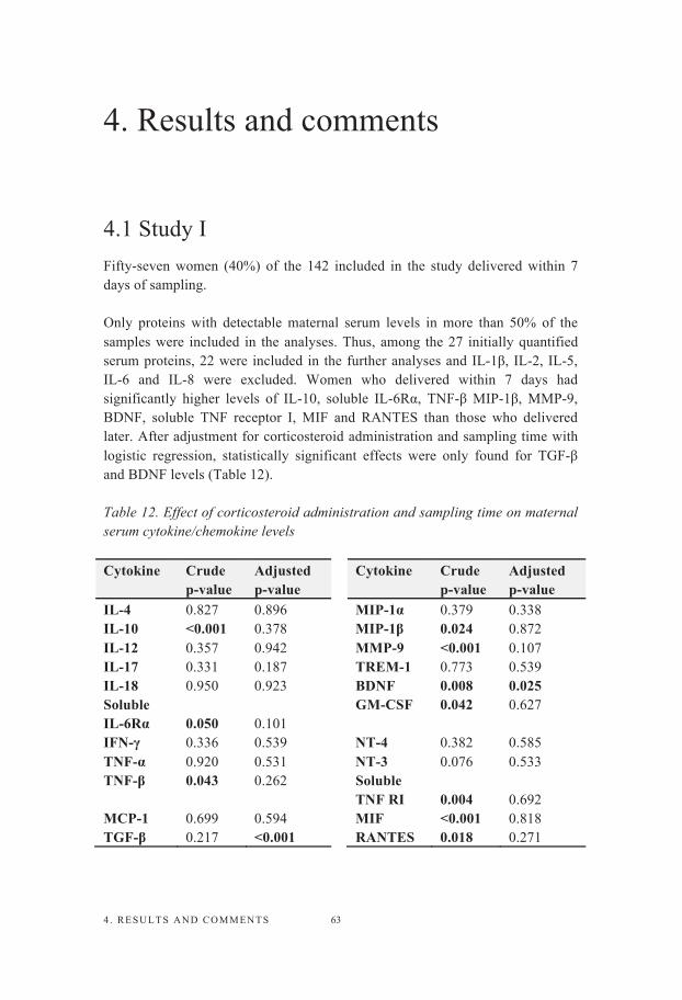
4 . RESULTS AND COMMENTS 63
4. Results and comments
4.1 Study I Fifty-seven women (40%) of the 142 included in the study delivered within 7 days of sampling.
Only proteins with detectable maternal serum levels in more than 50% of the samples were included in the analyses. Thus, among the 27 initially quantified serum proteins, 22 were included in the further analyses and IL-1β, IL-2, IL-5, IL-6 and IL-8 were excluded. Women who delivered within 7 days had significantly higher levels of IL-10, soluble IL-6Rα, TNF-β MIP-1β, MMP-9, BDNF, soluble TNF receptor I, MIF and RANTES than those who delivered later. After adjustment for corticosteroid administration and sampling time with logistic regression, statistically significant effects were only found for TGF-β and BDNF levels (Table 12).
Table 12. Effect of corticosteroid administration and sampling time on maternal serum cytokine/chemokine levels
Cytokine Crude p-value
Adjusted p-value
Cytokine Crude p-value
Adjusted p-value
IL-4 0.827 0.896 MIP-1α 0.379 0.338 IL-10 <0.001 0.378 MIP-1β 0.024 0.872 IL-12 0.357 0.942 MMP-9 <0.001 0.107 IL-17 0.331 0.187 TREM-1 0.773 0.539 IL-18 0.950 0.923 BDNF 0.008 0.025 Soluble IL-6Rα 0.050 0.101
GM-CSF 0.042 0.627
IFN-γ 0.336 0.539 NT-4 0.382 0.585 TNF-α 0.920 0.531 NT-3 0.076 0.533 TNF-β 0.043 0.262 Soluble
TNF RI 0.004 0.692 MCP-1 0.699 0.594 MIF <0.001 0.818 TGF-β 0.217 <0.001 RANTES 0.018 0.271

4 . RESULTS AND COMMENTS 64
Cervical length was significantly shorter among women who delivered within 7 days (5.5 mm vs 21 mm; p <0.001), compared to women who delivered later.
The ROC curve analysis used to calculate the best cut-off value and the AUC for every single protein and cervical length showed that the highest AUC values were for IL-10 (AUC = 0.69; p <0.001), MMP-9 (AUC = 0.74; p <0.001), MIF (AUC = 0.71; p <0.001) and cervical length (AUC = 0.77; p <0.001).
The highest crude ORs for delivery within 7 days were for IL-10 (OR 4.6; C.I. 2.2-9.7), MMP-9 (OR 6.0; C.I. 2.9-12.7), MIF (4.7; C.I. 2.2-9.9), RANTES (OR 3.9; C.I. 1.8-8.6) and cervical length (OR 21.2; C.I. 6.0-74.7).
Based on a stepwise multivariable logistic regression of dichotomous variables, univariate analyses were performed for the most significant serum proteins alone, then cervical length alone and, lastly, the combination of serum proteins and cervical length, in order to construct the best prediction model.
We found that the model combining proteins and cervical length had the best predictive capacity. In the multivariable model combining maternal serum proteins and cervical length, high levels of IL-10 (≥ 48 pg/ml; OR 5.0; 95% CI 1.7–14.3), RANTES (≥ 49293 pg/ml; OR 9.9; 95% CI 2.5–39.2) and short cervical length (≤18 mm; OR 43.5; 95% CI 8.9–211.3) were statistically significant contributors to the prediction of spontaneous PTD within 7 days of sampling.
Using this combined model, the ROC curve analysis showed an AUC of 0.88, sensitivity 74%, specificity 87%, positive predictive value 76%, negative predictive value 86%, + LR 5.83 and - LR 0.30.
The Hosmer-Lemeshow test of goodness of fit of the combined model yielded a p-value of 0.91. The regression equation for the combined model is Logit P= -4.71 + 1.62xIL-10 + 2.29xRANTES + 3.77xCL (CL is cervical length).
In the regression equation, all the variables are binary values. In order to ascertain whether adding continuous variables to the regression model would be a more powerful approach, an analysis with the variables as continuous variables in the combined model was performed (Table 13).

4 . RESULTS AND COMMENTS 65
Table 13. Predictive capacity of the combined model, comparison of binary and continuous variables
Binary approach Continuous approach
Correctly predicted 82.6% 77.6%
Sensitivity 73.8% 73.8% Specificity 87.3% 82.2% Positive predictive value 75.6% 68.9% Negative predictive value 86.2% 85.5% +LR 5.83 4.16 - LR 0.30 0.32 AUC 0.88 (0.84-0.95) 0.86 (0.79-0.92)
The binary approach has a predictive capacity resembling that of the continuous approach, as shown above. Moreover, the binary approach would be easier for clinicians to use bedside. Since the continuous variables in this study can have very extreme values, OR estimation can be very uncertain. For a more precise estimation of the continuous variables, more observations including many extreme values would be required.
In order to validate the combined model in this study, two resampling methods were used, i.e. bootstrapping (Table 14) and cross-validation. In practice, resampling is based on repeatedly drawing samples from a training set of observations and refitting a model on each sample in order to obtain additional information about the fitted model.
Table 14. Validation of the combined prediction model with bootstrapping, simulation of 1000 studies
Combined model OR (95% CI)
Bootstrapping Mean OR
Cervical length High RANTES, plasma
43.5 (8.9-211.3) 9.9 (2.5-39.2)
54.9 11.1
High IL-10, plasma 5.0 (1.7-14.3) 5.6 AUC (95% CI) Mean AUC (95% CI) 0.88 (0.84-0.95) 0.89 (0.84-0.94)

4 . RESULTS AND COMMENTS 66
According to these results, the mean AUC resulting from bootstrapping is approximately the same as the AUC from the original analysis. The mean ORs for cervical length, RANTES and IL-10 are higher than those found in the original analysis.
The cross-validation method (leave-one-out method) yielded an AUC of 0.83 (0.76-0.91). This indicates that performing the same study on another cohort of women with imminent spontaneous PTD might have led to a somewhat lower AUC for the same combined predictive model.
However, the results with the resampling methods suggest that the combined model is robust in predicting spontaneous PTD within 7 days of sampling.
4.2 Study IIAs in Study I, only proteins with detectable maternal serum levels in more than 50% of the samples were included in the analyses. Thus, among the 27 initially quantified serum proteins, 25 were included in the further analyses and IL-2 and IL-5 were excluded.
The median gestational age at sampling and delivery were 32+0 (range 22+6–33+6) and 34+5 weeks (range 32+1–43+0), respectively. The overall rate of MIAC was 29% (21 of 73) in women with PPROM and 19% (22 of 116) in women with PTL.
To evaluate the effect of the interval (days) from blood sample storage to multiple immunoassay analysis on the outcome presence or absence of MIAC, a linear regression (one-way ANOVA) was performed, revealing no differences, either for the PPROM group (F=0.304; p=0.583) or the PTL group (F=1.122; p=0.292).
Women with PPROM and PTL were evaluated separately. In both groups, the data were classified according to the whole study population group (all PPROM and all PTL), women with gestational age at admission <32+0 weeks and women with gestational age at admission ≥32+0 weeks.
High levels of maternal serum IL-18 (451 vs 335; p=0.031) and low levels of MCP-1 (139 vs 213, P=0.039) were observed in the PPROM with MIAC group. In the subgroup of women with PPROM at < 32+0 weeks, maternal serum IL-18

4 . RESULTS AND COMMENTS 67
levels were significantly higher (654 vs 361; p=0.003) and levels of IL-1β were significantly lower (9 vs 20; p=0.008) in women with MIAC than in those without. No significant association was found between MIAC and any of the cytokine levels at over 32+0 weeks of gestation.
In the group of women with PTL, there were no significant differences in the inflammatory response in those with MIAC. Only IL-6 levels were significantly higher (21 vs 14, P=0.019) in the subgroup of women with PTL at <32+0 weeks of gestation. No significant association was observed with any cytokine at over 32+0 weeks of gestation.
The stratification into two gestational age subgroups (less and more than 32 weeks) in this study was based on the finding from a study by Kacerovsky et al. [52] that the intra-amniotic inflammatory response to bacteria in pregnancies complicated by PPROM seems to be different at gestational ages above and below 32 weeks.
After adjustment for corticosteroid administration and sampling time using logistic regression, the only significant effect was on IL-6 levels in the PPROM group (Table 15).
Table 15. Effect of corticosteroid administration and sampling time on maternal serum cytokine/chemokine levels
Cytokine PTL Crude p-value
Adjusted p-value
PPROM Crude p-value
Adjusted p-value
IL-1β 0.441 0.176 0.135 0.226 IL-4 0.974 0.577 0.244 0.227 IL-6 0.124 0.289 0.226 0.037 IL-8 0.178 0.680 0.386 0.278 IL-10 0.249 0.295 0.316 0.801 IL-12 0.635 0.345 0.168 0.209 IL-17 0.050 0.946 0.873 0.461 IL-18 0.082 0.129 0.031 0.071 Soluble IL-6Rα 0.116 0.090 0.888 0.954 IFN-γ 0.598 0.184 0.709 0.533 TNF-α 0.572 0.214 0.484 0.270 TNF-β 0.266 0.216 0.939 0.976 MCP-1 0.779 0.460 0.039 0.051

4 . RESULTS AND COMMENTS 68
TGF-β 0.740 0.152 0.538 0.885 MIP-1α 0.297 0.239 0.334 0.129 MIP-1β 0.290 0.323 0.788 0.694 MMP-9 0.373 0.430 0.636 0.478 TREM-1 0.174 0.159 0.611 0.853 BDNF 0.157 0.936 0.183 0.269 GM-CSF 0.470 0.104 0.418 0.423 NT-4 0.214 0.355 0.176 0.242 NT-3 0.509 0.710 0.238 0.470 Soluble TNF RI 0.330 0.736 0.573 0.676 MIF 0.322 0.113 0.384 0.340 RANTES 0.412 0.466 0.711 0.873
No statistically significant differences were found between the PPROM and PTL groups regarding the type of microorganism isolated from the amniotic fluid in MIAC-positive cases (p=0.250) (Table 16).
Table 16. Microorganisms isolated in amniotic fluid of MIAC-positive cases
PPROM PTL Fungi 0 1 Gram-negative bacilli 3 5 Gram-positive bacilli 2 3 Gram-positive cocci 7 10 Ureaplasma urealyticum 9 3
4.3 Study III In this study, IL-6 concentrations were assayed in 21 amniotic fluid samples, divided into nine groups that were handled with different protocols. The demographics of the study population are displayed in Table 17.
Table 17. Maternal demographics. Values are shown as median, 25th (Q1) and 75th (Q3) quartiles
Maternal age, years (median, Q1–Q3) 34 (30–36)
Nulliparous 9 (43%)

4 . RESULTS AND COMMENTS 69
Gestational age at cesarean section (median, Q1–Q3) 39+0 (38+5-39+2)
Smoker 2 (10%)
Maternal BMI (median, Q1–Q3) 25 (22–27)
The median and quartile (Q1 and Q3) values of the amniotic fluid IL-6 for each group are listed in Table 18 and presented graphically in Figure 6.
Table 18. Amniotic fluid IL-6. Values are shown as median, 25th (Q1) and 75th (Q3) quartiles
Figure 6. Amniotic fluid IL-6 concentrations (pg/mL), presented by group
Median (pg/mL) Q1 (pg/mL) Q3 (pg/mL) Standard group 615 342 1040 Group 1a 585 354 1052 Group 1b 610 351 1002 Group 2a 610 355 977 Group 2b 615 358 1015 Group 3 580 350 995 Group 4 605 346 1000 Group 5 605 343 1000 Group 6 606 349 1005

4 . RESULTS AND COMMENTS 70
The IL-6 concentrations in the standard group ranged between 211 and 2005 pg/mL, with a median value of 615 pg/mL. No significant differences were found, in the median or dispersion values of the IL-6 concentrations, between the standard group and the six test groups.
The differences between IL-6 concentrations in each test group and IL-6 concentrations in the standard group were computed (Figure 7).
Figure 7. Schematic presentations of the differences in IL-6 concentrations between the test groups and the standard group. p-values are calculated with Wilcoxon signed-rank test. The markers (o) in the diagram represent outliers and ( ) extreme values [265]
There were no significant differences in amniotic fluid IL-6 concentrations between the test groups and the standard group.

4 . RESULTS AND COMMENTS 71
4.4 Study IV A total of 231 women with diagnosed PPROM were included in the analyses. One hundred forty-two cases (61%) were HCA-positive and 89 cases (39%) were HCA-negative. Among the HCA-positive cases, 19% were also positive for funisitis.
In this study, gestational age at delivery, latency from PPROM to delivery and birth weight cannot be considered as outcomes, because of the active management of PPROM pregnancies in the Czech Republic.
Women with HCA had neonates with significantly lower birth weight (1,840 g vs 2190 g; p<0.001) and higher rates of composite neonatal morbidity (75 cases vs 30 cases; p=0.005), RDS (52 cases vs 18 cases; p=0.008), BPD (18 cases vs 2 cases; p=0.006) and EOS (15 cases vs 1 case; p=0.001), than those without HCA.
After adjustment for gestational age at delivery, EOS differed significantly between these two groups [15 (11%) vs 1 (1%); p=0.011]. After adjustment for corticosteroid and antibiotics administration, EOS (p=0.027) and birth weight (p=0.032) differed significantly between the two groups. This significant difference remained only for EOS (p=0.029) after adjustment for corticosteroids and antibiotics administration and gestational age at delivery (Table 19).
Table 19. Neonatal outcomes in the groups with and without HCA. Crude p-value (P1); adjusted for gestational age at delivery (P1); adjusted for corticosteroid and antibiotics administration (P3) and adjusted for corticosteroid and antibiotics administration and gestational age at delivery (P4)
P1 P2 P3 P4 Birth weight (g) <0.001 0.153 0.032 0.158 5-min Apgar score <7 0.109 0.890 0.159 0.608 10-min Apgar score <7 0.155 0.788 0.179 0.620 Affected neonatal WBC count 0.615 0.229 0.117 0.120 Positive hemoculture 0.259 0.526 0.386 0.461 Neonatal composite morbidity 0.005 0.527 0.077 0.423 NICU stay ≥1 d 0.116 0.659 0.593 0.706 Tracheal intubation 0.014 0.530 0.077 0.303 Duration tracheal intubation ≥1 d 0.011 0.472 0.348 0.554

4 . RESULTS AND COMMENTS 72
Duration nCPAP ≥1 d 0.067 0.887 0.319 0.899 RDS 0.008 0.574 0.163 0.606 Perinatal death 0.058 0.810 0.136 0.390 IVH 0.479 0.959 0.964 0.814 IVH grade 1-2 0.501 0.904 0.922 0.932 IVH grade 2-4 0.575 0.764 0.871 0.810 NEC 0.389 0.797 0.667 0.817 Intestinal perforation 0.945 0.342 0.569 0.318 Patent ductus arteriosus 0.175 0.175 0.443 0.717 ROP 0.187 0.798 0.547 0.906 BPD 0.006 0.332 0.071 0.196 Pneumonia 0.181 0.490 0.217 0.405 Pulmonary hypertension 0.181 0.426 0.368 0.442 EOS 0.006 0.011 0.027 0.029 LOS 0.704 0.234 0.704 0.241 HCA: Histological chorioamnionitis; WBC: White blood cells; NICU: Neonatal intensive care unit; nCPAP: Nasal continuous positive airway pressure; RDS: Respiratory distress syndrome; IVH: Intraventricular hemorrhage; NEC: Necrotizing enterocolitis; ROP: Retinopathy of prematurity; BPD: bronchopulmonary dysplasia; EOS: early-onset sepsis; LOS: late-onset sepsis
Neonates with funisitis had significantly lower birth weight (1,630 g vs 2,080 g; p <0.001) and Apgar scores at 5 (10 cases vs 12 cases; p=0.001) and 10 min (6 cases vs 5 cases; p=0.002). The neonates with funisitis had higher rates of composite neonatal morbidity (33 cases vs 72 cases; p <0.001), perinatal death (11% vs 3%; p=0.001), RDS (24 cases vs 46 cases; p <0.001), ROP (10 cases vs 7 cases; p <0.001), BPD (11 cases vs 9 cases; p <0.001), EOS (18% vs 4%; p=0.001) and LOS (11% vs 4%; p=0.040).
After adjustment for gestational age at delivery, ROP [10 (23%) vs 7 (4%); p=0.014] and EOS [8 (18%) vs 8 (4%); p=0.002] differed significantly between these two groups. After adjustment for corticosteroid and antibiotics administration, birth weight (p<0.001), 5-min Apgar score <7 (p=0.004), neonatal composite morbidity (p<0.001), NICU stay ≥1 day (p=0.017), tracheal intubation (p=0.014), duration tracheal intubation ≥1 day (p=0.020), duration nCPAP ≥1 day (p=0.013), perinatal death (p=0.046), ROP (p=0.002), BPD (p=0.004) and EOS (p=0.001) differed significantly between the two groups. This significant difference remained only for ROP (p=0.018) and EOS (p=0.001) after adjustment for corticosteroid and antibiotics administration and gestational age at delivery (Table 20).

4 . RESULTS AND COMMENTS 73
Table 20. Neonatal outcomes in the group of women with and without funisitis. Crude p-value (P1); adjusted for gestational age at delivery (P2); adjusted for corticosteroid and antibiotics administration (P3) and corticosteroid and antibiotics administration and gestational age at delivery (P4)
P1 P2 P3 P4 Birth weight (g) <0.001 0.255 <0.001 0.339 5-min Apgar score <7 0.001 0.331 0.004 0.516 10-min Apgar score <7 0.002 0.426 0.007 0.646 Affected neonatal WBC count 0.725 0.204 0.168 0.170 Positive hemoculture 0.042 0.193 0.124 0.317 Neonatal composite morbidity <0.001 0.220 <0.001 0.110 NICU stay ≥1 d 0.002 0.827 0.017 0.578 Tracheal intubation 0.001 0.436 0.014 0.684 Duration tracheal intubation ≥1 d 0.003 0.886 0.020 0.152 Duration nCPAP ≥1 d 0.002 0.722 0.013 0.510 RDS <0.001 0.378 0.009 0.527 Perinatal death 0.011 0.897 0.046 0.850 IVH 0.394 0.756 0.813 0.848 IVH grade 1-2 0.792 0.529 0.787 0.646 IVH grade 2-4 0.112 0.694 0.285 0.831 NEC 0.228 0.738 0.470 0.801 Intestinal perforation 0.228 0.993 0.470 0.941 Patent ductus arteriosus 0.101 0.965 0.318 0.961 ROP <0.001 0.014 0.002 0.018 BPD <0.001 0.066 0.004 0.271 Pneumonia 0.192 0.686 0.471 0.639 Pulmonary hypertension 0.515 0.843 0.988 0.758 EOS 0.001 0.002 0.001 0.001 LOS 0.040 0.949 0.199 0.837 HCA: Histological chorioamnionitis; WBC: White blood cells; NICU: Neonatal intensive care unit; nCPAP: Nasal continuous positive airway pressure; RDS: Respiratory distress syndrome; IVH: Intraventricular hemorrhage; NEC: Necrotizing enterocolitis; ROP: Retinopathy of prematurity; BPD: bronchopulmonary dysplasia; EOS: early-onset sepsis; LOS: late-onset sepsis

74

5 . DISCUSSION 75
5. Discussion
PTD, accounting worldwide for more than 15 million preterm neonates annually, is a major perinatal health problem, not only in terms of associated mortality, but also with regard to morbidity and its economic implications [12].
Despite significant advances in obstetric and perinatal care, there has been little progress in reducing the global PTD rate [15]. Because of the complex nature of spontaneous PTD and the involvement of several pathways in its pathogenesis, prediction and prevention remain difficult and pose a continuing and significant challenge in maternal and fetal medicine.
Major effort has been devoted to identifying inflammatory biomarkers to predict spontaneous PTD in both asymptomatic and symptomatic women and to improve our understanding of the mechanisms and pathways leading to this condition. While several biomarkers have been tested as predictors of spontaneous PTD, few have proven useful for clinical purposes [15, 244]. So far, among the biomarkers evaluated, fetal fibronectin (fFN) in the cervico-vaginal secretion, together with short cervical length on transvaginal ultrasound has been found to be the most powerful and consistent predictor of spontaneous PTD (sensitivity around 80%, specificity around 61%, positive predictive value around 17% and negative predictive value around 97%) [41, 95, 266-269]. The clinical usefulness of both tests lies primarily in their negative predictive value, aiding in avoiding unnecessary interventions and guiding clinicians in decision-making regarding in utero transfer, administration of antenatal corticosteroids and/or tocolysis.
To progress in our understanding and prediction of spontaneous PTD, we must acknowledge that it is not one disease with a single solution or cure, but rather the product of overlapping pathophysiological pathways [39]. Indeed, it is has been suggested that a single biomarker cannot accurately predict spontaneous PTD [244, 270, 271]. Since the pathophysiology of spontaneous PTD is multifactorial with different pathways and biomarkers involved, multiplex immunoassay analysis have been introduced in the last decade, with the aim of simultaneously using several biomarkers from different and distinct biological pathways to better predict the risk [272]. These multiplex analyses of biomarker

5 . DISCUSSION 76
panels thus constitute the future direction of biomarker research [244]. Studies have shown that a combination of biomarkers in minimally invasive samples (cervical fluid, serum) and cervical length measurement could help to better predict MIAC [273] and spontaneous PTD in women with PPROM [274] and PTL [260, 275]. The development of predictive models based on the analysis of multiple biomarkers seems feasible and allows early identification, and potentially early intervention, before the onset of labor, in women at risk.
A perfect predictor of spontaneous PTD must have good specificity, sensitivity and high accuracy in order to increase the probability of diagnosis in women with a positive test result, as well as to rule out the condition when test results are negative [243, 272, 276]. Feasibility, accessibility, patient convenience and minimally invasive sampling are also important. Although a non-invasive approach is preferable for predicting spontaneous PTD, it is currently unclear which non-invasively sampled body fluid best represents the intra-amniotic environment and whether biomarkers in non-invasive samples reflect the intra-amniotic compartment adequately [272]. The accuracy of serum or plasma biomarkers for predicting spontaneous PTD in asymptomatic women has been shown to be very low [270].
In the following, the main findings of the studies in this thesis and the main findings concerning the analyzed biomarkers are briefly discussed. More detailed discussion is found in the Discussion section of each publication.
Study I
In the last decade, there have been efforts to predict spontaneous PTD in women with PTL using cervical length, alone [257, 277-284] or in combination with cervical biomarkers [260, 266, 285-291]. A cervical length of <15 mm (found in less than 10% of symptomatic women) detects approximately 60% of those who will deliver within 7 days and represents a 5.7-fold increased risk of delivering within 7 days [292]. Few studies have investigated the relationship between biomarkers in maternal serum and spontaneous PTD in women with PTL, suggesting that effective prediction of spontaneous PTD may be possible with a non-invasive approach [293-302]. In Study I, it was found that a multivariable prediction model, combining maternal serum biomarkers (IL-10 and RANTES) with cervical length, had predictive power equal to that of invasive models for delivery within 7 days from the beginning of labor [275].

5 . DISCUSSION 77
IL-10 and RANTES were statistically significant contributors to the prediction of spontaneous PTD within 7 days of sampling in Study I. IL-10 is likely to be an important cytokine because it suppresses the production of pro-inflammatory cytokines by other cells and numerous studies have documented its production at the maternal-fetal interface [303-305]. Pregnancy is not dependent on IL-10 production, however; IL-10 (-/-) mice are still fertile [306]. Up-regulation of this anti-inflammatory cytokine leads to suppression of NK cells and T lymphocytes against fetal alloantigens [307, 308]. In Study I, maternal serum IL-10 levels were significantly higher in women who delivered preterm (within 7 days of sampling) than in women who delivered later. We can therefore hypothesize that IL-10 can lessen the pathological effect of inflammation prevalent in spontaneous PTD. The other cytokine in our prediction model is RANTES. Maternal serum RANTES levels were significantly higher in women who delivered preterm (within 7 days of sampling), compared with women who delivered later. This pro-inflammatory cytokine that chemoattracts monocytes, with additional activity toward T-cells, has been found in the decidua of pregnancies complicated with spontaneous PTD. Its expression in this tissue is correlated with both macrophage and neutrophil abundance, suggesting that these immune cell subtypes are a major production source [309]. However, there are few studies describing the association between high levels of RANTES in maternal serum and spontaneous PTD [309-311].
Study II
There are few studies, with contradictory results, on the role of maternal serum biomarkers as predictors of IAI and fetal membrane inflammation [249, 312-316]. In Study II, the maternal serum inflammatory response was evaluated according to the presence of MIAC in women with PTL and PPROM. We found that the inflammatory status of the amniotic cavity is not accurately reflected in maternal serum. Studies have shown that sampling from maternal serum and other non-invasive procedures is effective in predicting PTD [293-302], but it seems that maternal serum sampling is not so effective in predicting MIAC. A weak maternal inflammatory response in women with MIAC was found and differences in the biomarker levels were only evident at early gestational ages (<32 weeks). This finding is in accordance with previous studies showing that the intra-amniotic inflammatory response to bacteria is different below and above 32 weeks of gestation. This process seems to be heterogeneous as a result of a broad spectrum of factors that can affect cytokine production in amniotic

5 . DISCUSSION 78
fluid, including the duration of MIAC, the type of bacteria, the microbial burden and bacterial virulence [52, 317].
In Study II, a higher but weak expression of serum IL-6 in women with PTL and MIAC was observed. Despite the fact that IL-6 is a classical marker of inflammation, no differences in the concentration of this serum protein were found between PPROM cases with or without MIAC. Higher IL-6 concentrations have been observed in the amniotic fluid of asymptomatic women delivering preterm, compared to those with term delivery [244], but a temporal relationship between IL-6 and interval until delivery has yet not been established [318]. The test accuracy of IL-6 in amniotic fluid seems to be especially good for both asymptomatic and symptomatic women, but is generally poor in other biological fluids (e.g. blood, cervico-vaginal fluid) [270, 276]. However, from a clinical point of view, the main limitation of this biomarker is its lack of specificity in predicting disease, because it is overexpressed in maternal serum in both infectious and non-infectious conditions [319, 320].
There have been no previous studies on the role of maternal serum IL-18 in women with MIAC. In Study II, higher levels of maternal serum IL-18 were found in women with PPROM who had MIAC, which suggests a pro-inflammatory effect of IL-18 in amniotic fluid and maternal serum in the presence of infection. It has previously been shown that IL-18 in amniotic fluid is associated with MIAC and spontaneous PTD in women with PTL [321]. IL-18 is a cytokine with pleiotropic qualities that regulate both the innate and acquired immune responses and it can stimulate both Th 1 and 2 responses, depending on the local cytokine environment [322, 323]. IL-18 is synthesized as a pro-form and is activated through cleavage by caspase-1 [324]. It is important in the host defense against severe infections via induction of other cytokines and effector cells and molecules. It enhances the inflammatory process by stimulating the production of IFN-γ, TNF-α and IL-1β [323]. IL-18 can also activate apoptosis by enhancing Fas ligand and Fas expression [325] and is found in both amniotic fluid and maternal and fetal plasma [326-328].
There is a lack of previous studies evaluating the importance of IL-1β and MCP-1 as risk factors for MIAC in maternal serum. In Study II, low levels of IL-1β and MCP-1 were observed in women with PPROM who had MIAC, indicating only a minor maternal serum inflammatory response mediated by those two cytokines. IL-1 plays a central role in the pro-inflammatory response. It is a prototypical pro-inflammatory cytokine that regulates a wide spectrum of immunological processes and it exists in two forms, IL-1α and IL-1β, both of

5 . DISCUSSION 79
which bind to two distinct membrane receptors [329]. IL-1β induces a systemic and local response to pathogenic invasion or tissue injury by inducing transcription or enhancing mRNA stability of a wide variety of pro-inflammatory genes. It has been shown to be a major factor in the initiation of infection-related PTL and spontaneous PTD [330]. IL-1β levels are elevated in the amniotic fluid of pregnancies complicated by microbial-associated IAI and it may be capable of causing PTL by up-regulating prostaglandin production, leading to myometrial contractions [331-333]. IL-1β also enhances MMP-1 and MMP-3 expression in decidual cells, possibly leading to PPROM [334]. MCP-1 stimulates chemotaxis of monocytes and several associated cellular events, including Ca+2 flux and integrin expression. It is also a weak inducer of cytokine expression in monocytes and, at high concentrations, elicits a respiratory burst leading to generation of reactive oxygen species [335].
Studies I and II have several strengths. We evaluated several biomarkers in maternal serum that can be easily tested during pregnancy. A non-invasive approach for predicting spontaneous PTD is relevant for clinical practice. Moreover, we adjusted for a possible confounding effect of gestational age at sampling and corticosteroid and antibiotics administration, in order to determine the respective effect of those covariates on serum levels of the inflammatory cytokines. Despite these strengths, an important limitation of these studies arises from the pre-analytical variability in sampling and storage procedures that has been shown to affect assayed biomarker concentrations. The best way to limit the effect of pre-analytical factors is to start sample processing immediately and to standardize it as much as possible. This approach was not, however, feasible in our studies for logistic reasons.
Study III
It is known that cytokines are fragile, that plasma and serum samples should be frozen promptly for long-term storage and that repeated freeze-thaw cycles should be avoided [336]. Some evidence indicates that a partial degradation of proteins occurs over time, even when stored at -80oC [337]. Amniotic fluid specimens are commonly frozen and stored for research purposes. During the last two decades, several studies of poor obstetric outcomes have been based on data from analysis of cytokine levels in frozen amniotic fluid [314, 338, 339]. Some studies have examined the stability of certain cytokines stored in amniotic fluid samples for prolonged periods of time [337, 340], revealing some degradation despite optimal freezing conditions. However, the impact of sample

5 . DISCUSSION 80
age and storage time on levels of inflammatory biomarkers is often inadequately addressed in studies.
On the other hand, little is known about how pre-analytical handling procedures affect cytokine levels. Skogstrand et al. [341] showed that trustworthy measurements of various inflammatory proteins in serum, plasma and whole blood relies on preparation and interval before long-term storage. According to this study, 4oC was the best temperature at which to preserve blood before separation into plasma and serum. It should, however, be noted that, even at this low temperature, measurable concentrations of several of the inflammatory markers increased significantly during the storage period. However, there are no data evaluating cytokine stability in amniotic fluid after different pre-analytical handling procedures. Study III showed that the investigated pre-analytical handling procedures ̶ latency between sampling and analysis, centrifugal conditions (centrifugal force, time, and temperature), supernatant filtration and addition of protease inhibitor ̶ did not seem to affect IL-6 concentrations in amniotic fluid.
The strength of Study III is the analysis of amniotic fluid samples after a short storage time in the freezer. Nonetheless, a limitation of the study is that only amniotic fluid from uncomplicated term pregnancies was used, instead of from second- and/or early third-trimester pregnancies, or from women with symptoms of PTL or with PPROM. Moreover, this study does not allow prediction of the degree of change in IL-6 levels in the samples after a prolonged storage time. The influence of the studied handling procedures on other amniotic fluid cytokines and the effect of storage time on the IL-6 concentrations after different pre-analytical handling procedures were not evaluated in this study and thus require investigation in future.
Study IV
The few studies evaluating the influence of MIAC on short-term neonatal outcome in women with PPROM have found a negative impact on neonatal morbidity [342-347]. However, a recent study showed that gestational age at delivery seems to be more important for short-term neonatal outcome than MIAC or IAI in PPROM pregnancies [348]. Furthermore, HCA and funisitis have been linked with EOS in many studies [349-353]. Both MIAC and HCA have also been associated with increased risk of EOS in pregnancies complicated by PPROM before 34 weeks of gestation [346]. It has been also shown that the

5 . DISCUSSION 81
higher the stage (or grade) of either HCA or funisitis and the earlier the gestational age at delivery, the higher the incidence of neonatal morbidity [354-356].
Study IV confirmed that some adverse neonatal outcomes (EOS and ROP) are associated with HCA and funisitis in PPROM pregnancies. The results of this study are in accordance with the results of a nationwide population-based study by Morken et al. [2], based on data from the Swedish Medical Birth Register and the Swedish Hospital Discharge Register. This study showed that preterm infants were at increased risk of sepsis at gestational age 32-33 weeks (Hazard Ratio 1.58; 95% CI: 1.12, 3.10) [13].
One of the strengths of Study IV is the relatively large number of women included, with a clearly defined phenotype of exclusively PPROM-induced PTD and a well-defined classification of HCA and funisitis. The active management of women with PPROM in the Czech Republic gave us the ability to compare short-term neonatal outcome, without the influence of long latencies on the evaluation of HCA and funisitis. Another strength of this study is the fact that the same pathologist examined all placentas, thus eliminating inter-observer variability. Limitations of the study are the facts that the infectious or non-infectious nature of the HCA was confirmed with non-cultivation techniques in amniotic fluid and that not all EOS cases were culture-positive.

82

6 . CONCLUSIONS AND FUTURE PERSPECTIVES 83
6. Conclusions and future perspectives
1. A combined prediction model, including non-invasive serum biomarkers and cervical length measurement by transvaginal sonography, is potentially useful for clinicians in determining which women with PTL symptoms have higher risk to spontaneously deliver preterm. This model must, however, be tested in a new cohort of high-risk women in order to confirm its predictive ability. If the moderately high positive predictive value and negative predictive value are thus confirmed, it could help identify women at considerable risk of delivering preterm, who could then be targeted for treatment (tocolytics to delay delivery, enabling transfer to a specialist unit, and corticosteroids to reduce neonatal morbidity and mortality). Likewise, women at low risk could be sent home without over-treatment.
2. The identification of inflammatory status in maternal serum in PPROM and PTL cases is not as accurate as analyzing amniotic fluid in predicting the occurrence of MIAC. A weak maternal inflammatory response in maternal serum is evident only at early gestational ages. This indicates the need for further research to establish the potential contribution of inflammatory cytokines that can be determined with non-invasive methods, in combination with those identified in amniotic fluid, to detect microbial-associated IAI in women with PPROM and PTL.
3. Amniotic fluid IL-6 levels appear to be stable regardless of sample handling prior to analysis, when analyses are performed after a short storage period.
4. HCA and funisitis in PPROM pregnancies are associated with increased rates of EOS and funisitis is associated with increased rates of EOS and ROP. From a clinical perspective, it is important that future studies clarify the role of placental pathology, MIAC, IAI, type of microorganism and gestational age for short- and long-term neonatal outcomes in spontaneous PTD with either PPROM or PTL onset.

84

ACKNOWLEDGEMENTS 85
Acknowledgements
I wish to express my sincere gratitude to:
Bo Jacobsson, my supervisor and colleague, for your never-ending enthusiasm, ideas, encouragement and support. Thank you for introducing me to the fascinating world of medical and clinical research and for staying with me throughout the whole journey! I also thank you for providing me with resources during my PhD studies and the possibility to meet so many interesting researchers during these years.
Karin Sävman, my co-supervisor and colleague, for kind support and encouragement. Designing studies with you has been a fascinating and engaging experience!
Marian Kacerovsky, my co-supervisor, for support, good advice and valuable input. Thank you for being so generous, both in research and privately, for creating such a great atmosphere and for advising me on small and big issues! Your prompt responses have been so important to me during my research journey.
Teresa Cobo, my co-supervisor, for endless support, advice and inspiring discussions, for generously sharing your extensive knowledge of preterm delivery, for practical help with research issues and, of course, for top-quality guidance in the different phases of my PhD studies.
Maria Hallingström, head of the Perinatal Laboratory, for kind and prompt support, help with practical issues and valuable input in organizing and analyzing samples and databases.
Jaroslava Johansson, colleague, friend and tutor during my residency, for listening and giving valuable support and advice. I would especially like to thank you for establishing the first contact with my research supervisor.

ACKNOWLEDGEMENTS 86
Rose-Marie Holst, co-author and colleague, for the enthusiastic support that you gave me during the years we worked together, both concerning research and clinical issues.
Jonas Bacelis, PhD student, for inspiring discussions and important statistical ideas and input during my studies.
Verena Sengpiel and Linda Englund-Ögge, wonderful research and clinical colleagues, for support, good advice and practical help with research issues.
All other co-authors, no one to be forgotten!
Collaborators and colleagues at the Perinatal Laboratory, for supportive talks and help with getting things done.
Ali Khatibi, friend and colleague, for good advice on small and big issues.
Andreas Fredriksson, friend and colleague, for help with the Swedish abstract of the thesis.
All friends and colleagues at the Department of Obstetrics and Gynecology, Sahlgrenska University Hospital, Gothenburg.
Vasilis, my best friend, for walking by my side all these years, encouraging and inspiring me in my life choices, and for kind and endless support whenever I needed it. Your life philosophy is going to accompany me for the rest of my life.
My parents, grandmother and brother, for always encouraging, inspiring and supporting me during my life. Thank you for making me who I am!
Dimitris, my friend and teacher, for support, advice and inspiration for my life as an adult.

REFERENCES 88
References
1. Pennell, C.E., et al., Genetic epidemiologic studies of preterm birth: guidelines for research. Am J Obstet Gynecol, 2007. 196(2): p. 107-18.
2. Morken, N.H., K. Kallen, and B. Jacobsson, Outcomes of preterm children according to type of delivery onset: a nationwide population-based study. Paediatr Perinat Epidemiol, 2007. 21(5): p. 458-64.
3. Nour, N.M., Premature delivery and the millennium development goal. Rev Obstet Gynecol, 2012. 5(2): p. 100-5.
4. Feng, Y., et al., Causes of death in infants admitted to Australian neonatal intensive care units between 1995 and 2006. Acta Paediatr, 2013. 102(1): p. e17-23.
5. Morken, N.H., et al., Reference population for international comparisons and time trend surveillance of preterm delivery proportions in three countries. BMC Womens Health, 2008. 8: p. 16.
6. Martin, J.A., B.E. Hamilton, and M.J. Osterman, Births in the United States, 2015. NCHS Data Brief, 2016(258): p. 1-8.
7. Yuan, W., et al., Analysis of preterm deliveries below 35 weeks' gestation in a tertiary referral hospital in the UK. A case-control survey. BMC Res Notes, 2010. 3: p. 119.
8. Romero, R., et al., The preterm parturition syndrome. BJOG, 2006. 113 Suppl 3: p. 17-42.
9. Jacobsson, B., et al., Microbial invasion and cytokine response in amniotic fluid in a Swedish population of women in preterm labor. Acta Obstet Gynecol Scand, 2003. 82(2): p. 120-8.
10. Jacobsson, B., et al., Microbial invasion and cytokine response in amniotic fluid in a Swedish population of women with preterm prelabor rupture of membranes. Acta Obstet Gynecol Scand, 2003. 82(5): p. 423-31.
11. Combs, C.A., et al., Amniotic fluid infection, inflammation, and colonization in preterm labor with intact membranes. Am J Obstet Gynecol, 2014. 210(2): p. 125 e1-125 e15.
12. Beck, S., et al., The worldwide incidence of preterm birth: a systematic review of maternal mortality and morbidity. Bull World Health Organ, 2010. 88(1): p. 31-8.
13. Blencowe, H., et al., National, regional, and worldwide estimates of preterm birth rates in the year 2010 with time trends since 1990 for selected countries: a systematic analysis and implications. Lancet, 2012. 379(9832): p. 2162-72.
14. Kramer, M.S., et al., Challenges in defining and classifying the preterm birth syndrome. Am J Obstet Gynecol, 2012. 206(2): p. 108-12.
87

REFERENCES 89
15. Goldenberg, R.L., et al., Epidemiology and causes of preterm birth. Lancet, 2008. 371(9606): p. 75-84.
16. Leading Health Indicators for Healthy People 2020: Letter Report. 2011: Washington (DC).
17. Marlow, N., et al., Neurologic and developmental disability at six years of age after extremely preterm birth. N Engl J Med, 2005. 352(1): p. 9-19.
18. Khan, K.S. and H. Honest, Risk screening for spontaneous preterm labour. Best Pract Res Clin Obstet Gynaecol, 2007. 21(5): p. 821-30.
19. Davey, M.A., et al., Risk scoring systems for predicting preterm birth with the aim of reducing associated adverse outcomes. Cochrane Database Syst Rev, 2011(11): p. CD004902.
20. Steer, P., The epidemiology of preterm labor--a global perspective. J Perinat Med, 2005. 33(4): p. 273-6.
21. McIntire, D.D. and K.J. Leveno, Neonatal mortality and morbidity rates in late preterm births compared with births at term. Obstet Gynecol, 2008. 111(1): p. 35-41.
22. Raju, T.N., et al., Optimizing care and outcome for late-preterm (near-term) infants: a summary of the workshop sponsored by the National Institute of Child Health and Human Development. Pediatrics, 2006. 118(3): p. 1207-14.
23. Gyamfi-Bannerman, C. and C.V. Ananth, Trends in spontaneous and indicated preterm delivery among singleton gestations in the United States, 2005-2012. Obstet Gynecol, 2014. 124(6): p. 1069-74.
24. Institute of Medicine Committee on Understanding Premature, B. and O. Assuring Healthy, The National Academies Collection: Reports funded by National Institutes of Health, in Preterm Birth: Causes, Consequences, and Prevention, R.E. Behrman and A.S. Butler, Editors. 2007, National Academies Press (US). National Academy of Sciences.: Washington (DC).
25. Tronnes, H., et al., The association of preterm birth with severe asthma and atopic dermatitis: a national cohort study. Pediatr Allergy Immunol, 2013. 24(8): p. 782-7.
26. Aarnoudse-Moens, C.S., et al., Meta-analysis of neurobehavioral outcomes in very preterm and/or very low birth weight children. Pediatrics, 2009. 124(2): p. 717-28.
27. Delobel-Ayoub, M., et al., Behavioral problems and cognitive performance at 5 years of age after very preterm birth: the EPIPAGE Study. Pediatrics, 2009. 123(6): p. 1485-92.
28. Van Lieshout, R.J., et al., Mental health of extremely low birth weight survivors in their 30s. Pediatrics, 2015. 135(3): p. 452-9.
29. Hovi, P., et al., Glucose regulation in young adults with very low birth weight. N Engl J Med, 2007. 356(20): p. 2053-63.
30. Rotteveel, J., et al., Infant and childhood growth patterns, insulin sensitivity, and blood pressure in prematurely born young adults. Pediatrics, 2008. 122(2): p. 313-21.
88

REFERENCES 90
31. Singer, L.T., et al., Maternal psychological distress and parenting stress after the birth of a very low-birth-weight infant. Jama, 1999. 281(9): p. 799-805.
32. Moutquin, J.M., Classification and heterogeneity of preterm birth. BJOG, 2003. 110 Suppl 20: p. 30-3.
33. Morken, N.H., et al., Preterm birth in Sweden 1973-2001: rate, subgroups, and effect of changing patterns in multiple births, maternal age, and smoking. Acta Obstet Gynecol Scand, 2005. 84(6): p. 558-65.
34. Mazaki-Tovi, S., et al., Recurrent preterm birth. Semin Perinatol, 2007. 31(3): p. 142-58.
35. Tucker, J. and W. McGuire, Epidemiology of preterm birth. BMJ, 2004. 329(7467): p. 675-8.
36. Morken, N.H., P. Magnus, and B. Jacobsson, Subgroups of preterm delivery in the Norwegian Mother and Child Cohort Study. Acta Obstet Gynecol Scand, 2008. 87(12): p. 1374-7.
37. Goldenberg, R.L., et al., The preterm birth syndrome: issues to consider in creating a classification system. Am J Obstet Gynecol, 2012. 206(2): p. 113-8.
38. Manuck, T.A., et al., The phenotype of spontaneous preterm birth: application of a clinical phenotyping tool. Am J Obstet Gynecol, 2015. 212(4): p. 487 e1-487 e11.
39. Villar, J., et al., The preterm birth syndrome: a prototype phenotypic classification. Am J Obstet Gynecol, 2012. 206(2): p. 119-23.
40. Barros, F.C., et al., The distribution of clinical phenotypes of preterm birth syndrome: implications for prevention. JAMA Pediatr, 2015. 169(3): p. 220-9.
41. Berghella, V., J.K. Baxter, and N.W. Hendrix, Cervical assessment by ultrasound for preventing preterm delivery. Cochrane Database Syst Rev, 2009(3): p. CD007235.
42. Romero, R., et al., Infection in the pathogenesis of preterm labor. Semin Perinatol, 1988. 12(4): p. 262-79.
43. Romero, R., et al., Inflammation in preterm and term labour and delivery. Semin Fetal Neonatal Med, 2006. 11(5): p. 317-26.
44. Timmons, B., M. Akins, and M. Mahendroo, Cervical remodeling during pregnancy and parturition. Trends Endocrinol Metab, 2010. 21(6): p. 353-61.
45. Holt, R., et al., The molecular mechanisms of cervical ripening differ between term and preterm birth. Endocrinology, 2011. 152(3): p. 1036-46.
46. Larsen, B. and J. Hwang, Progesterone interactions with the cervix: translational implications for term and preterm birth. Infect Dis Obstet Gynecol, 2011. 2011: p. 353297.
47. Romero, R., S.K. Dey, and S.J. Fisher, Preterm labor: one syndrome, many causes. Science, 2014. 345(6198): p. 760-5.
48. Mercer, B.M., et al., The Preterm Prediction Study: prediction of preterm premature rupture of membranes through clinical findings and
89

REFERENCES 91
ancillary testing. The National Institute of Child Health and Human Development Maternal-Fetal Medicine Units Network. Am J Obstet Gynecol, 2000. 183(3): p. 738-45.
49. Mercer, B.M., Preterm premature rupture of the membranes. Obstet Gynecol, 2003. 101(1): p. 178-93.
50. Parry, S. and J.F. Strauss, 3rd, Premature rupture of the fetal membranes. N Engl J Med, 1998. 338(10): p. 663-70.
51. Murtha, A.P. and R. Menon, Regulation of fetal membrane inflammation: a critical step in reducing adverse pregnancy outcome. Am J Obstet Gynecol, 2015. 213(4): p. 447-8.
52. Kacerovsky, M., et al., Intraamniotic inflammatory response to bacteria: analysis of multiple amniotic fluid proteins in women with preterm prelabor rupture of membranes. J Matern Fetal Neonatal Med, 2012. 25(10): p. 2014-9.
53. Kacerovsky, M., et al., The impact of the microbial load of genital mycoplasmas and gestational age on the intensity of intraamniotic inflammation. Am J Obstet Gynecol, 2012. 206(4): p. 342 e1-8.
54. Mercer, B.M., et al., Antibiotic therapy for reduction of infant morbidity after preterm premature rupture of the membranes. A randomized controlled trial. National Institute of Child Health and Human Development Maternal-Fetal Medicine Units Network. JAMA, 1997. 278(12): p. 989-95.
55. Ananth, C.V., J.M. Guise, and J.M. Thorp, Jr., Utility of antibiotic therapy in preterm premature rupture of membranes: a meta-analysis. Obstet Gynecol Surv, 1996. 51(5): p. 324-8.
56. Kenyon, S.L., et al., Broad-spectrum antibiotics for preterm, prelabour rupture of fetal membranes: the ORACLE I randomised trial. ORACLE Collaborative Group. Lancet, 2001. 357(9261): p. 979-88.
57. Gopalani, S., et al., Contemporary management of preterm premature rupture of membranes: determinants of latency and neonatal outcome. Am J Perinatol, 2004. 21(4): p. 183-90.
58. Kenyon, S., et al., Childhood outcomes after prescription of antibiotics to pregnant women with preterm rupture of the membranes: 7-year follow-up of the ORACLE I trial. Lancet, 2008. 372(9646): p. 1310-8.
59. Behnia, F., S. Sheller, and R. Menon, Mechanistic Differences Leading to Infectious and Sterile Inflammation. Am J Reprod Immunol, 2016. 75(5): p. 505-18.
60. Ward, K., et al., The heritability of preterm delivery. Obstet Gynecol, 2005. 106(6): p. 1235-9.
61. Treloar, S.A., et al., Genetic influences on premature parturition in an Australian twin sample. Twin Res, 2000. 3(2): p. 80-2.
62. Clausson, B., P. Lichtenstein, and S. Cnattingius, Genetic influence on birthweight and gestational length determined by studies in offspring of twins. Bjog, 2000. 107(3): p. 375-81.
63. Porter, T.F., et al., The risk of preterm birth across generations. Obstet Gynecol, 1997. 90(1): p. 63-7.
90

REFERENCES 92
64. Goffinet, F., Primary predictors of preterm labour. BJOG, 2005. 112 Suppl 1: p. 38-47.
65. Iams, J.D., et al., Primary, secondary, and tertiary interventions to reduce the morbidity and mortality of preterm birth. Lancet, 2008. 371(9607): p. 164-75.
66. Hua, M., et al., Congenital uterine anomalies and adverse pregnancy outcomes. Am J Obstet Gynecol, 2011. 205(6): p. 558 e1-5.
67. Chan, Y.Y., et al., Reproductive outcomes in women with congenital uterine anomalies: a systematic review. Ultrasound Obstet Gynecol, 2011. 38(4): p. 371-82.
68. Airoldi, J., et al., Transvaginal ultrasonography of the cervix to predict preterm birth in women with uterine anomalies. Obstet Gynecol, 2005. 106(3): p. 553-6.
69. Cnattingius, S., The epidemiology of smoking during pregnancy: smoking prevalence, maternal characteristics, and pregnancy outcomes. Nicotine Tob Res, 2004. 6 Suppl 2: p. S125-40.
70. Naeye, R.L., Effects of maternal cigarette smoking on the fetus and placenta. Br J Obstet Gynaecol, 1978. 85(10): p. 732-7.
71. Hadley, C.B., D.M. Main, and S.G. Gabbe, Risk factors for preterm premature rupture of the fetal membranes. Am J Perinatol, 1990. 7(4): p. 374-9.
72. Thompson, J.M., et al., Secular trends in socio-economic status and the implications for preterm birth. Paediatr Perinat Epidemiol, 2006. 20(3): p. 182-7.
73. Kaufman, J.S., et al., Modeling community-level effects on preterm birth. Ann Epidemiol, 2003. 13(5): p. 377-84.
74. O'Campo, P., et al., Neighborhood deprivation and preterm birth among non-Hispanic Black and White women in eight geographic areas in the United States. Am J Epidemiol, 2008. 167(2): p. 155-63.
75. Pickett, K.E., et al., Neighborhood socioeconomic status, maternal race and preterm delivery: a case-control study. Ann Epidemiol, 2002. 12(6): p. 410-8.
76. Lu, M.C. and B. Chen, Racial and ethnic disparities in preterm birth: the role of stressful life events. Am J Obstet Gynecol, 2004. 191(3): p. 691-9.
77. Kramer, M.R., et al., Preconceptional stress and racial disparities in preterm birth: an overview. Acta Obstet Gynecol Scand, 2011. 90(12): p. 1307-16.
78. Lumley, J., The epidemiology of preterm birth. Baillieres Clin Obstet Gynaecol, 1993. 7(3): p. 477-98.
79. Hauger, M.S., et al., Prepregnancy weight status and the risk of adverse pregnancy outcome. Acta Obstet Gynecol Scand, 2008. 87(9): p. 953-9.
80. Hoellen, F., et al., Does Maternal Underweight Prior to Conception Influence Pregnancy Risks and Outcome? In Vivo, 2014. 28(6): p. 1165-1170.
91

REFERENCES 93
81. Khatibi, A., et al., Prepregnancy maternal body mass index and preterm delivery. Am J Obstet Gynecol, 2012. 207(3): p. 212 e1-7.
82. Englund-Ogge, L., et al., Association between intake of artificially sweetened and sugar-sweetened beverages and preterm delivery: a large prospective cohort study. Am J Clin Nutr, 2012. 96(3): p. 552-9.
83. Myhre, R., et al., Intake of probiotic food and risk of spontaneous preterm delivery. Am J Clin Nutr, 2011. 93(1): p. 151-7.
84. DeFranco, E., S. Ehrlich, and L. Muglia, Influence of interpregnancy interval on birth timing. BJOG, 2014. 121(13): p. 1633-40.
85. de Weger, F.J., et al., Advanced maternal age, short interpregnancy interval, and perinatal outcome. Am J Obstet Gynecol, 2011. 204(5): p. 421 e1-9.
86. Getahun, D., et al., Recurrence of preterm premature rupture of membranes in relation to interval between pregnancies. Am J Obstet Gynecol, 2010. 202(6): p. 570 e1-6.
87. Goncalves, L.F., T. Chaiworapongsa, and R. Romero, Intrauterine infection and prematurity. Ment Retard Dev Disabil Res Rev, 2002. 8(1): p. 3-13.
88. Lee, J., et al., A signature of maternal anti-fetal rejection in spontaneous preterm birth: chronic chorioamnionitis, anti-human leukocyte antigen antibodies, and C4d. PLoS One, 2011. 6(2): p. e16806.
89. Lee, J., et al., Characterization of the fetal blood transcriptome and proteome in maternal anti-fetal rejection: evidence of a distinct and novel type of human fetal systemic inflammatory response. Am J Reprod Immunol, 2013. 70(4): p. 265-84.
90. Miyoshi, H., et al., Voltage-clamp studies of gap junctions between uterine muscle cells during term and preterm labor. Biophys J, 1996. 71(3): p. 1324-34.
91. Orsino, A., C.V. Taylor, and S.J. Lye, Connexin-26 and connexin-43 are differentially expressed and regulated in the rat myometrium throughout late pregnancy and with the onset of labor. Endocrinology, 1996. 137(5): p. 1545-53.
92. Buhimschi, C.S., et al., Novel insights into molecular mechanisms of abruption-induced preterm birth. Expert Rev Mol Med, 2010. 12: p. e35.
93. Ananth, C.V., et al., Placental abruption and adverse perinatal outcomes. JAMA, 1999. 282(17): p. 1646-51.
94. Lockwood, C.J. and E. Kuczynski, Risk stratification and pathological mechanisms in preterm delivery. Paediatr Perinat Epidemiol, 2001. 15 Suppl 2: p. 78-89.
95. Iams, J.D., et al., The length of the cervix and the risk of spontaneous premature delivery. National Institute of Child Health and Human Development Maternal Fetal Medicine Unit Network. N Engl J Med, 1996. 334(9): p. 567-72.
92

REFERENCES 94
96. Menon, R., Spontaneous preterm birth, a clinical dilemma: etiologic, pathophysiologic and genetic heterogeneities and racial disparity. Acta Obstet Gynecol Scand, 2008. 87(6): p. 590-600.
97. Norman, J.E., et al., Inflammatory pathways in the mechanism of parturition. BMC Pregnancy Childbirth, 2007. 7 Suppl 1: p. S7.
98. Romero, R., et al., The role of inflammation and infection in preterm birth. Semin Reprod Med, 2007. 25(1): p. 21-39.
99. Menon, R., Oxidative stress damage as a detrimental factor in preterm birth pathology. Front Immunol, 2014. 5: p. 567.
100. Goldenberg, R.L., J.C. Hauth, and W.W. Andrews, Intrauterine infection and preterm delivery. N Engl J Med, 2000. 342(20): p. 1500-7.
101. Watts, D.H., et al., The association of occult amniotic fluid infection with gestational age and neonatal outcome among women in preterm labor. Obstet Gynecol, 1992. 79(3): p. 351-7.
102. Yoon, B.H., et al., Clinical significance of intra-amniotic inflammation in patients with preterm labor and intact membranes. Am J Obstet Gynecol, 2001. 185(5): p. 1130-6.
103. Hillier, S.L., et al., Microbiologic causes and neonatal outcomes associated with chorioamnion infection. Am J Obstet Gynecol, 1991. 165(4 Pt 1): p. 955-61.
104. DiGiulio, D.B., et al., Microbial invasion of the amniotic cavity in pregnancies with small-for-gestational-age fetuses. J Perinat Med, 2010. 38(5): p. 495-502.
105. Goldenberg, R.L., W.W. Andrews, and J.C. Hauth, Choriodecidual infection and preterm birth. Nutr Rev, 2002. 60(5 Pt 2): p. S19-25.
106. Steel, J.H., et al., Bacteria and inflammatory cells in fetal membranes do not always cause preterm labor. Pediatr Res, 2005. 57(3): p. 404-11.
107. Galask, R.P., et al., Bacterial attachment to the chorioamniotic membranes. Am J Obstet Gynecol, 1984. 148(7): p. 915-28.
108. Kim, M.J., et al., Widespread microbial invasion of the chorioamniotic membranes is a consequence and not a cause of intra-amniotic infection. Lab Invest, 2009. 89(8): p. 924-36.
109. Kacerovsky, M., et al., The microbial load with genital mycoplasmas correlates with the degree of histologic chorioamnionitis in preterm PROM. Am J Obstet Gynecol, 2011. 205(3): p. 213 e1-7.
110. Romero, R., et al., A novel molecular microbiologic technique for the rapid diagnosis of microbial invasion of the amniotic cavity and intra-amniotic infection in preterm labor with intact membranes. Am J Reprod Immunol, 2014. 71(4): p. 330-58.
111. Romero, R., et al., Sterile and microbial-associated intra-amniotic inflammation in preterm prelabor rupture of membranes. J Matern Fetal Neonatal Med, 2015. 28(12): p. 1394-409.
112. Romero, R., et al., Infection and labor. V. Prevalence, microbiology, and clinical significance of intraamniotic infection in women with
93

REFERENCES 95
preterm labor and intact membranes. Am J Obstet Gynecol, 1989. 161(3): p. 817-24.
113. DiGiulio, D.B., et al., Microbial prevalence, diversity and abundance in amniotic fluid during preterm labor: a molecular and culture-based investigation. PLoS One, 2008. 3(8): p. e3056.
114. Jalava, J., et al., Bacterial 16S rDNA polymerase chain reaction in the detection of intra-amniotic infection. Br J Obstet Gynaecol, 1996. 103(7): p. 664-9.
115. DiGiulio, D.B., et al., Prevalence and diversity of microbes in the amniotic fluid, the fetal inflammatory response, and pregnancy outcome in women with preterm pre-labor rupture of membranes. Am J Reprod Immunol, 2010. 64(1): p. 38-57.
116. Kacerovsky, M., et al., Amniotic fluid protein profiles of intraamniotic inflammatory response to Ureaplasma spp. and other bacteria. PLoS One, 2013. 8(3): p. e60399.
117. Oh, K.J., et al., Intraamniotic infection with genital mycoplasmas exhibits a more intense inflammatory response than intraamniotic infection with other microorganisms in patients with preterm premature rupture of membranes. Am J Obstet Gynecol, 2010. 203(3): p. 211 e1-8.
118. Kasper, D.C., et al., The bacterial load of Ureaplasma parvum in amniotic fluid is correlated with an increased intrauterine inflammatory response. Diagn Microbiol Infect Dis, 2010. 67(2): p. 117-21.
119. Menon, R., et al., Diversity in cytokine response to bacteria associated with preterm birth by fetal membranes. Am J Obstet Gynecol, 2009. 201(3): p. 306 e1-6.
120. Romero, R., et al., Prevalence and clinical significance of sterile intra-amniotic inflammation in patients with preterm labor and intact membranes. Am J Reprod Immunol, 2014. 72(5): p. 458-74.
121. Jacobsson, B., et al., Quantification of Ureaplasma urealyticum DNA in the amniotic fluid from patients in PTL and pPROM and its relation to inflammatory cytokine levels. Acta Obstet Gynecol Scand, 2009. 88(1): p. 63-70.
122. Romero, R., et al., The diagnostic and prognostic value of amniotic fluid white blood cell count, glucose, interleukin-6, and gram stain in patients with preterm labor and intact membranes. Am J Obstet Gynecol, 1993. 169(4): p. 805-16.
123. Hillier, S.L., et al., The relationship of amniotic fluid cytokines and preterm delivery, amniotic fluid infection, histologic chorioamnionitis, and chorioamnion infection. Obstet Gynecol, 1993. 81(6): p. 941-8.
124. Jacobsson, B., I. Mattsby-Baltzer, and H. Hagberg, Interleukin-6 and interleukin-8 in cervical and amniotic fluid: relationship to microbial invasion of the chorioamniotic membranes. BJOG, 2005. 112(6): p. 719-24.
94

REFERENCES 96
125. Figueroa, R., et al., Evaluation of amniotic fluid cytokines in preterm labor and intact membranes. J Matern Fetal Neonatal Med, 2005. 18(4): p. 241-7.
126. Hitti, J., et al., Amniotic fluid infection, cytokines, and adverse outcome among infants at 34 weeks' gestation or less. Obstet Gynecol, 2001. 98(6): p. 1080-8.
127. Coultrip, L.L., et al., The value of amniotic fluid interleukin-6 determination in patients with preterm labor and intact membranes in the detection of microbial invasion of the amniotic cavity. Am J Obstet Gynecol, 1994. 171(4): p. 901-11.
128. Kim, S.M., et al., The frequency and clinical significance of intra-amniotic inflammation in women with preterm uterine contractility but without cervical change: do the diagnostic criteria for preterm labor need to be changed? J Matern Fetal Neonatal Med, 2012. 25(8): p. 1212-21.
129. Romero, R., et al., Amniotic fluid interleukin-6 determinations are of diagnostic and prognostic value in preterm labor. Am J Reprod Immunol, 1993. 30(2-3): p. 167-83.
130. Han, Y.W., et al., Uncultivated bacteria as etiologic agents of intra-amniotic inflammation leading to preterm birth. J Clin Microbiol, 2009. 47(1): p. 38-47.
131. Yoon, B.H., et al., The clinical significance of detecting Ureaplasma urealyticum by the polymerase chain reaction in the amniotic fluid of patients with preterm labor. Am J Obstet Gynecol, 2003. 189(4): p. 919-24.
132. Romero, R., et al., A comparative study of the diagnostic performance of amniotic fluid glucose, white blood cell count, interleukin-6, and gram stain in the detection of microbial invasion in patients with preterm premature rupture of membranes. Am J Obstet Gynecol, 1993. 169(4): p. 839-51.
133. Chaemsaithong, P., et al., A point of care test for interleukin-6 in amniotic fluid in preterm prelabor rupture of membranes: a step toward the early treatment of acute intra-amniotic inflammation/infection. J Matern Fetal Neonatal Med, 2016. 29(3): p. 360-7.
134. Chaemsaithong, P., et al., A rapid interleukin-6 bedside test for the identification of intra-amniotic inflammation in preterm labor with intact membranes. J Matern Fetal Neonatal Med, 2016. 29(3): p. 349-59.
135. Kacerovsky, M., et al., Detection of intraamniotic inflammation in fresh and processed amniotic fluid samples with the interleukin-6 point of care test. Am J Obstet Gynecol, 2015. 213(3): p. 435-6.
136. Chaemsaithong, P., et al., A point of care test for the determination of amniotic fluid interleukin-6 and the chemokine CXCL-10/IP-10. J Matern Fetal Neonatal Med, 2015. 28(13): p. 1510-9.
95

REFERENCES 97
137. Buhimschi, C.S., et al., Proteomic profiling of the amniotic fluid to detect inflammation, infection, and neonatal sepsis. PLoS Med, 2007. 4(1): p. e18.
138. Jacobsson, B., Intra-amniotic infection and inflammation in preterm birth--is bacteria always the connection? Commentary on the article by Miralles et al. on page 570. Pediatr Res, 2005. 57(4): p. 473-4.
139. Christiaens, I., et al., Inflammatory processes in preterm and term parturition. J Reprod Immunol, 2008. 79(1): p. 50-7.
140. Espinoza, J., et al., Lipopolysaccharide-binding protein in microbial invasion of the amniotic cavity and human parturition. J Matern Fetal Neonatal Med, 2002. 12(5): p. 313-21.
141. Friel, L.A., et al., The calcium binding protein, S100B, is increased in the amniotic fluid of women with intra-amniotic infection/inflammation and preterm labor with intact or ruptured membranes. J Perinat Med, 2007. 35(5): p. 385-93.
142. Romero, R., et al., Soluble receptor for advanced glycation end products (sRAGE) and endogenous secretory RAGE (esRAGE) in amniotic fluid: modulation by infection and inflammation. J Perinat Med, 2008. 36(5): p. 388-98.
143. Chaiworapongsa, T., et al., Amniotic fluid heat shock protein 70 concentration in histologic chorioamnionitis, term and preterm parturition. J Matern Fetal Neonatal Med, 2008. 21(7): p. 449-61.
144. Romero, R., et al., Damage-associated molecular patterns (DAMPs) in preterm labor with intact membranes and preterm PROM: a study of the alarmin HMGB1. J Matern Fetal Neonatal Med, 2011. 24(12): p. 1444-55.
145. Tang, D., et al., PAMPs and DAMPs: signal 0s that spur autophagy and immunity. Immunol Rev, 2012. 249(1): p. 158-75.
146. Romero, R., et al., Clinical chorioamnionitis is characterized by changes in the expression of the alarmin HMGB1 and one of its receptors, sRAGE. J Matern Fetal Neonatal Med, 2012. 25(6): p. 558-67.
147. Menon, R., et al., Cigarette smoke induces oxidative stress and apoptosis in normal term fetal membranes. Placenta, 2011. 32(4): p. 317-22.
148. Menon, R., et al., Histological evidence of oxidative stress and premature senescence in preterm premature rupture of the human fetal membranes recapitulated in vitro. Am J Pathol, 2014. 184(6): p. 1740-51.
149. Brancho, D., et al., Mechanism of p38 MAP kinase activation in vivo. Genes Dev, 2003. 17(16): p. 1969-78.
150. Torres, M., Mitogen-activated protein kinase pathways in redox signaling. Front Biosci, 2003. 8: p. d369-91.
151. Coppe, J.P., et al., The senescence-associated secretory phenotype: the dark side of tumor suppression. Annu Rev Pathol, 2010. 5: p. 99-118.
96

REFERENCES 98
152. Dutta, E.H., et al., Oxidative stress damage-associated molecular signaling pathways differentiate spontaneous preterm birth and preterm premature rupture of the membranes. Mol Hum Reprod, 2016. 22(2): p. 143-57.
153. Tsolou, A., et al., ssDNA fragments induce cell senescence by telomere uncapping. Exp Gerontol, 2008. 43(10): p. 892-9.
154. Kacerovsky, M., et al., Amniotic fluid soluble Toll-like receptor 4 in pregnancies complicated by preterm prelabor rupture of the membranes. J Matern Fetal Neonatal Med, 2012. 25(7): p. 1148-55.
155. Romero, R., et al., The relationship between acute inflammatory lesions of the preterm placenta and amniotic fluid microbiology. Am J Obstet Gynecol, 1992. 166(5): p. 1382-8.
156. Redline, R.W., et al., Amniotic infection syndrome: nosology and reproducibility of placental reaction patterns. Pediatr Dev Pathol, 2003. 6(5): p. 435-48.
157. Redline, R.W., Placental inflammation. Semin Neonatol, 2004. 9(4): p. 265-74.
158. Pacora, P., et al., Funisitis and chorionic vasculitis: the histological counterpart of the fetal inflammatory response syndrome. J Matern Fetal Neonatal Med, 2002. 11(1): p. 18-25.
159. Kim, C.J., et al., Acute chorioamnionitis and funisitis: definition, pathologic features, and clinical significance. Am J Obstet Gynecol, 2015. 213(4 Suppl): p. S29-52.
160. Russell, P., Inflammatory lesions of the human placenta. I. Clinical significance of acute chorioamnionitis. Am J Diagn Gynecol Obstet, 1979. 1(2): p. 127-37.
161. Guzick, D.S. and K. Winn, The association of chorioamnionitis with preterm delivery. Obstet Gynecol, 1985. 65(1): p. 11-6.
162. Newton, E.R., Preterm labor, preterm premature rupture of membranes, and chorioamnionitis. Clin Perinatol, 2005. 32(3): p. 571-600.
163. Edwards, R.K., Chorioamnionitis and labor. Obstet Gynecol Clin North Am, 2005. 32(2): p. 287-96, x.
164. Park, C.W., et al., The involvement of human amnion in histologic chorioamnionitis is an indicator that a fetal and an intra-amniotic inflammatory response is more likely and severe: clinical implications. Placenta, 2009. 30(1): p. 56-61.
165. Aziz, N., Y.W. Cheng, and A.B. Caughey, Neonatal outcomes in the setting of preterm premature rupture of membranes complicated by chorioamnionitis. J Matern Fetal Neonatal Med, 2009. 22(9): p. 780-4.
166. Moscuzza, F., et al., Correlation between placental histopathology and fetal/neonatal outcome: chorioamnionitis and funisitis are associated to intraventricular haemorrage and retinopathy of prematurity in preterm newborns. Gynecol Endocrinol, 2011. 27(5): p. 319-23.
167. Tsiartas, P., et al., The association between histological chorioamnionitis, funisitis and neonatal outcome in women with
97

REFERENCES 99
preterm prelabor rupture of membranes. J Matern Fetal Neonatal Med, 2013. 26(13): p. 1332-6.
168. Lee, S.D., et al., Chorionic plate vessels as an origin of amniotic fluid neutrophils. Pathol Int, 2004. 54(7): p. 516-22.
169. Steel, J.H., et al., Maternal origin of inflammatory leukocytes in preterm fetal membranes, shown by fluorescence in situ hybridisation. Placenta, 2005. 26(8-9): p. 672-7.
170. McNamara, M.F., et al., Determining the maternal and fetal cellular immunologic contributions in preterm deliveries with clinical or subclinical chorioamnionitis. Infect Dis Obstet Gynecol, 1997. 5(4): p. 273-9.
171. Salafia, C.M., C. Weigl, and L. Silberman, The prevalence and distribution of acute placental inflammation in uncomplicated term pregnancies. Obstet Gynecol, 1989. 73(3 Pt 1): p. 383-9.
172. Kim, C.J., et al., Histo-topographic distribution of acute inflammation of the human umbilical cord. Pathol Int, 2001. 51(11): p. 861-5.
173. Kim, C.J., et al., Umbilical arteritis and phlebitis mark different stages of the fetal inflammatory response. Am J Obstet Gynecol, 2001. 185(2): p. 496-500.
174. Medzhitov, R., Toll-like receptors and innate immunity. Nat Rev Immunol, 2001. 1(2): p. 135-45.
175. Gomez, R., et al., The fetal inflammatory response syndrome. Am J Obstet Gynecol, 1998. 179(1): p. 194-202.
176. Yoon, B.H., et al., A systemic fetal inflammatory response and the development of bronchopulmonary dysplasia. Am J Obstet Gynecol, 1999. 181(4): p. 773-9.
177. Chaiworapongsa, T., et al., Evidence for fetal involvement in the pathologic process of clinical chorioamnionitis. Am J Obstet Gynecol, 2002. 186(6): p. 1178-82.
178. Kim, Y.M., et al., Dermatitis as a component of the fetal inflammatory response syndrome is associated with activation of Toll-like receptors in epidermal keratinocytes. Histopathology, 2006. 49(5): p. 506-14.
179. Kim, S.K., et al., Evidence of changes in the immunophenotype and metabolic characteristics (intracellular reactive oxygen radicals) of fetal, but not maternal, monocytes and granulocytes in the fetal inflammatory response syndrome. J Perinat Med, 2009. 37(5): p. 543-52.
180. Madsen-Bouterse, S.A., et al., The transcriptome of the fetal inflammatory response syndrome. Am J Reprod Immunol, 2010. 63(1): p. 73-92.
181. Romero, R., et al., Hematologic profile of the fetus with systemic inflammatory response syndrome. J Perinat Med, 2012. 40(1): p. 19-32.
182. Romero, R., et al., Blood pH and gases in fetuses in preterm labor with and without systemic inflammatory response syndrome. J Matern Fetal Neonatal Med, 2012. 25(7): p. 1160-70.
98

REFERENCES 100
183. Born too soon: the global action report on preterm birth. 2012, World Health Organization.
184. www.who.int/mediacentre/factsheets/fs333/en/.Newborn:reducing mortality. 2016.
185. Eichenwald, E.C. and A.R. Stark, Management and outcomes of very low birth weight. N Engl J Med, 2008. 358(16): p. 1700-11.
186. Stoll, B.J., et al., Neonatal outcomes of extremely preterm infants from the NICHD Neonatal Research Network. Pediatrics, 2010. 126(3): p. 443-56.
187. Smith, V.C., et al., Rehospitalization in the first year of life among infants with bronchopulmonary dysplasia. J Pediatr, 2004. 144(6): p. 799-803.
188. Baraldi, E. and M. Filippone, Chronic lung disease after premature birth. N Engl J Med, 2007. 357(19): p. 1946-55.
189. Jobe, A.H., "Miracle" extremely low birth weight neonates: examples of developmental plasticity. Obstet Gynecol, 2010. 116(5): p. 1184-90.
190. Eriksson, L., et al., Perinatal conditions related to growth restriction and inflammation are associated with an increased risk of bronchopulmonary dysplasia. Acta Paediatr, 2015. 104(3): p. 259-63.
191. Lin, P.W. and B.J. Stoll, Necrotising enterocolitis. Lancet, 2006. 368(9543): p. 1271-83.
192. Pike, K., et al., Outcomes at 7 years for babies who developed neonatal necrotising enterocolitis: the ORACLE Children Study. Arch Dis Child Fetal Neonatal Ed, 2012. 97(5): p. F318-22.
193. Gilbert, C., Retinopathy of prematurity: a global perspective of the epidemics, population of babies at risk and implications for control. Early Hum Dev, 2008. 84(2): p. 77-82.
194. Torloni, M.R., et al., Maternal BMI and preterm birth: a systematic review of the literature with meta-analysis. J Matern Fetal Neonatal Med, 2009. 22(11): p. 957-70.
195. Holmstrom, G.E., et al., Swedish national register for retinopathy of prematurity (SWEDROP) and the evaluation of screening in Sweden. Arch Ophthalmol, 2012. 130(11): p. 1418-24.
196. Holmstrom, G., et al., Evaluation of new guidelines for ROP screening in Sweden using SWEDROP - a national quality register. Acta Ophthalmol, 2015. 93(3): p. 265-8.
197. Kurinczuk, J.J., M. White-Koning, and N. Badawi, Epidemiology of neonatal encephalopathy and hypoxic-ischaemic encephalopathy. Early Hum Dev, 2010. 86(6): p. 329-38.
198. Jacobs, S., et al., Cooling for newborns with hypoxic ischaemic encephalopathy. Cochrane Database Syst Rev, 2007(4): p. CD003311.
199. Ballabh, P., Pathogenesis and prevention of intraventricular hemorrhage. Clin Perinatol, 2014. 41(1): p. 47-67.
200. Fanaroff, A.A., et al., Trends in neonatal morbidity and mortality for very low birthweight infants. Am J Obstet Gynecol, 2007. 196(2): p. 147 e1-8.
99

REFERENCES 101
201. Jain, N.J., et al., Impact of mode of delivery on neonatal complications: trends between 1997 and 2005. J Matern Fetal Neonatal Med, 2009. 22(6): p. 491-500.
202. Sherlock, R.L., et al., Neurodevelopmental sequelae of intraventricular haemorrhage at 8 years of age in a regional cohort of ELBW/very preterm infants. Early Hum Dev, 2005. 81(11): p. 909-16.
203. Luu, T.M., et al., Lasting effects of preterm birth and neonatal brain hemorrhage at 12 years of age. Pediatrics, 2009. 123(3): p. 1037-44.
204. Camacho-Gonzalez, A., P.W. Spearman, and B.J. Stoll, Neonatal infectious diseases: evaluation of neonatal sepsis. Pediatr Clin North Am, 2013. 60(2): p. 367-89.
205. Hornik, C.P., et al., Early and late onset sepsis in very-low-birth-weight infants from a large group of neonatal intensive care units. Early Hum Dev, 2012. 88 Suppl 2: p. S69-74.
206. Bizzarro, M.J., et al., Seventy-five years of neonatal sepsis at Yale: 1928-2003. Pediatrics, 2005. 116(3): p. 595-602.
207. Baltimore, R.S., et al., Early-onset neonatal sepsis in the era of group B streptococcal prevention. Pediatrics, 2001. 108(5): p. 1094-8.
208. Cohen-Wolkowiez, M., et al., Early and late onset sepsis in late preterm infants. Pediatr Infect Dis J, 2009. 28(12): p. 1052-6.
209. Stoll, B.J., et al., Early onset neonatal sepsis: the burden of group B Streptococcal and E. coli disease continues. Pediatrics, 2011. 127(5): p. 817-26.
210. Hakansson, S. and K. Kallen, Impact and risk factors for early-onset group B streptococcal morbidity: analysis of a national, population-based cohort in Sweden 1997-2001. Bjog, 2006. 113(12): p. 1452-8.
211. Schuchat , A., Neonatal Group B Streptococcal Disease — Screening and Prevention. New England Journal of Medicine, 2000. 343(3): p. 209-210.
212. Stoll, B.J., et al., Late-onset sepsis in very low birth weight neonates: the experience of the NICHD Neonatal Research Network. Pediatrics, 2002. 110(2 Pt 1): p. 285-91.
213. Shim, G.H., et al., Trends in epidemiology of neonatal sepsis in a tertiary center in Korea: a 26-year longitudinal analysis, 1980-2005. J Korean Med Sci, 2011. 26(2): p. 284-9.
214. Tsai, M.H., et al., Incidence, clinical characteristics and risk factors for adverse outcome in neonates with late-onset sepsis. Pediatr Infect Dis J, 2014. 33(1): p. e7-e13.
215. Engle, W.A., Morbidity and mortality in late preterm and early term newborns: a continuum. Clin Perinatol, 2011. 38(3): p. 493-516.
216. Colin, A.A., C. McEvoy, and R.G. Castile, Respiratory morbidity and lung function in preterm infants of 32 to 36 weeks' gestational age. Pediatrics, 2010. 126(1): p. 115-28.
217. Hibbard, J.U., et al., Respiratory morbidity in late preterm births. Jama, 2010. 304(4): p. 419-25.
100

REFERENCES 102
218. Altman, M., et al., Neonatal morbidity in moderately preterm infants: a Swedish national population-based study. J Pediatr, 2011. 158(2): p. 239-44.e1.
219. Moore, T., et al., Neurological and developmental outcome in extremely preterm children born in England in 1995 and 2006: the EPICure studies. Bmj, 2012. 345: p. e7961.
220. McEniery, C.M., et al., Cardiovascular consequences of extreme prematurity: the EPICure study. J Hypertens, 2011. 29(7): p. 1367-73.
221. Johnson, S., et al., Psychiatric disorders in extremely preterm children: longitudinal finding at age 11 years in the EPICure study. J Am Acad Child Adolesc Psychiatry, 2010. 49(5): p. 453-63.e1.
222. Fawke, J., et al., Lung function and respiratory symptoms at 11 years in children born extremely preterm: the EPICure study. Am J Respir Crit Care Med, 2010. 182(2): p. 237-45.
223. Costeloe, K., EPICure: facts and figures: why preterm labour should be treated. Bjog, 2006. 113 Suppl 3: p. 10-2.
224. Bracewell, M.A., et al., The EPICure study: growth and blood pressure at 6 years of age following extremely preterm birth. Arch Dis Child Fetal Neonatal Ed, 2008. 93(2): p. F108-14.
225. Mwaniki, M.K., et al., Long-term neurodevelopmental outcomes after intrauterine and neonatal insults: a systematic review. Lancet, 2012. 379(9814): p. 445-52.
226. Serenius, F., et al., Neurodevelopmental Outcomes Among Extremely Preterm Infants 6.5 Years After Active Perinatal Care in Sweden. JAMA Pediatr, 2016. 170(10): p. 954-963.
227. Allen, M.C., E.A. Cristofalo, and C. Kim, Outcomes of preterm infants: morbidity replaces mortality. Clin Perinatol, 2011. 38(3): p. 441-54.
228. Robertson, C.M., M.J. Watt, and Y. Yasui, Changes in the prevalence of cerebral palsy for children born very prematurely within a population-based program over 30 years. Jama, 2007. 297(24): p. 2733-40.
229. Wilson-Costello, D., et al., Improved neurodevelopmental outcomes for extremely low birth weight infants in 2000-2002. Pediatrics, 2007. 119(1): p. 37-45.
230. Himpens, E., et al., Prevalence, type, distribution, and severity of cerebral palsy in relation to gestational age: a meta-analytic review. Dev Med Child Neurol, 2008. 50(5): p. 334-40.
231. McGowan, J.E., et al., Early childhood development of late-preterm infants: a systematic review. Pediatrics, 2011. 127(6): p. 1111-24.
232. Carlo, W.A., et al., Cytokines and neurodevelopmental outcomes in extremely low birth weight infants. J Pediatr, 2011. 159(6): p. 919-25 e3.
233. Shatrov, J.G., et al., Chorioamnionitis and cerebral palsy: a meta-analysis. Obstet Gynecol, 2010. 116(2 Pt 1): p. 387-92.
101

REFERENCES 103
234. Soraisham, A.S., et al., Histological chorioamnionitis and neurodevelopmental outcome in preterm infants. J Perinatol, 2013. 33(1): p. 70-5.
235. Salas, A.A., et al., Histological characteristics of the fetal inflammatory response associated with neurodevelopmental impairment and death in extremely preterm infants. J Pediatr, 2013. 163(3): p. 652-7.e1-2.
236. Saigal, S. and L.W. Doyle, An overview of mortality and sequelae of preterm birth from infancy to adulthood. Lancet, 2008. 371(9608): p. 261-9.
237. Uria-Avellanal, C., N. Marlow, and J.M. Rennie, Outcome following neonatal seizures. Semin Fetal Neonatal Med, 2013. 18(4): p. 224-32.
238. Nardou, R., D.C. Ferrari, and Y. Ben-Ari, Mechanisms and effects of seizures in the immature brain. Semin Fetal Neonatal Med, 2013. 18(4): p. 175-84.
239. Goldenberg, R.L., et al., The Preterm Prediction Study: toward a multiple-marker test for spontaneous preterm birth. Am J Obstet Gynecol, 2001. 185(3): p. 643-51.
240. Iams, J.D., et al., The preterm prediction study: can low-risk women destined for spontaneous preterm birth be identified? Am J Obstet Gynecol, 2001. 184(4): p. 652-5.
241. Benford, D.J., Biomarkers as Predictive Tools in Toxicity Testing. Altern Lab Anim, 2000(28): p. 119-31.
242. Esplin, M.S. and M.W. Varner, Genetic factors in preterm birth--the future. BJOG, 2005. 112 Suppl 1: p. 97-102.
243. Goldenberg, R.L., A.R. Goepfert, and P.S. Ramsey, Biochemical markers for the prediction of preterm birth. Am J Obstet Gynecol, 2005. 192(5 Suppl): p. S36-46.
244. Menon, R., et al., Biomarkers of spontaneous preterm birth: an overview of the literature in the last four decades. Reprod Sci, 2011. 18(11): p. 1046-70.
245. Bhat, G., et al., Biomarker interactions are better predictors of spontaneous preterm birth. Reprod Sci, 2014. 21(3): p. 340-50.
246. Abbott, D.S., et al., Quantitative fetal fibronectin to predict preterm birth in asymptomatic women at high risk. Obstet Gynecol, 2015. 125(5): p. 1168-76.
247. Crane, J.M. and D. Hutchens, Transvaginal sonographic measurement of cervical length to predict preterm birth in asymptomatic women at increased risk: a systematic review. Ultrasound Obstet Gynecol, 2008. 31(5): p. 579-87.
248. Romero, R., et al., The use of high-dimensional biology (genomics, transcriptomics, proteomics, and metabolomics) to understand the preterm parturition syndrome. BJOG, 2006. 113 Suppl 3: p. 118-35.
249. Gravett, M.G., et al., Diagnosis of intra-amniotic infection by proteomic profiling and identification of novel biomarkers. Jama, 2004. 292(4): p. 462-9.
102

REFERENCES 104
250. Pereira, L., et al., Identification of novel protein biomarkers of preterm birth in human cervical-vaginal fluid. J Proteome Res, 2007. 6(4): p. 1269-76.
251. Romero, R., et al., Isobaric labeling and tandem mass spectrometry: a novel approach for profiling and quantifying proteins differentially expressed in amniotic fluid in preterm labor with and without intra-amniotic infection/inflammation. J Matern Fetal Neonatal Med, 2010. 23(4): p. 261-80.
252. Buhimschi, C.S., et al., Fetal inflammatory response in women with proteomic biomarkers characteristic of intra-amniotic inflammation and preterm birth. BJOG, 2009. 116(2): p. 257-67.
253. Buhimschi, I.A., R. Christner, and C.S. Buhimschi, Proteomic biomarker analysis of amniotic fluid for identification of intra-amniotic inflammation. BJOG, 2005. 112(2): p. 173-81.
254. Bujold, E., et al., Proteomic profiling of amniotic fluid in preterm labor using two-dimensional liquid separation and mass spectrometry. J Matern Fetal Neonatal Med, 2008. 21(10): p. 697-713.
255. Kacerovsky, M., et al., Proteomic biomarkers for spontaneous preterm birth: a systematic review of the literature. Reprod Sci, 2014. 21(3): p. 283-95.
256. Polettini, J., et al., Biomarkers of spontaneous preterm birth: a systematic review of studies using multiplex analysis. J Perinat Med, 2016.
257. Holst, R.M., et al., Cervical length in women in preterm labor with intact membranes: relationship to intra-amniotic inflammation/microbial invasion, cervical inflammation and preterm delivery. Ultrasound Obstet Gynecol, 2006. 28(6): p. 768-74.
258. Skogstrand, K., et al., Simultaneous measurement of 25 inflammatory markers and neurotrophins in neonatal dried blood spots by immunoassay with xMAP technology. Clin Chem, 2005. 51(10): p. 1854-66.
259. Skogstrand, K., Multiplex assays of inflammatory markers, a description of methods and discussion of precautions - Our experience through the last ten years. Methods, 2012. 56(2): p. 204-12.
260. Holst, R.M., et al., Prediction of spontaneous preterm delivery in women with preterm labor: analysis of multiple proteins in amniotic and cervical fluids. Obstet Gynecol, 2009. 114(2 Pt 1): p. 268-77.
261. Cobo, T., et al., Intra-amniotic inflammatory response in subgroups of women with preterm prelabor rupture of the membranes. PLoS One, 2012. 7(8): p. e43677.
262. Simmonds, M., et al., Intraobserver and interobserver variability for the histologic diagnosis of chorioamnionitis. Am J Obstet Gynecol, 2004. 190(1): p. 152-5.
263. Kramer, M.S., et al., Intra- and interobserver agreement and statistical clustering of placental histopathologic features relevant to preterm birth. Am J Obstet Gynecol, 2006. 195(6): p. 1674-9.
103

REFERENCES 105
264. Papile, L.A., et al., Incidence and evolution of subependymal and intraventricular hemorrhage: a study of infants with birth weights less than 1,500 gm. J Pediatr, 1978. 92(4): p. 529-34.
265. Tsiartas, P., et al., The effect of latency of time, centrifugation conditions, supernate filtration, and addition of protease inhibitors on amniotic fluid interleukin-6 concentrations. Am J Obstet Gynecol, 2015. 213(2): p. 247-8.
266. Bruijn, M.M., et al., The predictive value of quantitative fibronectin testing in combination with cervical length measurement in symptomatic women. Am J Obstet Gynecol, 2016.
267. Berghella, V., J.K. Baxter, and N.W. Hendrix, Cervical assessment by ultrasound for preventing preterm delivery. Cochrane Database Syst Rev, 2013(1): p. Cd007235.
268. Audibert, F., et al., Contingent use of fetal fibronectin testing and cervical length measurement in women with preterm labour. J Obstet Gynaecol Can, 2010. 32(4): p. 307-12.
269. Berghella, V., et al., Fetal fibronectin testing for reducing the risk of preterm birth. Cochrane Database Syst Rev, 2008(4): p. CD006843.
270. Conde-Agudelo, A., et al., Novel biomarkers for the prediction of the spontaneous preterm birth phenotype: a systematic review and meta-analysis. BJOG, 2011. 118(9): p. 1042-54.
271. Taylor, B.D., et al., Inflammation biomarkers in vaginal fluid and preterm delivery. Hum Reprod, 2013. 28(4): p. 942-52.
272. Hanna, N. and D. Kiefer, A translational view of biomarkers in preterm labor. Am J Reprod Immunol, 2012. 67(4): p. 268-72.
273. Holst, R.M., et al., Prediction of microbial invasion of the amniotic cavity in women with preterm labour: analysis of multiple proteins in amniotic and cervical fluids. BJOG, 2011. 118(2): p. 240-9.
274. Park, K.H., et al., Prediction of imminent preterm delivery in women with preterm premature rupture of membranes. J Perinat Med, 2012. 40(2): p. 151-7.
275. Tsiartas, P., et al., Prediction of spontaneous preterm delivery in women with threatened preterm labour: a prospective cohort study of multiple proteins in maternal serum. BJOG, 2012. 119(7): p. 866-73.
276. Honest, H., et al., Screening to prevent spontaneous preterm birth: systematic reviews of accuracy and effectiveness literature with economic modelling. Health Technol Assess, 2009. 13(43): p. 1-627.
277. Weichert, A., et al., Cervical Sonoelastography and Cervical Length Measurement but not Cervicovaginal Interleukin-6 Are Predictors for Preterm Birth. Ultrasound Int Open, 2016. 2(3): p. E83-9.
278. Wagner, P., et al., Repeat Measurement of Cervical Length in Women with Threatened Preterm Labor. Geburtshilfe Frauenheilkd, 2016. 76(7): p. 779-784.
279. Melamed, N., et al., Is measurement of cervical length an accurate predictive tool in women with a history of preterm delivery who present
104

REFERENCES 106
with threatened preterm labor? Ultrasound Obstet Gynecol, 2014. 44(6): p. 661-8.
280. Demirci, O., et al., Sonographic measurement of cervical length and risk of preterm delivery. J Obstet Gynaecol Res, 2011. 37(7): p. 809-14.
281. Schmitz, T., et al., Selective use of sonographic cervical length measurement for predicting imminent preterm delivery in women with preterm labor and intact membranes. Ultrasound Obstet Gynecol, 2008. 31(4): p. 421-6.
282. Tsoi, E., et al., Sonographic measurement of cervical length in threatened preterm labor in singleton pregnancies with intact membranes. Ultrasound Obstet Gynecol, 2005. 25(4): p. 353-6.
283. Fuchs, I.B., et al., Sonographic cervical length in singleton pregnancies with intact membranes presenting with threatened preterm labor. Ultrasound Obstet Gynecol, 2004. 24(5): p. 554-7.
284. Tsoi, E., et al., Ultrasound assessment of cervical length in threatened preterm labor. Ultrasound Obstet Gynecol, 2003. 21(6): p. 552-5.
285. Bruijn, M., et al., Quantitative fetal fibronectin testing in combination with cervical length measurement in the prediction of spontaneous preterm delivery in symptomatic women. Bjog, 2016. 123(12): p. 1965-1971.
286. Jung, E.Y., et al., Prediction of impending preterm delivery based on sonographic cervical length and different cytokine levels in cervicovaginal fluid in preterm labor. J Obstet Gynaecol Res, 2016. 42(2): p. 158-65.
287. Raiche, E., et al., Short and inflamed cervix predicts spontaneous preterm birth (COLIBRI study). J Matern Fetal Neonatal Med, 2014. 27(10): p. 1015-9.
288. van Baaren, G.J., et al., Predictive value of cervical length measurement and fibronectin testing in threatened preterm labor. Obstet Gynecol, 2014. 123(6): p. 1185-92.
289. Danti, L., et al., The combination of short cervical length and phIGFBP-1 in the prediction of preterm delivery in symptomatic women. J Matern Fetal Neonatal Med, 2011. 24(10): p. 1262-6.
290. Paternoster, D., et al., Phosphorylated insulin-like growth factor binding protein-1 in cervical secretions and sonographic cervical length in the prediction of spontaneous preterm delivery. Ultrasound Obstet Gynecol, 2009. 34(4): p. 437-40.
291. Tanir, H.M., T. Sener, and Z. Yildiz, Cervicovaginal fetal fibronectin (FFN) for prediction of preterm delivery in symptomatic cases: a prospective study. Clin Exp Obstet Gynecol, 2008. 35(1): p. 61-4.
292. Sotiriadis, A., et al., Transvaginal cervical length measurement for prediction of preterm birth in women with threatened preterm labor: a meta-analysis. Ultrasound Obstet Gynecol, 2010. 35(1): p. 54-64.
293. Rzepka, R., et al., Diagnostic Potential of Evaluation of SDF-1alpha and sRAGE Levels in Threatened Premature Labor. Biomed Res Int, 2016. 2016: p. 2719460.
105

REFERENCES 107
294. Bozoklu Akkar, O., et al., Evaluation of Maternal Serum 25-Hydroxyvitamin D, Paraoxonase 1 Levels, and Neutrophil-to-Lymphocyte Ratio in Spontaneous Preterm Birth. Med Sci Monit, 2016. 22: p. 1238-43.
295. Fischer-Suarez, N., et al., Maternal serum 25-hydroxyvitamin D and C-reactive protein levels in pregnancies complicated with threatened preterm labour. Gynecol Endocrinol, 2016: p. 1-5.
296. Hudic, I., et al., Maternal serum progesterone-induced blocking factor (PIBF) in the prediction of preterm birth. J Reprod Immunol, 2015. 109: p. 36-40.
297. Heng, Y.J., et al., Albumin decrease is associated with spontaneous preterm delivery within 48 h in women with threatened preterm labor. J Proteome Res, 2015. 14(1): p. 457-66.
298. Shahshahan, Z. and L. Hashemi, Maternal serum cytokines in the prediction of preterm labor and response to tocolytic therapy in preterm labor women. Adv Biomed Res, 2014. 3: p. 126.
299. Laudanski, P., et al., Assessment of the selected biochemical markers in predicting preterm labour. J Matern Fetal Neonatal Med, 2012. 25(12): p. 2696-9.
300. Kim, M.A., et al., Serum markers for prediction of spontaneous preterm delivery in preterm labour. Eur J Clin Invest, 2011. 41(7): p. 773-80.
301. Ekelund, C.K., et al., Interleukin-18 and interleukin-12 in maternal serum and spontaneous preterm delivery. J Reprod Immunol, 2008. 77(2): p. 179-85.
302. Florio, P., et al., Prediction of preterm delivery based on maternal plasma urocortin. J Clin Endocrinol Metab, 2007. 92(12): p. 4734-7.
303. Lin, H., et al., Synthesis of T helper 2-type cytokines at the maternal-fetal interface. J Immunol, 1993. 151(9): p. 4562-73.
304. Bennett, W.A., et al., First-trimester human chorionic villi express both immunoregulatory and inflammatory cytokines: a role for interleukin-10 in regulating the cytokine network of pregnancy. Am J Reprod Immunol, 1999. 41(1): p. 70-8.
305. Bennett, W.A., et al., Expression and production of interleukin-10 by human trophoblast: relationship to pregnancy immunotolerance. Early Pregnancy, 1997. 3(3): p. 190-8.
306. Kuhn, R., et al., Interleukin-10-deficient mice develop chronic enterocolitis. Cell, 1993. 75(2): p. 263-74.
307. Scapini, P., et al., B cell-derived IL-10 suppresses inflammatory disease in Lyn-deficient mice. Proc Natl Acad Sci U S A, 2011. 108(41): p. E823-32.
308. Sabat, R., et al., Biology of interleukin-10. Cytokine Growth Factor Rev, 2010. 21(5): p. 331-44.
309. Hamilton, S.A., C.L. Tower, and R.L. Jones, Identification of chemokines associated with the recruitment of decidual leukocytes in human labour: potential novel targets for preterm labour. PLoS One, 2013. 8(2): p. e56946.
106

REFERENCES 108
310. Chow, S.S., et al., Differences in amniotic fluid and maternal serum cytokine levels in early midtrimester women without evidence of infection. Cytokine, 2008. 44(1): p. 78-84.
311. Yang, Q., et al., Multiple cytokine profile in plasma and amniotic fluid in a mouse model of pre-term labor. Am J Reprod Immunol, 2009. 62(5): p. 339-47.
312. Murtha, A.P., et al., Maternal serum cytokines in preterm premature rupture of membranes. Obstet Gynecol, 2007. 109(1): p. 121-7.
313. Oh, K.J., et al., Predictive value of intra-amniotic and serum markers for inflammatory lesions of preterm placenta. Placenta, 2011. 32(10): p. 732-6.
314. Cobo, T., et al., Systemic and local inflammatory response in women with preterm prelabor rupture of membranes. PLoS One, 2014. 9(1): p. e85277.
315. Rzepka, R., et al., Soluble and Endogenous Secretory Receptors for Advanced Glycation End Products in Threatened Preterm Labor and Preterm Premature Rupture of Fetal Membranes. Biomed Res Int, 2015. 2015: p. 568042.
316. Stepan, M., et al., Maternal Serum C-Reactive Protein in Women with Preterm Prelabor Rupture of Membranes. PLoS One, 2016. 11(3): p. e0150217.
317. Kacerovsky, M., et al., The impact of the microbial load of genital mycoplasmas and gestational age on the intensity of intraamniotic inflammation. Am J Obstet Gynecol, 2012. 206(4): p. 342.e1-8.
318. Krupa, F.G., et al., Predictors of preterm birth. Int J Gynaecol Obstet, 2006. 94(1): p. 5-11.
319. Lewis, D.F., et al., Detection of interleukin-6 in maternal plasma predicts neonatal and infectious complications in preterm premature rupture of membranes. Am J Perinatol, 2001. 18(7): p. 387-91.
320. Pfeiffer, K.A., et al., Clinical application of maternal serum cytokine determination in premature rupture of membranes--interleukin-6, an early predictor of neonatal infection? Acta Obstet Gynecol Scand, 1999. 78(9): p. 774-8.
321. Jacobsson, B., et al., Interleukin-18 in cervical mucus and amniotic fluid: relationship to microbial invasion of the amniotic fluid, intra-amniotic inflammation and preterm delivery. BJOG, 2003. 110(6): p. 598-603.
322. Nakanishi, K., et al., Interleukin-18 is a unique cytokine that stimulates both Th1 and Th2 responses depending on its cytokine milieu. Cytokine Growth Factor Rev, 2001. 12(1): p. 53-72.
323. McInnes, I.B., et al., Interleukin 18: a pleiotropic participant in chronic inflammation. Immunol Today, 2000. 21(7): p. 312-5.
324. Dinarello, C.A., Interleukin-18. Methods, 1999. 19(1): p. 121-32. 325. Hashimoto, W., et al., Differential antitumor effects of administration of
recombinant IL-18 or recombinant IL-12 are mediated primarily by
107

REFERENCES 109
Fas-Fas ligand- and perforin-induced tumor apoptosis, respectively. J Immunol, 1999. 163(2): p. 583-9.
326. Pacora, P., et al., Participation of the novel cytokine interleukin 18 in the host response to intra-amniotic infection. Am J Obstet Gynecol, 2000. 183(5): p. 1138-43.
327. Menon, R., S.J. Lombardi, and S.J. Fortunato, IL-18, a product of choriodecidual cells, increases during premature rupture of membranes but fails to turn on the Fas-FasL-mediated apoptosis pathway. J Assist Reprod Genet, 2001. 18(5): p. 276-84.
328. Ida, A., et al., IL-18 in pregnancy; the elevation of IL-18 in maternal peripheral blood during labour and complicated pregnancies. J Reprod Immunol, 2000. 47(1): p. 65-74.
329. Sims, J.E., et al., Interleukin 1 signaling occurs exclusively via the type I receptor. Proc Natl Acad Sci U S A, 1993. 90(13): p. 6155-9.
330. Baggia, S., et al., Interleukin-1 beta intra-amniotic infusion induces tumor necrosis factor-alpha, prostaglandin production, and preterm contractions in pregnant rhesus monkeys. J Soc Gynecol Investig, 1996. 3(3): p. 121-6.
331. Romero, R., et al., Interleukin-1 alpha and interleukin-1 beta in preterm and term human parturition. Am J Reprod Immunol, 1992. 27(3-4): p. 117-23.
332. Romero, R., et al., The natural interleukin-1 receptor antagonist in term and preterm parturition. Am J Obstet Gynecol, 1992. 167(4 Pt 1): p. 863-72.
333. Witkin, S.S., et al., Induction of interleukin-1 receptor antagonist in rhesus monkeys after intraamniotic infection with group B streptococci or interleukin-1 infusion. Am J Obstet Gynecol, 1994. 171(6): p. 1668-72.
334. Dunn, C.L., R.W. Kelly, and H.O. Critchley, Decidualization of the human endometrial stromal cell: an enigmatic transformation. Reprod Biomed Online, 2003. 7(2): p. 151-61.
335. Jiang, Y., et al., Monocyte chemoattractant protein-1 regulates adhesion molecule expression and cytokine production in human monocytes. J Immunol, 1992. 148(8): p. 2423-8.
336. Flower, L., et al., Effects of sample handling on the stability of interleukin 6, tumour necrosis factor-alpha and leptin. Cytokine, 2000. 12(11): p. 1712-6.
337. Spong, C.Y., et al., Are the cytokines interleukin-6 and angiogenin stable in frozen amniotic fluid? Am J Obstet Gynecol, 1998. 178(4): p. 783-6.
338. Kacerovsky, M., et al., Prelabor rupture of membranes between 34 and 37 weeks: the intraamniotic inflammatory response and neonatal outcomes. Am J Obstet Gynecol, 2014. 210(4): p. 325 e1-325 e10.
339. Bamberg, C., et al., Correlation of midtrimester amniotic fluid cytokine concentrations with adverse pregnancy outcome in terms of
108

REFERENCES 110
spontaneous abortion, preterm birth, and preeclampsia. J Matern Fetal Neonatal Med, 2012. 25(6): p. 812-7.
340. Porter, A.E., et al., Optimization of cytokine stability in stored amniotic fluid. Am J Obstet Gynecol, 2001. 185(2): p. 459-62.
341. Skogstrand, K., et al., Effects of blood sample handling procedures on measurable inflammatory markers in plasma, serum and dried blood spot samples. J Immunol Methods, 2008. 336(1): p. 78-84.
342. Buhimschi, C.S., et al., Proteomic biomarkers of intra-amniotic inflammation: relationship with funisitis and early-onset sepsis in the premature neonate. Pediatr Res, 2007. 61(3): p. 318-24.
343. Shim, S.S., et al., Clinical significance of intra-amniotic inflammation in patients with preterm premature rupture of membranes. Am J Obstet Gynecol, 2004. 191(4): p. 1339-45.
344. Cobo, T., et al., Clinical and inflammatory markers in amniotic fluid as predictors of adverse outcomes in preterm premature rupture of membranes. Am J Obstet Gynecol, 2011. 205(2): p. 126 e1-8.
345. Erdemir, G., et al., Histological chorioamnionitis: effects on premature delivery and neonatal prognosis. Pediatr Neonatol, 2013. 54(4): p. 267-74.
346. Stepan, M., et al., Neonatal outcomes in subgroups of women with preterm prelabor rupture of membranes before 34 weeks. J Matern Fetal Neonatal Med, 2016. 29(14): p. 2373-7.
347. Kacerovsky, M., et al., Prelabor rupture of membranes between 34 and 37 weeks: the intraamniotic inflammatory response and neonatal outcomes. Am J Obstet Gynecol, 2014. 210(4): p. 325.e1-325.e10.
348. Rodriguez-Trujillo, A., et al., Gestational age is more important for short-term neonatal outcome than microbial invasion of the amniotic cavity or intra-amniotic inflammation in preterm prelabor rupture of membranes. Acta Obstet Gynecol Scand, 2016. 95(8): p. 926-33.
349. Lau, J., et al., Chorioamnionitis with a fetal inflammatory response is associated with higher neonatal mortality, morbidity, and resource use than chorioamnionitis displaying a maternal inflammatory response only. Am J Obstet Gynecol, 2005. 193(3 Pt 1): p. 708-13.
350. Pappas, A., et al., Chorioamnionitis and early childhood outcomes among extremely low-gestational-age neonates. JAMA Pediatr, 2014. 168(2): p. 137-47.
351. Soraisham, A.S., et al., A multicenter study on the clinical outcome of chorioamnionitis in preterm infants. Am J Obstet Gynecol, 2009. 200(4): p. 372 e1-6.
352. Lee, S.Y. and C.W. Leung, Histological chorioamnionitis - implication for bacterial colonization, laboratory markers of infection, and early onset sepsis in very-low-birth-weight neonates. J Matern Fetal Neonatal Med, 2012. 25(4): p. 364-8.
353. Ofman, G., N. Vasco, and J.B. Cantey, Risk of Early-Onset Sepsis following Preterm, Prolonged Rupture of Membranes with or without Chorioamnionitis. Am J Perinatol, 2016. 33(4): p. 339-42.
109

REFERENCES 111
354. Lee, Y., et al., Is there a stepwise increase in neonatal morbidities according to histological stage (or grade) of acute chorioamnionitis and funisitis?: effect of gestational age at delivery. J Perinat Med, 2015. 43(2): p. 259-67.
355. Xie, A., et al., Related factors and adverse neonatal outcomes in women with preterm premature rupture of membranes complicated by histologic chorioamnionitis. Med Sci Monit, 2015. 21: p. 390-5.
356. Yamada, N., et al., Histological severity of fetal inflammation is useful in predicting neonatal outcome. Placenta, 2015. 36(12): p. 1490-3.
110