material analysis using image processing
DESCRIPTION
A Review Paper on Material Analysis Using ImageProcessingTRANSCRIPT

International Journal of Science and Research (IJSR) ISSN (Online): 2319-7064
Impact Factor (2012): 3.358
Volume 3 Issue 10, October 2014 www.ijsr.net
Licensed Under Creative Commons Attribution CC BY
A Review Paper on Material Analysis Using Image Processing
Amanpreet Kaur
M. Tech CSE, Global institute of Management and Technology, Amritsar, India
Abstract: Material analysis using digital Image processing becomes an interesting topic. The aim of material analysis is to develop the methods of digital image processing to identify the organic and inorganic compounds. The art restoration is an important application area in image processing. In this paper, I have given a review on material analysis using digital image processing. Keywords: Material, 2D, 3D, MICROCT, SEM, US, VS, Artwork, Pigment, Spectrum 1. Introduction Material analysis is used to identify inorganic and inorganic compounds with the help of micro analytical methods. Analysis of material’s microinstructions is necessary for quality control, choice and design of products. Image processing methods plays important role material analysis’s areas. Many algorithms are used to improve the quality and ability of material’s art work. Scientists use variety of microscopy methods for many years. Images of materials are obtained from different types of metallic materials using electronic microscope. The metals having different degradation levels in the surface and then these levels exposed to corrosive environment conditions. In theses, different degradation levels, different types of pits or holes are formed. The art restoration is an important application area in image processing. Now a days, the standard method of materialograph is used to prepare two dimensional section of materials using two dimensional images of these sections and then analyze the image. This technique is used in metals, ceramics, plastics etc. This possibility is backed in [1]: It is my strong belief that all the branches of materials science, including “materials” such as snow, ice and biological tissue, from a very important field of application of the methods based image analysis and spatial statistics, which aims to describe irregular structures statistically by numerical or functional summary characteristics.
Figure 1.1 [5]: Displacement and strain analysis of
speckle painted materials under mechanical stress using sub- pixel motion estimation techniques
2. Visualization 2.1 Volume Rendering This technique is used to represent and analyze the volume data. In case of volume rendering, a set of techniques are used which display a two dimensional projects of a three dimensional discretely sampled data set. The combination of two dimensional sliced images acquired from MICROCT scanner called a typical 3D data set. It includes pixel operations, local filtering, morphology and more. The techniques of volume rendering can be valuable in medical like diagnosis and preoperative planning. In volume rendering, we have two basic parts: a) Surface extraction algorithms b) Direct volume rendering algorithms
Paper ID: OCT14232 624
International Journal of Science and Research (IJSR) ISSN (Online): 2319-7064
Impact Factor (2012): 3.358
Volume 3 Issue 10, October 2014 www.ijsr.net
Licensed Under Creative Commons Attribution CC BY
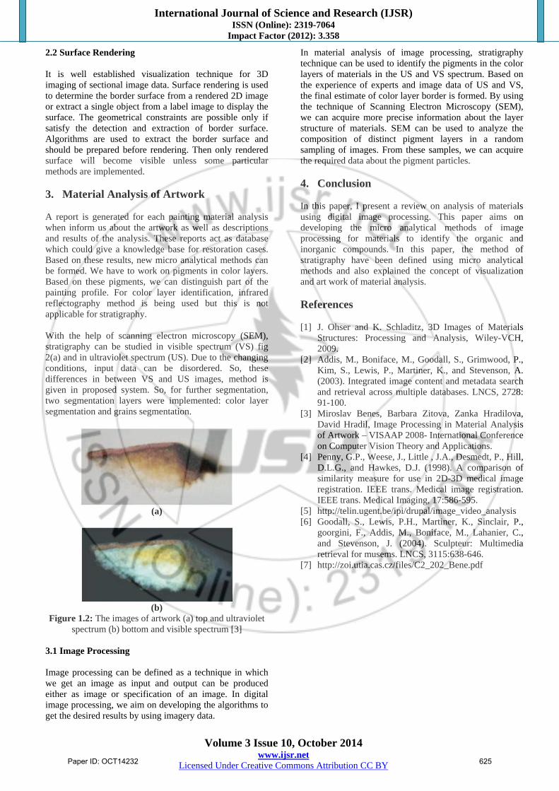
2.2 Surface Rendering It is well established visualization technique for 3D imaging of sectional image data. Surface rendering is used to determine the border surface from a rendered 2D image or extract a single object from a label image to display the surface. The geometrical constraints are possible only if satisfy the detection and extraction of border surface. Algorithms are used to extract the border surface and should be prepared before rendering. Then only rendered surface will become visible unless some particular methods are implemented. 3. Material Analysis of Artwork A report is generated for each painting material analysis when inform us about the artwork as well as descriptions and results of the analysis. These reports act as database which could give a knowledge base for restoration cases. Based on these results, new micro analytical methods can be formed. We have to work on pigments in color layers. Based on these pigments, we can distinguish part of the painting profile. For color layer identification, infrared reflectography method is being used but this is not applicable for stratigraphy. With the help of scanning electron microscopy (SEM), stratigraphy can be studied in visible spectrum (VS) fig 2(a) and in ultraviolet spectrum (US). Due to the changing conditions, input data can be disordered. So, these differences in between VS and US images, method is given in proposed system. So, for further segmentation, two segmentation layers were implemented: color layer segmentation and grains segmentation.
(a)
(b)
Figure 1.2: The images of artwork (a) top and ultraviolet spectrum (b) bottom and visible spectrum [3]
3.1 Image Processing Image processing can be defined as a technique in which we get an image as input and output can be produced either as image or specification of an image. In digital image processing, we aim on developing the algorithms to get the desired results by using imagery data.
In material analysis of image processing, stratigraphy technique can be used to identify the pigments in the color layers of materials in the US and VS spectrum. Based on the experience of experts and image data of US and VS, the final estimate of color layer border is formed. By using the technique of Scanning Electron Microscopy (SEM), we can acquire more precise information about the layer structure of materials. SEM can be used to analyze the composition of distinct pigment layers in a random sampling of images. From these samples, we can acquire the required data about the pigment particles. 4. Conclusion In this paper, I present a review on analysis of materials using digital image processing. This paper aims on developing the micro analytical methods of image processing for materials to identify the organic and inorganic compounds. In this paper, the method of stratigraphy have been defined using micro analytical methods and also explained the concept of visualization and art work of material analysis. References [1] J. Ohser and K. Schladitz, 3D Images of Materials
Structures: Processing and Analysis, Wiley-VCH, 2009.
[2] Addis, M., Boniface, M., Goodall, S., Grimwood, P., Kim, S., Lewis, P., Martiner, K., and Stevenson, A. (2003). Integrated image content and metadata search and retrieval across multiple databases. LNCS, 2728: 91-100.
[3] Miroslav Benes, Barbara Zitova, Zanka Hradilova, David Hradil, Image Processing in Material Analysis of Artwork – VISAAP 2008- International Conference on Computer Vision Theory and Applications.
[4] Penny, G.P., Weese, J., Little , J.A., Desmedt, P., Hill, D.L.G., and Hawkes, D.J. (1998). A comparison of similarity measure for use in 2D-3D medical image registration. IEEE trans. Medical image registration. IEEE trans. Medical Imaging, 17:586-595.
[5] http://telin.ugent.be/ipi/drupal/image_video_analysis [6] Goodall, S., Lewis, P.H., Martiner, K., Sinclair, P.,
goorgini, F., Addis, M., Boniface, M., Lahanier, C., and Stevenson, J. (2004). Sculpteur: Multimedia retrieval for musems. LNCS, 3115:638-646.
[7] http://zoi.utia.cas.cz/files/C2_202_Bene.pdf
Paper ID: OCT14232 625