mastication and wear in lophiodon (perissodactyla ... · ann. zool. fennici vol. 51 • mastication...
TRANSCRIPT
Ann. Zool. Fennici 51: 162–176 ISSN 0003-455X (print), ISSN 1797-2450 (online)Helsinki 7 April 2014 © Finnish Zoological and Botanical Publishing Board 2014
Mastication and wear in Lophiodon (Perissodactyla, Mammalia) compared with lophodont dentitions in some other mammals
Wighart von Koenigswald
Steinmann-Institut (Paläontologie) Universität Bonn, Nussallee 8, D-53115 Bonn, Germany ([email protected])
Received 3 June 2013, final version received 26 Aug. 2013, accepted 20 Sep. 2013
Koenigswald, W. v. 2014: Mastication and wear in Lophiodon (Perissodactyla, Mammalia) com-pared with lophodont dentitions in some other mammals. — Ann. Zool. Fennici 51: 162–176.
Three functional aspects (jaw movement, collapse of lophs by wear, and molar wear gradient) are described for Lophiodon and compared with typical lophodont dentitions in other mammals, e.g., Macropus, Pyrotherium and Deinotherium. The jaw move-ment is deduced from striations and guiding rails. In Lophiodon only phase I of the power stroke is documented by facets, whereas a minor phase II occurs in Macropus. During phase I the lophs perform two functions: cutting when the crests are passing each other, and compressing the bolus during further interlocking. When dentine is exposed the lophs collapse and lose their trenchant function and are grinding only. The sudden collapse of the lophs is partially due to the abrasion within the compres-sion chamber, as seen in Dendrolagus. The molar wear gradient (the differential wear between the first and last molars) is low in Pyrotherium, intermediate in Lophiodon, and Deinotherium as compared with a low molar wear gradient in Tapirus or a high gradient in Macropus.
Introduction
This volume is dedicated to Mikael Fortelius. At the 1979 dental meeting organized by B. Kurtén in Turku, I met Mikael and have vivid memories of our discussion on mastication in fossil and extant rhinos, the favourite objects of his early studies (Fortelius 1982, 1985). Remembering our long-lasting common interest, the mastica-tion of the fossil Lophiodon shall be discussed in this paper.
Lophiodon is a middle to large sized herbiv-ore that occurred in Europe during the middle
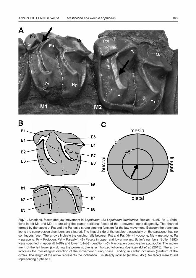
and upper Eocene. One of the latest species, the Bartonian Lophiodon lautricense from Robiac (Gard, France), reached the body size of a horse or a small rhino. This species is especially suited for studying the jaw movements during mastica-tion since the teeth show very clear facets with deep striations (Fig. 1A).
Lophiodontidae are basal Perissodactyla, and were regarded as closely related to Tapiroi-dea within Ceratomopha due to the similari-ties in the lophodont dentition (Cuvier 1822, Radinsky 1963, McKenna & Bell 1997). Hooker (1984, 2005) and Hooker and Dashzeveg (2004) discussed a closer relationship with chalicoth-
Ann. Zool. Fennici Vol. 51 • Mastication and wear in Lophiodon 163
Fig. 1. Striations, facets and jaw movement in Lophiodon. (A) Lophiodon lautricense, Robiac, HlMD-Ro 2. Stria-tions in left M1 and M2 are crossing the planar attritional facets of the transverse lophs diagonally. The channel formed by the facets of Pst and the Pa has a strong steering function for the jaw movement. Between the trenchant lophs the compression chambers are situated. The lingual side of the ectoloph, especially on the paracone, has no continuous facet. The arrows indicate the guiding rails between Pst and Pa. (Hy = hypocone, Me = metacone, Pa = paracone, Pr = Protocon, Pst = Parastyl). (B) Facets in upper and lower molars, Butler’s numbers (Butler 1952) were specified in upper (B1–B8) and lower (b1–b8) dentition. (C) Mastication compass for Lophiodon. The move-ment of the left lower jaw during the power stroke is symbolized following Koenigswald et al. (2013). The arrow indicates the mesiolingual direction of the movement during phase i ending in centric occlusion (centrum of the circle). The length of the arrow represents the inclination. it is steeply inclined (at about 45°). no facets were found representing a phase ii.
164 Koenigswald • Ann. Zool. Fennici Vol. 51
eriids within Ancylopoda. This relationship was accepted by Froehlich (1999), Rose and Archibald (2005), and Rose (2006), whereas Holbrook (2009) investigated cranial, postcra-nial, and dental characters and concluded that Lophiodontidae are best classified as the sister-taxon to Ceratomorpha (Tapiroidea plus Rhi-nocerotoidea), and thus that Lophiodon is closer to Tapiroidea than to Chalicotheriidae. Several species were described for the genus Lophiodon that differ mainly in size, but the variability within the species is very great (M. Godinot pers. comm.).
The topic of this paper, however, is nei-ther the systematic nor the phylogenetic position of Lophiodon, but focuses on the functional morphology of the postcanine dentition. Three aspects are investigated, the direction of mastica-tion movements, the typical wear of the trans-verse lophs, and the gradient of wear within the molar dentition. Restricting discussion to the postcanine dentition seems to be adequate, since its function differs strongly from that in the ante-rior region. This functional separation is stressed by the heterodonty and the diastema between the canine and the premolars in many lophodont dentitions.
The Lophiodon dentition is regarded as a typical lophodont, as indicated by the name, given by Cuvier in 1822. The comparison with other lophodont dentitions shows that the func-tion of lophodont dentitions is not uniform. Thus a more detailed knowledge of Lophiodon sets the pattern for the diversity within this classical type of mammalian dentitions.
Methods
Three functional aspects were investigated: the movement of the lower jaw during mastication, the collapse of transverse lophs by wear, and the gradient of wear between M1 and M3.
Mastication movements were deduced from the orientation of facets and from the striations on these facets. For the identification of the vari-ous facets, the numbering proposed by Butler (1952) is used. Other authors have numbered the wear facets differently (e.g. Crompton 1971). To avoid misunderstanding we mark Butler’s num-
bers with a “B” or “b” and differentiate facets in upper teeth (B1, B2 …) from those in lower ones (b1, b2 …), respectively (Fig. 1B).
The orientation and inclination of facets rep-resent only one aspect of the jaw movement, since movements can happen in any direction across a plane. Striations on planar attrition facets are the traces of the actual movement and document the direction. Where two facets with a different orientation meet the channel in between them, this forms a guiding rail. Such rails pro-vide the best evidence for the reconstruction of jaw movement. The polarity of the move-ment has to be reconstructed from the general tooth morphology. In contrast to reconstructions of jaw movements based of electromyographic investigations, the striations on facets and the guiding rails are traces of the actual movements (see Discussion).
The traditional terms, like “orthal”, “hori-zontal”, or “transverse” describe orthogonal Car-tesian coordinates, but the real movements are almost always oblique to these axes (Koenig-swald et al. 2013). Therefore, the mastication movements were described and depicted in the mastication compass (Fig. 1C). It illustrates the movement of the left lower jaw and differenti-ates clearly between direction and inclination for the two (possible) phases of the power stroke. The arrow of phase I ends in the center of the compass that represents centric occlusion. If a phase II is present and documented by facets, a second arrow starts in the center. The arrow indicates the direction of the jaw movement according to the compass rose. The inclination is indicated by the length of the arrow.
Exact measurements of the angles are dif-ficult because of the lack of consistent reference planes. For isolated teeth the base of the crown is used, but the plane differs from one tooth to the next in the dental row. In the verbal description the directions are indicated as mesial, lingual, or distal, with subdivisions similar to those in the compass rose. The inclination differentiates between horizontal, steep (about 45°), and very steep (more than 60°). Estimated directions and inclinations as deduced from visual inspection are very useful for general information, but for detailed analyses more exact measurements are indispensable.

Ann. Zool. Fennici Vol. 51 • Mastication and wear in Lophiodon 165
The wear of transverse lophs in Lophiodon is of interest, since these lophs are worn down dif-ferently from cusps. These trenchant lophs wear into flat grinding surfaces and change their func-tion. The specific kind of collapse was observed in transverse lophs of other taxa as well.
The term “molar gradient wear” describes the difference in wear between the first and the last molars and was used by Damuth et al. (2012). It differs from the “wear gradient” introduced by Fortelius and Solounias (2000) by comparing the molar profile and not the wear stages of specific teeth. Despite the fact that the first molar is gen-erally worn more heavily than the last one, dif-ferent species can be characterized by a smaller or larger molar wear gradient. A “low molar wear gradient” is characterized by similar wear in all molars, indicating that the entire molar tooth row is functioning simultaneously.
The other extreme is a “high molar wear gra-dient”, when only one molar or the posterior half of one and the anterior part of the subsequent molar are functioning, and abraded teeth are shed anteriorly. There is a wide range in between these two extremes that is called here “interme-diate molar wear gradient”. For characterizing this type the eruption sequence has to be consid-ered. Normally the premolars replace the decidu-ous teeth prior to the eruption of the last molar. But this is the topic of another ongoing study. For the comparison of Lophiodon with some other lophodont dentitions the three grades: low, intermediate and high, are adequate.
Material
For the study of mastication and wear, tooth rows are more suitable than isolated teeth. Two fossil localities produced very suitable denti-tions of the genus Lophiodon. One is the former coal mines of the Geiseltal near Halle from the middle Eocene (MP 13), which produced rich materials of Lophiodon, predominantly from L. remensis (Fischer 1977). The material is housed in the Geiseltalmuseum in Halle.
The second site is Robiac (Grad, France), representing the Bartonian of the upper Eocene (MN16). Rich material attributed to Lophiodon was collected in the early years of the 20th
century and is distributed in several museums. The identification of the species as Lophiodon lautricense from Robiac was confirmed by Sudre (1971). Some additional material of other spe-cies of Lophiodon and from various other locali-ties was considered:
— Lophiodon remensis Lemoine, 1978; Geisel-tal near Halle, Germany:• left maxilla with P2–M3; GMH XXXVIII
69 1964,• left mandible with p3–m3; GMH
XXXVIII 69 1964,• left and right maxilla; GMH LIX 15 a + b• right and left maxilla with P2–M3; GMH
no number.— Lophiodon sp.; Geiseltal near Halle, Ger-
many:• left mandible with p3–m3; GMH XXXIII
24ab 1962.— Lophiodon isselensis Fischer, 1829; Issel
(Aude) France:• left maxilla with P2–M3; MNHN —
1903/20,• left maxilla with P2–M3; MNHN — no
number,• right mandible with p3–m3, MNHN —
EBA 32.— Lophiodon lautricense Noulet, 1852; Robiac,
St. Mamert, Gard, France:• left mandible with p3–m3; HLMD-Ro 3,• right maxilla with P2–M3; HLMD-Ro 2,• left M3; HLMD-Ro 17c,• left maxilla with p2–M3; NHMW
1902/0002/0002 (old no. NHMW 1902/II/2),
• left mandible with p2–m3; NHMW 1902/0002/0001 (old no. NHMW 1902/II/1).
For comparison, the following taxa were included:
— Macropus div. sp., Recent, NSW Australia; QMB, Brisbane and STIPB, Bonn.
— Pyrotherium romeroi and macfaddeni, Oli-gocene, Bolivia; MNHN, Paris; UF Gaines-ville; FM Chicago, and Yale.
— Dendrolagus sp., Recent, Papua New Guinea; STIPB, Bonn.
166 Koenigswald • Ann. Zool. Fennici Vol. 51
— Dorcopsulus vanheuerni, Recent, Papua New Guinea; STIPB, Bonn.
— Macropus rufus, Recent, Queensland, Aus-tralia; STIB, Bonn.
— Diprotodon opatum, Pleistocene, New South Wales, Australia; QMB, Brisbane.
— Platygonus vetus, Pleistocene, Texas and Florida; TU, Austin and UF, Gainesville.
— Phanourios minor, Pleistocene, Cyprus; Uni-versity of Athens.
— Tapirus lundeliusi, Pleistocene, Florida; UF, Gainesville.
— Tapirus terrestris, Recent; ZMUH, Hamburg,— Prodeinotherium bavaricum and Deinothe-
rium giganteum, Miocene, Germany; SMNS, Stuttgart, BSPM, Munich, NHMW, Vienna.
— Daouitherium rebouli Paleocene, Morocco; MNHN, Paris.
— Phosphatherium escuillei, Paleocene, Morocco; MNHN, Paris.
Virtual 3D models of some dentitions were established by scanning in the µCT scanner v/tome/xs (GE sensing Inspection technolo-gies GmbH phoenix/X-ray) or optically with the Breukmann optoTOP-He device, both at the Steinmann Institut für Geologie, Mineralogie und Paläontologie, University of Bonn. Virtual 3D models were processed with the Breukmann software Optocat 2007 R3 (ver. 7.20.03-1226). Using the virtual 3D models the figures were established with the programs Polyworks and Volume Graphics VG Studio MAX 2.0. For the dentitions of Tapirus lundeliusi casts were made in Gainesville, Florida. Casts of L. remensis, and Tapirus terrestris were kindly provided by Prof. Th. Kaiser, ZMUH.
Abbreviations for collections:
AMS: Australian Museum, Sydney, Australia.BSPG: Bayerische Staatssammlung für Palä-
ontologie etc., Munich.FM: Field Museum, Chicago.GMH: Geiseltalmuseum, Halle.HMLD: Hessisches Landesmuseum Darmstadt.NHMW: Naturhistorisches Museum, Vienna.MNHN: Muséum national d’Histoire naturelle,
Paris.PU: Princeton University Collection, Field
Museum, Chicago.QMB: Queensland Museum, Brisbane, Aus-
traliaSTIPB: Steinmann Institute — Paleontology,
University of Bonn.TU: University of Texas, Austin.UF: University of Florida, Gainesville.ZMUH: Zoologische Museum der Universität
Hamburg.
Results
The dentition of Lophiodon
In the maxilla there are six postcanines (three premolars and three molars) separated from the anterior dentition by a short diastema. The molars have the typical π-structure formed by a protoloph, a metaloph and an ectoloph (Fig. 2). The ectoloph is, however, not as continuous as in rhinos. The parastyle, the paracone, and the metacone are bulbous and well separated. The molarization of the premolars is used for taxo-nomic purposes, but their variability is of minor significance here.
The lower molars are bilophodont, formed by a protolophid that links the protoconid with the metaconid and a hypolophid that spans the hypoconid and entoconid (Fig. 3). The paralo-phid is directed mesially from the protoconid on the buccal side. The m3 has an additional loph that is relatively much smaller. The lower p4 is rather molariform, and p3 and p2 differ between taxa in their degree of molarization.
The sequence of tooth eruption is incom-pletely known for Lophiodon, but according to the maxilla of Lophiodon lautricence (Fig. 4A) the upper premolars have replaced the deciduous teeth prior to the eruption of the M3. This corre-sponds with a mandible figured by Filhol (1888: pl. 3: 2) the p2, p3 and p4 are erupting when the m3 is still hidden deeply in the crypt.
The reconstructed mastication movement
The lower molars of Lophiodon are character-ized by two parallel transverse lophs. These

Ann. Zool. Fennici Vol. 51 • Mastication and wear in Lophiodon 167
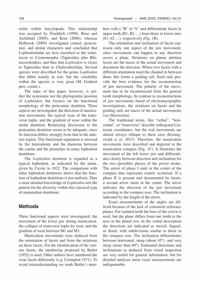
Fig. 2. Upper dentitions in Lophiodon remensis from the Geiseltal near Halle in different stages of wear (virtual images based on optical scans of the casts). After the M1 is worn out the area of mastication, the center of wear shifts backwards with increasing age. The two maxillae document the shift from M1 to M2. The distinct wear gradi-ent between M1 and M3 is very obvious. (A) left maxilla with P2–M3; GMH XXXViii 69 (1964). (B) left maxilla with P2–M3; GMH no number. For easier comparison, virtual images were scaled to the same length. The numbers of the lophs are indicated in each molar.
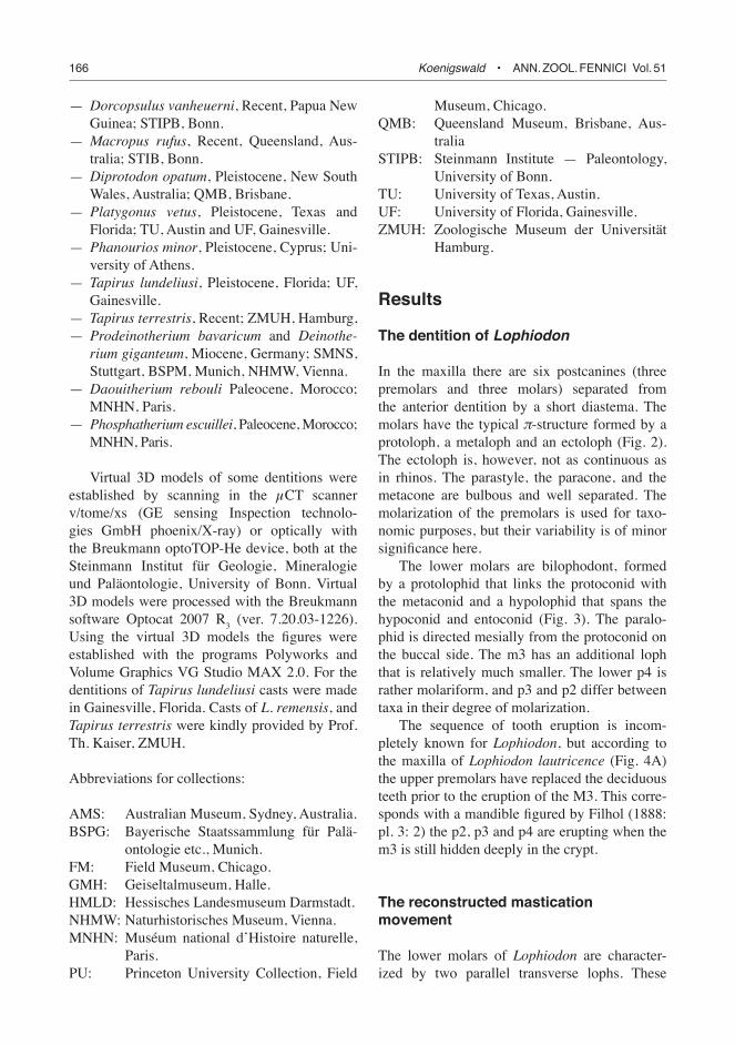
Fig. 3. lower dentitions in (A) Lophiodon remensis and (B) L. sp. from the Geiseltal near Halle in different stages of wear (virtual images based on optical scans of the casts). (A) left mandible with p2–m3; GMH XXXViii 69 (1964), left mandible. (B) left mandible with p3–m3; GMH XXXiii 24ab (1962). For easier comparison, virtual images were brought to the same length of m1–m3. The numbers of the lophs are indicated in each molar.

168 Koenigswald • Ann. Zool. Fennici Vol. 51
Fig. 4. Lophiodon lautricense, Robiac, St. Mamert, Gard, France. (A) Right maxilla with P2–M3; the M3 is not yet fully erupted; HlMD-Ro 3 (photo). The M1 has distinct wear facets while M3 is pristine. (B) left mandible with p3–m3; HlMD-Ro 2. Virtual image based on optical scans of the original. note the decrease in wear in the lophs from M1/m1 to M3/m3. The numbers of the lophs are indicated in each molar.
lophs are not perpendicular to the longitudinal tooth axis, but the buccal edge is rotated about 20° anteriorly. In moderately worn teeth the fol-lowing facets are present (Fig. 3A and B). On the distal slope of both the protolophid and the hypolophid, a planar attritional facet is domi-nant. These facets are steeply inclined distally (about 45°). In the terminology of Butler (1952) these facets combine b2 and b3 on the protolo-phid and b7 and b8 on the hypolophid (Fig. 1B).
The crests of the lophs are trenchant as long as the facets are covered by enamel. The unworn crest of each transverse loph is bent slightly backwards but is not crenulated, e.g., preserved in the m3 of L. remensis (Fig. 3A) and L. lautri-cense (Fig. 4B). Due to this overhanging crest the area of the first contact with the antagonistic tooth is very small, and the initial facet can be formed easily in the optimal orientation. With wear the facets elongate to the full width of the loph. On the buccal side the paralophid extends mesially from the protoconid. It is better devel-
oped in L. lautricense (Fig. 4B) than in L. remen-sis (Fig. 3A). This facet (b1) steeply inclines towards the buccal side.
The upper molars have dominating facets on the mesial side of each transverse loph (B3 + B2 and B8 + B7, respectively). The facets are steeply inclined mesially and correspond to the facets of the lower molars. Corresponding to the lower molars, the crests of unworn teeth are bent forward (see M3 in Figs. 2A and 4A). The ectoloph has no continuous facet on the lingual side, e.g., as in rhinoceroses. Several independ-ent facets occur in slightly worn teeth that are parts of the dominant facets of the two transverse lophs. On the protoloph the facet B2 grows to the buccal side, onto the mesiolingual side of the paracone. On the metaloph the facet B7 expands to the mesiolingual side of the metacone. The lingual side of the paracone has no facet in that stage of wear (see M2 in Figs. 2A and 4A). A functionally important facet occurs on the dis-tolingual side of the prominent parastyle. It is

Ann. Zool. Fennici Vol. 51 • Mastication and wear in Lophiodon 169
inclined mesiolingually and forms a guiding rail for the antagonistic protoconid. When the upper molars are more deeply worn the mesial side of the paracone (B1) contributes to this guiding rail (Fig. 1A).
The other facets on the ectoloph also grow with wear. On the distal side of the paracone a facet occurs (B6) and joins B7 on the mesial side of the metacone. It is inclined in mesiolingual direction. On the distal side of the metacone another facet (B1) articulates with the paralophid (b1) of the lower molars.
Because these facets are inclined differently they allow the antagonist to move only in a single direction: the lophs of the lower molars slide with a steep inclination in a mesiolingual direction into the valleys of the upper molars (Fig. 1B). This movement crosses the planar attritional facets at an oblique angle and not parallel to the inclination, as clearly indicated by distinct striations in all facets in L. lautricense (Figs. 1A and 4A).
The planar attritional facets of the upper lophs that are inclined mesially, occlude with the distally inclined facets of the lower lophs. Due to the mesial shift of the lower molars, the facet of the anterior loph in upper molars (B2–B3) occludes with the distal loph of the lowers (b7–b8) and thus the posterior loph of the uppers (B7–B8) occludes with the anterior loph of the subsequent lower molar (b2–b3).
During the occlusion two different functions can be deduced from the morphology. When the sharp crests of the upper and lower lophs meet, they cut the plant material. It is astonishing that these cutting edges are not rounded by abra-sion — at least attrition dominates and keeps them sharp. The two facets pass each like scissor blades and thereafter steer the movement of the antagonistic loph until they interlock each other. How deeply the lophs interlock may depend on the amount of bolus in between. The second function of this movement is the compression of the bolus between the distal slope of the upper and the mesial slope of the lower lophs. In upper teeth the lophs are slightly concave on the distal side and form a compression chamber to each loph. On the buccal side the ectoloph and the parastyle form a barrier and on the lingual sides the protocone and hypocone are bulbous. They
prevent the bolus from escaping during compres-sion, but the gaps between the latter cones allow the liquefied bolus to flow into the mouth.
The movement along the facets ends when the lophs are fully interlocked in centric occlu-sion. Thus the entire movement represents the phase I of the power stroke. After centric occlu-sion the jaw likely opens directly into the recov-ery stroke. At least, no facet indicates any phase II during the power stroke (Fig. 1B).
The strong transverse lophs of the dentition allow an estimate of the horizontal shift of the lower jaw during mastication. When the occlusal surfaces of the dentition are flat, as they are in elephants, the amount of horizontal movement can not be observed and thus it might be over-estimated. In Lophiodon the horizontal shift is distinctly less than the mesio-distal length of a single loph.
As long as the lophs wear their facets, the reconstructed jaw movement can be precisely oriented. With increasing age, jaw movements generally become washy and are less easy to discern (Anders 2012). Therefore, special atten-tion was paid to the kind of wear. The heavily worn teeth function as a grinding surface with a slightly projecting ring of marginal enamel (M1 in Fig. 2B).
The wear of the lophs and the molar wear gradient
In terms of functional morphology it is of inter-est that the lophs are worn down differently from cusps. Apical wear of the cusps was not found in fresh lophs. The crests of the transverse lophs are slightly overhanging, in uppers mesially and in lowers distally, according to rules of functional symmetry (Koenigswald et al. 1994). These crests ease the subsequent formation and orienta-tion of the facets (Fig. 5). The very steep facet cuts into this overhanging crest and forms a well oriented planar attritional facet that is conse-quently worn obliquely into the loph. The upper rim of the facet forms a sharp crest of enamel.
During continuous wear the dentine core is exposed in upper teeth on the lingual side and in lower teeth on the buccal side near the pro-toconid. When the dentine field is opened in the

170 Koenigswald • Ann. Zool. Fennici Vol. 51
entire length of the loph, the angle of inclina-tion turns quickly into horizontal (Fig. 4B, m1 loph 2). The cutting character of the loph has changed into a grinding function with the ero-sion of the loph (Fig. 5).
These various stages of wear are represented in the same jaw due to the differential wear between the first and the last molar. Among the figured specimen the unworn loph with an overhanging crest or with the first incomplete facet are visible in L. rem. (M3 loph 2 in Figs. 2A and 4A) and in L. laut. (both lophs of M3/m3 in Fig. 4A and B). The planar attritional facet has reached the full length of the loph in L. rem. (e.g., in loph 1 of M3 in Fig. 2A, loph 2 of M2 and loph 1 of M3 in Fig. 3A and loph 2 of m3 in Fig. 3B). The dentine field is initially exposed by the facet in L. rem. (loph 1 in M2 in Fig. 2A, and loph 1 of M3 in Fig. 2B) and in L. laut. (loph 1 and 2 of M1 in Fig. 4A). The facet has lost the inclination and is more or less horizontal in L. rem (loph 1 and 2 of M1 in Fig. 2A; loph 1 of M 3 in Fig. 2B; loph 2 in Fig. 3A) and in L. laut. (loph 1 of m1 in Fig. 4B). The lophs are totally collapsed in L. rem. (M1 in Fig. 2B and 3B). Note that the lophs with planar attritional facets are next to worn out lophs.
A mandible of Lophiodon isselensis (MNHN — EBA 32) from Issel (Aude, Languedoc-Rous-sillon), shows the entire sequence of the collapse of the lophs. The m1 is totally flattened as seen in the figure of Filhol (1888: pl. 1, figs. 1 and 2). [Since Filhol, part of the m1 is broken off.] In the m2, the loph 1 has a widely opened den-tine field that is inclined distally. In the loph 2 of the m2, the dentine field is widely opened as well but the inclination is steeper. In the m3, the loph 1 has a steeply inclined enamel facet that does not open the dentine. Striations are visible only on this facet confirming the movement in a mesio-lingual direction. The loph 2 of the m3 is absolutely fresh.
The two maxillas from Issel (MNHM — 1903/27 and MNHN — no number) represent a younger individual age. Accordingly the wear is less intensive. Other material of a smaller Lophiodon from Rouzilhac close to Issel (Aude) was made available by M. Godinot (unpubl.). This material contains maxillae from fairly old individuals with transverse lophs that are pro-
gressively collapsing from front to back. Figures given by Deperét (1903) for Lophiodon from Minervois (Aude, Languedoc-Roussillon) depict the same wear pattern. This confirms that the collapse of trenchant lophs by wear is a general pattern of the upper and lower molars in Lophi-odon. A very similar pattern of collapse (Fig. 5) of the lophs occurs in some macropodids, as dis-cussed further later in this paper.
In Lophiodon remensis the intermediate gra-dient of molar wear is very obvious. In the maxilla (GMH XXXVIII 69; Fig. 2B) the M1 the lophs are flattened while in M3 the lophs have initial attritional facets only. The mandible (GMH no number; Fig. 2B) shows a heavily worn m1 with no internal ridges, whereas the enamel covers almost the entire length in both facets of the m3. Thus the difference in wear between M1 and M3 is significant. In the two mandibles (Fig. 3) the same difference occurs. Lophiodon lautricense presents a similar picture (Fig. 4A), although at a younger age. In this specimen the M3 is not fully erupted although the dentine is exposed in the lophs of the M1. Several other tooth rows confirm the difference of wear between the first and the last molars. The eruption of the last molar is slight retarded, at least in Lophiodon lautricense (Fig. 4A).
The molar wear gradient can be evaluated only in comparison to other taxa (see Discussion for details). In comparison, Tapirus lundeliusi and T. terrestris (Fig. 6) have a low molar wear gradient where the entire tooth row functions simultaneously with well-persevered cutting edges. Macropus rufus has a high molar wear gradient with late erupting posterior molars. Thus the molar wear gradient in Lophiodon is
Fig. 5. Schematic illustration of the collapse of lophs in Lophiodon by wear: (A) unworn loph with overhang-ing crest; (B) trenchant facet fully within the enamel, strongly inclined; (C) trenchant facet with initial dentine field, strongly inclined; (D) horizontal facet with wide dentine field; (E) worn out rudimentary loph. note the abrupt change of the inclination of the facet when the dentine is opened (between C and D). it marks the col-lapse of the loph.

Ann. Zool. Fennici Vol. 51 • Mastication and wear in Lophiodon 171
intermediate. A preliminary definition is pro-vided in the discussion chapter below.
Comparisons and discussion
Lophodont dentitions evolved in some marsu-pial and in many placental orders by combin-ing cusps into lophs and sometimes including cingula. During the Paleocene/Eocene several independent lineages invented “brachydont-transverse-lophodont-teeth” Hooker (2000). The typical lophodont dentitions are brachydont, but several taxa changed the construction of the lophodont teeth by introducing hypsodonty. This was made possible by a modified tooth ontog-eny (Koenigswald 2011). Few taxa developed poly-lophodont teeth, but the majority retained the bilophodont tooth type, with two lophs per tooth. From this great variety of the bilophodont-brachydont dentitions only a small number of typical taxa was selected for a comparison with Lophiodon.
Marsupials are represented in this study by the vombatoid Diprotodon from the Pleistocene and some extant macropodoids (Dendrolagus, Dorcopsulus, Dorcopsis and Macropus; Sanson 1980, Crompton et al. 2008). Among placentals the South-American Pyrotherium (Billet 2010), the perissodactyl Tapirus and the proboscideans Daouitherium and Phosphatherium (Gheerbrant et al. 2005) as well as Deinotherium (Gräf 1956, Harris 1975) are discussed. In addition, some general remarks on lophodont primates and arti-odactyls are provided.
The jaw movement
Several techniques were applied for reconstruct-ing jaw movement. In living mammals the activ-ity of muscles can be investigated by electro-myographic analysis (e.g. Crompton et al. 2010). In fossil mammals the direction of forces caused by the musculature can be reconstructed from localizing the muscle attachments (e.g., Turn-
Fig. 6. Upper tooth rows of (A) Tapirus lundeliusi (UF 274674) and (B) Tapirus terrestris (ZMUH 2760-txl) at slightly different wear stages. note the relatively low wear gradient between M1 and M3. The planar attritional facets are not restricted to the molars but are well developed in P3 and P4. in a more intensively-worn mandible (B) the lophs of M1 and P3 are collapsed, while P4 shows better preserved lophs. P4 obviously erupted later than P3. Virtual images based on optical scans of the casts. The numbers of the lophs are indicated in each molar.

172 Koenigswald • Ann. Zool. Fennici Vol. 51
bull 1970, Harris 1975). The stresses caused by the musculatory motor, however, are only one factor controlling the general jaw movement. The second factor is redirection by the antago-nistic teeth. When the teeth come into occlusion, the morphology of antagonistic teeth controls the jaw movement in detail (Koenigswald et al. 2013). Thus guiding rails and striations on attri-tional facets represent the actual movements.
The gross morphology in lophodont denti-tions with high lophs allows movements in two different directions and the subsequent com-bination of the two during mastication. The antagonistic lophs move either strictly parallel to each other with a low inclination in lingual direction, or the lophs pass each other with a high inclination in a mainly mesial or mesio-lingual direction. In several lophodont primates (e.g., Cercopithecus), and some marsupials (e.g., Phalanger) the bilophodont teeth do not show facets indicating an intensive mesial or lingual movement during mastication (Fig. 7A).
A movement parallel to the lophs with little inclination was found in the tayassuid Platygonus and hippopotamid Phanourios (Sondaar 1977, Hulbert 2001). The lophs interlock each other, but they do not wear sharp transverse crests (Fig. 7B). Facets occur on both sides of the lophs and carry horizontal striations according to the lower jaw moving in lingual direction. Striations on the bottom of the valleys stress their function
as guiding rails. Such a lateral jaw movement is not restricted to artiodactyls since horizontal striations were found in the lophodont deciduous molars of Mammut americanum as well (Green & Hulbert 2005). Lophodont dentitions showing mainly a transverse movement are very different from those in Lophiodon and thus they are of minor significance for the comparison here.
More similar to Lophiodon (Fig. 1C) is the mastication pattern in Diprotodon, Macropus, Dendrolagus, Dorcopsulus, Pyrotherium, Tapi-rus, Daouitherium, Phosphatherium, and Dein-otherium. They all carry distinct planar attrition facets on the distal side of the lophs in lower teeth and on the mesial side in upper teeth. The mandible moves generally in mesial to mesio-lingual direction with a steep inclination (Harris 1975, Sanson 1980, Gheerbrant et al. 2005, Crompton et al. 2008). Some but not all of these facets show striations. They indicate a mesial to mesiolingual direction with a steep inclination (Fig. 7C).
Some genera, e.g., Lophiodon and Tapirus, have very distinct guiding rails between para-style and paracone, in the upper molars. They determine the mesiolingual direction and a steep inclination. In Deinotherium only the upper premolars have an ectoloph that controls the jaw movement in a mesio-mesiolingual direction. The lower premolars have antagonistic facets (b1) on the paralophid that are steeply inclined.
Fig. 7. Mastication compasses of various bilophodont and polylophodont dentitions representing the diversity of jaw movements. The mastication compass indicates the direction (arrow) and the inclination (length of the arrow) in the two phases of the left lower jaw movement. Phase i ends in the center of the compass representing centric occlusion. For further explanation of the mastication compass see caption to Fig. 1c and Koenigswald et al. (2013). (A) An almost orthal occlusion characterises some bilophodont primates and marsipials. (B) A horizontal movement in lingual direction was found in the bilophodont hippo Phanourious. Phase i and ii cannot be distinguished. (C) Many bilophodont dentitions share a dominant phase i that is steeply inclined and directed almost mesially. The jaw motion ends with phase i in centric occlusion. (D) in Macropus the steep and mesial movement in phase i is fol-lowed by an additional phase ii in lingual direction and horizontal inclination. (E) in the polylophodont elephants the horizontal movement is directed mesially.
Ann. Zool. Fennici Vol. 51 • Mastication and wear in Lophiodon 173
In most of these bilophodont dentitions the facets document a movement only in phase I that ends in the centric occlusion. There is no evidence for a phase II movement in most of these bilophodont mammals. However, an additional movement was found in Macropus (Sanson 1980), where after the end of phase I a subsequent lateral movement can be deduced from facets with striations on the longitudinal central crest. They represent phase II with a movement in the lingual direction according to the inclination of the facets (Fig. 7D). This transverse movement of phase II was confirmed in the electromyographic analyses of two species of Macropus (Crompton et al. 2008). Similarly in Pyrotherium macfaddeni (PU 20684) some valleys between the lophs show at the base trans-verse oriented striations that indicate a possible mesial movement during a phase II of the mas-tication circle.
These two cases advise caution against excluding any lingual movement after centric occlusion. The presence or absence of facets related to phase II shows the limitation for inter-preting the jaw movement from facets only. But even if such a lingual movement occurs in bilo-phodont dentitions, the functional significance of such a phase II movement is most probably small according to the very limited contact of teeth.
The two functional components, cutting and compressing, during phase I that were distin-guished for Lophiodon can be postulated for the Macropus, Pyrotherium, Tapirus and Dein-otherium as well. The sharp crests of the lophs form a cutting device, and the consequent move-ment compresses the bolus. In all listed genera the lophs of upper and lower teeth show planar attritional facets on one side, and the other side is consistently slightly concave contributing to the compression chamber. The gross morphol-ogy provides a measure of how the mandible is shifted in the mesial direction during the power stroke. This shift does not exceed more than half the mesio-distal distance between two lophs. This short distance should be considered when the horizontal aspect of movement is dis-cussed for animals with flat occlusal surfaces, like in elephants. There the movement may cross several lophs, but certainly not the entire
tooth length. The maximal distance of transverse movements is limited by the width of the tooth, but it is mostly smaller.
The lophs and their wear
The shearing blade
In bilophodont dentitions comparable to Lophio-don, the lophs are characterized by a broad and steeply inclined planar attrition facet on one side. In unworn teeth of Lophiodon each loph has an overhanging crest where the facet will be formed. This facilitates the formation of a pre-cisely oriented facet. Similar overhanging crests occur in other species, e.g., in Diprotodon, Mac-ropus, Pyrotherium, and Deinotherium. But such overhanging crests are not essential for the for-mation of such facets since overhanging crests are missing in Daouitherium, Phosphatherium, and Tapirus terrestris. Another way to reduce the surface of the initial facet is a crenulated ridge of enamel. That is not present in Lophiodon but found in Pyrotherium, Daouitherium and Phos-phatherium. In Diprotodon, and Deinotherium both traits are combined as overhanging and crenulated crests. This feature was already fig-ured for the latter genus by Cuvier (1836: pl. 74: 2–3). The mosaic of these two characters reflects their parallel evolution.
The sharp crests in the upper rim of the planar attritional facet seem to be highly impor-tant, since they occur in all discussed taxa. They are formed by wear, especially attrition (tooth–tooth contact) during normal mastication move-ments. For sharpening, no “thegosis,” defined as a sharpening process with a movement in the opposite direction, is necessary (Murray & Sanson 1998).
Despite the attrition, the wear on the crest is surprisingly limited. This might be due pri-marily to a fairly soft diet. Additionally, the enamel microstructure in Lophiodon emphasizes the crests. The curved Hunter-Schreger bands that are oriented perpendicular to the crests might strengthen the cutting edge. This character is shared by Tapirus and chalicotheriids (Koenigs-wald et al. 2011). Among the other bilophodont dentitions, in Macropus a specialized structure,

174 Koenigswald • Ann. Zool. Fennici Vol. 51
the “zipper enamel”, was found to be related to the crests (Koenigswald 1994). In Pyrotherium another modification occurs in the enamel of the shearing blades: vertical Hunter-Schreger-bands (but irregular), as listed by Fortelius (1984, 1985) for this genus. No specializations, however, were found in the remaining lophodont dentitions indi-cating that no enamel specializations are essential for the function of the shearing blade.
The collapse of the lophs
Once the initial planar attritional facet is formed, the steep inclination is retained as along as the facet cuts through enamel. During progressive wear the dentine is opened and very soon the facet loses its trenchant character as described for Lophiodon (Fig. 5). A similar decay of the lophs can be observed in Dendrolagus, Dorcop-silus and Macropus. In the mandibular lophs of Dendrolagus the dentine core is opened regu-larly to the mesial side. Thus the loph is not destroyed by the planar facet on the distal side but by the abrasion related to the compres-sion chamber. This explains why the lophs col-lapse so quickly when the dentine is opened. In Lophiodon the abrasion of the enamel in the compression chamber is less obvious, but most probably the abrasion there contributes to the collapse of the lophs. When the lophs collapse, the dentine plate and the surrounding enamel band form a grinding tool (Fig. 2B).
In Deinotherium the lophs are slightly more solid and retain the shearing blade somewhat longer due to the very thick enamel, but the col-lapse of the lophs is very similar. In contrast, the wear of the very solid lophs in Pyrotherium differs greatly. The steeply inclined planar attri-tional facet of the lophs seem to last very long throughout life time, since they can be observed in almost all jaws. Only the angle of the inclina-tion of the facet is slightly reduced.
The molar wear gradient
The wear of the various lophs progresses from front to rear. Three levels of the molar wear gra-dient are differentiated for this paper.
A “low wear gradient” is characterized by an almost equal wear of all lophs as in Tapirus ter-restris and T. lundeliusi (Fig. 7), or the macropo-dids Dendrolagus and Dorcopsulus
A “high wear gradient” is characterized by a great difference between the wear of M1/m1 and that of M3/m3. That includes cases where the M1/m1 are worn out and shed before the M3/m3 erupts. Due to a horizontally displacement the posterior molars come into function. It means that the functional area as compared with the entire tooth row is reduced (Sanson 1989).
Typical examples for this extreme are gom-photheres, elephantids and derived macropodids as Macropus rufus and M. giganteus.
The wide range between a low and a high molar wear gradient is regarded as an “inter-mediate wear gradient”. As in Lophiodon adult individuals have the entire tooth row in use simultaneously, but the M1/m1 are more inten-sively worn while the M3/m3 are almost pris-tine. Another character defines this group: The premolars replace the deciduous teeth prior to the eruption of the M3/m3. By the delay of the eruption of the last molar, the first molar shows much more intensive wear traces than the last molar (Fig. 2B).
The unequal wear of the molars indicates that the main work of mastication is done initially by the last premolar and the first molar, but shifts backwards from one tooth to the next during ontogeny as documented by the two maxillas of Lophiodon remensis (Fig. 2A and B). The func-tional significance of a reduced functional area is the increase of chewing stress without increasing the chewing forces (Sanson 1989).
Another mammal with a typical lophodont dentition and an intermediate molar wear gradi-ent is Deinotherium. In several jaws of Deinoth-erium giganteum, the three lophs of the m1 show a wide-open and horizontal dentine field when the m3 has just erupted but the crenulated ridges of the lophs are still present. This differential wear in Deinotherium was observed by Harris (1975: 360), and he concluded for Deinotherium that “the cheek teeth were deployed in two dis-tinct functional batteries: an anterior crushing battery and a posterior shearing battery”. He is certainly right that the flattened and worn teeth have lost most of their function as a cutting

Ann. Zool. Fennici Vol. 51 • Mastication and wear in Lophiodon 175
device, but the difference is more triggered by intensive wear than by a constructional pattern in a bimodal function.
In Pyrotherium differential wear between M1/m1 and M3/m3 and thus the shift of the center of mastication is less obvious, due to the very solid lophs that do not collapse like those of Lophiodon, Deinotherium or Macropus. However, in a mandible from Salla the lophs of the premolars and the m1 are well-worn while the last loph in m3 seems to be pristine (Mac-Fadden & Frailey 1984). A reinvestigation of Pyrotherium material in the NMHN showed that Pyrotherium is most probably best classified as having a low molar-wear gradient.
An intermediate wear gradient, with the center of mastication shifting backwards to the less worn teeth, is not restricted to lophodont dentitions. It occurs in many other groups, and was described for Sus scrofa or Hippopotamus amphibius (Anders et al. 2011).
A backwards shift of the main functional area as it occurs in dentitions with an interme-diate molar wear gradient can be compensated by a horizontal displacement of the molars in the mesial direction. Thus the reduced center of mastication stays in place as it occurs in Phacoerus and in elephants. This is a specialized modification of a high molar wear gradient.
The study of the lophodont dentitions started initially with the preliminary assumption that the function in lophodont dentitions might be simi-lar. But the comparison shows an unexpected diversity due to differences in the occlusal pat-tern and the molar wear gradient.
Acknowledgements
This paper is part of a project on molar wear gradients and lophodont dentitions within the DFG Research Unit 771 “Function and performance enhancement in the mam-malian dentition of phylogenetic and ontogenetic impact on the masticatory apparatus” and carries the publication no. 62. Many thanks to the Deutsche Forschungsgemein-schaft (DFG, German Research Foundation) for funding. I enjoyed the great help of various curators that made valu-able material accessible. I want to name particularly those especially related to materials mentioned in this paper: G. Billet, E. Gheerbrant, M. Godinot, and P. Tassy, all MNHN, Paris; C. Doukas, University of Athens; U. Göhlich, NHMW Vienna; G. Gruber, HLMD Darmstadt; M. Hellmund, GMH
Halle; R.C. Hulbert, UF Gainesville; T. Kaiser, Universität Hamburg; E. Lundelius, University of Texas, Austin; R. Molnar, Queensland Museum, Brisbane; G. Rößner, BSPM München; R. Ziegler, SMNS Stuttgart. English is not my mother tongue, and the text was kindly revised by L. Hol-brook, Rowan University, Glassboro. For technical assistance especially with photos, CT scanning and computer programs I am indebted to P. Göddertz, G. Oleschinski, T. Engler and L. Schwermann, all STIPB in Bonn. I am indebted to two unknown reviewers for their comments.
References
Anders, U. 2012: Funktionsmorphologische Veränderungen und Funktionalitätserhaltung in bunodonten, selenodon-ten und secodonten Gebissen. — Dissertation, Uni-versity of Bonn [available at http://hss.ulb.uni-bonn.de/2011/2736/2736.pdf].
Anders, U., Koenigswald, W. v., Ruf, I. & Smith, B. H. 2011: Generalized individual dental age stages (IDAS) for fossil and extant placental mammals. — Paläontologi-sche Zeitschrift 85: 321–341.
Billet, G. 2010: New observations on the skull of Pyroth-erium (Pyrotheria, Mammalia) and new phylogenetic hypotheses on South American Ungulates. — Journal of Mammalian Evolution 17: 21–59.
Butler, P. M. 1952. The milk-molars of Perissodactyla, with remarks on molar occlusion. — Proceedings of the Zoo-logical Society 121: 777–817.
Crompton, A. W. 1971: The origin of the tribosphenic molar. In: Kermack, D. M. & Kermack, K. A. (eds.), Early mammals. — Zoological Journal of the Linnean Society 50 (Suppl. 1): 65-87.
Crompton, A. W., Owerkowicz, T. & Skinner, J. 2010: Masticatory motor pattern in the koala (Phascolarctos cinerus): a comparison of jaw movements in marsupial and placental herbivores. — Journal of Experimental Zoology 313A: 564–578.
Crompton, A. W., Barnet, J., Liebermann, D. E., Owerkow-icz, T., Skinner, J. & Baudinette, R. V. 2008: Control of jaw movements in two species of macropodines (Mac-ropus eugenii and Macropus rufus). — Comparative Biochemistry and Physiology A 150: 109–123.
Cuvier, G. 1822: Recherches sur le ossemens fossiles, Nou-velle edition, vol: 2/1: 1–232. — Dufour & d’Ocagne, Paris.
Cuvier, G. 1834–1836: Recherches sur les ossemens fossiles, vols: 1–10, Atlas 1–2, 4th ed. — Edmond d’Ocagne, Paris.
Damuth, J., Janis, C., Travouillon. K., Archer, M. & Hand, S. 2012: Molar wear gradient analysis in extant and fossil kangaroos (Marsupialia, Macropodoidea). — SVP Abstracts 2012: 81.
Deperét, Ch. 1903: Études paléontologiques sur les Lophio-don du Minervois. — Archives du Museum d’Histoire Naturelle de Lyon 9: 1–48.
Filhol, H. 1888: Étude sur les vertébrés fossiles d’Issel (Aude). — Mémoirs societé géologique de la France 3:
176 Koenigswald • Ann. Zool. Fennici Vol. 51
1–188, pls. 1–21.Fischer, K. H. 1977: Neue Funde von Rhinocerolophiodon
(n. gen.), Lophiodon, und Hyrachyus (Ceratomorpha, Perissodactyla, Mammalia) aus dem Eozän des Gei-seltals bei Halle (DDR). — Zeitschrift für geologische Wissenschaft, Berlin 5: 909–919 and 1129–1152.
Fortelius, M. 1982: Ecological aspects of dental functional morphology in the Plio-Pleistocene rhinoceroses of Europe. — In: Kurtén, B. (ed.), Teeth: form, function & evolution: 163–181. Columbia Univ. Press, New York.
Fortelius, M. 1984. Vertical decussation of enamel, prisms in lophodont ungulates. — In: Fearnhead, R. & Suga, S. (eds.), Tooth enamel, IV: 427–431. Elsevier, Amsterdam.
Fortelius, M. 1985: Ungulate cheek teeth: developmental, functional and evolutionary interrelations. — Acta Zoo-logica Fennica 180: 1–76.
Fortelius, M. & Solounias, N. 2000. Functional characteriza-tion of ungulate molars using the abrasion-attrition wear gradient: A new method for reconstructing paleodiets. — American Museum Novitates 3301: 1–36.
Froehlich, D. J. 1999: Phylogenetic systematics of basal perissodactyls. — Journal of Vertebrate Palaeontology 19: 140–159.
Gheerbrant, E., Sudre, J., Tassy, P., Amaghzaz, M., Bouya, B. & Iarochène, M. 2005: Nouvelles données sur Phospha-totherium escuillei (Mammalia, Proboscidea) de l’Éo-cène inférieur du Maroc, apports à la phylogénie des Proboscidea et des oungulés lophodontes. — Geodiver-sitas 27: 239–333.
Gräf, I. E. 1956: Die Kaubewegung von Deinotherium. — Neues Jahrbuch für Mineralogie, Geologie, Paläontolo-gie, Abhandlungen, Ser. B 103: 80–90.
Green, J. L & Hulbert, R. C. 2005: The deciduous premolars of Mammut americanum (Mammalia, Proboscidea). — Journal of Vertebrate Paleontology 25: 702–715.
Harris, J. M. 1975: Evolution of feeding mechanisms in the family Deinotheriidae (Mammalia: Proboscidea). — Zoological Journal of the Linnean Socety 56: 331–362.
Holbrook, L. 2009. Osteology of Lophiodon Cuvier, 1822 (Mammalia, Perissodactyla) and its phylogenetic impli-cations. — Journal of Vertebrate Paleontology 29: 212–230.
Hooker, J. J. 1984. A primitive ceratomorph (Perissodactyla, Mammalia) from the early Tertiary of Europe. — Zoo-logical Journal of the Linnean Society 82: 229–244.
Hooker. J. J. 2000: Ecological response of mammals to global warming in the late Paleocene and early Eocene. — Geologiska föreningens förhandlingen GFF 122: 77–79.
Hooker, J. J. 2005: Perissodactyla. — In: Rose, K. D. & Archibald, J. D. (eds.), The rise of placental mammals: origins and relationships of the major extant clades: 199–214. Johns Hopkins University Press, Baltimore.
Hooker, J. J. & Dashzeveg, D. 2004: The origin of chalicoth-eres (Perissodactyla, Mammalia). — Palaeontology 47:
1363–1386.Hulbert, R. C. (ed.) 2001: The fossil vertebrates of Florida.
— University of Florida, Gainesville.Koenigswald, W. v. 1994: Differenzierungen im Zahn-
schmelz der Marsupialia im Vergleich zu den Verhält-nissen bei den Placentalia (Mammalia). — Berliner Geowissenschaftliche Abhandlungen E13: 45–81.
Koenigswald, W. v. 2011: Diversity of hypsodont teeth in mammalian dentitions — construction and classification. — Palaeontographica A 294: 63–94.
Koenigswald, W. v., Holbrook, L. & Rose, K. D. 2011: The variation of Hunter-Schreger band configuration in tooth enamel of Perissodactyla (Mammalia) and its evolution-ary implications. — Acta Palaeontolonica Polonica 56: 1–18.
Koenigswald, W. v., Sander, M., Leite, M., Mörs, T. & Santel, W. 1994: Functional symmetries in the schmelz-muster and morphology in rootless rodent molars. — Zoological Journal of the Linnean Society 110: 141–179.
Koenigswald W. v., Anders, U., Engels, S., Schultz, J.A. & Kullmer, O. 2013: Jaw movement in fossil mammals: analysis, description and visualization. — Paläontologi-sche Zeitschrift 87: 141–159.
MacFadden, B. & Frailey, C. D. 1984: Pyrotherium, a large enigmatic ungulate (Mammalia, incertae sedis) from the Deseadan (Oligocene) of Salla, Bolivia. — Palaeontol-ogy 27: 976–874.
McKenna, M. C. & Bell, S. 1997: Classification of mammals above the species level. — Columbia University Press, New York.
Murray, C. G. & Sanson, D. S. 1998: Thegosis — a critical review. — Australian Dental Journal 43: 192–198.
Radinsky, L. 1963: Origin and evolution of North American Tapiroidea. — Peabody Museum of Natural History Yale University Bulletin 17: 1–106.
Rose, K. D. 2006: The beginning of the age of mammals. — Johns Hopkins University Press, Baltimore.
Rose, K. D. & Archibald, J. D. 2005: The rise of placental mammals: origins and relationships of the major extant clades. — Johns Hopkins University Press, Baltimore.
Sanson, G. D. 1980. The morphology and occlusion of the molariform cheek teeth in some Macropodinae (Marsu-pialia: Macropodidae). — Australian Journal of Zoolol-ogy 28: 341–365.
Sanson, G. D. 1989. Morphological adaptations of teeth to diets and feeding in the Macropodoidea. — In: Grigg, G., Jarman, P. & Hume, I. (eds.), Kangaroos, wallabies and rat-kangaroos: 155–168. Surrey Beatty & Sons, Sydney.
Sondaar, P. 1977. Insularity and its effect on mammalian evo-lution. — NATO Advances Study Series A14: 671–707.
Sudre, J. 1971. Étude de la variabilité chez Lophiodon lautri-cense Noulet. — Palaeovertebrata 4: 67–95.
Turnbull, W. D. 1970. Mammalian mastication apparatus. — Fieldiana: Geology 18: 149–356.
This article is also available at http://www.annzool.net/