massive destruction of malaria-parasitized red blood cells despite spleen closure
TRANSCRIPT

INFECTION AND IMMUNITY, Oct. 2005, p. 6390–6398 Vol. 73, No. 100019-9567/05/$08.00�0 doi:10.1128/IAI.73.10.6390–6398.2005Copyright © 2005, American Society for Microbiology. All Rights Reserved.
Massive Destruction of Malaria-Parasitized Red Blood Cells despiteSpleen Closure
Jurgen Krucken,1 Liv I. Mehnert,1 Mohamed A. Dkhil,2 Manal El-Khadragy,1 W. Peter M. Benten,1Horst Mossmann,3 and Frank Wunderlich1*
Division of Molecular Parasitology and Centre for Biological and Medical Research, Heinrich Heine University,40225 Dusseldorf,1 and Max Planck Institute of Immunobiology, 79108 Freiburg,3 Germany,
and Zoology Department, Helwan University, Ein Helwan, Cairo, Egypt2
Received 17 December 2004/Returned for modification 30 January 2005/Accepted 23 May 2005
It is currently accepted that malaria-parasitized red blood cells (pRBC) are eliminated, like senescenterythrocytes, phagocytically by macrophages in the red pulp of the spleen. Here, however, we show thatself-healing Plasmodium chabaudi malaria activates spleen closure in C57BL/6 mice. Confocal laser scanningmicroscopy revealed that spleen closing was manifested by elimination of entry into the red pulp of 3-�mpolystyrol particles, pRBC, and nonparasitized red blood cells but not of bovine serum albumin. This spleenclosure did not reflect a reduction in the number of phagocytic cells, as shown by flow cytometry, whereasmarginal zone macrophages (MZM) were lost and red pulp macrophages entered the white pulp. Splenictrapping of pBRC was strongly reduced in the absence of MZM and marginal metallophilic macrophages(MMM), as it is in noninfected mice with a disrupted lymphotoxin � receptor (LT�R�/�), and it was stillsignificantly reduced when the number of MZM and MMM was diminished, as in tumor necrosis factoralpha-deficient (TNF-��/�) mice. Moreover, mice deficient in TNF-�, tumor necrosis factor receptor I(TNFRI�/�), and LT�R exhibited progressive impairment in malaria-induced spleen closing. Treatment ofC57BL/6 mice with TNF-� induced loss of MZM and spleen closing by about 20%. Our data indicate thatTNF/TNFRI signaling is involved in regulating malaria-induced spleen closure, which is maximal during crisis,when parasitemia declines more than 100-fold. Consequently, the vast majority of pRBC cannot be destroyedby the spleen during crisis, suggesting that the known sophisticated sequestration system of Plasmodiumparasites did not evolve to avoid splenic clearance.
Natural immunity to malaria is directed against the bloodstages of Plasmodium parasites, and the spleen is a key effectorfor parasite killing through production of free radicals and phago-cytosis by activated macrophages. Currently, the spleen is thoughtto destroy parasites basically by making use of the same phago-cytic mechanisms which evolved to clear senescent and otheraberrant erythrocytes (5, 18). It is a widely accepted, althoughnever formally proven, view that parasitized red blood cells(pRBC) escape splenic trapping and phagocytic clearance by se-questration at endothelia of postcapillary venuoles (3, 9, 13, 17,25) which is mediated by a large family of antigens on the surfaceof the pRBC undergoing antigenic variation.
In the spleen, there are two regions with intense phagocyticactivity, the marginal zone (MZ), which is responsible for elim-ination of inert particles, bacteria, and viruses (19), and the redpulp, which is engaged in removal of senescent and aberrantred blood cells (RBC) (19, 24). Blood enters the spleenthrough the splenic artery, which branches and gives rise toarterioles and capillaries. The latter terminate either in themarginal sinus, in the MZ, or in the red pulp (14). While thelarger portion of blood takes a fast route directly from themarginal sinus to the draining veins, about 90% of the bloodenters the extravascular beds of the “open” circulation of theMZ and red pulp. These beds contain about 91% of the total
splenic blood and are characterized by a high hematocrit, a verylow flow rate, and close contact between resident macrophagesand blood-borne material. Passage through these extravascularbeds results in efficient percolation and filtration of blood.
During avirulent Plasmodium yoelii 17XNL malaria inBALB/c mice, the ability of the spleen to trap carbon particleswas found to be transiently reduced (32). In the so-calledprecrisis phase, when parasitemia is continuously rising, fusion ofactivated stromal cells has been reported to give rise to barriercells which restrict entrance to the extravascular beds of the redpulp. When the precrisis phase culminates in peak parasitemia,reopening of the filtration beds is associated with an increasedtrapping activity that is presumably due to malaria-inducedsplenomegaly (32), and this has been correlated with rapidly de-creasing parasitemia in the following crisis phase. In anotheravirulent malaria model, Plasmodium chabaudi adami infection ofBALB/c mice, such transient changes in trapping were furthersubstantiated using fluorescent polystyrol particles (2). By con-trast, Yadava et al. (36) did not find, with exactly the same par-asite/host combination, any evidence for reduced splenic trappingof either Salmonella enterica serovar Typhimurium in the MZ orpRBC in the red pulp, and they concluded that the pRBC avoida fully functional MZ.
In contrast to previous studies, we show here that malariainduces dysfunction of the MZ and activates a spleen-inherentgating mechanism that locks out pRBC, particularly in thecrisis phase of infection characterized by massive destructionof pRBC. As a model we chose a more virulent strain of P.chabaudi, a parasite species that exhibits sequestration and
* Corresponding author. Mailing address: Heinrich Heine Univer-sity of Dusseldorf, Division of Molecular Parasitology, Universitats-str. 1, 40225 Dusseldorf, Germany. Phone: 49(0)211-8113401. Fax:49(0)211-8114734. E-mail: [email protected].
6390
Dow
nloa
ded
from
http
s://j
ourn
als.
asm
.org
/jour
nal/i
ai o
n 15
Dec
embe
r 20
21 b
y 13
4.23
6.17
.135
.

antigenic variation similar to the sequestration and antigenicvariation of Plasmodium falciparum (12).
MATERIALS AND METHODS
Antibodies. Fluorescein isothiocyanate-labeled rat primary antibodies L3T4(anti-CD4), Ly-2 (anti-CD8), Ra3-6B2 (anti-CD45R/B220), and RB6-8C5 (anti-Gr-1) (all obtained from BD PharMingen, Heidelberg, Germany), as well asF4/80 (ImmunoKontact, Wiesbaden, Germany), were used. In addition, uncon-jugated monoclonal antibodies MOMA1 and ER-TR7 (both obtained fromBMA Biomedicals, Augst, Switzerland), 3D6.112 (anti-CD169; Serotec, Dussel-dorf, Germany), and biotinylated ER-TR9 (BMA Biomedicals) were used.
Animals. C57BL/6 mice, mice with a disrupted lymphotoxin � receptor(LT�R�/�), mice deficient in tumor necrosis factor receptor I (TNFRI�/�), andtumor necrosis factor alpha-deficient (TNF-��/�) mice with a C57BL/6 back-ground were bred under specific-pathogen-free conditions in our central animalfacilities. Experiments were performed with 12- to 14-week-old female micewhich were housed in plastic cages and received a standard diet (Wohrlin, BadSalzuflen, Germany) and water ad libitum. The research complied with all rel-evant guidelines and institutional policies.
Infections and TNF-� treatment. We used a nonclonal line of P. chabaudi (33)which exhibits a restriction fragment length polymorphism pattern that is verysimilar but not identical to that of P. chabaudi chabaudi AS (G. Snounou and F.Wunderlich, unpublished data). Blood stages of P. chabaudi were weekly pas-saged in NMRI mice (34). Experimental animals were challenged with 1 � 106
P. chabaudi-parasitized erythrocytes. Parasitemia was evaluated in Giemsa-stained blood smears, and total erythrocytes were counted in a Neubauer cham-ber. For treatment with TNF-�, mice were injected intraperitoneally with 104 or105 U recombinant mouse TNF-� (Roche, Mannheim, Germany) in 200 �lphosphate-buffered saline (PBS) on three consecutive days.
Isolation and labeling of pRBC. Isolation of pRBC from the blood of infectedNMRI mice with 30 to 40% parasitemia was performed as described previously(33). For labeling with PKH-26 (Sigma, Taufkirchen, Germany), 1 � 1010 RBCor pRBC were resuspended in 1 ml diluent C and mixed with 1 ml diluent Ccontaining 4 �M PKH-26. Cells were incubated for 5 min at room temperaturein the dark before 2 ml of a bovine serum albumin (BSA) solution (1% in PBS)was added to stop the reaction. After three washing steps with IMDM cellculture medium (Invitrogen, Karlsruhe, Germany) containing 5% fetal calf se-rum (PAA Laboratories, Colbe, Germany), cells were washed once with PBS andfinally resuspended in PBS.
Determination of filtration capacity. Mice were anesthetized with diethyl etherbefore 200 �l PBS containing 1.3 � 108 green fluorescent beads (Duke Scientific,Palo Alto, CA) with a diameter of 3 �m were injected intravenously (i.v.). After5 min, the mice were killed by cervical dislocation. The spleen of each mouse wasremoved, weighed, and squeezed with a glass rod in 8 ml 2.3 M KOH, 0.5%Tween 80 in ethanol. After addition of 5 � 105 red beads (diameter, 2.9 �m;Duke Scientific) as a control for losses during extraction, the tissues were dis-solved by shaking at 50°C for 48 h interrupted several times by vigorous vortex-ing. Samples were centrifuged at 2,000 � g at 20°C for 20 min, and each pelletwith beads was washed with 8 ml 1% Triton X-100 and then with 8 ml PBS andfinally resuspended in 1 ml distilled water. Fluorescence intensity was measuredwith a luminescence spectrometer (LS 55; Perkin-Elmer, Langen, Germany) atexcitation and emission wavelengths of 450 and 480 nm, respectively, for greenbeads and 520 and 590 nm, respectively, for red beads. The filtration capacity forinfected erythrocytes was measured by i.v. injection of 1 � 109 PKH-26-labeledP. chabaudi-parasitized erythrocytes. Splenic uptake of BSA was quantified byi.v. injection of 500 �g red fluorescent BSA-tetramethyl rhodamine isocyanate(TRITC) conjugate (Molecular Probes, Karlsruhe, Germany) in 200 �l PBS asdescribed by Nolte et al. (23). After 30 min, the mice were killed, and the spleenswere processed for immune fluorescence microscopy. Sections without any coun-terstaining were used for quantification of trapping. Pictures of red fluorescencewere taken with a Leica DMLB microscope (Leica Microsysteme Vertrieb,Bensheim, Germany). Signal intensity was evaluated using the ImageJ software.
Isolation of spleen cells. Spleens were gently dissociated through a stainlesssteel sieve into RPMI medium (Invitrogen, Karlsruhe, Germany) supplementedwith 5% fetal calf serum. Erythrocytes were removed by NH4Cl lysis as describedpreviously (4). Total leukocytes were counted in a Neubauer chamber.
Flow cytometry. Splenic leukocytes were labeled with antibodies and analyzedby flow cytometry (FACScan; BD Biosciences, Heidelberg, Germany) as detailedpreviously (4). Specifically, cells were preincubated with anti-mouse CD16/CD32(FcIII/II receptor) Fc block (BD PharMingen, Heidelberg, Germany) for 3 minand then labeled with anti-mouse F4/80 (ImmunoKontact, Wiesbaden, Ger-many) or anti-mouse Gr-1 (BD PharMingen) fluorescein isothiocyanate-labeled
monoclonal antibodies. Fluorescence-activated cell sorting analysis was donewith a sample size of 10,000 cells gated on the basis of forward and side scatter.Data were stored and processed using the Cell Quest Pro software (BD Bio-sciences).
Fluorescence microscopy. Spleen pieces were embedded in OCT compound(Sakura, Zouterwede, The Netherlands) and snap frozen on dry ice before serialsections were mounted on superfrost plus slides (Menzel-Glaser, Braunschweig,Germany). Slides were air dried at room temperature for at least 1 h, fixed inacetone for 20 min, and washed with PBS. For localization of fluorescent beads,sections were counterstained with hematoxylin and mounted in Mowiol (Poly-science, Niles, IL) containing 2.5% DABCO (Merck, Darmstadt, Germany), andphotographs were taken with a Leica DMLB fluorescence microscope. After injec-tion of PKH-26-labeled P. chabaudi-parasitized erythrocytes, sections were fixed for20 min in 1% paraformaldehyde in PBS and mounted without counterstaining.Evaluation of fluorescence intensity was performed using the ImageJ software.
For localization of cell populations in the spleen, sections were air dried, fixedfor 20 min in 1% paraformaldehyde in PBS, and blocked for 20 min with Fc block(1:200 in PBS) before incubation for 1 h with primary antibodies diluted 1:20(anti-CD4, anti-CD8), 1:50 (F4/80), or 1:100 (ER-TR7, MOMA-1, anti-CD169,B220, GR-1, biotinylated ER-TR9) in PBS. Signal amplification was performedusing a biotinylated secondary mouse anti-rat antibody (Dianova, Hamburg,Germany) and Alexa fluor 488-coupled streptavidin (Molecular Probes). Thebiotinylated ER-TR9 antibody was directly detected with Alexa fluor 488-cou-pled streptavidin. After counterstaining with hematoxylin, sections mounted inMowiol-DABCO were analyzed with a Leica confocal laser scanning microscope(Leica Lasertechnik, Heidelberg, Germany). Linear enhancement of the bright-ness, contrast, and intensity of red fluorescence pictures was performed usingCorel PhotoPaint 10. All pictures were treated in exactly the same way.
RESULTS
Malaria-induced spleen closing. Blood stage infections withP. chabaudi took a self-healing course in female C57BL/6 mice(Fig. 1A). The precrisis phase culminated in peak parasitemiaof about 30% on day 8 postinfection (p.i.), and the subsequentcrisis phase was characterized by massive destruction of pRBCwhich resulted in less than 1% pRBC on days 12 and 13 p.i.,before passage into a chronic phase of persistent low-gradeparasitemia controlled by protective immune mechanisms. Fig-ure 1B shows that P. chabaudi infections induced dramaticsplenomegaly and that the maximal size, with an approximatelysevenfold increase in spleen weight, was reached shortly afterthe crisis phase on day 14 p.i. In the precrisis phase, there wasa dramatic drop in the total and specific capacities of thespleen to trap injected 3-�m fluorescent polystyrol beads (Fig.1B and C). This state of the spleen, referred to as “closed”below, has been reported to occur transiently in nonlethalinfections with P. yoelii 17XNL (32) and P. chabaudi adami (2).However, the data in Fig. 1 show that the closed state was main-tained throughout the entire crisis phase and even after the crisisphase. On day 21 p.i., slight reopening occurred, but even on day35 p.i. the spleen exhibited less than 25% of its initial specifictrapping capacity. Fluorescence microscopy of spleen sectionsconfirmed that there was reduced trapping of beads on days 8 and35 p.i. (Fig. 1D). While only a few beads appeared to be randomlyscattered on day 8 p.i., trapping of beads occurred preferentiallyin the MZ on days 0 and 35 p.i. (Fig. 1D). This is consistent withthe view that the MZ is responsible for removal of inert particles,various bacteria, and viruses by MZ macrophages (MZM) (1, 19)and demonstrated that there was a loss of functional MZM dur-ing the crisis phase.
Splenic exclusion of pRBC during the crisis phase. In con-trast to inert particles, it has been shown that pRBC avoid afully functional MZ and the spleen forms no blood-spleenbarrier against P. chabaudi adami-infected pRBC (36). How-
VOL. 73, 2005 MALARIA PARASITE DESTRUCTION DESPITE SPLEEN CLOSURE 6391
Dow
nloa
ded
from
http
s://j
ourn
als.
asm
.org
/jour
nal/i
ai o
n 15
Dec
embe
r 20
21 b
y 13
4.23
6.17
.135
.

ever, our data demonstrate that malaria-induced spleen closingalso occurs for pRBC. After injection of red fluorescent PKH-26-labeled pRBC, noninfected mice displayed bright fluores-cence in the red pulp (Fig. 2A). The uptake of pRBC wasapproximately twofold greater than the uptake of noninfectederythrocytes (Fig. 2A, B, and D). Presumably, this was due to
decreased deformability of pRBC and altered surface proper-ties compared to those of RBC, which facilitated trapping inthe red pulp meshwork and interaction with macrophages (6,28). On day 4 p.i., pRBC trapping by the red pulp was alreadydecreased, and it even ceased during the crisis phase betweendays 8 and 12 p.i. On day 35 p.i., the spleen had largely recov-
FIG. 1. Malaria-induced spleen closing. (A) Parasitemia and survival after challenge of female C57BL/6 mice (n � 12) with 1 � 106 P. chabaudipRBC. (B) Splenic trapping of green fluorescent polystyrol particles. Spleen weight (circles) and total splenic uptake of particles (triangles) weredetermined. (C) Specific uptake of particles per 100 mg spleen. (D) Localization of green fluorescent particles in splenic cryosections stained withhematoxylin and eosin. All values are means � standard errors of the means.
6392 KRUCKEN ET AL. INFECT. IMMUN.
Dow
nloa
ded
from
http
s://j
ourn
als.
asm
.org
/jour
nal/i
ai o
n 15
Dec
embe
r 20
21 b
y 13
4.23
6.17
.135
.

ered and was again able to trap pRBC (Fig. 2A and D). Re-markably, spleen closing also occurred with noninfected RBC(Fig. 2B and D), indicating the independence of the closingfrom pathogen-associated molecular patterns. By contrast, en-try of soluble material was not significantly impaired during thecrisis phase (Fig. 2C and D). Injected BSA-TRITC was foundpredominantly in the red pulp before infection and on day8 p.i., demonstrating that the barrier between the blood andwhite pulp remained intact and confined soluble material tonarrow conduits in the white pulp (23).
Redistribution of phagocytic cells during spleen closing.The reduced trapping activity of the spleen at peak parasitemiawas not due to a decrease in the number of phagocytic cells. Bycontrast, there was even a significant increase in the proportionof F4/80� macrophages and Gr-1�granulocytes (Fig. 2E).However, phagocytic cells became dramatically redistributedin the spleen during malaria.
On day 0 p.i., injected red fluorescent pRBC colocalized
predominantly with F4/80� macrophages and only rarely withGr-1� granulocytes and the ER-TR7� reticular meshwork inthe red pulp of the spleen (Fig. 3). Moreover, pRBC colocal-ized with CD169�/MOMA1� marginal metallophilic macro-phages (MMM) between the marginal sinus and white pulp butonly very rarely with the ER-TR9� MZM localized betweenthe marginal sinus and red pulp.
With the beginning of spleen closing on day 4 p.i., F4/80�
macrophages entered the white pulp, and the labeling patternsfor T cells, MMM, and MZM became slightly diffuse (Fig. 3).Only a few MMM and some F4/80� macrophages colocalizedwith PKH-26-labeled pRBC since uptake of the latter wasalready significantly reduced. ER-TR9� MZM completely dis-appeared from the spleen, as reported previously for miceinfected with P. chabaudi (29, 36) or Leishmania donovani (8).
In the closed state on days 8 and 12 p.i., B cells, T cells, andthe reticular meshwork remained largely confined to the whitepulp and red pulp, respectively, while there was a dramatic
FIG. 2. (A to C) Trapping capacity of the spleen during P. chabaudi infection. Splenic uptake of pRBC (A), RBC (B), and BSA-TRITC (C) wasanalyzed by fluorescence microscopy on the days indicated (d0 to d35) after injection of 1 � 109 PKH-26-labeled pRBC or noninfected RBC or500 �g BSA-TRITC. (D) Semiquantitative evaluation of fluorescence intensity for 10 cryosections per mouse with three to five mice per time afterinjection of PKH-26-labeled pRBC (red bars), noninfected RBC (green bars), and BSA-TRITC (blue bars). Each bar indicates the mean, and eacherror bar indicates 0.5 standard error of the mean. The horizontal lines indicate means � standard errors of the means for background fluorescencefor cryosections of mice that did not receive an injection. (E) Numbers of F4/80� macrophages (black bars) and Gr-1� granulocytes (red bars)expressed as percentages of the total spleen cells. Each bar indicates the mean, and each error bar indicates 0.5 standard error of the mean. Oneasterisk, P 0.05 compared with day 0 p.i.; two asterisks, P 0.01 compared with day 0 p.i.
VOL. 73, 2005 MALARIA PARASITE DESTRUCTION DESPITE SPLEEN CLOSURE 6393
Dow
nloa
ded
from
http
s://j
ourn
als.
asm
.org
/jour
nal/i
ai o
n 15
Dec
embe
r 20
21 b
y 13
4.23
6.17
.135
.

6394
Dow
nloa
ded
from
http
s://j
ourn
als.
asm
.org
/jour
nal/i
ai o
n 15
Dec
embe
r 20
21 b
y 13
4.23
6.17
.135
.

redistribution of Gr-1� granulocytes and F4/80� macrophages,which were scattered almost uniformly throughout the spleen(Fig. 3). Labeling of MMM for MOMA1 was only weak, indi-cating that expression of the MOMA1 antigen is downregu-lated in MMM during infection, as recently also suggested forinfections with L. donovani (8). However, a thin, perforatedring of MMM persisted around the white pulp throughoutinfection, as observed with anti-CD169 (Fig. 3).
On day 35 p.i., the spleen architecture and trapping activityfor pRBC had largely recovered (Fig. 2 and 3). A few F4/80�
macrophages were still detectable in the white pulp, but themajority were found in the red pulp, with considerable colo-calization with PKH-26-labeled pRBC. However, this colocaliza-tion was not as pronounced as that on day 0 p.i., since the redfluorescence of pRBC was brighter in the MZ than in the redpulp (Fig. 2). An inner MZ ring composed of CD169�/MOMA1� MMM was clearly visible, again showing remarkablecolocalization with pRBC. ER-TR9� MZM were still absentfrom the spleen, which is consistent with the low trapping activityfor polystyrol beads at this time (Fig. 1). Furthermore, there wasconsiderable colocalization of pRBC with B220� B cells and theER-TR7� reticular meshwork in the MZ.
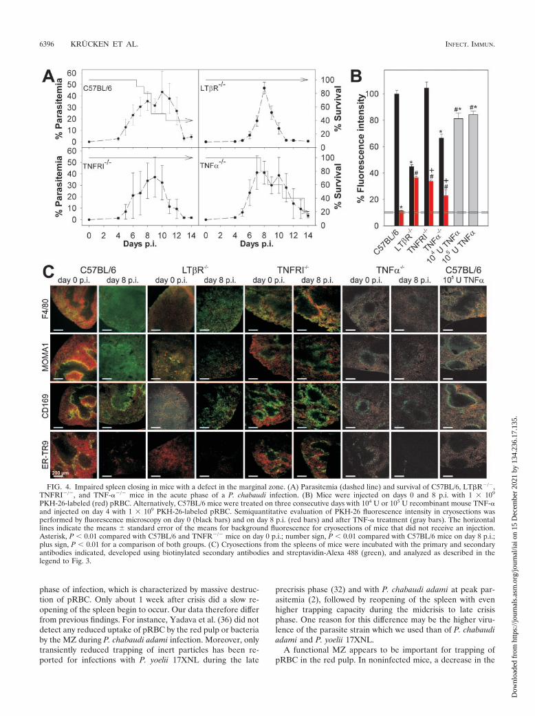
Role of TNF/TNFR family members in spleen closing. Thedata described above suggest that the MZ plays a critical rolein the control of spleen closing. This view was further sup-ported by analyzing mice with different defects in their MZ.For instance, LT�R�/� mice completely lack MZM andMMM (10, 11). All LT�R�/� mice survived an acute P.chabaudi infection, while a large proportion of C57BL/6 mice,used as controls in this experiment, succumbed to infection(Fig. 4A). The latter observation resulted from an abrupt in-crease in virulence of the parasite strain used, which occasion-ally occurs after more than 40 passages. Although this effectcomplicates comparison of the survival data shown in Fig. 1and 4, it extends the difference in susceptibility betweenLT�R�/� mice and C57BL/6 mice that we reported recently(35), thus allowing better correlation of changes in the MZ andsplenic trapping activity with resistance to blood stage malaria.A constitutive property of the LT�R�/� mice was their signif-icantly diminished ability to trap pRBC on day 0 p.i. (P 0.01)(Fig. 4B and C), and no significant spleen closing occurredafter infection on day 8 p.i.
This finding was further corroborated with TNFRI�/� mice,which were also highly resistant to P. chabaudi (Fig. 4A). Incontrast to LT�R�/� mice, TNFRI�/� mice have been re-ported to have an MZ with MZM and MMM, although thenumber was reduced (26). Moreover, the two cell populationsare not separated into two layers but are intermingled. How-ever, our confocal laser scanning microscopy analysis did notreveal the expected reduction in labeling for ER-TR9� MZMor CD169� MMM, although the labeling pattern was morediffuse than that in C57BL/6 mice and expression of MMM onMOMA1 was very weak (Fig. 4C). One reason for this differ-ence might be an effect of the different genetic backgrounds of
the mice used. TNFRI�/� mice exhibited normal pRBC trap-ping activity on day p.i. (Fig. 4B and C), but their spleens wereonly partially closed upon infection on day 8 p.i., with aboutone-third of the uptake occurring on day 0 p.i.. This indicatesthat signaling through TNFRI is involved in the regulation ofmalaria-inducible spleen closing. Conspicuously, TNFRI�/�
mice did not exhibit any malaria-induced redistribution of F4/80� macrophages and exhibited no alterations in labeling pat-terns of CD169/MOMA1� MMM and ER-TR9� MZM atpeak parasitemia (Fig. 4C). In contrast to C57BL/6 mice, co-localization of PKH-26-labeled pRBC with F4/80� red pulpmacrophages and MMM was observed even on day 8 p.i.,which further supports the hypothesis that the MZ play a rolein the control of pRBC entry into the red pulp.
TNFRI is known to mediate not only the effects of TNF-�but also the effects of lymphotoxin alpha (LT�) (16). There-fore, we also investigated TNF-��/� mice to determine if pro-duction of TNF-� is sufficient or necessary to provoke thechanges in histology and function associated with malaria-in-duced spleen closing. In contrast to TNFRI�/� mice, the pro-portion of TNF-��/� mice which succumbed to P. chabaudiinfection during the crisis phase was similar to the proportionof wild-type C57BL/6 mice which succumbed (Fig. 4A). Also incontrast to the TNFRI�/� mice, the TNF-��/� mice showedonly weak labeling for MMM and MZM that was correlatedwith significantly reduced trapping of pRBC on day 0 p.i. (Fig.4C). On day 8 p.i., however, the redistribution of F4/80� mac-rophages, the loss of ER-TR9� MZM, and the downregulationof MOMA1 antigen on MMM were similar to the redistribu-tion of F4/80� macrophages, the loss of ER-TR9� MZM, andthe downregulation of MOMA1 antigen on MMM in wild-typecontrol mice, indicating that LT� signaling is sufficient to causethese malaria-induced effects. Moreover, the uptake of pRBCwas reduced to about one-third that on day 0 p.i. which resem-bles the situation in TNFRI�/� mice. The uptake of PKH-26-labeled pRBC at peak parasitemia was lower in TNF-��/�
mice than in TNFRI�/� mice but still significantly higher thanthe uptake in wild-type mice (Fig. 4B).
Since disruption of the TNF-� gene impaired spleen trap-ping, we analyzed the possibility that TNF-� has a direct rolein spleen closing by injecting 104 U or 105 U TNF-� per day for3 days into noninfected C57BL/6 mice. This treatment resultedin a small but significant reduction in pRBC uptake (about20%) (Fig. 4B) and in a loss of ER-TR9� MZM (Fig. 4C),indicating that both the absence and strong activation ofTNFRI signaling lead to changes in the MZ.
DISCUSSION
This study demonstrated that there is a malaria-induciblespleen-inherent closing mechanism that controls the uptake ofboth inert beads and parasitized and nonparasitized RBC. Thespleen became largely closed shortly before peak parasitemiaand remained in the closed state throughout and after the crisis
FIG. 3. Localization of cell types in the spleen. Mice were injected with 1 � 109 PKH-26-labeled (red) pRBC. Cryosections were incubated withthe primary antibodies indicated and were developed using biotinylated secondary antibodies and streptavidin-Alexa 488 (green). Separate greenand red images were taken by confocal laser scanning microscopy and merged.
VOL. 73, 2005 MALARIA PARASITE DESTRUCTION DESPITE SPLEEN CLOSURE 6395
Dow
nloa
ded
from
http
s://j
ourn
als.
asm
.org
/jour
nal/i
ai o
n 15
Dec
embe
r 20
21 b
y 13
4.23
6.17
.135
.
phase of infection, which is characterized by massive destruc-tion of pRBC. Only about 1 week after crisis did a slow re-opening of the spleen begin to occur. Our data therefore differfrom previous findings. For instance, Yadava et al. (36) did notdetect any reduced uptake of pRBC by the red pulp or bacteriaby the MZ during P. chabaudi adami infection. Moreover, onlytransiently reduced trapping of inert particles has been re-ported for infections with P. yoelii 17XNL during the late
precrisis phase (32) and with P. chabaudi adami at peak par-asitemia (2), followed by reopening of the spleen with evenhigher trapping capacity during the midcrisis to late crisisphase. One reason for this difference may be the higher viru-lence of the parasite strain which we used than of P. chabaudiadami and P. yoelii 17XNL.
A functional MZ appears to be important for trapping ofpRBC in the red pulp. In noninfected mice, a decrease in the
FIG. 4. Impaired spleen closing in mice with a defect in the marginal zone. (A) Parasitemia (dashed line) and survival of C57BL/6, LT�R�/�,TNFRI�/�, and TNF-��/� mice in the acute phase of a P. chabaudi infection. (B) Mice were injected on days 0 and 8 p.i. with 1 � 109
PKH-26-labeled (red) pRBC. Alternatively, C57BL/6 mice were treated on three consecutive days with 104 U or 105 U recombinant mouse TNF-�and injected on day 4 with 1 � 109 PKH-26-labeled pRBC. Semiquantitative evaluation of PKH-26 fluorescence intensity in cryosections wasperformed by fluorescence microscopy on day 0 (black bars) and on day 8 p.i. (red bars) and after TNF-� treatment (gray bars). The horizontallines indicate the means � standard error of the means for background fluorescence for cryosections of mice that did not receive an injection.Asterisk, P 0.01 compared with C57BL/6 and TNFR�/� mice on day 0 p.i.; number sign, P 0.01 compared with C57BL/6 mice on day 8 p.i.;plus sign, P 0.01 for a comparison of both groups. (C) Cryosections from the spleens of mice were incubated with the primary and secondaryantibodies indicated, developed using biotinylated secondary antibodies and streptavidin-Alexa 488 (green), and analyzed as described in thelegend to Fig. 3.
6396 KRUCKEN ET AL. INFECT. IMMUN.
Dow
nloa
ded
from
http
s://j
ourn
als.
asm
.org
/jour
nal/i
ai o
n 15
Dec
embe
r 20
21 b
y 13
4.23
6.17
.135
.

number of MZM and MMM in TNF-��/� mice and the com-plete absence of MZM and MMM in LT�R�/� mice correlatewith weakly reduced and strongly reduced trapping, respec-tively. In addition, the MZ appears to be involved in malaria-induced spleen closing since no closing occurred in LT�R�/�
mice during malaria, although the red pulp is apparently nor-mal in these mice. Moreover, our data demonstrate that spleenclosure is partially mediated by signaling through TNFRI.Thus, disrupted signaling through TNFRI impaired splenicclosing, and, coincidently, it prevented loss of MZM and mi-gration of F4/80� macrophages into the white pulp. In accor-dance with this view, treatment of C57BL/6 mice with recom-binant TNF-� diminished splenic trapping and eliminatedMZM, whereas no migration of F4/80� macrophages and nochanges in localization of MMM were observed. AlthoughMZM were implicated in regulation of lymphocyte entry intothe white pulp (22), they presumably also contribute to entry ofpRBC into the red pulp. In the absence of MZM (e.g., aftertreatment of noninfected C57BL/6 mice with recombinantTNF-� or on day 35 p.i. in C57BL/6 mice), splenic trapping ofpRBC was reduced by at least 20%. In addition, MMM arealso candidates for regulation of entry into the red pulp, sincesignificant colocalization between MMM and pRBC was al-ways observed in an open spleen. In this context, it is alsonoteworthy that, besides TNF-�/LT�, additional proinflamma-tory cytokines, such as interleukin-1, have been found to beinvolved in malaria-induced spleen closure (2).
Spleen closing may have both protective and adverse effectson the malaria-infected host. In the precrisis phase, severalprotective effects of spleen closing can be envisaged (2, 32).First, the release of toxic and proinflammatory products upondestruction of pRBC by activated macrophages in the spleenmight be limited, and, therefore, the inflammatory responsecould be mitigated. Second, exclusion of pRBC allows adaptiveimmune responses to develop largely unperturbed in thespleen. For instance, adhesion of pRBC to dendritic cells im-pairs their activation in vitro, and this effect has been invokedto explain T-cell anergy at peak parasitemia (30, 31), involvingperturbed expansion of malaria-specific T- and B-cell popula-tions. By contrast, however, fully functional dendritic cells werefound in vivo in the spleens of malaria-infected mice (20, 21,27), which is in accordance with our findings that the spleenbecame largely closed throughout the crisis phase and there-fore locked out pRBC. Third, the spleen has been shown to bethe major erythropoietic organ in malaria-infected mice (15),and exclusion of parasites from the spleen could efficientlyprotect splenic reticulocytes from infection. Fourth, it is knownthat the spleen also destroys noninfected RBC during malariaand thus contributes to malaria-induced anemia (5, 7), whichwould therefore be diminished by spleen closure.
At peak parasitemia, however, spleen closure may be a dis-advantage for the host, as indicated by our results. Indeed,TNFRI�/� and LT�R�/� mice displayed only incompletesplenic closing, which correlated with increased resistance toblood stage malaria, compared with substantial spleen closingand higher susceptibility in TNF-��/� and wild-type C57BL/6mice. It is therefore tempting to speculate that incompleteclosing, like that in P. chabaudi-infected LT�R�/� andTNFRI�/� mice, is optimal for survival of the host. This viewis also supported by the finding that interleukin-1 treatment of
P. chabaudi adami-infected BALB/c mice causes an additionaldrop in particle trapping activity compared to untreated in-fected mice, which inversely correlates with an approximatelytwofold increase in peak parasitemia (2).
Although we are only beginning to understand the mecha-nisms that regulate the so-called closing of the spleen, our dataindicate at least that P. chabaudi infections induce spleen clo-sure which is manifested by a locking out of pRBC, particularlyduring the crisis phase. Therefore, the spleen cannot destroypRBC during the crisis phase when parasitemia decreases by atleast 100-fold within 4 days. This indicates that the sophisti-cated sequestration system of Plasmodium parasites did notevolve to avoid clearance by the spleen.
REFERENCES
1. Aichele, P., J. Zinke, L. Grode, R. A. Schwendener, S. H. Kaufmann, and P.Seiler. 2003. Macrophages of the splenic marginal zone are essential fortrapping of blood-borne particulate antigen but dispensable for induction ofspecific T cell responses. J. Immunol. 171:1148–1155.
2. Alves, H. J., W. Weidanz, and L. Weiss. 1996. The spleen in murine Plas-modium chabaudi adami malaria: stromal cells, T lymphocytes, and hema-topoiesis. Am. J. Trop. Med. Hyg. 55:370–378.
3. Baruch, D. I., S. J. Rogerson, and B. M. Cooke. 2002. Asexual blood stagesof malaria antigens: cytoadherence. Chem. Immunol. 80:144–162.
4. Benten, W. P. M., U. Bettenhaeuser, F. Wunderlich, E. Van Vliet, and H.Mossmann. 1991. Testosterone-induced abrogation of self-healing of Plas-modium chabaudi malaria in B10 mice: mediation by spleen cells. Infect.Immun. 59:4486–4490.
5. Connor, J., C. C. Pak, and A. J. Schroit. 1994. Exposure of phosphatidyl-serine in the outer leaflet of human red blood cells. Relationship to celldensity, cell age, and clearance by mononuclear cells. J. Biol. Chem. 269:2399–2404.
6. Cooke, B. M., N. Mohandas, and R. L. Coppel. 2004. Malaria and the redblood cell membrane. Semin. Hematol. 41:173–188.
7. Ekvall, H. 2003. Malaria and anemia. Curr. Opin. Hematol. 10:108–114.8. Engwerda, C. R., M. Ato, S. E. Cotterell, T. L. Mynott, A. Tschannerl, P. M.
Gorak-Stolinska, and P. M. Kaye. 2002. A role for tumor necrosis factor-alpha in remodeling the splenic marginal zone during Leishmania donovaniinfection. Am. J. Pathol. 161:429–437.
9. Flick, K., and Q. Chen. 2004. var genes, PfEMP1 and the human host. Mol.Biochem. Parasitol. 134:3–9.
10. Fu, Y. X., and D. D. Chaplin. 1999. Development and maturation of sec-ondary lymphoid tissues. Annu. Rev. Immunol. 17:399–433.
11. Futterer, A., K. Mink, A. Luz, M. H. Kosco-Vilbois, and K. Pfeffer. 1998. Thelymphotoxin beta receptor controls organogenesis and affinity maturation inperipheral lymphoid tissues. Immunity 9:59–70.
12. Gilks, C. F., D. Walliker, and C. I. Newbold. 1990. Relationships betweensequestration, antigenic variation and chronic parasitism in Plasmodiumchabaudi chabaudi—a rodent malaria model. Parasite Immunol. 12:45–64.
13. Gratepanche, S., B. Gamain, J. D. Smith, B. A. Robinson, A. Saul, and L. H.Miller. 2003. Induction of crossreactive antibodies against the Plasmodiumfalciparum variant protein. Proc. Natl. Acad. Sci. USA 100:13007–13012.
14. Groom, A. C., E. E. Schmidt, and I. C. MacDonald. 1991. Microcirculatorypathways and blood flow in spleen: new insights from washout kinetics,corrosion casts, and quantitative intravital videomicroscopy. Scanning Mi-crosc. 5:159–173.
15. Halder, R. C., T. Abe, M. K. Mannoor, S. R. Morshed, A. Ariyasinghe, H.Watanabe, H. Kawamura, H. Sekikawa, H. Hamada, Y. Nishiyama, H. Ishi-kawa, K. Toba, and T. Abo. 2003. Onset of hepatic erythropoiesis aftermalarial infection in mice. Parasitol. Int. 52:259–268.
16. Hehlgans, T., and K. Pfeffer. 2005. The intriguing biology of the tumournecrosis factor/tumour necrosis factor receptor superfamily: players, rulesand the games. Immunology 115:1–20.
17. Ho, M., and N. J. White. 1999. Molecular mechanisms of cytoadherence inmalaria. Am. J. Physiol. 276:C1231–C1242.
18. Janicik, J. M., R. Schauer, K. H. Andres, and M. von During. 1978. Seques-tration of neuraminidase-treated erythrocytes. Studies on its topographic,morphologic and immunologic aspects. Cell Tissue Res. 186:209–226.
19. Kraal, G. 1992. Cells in the marginal zone of the spleen. Int. Rev. Cytol.132:31–74.
20. Langhorne, J., F. R. Albano, M. Hensmann, L. Sanni, E. Cadman, C.Voisine, and A. M. Sponaas. 2004. Dendritic cells, pro-inflammatory re-sponses, and antigen presentation in a rodent malaria infection. Immunol.Rev. 201:35–47.
21. Leisewitz, A. L., K. A. Rockett, B. Gumede, M. Jones, B. Urban, and D. P.Kwiatkowski. 2004. Response of the splenic dendritic cell population tomalaria infection. Infect. Immun. 72:4233–4239.
VOL. 73, 2005 MALARIA PARASITE DESTRUCTION DESPITE SPLEEN CLOSURE 6397
Dow
nloa
ded
from
http
s://j
ourn
als.
asm
.org
/jour
nal/i
ai o
n 15
Dec
embe
r 20
21 b
y 13
4.23
6.17
.135
.

22. Lyons, A. B., and C. R. Parish. 1995. Are murine marginal-zone macro-phages the splenic white pulp analog of high endothelial venules? Eur.J. Immunol. 25:3165–3172.
23. Nolte, M. A., J. A. Belien, I. Schadee-Eestermans, W. Jansen, W. W. Unger,N. van Rooijen, G. Kraal, and R. E. Mebius. 2003. A conduit system dis-tributes chemokines and small blood-borne molecules through the splenicwhite pulp. J. Exp. Med. 198:505–512.
24. Oldenborg, P. A., A. Zheleznyak, Y. F. Fang, C. F. Lagenaur, H. D. Gresham,and F. P. Lindberg. 2000. Role of CD47 as a marker of self on red bloodcells. Science 288:2051–2054.
25. Pasloske, B. L., and R. J. Howard. 1994. Malaria, the red cell, and theendothelium. Annu. Rev. Med. 45:283–295.
26. Pasparakis, M., S. Kousteni, J. Peschon, and G. Kollias. 2000. Tumornecrosis factor and the p55TNF receptor are required for optimal develop-ment of the marginal sinus and for migration of follicular dendritic cellprecursors into splenic follicles. Cell. Immunol. 201:33–41.
27. Perry, J. A., A. Rush, R. J. Wilson, C. S. Olver, and A. C. Avery. 2004.Dendritic cells from malaria-infected mice are fully functional APC. J. Im-munol. 172:475–482.
28. Sherman, I. W., S. Eda, and E. Winograd. 2004. Erythrocyte aging andmalaria. Cell. Mol. Biol. 50:159–169.
29. Stevenson, M. M., and G. Kraal. 1989. Histological changes in the spleen andliver of C57BL/6 and A/J. mice during Plasmodium chabaudi AS infection.Exp. Mol. Pathol. 51:80–95.
30. Urban, B. C., D. J. P. Ferguson, A. Pain, N. Willcox, M. Plebanski, J. M.Austyn, and D. J. Roberts. 1999. Plasmodium falciparum-infected erythro-cytes modulate the maturation of dendritic cells. Nature 400:73–77.
31. Urban, B. C., and D. J. Roberts. 2002. Malaria, monocytes, macrophages andmyeloid dendritic cells: sticking of infected erythrocytes switches off hostcells. Curr. Opin. Immunol. 14:458–465.
32. Weiss, L., U. Geduldig, and W. Weidanz. 1986. Mechanisms of spleniccontrol of murine malaria: reticular cell activation and the development of ablood-spleen barrier. Am. J. Anat. 176:251–285.
33. Wunderlich, F., H. Stubig, and E. Konigk. 1982. Development of Plasmo-dium chabaudi in mouse red blood cells: structural properties of the host andparasite membranes. J. Protozool. 29:60–66.
34. Wunderlich, F., G. Schillinger, and M. Helwig. 1985. Fractionation of Plas-modium chabaudi-infected erythrocytes into parasites and ghosts. Z. Para-sitenkd. 71:545–551.
35. Wunderlich, F., M. A. Dkhil, L. I. Mehnert, J. V. Braun, M. El-Khadragy, E.Borsch, D. Hermsen, W. P. M. Benten, K. Pfeffer, H. Mossmann, and J.Krucken. 2005. Testosterone responsiveness of spleen and liver in femalelymphotoxin � receptor-deficient mice resistant to blood-stage malaria. Mi-crobes Infect. 7:399–409.
36. Yadava, A., S. Kumar, J. A. Dvorak, G. Milon, and L. H. Miller. 1996.Trafficking of Plasmodium chabaudi adami-infected erythrocytes within themouse spleen. Proc. Natl. Acad. Sci. USA 93:4595–4599.
Editor: W. A. Petri, Jr.
6398 KRUCKEN ET AL. INFECT. IMMUN.
Dow
nloa
ded
from
http
s://j
ourn
als.
asm
.org
/jour
nal/i
ai o
n 15
Dec
embe
r 20
21 b
y 13
4.23
6.17
.135
.