marjolin's ulcer: mismanaged chronic cutaneous ulcers
TRANSCRIPT
Case Report Open Access
Asuquo et al., J Clin Exp Dermatol Res 2013, S6 DOI: 10.4172/2155-9554.S6-015
J Clin Exp Dermatol Res Dermatology: Case Reports ISSN: 2155-9554 JCEDR, an open access journal
*Corresponding author: Maurice E Asuquo, Department of Surgery, Universityof Calabar, Calabar, Nigeria, GPO Box 1891 Calabar, 540001, Nigeria, E-mail:[email protected], [email protected]
Received May 29, 2013; Accepted July 14, 2013; Published July 23, 2013
Citation: Asuquo ME, Nwagbara VI, Omotoso A, Asuquo IM (2013) Marjolin’s Ulcer: Mismanaged Chronic Cutaneous Ulcers. J Clin Exp Dermatol Res S6: 015. doi:10.4172/2155-9554.S6-015
Copyright: © 2013 Asuquo ME, et al. This is an open-access article distributed under the terms of the Creative Commons Attribution License, which permits unrestricted use, distribution, and reproduction in any medium, provided the original author and source are credited.
AbstractBackground: In developing countries, chronic cutaneous ulcers, scars, and osteomyelitic sinuses are common
lesions. Marjolin’s ulcer is a malignant tumour that may arise from any of the above lesions. It may be misdiagnosed for infection and mismanaged as such.
Method: We evaluated the clinical histories, treatment, and outcomes of patients who presented to the University of Calabar Teaching Hospital, Calabar with histologic diagnosis of Marjolin’s ulcer from January 2010 to December 2012.
Results: The seven patients were 4 males and 3 females whose ages ranged from 28-70 years (mean 45.7 years). Trauma was the leading cause of injury resulting in ulceration [road traffic injuries- 4(57%), burns-2(29%)] while the other patient suffered diabetic foot ulcer. All the injuries involved the limbs [upper-1(14%), lower -6(86%)]. The histologic diagnosis in all the patients was squamous cell carcinoma with a mean latency period of 16.3 years. Late presentation with advanced loco-regional disease precluded curative surgery with poor outcomes.
Conclusion: Marjolin’s ulcer is preventable and areas of intervention have been highlighted. Educations concerning prevention, risk of chronic ulceration, early presentation, prompt and proper treatment is advocated for improved outcome.
Marjolin’s Ulcer: Mismanaged Chronic Cutaneous UlcersMaurice E Asuquo1*, Victor I Nwagbara1, Ayodele Omotoso2 and Inyang M Asuquo3
1Department of Surgery, University of Calabar, Calabar, Nigeria2Department of Histopathology, University of Calabar, Calabar, Nigeria3Department of Curriculum and Teaching, University of Calabar, Nigeria
Keywords: Marjolin’s ulcer; Chronic cutaneous ulcers;Mismanagement
IntroductionMarjolin’s ulcer (MU) is a malignancy arising in cutaneous scars,
chronic wounds, and osteomyelitic sinuses [1]. Jean-Nicholas Marjolin (1828) first described this in a burn scar [2]. Chronic ulcers, scars, and osteomyelitic sinuses are commonplace in developing countries [3,4]. It is not uncommon for Marjolin’s ulcer to be misdiagnosed for infection and mismanaged as such [5]. Theories concerning pathogenesis remain unclear, controversial, and not explained by one. They include Arons theory of transformation of chronic irritation to atypia and carcinoma, radiation/toxin-induced alteration of mitosis, implantation of epidermal cells into the dermis, induction of dormant neoplastic cells. Ultraviolet rays, ischemia, decreased immunity of affected skin/scar tissue, environmental and genetic interaction [6-8]. Diagnosis is histologic, most common squamous cell carcinoma (SCC) followed by basal cell carcinoma [9]. In the authors’ setting, traditional healers enjoy the patronage of the inhabitants because of strong socio-cultural beliefs and majority of times, patients seek the physician too late. Physician may also underestimate case with further loss of time [1]. Consequent on the above, majority of patients in the developing world presented with advanced lesions that precluded curative surgery [3]. We present this study to highlight the sequelae of mismanaged chronic cutaneous ulcers and recommend solutions for prevention and improved outcome of management.
Patients and Methods Patients who presented to the University of Calabar Teaching
Hospital (UCTH), Calabar from January 2010 to December 2012 with histologic diagnosis of Marjolin’s ulcer were studied. Indices analyzed were age, sex, aetiology, clinical presentation, latency period, histology, treatment, and outcome. This was compared with total number of SCC and skin malignancy seen during the same period and compared with previous reports from this facility.
ResultsSeven patients presented to UCTH, Calabar with Marjolin’s ulcer
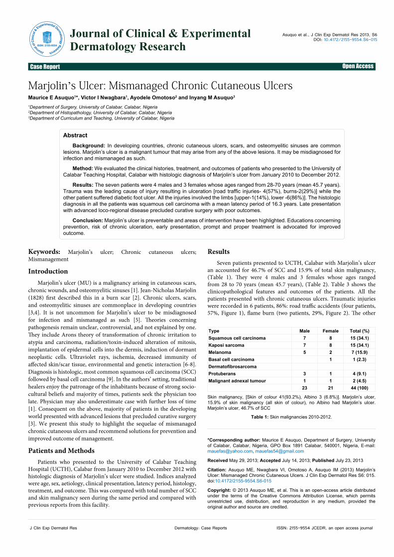
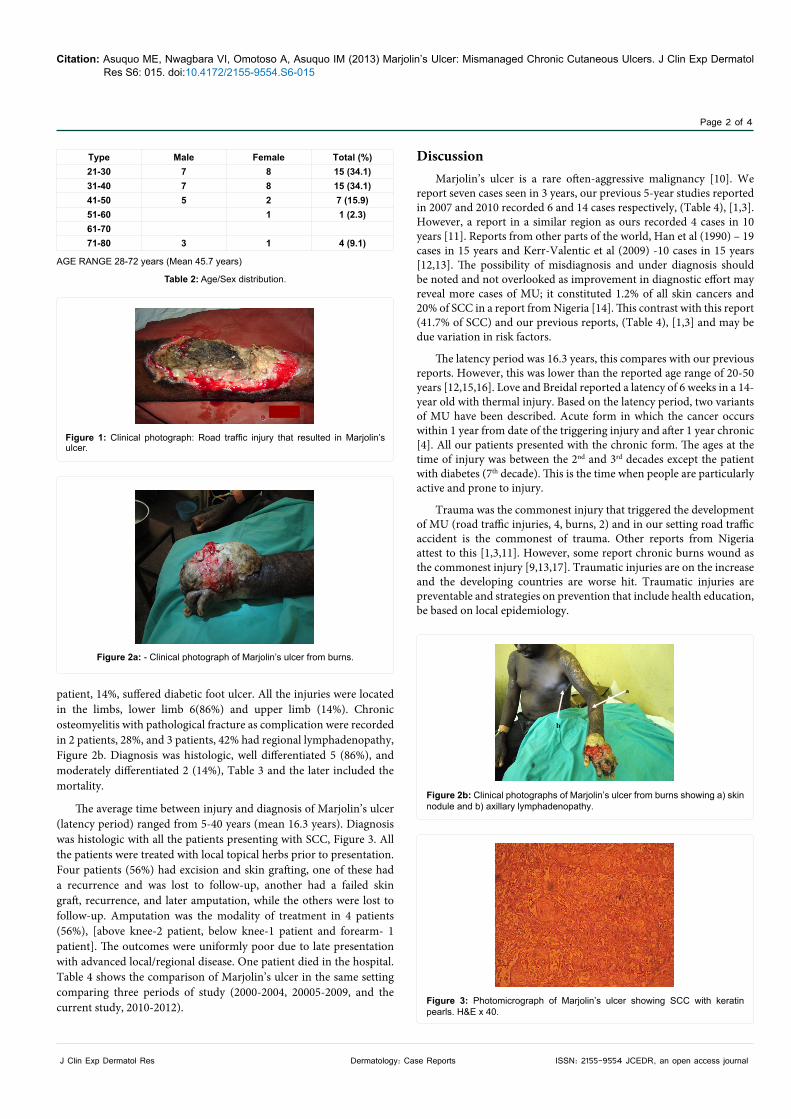
an accounted for 46.7% of SCC and 15.9% of total skin malignancy, (Table 1). They were 4 males and 3 females whose ages ranged from 28 to 70 years (mean 45.7 years), (Table 2). Table 3 shows the clinicopathological features and outcomes of the patients. All the patients presented with chronic cutaneous ulcers. Traumatic injuries were recorded in 6 patients, 86%: road traffic accidents (four patients, 57%, Figure 1), flame burn (two patients, 29%, Figure 2). The other
Type Male Female Total (%)Squamous cell carcinoma 7 8 15 (34.1)Kaposi sarcoma 7 8 15 (34.1)Melanoma 5 2 7 (15.9)Basal cell carcinoma 1 1 (2.3)DermatofibrosarcomaProtuberans 3 1 4 (9.1)Malignant adnexal tumour 1 1 2 (4.5)
23 21 44 (100)
Skin malignancy, [Skin of colour 41(93.2%), Albino 3 (6.8%)]. Marjolin’s ulcer, 15.9% of skin malignancy (all skin of colour), no Albino had Marjolin’s ulcer. Marjolin’s ulcer, 46.7% of SCC
Table 1: Skin malignancies 2010-2012.
Journal of Clinical & ExperimentalDermatology ResearchJourna
l of C
linic
al &
Experimental Dermatology Research
ISSN: 2155-9554
Citation: Asuquo ME, Nwagbara VI, Omotoso A, Asuquo IM (2013) Marjolin’s Ulcer: Mismanaged Chronic Cutaneous Ulcers. J Clin Exp Dermatol Res S6: 015. doi:10.4172/2155-9554.S6-015
Page 2 of 4
J Clin Exp Dermatol Res Dermatology: Case Reports ISSN: 2155-9554 JCEDR, an open access journal
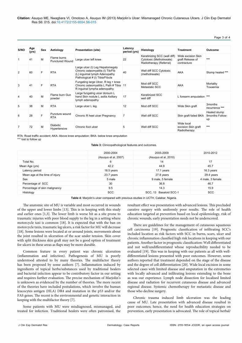
patient, 14%, suffered diabetic foot ulcer. All the injuries were located in the limbs, lower limb 6(86%) and upper limb (14%). Chronic osteomyelitis with pathological fracture as complication were recorded in 2 patients, 28%, and 3 patients, 42% had regional lymphadenopathy, Figure 2b. Diagnosis was histologic, well differentiated 5 (86%), and moderately differentiated 2 (14%), Table 3 and the later included the mortality.
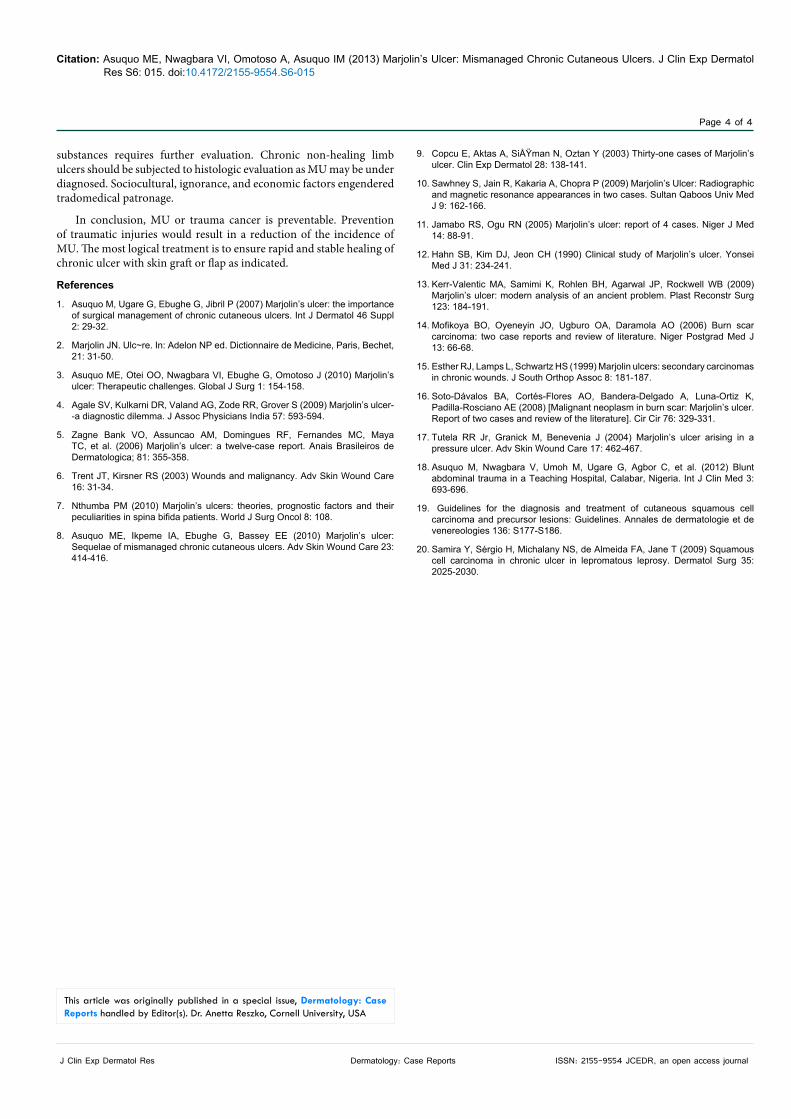
The average time between injury and diagnosis of Marjolin’s ulcer (latency period) ranged from 5-40 years (mean 16.3 years). Diagnosis was histologic with all the patients presenting with SCC, Figure 3. All the patients were treated with local topical herbs prior to presentation. Four patients (56%) had excision and skin grafting, one of these had a recurrence and was lost to follow-up, another had a failed skin graft, recurrence, and later amputation, while the others were lost to follow-up. Amputation was the modality of treatment in 4 patients (56%), [above knee-2 patient, below knee-1 patient and forearm- 1 patient]. The outcomes were uniformly poor due to late presentation with advanced local/regional disease. One patient died in the hospital. Table 4 shows the comparison of Marjolin’s ulcer in the same setting comparing three periods of study (2000-2004, 20005-2009, and the current study, 2010-2012).
Discussion Marjolin’s ulcer is a rare often-aggressive malignancy [10]. We
report seven cases seen in 3 years, our previous 5-year studies reported in 2007 and 2010 recorded 6 and 14 cases respectively, (Table 4), [1,3]. However, a report in a similar region as ours recorded 4 cases in 10 years [11]. Reports from other parts of the world, Han et al (1990) – 19 cases in 15 years and Kerr-Valentic et al (2009) -10 cases in 15 years [12,13]. The possibility of misdiagnosis and under diagnosis should be noted and not overlooked as improvement in diagnostic effort may reveal more cases of MU; it constituted 1.2% of all skin cancers and 20% of SCC in a report from Nigeria [14]. This contrast with this report (41.7% of SCC) and our previous reports, (Table 4), [1,3] and may be due variation in risk factors.
The latency period was 16.3 years, this compares with our previous reports. However, this was lower than the reported age range of 20-50 years [12,15,16]. Love and Breidal reported a latency of 6 weeks in a 14-year old with thermal injury. Based on the latency period, two variants of MU have been described. Acute form in which the cancer occurs within 1 year from date of the triggering injury and after 1 year chronic [4]. All our patients presented with the chronic form. The ages at the time of injury was between the 2nd and 3rd decades except the patient with diabetes (7th decade). This is the time when people are particularly active and prone to injury.
Trauma was the commonest injury that triggered the development of MU (road traffic injuries, 4, burns, 2) and in our setting road traffic accident is the commonest of trauma. Other reports from Nigeria attest to this [1,3,11]. However, some report chronic burns wound as the commonest injury [9,13,17]. Traumatic injuries are on the increase and the developing countries are worse hit. Traumatic injuries are preventable and strategies on prevention that include health education, be based on local epidemiology.
Figure 1: Clinical photograph: Road traffic injury that resulted in Marjolin’s ulcer.
Figure 2a: - Clinical photograph of Marjolin’s ulcer from burns.
AGE RANGE 28-72 years (Mean 45.7 years)
Table 2: Age/Sex distribution.
Type Male Female Total (%)21-30 7 8 15 (34.1)31-40 7 8 15 (34.1)41-50 5 2 7 (15.9)51-60 1 1 (2.3)61-7071-80 3 1 4 (9.1)
a
b
Figure 2b: Clinical photographs of Marjolin’s ulcer from burns showing a) skin nodule and b) axillary lymphadenopathy.
Figure 3: Photomicrograph of Marjolin’s ulcer showing SCC with keratin pearls. H&E x 40.
Citation: Asuquo ME, Nwagbara VI, Omotoso A, Asuquo IM (2013) Marjolin’s Ulcer: Mismanaged Chronic Cutaneous Ulcers. J Clin Exp Dermatol Res S6: 015. doi:10.4172/2155-9554.S6-015
Page 3 of 4
J Clin Exp Dermatol Res Dermatology: Case Reports ISSN: 2155-9554 JCEDR, an open access journal
The anatomic site of MU is variable and most occurred in wounds of the upper and lower limbs [13]. This is in keeping with this study and earlier ones [1,3]. The lower limb is worse hit as a site prone to traumatic injuries with poor blood supply to the leg in a setting where motorcycle taxi is common [18]. It is expected that with the ban on motorcycle taxis, traumatic leg ulcers, a risk factor for MU will decrease [18]. Some lesions were located at or around joints, movements about the joint resulted in ulceration of the scar under tension. Skin cover with split thickness skin graft may not be a good option of treatment for ulcers in these areas as flaps may be more durable.
Common feature in every patient was chronic ulceration (inflammation and infection). Pathogenesis of MU is poorly understood attested to by many theories. The multifactor theory has been proposed by some authors [7]. Inflammation induced by ingredients of topical herbs/substances used by traditional healers and bacterial infection appear to be contributory factor in our setting and requires further evaluation. The precise mechanism of Marjolin’s is unknown as evidenced by the number of theories. The more recent of the theories have included postulations, which involve the human leucocytes antigen (HLA) DR4 and mutation in the p53 and/or the FAS genes. The recent is the environmental and genetic interaction in keeping with the multifactor theory [7].
Some patients with MU were misdiagnosed, mismanaged, and treated for infection. Traditional healers were often patronised, the
resultant effect was presentation with advanced lesions. This precluded curative surgery with uniformly poor results. The role of health education targeted at prevention based on local epidemiology, risk of chronic wounds, early presentation needs not be underscored.
There are guidelines for the management of cutaneous squamous cell carcinoma [19]. Prognostic classification of infiltrating SCC’s included location as risk factors with SCC in burns, scars, ulcer and chronic inflammation classified high risk locations in keeping with our patients. Another factor in prognostic classification: Well differentiated and not well/undifferentiated whose reproducibility needed to be evaluated [19]. This was in keeping with our patients as all with well-differentiated lesions presented with poor outcomes. However, some authors reported that treatment depended on the stage of the disease and the degree of cell differentiation [20]. Wide local excision in some selected cases with limited disease and amputation in the extremeties with locally advanced and infiltrating lesions extending to the bone as was our experience. Lymph node dissection for localised limited disease and radiation for recurrent cutaneous disease and advanced regional disease. Systemic chemotherapy for metastatic disease and those who decline surgery.
Chronic trauma induced limb ulceration was the leading cause of MU. Late presentation with advanced disease resulted in poor outcomes; hence, the need for health education strategies on prevention, early presentation is advocated. The role of topical herbal/
RTA: Road traffic accident; AKA- Above knee amputation; BKA- below knee amputation*** lost to follow up
Table 3: Clinicopathological features and outcomes.
S/NO Age (yrs) Sex Aetiology Presentation (site) Latency
period (yrs) Histology Treatment Outcome
1 41 M Flame burns Punctured Wound Large ulcer left knee 22
Keratinizing SCC (well diff) Cytotoxic (Methotrexate) Radiotherapy (Referral)
Wide excision Skin graft Release of contracture
***
2 60 F RTA
Large ulcer (l) Leg Hepatomegaly Chronic osteomyelitis (l) Tib/Fib (L) inguimal lymph Adenopathy Pathological # (l) Tibia/Fibula
40 Well diff SCC Cytotoxic (methotrexate) AKA Stump healed ***
3 41 F RTAFungating large Ulcer, R leg + knee Chronic osteomyelitis L Path # Tibia R inguinal lympha adenopathy
11 Mod diff SCCMetastatic SCC AKA Mortality
Toxaemia
4 40 M Flame burn Gun powder
Large fungating ulcer dorsum L hand Skin nodule L axilla Axillary lymph adenopathy
17 Keratinized SCCwell diff L forearm amputation ***
5 38 M RTA Large ulcer L leg 12 Mod diff SCC Wide Skin graft 3months recurrence ***
6 28 F Puncture wound RTA Chronic R heel ulcer Pregnancy 7 Well diff SCC Skin graft failed BKA
Healed stump 9months Follow-up
7 72 M Diabetic Hypertensive Chronic foot ulcer 5 Well diff SCC
Wide local excision Skin graft Radiotherapy
***
Table 4: Marjolin’s ulcer compared with previous studies in UCTH, Calabar, Nigeria.
2000-2004 2005-2009 2010-2012 (Asuquo et al, 2007) (Asuquo et al, 2010) Total No. 6 14 17 Mean Age (yrs) 42.2 44.9 45.7 Latency period 18.5 years 17.1 years 16.3 years Mean age at the time of injury 23.7 years 27.8 years 29.4 years Sex 5 male 9 male, 5 female 4 male, 3 female Percentage of SCC 30 36.8 46.7 Percentage of skin malignancy 9.5 14.3 15.9 Histology SCC SCC, 13 Basaloid SCC-1 SCC
Citation: Asuquo ME, Nwagbara VI, Omotoso A, Asuquo IM (2013) Marjolin’s Ulcer: Mismanaged Chronic Cutaneous Ulcers. J Clin Exp Dermatol Res S6: 015. doi:10.4172/2155-9554.S6-015
Page 4 of 4
J Clin Exp Dermatol Res Dermatology: Case Reports ISSN: 2155-9554 JCEDR, an open access journal
substances requires further evaluation. Chronic non-healing limb ulcers should be subjected to histologic evaluation as MU may be under diagnosed. Sociocultural, ignorance, and economic factors engendered tradomedical patronage.
In conclusion, MU or trauma cancer is preventable. Prevention of traumatic injuries would result in a reduction of the incidence of MU. The most logical treatment is to ensure rapid and stable healing of chronic ulcer with skin graft or flap as indicated.
References
1. Asuquo M, Ugare G, Ebughe G, Jibril P (2007) Marjolin’s ulcer: the importance of surgical management of chronic cutaneous ulcers. Int J Dermatol 46 Suppl2: 29-32.
2. Marjolin JN. Ulc~re. In: Adelon NP ed. Dictionnaire de Medicine, Paris, Bechet, 21: 31-50.
3. Asuquo ME, Otei OO, Nwagbara VI, Ebughe G, Omotoso J (2010) Marjolin’sulcer: Therapeutic challenges. Global J Surg 1: 154-158.
4. Agale SV, Kulkarni DR, Valand AG, Zode RR, Grover S (2009) Marjolin’s ulcer--a diagnostic dilemma. J Assoc Physicians India 57: 593-594.
5. Zagne Bank VO, Assuncao AM, Domingues RF, Fernandes MC, Maya TC, et al. (2006) Marjolin’s ulcer: a twelve-case report. Anais Brasileiros deDermatologica; 81: 355-358.
6. Trent JT, Kirsner RS (2003) Wounds and malignancy. Adv Skin Wound Care 16: 31-34.
7. Nthumba PM (2010) Marjolin’s ulcers: theories, prognostic factors and theirpeculiarities in spina bifida patients. World J Surg Oncol 8: 108.
8. Asuquo ME, Ikpeme IA, Ebughe G, Bassey EE (2010) Marjolin’s ulcer:Sequelae of mismanaged chronic cutaneous ulcers. Adv Skin Wound Care 23: 414-416.
9. Copcu E, Aktas A, SiÅŸman N, Oztan Y (2003) Thirty-one cases of Marjolin’s ulcer. Clin Exp Dermatol 28: 138-141.
10. Sawhney S, Jain R, Kakaria A, Chopra P (2009) Marjolin’s Ulcer: Radiographic and magnetic resonance appearances in two cases. Sultan Qaboos Univ MedJ 9: 162-166.
11. Jamabo RS, Ogu RN (2005) Marjolin’s ulcer: report of 4 cases. Niger J Med14: 88-91.
12. Hahn SB, Kim DJ, Jeon CH (1990) Clinical study of Marjolin’s ulcer. Yonsei Med J 31: 234-241.
13. Kerr-Valentic MA, Samimi K, Rohlen BH, Agarwal JP, Rockwell WB (2009) Marjolin’s ulcer: modern analysis of an ancient problem. Plast Reconstr Surg123: 184-191.
14. Mofikoya BO, Oyeneyin JO, Ugburo OA, Daramola AO (2006) Burn scar carcinoma: two case reports and review of literature. Niger Postgrad Med J13: 66-68.
15. Esther RJ, Lamps L, Schwartz HS (1999) Marjolin ulcers: secondary carcinomas in chronic wounds. J South Orthop Assoc 8: 181-187.
16. Soto-Dávalos BA, Cortés-Flores AO, Bandera-Delgado A, Luna-Ortiz K, Padilla-Rosciano AE (2008) [Malignant neoplasm in burn scar: Marjolin’s ulcer. Report of two cases and review of the literature]. Cir Cir 76: 329-331.
17. Tutela RR Jr, Granick M, Benevenia J (2004) Marjolin’s ulcer arising in apressure ulcer. Adv Skin Wound Care 17: 462-467.
18. Asuquo M, Nwagbara V, Umoh M, Ugare G, Agbor C, et al. (2012) Bluntabdominal trauma in a Teaching Hospital, Calabar, Nigeria. Int J Clin Med 3:693-696.
19. Guidelines for the diagnosis and treatment of cutaneous squamous cellcarcinoma and precursor lesions: Guidelines. Annales de dermatologie et devenereologies 136: S177-S186.
20. Samira Y, Sérgio H, Michalany NS, de Almeida FA, Jane T (2009) Squamous cell carcinoma in chronic ulcer in lepromatous leprosy. Dermatol Surg 35:2025-2030.
Thisarticlewasoriginallypublishedinaspecial issue,Dermatology: Case Reports handledbyEditor(s).Dr.AnettaReszko,CornellUniversity,USA