marc e. gottlieb, md, facs plastic reconstructive surgery · 1 this presentation will give an...
TRANSCRIPT

Office: 1415 N. 7th Avenue Phoenix, AZ 85007 Phone 602-252-3354 Fax 602-254-7891 [email protected]
Specializing in the treatment, reconstruction, and management ofAcute and chronic wounds Diseases and defects of the soft tissues Injuries, diseases, and defects of the hand and extremities Defects of the head and trunk
PLASTIC & RECONSTRUCTIVE SURGERYBoard Certification Plastic Surgery Hand Surgery General Surgery
Marc E. Gottlieb, MD, FACSA Professional Corporation
Marc E. Gottlieb, MD, FACS
1415 N. 7th AvenuePhoenix, AZ 85007
Phone 602-252-3354Fax 602-254-7891
Copyright © 2005, Marc E. Gottlieb, MD
Content may be used for non-commercial educational purposes.Content may not be published or used for commercial purposes without prior license or permission.
Closing the Foot - Repair, Reconstruction, and Plastic Surgery of the Lower Extremity
Original presentation August 23, 2004, Boston, MA, ay the American Podiatric Medical Association.
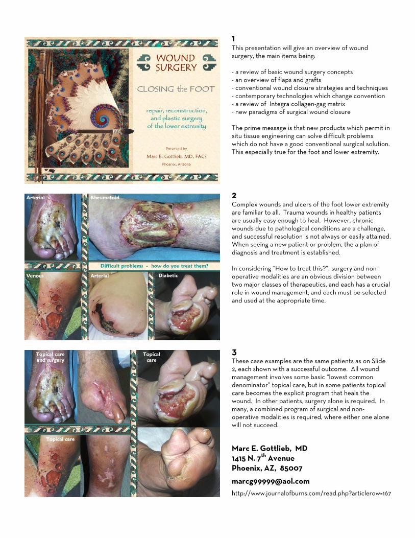
1 This presentation will give an overview of wound surgery, the main items being: - a review of basic wound surgery concepts - an overview of flaps and grafts - conventional wound closure strategies and techniques - contemporary technologies which change convention - a review of Integra collagen-gag matrix - new paradigms of surgical wound closure The prime message is that new products which permit in situ tissue engineering can solve difficult problems which do not have a good conventional surgical solution. This especially true for the foot and lower extremity.
2 Complex wounds and ulcers of the foot lower extremity are familiar to all. Trauma wounds in healthy patients are usually easy enough to heal. However, chronic wounds due to pathological conditions are a challenge, and successful resolution is not always or easily attained. When seeing a new patient or problem, the a plan of diagnosis and treatment is established. In considering “How to treat this?”, surgery and non-operative modalities are an obvious division between two major classes of therapeutics, and each has a crucial role in wound management, and each must be selected and used at the appropriate time.
3 These case examples are the same patients as on Slide 2, each shown with a successful outcome. All wound management involves some basic “lowest common denominator” topical care, but in some patients topical care becomes the explicit program that heals the wound. In other patients, surgery alone is required. In many, a combined program of surgical and non-operative modalities is required, where either one alone will not succeed.
Marc E. Gottlieb, MD 1415 N. 7th Avenue Phoenix, AZ, 85007
http://www.journalofburns.com/read.php?articlerow=167

4 For any physician treating any wound, effective care begins with are some fundamental questions that must be answered about the wound and the patient. When it comes time to choose treatment, the choices are easy to understand and easy to make – once you understand the art and science. For the average physician though, wound management seems like a mysterious black art. And even for accomplished wound physicians, and yet again for surgeons who are not plastic surgeons, when to do wound surgery and which technique to choose is a mysterious thing. Understanding when to choose which option begins with understanding the basic rules of wounds and wound surgery.
5 When surgery for a wound is required or desirable – once that question is answered – then the subsequent questions all concern timing and technique . . . and a few golden rules.
6 The distinction must always be made between benign, simple, acute wounds, and complex, pathological, and chronic wounds due to underlying illness and risk factors. Effective, uncomplicated surgery depends on a competent wound healing process. Surgery done on healthy wounds can usually be counted on to heal. Doing surgery on problem wounds often fails or make things worse. Thus, it is crucial to know when not to do surgery – perhaps not ever for a particular wound – or perhaps only after a period of adequate preparation and control of disease and risk factors. However, it is equally important to know when to surgery – when it is that the wound has no chance of healing without it.

7 The hand wound is in a healthy young man after flexor tenosynovitis from a penetrating injury. It is healthy, and it heals by topical care alone. Surgery serves no purpose. Only the most rudimentary hygienic care is required to keep the wound healthy, and healing the way it is programmed to do.
The leg wound is also in a healthy patient after minor skin trauma. An interval of grossly incompetent care by a skilless physician resulted in severe dermatitis, with perforation and abscess into the anterior compartment, after which the patient was scheduled for a leg amputation – honest folks – you can’t make this stuff up! Two weeks of good care resolved the acute problems. A small flap then closed the ankle tendons. Interval from 1st to 3rd picture = 8 weeks. As hard as it is to believe, you CAN mess up a good wound in a healthy patient – so don’t.
8 The hand problem is due to advanced diabetes-related atherosclerosis. Multiple debridements and amputations made the problem worse. The bilateral ankle ulcers are chronic, from undiagnosed Sjögren’s syndrome. The tibial ulcer is from atherosclerosis and the adverse mechanics of tendon shearing. All of these problems were fixed after multiple treatments had failed or had made the patients worse. Good results depend on first making the correct diagnosis, and then planning a program of comprehensive systematic care. In all of these cases, it was essential to know when to NOT do surgery, when to do no further surgery until disease and conditions were controlled. Then, it was crucial to know WHAT kind of surgery was required.
9 Assume now you have decided that surgery must be done for a particular wound. the question is, what techniques are required?
The principles of surgery that apply to healthy, healing competent wounds are very different than the rules required for chronic and pathological wounds. Sadly, the typical surgeon learns only the rules for healthy wounds, rules which DON’T necessarily work with problem wounds. Chronic and pathological wounds are their own special problem, with more detailed rules, and with fundamentally different strategies of care.
But, when it comes to basic tactics and techniques, there are robust, well-developed concepts of surgical wound closure which are relevant to all wounds. We will now start to look at these tactics and techniques of surgical wound closure.

10 “Wound repair” means several things, context-dependent. It can be (1) the physiological process of wound healing, (2) a generic reference to any kind of wound closure surgery, or (3) the more specific technique or simple wound closure. Simple healthy wounds can heal, and topical care in lieu of surgery is often the best choice. But surgery ought to be done for many good reasons, as listed. But, there are times when a wound cannot heal on its own, or should not be allowed to heal on its own (such as to prevent scar contractures across a joint). This is when one has to choose a method of closure. Simple repairs, grafts, and flaps are the conventional categorical methods of closure, but new technologies are expanding the options.
11 These cases illustrate the basic choices of simple repair.
Top case – This leg wound healed by natural contraction and epithelialization, supported by good topical care. Surgery would have contributed nothing. Simple debridement is the must fundamental of all wound “surgery”.
Middle case – Dog bite of thigh in a young boy. After a few more days of good care, it was ready to close. Surgical repair was not mandatory - it would have healed on its own - but simple repair was preferable, to simplify and expedite care and minimize symptoms.
Bottom case – Surgery of the chest and abdomen. Regardless what is done inside, closure of the skin and fascias is a “simple repair”. However, it is not really optional (unless there is a risk of infection, in which case they must be left open, and properly cared for.)
NOTES:
NOTE All wounds, prior to surgery, must meet basic criteria of suitability-for-closure. These are based on good hygiene, control of disease and inflammation, thorough debridement and reduction of bio-burden, and relief of other risks and injury. The basic principles of good care which allow a wound to heal by topical care alone are the same principles which prepare a wound for surgical closure by any method. Simple debridement is the must fundamental of all wound “surgery”, and this DOES apply to all wounds.

12 Getting past the simple repairs, the two main categories of wound closure are flaps and grafts. Non-plastic surgeons are often confused and perplexed about what these mean and when they are used. Important principles govern their use, and the necessity of one or the other is almost always “self-evident”, once the principles are understood. This section will discuss grafts: what makes a graft a graft, what conditions they need to be successful, and the three general reasons they are used: convenient closure, biological coverage, and specialized reconstruction.
13 Grafts are completely detached from the body. They have no circulation of their own, and they cannot live independently away from a recipient wound.
Skin grafts can only be used on a wound which is healthy and capable of healing. - Grafts do not carry the cellular machinery of repair. - Grafts depend on the recipient wound to heal. - They do not survive if the wound is incompetent. - The wound must be healthy and properly prepared.
Technique matters, especially fixation of the graft. - The graft must be in firm contact with the wound. - The graft must be suitably thin to stay alive.
Contracture can be expected in susceptible areas. - A healed skin graft is epidermis on scar == problems.
14 These cases illustrate the above principles of skin grafts.
Top case – This leg burn has healed skin grafts on healthy muscle, but grafts died over dead bone. The abdomen shows the design of a free flap which will be used to close the tibia.
Middle case – This ankle wound in an old atherosclerotic man took many weeks of preparatory care until there was a surface that would support a skin graft. Skin grafts that are too thick cannot survive, especially important in the face of arterial insufficiency.
Bottom case – A heel pressure ulcer with necrotic calcaneus. A flap was used to close the bone – a skin graft would likely have had problems – but a skin graft was used to close the healthy tissues of the flap donor site.

15 Any kind of tissue can be grafted, such as bone and cartilage and fascias for reconstructive purposes. Skin grafts are of two varieties – split and full thickness:
Split thickness grafts are easy, convenient, abundant (except in large burns), and renewable. But, they make scar and contract. They are good for expeditious wound closure when there are no special issues other than wound closure per se, when quality of the result is not a concern, or when patient safety demands expediency.
Full thickness grafts are of limited availability, but they usually give a high quality reconstruction without contractures. They are used for elective reconstruction, or for closure of small wounds where expediency, a closed donor site, and cosmesis are important or opportune.
16There are three general reasons to use grafts:
Top – convenient wound closure – Venous ulcer of the leg, excised and skin grafted. When quick closure is needed for large healthy areas, when there are o special concerns or caveats, then split thickness skin grafts get the job done.
Middle – biological dressing – These are extensive life-threatening wounds from necrotizing fasciitis. The wounds may not be completely healthy and ready to heal a skin graft, but sometimes closure with a graft, even if temporary, helps stabilize a wound and improve the patient. Biological dressings can be done with a variety of living and engineered materials.
Bottom case – specialized reconstruction – This case shows reconstruction of the nasal septum after trauma – a typical use of cartilage grafts.
17 This section will discuss flaps: what makes a flap a flap, what conditions they need to be successful, and the four general reasons they are used: convenient closure, general reconstruction, essential coverage, and closure of incompetent wounds.

18 Flaps are flaps because they remain attached to the body. The have their own circulation, and they live independently of anything other than their own pedicle. Flaps have these desirable properties: - They can transport large volumes of tissue. - They retain their original characteristics, so they satisfy
many functional, cosmetic, and reconstructive needs. - Assuming that they are made from healthy tissues (to
do otherwise would be a foolish waste), they can do the healing for a wound which cannot.
The caveat is that flaps require finesse and technical precision, or they can die. Done well, they solve many of the problems in wound and reconstructive surgery.
19 These cases illustrate the above principles of flaps.
Top case – This large flap from the posterior thigh will close an ischial ulcer. Note the large volume of tissue and the small pedicle. Depending on the flap and its anatomy, a flap’s pedicle can be nothing more than a single small artery and one vena comitans.
Middle case – Raising a flap on isolated vessels means that the vessels can be divided, then anastomosed (with a microscope) to recipient vessels in the target area, a “free flap”, as was done here to move the abdominal flap to the burned leg (same case as Slide 14).
Bottom case – This is a buttock flap, used to close a sacral ulcer, a relatively simple design and advancement. Regardless of the technical simplicity or complexity of the flap, if done properly, the flap only knows that it must heal its perimeter, an easy job for healthy tissues.
20There are four general reasons to use flaps:
1 – convenient wound closure – oftentimes, typically for minor trauma wounds, excision of skin lesions, and other minor surgery, small skin flaps are the simplest and best way to close the defect (not illustrated). 2 – general reconstruction – flaps are the workhorses of reconstructive surgery, replacing soft tissues wherever they are needed, for whatever purpose, as in this case of an ear after cancer excision. 3 – essential coverage – when viscera and skeletal structures demand coverage, to preserve health and function, flaps are mandatory, as in this open ankle after trauma (closed with a rectus abdominis free flap). 4 – wound healing incompetence – if the wound is pathological and incapable of healing, for whatever reason, a healthy flap does the healing, as in this case of adriamycin infiltration (closed with a groin flap).

21 Flaps require technical precision, or else . . .
There are two general risks, very real, with flaps: (1) They may not reach the target. (2) They may not live (inadequate perfusion). The surgeon must not let these happen – this is the art of flaps. When the risks are high, many strategies and methods ensure a good outcome, if properly practiced. Avoiding trouble depends on:
There are two general flap designs: (1) Random - based on target geometry and local soft tissue mechanics. (2) Anatomical – based on specific angiosomes (vascular territories). Tissue mechanics and circulation govern how these flaps move and behave.
There are special strategies: Among them, the use of multiple flaps and grafts for one target, and sequential procedures to incrementally elevate, “train”, and transfer a flap (delay procedures).
22Here are illustrations of some of these principles.
Top left – closure of a tibial pseudarthrosis, after rods and bone grafts. The bone grafts demand a flap. A small adjacent random flap works perfectly. Skin grafts close the flap donor site. You cannot eliminate the middleman – NO skin grafts on bone graft.
Bottom left – An open knee after fasciitis, covered by a gastrocnemius skin and muscle flap. The flap donor site and non-essential parts of the original wound are covered by Integra, an artificial skin.
Right – A radiation ulcer of the back in a child, after spinal cord tumor. The lumbar area is challenged by a lack of directly arterialized skin – no easy flaps. Closure was done in several stages – delays and intermediate transfers - incrementally moving lumbar skin from the good side to the bad side.
NOTES:
NOTE, reprise All wounds, prior to surgery, must meet basic criteria of suitability-for-closure. These are based on good hygiene, control of disease and inflammation, thorough debridement and reduction of bio-burden, and relief of other risks and injury. The basic principles of good care which allow a wound to heal by topical care alone are the same principles which prepare a wound for surgical closure by any method. Simple debridement is the must fundamental of all wound “surgery”, and this DOES apply to all wounds.

23 Flaps are the romantic heroes of plastic surgery – the “big”, challenging, important cases coveted by every red-blooded resident. Flaps have a pivotal role in the closure of complex wounds. When the stakes are high for successful closure, good flaps get the job done. Flaps rule. Flaps are boss . . . you get the idea.
. . . BUT . . .
There are times when flaps simply cannot be done or will not survive. THEN WHAT?
24Some wounds are thorny problems. Their circumstances may mean that: There are no good flaps available. Any potential flap is in the zone of injury. General or systemic diseases create the same risks for flaps as they do for the target wound. The patient may be too sick for the preferred procedure.
The 1970s and 1980s were the era of flaps, when the vascular anatomy of the soft tissues was completely mapped, when the techniques of microvascular free flaps were developed, and when sound methods were established for closing ANY kind of acute wound, or reconstructing nearly any kind of defect.
BUT . . . there is still a frontier – the chronic pathological wound. Flaps can sometimes solve these problems, sometimes not. When they cannot, new technologies of the 1990s and 2000s are coming to the rescue.
25This leg has chronic and active ulceration due to severe untreated rheumatoid disease, compounded by severe arterial insufficiency. Concurrent post-phlebitic venous hypertension probably reflects a hypercoagulability. This wound is wound healing incompetent, with exposed structures – a skin graft will die. A flap is needed, but there are many reasons why a flap will fail:
Not enough local skin; available skin flaps too small or will not reach; no nearby anatomical flaps; a flap is likely to die from the vascular disease; atherosclerosis will prevent micro-anastomosis of a free flap; potential flaps are within zone of injury; hematological disorders can kill a flap; inflammation and disease threaten a flap; comorbidities make patient too high risk; risk for donor site complications; a failed flap wastes anatomy and limits further options. muscle flaps might sacrifice useful anatomy needed after an amputation (eg, latissimus).

26 The new technologies of the past 10 – 20 years have created new options and new strategies to fix those problems that cannot be fixed by conventional wound surgery, even flaps. This section will look at some of these new technologies and methods, including advanced surgical strategies, wound healing pharmaceuticals, and bio-integrated artificial skin.
27Bottom case – A non-healing abdomen after lymphoma staging and radiation. The wound failed multiple naive attempts to reclose it. Since radiation kills wound healing cells, flaps are normally used to close these incompetent wounds. However, at the low dose of 2300 cGy, some wound healing potential remains. It was coaxed out by stimulating the wound with a wound active cytokine – PDGF.
Top case – High dose radiation for skin lymphoma caused this ankle ulcer, which was completely devoid of wound healing activity. There are no local flaps which can cover this large area, and atherosclerosis precludes a free flap. Using granulation tissue from a remote donor wound (an implanted plastic chamber), a graft was prepared of wound activated healthy cells. After two harvests / injections into the wound, it proliferated until it could accept a skin graft.
28 Another modern technology that solves complex problems is Apligraf, a living, in vitro engineered skin. It does not function as a skin graft, but rather as a pharmacological agent packaged in a living material. It is used for problem wounds that fail to heal in spite of all best efforts to control risks and protect the wound. When a wound should be healing but isn’t, Apligraf has a predictable effect to stimulate proliferative healing. In the illustration, a chronic trauma related plantar ulcer failed to heal after several months of appropriate care (with the patient still walking and working). Within three weeks of Apligraf placement, it was healed. Apligraf is comparable to PDGF in its effects, and the clinical indications are similar for the two agents.

29 This is another case, a chronic Achilles ulcer, where Apligraf stimulated a wound that would not budge after many months of care. This series shows the typical progression of observable effects, usually running its course over a 4 – 6 week period. Many wounds heal after one application, but if the response was good but incomplete, as in this case, a second “dose” can be placed. In this case, the wound healed by the 9th week.
30 The above technologies are useful for many types of chronic and pathological wounds, but they have their limitations. They are pharmaceuticals, agents of topical care for a wound which will be managed open. They can be used whenever topical care is appropriate for a stalled wound. They can be used in lieu of surgery where the wound has a chance of healing by stimulated natural processes. However, some wounds mandate surgery – period. This implies flaps. But when flaps are not available or will not work, is there an option? Yes. The technology product that replaces or bests flaps is an artificial “skin”, Integra® (Integra Life Sciences, New Jersey). As an implant applied to the surface, then bio-integrated by embryonic processes unrelated to the normal wound healing process, it fixes those situations which flaps cannot.
31 Integra structure - A working layer of a collagen-chondroitin sponge (the matrix in which histogenesis occurs), topped by a silicone rubber “epidermis”. Applied to a wound, tissue regenerates in the sponge. A few weeks later, thin skin grafts replace the silicone to complete the reconstruction.
Two main modes of use – (1) a high quality artificial skin for acute closure – the body believes it to be real skin; (2) a dermal regenerant used as an agent of reconstruction.
Unique properties lead to remarkable results - not being alive, it is tolerant of conditions which kill grafts; it completely suppresses inflammation; no inflammation means no conventional wound healing means no scar; it can conduct tissue through the sponge, covering exposed structures and voids; there is no contraction.

32 These examples illustrate some of these properties.
Top – Integra eliminates inflammation. The histology, a few days after placement, shows early histogenetic cells. Neutrophils and other inflammation NEVER appear in the matrix. Clinically, refractory periwound inflammation subsides quickly. Middle – Compare normal histogenetic tissue in the Integra versus normal granulation tissue in a seam between two pieces. When healed, the differences between Integra skin and normal scar are striking. Bottom – Normal scar is stiff, non-compliant, and contracts, causing scar fracture ulcers, such as across this burned ankle. Integra has normal skin compliance and remains highly pliable, as seen on the dorsum of the hand. Normal dermatogenesis results from the large syncytial fibroblasts seen in the matrix, an embryonic cell that NEVER appears during normal wound healing.
33Reprise of Slide 25 – a list of reasons why a flap might not work, why there needs to be alternative options.
Here is an open MCP joint in a severe scleroderma patient. According to convention, a flap is mandatory for closure, but: even in a normal person, flaps to cover this position are limited without collateral injury to the hand; non-compliant sclerotic scleroderma skin cannot move; angiopathy puts any potential flap at risk to die; inflammatory disease threatens any autogenous reconstruction; donor site complications are likely, and failed flaps will make the whole problem worse.
Integra succeeded with only a minor low-risk outpatient program of care.
Understanding when a flap should but cannot be used is to understand when Integra should be used.
34 Integra is a new paradigm of surgery. It’s technical use as a sheet material is comparable to skin grafts, but it is not a graft. It solves many of the problems that flaps can, but it also solves many problems that a flap cannot (and vice versa). It is an implant, applied to the surface. It induces generation of new tissue via embryonic processes. Flaps remain the great heroes of plastic surgery for reconstructive needs, but for closing problem wounds, Integra is the modern Excalibur.
Integra is a completely new paradigm of surgery:
in-situ tissue engineering
part of the providence of modern biotechnology.

35The following series of slides illustrate the use of Integra for problem wounds, beginning with general cases from any part of the body. All Integra use falls into 4 conceptual categories:
36 Acute & critical coverage (left) – Life-saving coverage in this patient with necrotizing fasciitis. Developed for burns, Integra also improves survival and results for degloving injuries and any other extensive injury. Essential coverage (middle) – Closure of extensor tendons of hand and wrist, a superior result compared to flaps, because late revision is not needed. The closure of working parts conventionally requires flaps, but Integra solves these problems when flaps cannot. Wound control (right) – Like any biological dressing, Integra can tame a wild wound, but unlike the others, it is not an interim modality. It remains on the wound and regenerates healthy new tissue, as in this patient with a chronic granulomatous ulcer that healed after numerous skin grafts failed. Reconstruction (bottom) – Because it does not scar and contract, Integra is good for reconstruction, as in this patient with burn contractures of elbow and wrist.
37 Left – Necrosis of the hand (see Slide 8). Vascular disease, high risk for any surgery, and open flexor tendons are the challenges. There are no good flaps, and if there were, they would die. Integra solves the problem easily, as an outpatient. Right – Because Integra eliminates normal wound repair and scar, it can solve that most persnickety of abnormal scar problems, the keloid, as shown here after resurfacing the back of the ear.
NOTES:
NOTE, reprise All wounds, prior to surgery, must meet basic criteria of suitability-for-closure. These are based on good hygiene, control of disease and inflammation, thorough debridement and reduction of bio-burden, and relief of other risks and injury. The basic principles of good care which allow a wound to heal by topical care alone are the same principles which prepare a wound for surgical closure by any method.
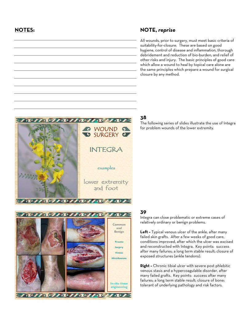
38 The following series of slides illustrate the use of Integra for problem wounds of the lower extremity.
39 Integra can close problematic or extreme cases of relatively ordinary or benign problems. Left – Typical venous ulcer of the ankle, after many failed skin grafts. After a few weeks of good care, conditions improved, after which the ulcer was excised and reconstructed with Integra. Key points: success after many failures; a long term stable result; closure of exposed structures (ankle tendons). Right – Chronic tibial ulcer with severe post phlebitic venous stasis and a hypercoagulable disorder, after many failed grafts. Key points: success after many failures; a long term stable result; closure of bone; tolerant of underlying pathology and risk factors.

40 Left – Severe trauma and degloving of lower extremity, in a child. Key points: critical contribution to salvage of an extremity that all of the brand-X doctors wanted to amputate; closure of multiple exposed structures; compliant skin with full range of motion across knee. Right – Decades long ulceration of diabetic necrobiosis lipoidica, after many failed attempts to treat. Excision and skin reconstruction with Integra gives long term stable results. Key points: success after many failures; a long term stable result; tolerant of underlying pathology and risk factors; managing large area wounds as an outpatient.
41 Integra can close problematic wounds due to active ulcerative pathologies, especially connective tissue disorders and other chronic inflammatory, vascular, hematological, and necrotizing disorders. Case - This woman developed refractory ankle ulceration due to severe rheumatoid arthritis. FactorV Leiden and protein C and S abnormalities worsened the problem. Figures: original; inflammation subsided after good topical care, but no wound healing; excised ulcer; two weeks into the Integra reconstruction; early after the split thickness overgrafts; a healed stable result one year later (note the Integra behind the other ankle, for new ulcers with disease flare-up). Key points: success after failures; a long term stable result; tolerant of underlying pathology; tolerant of disease flare-up.
42 Top – Chronic ulceration of the ankle in a rheumatoid patient – typical ulceration over bursas and tendon sheaths due to rheumatoid synovitis. refractory to topical care, it healed with Integra. Key points: success after failed care; tolerant of underlying pathology; closure of tendons and joints. Bottom – Refractory chronic ulceration of ankle. Old malleolar fracture and pseudarthrosis was the only identifiable pathology. Key points: success after failed care; closure of tendons and joints.

43 Left – A patient with chronic ankle ulceration due to rheumatoid and Sweet’s neutrophilic dermatitis. Top, just prior to severe inflammatory lysis of old skin grafts, with perforation into ankle. Below, a stable Integra reconstruction several years later. There has been no further pathology in the reconstructed skin.
Right – Rapid progressive ulceration of the ankle after biopsy of a skin lesion, worsened by attempts to reclose the wound. Lab and histology show protein C deficient hypercoagulability. Warfarin and Integra arrest further ulceration. Integra regenerated over the lateral compartment tendons, yielding a stable long term, result here after a few years.
Key points - Success after many failures; a long term stable results; tolerant of underlying pathology and risk factors.
44 Left – Ankle ulceration of many years, treated as “venous”, her rheumatoid diagnosis missed. Once the correct diagnosis was made and treatment started, the ulcer was excised, and skin reconstructed over bone and tendon, yielding a stable result of years duration, remaining stable when disease flare caused new ulcers.
Right – Forty year history of leg ulceration, due to a missed diagnosis of Sjögren’s. Once the correct diagnosis was made and treatment started, the ulcers were excised, and skin was reconstructed over bone and tendon, yielding a stable result of years duration.
Key points - Success after many years and many failures; a long term stable result; tolerant of underlying pathology and risk factors; managing large area wounds as an outpatient; accurate diagnosis and treatment are crucial; nearly normal quality of the reconstructed skin.
45 Case - This young woman ruptured her achilles while exercising. Recurrent necrosis and dehiscence resulted in many failed operations. Ultimately closed with a free flap, the skin grafts over the muscle flap developed characteristic hypertrophy and chronic ulceration. Attempts to revise this caused more dehiscence and necrosis. Lab workup and a history of retinal artery occlusion confirmed an antiphospholipid antibody syndrome and hypercoagulability. Warfarin and Integra arrested necrosis and ulceration. The regenerated skin has normal characteristics and biomechanics, and no further pathology. Graduated loading and therapy induced tendinous metaplasia in the old muscle flap, and her ankle is now essentially normal.
Key points - Success after many failures; a long term stable result; tolerant of underlying pathology; accurate diagnosis and treatment are crucial.

46 Left – Typical heel and achilles ulceration from pressure, in an infirm person. For large heel defects, simple posterior calcanectomy, and Integra over bone and tendon easily heals these as an outpatient.
Middle – Comparable case of heel and achilles ulceration, easily healed by debridement, calcanectomy, and Integra.
Right – Achilles ulcer in a patient with Wegener’s granulomatosis and sever pulmonary disease. Straightforward Integra reconstruction, with two minor outpatient procedures - healed.
Key points - All the usual stuff as above. Integra makes those traditionally “hard” problems easy to manage. No need for big operations. No need for inpatient care. No need for amputations.
47 With Integra, it is easy to close high risk wounds with no risk to the patient. It is thus easy to salvage “simple” problems that most surgeons indiscriminately amputate.
Case - Forefoot necrosis from vascular athero-emboli. The transtarsal debridement is a “good” amputation, because all major ankle motors are attached. But, there is not enough skin to close. Flaps are not there, or high risk. Atherosclerosis precludes a free flap. Recessing bone to get “enough” skin will destabilize the ankle, making foot salvage irrelevant. The average surgeon does an amputation. That is pointless, because this problem is easily fixed by a piece of Integra.
Key points - Don’t amputate for lack of a good flap. Don’t amputate just because you don’t know what else to do. Understanding when a flap should but cannot be used is to understand when Integra should be used.
48 Top – Transmetatarsal amputation after vascular complications. The foot is stable and well cared for, no acute jeopardy, but the wound will not heal, after months. Integra cannot correct vascular disease, but it controls inflammation and other adverse wound conditions enough to change the balance of factors to a state that permits reparative cellular processes to survive and complete their job. Bottom – Aorto-iliac vasculopathy, with toe necrosis. Progressive levels of amputation were all complicated by more necrosis – and now “hip disarticulation” is pending. Integra, in this case supported by hyperbaric oxygen, stabilizes the wound and allows it to heal. Key points – Don’t keep doing the same wrong thing that didn’t work the first time. Wound knowledge is important. Patience and Integra are virtues. Simple outpatient care.

49 Top – A below knee amputation with minor wound complications. You know what the average surgeon would do – an above knee amputation – you may have been guilty of this yourself. Good preparatory wound care followed by Integra preserves all available length of tibia, heals the wound, and permits a prosthesis. Bottom – Another vasculopathic foot. After popliteal-tibial bypass, circulation was excellent, there was no more necrosis, and the wound started to heal. But, the damage was already done, and most of those “other surgeons” would have done an amputation. But – the damage is only to skin and fascias. That is easily rescued and reconstructed. Healed after Integra. Key points – Don’t throw away perfectly good limbs. Knowledge is important. Patience and Integra are virtues. Simple outpatient care.
50 Case - Comparable to the case on Slide 47, this problem resulting from diabetic neuropathic forefoot ulceration. The patient wears a shoe insert and thin ankle orthosis, and he is essentially normal in his daily activities. Key points – Don’t throw away perfectly good limbs. Knowledge is important. Patience and Integra are virtues. Simple outpatient care.
51 Case - Diabetes and severe vasculopathy case loss of toes on the right foot. He subsequently developed gangrene of the left leg, and while recuperating from his amputation, he was allowed to develop a large right heel ulcer. Now, “somebody” wants to do another BKA. Simple posterior calcanectomy and Integra heal the problem. He wears a small orthosis, a sneaker, his contralateral leg prosthesis, and he uses a walker, and he is ambulatory and independent.
Key points – Don’t throw away perfectly good limbs. Knowledge is important. Patience and Integra are virtues. Simple outpatient care.
Hopefully, soon will be gone the days when impatient and improperly educated orthopedic and vascular surgeons will stop throwing away good limbs simply because they do not know how to fix the skin.

52 The art of wound surgery depends on the traditional options of topical care, simple repairs, grafts, and flaps, now augmented by technological products that stimulate wound healing or permit in situ tissue engineering by the induction of histogenetic processes.
How does a surgeon choose? For most cases, the best choice is usually obvious, all based on the principles discussed above. Sometimes there are choices to be made, based on safety, time, cost, complexity, lifestyle, and other patient, surgeon, and incidental factors. These goals should always be foremost, more or less in this order of importance: keep patient safe; control disease and symptoms; preserve or improve function and lifestyle; heal the wound; do so quickly and efficiently; minimize costs and resource utilization.
Choose whatever treatment best fulfills these goals.
53These cases illustrate some of the decision making art. Left top – Chronic ankle ulcer in a diabetic vasculo-pathic rheumatoid patient. Perfect for Integra, right? Yes, it would have worked, but none of his risks are severe or active, so wound healing should be normal. So, a reverse sural nerve flap was opted, for a one stage reconstruction – simpler, quicker, more efficient. Left bottom – Malleolar ulcer in a rheumatoid patient. Same issues – disease was quiet, and the ulcer was due to the biomechanics of bursas. A small local flap should do well - and it did do well, with the advantage of one stage and a more durable result in this high impact area. Right bottom – From Slide 22. Remember – you can’t eliminate the middle man – each method has its role, and you can’t cheat for lack of experience. Any case can use multiple methods, such as the skin grafts used to close the donor sites in each of these three patients.
54 Conventional wound surgery - repairs-grafts-flaps - is a sophisticated dependable art that can close virtually any healthy wound – wounds due to trauma or surgery in healthy people. Chronic and pathological wounds challenge these principles, but with new technology products and strategies, now almost any wound of any kind can be healed (with greater or lesser ease).
Integra is a new product with remarkable properties, efficacy, and range of applications. In a recent paper analyzing Integra in 120 patients, chronic wounds were closed successfully in 92%. Inpatient rates in these patients declined to zero, a testimonial to how well modern products and concepts not only cure the wound, but do so efficiently and easily. Integra represents a distinct new paradigm of wound closure surgery – in situ tissue regeneration – and surgeons must work this new mode into their decision schemas.

NOTES: