map6-fisatemperaturesensorthatdirectlybindstoand ... · at the cellular level, map6 have been found...
TRANSCRIPT

MAP6-F Is a Temperature Sensor That Directly Binds to andProtects Microtubules from Cold-induced Depolymerization*
Received for publication, July 5, 2012, and in revised form, August 6, 2012 Published, JBC Papers in Press, August 17, 2012, DOI 10.1074/jbc.M112.398339
Christian Delphin‡1, Denis Bouvier§, Maxime Seggio‡, Emilie Couriol‡, Yasmina Saoudi‡, Eric Denarier‡,Christophe Bosc‡, Odile Valiron‡, Mariano Bisbal‡2, Isabelle Arnal¶, and Annie Andrieux‡
From ‡Team 1 Physiopathology of Cytoskeleton; Commissariat a I’Energie Atomique, Institut National de la Santé et de laRecherche Médicale, U836-GIN iRTSV-GPC, Site Santé La Tronche, BP170, 38042 Grenoble, Cedex 9, France, §the EuropeanMolecular Biology Laboratory, Grenoble Outstation, 6 rue Jules Horowitz, BP181, 38042 Grenoble Cedex 9, France, and ¶Team 13Dynamic and Structural Regulation of Cytoskeleton, Institut National de la Santé et de la Recherche Médicale, U836-GIN, Site SantéLa Tronche, BP170, 38042 Grenoble, Cedex 9, France
Background:Microtubules are intrinsically cold-sensitive polymers, but cold-stable microtubules are observed in cells.Results: Progressive temperature-dependent conformational change inMAP6-F coincides with its binding tomicrotubules andwith its microtubule cold stabilization activity.Conclusion:MAP6-F is a temperature sensor that protects microtubules from cold-induced depolymerization at temperaturesranging from 4 to 20 °C.Significance: This work provides a better understanding of cellular microtubule stabilization under hypothermic stress.
Microtubules are dynamic structures that present the pecu-liar characteristic to be ice-cold labile in vitro. In vivo, microtu-bules are protected from ice-cold induced depolymerization bythe widely expressedMAP6/STOP family of proteins. However,the mechanism by which MAP6 stabilizes microtubules at 4 °Chas not been identified. Moreover, the microtubule cold sensi-tivity and therefore the needs for microtubule stabilization inthe wide range of temperatures between 4 and 37 °C areunknown. This is of importance as body temperatures of ani-mals can drop during hibernation or torpor covering a largerange of temperatures. Here, we show that in the absence ofMAP6, microtubules in cells below 20 °C rapidly depolymerizein a temperature-dependentmannerwhereas they are stabilizedin the presence ofMAP6.We further show that in cells,MAP6-Fbinding to and stabilization of microtubules is temperature-dependent and very dynamic, suggesting a direct effect of thetemperature on the formation of microtubule/MAP6 complex.We also demonstrate using purified proteins that MAP6-Fbinds directly to microtubules through its Mc domain. Thisbinding is temperature-dependent and coincides with progres-sive conformational changes of the Mc domain as revealed bycircular dichroism. Thus, MAP6 might serve as a temperaturesensor adapting its conformation according to the temperatureto maintain the cellular microtubule network in organismsexposed to temperature decrease.
Microtubules are essential components of the cell cytoskele-ton, being involved in cell division, cell migration and intracel-
lular trafficking. Microtubules result from the polymerizationof tubulin dimers in protofilaments that associate through lat-eral contacts (for reviews see Refs. 1–4). Microtubules aredynamic structures alternating growing and shrinking phasesended by catastrophes and rescues, respectively. In vitro,microtubule dynamics are under the control of the tubulin con-centration and numerous other physico-chemical parameters(5–8). Among them, temperature plays a crucial role as micro-tubules depolymerize upon a temperature shift from 37 to 4 °C.This could be due to the modification of different dynamicsparameters especially the increase of catastrophe and the dis-appearance of rescue events at such temperatures (9, 10). In acold environment ectothermic organisms express tubulin vari-ants able to assemble at temperatures below 4 °C and to resist tocold-induced depolymerization (11). The amino acid substitu-tions affect residues located at sites implicated in tubulin lateralcontact and are thought to increase interactions betweenprotofilaments (12). Among endotherms that contain cold-sen-sitive microtubules, some may undergo a significant drop intheir body temperature during natural events. Indeed, duringhibernation or torpor, body temperature can decrease by sev-eral degrees down to temperatures close to 0 °C depending onthe species (13, 14). In human, deep hypothermia can occurduring accidental events or be medically provoked to preservetissues. During such events, the maintenance of a minimal net-work ofmicrotubules is thought to be required for basal cellularfunctions. It has been shown that the main proteins able toprotect microtubules against cold-induced depolymerizationare the microtubule-associated proteins MAP63 (also calledSTOP for Stable Tubule Only Polypeptide) (15, 16). MAP6belongs to a family of proteins encoded by a single gene (16, 17).They are restricted to vertebrates and expressed in several tis-sues including brain, heart,muscle, kidney, lung, and testis (18).
* This work was supported by INSERM, CEA, University Joseph Fourier, andFrench National Research Agency Awards 2010Blan120201 CBioS (to A. A.).
1 To whom correspondence should be addressed: Inserm U836, Equipe 1 Bâti-ment Edmond J. Safra, Université Joseph Fourier, Site Santé à La Tronche,BP 170, 38042 GRENOBLE Cedex 9-France. Tel.: 33-456-520-539; Fax:33-456-520-657; E-mail: [email protected].
2 Recipient of the Roche Pharmaceutic RPF program (AA team and F Hoff-mann-La Roche Ltd, Basel, Switzerland).
3 The abbreviations used are: MAP, microtubule-associated protein; MEF,mouse embryonic fibroblasts; PIPES, piperazine-N,N�-bis(2-ethanesulfonicacid); EM, electron microscopy.
THE JOURNAL OF BIOLOGICAL CHEMISTRY VOL. 287, NO. 42, pp. 35127–35138, October 12, 2012© 2012 by The American Society for Biochemistry and Molecular Biology, Inc. Published in the U.S.A.
OCTOBER 12, 2012 • VOLUME 287 • NUMBER 42 JOURNAL OF BIOLOGICAL CHEMISTRY 35127
by guest on Decem
ber 28, 2019http://w
ww
.jbc.org/D
ownloaded from

At the cellular level, MAP6 have been found in neurons, astro-cytes, oligodendrocytes, fibroblasts, and pulmonary endothe-lium (16, 19–21). Depending onmouse strains,MAP6 isoformscontain a Mc domain composed of a 4 or 5 times tandemlyrepeated 46-residue motif. This Mc domain, when overex-pressed in HeLa cells (that do not express any MAP6 variants),is able to stabilize microtubules against exposure to 4 °C (22).In a previous study, it was shown that NIH/3T3 fibroblastic
cells (16) mainly expressed the smallest MAP6-F isoform. Inthese cells, at 37 °C, microtubules are dynamic, non stable (asrevealed by nocodazole sensitivity) (23) whereas they are stableat 4 °C. Concomitantly, MAP6-F relocalizes from the cytosol at37 °C to the microtubule network upon chilling at 4 °C (16).These data suggest thatmicrotubule stabilization byMAP6-F isdependent on the binding of MAP6-F to the microtubules.However, the cold sensitivity of cellular microtubules to tem-peratures between 37 and 4 °C and the effect of these tempera-tures on MAP6 binding to microtubules were unclear. More-over, the exactmechanisms involved in the regulation ofMAP6interaction with microtubules under cold exposure have yet tobe identified.Here, we show that in the absence of MAP6, cellular micro-
tubules depolymerize in a temperature-dependent manner assoon as the temperature decreases below 20 °C. In fibroblasts,which contain MAP6-F, microtubule cold resistance is directlycorrelatedwith a rapid and temperature-dependent interactionof MAP6-F with the microtubules. Using purified in vitro sys-tems, we provide evidence that MAP6-F stabilizes microtu-bules via a direct interaction through its Mc domain. Further-more, we found structural changes in the MAP6 Mc domainupon temperature modifications, indicating that MAP6behaves as a “temperature-sensor” adapting its conformation toallowmicrotubule binding and preservation upon temperaturedrop.
EXPERIMENTAL PROCEDURES
Cell Culture—Mouse fibroblastic NIH/3T3 cells were grownat 37 °C in DMEM 4.5 g/liter glucose (Invitrogen) supple-mented with 10% fetal bovine serum (Hyclone LaboratoriesInc.), 50 units/ml of penicillin and 50�g of streptomycin (Invit-rogen). Human epithelial HeLa cells and mouse embryonicfibroblasts (MEF) prepared from either wild-type or MAP6-null mice as in Ref. 24, were maintained as for NIH/3T3 cellsexcept that 1 g/liter glucose DMEM was used instead of 4.5g/liter glucose DMEM.Cell Cold Treatment and Permeabilization—For immuno-
fluorescence analysis of cellularmicrotubule content, cells wereplated 24 to 48 h before analysis on glass coverslips placed inB60 cell culture dishes. For analysis of time- and temperature-dependent effects on cellular microtubule content, coverslipswere placed in culture medium complemented with 10 mM
HEPES pH 7.4 in P24 culture dishes whose temperature waspre-equilibrated in water baths. For short incubation times of 1min or less, incubations were done in PBS instead of culturemedium. At the end of the indicated time, cells were washedwith PBS at the incubation temperature and then permeabi-lized to wash out the soluble cell content through two 30 sincubations inOPT buffer (100mM PIPES/KOHpH 6.75, 1mM
MgCl2, 1 mM EGTA, 10% glycerol, 0.5% Triton X-100) at theincubation temperature. Cells were then fixed in �20 °Cmeth-anol and processed for immunodetection. For immunofluores-cence analysis of MAP6-dependent microtubule cold stability,NIH/3T3 cells on coverslips were treated as described aboveexcept that cells were permeabilized and washed (2 � 30 s) inPEMT buffer (OPT buffer without glycerol) at the incubationtemperature and then dipped in 4 °C PEMT buffer for 10 minbefore methanol fixation.Detection and Quantification of Cellular Microtubule Con-
tent by Immunofluorescence—Methanol-fixed cells were incu-bated for 30 min in PBST (PBS buffer plus 0.3% Tween 20) plus5% (v/v) of normal goat serum (NGS). Mouse monoclonal anti-tubulin �3a1 antibody (24) (dilution 1/6,000) in PBST plus 1%NGS was then added and incubated for 1 h at room tempera-ture. Coverslips were washed five times with PBST and thenincubated for 45 min with Alexa488-coupled anti-mouse sec-ondary antibody (Invitrogen). Coverslips were washed fivetimeswith PBST, oncewith PBS, incubated for 15minwith PBScontaining 100 ng/ml of Hoechst 33258, briefly rinsed withPBS, ethanol dried, and mounted on glass slides using DAKOmounting medium. Images were acquired using Axioskop 50(Carl Zeiss Micro Imaging) microscope and Metamorph (Uni-versal ImagingCorp.) software. Formicrotubule quantification,at least 12 pictures of 30 to 50 cells were taken for each condi-tion. The surface covered bymicrotubules was quantified usingImageJ software (25) and reported to the number of cells in eachcorresponding picture. Statistical analysis and graphs weremade using Excel software (Microsoft).Immunofluorescence Analysis of MAP6-F Association with
theMicrotubule Network—MEF cells grown on coverslips wereplaced in culture medium complemented with 10 mM HEPESpH 7.4 and either kept at 37 °C or incubated at 4 °C for 30 min.Cells were permeabilized in OPT buffer and fixed with 3.7%paraformaldehyde, 0.5% glutaraldehyde, and 0.1% TritonX-100 in PBS, followed by a treatment with 1 mg/ml NaBH4 inPBS for 10 min. Tubulin was detected using �3A1 antibody(1/1,000) andMAP6-F with polyclonal 23C antibody (1/1,000).Images were recorded with an Axioskop 20 microscope (CarlZeiss) (Leica) coupled with Coolnaps ES camera (Roper Scien-tific) driven by Metaview Software (Universal Imaging).VideoMicroscopy Analysis of mCherry-MAP6-F Localization
in HeLa Cells—pmCherry-C2 vector was obtained by replacingthe EGFP sequence of pEGFP-C2 vector (Clontech) by themCherry sequence from pmCherry-C1 (Clontech) using AgeI/BsrG1 cloning sites. mCherry-MAP6-F expression vector wasthen constructed by inserting MAP6-F cDNA from p16C-APa(16) into pmCherry-C2 vector using XhoI/KpnI cloning sites.HeLa cells transfected with pmCherry-MAP6-F using Amaxatechnology (Lonza) were grown on coverslip for 24 h and thenplaced in a perfusion chamber at 37 °C. Cell culture mediumwas progressively cooled from 37 °C to 15 °C during the courseof the experiment (15min). Imageswere recorded everyminuteon a DMI6000 microscope (Leica) with an EMCCD Quantemcamera (Roper Scientific) driven byMetaMorphSoftware (Uni-versal Imaging).
Microtubule Stabilization by MAP6
35128 JOURNAL OF BIOLOGICAL CHEMISTRY VOLUME 287 • NUMBER 42 • OCTOBER 12, 2012
by guest on Decem
ber 28, 2019http://w
ww
.jbc.org/D
ownloaded from

Western Blot Analysis of MAP6-F Recruitment to CellularInsoluble Material—For analysis of the temperature-depen-dent solubility ofMAP6-F inNIH/3T3 cells, cells were grown inB35 cell culture dishes. The cell culture medium was renewedwith the same medium supplemented with 10 mM HEPES pH7.4, equilibrated at the temperature of the assay and the culturedishes were placed in a temperature-controlled water bath. Forincubation times of 1 min or less, PBS was used instead of cellculture medium. Dishes were incubated in water bath at thetemperature and for the time indicated. Dishes were rinsedwith 1 ml of PBS at incubation temperature and permeabilizedwith 400 �l of OPT at the incubation temperature. After 1 min,OPT medium was recovered and added to 100 �l of 5� Laem-mli buffer (soluble extract). Insoluble cell material was recov-ered in 500 �l of 1� Laemmli buffer. Extracts were run on 12%SDS-PAGE and transferred onto nitrocellulose membranes.Membranes were blocked with 5% nonfatmilk in TTBS (50mM
Tris-HCl, pH 7.4, 150 mM NaCl, 0.3% Tween 20). Membraneswere probed in the same buffer with affinity-purified rabbitanti-MAP6 antibody 23C (dilution 1/5,000) or monoclonalanti-tubulin antibody�3a1 (1/10,000).HRP-coupled secondaryantibodies were incubated in TTBS and probed using PierceECL Substrate (ThermoScientific) and light sensitive film (Fis-cher Scientific).Western Blot Analysis of MAP6 Expression in NIH/3T3,
HeLa, MEF�/�, and MEF�/�—Cells grown on B30 culturesdishes were washed with PBS and lysed by addition of 400 �l ofLaemmli buffer. Extracts were normalized for the quantity oftubulin by semi-quantitative Western blot using �3A1 anti-body.MAP6 proteins expressionwas then analyzed byWesternblot as described above using amixture of both anti-MAP6 23Nand 23C polyclonal antibodies at 1/5,000 dilution each.MAP6-F and Mc Domain Expression and Purification—
MAP6-F was cloned into pFAST-BAC HTb as previouslyreported (22). The Mc domain coding sequences were clonedintoNcoI/XhoI cloning sites of pFAST-BACHTb in framewiththe His6 tag. Recombinant baculoviruses were obtained andamplified in Sf9 insect cells (Invitrogen). Protein expressionswere carried out inHigh Five insect cells grown in suspension inExpress Five culture medium (Invitrogen). For His-taggedMAP6-F purification, 500 ml of infected High Five cell culturewere spun down at 4 °C for 5min at 125� g. Pellets were frozenand thawed in 50 ml of lysis buffer (20 mM PIPES-KOH pH 6.6,150 mM KCl, 0.1% Triton X-100, 10% glycerol, 1 mM MgCl2, 1mM EGTA 0.1 mM DTT, 50 �g/ml AEBSF, 5 �g/ml aprotinin,10 �g/ml leupeptin, 2.5 �g/ml pepstatin, and 10 �g/ml E64.Cells were lyzed by passing three times through a 23-gaugeneedle and cell debris were pelleted by centrifugation for 30min at 4 °C and 30,000 � g. Supernatant was diluted with onevolume of Q-Sepharose-adjusting buffer (100 mM HEPES pH8.0, 50 mM KCl, 0.9% Triton X-100, 1 mM EGTA, 1 mM MgCl2,0.1 mM DTT plus protease inhibitors and centrifuged at100,000 � g. The supernatant was loaded onto a 25 ml Q-Sep-harose column equilibrated in Q buffer (100 mM HEPES-KOHpH 8.0, 100 mM KCl, 0.5% Triton X-100, 1 mM EGTA, 1 mM
MgCl2, 0.1 mM DTT). Flow through and 25 ml wash with Qbuffer were collected, mixed with 1 volume of Ni-Sepharoseadjustment buffer (60 mM imidazole pH 8.0, 900 mM KCl, 0.5%
Triton X-100, 20% sucrose, 1 mM MgCl2, 1 mM EGTA, andprotease inhibitors) and incubated at 4 °C for 90 min with 1 mlof Ni-Sepharose beads previously equilibrated in Ni columnbuffer (50 mM HEPES-KOH pH 8.0, 500 mM KCl, 0.5% TritonX-100, 10% sucrose, 50 mM Imidazole, 0.1 mM DTT). Beadswere spun down at 100� g for 5min at 4 °C, washed twice withNi-column buffer, and packed in a disposable plastic column.The column was further washed with Ni-column buffer, equil-ibrated in equilibration buffer (50mMHEPES-KOHpH8.0, 250mM KCl, 0.1 mM DTT, 50 mM imidazole), and the protein waseluted with equilibration buffer containing 250 mM imidazole.Purified His-tagged MAP6-F was extensively dialyzed againstPEM buffer, aliquoted, frozen in liquid nitrogen, and stored at�80 °C. Protein purity was analyzed on Coomassie Blue-stained SDS-PAGE. Quantification was made with ImageJ andExcel softwares from scanned gel using bovine serum albuminas standard. For His-Mc domain purification, a 50ml culture ofinfected High Five cells was spun down and frozen as indicatedabove. Cells were lysed in 10ml of buffer (20mMHEPES pH7.4,300mMNaCl, 0.5% Triton X-100, and protease inhibitors), andcentrifuged at 30,000 � g for 30 min at 4 °C. The supernatantwas heated at 90 °C for 15 min, cooled on ice, and centrifugedagain. The supernatantwas adjusted to 2.5mMof imidazole andloaded on a 1ml cobalt column (Pierce) connected toDuo Flowapparatus (Bio-Rad) pre-equilibrated in buffer A (20 mM
HEPES pH 7.4, 300 mM NaCl, 2.5 mM imidazole). Column waswashed with 20 ml of Buffer A and the proteins were elutedusing a 2.5 to 200mM imidazole gradient. Fractionswith theMcdomain were pooled, concentrated with Ultracel-10K (Milli-pore), loaded on a S200 gel-filtration column equilibrated in 2�BRB buffer (160mMPIPES pH 6.75, 2mMEGTA, 2mMMgCl2).Elution fractions were analyzed, diluted with one volume ofwater, reconcentrated with Ultracel-10K, aliquoted, frozen inliquid nitrogen, and stored at �80 °C. For circular dichroismexperiments, the buffer containing the Mc domain waschanged to 50 mM potassium phosphate buffer (pH 6.75)through repeated concentrations and dilution steps withUltracel-10K.In Vitro Analysis of the Mc Domain Binding to Taxol-stabi-
lized Microtubules—Microtubules were polymerized from 50�M brain bovine PC-tubulin prepared as published (26). Tubu-lin was incubated for 20 min at 37 °C in PEM buffer (100 mM
PIPES pH 6.75, 1 mM MgCl2, 1 mM EGTA) supplemented with1mMofGTP.Mixturewas further incubated at 37 °C for 10minin the presence of Taxol (100�M) and then diluted to 2�M finalconcentration of tubulin. Five �l of microtubule solution wereincubatedwith 5�l of various concentrations of theMcdomainin PEM buffer plus 0.05% Tween 20 and 1 mM DTT. After 10min at the indicated temperature, microtubules were sedi-mented through centrifugation at 100,000� g for 10min at theincubation temperature. Pellets were recovered in 80 �l ofLaemmli buffer. Fifteen �l of each fraction were loaded on a12%SDS-PAGEgels. Aftermigration, 2 gel bandswere cut fromeach gel. The first at 45/60 kDa was Coomassie Blue stained toverify the even quantity of tubulin present in the assay. Thesecond, at 30–45 kDa was transferred onto a single nitrocellu-lose membrane at 100 V for 1 h in transfer buffer containing20% ethanol and 0.05% SDS. The Mc domain detection was
Microtubule Stabilization by MAP6
OCTOBER 12, 2012 • VOLUME 287 • NUMBER 42 JOURNAL OF BIOLOGICAL CHEMISTRY 35129
by guest on Decem
ber 28, 2019http://w
ww
.jbc.org/D
ownloaded from

carried out as described forWestern blot detection of MAP6-Fexcept that pico-ECL kit (Pierce) was used, and the signal wasrecorded with Chemidoc apparatus (Bio-Rad). Signal intensi-ties were quantified with ImageJ software, and the correspond-ing amount of protein present in the extract was determined bycomparison with the ECL signal obtained with a ladder of givenquantity of the protein using Excel software.In Vitro Analysis of Microtubules Cold Stability—Microtu-
bules were polymerized from 60 �M of bovin brain tubulin inPEM buffer plus 1 �M GTP at 37 °C for 30 min. Three �l ofmicrotubule solution were mixed with 3 �l of PEM solutioncontaining either 100 �M taxol or increasing concentration ofpurified MAP6-F or Mc domain and incubated on ice for 10min. Five �l of the mix were loaded on 100 �l of a 30% sucrosecushion in PEMbuffer, centrifuged at 100,000� g for 30min at4 °C. The pellets were recovered in Laemmli buffer and ana-lyzed by SDS-PAGE and Coomassie Blue staining.Cryo-electron Microscopy Analysis of Microtubule Ends at
4 °C—Microtubules were grown from 80 �M of bovine braintubulin in PEM-GTP buffer for 30 min at 37 °C. Microtubuleswere then diluted at 37 °C to 55 �M either with PEM bufferalone or supplemented with the Mc domain (5.5 �M final con-centration). After 10min at 37 °C, themixes were incubated onice for either 1 min in the control condition (rapid depolymer-ization) or for 20min whenmicrotubules were stabilized by theMc domain. Five �l of chilled mixes were loaded onto holeycarbon-coated grids, briefly blotted and plunged into liquidethane using the vitrification robot Vitrobot MARK IV (FEICo.). Specimens were observed with a Phillips CM200 using aGatan G26 cryoholder. Images were recorded on Kodak SO163films under low dose conditions at defocus in the range of 2 to 3�m and at a magnification of �27,500. Micrographs were dig-itized with an Epson scanner at 1600 dpi.Thermostability Analysis of MAP6-F—Forty �l of PEM
buffer containing 1 �g of MAP6-F were either kept on ice orincubated for 30 min at 90 °C. Heated solutions were cooleddown on ice and centrifuged at 100,000 � g for 15 min at 4 °C.Supernatants were recovered and mixed with 10 �l of 5�Laemmli buffer, while pellets were resuspended in 50 �l ofLaemmli buffer. As control for the total amount of MAP6-F,40 �l of non-centrifuged protein solution were treated asdescribed for the supernatants. Protein solutionswere analyzedby SDS-PAGE on 12% gel and Coomassie Blue staining.Circular Dichroism Analysis—Circular dichroism spectra
were recorded with a Jasco J-810 spectropolarimeter interfacedwith a Peltier temperature control unit. The Mc domain con-centration was 9 �M in 50 mM potassium phosphate buffer atpH 6.75. The path length of the cuvette was 1 mm. Thermalfolding was followed by monitoring the CD signal at 220 nmand 208 nmwhile changing the temperature from 5 to 80 °C by1 °C steps. The structural changes observed while varying thetemperature was emphasized by subtracting the CD spectrumat 5 °C from the CD spectra obtained at given temperatures.
RESULTS
MAP6 Protects Microtubules against Temperature-depen-dent Depolymerization in Cells—Previous experiments haveshown that in HeLa cells that do not express MAP6 proteins,
microtubules depolymerize when incubated on ice for a pro-longed time (23). However, no data were available on the effectof moderate chilling conditions on microtubules. To assess theeffect of intermediate drops in temperature on cellular micro-tubules, we incubated HeLa cells grown on coverslips at tem-peratures varying from 37 to 4 °C before rapid permeabilizationand fixation (23). The remaining microtubules were observedand quantified (Fig. 1, A and B). The quantity of microtubuleswas constant for temperatures above 25 °C. This quantity thenprogressively decreased with lower temperatures to reach avery low level at 4 °C. Similar results were obtained withmouse embryonic fibroblasts derived from MAP6-null mice(MEF�/�) (Fig. 1, A and C) albeit with stronger chilling effecton microtubules. In MEF�/�, significant microtubule lossappeared below 25–20 °C with a rapid drop between 20 and15 °C with only about 20% of microtubule left and a completemicrotubule depolymerization below 10 °C (Fig. 1C).On the contrary, in NIH/3T3 cells or MEF wild type
(MEF�/�) that express MAP6, microtubules were mostlyresistant to cold-induced depolymerization (Fig. 1A). In NIH/3T3 cells, close to 80% of the total amount of microtubules waspreserved at temperatures below 25 °C (Fig. 1B). In MEF�/�,microtubule resistance to cold-induced depolymerization wasnot as strong as inNIH/3T3 cells, with 80%of remainingmicro-tubules at 15 °C and 30% under 10 °C (Fig. 1C). This discrep-ancy between NIH/3T3 and MEF�/� could be correlated withthe difference of MAP6-F expression as revealed by Westernblot analysis (Fig. 1D).We then analyzed the kinetics of cold-induced microtubule
depolymerization in HeLa cells that do not express MAP6 pro-teins at 4 °C and 15 °C (Fig. 1E). At 4 °C, microtubules depo-lymerized rapidly and completely in 2 min. At 15 °C, microtu-bule amount dropped by about 40% in a few minutes butremained stable thereafter. This suggested that, in cells, adecrease in temperature induces a change from an equilibriumstate at 37 °C to a new one with a lower quantity of microtu-bules. Altogether, these experiments indicated that below25–20 °C, cellular microtubules depolymerize rapidly in a tem-perature-dependent manner unless they are protected by thepresence of MAP6.MAP6-F Binding and Stabilization of Microtubules under
Cold Exposure Are Dynamic and Temperature-dependent—Tobetter understand the mechanisms involved in microtubulestabilization by MAP6 upon chilling, we intended to correlatethe cold conditions required formicrotubule depolymerizationto those required for MAP6 binding to and stabilization ofmicrotubules.It was shown that upon chilling, MAP6-F of NIH/3T3 cells
re-localized from the cytoplasm to the microtubule network(16). Cold-dependent association of MAP6-F with microtu-bules was also observed in MEF�/� as revealed by immunoflu-orescence analysis (Fig. 2). Indeed, after cell permeabilization,MAP6-Fwas found associatedwith themicrotubule network at4 °C but not at 37 °C (upper panels). The low MAP6-F nuclearlabeling visible at 37 °C correspond to background as observedinMEF�/� (lower panels). In addition, temperature-dependenttranslocation of mCherry-MAP6-F from the cytoplasm tomicrotubules in Hela cells was observed using live video
Microtubule Stabilization by MAP6
35130 JOURNAL OF BIOLOGICAL CHEMISTRY VOLUME 287 • NUMBER 42 • OCTOBER 12, 2012
by guest on Decem
ber 28, 2019http://w
ww
.jbc.org/D
ownloaded from

microscopy analysis (Fig. 3). Results revealed a progressivelocalization of the protein onmicrotubules during temperatureshift from 37 to 15 °C.
To further characterize the association of MAP6-F withmicrotubules, we performed cell fractionation and analyzedMAP6-F presence in soluble versus insoluble fractions. In par-
FIGURE 1. MAP6-F protects fibroblastic microtubules from temperature- and time-dependent cold-induced depolymerization. A, immunofluorescenceanalysis of microtubules of NIH/3T3 cells, HeLa cells, MEF�/�, and MEF�/� exposed to the indicated temperature. B and C, quantification of microtubule surfacein immunofluorescence experiments as shown in A for each cell line (NIH/3T3 and HeLa in B, MEF�/� and MEF�/� in C). D, NIH/3T3, HeLa, MEF�/� and MEF�/�
cell extracts were normalized for their content in tubulin and then analyzed for the expression of MAP6-F by Western blot. * indicates minor MAP6 isoforms (16,19). E, quantification of microtubules remaining in HeLa cells after incubation for the indicated time at 4 or 15 °C. In B, C, and E, data represent mean � S.E. (n> 12) and are expressed as a percentage of the signal obtained without cold treatment (37 °C).
Microtubule Stabilization by MAP6
OCTOBER 12, 2012 • VOLUME 287 • NUMBER 42 JOURNAL OF BIOLOGICAL CHEMISTRY 35131
by guest on Decem
ber 28, 2019http://w
ww
.jbc.org/D
ownloaded from

FIGURE 2. Cold-induced association of MAP6-F with the microtubule network in MEF. MEF�/� or MEF�/� were either kept at 37 °C or incubated at 4 °C for30 min as indicated. Cells were then permeabilized with OPT at the incubation temperature, fixed, and processed for immunodetection of tubulin (green) orMAP6-F (red).
FIGURE 3. Cooling cells from 37 to 15 °C induces a progressive accumulation of MAP6-F-mCherry on microtubule network in HeLa cells. HeLa cellsoverexpressing MAP6-F fused to mCherry were placed in a perfusion chamber at 37 °C. Cell culture medium was progressively cooled from 37 to 15 °C duringthe course of the experiment (15 min:30 s). Upper panels are recorded images of MAP6-F-mCherry at the indicated time (min:s). Lower panels are enlargements(3�) of part of the images indicated by white squares.
Microtubule Stabilization by MAP6
35132 JOURNAL OF BIOLOGICAL CHEMISTRY VOLUME 287 • NUMBER 42 • OCTOBER 12, 2012
by guest on Decem
ber 28, 2019http://w
ww
.jbc.org/D
ownloaded from
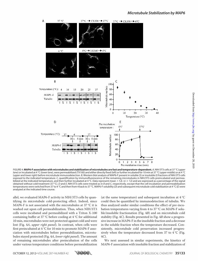
allel, we evaluated MAP6-F activity in NIH/3T3 cells by quan-tifying its microtubule cold-protecting effect. Indeed, sinceMAP6-F is not associated with the microtubules at 37 °C it iswashed out upon cell permeabilization. Thus, when NIH/3T3cells were incubated and permeabilized with a Triton X-100containing buffer at 37 °C before cooling at 4 °C for additional10min, microtubules were not protected against cold and werelost (Fig. 4A, upper right panel). In contrast, when cells werefirst preincubated at 4 °C for 10 min to promote MAP6-F asso-ciation with microtubules before permeabilization, microtu-bules stayed protected (Fig. 4A, lower right panel). The amountof remaining microtubules after preincubation of the cellsunder various temperature conditions before permeabilization
(at the same temperature) and subsequent incubation at 4 °Ccould then be quantified by immunodetection of tubulin. Wethus analyzed under similar conditions the effect of pre-incu-bation temperatures varying from 4 to 37 °C on MAP6-F solu-ble/insoluble fractionation (Fig. 4B) and on microtubule coldstability (Fig. 4C). Results presented in Fig. 4B show a progres-sive increase inMAP6-F in the insoluble fraction and a decreasein the soluble fraction when the temperature decreased. Con-sistently, microtubule cold preservation increased progres-sively when the temperature decreased from 37 to 4 °C (Fig.4C).We next assessed in similar experiments, the kinetics of
MAP6-F associationwith insoluble fraction and stabilization of
FIGURE 4. MAP6-F association with microtubules and stabilization of microtubules are fast and temperature-dependent. A, NIH/3T3 cells at 37 °C (upperlane) or incubated at 4 °C (lower lane), were permeabilized (TX100) and either directly fixed (left) or further incubated for 10 min at 37 °C (upper middle) or at 4 °C(upper and lower right) before microtubule immunodetection. B, Western blot analysis of MAP6-F present in soluble (S) or insoluble (I) fractions of NIH/3T3 cellsexposed to the indicated temperature. C, quantification by immunofluorescence of the remaining microtubules in NIH/3T3 cells preincubated and permea-bilized at the indicated temperature, and then further incubated at 4 °C. Data represent mean � S.E. (n � 12) and are expressed as a percentage of the signalobtained without cold treatment (37 °C). D and E, NIH/3T3 cells were treated as in B and C, respectively, except that the cell incubation and permeabilizationtemperatures were switched from 37 to 4 °C and then from 4 back to 37 °C. MAP6-F solubility (D) and subsequent microtubule cold stabilization at 4 °C (E) wereanalyzed at the indicated time course.
Microtubule Stabilization by MAP6
OCTOBER 12, 2012 • VOLUME 287 • NUMBER 42 JOURNAL OF BIOLOGICAL CHEMISTRY 35133
by guest on Decem
ber 28, 2019http://w
ww
.jbc.org/D
ownloaded from

microtubules at 4 °C. As shown in Fig. 4D, an incubation at 4 °Cas short as 30 s was sufficient for maximum MAP6-F associa-tion with the insoluble fraction and this association wasreversible with apparently similar fast kinetics when cellswere transferred back from 4 to 37 °C. As for the tempera-ture-dependence experiment, induction of microtubule coldstabilization followed the localization of MAP6-F in the insol-uble fraction (Fig. 4E). In addition, analysis of early time pointsshowed that cold-induced stabilization of microtubules wasmostly achieved within 15 s.These results indicate that under cold exposure, MAP6-F
interaction with microtubules is very fast and dynamic. Thisprobably involves a direct effect of the temperature on one ofthe binding partners rather than a post-translational modifica-tion. Furthermore, the concomitant cold exposure conditionsfor microtubule depolymerization (Fig. 1) and MAP6-F associ-ation with microtubules (Fig. 4) suggested that microtubulesand/or MAP6-F and/or other partners involved in the interac-tion might undergo temperature-dependent conformationalchanges.MAP6-F Is a Heat-stable Protein That Protects Microtubules
from Cold-induced Depolymerization through Direct Interac-tion via Its Mc Domain—To test whether MAP6-F by itself iscapable of stabilizingmicrotubules and inwhich conditions, wecarried out the purification of theMAP6-F protein as well as ofits Mc domain that constitute two third of the molecule (189/
306 amino acids) and has been shown to specifically protectmicrotubules from cold when ectopically expressed in HeLacells (22). Both polypeptides expressed in fusion with a His6 tagusing baculovirus-insect cells system were purified to purityclose to 95% (Fig. 5A). During the course of the purification, wealso observed that MAP6-F is a heat stable protein. Indeed,contrary tomost proteins that denature and precipitate at tem-peratures above 50 °C, heatingMAP6-F to 90 °C for 30 min didnot lead to protein aggregation (Fig. 5B).Using a microtubule sedimentation assay, we analyzed the
capacity of purified MAP6-F to protect in vitro polymerizedmicrotubules from cold-induced depolymerization. Fig. 5Cshows that, as expected, microtubules grown at 37 °C weremostly lost when incubated at 4 °C alone (lane 2) as comparedwith the total amount of microtubules stabilized with taxol(lane 1). Addition of MAP6-F just before cooling at 4 °C pro-tected microtubules from cold-induced depolymerization in adose-dependent manner (lanes 3–5). The same held true whenheated MAP6-F was used, confirming the heat stability ofMAP6-F (lanes 7–9). TheMcdomainwas also used in this assayand presented the same ability as the full-length molecule formicrotubule stabilization (lanes 11–13). It is interesting to notethat the Mc domain was more soluble and did not sediment inthe absence of microtubules (compare lane 14with lanes 6 and10). Altogether, these results demonstrated for the first timethat MAP6-F stabilizes microtubules exposed to cold via a
FIGURE 5. MAP6-F is a heat stable protein that stabilizes microtubules in vitro through interaction with its Mc domain. A, Coomassie Blue-stainedSDS-PAGE gel of the purification products of MAP6-F and the Mc domain expressed as His-tagged proteins in insect cells. B, solubility analysis of purifiedMAP6-F at 4 °C or after heating for 30 min at 90 °C. Proteins in the 100,000 � g pellet (P) or supernatant (S) were analyzed in Coomassie Blue-stained SDS-PAGEgel. T: total. C, microtubule cold-stabilizing activity of MAP6-F, heated MAP6-F, and the Mc domain. Microtubules were grown from pure tubulin in vitro, mixedwith either taxol (60 �M) or increasing concentration of MAP6-F, 90 °C-treated MAP6-F, or with the Mc repeat domain as indicated and then incubated on icefor 10 min. Cold-resistant microtubules were isolated by centrifugation, and the pellets were analyzed by SDS-PAGE and Coomassie Blue staining.
Microtubule Stabilization by MAP6
35134 JOURNAL OF BIOLOGICAL CHEMISTRY VOLUME 287 • NUMBER 42 • OCTOBER 12, 2012
by guest on Decem
ber 28, 2019http://w
ww
.jbc.org/D
ownloaded from

direct interaction. TheMc domain is able by itself to fully reca-pitulate this activity and is thus the main domain involved inthis function.Characterization of the Mc Domain Binding to Microtubules
at 4 °C—To further characterize the effect of the temperatureon MAP6-F interaction with microtubules, we performed atitration binding curve of the Mc domain to Taxol-stabilizedmicrotubules. Results presented in Fig. 6A show that, at 4 °C,the Mc domain binding to 1 �M of microtubules was titrablewith a binding stoichiometry of 1 mol of the Mc domain for 25mol of tubulin dimer. The apparent affinity determined was 70nM. On the contrary, at 37 °C, no specific interaction wasdetected (Fig. 6A). Using a Mc domain concentration close tothe apparent dissociation constant (Kd), we analyzed the effectof various temperatures on the Mc domain interaction withmicrotubules in the same conditions. Results presented in Fig.6B show that, as for MAP6-F association with and stabilizationof microtubule in NIH/3T3 cells (Fig. 4, B and C), in vitromicrotubule binding of the Mc domain progressively increaseswith the fall of temperature. The interaction started to bedetectable at 25 °C, rapidly increased between 20 °C and 15 °Cand then was further strengthened between 15 °C and 4 °C.
CircularDichroismAnalysis of Temperature-dependentCon-formational Changes in the Mc Domain—To investigatewhether the temperature-associated changes in the bindingaffinity between the Mc domain and microtubules could beassociated to conformational changes in the Mc domain, wecarried out circular dichroism experiments (Fig. 7). Absorptionspectra from 190 to 250 nm, obtained at different temperatures(Fig. 7A), indicated that theMcdomainwas essentially unstruc-tured with mainly random coiled signal (negative absorptionpeak near 195 nm) (27). Nevertheless, with increasing temper-atures, the intensity of the negative bands at 198 nm decreasedand shifted to slightly higher wavelengths while absorption at219 nm appeared. Moreover, the presence of an isodichroicpoint at 208 nm indicated a two conformational state system.To characterize this structure, subtraction spectra were drawn(Fig. 7B). The difference spectra showed the apparition withheating of negative bands at 219 nm and positive bands at 195nm. This signal is characteristic of beta structures (28). Tem-perature-dependent Mc domain absorption spectra at 220 nm(Fig. 7A, inset) indicated that the conformational changebetween 5 and 80 °C was linear and reversible. Altogether thesedata indicated that the Mc domain which contains beta struc-tures progressively unfolded upon chilling from 37 to 5 °C.Structural Analysis of Microtubules Stabilized at 4 °C by the
Mc Domain—To gain insight into the mechanisms by whichMAP6-F protects microtubules from depolymerization undercold exposure, we used cryo-electron microscopy to analyzemicrotubules at 4 °C in the absence or in the presence of theMcdomain. Growing and shrinking microtubules assume variousconformations at their extremities, including sheet-like exten-sions and blunt ends during polymerization and outwardlycurled protofilaments in disassembling conditions (29, 30). AsMAP6-F inhibits cold-induced microtubule disassembly, wewondered whether it could affect the global structure of micro-tubule ends. Microtubules were polymerized at 37 °C and thenincubated at 4 °C with or without the Mc domain. In our con-ditions, we could not observe the Mc domain bound to thestabilized microtubules. The main observed differences con-cerned the structures of the microtubule ends that presentedmostly curled depolymerizing protofilaments in the absence ofthe Mc domain and about 50% of blunt extremities in the pres-ence of the Mc domain (Fig. 8). These data indicated that theMc domain stabilized the extremities of microtubule exposedto cold. However, one would have expected a near to completestabilization of microtubules without curled ends in the pres-ence of the Mc domain. The remaining 50% of curled microtu-bule ends indicates that the microtubule stabilization is notcomplete and that theMc domainmight only dramatically slowdowndepolymerization and/or allow very slowdynamics at lowtemperatures.
DISCUSSION
MAP6 proteins have been shown to be essential for the sta-bilization of microtubules in cells at 4 °C. In fibroblastic cells,the main isoform, MAP6-F, was shown to relocalize from thecytoplasm to the microtubule network upon chilling to 4 °C. Ithas been suggested that the changes in MAP6-F bindingto microtubules relied on temperature-induced changes in
FIGURE 6. In vitro analysis of temperature-dependent binding of theMAP6 Mc domain with taxol-stabilized microtubules. A, microtubuleswere grown at 37 °C from 60 �M pure tubulin, stabilized with Taxol anddiluted to 1 �M in the final binding reaction mix. The Mc domain was added atincreasing concentrations and incubated at 4 or 37 °C for 10 min. Microtu-bules were isolated by centrifugation and the associated Mc domain wasquantified by Western blot using Chemidoc apparatus, ImageJ, and Excelsoftwares. 4 and 37 °C controls (Ctrl) are the amount of the Mc domain pel-leted in the absence of microtubules. Insert, Lineweaver-Burk plot analysis ofthe data shown in A gave 70 nM as the apparent dissociation constant (Kdapp)for the binding of the Mc domain to microtubules at 4 °C. B, the Mc domain(50 nM) binding to microtubules was analyzed as in A at the indicated tem-peratures. Data represent mean � S.E. (n � 4).
Microtubule Stabilization by MAP6
OCTOBER 12, 2012 • VOLUME 287 • NUMBER 42 JOURNAL OF BIOLOGICAL CHEMISTRY 35135
by guest on Decem
ber 28, 2019http://w
ww
.jbc.org/D
ownloaded from

MAP6-F post-translational modifications (16). We show herethat under cold exposure, MAP6-F recruitment to microtu-bules and stabilization is highly dynamic, indicating a directeffect of the temperature on the interaction. In vitro, purifiedMAP6-F can directly interact with and stabilize microtubulesagainst cold-induced depolymerization through itsMcdomain.As observed in vivo for MAP6-F, in vitro binding of the Mcdomain to microtubules is temperature-dependent. The bind-ing was very low at 37 °C and increased significantly when the
temperature decreased below 20 °C to reach a high affinity(Kdapp � 70 nM) at 4 °C. Thus, in vivo and in vitro, the Mcdomain of MAP6 recognizes and stabilizes microtubules assoon as the temperature drops under 20 °C.What Is the Structural Element that Regulates the Interaction
between Microtubules and the Mc Domain of MAP6?—Ourresults reveal that lowering of temperature is accompanied bythe recruitment of MAP6-F on microtubules both in cells andin vitro. This suggests that MAP6-F adopts a cold-dependentconformation enabling its interaction with the polymers,and/or that MAP6-F recognizes a structural feature of micro-tubules depolymerizing under cold exposure. Our data providestrong evidence that variations in temperatures induce confor-mational changes ofMAP6-F.MAP6-F exhibits beta structuresat 37 °C and progressively unfolds as the temperature de-creases, enabling its interaction with microtubules and theirsubsequent stabilization. MAP6-F binding to microtubules atlow temperatures could also involve the recognition of a spe-cific feature of cold-induced depolymerizing polymers. In par-ticular, shrinkingmicrotubules display curled protofilaments attheir ends, butwe did not observe any accumulation ofMAP6-Fat the extremity of disassembling microtubules, neither in cells(Figs. 2 and 3) nor in purified assays (Fig. 8), suggesting thatspecific recognition of the outwardly curled protofilaments byMAP6-F is an unlikely mechanism. MAP6-F might associatepreferentially to a cold-sensitive lattice region, although littleinformation is available on changes occurring at low tempera-tures within the microtubule wall. High-resolution structuresof MAP6-F and MAP6-F/microtubule complexes will berequired to visualize the Mc domain and its precise bindingsites to the microtubule lattice.HowDoes theMcDomain ofMAP6 StabilizeMicrotubules at
4 °C?—As suggested above, under cold exposure, the Mcdomain of MAP6 might bridge adjacent protofilaments. This,in turn, would decrease the catastrophe frequency thought tobe involved in cold-inducedmicrotubule depolymerization (9).Reinforcing lateral interactions has already been proposed to bea way of stabilizing microtubules in cold-living organisms (12).Such a mechanism is reminiscent of the doublecortin microtu-
FIGURE 7. Temperature induces secondary structure changes in the Mc domain of MAP6. A, circular dichroism spectra at 5 °C showed a clear minimum forthe spectrum at 198 nm, indicating a mostly random coiled structure. This minimum is shifted with increasing temperature toward higher wavelengths and aminimum at 219 nm appears. This indicates an increase in structuration. In the inset, the structuration has been monitored at 220 nm from 5 to 85 °C asindicated by the black arrow. The folding reversibility from 85 °C to 5 °C is indicated by the open arrow. The isodichroic point at 208 nm suggests a two-statesystem characterizing the secondary structure of the Mc domain. B, difference CD spectra from A. The subtraction of the CD spectrum at 5 °C from CD spectraat 10, 20, 40, and 70 °C gave the structure gain during the temperature titration. The spectra obtained correspond to �-sheet conformation.
FIGURE 8. Microtubule cold-stabilization by the Mc domain is associatedwith an increase of blunt-ended microtubules. Microtubules were grownfrom 80 �M of pure tubulin at 37 °C, mixed at a final concentration of 55 �M
with buffer alone (Ctrl) or with 5.5 �M final of the Mc domain (Mc). After 10 minat 37 °C, microtubules were chilled on ice for 1 min to allow partial depo-lymerization of microtubules alone or for 20 min for the Mc domain-stabilizedmicrotubules. Microtubules were loaded on grids, frozen in liquid ethane, andobserved by cryo-electron microscopy. A, pictures and schematic represen-tations of microtubule curled and blunt ends. B, quantitative analysis of theproportion of blunt versus curled ends in control condition or in the presenceof the Mc domain.
Microtubule Stabilization by MAP6
35136 JOURNAL OF BIOLOGICAL CHEMISTRY VOLUME 287 • NUMBER 42 • OCTOBER 12, 2012
by guest on Decem
ber 28, 2019http://w
ww
.jbc.org/D
ownloaded from

bule stabilization mode at 37 °C (31). Doublecortin is an anti-catastrophe factor that stabilizes microtubules by linking adja-cent protofilaments and counteracting their outward bendingin depolymerizing microtubules (32). The analysis of the Mcdomain effects on microtubule dynamics at low temperatureshould be very informative but such experiments which requirea controlled cold environment are still technically challenging.A Link Between the Mc Domain Structure and MAP6 Aggre-
gation in Human Pathology?—During our in vitro studies wediscovered that MAP6-F is a weakly structured molecule asrevealed by its heat stability, a feature shared by other MAPssuch as MAP1, MAP2, and Tau (33). The folding of MAP6-Finto beta structures represents a peculiar behavior alreadyreported for the Tau protein (34). These beta structures havebeen implicated in the formation of pathologic amyloid aggre-gates (35) and beta structures of MAP6 might be implicated inthe aggregation of MAP6 in Lewis bodies during amyotrophiclateral sclerosis (36).A Physiological Function for MAP6 Stabilization of Microtu-
bules under Cold Exposure?—InHeLa cells, that do not expressMAP6proteins,microtubules depolymerized in a temperature-dependentmanner as soon as the temperature decreased below20 °C with virtually no microtubules left at 4 °C. On the con-trary, inMAP6-containingNIH/3T3 cells, 80% ofmicrotubuleswere preserved upon cold exposure from 20 to 4 °C. In morephysiological conditions, MAP6 of MEF�/� allowed to pre-serve about 80% of microtubules at 15 °C and 30% at tempera-tures below 10 °C. In animals, such low temperatures could beachieved during episodes of torpor or hibernation and MAP6could be required to preserve aminimal level ofmicrotubules insuch challenging conditions. In this study we considered thewhole Mc domain without analyzing the contribution of eachindividual repeat. However, one can speculate that the compo-sition in repeats of the Mc domain would determine thestrength of the cold response. Along this line, we found a rela-tively good correlation between the number of repeats (from 1to 9) in the Mc domain of mammals and the ability of the ani-mals to hibernate or to make torpor. For example, almost allgenome-sequenced rodents (house mice, Norway rat, thirteen-lined ground squirrel) which exhibit MAP6 with 3 to 9 Mcrepeats are able to hibernate/torpor, whereas the naked molerat which exhibits only one repeat is very sensitive to cold expo-sure (main cause of lethality).Also, in primates, only the lemurian branch which contains
several Mc repeats (3 to 6 for small-eared galago and graymouse lemur) can enter torpor whereas all other primatesincluding human, Sumatran ourangutan, chimpanzee, gorilla,Rhesus monkey, and Hamadryas baboon do not enter torporand express MAP6 containing only one repeat. In human,MAP6 stabilization of microtubules can still be useful duringaccidental hypothermic episodes or even during perioperativehypothermia following anesthesia.In conclusion, the Mc domain of MAP6 behaves as a sensor
of temperature able to protect microtubules from temperaturevariations. How microtubule stabilization upon temperaturereduction is involved and whether it is crucial to adapt cold-de-pendent cell responses such as metabolic changes are excitingquestions.
Acknowledgments—We thank D. Job for helpful discussions on thework and to L. Aubry, J. Brocard, F. Costagliola, and J.C, Deloulme forcritical reading of the manuscript. We are grateful to the Institute ofStructural Biology Jean-Pierre Ebel (Grenoble, France) for the use ofits EM facility.
REFERENCES1. Akhmanova, A., and Steinmetz, M. O. (2008) Tracking the ends: a dy-
namic protein network controls the fate of microtubule tips. Nat. Rev.Mol. Cell Biol. 9, 309–322
2. Howard, J., andHyman, A. A. (2009) Growth, fluctuation and switching atmicrotubule plus ends. Nat. Rev. Mol. Cell Biol. 10, 569–574
3. Nogales, E., and Wang, H. W. (2006) Structural intermediates in micro-tubule assembly and disassembly: how and why? Curr. Opin Cell Biol. 18,179–184
4. Wade, R. H. (2009) On and around microtubules: an overview. Mol Bio-technol 43, 177–191
5. Lee, J. C., and Timasheff, S. N. (1977) In vitro reconstitution of calf brainmicrotubules: effects of solution variables. Biochemistry 16, 1754–1764
6. Olmsted, J. B., and Borisy, G. G. (1975) Ionic and nucleotide requirementsfor microtubule polymerization in vitro. Biochemistry 14, 2996–3005
7. Regula, C. S., Pfeiffer, J. R., and Berlin, R. D. (1981) Microtubule assemblyand disassembly at alkaline pH. J. Cell Biol. 89, 45–53
8. Schilstra,M. J., Bayley, P.M., andMartin, S. R. (1991)The effect of solutioncomposition on microtubule dynamic instability. Biochem. J. 277,839–847
9. Fygenson, D. K., Braun, E., and Libchaber, A. (1994) Phase diagram ofmicrotubules. Phys. Rev. E. Stat. Phys. Plasmas Fluids Relat InterdiscipTopics 50, 1579–1588
10. Modig, C.,Wallin,M., andOlsson, P. E. (2000) Expression of cold-adapted�-tubulins confer cold-tolerance to human cellular microtubules.Biochem. Biophys. Res. Commun. 269, 787–791
11. Detrich, H. W., 3rd. (1997) Microtubule assembly in cold-adapted organ-isms: functional properties and structural adaptations of tubulins fromantarctic fishes. Comp. Biochem. Physiol. A. Physiol. 118, 501–513
12. Detrich, H. W., 3rd, Parker, S. K., Williams, R. C., Jr., Nogales, E., andDowning, K. H. (2000) Cold adaptation of microtubule assembly and dy-namics. Structural interpretation of primary sequence changes present inthe�- and�-tubulins of Antarctic fishes. J. Biol. Chem. 275, 37038–37047
13. Geiser, F. (1988) Reduction of metabolism during hibernation and dailytorpor in mammals and birds: temperature effect or physiological inhibi-tion? J. Comp Physiol. B. 158, 25–37
14. Heldmaier, G., Ortmann, S., and Elvert, R. (2004) Natural hypometabo-lism during hibernation and daily torpor in mammals. Respir. Physiol.Neurobiol. 141, 317–329
15. Andrieux, A., Salin, P. A., Vernet,M., Kujala, P., Baratier, J., Gory-Fauré, S.,Bosc, C., Pointu, H., Proietto, D., Schweitzer, A., Denarier, E., Klumper-man, J., and Job, D. (2002) The suppression of brain cold-stable microtu-bules in mice induces synaptic defects associated with neuroleptic-sensi-tive behavioral disorders. Genes Dev. 16, 2350–2364
16. Denarier, E., Fourest-Lieuvin, A., Bosc, C., Pirollet, F., Chapel, A., Marg-olis, R. L., and Job, D. (1998) Nonneuronal isoforms of STOP protein areresponsible for microtubule cold stability in mammalian fibroblasts. Proc.Natl. Acad. Sci. U.S.A. 95, 6055–6060
17. Bosc, C., Andrieux, A., and Job, D. (2003) STOPproteins.Biochemistry 42,12125–12132
18. Aguezzoul,M., Andrieux, A., andDenarier, E. (2003)Overlap of promoterand coding sequences in the mouse STOP gene (Mtap6). Genomics 81,623–627
19. Galiano, M. R., Bosc, C., Schweitzer, A., Andrieux, A., Job, D., and Hallak,M. E. (2004) Astrocytes and oligodendrocytes express different STOPprotein isoforms. J. Neurosci. Res. 78, 329–337
20. Guillaud, L., Bosc, C., Fourest-Lieuvin, A., Denarier, E., Pirollet, F., Lafan-echère, L., and Job, D. (1998) STOP proteins are responsible for the highdegree of microtubule stabilization observed in neuronal cells. J. Cell Biol.142, 167–179
Microtubule Stabilization by MAP6
OCTOBER 12, 2012 • VOLUME 287 • NUMBER 42 JOURNAL OF BIOLOGICAL CHEMISTRY 35137
by guest on Decem
ber 28, 2019http://w
ww
.jbc.org/D
ownloaded from

21. Ochoa, C. D., Stevens, T., and Balczon, R. (2011) Cold exposure revealstwo populations of microtubules in pulmonary endothelia. Am. J. Physiol.Lung Cell Mol. Physiol. 300, L132–L138
22. Bosc, C., Frank, R., Denarier, E., Ronjat, M., Schweitzer, A., Wehland, J.,and Job, D. (2001) Identification of novel bifunctional calmodulin-bindingand microtubule-stabilizing motifs in STOP proteins. J. Biol. Chem. 276,30904–30913
23. Lieuvin, A., Labbé, J. C., Dorée, M., and Job, D. (1994) Intrinsic microtu-bule stability in interphase cells. J. Cell Biol. 124, 985–996
24. Erck, C., Peris, L., Andrieux, A., Meissirel, C., Gruber, A. D., Vernet, M.,Schweitzer, A., Saoudi, Y., Pointu, H., Bosc, C., Salin, P. A., Job, D., andWehland, J. (2005) A vital role of tubulin-tyrosine-ligase for neuronalorganization. Proc. Natl. Acad. Sci. U.S.A. 102, 7853–7858
25. Schneider, C. A., Rasband, W. S., and Eliceiri, K. W. (2012) NIH Image toImageJ: 25 years of image analysis. Nat. Methods 9, 671–675
26. Paturle-Lafanechère, L., Eddé, B., Denoulet, P., Van Dorsselaer, A., Maz-arguil, H., LeCaer, J. P.,Wehland, J., and Job, D. (1991) Characterization ofa major brain tubulin variant which cannot be tyrosinated. Biochemistry30, 10523–10528
27. Venyaminov, S. Y., Baikalov, I. A., Shen, Z. M., Wu, C. S., and Yang, J. T.(1993) Circular dichroic analysis of denatured proteins: inclusion of dena-tured proteins in the reference set. Anal. Biochem. 214, 17–24
28. Greenfield, N., and Fasman, G. D. (1969) Computed circular dichroismspectra for the evaluation of protein conformation. Biochemistry 8,4108–4116
29. Arnal, I., Karsenti, E., and Hyman, A. A. (2000) Structural transitions atmicrotubule ends correlate with their dynamic properties in Xenopus egg
extracts. J. Cell Biol. 149, 767–77430. Chrétien, D., Fuller, S. D., and Karsenti, E. (1995) Structure of growing
microtubule ends: two-dimensional sheets close into tubes at variablerates. J. Cell Biol. 129, 1311–1328
31. Moores, C. A., Perderiset, M., Francis, F., Chelly, J., Houdusse, A., andMilligan, R. A. (2004) Mechanism of microtubule stabilization by dou-blecortin.Mol. Cell 14, 833–839
32. Moores, C. A., Perderiset, M., Kappeler, C., Kain, S., Drummond, D., Per-kins, S. J., Chelly, J., Cross, R., Houdusse, A., and Francis, F. (2006) Distinctroles of doublecortin modulating the microtubule cytoskeleton. EMBO J.25, 4448–4457
33. Vera, J. C., Rivas, C. I., andMaccioni, R. B. (1988) Heat-stablemicrotubuleproteinMAP-1 binds tomicrotubules and inducesmicrotubule assembly.FEBS Lett. 232, 159–162
34. von Bergen, M., Friedhoff, P., Biernat, J., Heberle, J., Mandelkow, E. M.,andMandelkow, E. (2000) Assembly of tau protein into Alzheimer pairedhelical filaments depends on a local sequence motif ((306)VQIVYK(311))forming beta structure. Proc. Natl. Acad. Sci. U.S.A. 97, 5129–5134
35. Mukrasch, M. D., Biernat, J., von Bergen, M., Griesinger, C., Mandelkow,E., and Zweckstetter, M. (2005) Sites of tau important for aggregationpopulate {beta}-structure and bind tomicrotubules and polyanions. J. Biol.Chem. 280, 24978–24986
36. Letournel, F., Bocquet, A., Dubas, F., Barthelaix, A., and Eyer, J. (2003)Stable tubule only polypeptides (STOP) proteins co-aggregate with spher-oid neurofilaments in amyotrophic lateral sclerosis. J. Neuropathol. Exp.Neurol. 62, 1211–1219
Microtubule Stabilization by MAP6
35138 JOURNAL OF BIOLOGICAL CHEMISTRY VOLUME 287 • NUMBER 42 • OCTOBER 12, 2012
by guest on Decem
ber 28, 2019http://w
ww
.jbc.org/D
ownloaded from

Annie AndrieuxEric Denarier, Christophe Bosc, Odile Valiron, Mariano Bisbal, Isabelle Arnal and
Christian Delphin, Denis Bouvier, Maxime Seggio, Emilie Couriol, Yasmina Saoudi,Microtubules from Cold-induced Depolymerization
MAP6-F Is a Temperature Sensor That Directly Binds to and Protects
doi: 10.1074/jbc.M112.398339 originally published online August 17, 20122012, 287:35127-35138.J. Biol. Chem.
10.1074/jbc.M112.398339Access the most updated version of this article at doi:
Alerts:
When a correction for this article is posted•
When this article is cited•
to choose from all of JBC's e-mail alertsClick here
http://www.jbc.org/content/287/42/35127.full.html#ref-list-1
This article cites 36 references, 13 of which can be accessed free at
by guest on Decem
ber 28, 2019http://w
ww
.jbc.org/D
ownloaded from