mansoura vet. med. j. vol. xviii, no. 1, des. 2017 51
TRANSCRIPT

Mansoura Vet. Med. J. Vol. XVIII, No. 1, Des. 2017 ISSN 1110-7219
51
Mansoura Veterinary Medical Journal
PLAIN AND CONTRAST RADIOGRAPHIC EVALUATION OF THE DIGIT IN DONKEYS
(EQUUS ASINUS)
Mohamed Salem, El-Sayed El-Shafaey, Esam Mosbah, Adel E.I. Zaghloul*
*Department of Surgery, Anesthesiology and Radiology, Fac. of. Vet. Medicine Mansoura University
ABSTRACT Radiography of the digit is a golden standard technique allows the veterinarian to render a
subjective evaluation of the digit in donkeys. The present study was planned to evaluate the bony tissues and blood circulation in donkey’s digit using plain and contrast radiography (angiography and venography). The digits of ten clinically and orthopedic healthy donkeys were subjected to plain and contrast radiographic examination of the digit region using four standard views (dorsopalmar, anterior-posterior, lateromedial and oblique views). Plain radiographic examinations revealed fully descriptions of the bony structures and joint surfaces of the donkey’s digits. While, the digital angiography and venography of the donkey digit illustrate clearly evident digital arteries and veins network that are course from the carpus/tarsus to the terminal arch. In conclusion, radiography provides a pierce non-invasive technique for evaluation of the hard tissue and digital blood circulation of the digit in donkeys. Keywords: Angiography, Donkey, Digit, Radiography, Venography
INTRODUCTION
Donkey is one of the most popular animals in Egypt that used as draft, transportation and working animal. Donkeys play an important role in Egyptian livestock, agriculture, economy, where they are used as mounts in agriculture and transport. The digit is a vital structure to the sound gait of the donkey limb during both support and swing phases of the stride. Any disorder that causes pain or reduction in the range of motion may extremely affect performance (Semieka and Magda, 2012). The digit is the most area that contributed to injuries and lameness in donkeys as a result of its anatomical nature because it is free from muscles (Good ship and Birch, 1996). Diagnosis of lameness is a challengeable process and needs conjunction
between the clinical examinations and the different diagnostic imaging modalities (Tucker and Sande, 2001; Dyson and Murray, 2007; and Sherlock et al., 2007).
Radiography plays an important role in the diagnosis of digit disorders in donkeys (Fogarty et al., 2011). The digit is clinically important area that examined radiographically with exposure factors 56 – 60 KV and 10 – 12 mAs (Vanderperren and Saunders, 2009). X-ray is a vital part of the purchase examination because skeletal disease that detected during physical examination can be further assessed and occult disease can be uncovered. Additionally, the radiographic assessment should clearly describe lesions and diagnoses so that a more accurate prognosis for use of the donkeys can be concluded. Although the development of the different diagnostic

Mohamed Salem. et al…
Mansoura Vet. Med. J. Vol. XVIII, No. 1, 2017
52
imaging techniques, radiography is still a unique method for diagnosis of lameness that contributed to the bony structures and joints of the digit in donkeys and horses (Brown, 2004 and Ross, 2016).
Digital venography and angiography are forms of contrast radiography that used to describe the blood circulation of the donkey digits. The technique is relatively simple and can be done stall side using standard radiographic equipment, so it can be used to assess a variety of clinical conditions of the donkey digit. Digital venography can greatly assist the clinician in making both diagnostic and therapeutic decisions, monitoring recovery, and offering a more accurate prognosis to the owner. The resulting image provides informations regarding the status of the vasculature and dermis of the foot (Rucker, 2010).
In the laminitic patient, venous compromise or occlusion results in a decline or absence of contrast in the affected area. Changes in the contrast pattern are obvious before radiographically displacement of the third phalanx, the degree of the vascular change being determined by the severity of pathology in the suspension apparatus of the third phalanx (the dermal-epidermal junction) (Baldwin and Pollitt, 2010 and Rucker, 2010).
Digit lameness is usually related to the bony tissues and blood circulation that needs calibration between plain and contrast radiographic diagnostic imaging modalities. Thus, the present study was designed to evaluate the hard tissue and vasculatures of the digit using plain radiography and contrast radiography, respectively.
MATERIALS AND METHODS
Animals: This study was carried out on ten
clinically and orthopedically healthy donkeys. The animals of this study were kept in the animal house of Mansoura Veterinary Teaching Hospital Fac. Vet. Med.; Mansoura University and were fed on a balanced mixed ration. Plain Radiographic Examination.
Radiographic images of donkey digits were taken using a 70 kVp, 2 mAs radiography unit (SY-31-100P, Samsung-dong, kingdom -KU, Seoul, Kore) with a 70 cm focal film distance. The donkey digit is divided into two anatomical regions, fetlock and pastern region and the foot. A standard radiographic examination of the equine fetlock and pastern region comprises three projections (palmaro -dorsal view (PD), lateromedial view (LM) and oblique view).While for the foot; there are another three projections (anterior-posterior, lateromedial and palmarodistal oblique views).
For radiographic examination; the digit was thoroughly cleansed with a stiff brush, soap and water. All debris was removed before the radiographic views are taken. The images were processed using digital processing unit (CR-30-X- AGFA-Germany) and digital printer (Dry star 5302- AGFA - Germany). The radiographic views were done According to Collins et al., 2011. Radiographic views: The fetlock and pastern region:
palmaro -dorsal view of the fetlock and pastern joints was made with the limbs extended forward and the hoofs elevated on a wooden block with the central beam directed at

Mohamed Salem. et al…
Mansoura Vet. Med. J. Vol. XVIII, No. 1, 2017
53
the pastern joint (Fig. 1A). For lateromedial view, the cassette was positioned at the medial side of digit and the central beam was directed to the lateral side of the digit (Fig. 1B). While for oblique view, the tube was positioned anterio- lateral and the cassette posterio- medial and made at approximately 45 angle (Fig. 1C). The foot:
For lateromedial view of the third phalanx and the navicular bone, the foot was elevated so the central beam is parallel to the floor and perpendicular to the third phalanx (Fig. 2A). While, for anterior posterior view, the foot was placed directly on cassette and the central beam was directed toward the floor perpendicular to the hoof wall and centered just distal to coronary (Fig. 2B). For palmarodistal oblique view, the film holder was placed on the ground and the hoof was placed on film holder, the tube was moved 45 medially or laterally and the central beam was centered 1 cm below the coronary band (Fig. 2C).
Contrast radiographic examinations:
The donkeys were sedated with xylazine (Xylaject 2% - ADWIA Co., Egypt) in a dose rate of 1mg/kg.b.wt/i/v injection. The hair was clipped from the carpus/tarsus till coronary band. A-Digital angiography:According to
(Moslemi et al., 2008). The tourniquet was applied firmly above
the carpus/tarsus. The butterfly catheter was secured in the medial metacarpal artery. 30 ml of contrast agent (scanlux®300, sanochemia pharmazeutika AG Boltzmanngasse 11, A-1091 Wien) was injected in the medial metacarpal artery (Fig. 3 A). The mediolateral and dorsopalmar views were taken in non-wight bearing position.
B- Digital Venography: According (Redden, 2001 and Rucker et al., 2006).
The fetlock was covered with a couple layers of adhesive plaster, which will serve as a non-slip base for the tourniquet. The tourniquet was applied firmly around the fetlock at the level of the proximal sesamoid bones. The butterfly catheter with extension tubing attached was carefully inserted into the distended palmar digital vein. The syringe was attached to the end of butterfly catheter. Thirty millimeter of contrast agent (scanlux®300, sanochemia pharmazeutika AG Boltzmanngasse 11, A-1091 Wien) was injected into distended vein. After all the contrast has been injected, the tubing was held in place with the free end of the adhesive plaster covering the tourniquet. The catheter was left in place until the radiographs had been taken. The mediolateral view was taken with the limb in non-wight bearing position (Fig. 3 B).
RESULTS Plain radiographic examinations: 1- Fetlock and pastern region
On normal lateromedial radiographs of fetlock joint, the joint surface of the distal cannon bone has a smooth curve, with a mild flattening at its palmarodistal aspect. The cannon bone was seen articulating with the first phalanx and the proximal sesamoid bone (Fig. 4). On the oblique view, the sesamoid bones have a smooth and rounded outline over the palmar aspect (Fig. 5).
While in palmaro - dorsal view, the fetlock joint is approximately symmetrical about the prominent sagittal ridge of the distal cannon bone. The sagittal ridge articulates with a groove in the proximal phalanx. The width of the fetlock joint was easily seen. The medial
Mohamed Salem. et al…
Mansoura Vet. Med. J. Vol. XVIII, No. 1, 2017
54
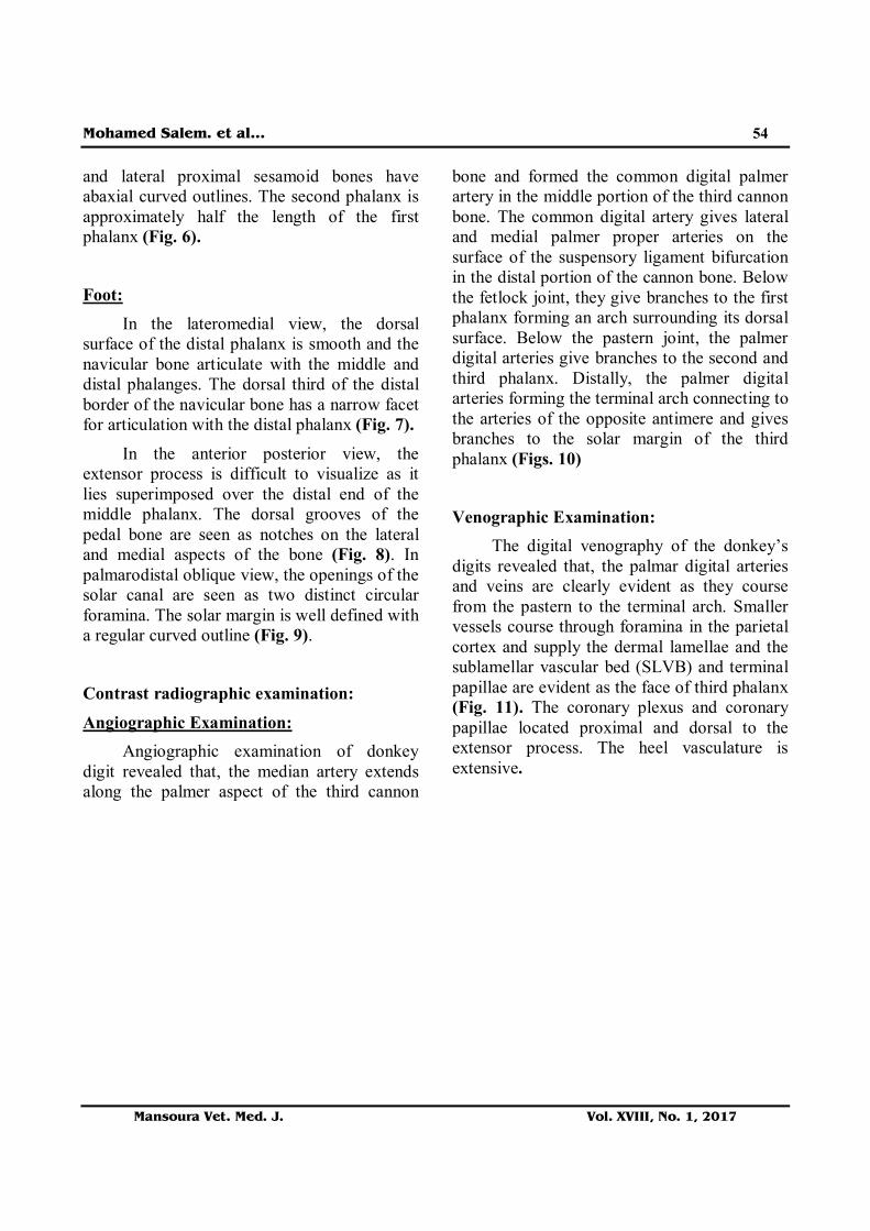
and lateral proximal sesamoid bones have abaxial curved outlines. The second phalanx is approximately half the length of the first phalanx (Fig. 6).
Foot: In the lateromedial view, the dorsal
surface of the distal phalanx is smooth and the navicular bone articulate with the middle and distal phalanges. The dorsal third of the distal border of the navicular bone has a narrow facet for articulation with the distal phalanx (Fig. 7).
In the anterior posterior view, the extensor process is difficult to visualize as it lies superimposed over the distal end of the middle phalanx. The dorsal grooves of the pedal bone are seen as notches on the lateral and medial aspects of the bone (Fig. 8). In palmarodistal oblique view, the openings of the solar canal are seen as two distinct circular foramina. The solar margin is well defined with a regular curved outline (Fig. 9).
Contrast radiographic examination: Angiographic Examination:
Angiographic examination of donkey digit revealed that, the median artery extends along the palmer aspect of the third cannon
bone and formed the common digital palmer artery in the middle portion of the third cannon bone. The common digital artery gives lateral and medial palmer proper arteries on the surface of the suspensory ligament bifurcation in the distal portion of the cannon bone. Below the fetlock joint, they give branches to the first phalanx forming an arch surrounding its dorsal surface. Below the pastern joint, the palmer digital arteries give branches to the second and third phalanx. Distally, the palmer digital arteries forming the terminal arch connecting to the arteries of the opposite antimere and gives branches to the solar margin of the third phalanx (Figs. 10) Venographic Examination:
The digital venography of the donkey’s digits revealed that, the palmar digital arteries and veins are clearly evident as they course from the pastern to the terminal arch. Smaller vessels course through foramina in the parietal cortex and supply the dermal lamellae and the sublamellar vascular bed (SLVB) and terminal papillae are evident as the face of third phalanx (Fig. 11). The coronary plexus and coronary papillae located proximal and dorsal to the extensor process. The heel vasculature is extensive.

Mohamed Salem. et al…
Mansoura Vet. Med. J. Vol. XVIII, No. 1, 2017
55
Figure (1): Radiographic views of fetlock and pastern region of adult donkey digit:
(A): Palmaro-dorsal view of the fetlock and pastern region, (B): Lateromedial view of the fetlock and pastern region, (C): Oblique view of the fetlock and pastern region.

Mohamed Salem. et al…
Mansoura Vet. Med. J. Vol. XVIII, No. 1, 2017
56
Figure (2): Radiographic views of the digit of adult donkey digit:
(A): Lateromedial view (B): Anterior-posterior view (C): Palmarodistal oblique view

Mohamed Salem. et al…
Mansoura Vet. Med. J. Vol. XVIII, No. 1, 2017
57
Figure (3): Contrast radiography in adult donkey digit showed:
(A): Mediolateral radiographic view after injection of contrast material in medial metacarpal artery for angiography of adult donkey digit.
(B): Mediolateral radiographic view after injection of contrast material in the medial palmer digital vein for venography of adult donkey digit.
Figure (4): Lateral radiographic view of the fetlock and pastern regions of adult donkey digit showed:
(1) Third cannon bone, (2) Proximal sesamoid bones, (3) Fetlock joint (4) First phalanx, (5) Pastern joint, (6) Second phalanx, (7) Coffin joint

Mohamed Salem. et al…
Mansoura Vet. Med. J. Vol. XVIII, No. 1, 2017
58
Figure (5): Oblique radiographic view of the fetlock and pastern region of adult donkey digit Showed:
1- Cannon bone, (2) Splint bone, (3) Proximal sesamoid bone, (4) Fetlock joint. (5) First phalanx, (6) Pastern joint, (7) Second phalanx, (8) Navicular bone, (9) Coffin joint.
Figure (6): Dorsopalmar radiographic view of the fetlock and pastern region of adult donkey digit Showed:
(1) Third cannon bone, (2) Proximal sesamoid bones, (3) Fetlock joint, (4) Pastern joint, (5) Second phalanx, (6) Coffin joint.

Mohamed Salem. et al…
Mansoura Vet. Med. J. Vol. XVIII, No. 1, 2017
59
Figure (7): Lateromedial view of the 3rd phalanx and NB of adult donkey digit Showed:
(1)Third cannon bone, (2) Proximal sesamoid bones, (3) Fetlock joint, (4) First phalanx, (5) Pastern joint, (6) Navicular bone, (7) Coffin joint, (8) Third phalanx.
Figure (8): Anterior posterior view of the 3rd phalanx and NB of adult donkey digit showing the dorsal
grooves of the pedal bone are seen as notches on the lateral and medial aspects of the bone (arrows): (1) Third cannon bone, (2) Fetlock joint, (3) first phalanx, (4) Second phalanx, (5) Third
phalanx.

Mohamed Salem. et al…
Mansoura Vet. Med. J. Vol. XVIII, No. 1, 2017
60
Figure (9): Palmarodistal oblique view of the foot of adult donkey digit showed the openings of the solar
canal are seen as two distinct circular foramina (Red arrows) . The solar margin is well defined with a regular curved outline.
1- Second phalanx. 2- Coffin joint. 3- Pedal bone.
Figure (10): (A) Dorsopalmar and (B) Lateromedial angiographic view of adult donkey digit.
(1) Lateral metacarpal artery, (2) Medial metacarpal artery, (3) Common digital palmer artery (4) medial proper digital artery, (5) lateral proper digital artery, (6) Anastomosing branch, (7) Medial digital artery, (8) Lateral digital artery, (9) Terminal arch, (10) Heel vasculatures, (11) Coronary plexus.

Mohamed Salem. et al…
Mansoura Vet. Med. J. Vol. XVIII, No. 1, 2017
61
Figure (11): Mediolateral view of foot of adult donkey after digital venography showed:
(1) Medial palmer digital vein, (2) Coronary plexus, (3) Sublamellar vascular bed, (4) Third phalanx, (5) Terminal papillae, (6) Terminal arch, (7) Heel vascularization.
DISCUSSION
The digit represents a vital structure to the sound movement of the donkey digit. The most common affections of the digit in donkeys are related to the bony structures, circulation and joint. Digit radiographs are acquired by veterinarian for pre-purchase examination, diagnosis of lameness and evaluation of digit affections. For each of these circumstances, gaining high quality normal radiographic views is important for veterinarians to enable them to supply truthful information to their customer (Vanderperren and Saunders, 2009). The diagnosis of these disorders needs conjunction between clinical
examination, nerve block and radiography (Semieka and Magda, 2012). Thus, the present study focused on providing detailed reference radiographic images for the donkey digit and provides radiographic interpretation of normal donkey’s digit as a reference base for distinguishing abnormal condition of donkey’s digit.
The digit in horses is clinically important area that examined radiographically with exposure factors 56 – 60 KV and 10 – 12 mAs (Vanderperren and Saunders, 2009). In the present study, the digit of the donkey is examined with exposure factors 70 KV and 2 mAs. This is attributed to that the bones of horse digit are thicker than those of donkeys.

Mohamed Salem. et al…
Mansoura Vet. Med. J. Vol. XVIII, No. 1, 2017
62
The digit positioning during radiographic examination is essential step for obtaining a representative and accurate radiographic view. In the present study, standard radiographic views were used for evaluation of each region of the digit. This including, dorsopalmar, anterio- posterior, lateromedial and oblique views. These results are in agreement with Richard and Alexander, 2007.
Plain radiography provides standard radiographic details for the bony and articular components of the donkey digit (Butler et al., 2000). In this study, the digit bones appeared clinically radiopaque structures with defined details and smooth outlines. The second phalanx is approximately half the length of the first phalanx and has a flexor tuberosity for insertion of superfacial digital flexor tendon. The extensor process of the third phalanx is difficult to be visualized as it superimposed over the distal end of the middle phalanx. Moreover, the joint surfaces of the digit are appeared asymmetrically with radiopaque smooth curved outlines. These findings were in agreements with Dyson, 2011.
Contrast radiography is an important teqnique for description of the blood circulation of the digit in equidae and diagnosis of laminitic cases (Redden, 1993). In the present study, digital angiography and venography were made by injection of the contrast material (scanlux) in the medial metacarpal/metatarsal artery and medial proper digital vein, respectively with the limb in non-wight bearing position. The palmar digital arteries and veins are clearly evident as they course from the carpus/tarsus to the terminal arch. Smaller vessels course through foramina
in the parietal cortex and supply the dermal lamellae. The heel vasculature is extensive. These findings were in similar to that reported by Rucker, 2010.
In horses, the lateral and medial digital palmer/ planter arteries are formed after the proximal portion of the third cannon bone then they give proximal, medial and distal branches (Keys et al,. 2006). However, in the present study, only the medial and lateral branches were observed in donkeys.
Numerous areas of anastomosis were observed in donkeys. This is explained by; the digital circulation arrangement in donkeys is more extensive than in horses to make the donkeys more adaptable to its type of work. However, in horses, digital palmer/planter arteries forming the terminal arch that gives branches to the solar surface of the distal phalanx (Rosenstein et al., 2000).
CONCLUSION
Plain radiography is the standard and economic method for evaluation of bony and articular surfaces of the donkey’s digit. The disadvantages of the plain radiography are that lack of 3D and cross sectional images with presence of superimposition of the structures over each others. The contrast radiography could be used in evaluation of the circulation of the donkey’s digit and therefore it is important tool in diagnosis of animals that suffered from laminitis or occlusion in the digital vasculature.
Mohamed Salem. et al…
Mansoura Vet. Med. J. Vol. XVIII, No. 1, 2017
63
REFERENCES
Baldwin, G. I. and Pollitt, C. C. (2010): Progression of venographic changes after experimentally induced laminitis. Vet Clin North Am; 26 (1): 135-140.
Brown, L.S. (2004): Radiography and the Equine Prepurchase Exam. Clin Tech Equine Pract; 3: 361-364.
Butler, J., Colles, C., Dyson, S., Kold, S. E. and Poulos, P. W. (2000): Foot, pastern and fetlock. Clinical radiology of the horse, 2nd ed. Oxford, Blackwell Scientific. PP: 27-130.
Collins, S. N., Dyson, R. C., Murray, F., Burden and Trawford. A. (2011): Radiological anatomy of the donkey’s foot: Objective characterisation of the normal and laminitic donkey foot. Equine Vet. J. 43 (4) 478-486.
Dyson, S. (2011): Radiological interpretation of the navicular bone. Equine Vet. Educ. 23 (2); 73-87.
Dyson, S., and Murray, R. (2007): Magnetic resonance imaging evaluation of 264 horses with foot pain: the podotrochlear apparatus, deep digital flexor tendon and collateral ligaments of the distal interphalangeal joint .Equine Vet J; 39 (4):340–343.
Fogarty, D. P., Reinhart, B., Tzvetkov, T., Nesch, I., and Williams, C. (2011): In-Laboratory Diffraction-Enhanced X-Ray Imaging of an Equine Hoof. Journal of Equine Veterinary Science; 31: 365-369.
Goodship, A. E.; and Birch, H. L. (1996): The pathophysiology of the flexor
tendons in the equine athlete. In: Rantanen, N.W., and Hauser, M.L. (eds). Proceedings Dubai International Equine Symp. Neyenesch Printers Inc.: PP:83-107.
Keys, G.J., Berry. D. B., Pleasant, S., Jones, J. C., and freeman, L.E. (2006): Vascular disruption of contrast medium during intraossus regional perfusion of the distal portion of the equine forelimb. American Journal of Veterinary Research, 67: 1445-1452.
Moslemi, H. R. S., Dehghani, S., and Kafshdouzan, KH. (2008): Digital angiography in camel. Proceeding, The 15th congress of FAVA. FAVA-OIE joint symposium on Emerging Diseases.pp-299-300.
Redden, R.F. (1993): The use of venograms as a diagnostic tool. In: Proceedings. Bluegrass Laminitis Symposium. Lexington (KY):1-6.
Redden, R.F. (2001): A technique for performing digital venography in the standing horse. Equine Vet Educ; 3: 172-178.
Sherlock, C.E., Kinns, J., Mair, T.S. (2007): Evaluation of foot pain in the standing horse by magnetic resonance imaging. Vet Rec; 161: 739–744.
Richard, E., and Alexander, K. (2007): Nonconventional radiographic projections in the equine orthopedic examination. Equine Veterinary Education 19, 551-559.
Rosenstein, D. S., Bowker, R.M., and Bartlett, P. C. (2000): Digital angiography of the feet of horses.

Mohamed Salem. et al…
Mansoura Vet. Med. J. Vol. XVIII, No. 1, 2017
64
American Journal of veterinary research, 61; 255-259.
Ross, M.W. (2016): Subchondral bone pain- the fetlock joint in racehorses and sport horses. AAEP proceedings. 58-64
Rucker, A. (2010): Clinical Applications of Digital Venography. Journal of Equine Veterinary Science, 30 (9):491-503.
Rucker A, Redden RF, Arthur EG, Reed, S.K., Hill, B.W., Dziuban, E.M., and Renfro, D.C. (2006): How to perform the digital venogram. In: Proceedings. Am Assoc Equine Pract. (52): 526-530.
Semieka, M.A., and Magda M. Ali. (2012): Radiography of Manus and Pes in Hard
Working Donkeys. Journal of Advanced Veterinary Research. 2: 32-37.
Tucker, R.L. and Sande, R.D. (2001): Computed tomography and magnetic resonance imaging of the equine musculoskeletal conditions. Vet. Clin. N. Am.: Equine Pract. 17, 145-157.
Vanderperren, K and. Saunders, J.H. (2009): Diagnostic imaging of the equine fetlock region using radiography and ultrasonography. Part 1: Soft tissues. The Veterinary Journal 181: 111–122.

Mohamed Salem. et al…
Mansoura Vet. Med. J. Vol. XVIII, No. 1, 2017
65
جامعة المنصورة- كلیة الطب البیطرى-خدیر والأشعةقسم الجراحة والت
إنقسمت الدراسة بالأشعة السینیة . من الحمیرالسلیمة والخالیة من الإصابات١٠أجریت ھذه الدراسة على عدد تم تقسیم منطقة الإصبع إلى منطقتین رئیستین ھما منقطة . تصویر بالأشعة العادیة وتصویر بالصبغة، الى قسمین
ضع الخلفى الأمامى الو: وقد تم إستخدام ثلاثة أوضاع لكل منطقة ھم. مفصل الرمانة ورسغ الدابة ومنطقة القدموالوضع الجانبى والوضع المائل لمنطقة مفصل الرمانة ورسغ الدابة والوضع الأمامى الخلفى والوضع المائل والوضع
وقد أوضحت الدراسة أن ھناك تشابھ بین الإصبع فى الخیول والحمیر غیر أن إصبع . الخلفى المائل لمنطقة القدمفقد تم حقن الصبغة فى شریان المشط الوسطى ووورید ) سكانلكس(الصبغة أما التصویر ب. الحصان أكبر حجما
وقد إستخلصنا من ھذه الدراسة ان الأشعھ . وقد أوضحت ھذه الدراسة الدورة الدمویة فى الإصبع. الإصبع الوسطى وصف السینیة العادیة لھا دور كبیر فى تشخیص إصابات العظم والمفاصل أن التصویربالصبغة لھ دورمھم فى
لإصبع فى الدورة الدمویة فى الإصبع فى الحمیروتشخیص مرض حمى الحافر والتغیرات فى الدوره الدمویة لمنطقة ا . الحمیر