manga.guide.to.biochemistry
TRANSCRIPT

PRaise for the manga guide series
“Highly Recommended.” — Choice Magazine
“Stimulus for the next generation of scientists.” — Scientific computing
“A great fit of FOrm and subject. Recommended.” — Otaku USA Magazine
Science, Romance, and Robot Cats!Kumi loves to eat, but she’s worried that
her passion for junk food is affecting her
health. Determined to unlock the secrets of
dieting, she enlists the help of her brainy
friend Nemoto and his beautiful biochemistry
professor, Dr. Kurosaka. And so the
adventure begins…
Follow along in The Manga Guide™ to Biochemistry as Kumi explores the
mysteries of her body’s inner workings.
With the help of RoboCat, the professor’s
friendly endoscopic robot, you’ll soar
through the incredible chemical machinery
that keeps us alive and get an up-close look
at biopolymers like DNA and proteins, the
metabolic processes that turn our food
into energy, and the enzymes that catalyze
our bodies’ chemical reactions.
As you dive into the depths of plant and
animal cells, you’ll learn about:
The metabolism of substances like carbo-
hydrates, lipids, proteins, and alcohol
How the energy powerhouses known as
mitochondria produce ATP
DNA transcription and the different types
of RNA that work together to translate
the genetic code into proteins
How enzyme kinetics are measured and how
enzyme inhibition works
Whether you’re an amateur scientist, a medical
student, or just curious about how your body
turns cupcakes into energy, The Manga Guide to Biochemistry is your key to
understanding the science of life.
Find more Manga Guides at www.nostarch.com/manga
$24.95 ($25.95 CDN) SHelve iN: Science/BioloGy
THe M
ANGA G
UiD
e™ TO
BiO
CHeM
iSTRY
TakeM
ura
kik
uyaro
off
ice S
aw
a
TH E F I N EST I N G E E K E NTE RTA I N M E NT™www.nostarch.com
BIOCHEMISTRYBiOCHeMiSTRYMasaharu TakemuraKikuyaroOffice sawa
The Manga Guide™ to comics
inside!


Praise for the Manga Guide series
“Highly recommended.”—choice magazine on the manga guide to databases
“Stimulus for the next generation of scientists.”—scientific computing on the manga guide to molecular biology
“A great fit of form and subject. Recommended.”—otaku usa magazine on the manga guide to physics
“The art is charming and the humor engaging. A fun and fairly painless lesson on what many consider to be a less-than-thrilling subject.”—school library journal on the manga guide to statistics
“This is really what a good math text should be like. Unlike the majority of books on subjects like statistics, it doesn’t just present the material as a dry series of pointless- seeming formulas. It presents statistics as something fun, and something enlightening.”—good math, bad math on the manga guide to statistics
“I found the cartoon approach of this book so compelling and its story so endearing that I recommend that every teacher of introductory physics, in both high school and college, consider using it.”—american journal of physics on the manga guide to physics
“The series is consistently good. A great way to introduce kids to the wonder and vastness of the cosmos.”—discovery.com on the manga guide to the universe
“A single tortured cry will escape the lips of every thirty-something biochem major who sees The Manga Guide to Molecular Biology: ‘Why, oh why couldn’t this have been written when I was in college?’”—the san francisco examiner
“Scientifically solid . . . entertainingly bizarre.”—chad orzel, author of how to teach physics to your dog, on the manga guide to relativity
“A lot of fun to read. The interactions between the char-acters are lighthearted, and the whole setting has a sort of quirkiness about it that makes you keep reading just for the joy of it.”—hack a day on the manga guide to electricity
Wow!

“The Manga Guide to Databases was the most enjoyable tech book I’ve ever read.”—rikki kite, linux pro magazine
“The Manga Guides definitely have a place on my bookshelf.”—smithsonian’s “surprising science”
“For parents trying to give their kids an edge or just for kids with a curiosity about their electronics, The Manga Guide to Electricity should definitely be on their bookshelves.”—sacramento book review
“This is a solid book and I wish there were more like it in the IT world.”—slashdot on the manga guide to databases
“The Manga Guide to Electricity makes accessible a very intimidating subject, letting the reader have fun while still delivering the goods.”—geekdad blog, wired.com
“If you want to introduce a subject that kids wouldn’t normally be very interested in, give it an amusing storyline and wrap it in cartoons.”—make on the manga guide to statistics
“A clever blend that makes relativity easier to think about—even if you’re no Einstein.”—stardate, university of texas, on the manga guide to relativity
“This book does exactly what it is supposed to: offer a fun, interesting way to learn calculus concepts that would otherwise be extremely bland to memorize.”—daily tech on the manga guide to calculus
“The art is fantastic, and the teaching method is both fun and educational.”—active anime on the manga guide to physics
“An awfully fun, highly educational read.”—frazzleddad on the manga guide to physics
“Makes it possible for a 10-year-old to develop a decent working knowledge of a subject that sends most college students running for the hills.”—skepticblog on the manga guide to molecular biology
“This book is by far the best book I have read on the subject. I think this book absolutely rocks and recommend it to anyone working with or just interested in databases.”—geek at large on the manga guide to databases
“The book purposefully departs from a traditional physics textbook and it does it very well.”—dr. marina milner-bolotin, ryerson university on the manga guide to physics
“Kids would be, I think, much more likely to actually pick this up and find out if they are interested in statistics as opposed to a regular textbook.”—geek book on the manga guide to statistics

The Manga Guide™ to Biochemistry


The Manga Guide™ to
BiocheMisTry
Masaharu Takemura, Kikuyaro, and office sawa

The Manga Guide to Biochemistry. Copyright © 2011 by Masaharu Takemura and Office Sawa.
The Manga Guide to Biochemistry is a translation of the Japanese original, Manga de wakaru seikagaku, published by Ohmsha, Ltd. of Tokyo, Japan, © 2009 by Masaharu Takemura and Office Sawa
This English edition is co-published by No Starch Press, Inc. and Ohmsha, Ltd.
All rights reserved. No part of this work may be reproduced or transmitted in any form or by any means, electronic or mechanical, including photocopying, recording, or by any information storage or retrieval system, without the prior written permission of the copyright owner and the publisher.
15 14 13 12 11 1 2 3 4 5 6 7 8 9
ISBN-10: 1-59327-276-6ISBN-13: 978-1-59327-276-0
Publisher: William PollockAuthor: Masaharu TakemuraIllustrator: KikuyaroProducer: Office SawaProduction Editor: Serena YangDevelopmental Editors: Keith Fancher and Sondra SilverhawkTranslator: Arnie RusoffTechnical Reviewers: Brandon Budde and Jordan GallinettiCompositor: Riley HoffmanCopyeditor: Kristina PottsProofreader: Alison LawIndexer: BIM Indexing & Proofreading Services
For information on book distributors or translations, please contact No Starch Press, Inc. directly:No Starch Press, Inc.38 Ringold Street, San Francisco, CA 94103phone: 415.863.9900; fax: 415.863.9950; [email protected]; http://www.nostarch.com/
Library of Congress Cataloging-in-Publication DataA catalog record of this book is available from the Library of Congress.
No Starch Press and the No Starch Press logo are registered trademarks of No Starch Press, Inc. Other product and company names mentioned herein may be the trademarks of their respective owners. Rather than use a trademark symbol with every occurrence of a trademarked name, we are using the names only in an editorial fashion and to the benefit of the trademark owner, with no intention of infringement of the trademark.
The information in this book is distributed on an “As Is” basis, without warranty. While every precaution has been taken in the preparation of this work, neither the authors nor No Starch Press, Inc. shall have any liability to any person or entity with respect to any loss or damage caused or alleged to be caused directly or indirectly by the infor-mation contained in it.
All characters in this publication are fictitious, and any resemblance to real persons, living or dead, is purely coincidental.

contents
Preface. . . . . . . . . . . . . . . . . . . . . . . . . . . . . . . . . . . . . . . . . . . . . . . . . . . . . . . . . . . . . . . . . . . xi
Prologue . . . . . . . . . . . . . . . . . . . . . . . . . . . . . . . . . . . . . . . . . . . . . . . . . . . . . . . . . . . . . . . . 1
1 What happens inside your Body? . . . . . . . . . . . . . . . . . . . . . . . . . . . . . . . . . . . . . 13
1. Cell Structure . . . . . . . . . . . . . . . . . . . . . . . . . . . . . . . . . . . . . . . . . . . . . . . . . . . . . . . . . . . . . . 14What Are the Components of a Cell? . . . . . . . . . . . . . . . . . . . . . . . . . . . . . . . . . . . . . . . . . . 16
2. What Happens Inside a Cell? . . . . . . . . . . . . . . . . . . . . . . . . . . . . . . . . . . . . . . . . . . . . . . . . . . 18Protein Synthesis . . . . . . . . . . . . . . . . . . . . . . . . . . . . . . . . . . . . . . . . . . . . . . . . . . . . . . . . . 19Metabolism . . . . . . . . . . . . . . . . . . . . . . . . . . . . . . . . . . . . . . . . . . . . . . . . . . . . . . . . . . . . . . 20Energy Production . . . . . . . . . . . . . . . . . . . . . . . . . . . . . . . . . . . . . . . . . . . . . . . . . . . . . . . . 22Photosynthesis . . . . . . . . . . . . . . . . . . . . . . . . . . . . . . . . . . . . . . . . . . . . . . . . . . . . . . . . . . . 24
3. A Cell Is the Location of Many Chemical Reactions . . . . . . . . . . . . . . . . . . . . . . . . . . . . . . . . . 26Biochemistry of Protein Synthesis . . . . . . . . . . . . . . . . . . . . . . . . . . . . . . . . . . . . . . . . . . . . 27Biochemistry of Metabolism . . . . . . . . . . . . . . . . . . . . . . . . . . . . . . . . . . . . . . . . . . . . . . . . . 29Biochemistry of Energy Production . . . . . . . . . . . . . . . . . . . . . . . . . . . . . . . . . . . . . . . . . . . 30Biochemistry of Photosynthesis . . . . . . . . . . . . . . . . . . . . . . . . . . . . . . . . . . . . . . . . . . . . . . 32
4. Fundamental Biochemistry Knowledge . . . . . . . . . . . . . . . . . . . . . . . . . . . . . . . . . . . . . . . . . . 36Carbon . . . . . . . . . . . . . . . . . . . . . . . . . . . . . . . . . . . . . . . . . . . . . . . . . . . . . . . . . . . . . . . . . 36Chemical Bonds . . . . . . . . . . . . . . . . . . . . . . . . . . . . . . . . . . . . . . . . . . . . . . . . . . . . . . . . . . 36Biopolymers . . . . . . . . . . . . . . . . . . . . . . . . . . . . . . . . . . . . . . . . . . . . . . . . . . . . . . . . . . . . . 36Enzymes . . . . . . . . . . . . . . . . . . . . . . . . . . . . . . . . . . . . . . . . . . . . . . . . . . . . . . . . . . . . . . . . 37Oxidation-Reduction . . . . . . . . . . . . . . . . . . . . . . . . . . . . . . . . . . . . . . . . . . . . . . . . . . . . . . . 37Respiration . . . . . . . . . . . . . . . . . . . . . . . . . . . . . . . . . . . . . . . . . . . . . . . . . . . . . . . . . . . . . . 37Metabolism . . . . . . . . . . . . . . . . . . . . . . . . . . . . . . . . . . . . . . . . . . . . . . . . . . . . . . . . . . . . . . 38
2 Photosynthesis and respiration . . . . . . . . . . . . . . . . . . . . . . . . . . . . . . . . . . . . . 39
1. Ecosystems and Cycles . . . . . . . . . . . . . . . . . . . . . . . . . . . . . . . . . . . . . . . . . . . . . . . . . . . . . . . 40Ecosystems and the Biogeochemical Cycle . . . . . . . . . . . . . . . . . . . . . . . . . . . . . . . . . . . . . . 40What Is the Biogeochemical Cycle?. . . . . . . . . . . . . . . . . . . . . . . . . . . . . . . . . . . . . . . . . . . . 43Carbon Cycle . . . . . . . . . . . . . . . . . . . . . . . . . . . . . . . . . . . . . . . . . . . . . . . . . . . . . . . . . . . . . 45
2. Let’s Talk Photosynthesis . . . . . . . . . . . . . . . . . . . . . . . . . . . . . . . . . . . . . . . . . . . . . . . . . . . . . 48The Importance of Plants . . . . . . . . . . . . . . . . . . . . . . . . . . . . . . . . . . . . . . . . . . . . . . . . . . . 48Chloroplast Structure . . . . . . . . . . . . . . . . . . . . . . . . . . . . . . . . . . . . . . . . . . . . . . . . . . . . . . 49Photosynthesis—The Photophosphorylation Reaction . . . . . . . . . . . . . . . . . . . . . . . . . . . . . 50Photosynthesis—Carbon Dioxide Fixation . . . . . . . . . . . . . . . . . . . . . . . . . . . . . . . . . . . . . . . 57
3. Respiration . . . . . . . . . . . . . . . . . . . . . . . . . . . . . . . . . . . . . . . . . . . . . . . . . . . . . . . . . . . . . . . . 60What Is a Carbohydrate?. . . . . . . . . . . . . . . . . . . . . . . . . . . . . . . . . . . . . . . . . . . . . . . . . . . . 60Saccharides and the “-ose” Suffix . . . . . . . . . . . . . . . . . . . . . . . . . . . . . . . . . . . . . . . . . . . . 63Why Do Monosaccharides Take a Cyclic Structure? . . . . . . . . . . . . . . . . . . . . . . . . . . . . . . . 63

viii Table of contents
Why Do We Need to Breathe? . . . . . . . . . . . . . . . . . . . . . . . . . . . . . . . . . . . . . . . . . . . . . . . 64Respiration Is a Reaction That Breaks Down Glucose to Create Energy . . . . . . . . . . . . . . . 66Stage 1: Glucose Decomposition by Glycolysis . . . . . . . . . . . . . . . . . . . . . . . . . . . . . . . . . . . 68Stage 2: Citric Acid Cycle (aka TCA Cycle) . . . . . . . . . . . . . . . . . . . . . . . . . . . . . . . . . . . . . . . 71Stage 3: Mass Production of Energy by the Electron Transport Chain. . . . . . . . . . . . . . . . . 74Conclusion. . . . . . . . . . . . . . . . . . . . . . . . . . . . . . . . . . . . . . . . . . . . . . . . . . . . . . . . . . . . . . . 79
4. ATP—The Common Currency of Energy. . . . . . . . . . . . . . . . . . . . . . . . . . . . . . . . . . . . . . . . . . 825. Types of Monosaccharides . . . . . . . . . . . . . . . . . . . . . . . . . . . . . . . . . . . . . . . . . . . . . . . . . . . . 83
Aldoses and Ketoses . . . . . . . . . . . . . . . . . . . . . . . . . . . . . . . . . . . . . . . . . . . . . . . . . . . . . . . 83Pyranose and Furanose . . . . . . . . . . . . . . . . . . . . . . . . . . . . . . . . . . . . . . . . . . . . . . . . . . . . 83D-form and L-form . . . . . . . . . . . . . . . . . . . . . . . . . . . . . . . . . . . . . . . . . . . . . . . . . . . . . . . 84
6. What Is CoA?. . . . . . . . . . . . . . . . . . . . . . . . . . . . . . . . . . . . . . . . . . . . . . . . . . . . . . . . . . . . . . . 85
3 Biochemistry in our everday Lives . . . . . . . . . . . . . . . . . . . . . . . . . . . . . . . . . . . 87
1. Lipids and Cholesterol . . . . . . . . . . . . . . . . . . . . . . . . . . . . . . . . . . . . . . . . . . . . . . . . . . . . . . . 88What Are Lipids? . . . . . . . . . . . . . . . . . . . . . . . . . . . . . . . . . . . . . . . . . . . . . . . . . . . . . . . . . . 88Fatty Acids. . . . . . . . . . . . . . . . . . . . . . . . . . . . . . . . . . . . . . . . . . . . . . . . . . . . . . . . . . . . . . . 95Cholesterol Is a Type of Steroid. . . . . . . . . . . . . . . . . . . . . . . . . . . . . . . . . . . . . . . . . . . . . . . 97Cholesterol’s Job . . . . . . . . . . . . . . . . . . . . . . . . . . . . . . . . . . . . . . . . . . . . . . . . . . . . . . . . . . 98Lipoproteins: Beyond Good and Evil . . . . . . . . . . . . . . . . . . . . . . . . . . . . . . . . . . . . . . . . . . 100What is Arteriosclerosis?. . . . . . . . . . . . . . . . . . . . . . . . . . . . . . . . . . . . . . . . . . . . . . . . . . . 103Mystery 1: Is Cholesterol Really Bad?. . . . . . . . . . . . . . . . . . . . . . . . . . . . . . . . . . . . . . . . . 105
2. Biochemistry of Obesity—Why Is Fat Stored? . . . . . . . . . . . . . . . . . . . . . . . . . . . . . . . . . . . . 106Ingested and Expended Energy . . . . . . . . . . . . . . . . . . . . . . . . . . . . . . . . . . . . . . . . . . . . . 106Animals Preserve Fat . . . . . . . . . . . . . . . . . . . . . . . . . . . . . . . . . . . . . . . . . . . . . . . . . . . . . 108Excess Saccharides Become Fat! . . . . . . . . . . . . . . . . . . . . . . . . . . . . . . . . . . . . . . . . . . . . 111When Fat Is Used as an Energy Source . . . . . . . . . . . . . . . . . . . . . . . . . . . . . . . . . . . . . . . 118Mystery 2: Why Do You Gain Weight If You Overeat? . . . . . . . . . . . . . . . . . . . . . . . . . . . . . 123
3. What Is Blood Type? . . . . . . . . . . . . . . . . . . . . . . . . . . . . . . . . . . . . . . . . . . . . . . . . . . . . . . . . 124Blood Type . . . . . . . . . . . . . . . . . . . . . . . . . . . . . . . . . . . . . . . . . . . . . . . . . . . . . . . . . . . . . 124How Is Blood Type Determined?. . . . . . . . . . . . . . . . . . . . . . . . . . . . . . . . . . . . . . . . . . . . . 125Mystery 3: What Is Blood Type? . . . . . . . . . . . . . . . . . . . . . . . . . . . . . . . . . . . . . . . . . . . . . 129
4. Why Does Fruit Get Sweeter as It Ripens?. . . . . . . . . . . . . . . . . . . . . . . . . . . . . . . . . . . . . . . 130What Types of Sugar Are in Fruit? . . . . . . . . . . . . . . . . . . . . . . . . . . . . . . . . . . . . . . . . . . . 130Monosaccharides, Oligosaccharides, and Polysaccharides . . . . . . . . . . . . . . . . . . . . . . . . . 131How Fruits Become Sweet . . . . . . . . . . . . . . . . . . . . . . . . . . . . . . . . . . . . . . . . . . . . . . . . . 133Mystery 4: Why Does Fruit Become Sweet?. . . . . . . . . . . . . . . . . . . . . . . . . . . . . . . . . . . . 135
5. Why Are Mochi Rice Cakes Springy? . . . . . . . . . . . . . . . . . . . . . . . . . . . . . . . . . . . . . . . . . . . 136Differences Between Normal Rice and Mochi Rice. . . . . . . . . . . . . . . . . . . . . . . . . . . . . . . 136The Difference Between Amylose and Amylopectin . . . . . . . . . . . . . . . . . . . . . . . . . . . . . . 138What Do the Numbers Mean in α(1→4) and α(1→6)? . . . . . . . . . . . . . . . . . . . . . . . . . 140Mystery 5: Why Are Mochi Rice Cakes Springy?. . . . . . . . . . . . . . . . . . . . . . . . . . . . . . . . . 145

Table of contents ix
4 enzymes Are the Keys to chemical reactions . . . . . . . . . . . . . . . . . . . . . 149
1. Enzymes and Proteins . . . . . . . . . . . . . . . . . . . . . . . . . . . . . . . . . . . . . . . . . . . . . . . . . . . . . . 150The Roles of Proteins . . . . . . . . . . . . . . . . . . . . . . . . . . . . . . . . . . . . . . . . . . . . . . . . . . . . . 151What Is an Enzyme? . . . . . . . . . . . . . . . . . . . . . . . . . . . . . . . . . . . . . . . . . . . . . . . . . . . . . . 153Proteins Are Formed from Amino Acids . . . . . . . . . . . . . . . . . . . . . . . . . . . . . . . . . . . . . . . 154Primary Structure of a Protein . . . . . . . . . . . . . . . . . . . . . . . . . . . . . . . . . . . . . . . . . . . . . . 158Secondary Structure of a Protein . . . . . . . . . . . . . . . . . . . . . . . . . . . . . . . . . . . . . . . . . . . . 159Tertiary Structure of a Protein . . . . . . . . . . . . . . . . . . . . . . . . . . . . . . . . . . . . . . . . . . . . . . 160Quaternary Structure of a Protein and Subunits . . . . . . . . . . . . . . . . . . . . . . . . . . . . . . . . 161
2. An Enzyme’s Job . . . . . . . . . . . . . . . . . . . . . . . . . . . . . . . . . . . . . . . . . . . . . . . . . . . . . . . . . . . 162Substrates and Enzymes . . . . . . . . . . . . . . . . . . . . . . . . . . . . . . . . . . . . . . . . . . . . . . . . . . 162Strict Enzyme? Relaxed Enzyme?. . . . . . . . . . . . . . . . . . . . . . . . . . . . . . . . . . . . . . . . . . . . 164Enzyme Classifications . . . . . . . . . . . . . . . . . . . . . . . . . . . . . . . . . . . . . . . . . . . . . . . . . . . . 166Transferases . . . . . . . . . . . . . . . . . . . . . . . . . . . . . . . . . . . . . . . . . . . . . . . . . . . . . . . . . . . . 168Glucosyltransferase Determines Blood Type. . . . . . . . . . . . . . . . . . . . . . . . . . . . . . . . . . . . 169Hydrolases . . . . . . . . . . . . . . . . . . . . . . . . . . . . . . . . . . . . . . . . . . . . . . . . . . . . . . . . . . . . . 172
3. Using Graphs to Understand Enzymes. . . . . . . . . . . . . . . . . . . . . . . . . . . . . . . . . . . . . . . . . . 174Why Are Enzymes Important for Chemical Reactions?. . . . . . . . . . . . . . . . . . . . . . . . . . . . 175What Is Activation Energy? . . . . . . . . . . . . . . . . . . . . . . . . . . . . . . . . . . . . . . . . . . . . . . . . . 176Enzymes Bring Down the “Wall” . . . . . . . . . . . . . . . . . . . . . . . . . . . . . . . . . . . . . . . . . . . . 177Maximum Reaction Rate. . . . . . . . . . . . . . . . . . . . . . . . . . . . . . . . . . . . . . . . . . . . . . . . . . . 178The Michaelis-Menten Equation and the Michaelis Constant. . . . . . . . . . . . . . . . . . . . . . . 180Let’s Calculate Vmax and Km! . . . . . . . . . . . . . . . . . . . . . . . . . . . . . . . . . . . . . . . . . . . . . . . . 182Why Do We Take Reciprocals? . . . . . . . . . . . . . . . . . . . . . . . . . . . . . . . . . . . . . . . . . . . . . . 186
4. Enzymes and Inhibitors . . . . . . . . . . . . . . . . . . . . . . . . . . . . . . . . . . . . . . . . . . . . . . . . . . . . . 193Allosteric Enzymes . . . . . . . . . . . . . . . . . . . . . . . . . . . . . . . . . . . . . . . . . . . . . . . . . . . . . . . 196
5 Molecular Biology and the Biochemistry of Nucleic acids. . . . . 199
1. What Is Nucleic Acid? . . . . . . . . . . . . . . . . . . . . . . . . . . . . . . . . . . . . . . . . . . . . . . . . . . . . . . . 202Nucleic Acid Basics . . . . . . . . . . . . . . . . . . . . . . . . . . . . . . . . . . . . . . . . . . . . . . . . . . . . . . . 202The Discovery of Nuclein . . . . . . . . . . . . . . . . . . . . . . . . . . . . . . . . . . . . . . . . . . . . . . . . . . 204Nucleic Acid and Nucleotides . . . . . . . . . . . . . . . . . . . . . . . . . . . . . . . . . . . . . . . . . . . . . . . 205Base Complementarity and DNA Structure . . . . . . . . . . . . . . . . . . . . . . . . . . . . . . . . . . . . 209DNA Replication and the Enzyme DNA Polymerase . . . . . . . . . . . . . . . . . . . . . . . . . . . . . . 212RNA Structure. . . . . . . . . . . . . . . . . . . . . . . . . . . . . . . . . . . . . . . . . . . . . . . . . . . . . . . . . . . 214
2. Nucleic Acid and Genes . . . . . . . . . . . . . . . . . . . . . . . . . . . . . . . . . . . . . . . . . . . . . . . . . . . . . 218DNA Is the Language of Genes. . . . . . . . . . . . . . . . . . . . . . . . . . . . . . . . . . . . . . . . . . . . . . 218RNA Has Several Jobs. . . . . . . . . . . . . . . . . . . . . . . . . . . . . . . . . . . . . . . . . . . . . . . . . . . . . 220mRNA . . . . . . . . . . . . . . . . . . . . . . . . . . . . . . . . . . . . . . . . . . . . . . . . . . . . . . . . . . . . . . . . . 222rRNA and tRNA. . . . . . . . . . . . . . . . . . . . . . . . . . . . . . . . . . . . . . . . . . . . . . . . . . . . . . . . . . 223Ribozymes. . . . . . . . . . . . . . . . . . . . . . . . . . . . . . . . . . . . . . . . . . . . . . . . . . . . . . . . . . . . . . 226

x Table of contents
3. Biochemistry and Molecular Biology . . . . . . . . . . . . . . . . . . . . . . . . . . . . . . . . . . . . . . . . . . . 228The Dirty Job of a Biochemist . . . . . . . . . . . . . . . . . . . . . . . . . . . . . . . . . . . . . . . . . . . . . . 228Early Biochemistry and Molecular Biology . . . . . . . . . . . . . . . . . . . . . . . . . . . . . . . . . . . . . 229Development of Recombinant DNA Techniques . . . . . . . . . . . . . . . . . . . . . . . . . . . . . . . . . 229Returning to Biochemistry . . . . . . . . . . . . . . . . . . . . . . . . . . . . . . . . . . . . . . . . . . . . . . . . . 230The Origin of the Cell . . . . . . . . . . . . . . . . . . . . . . . . . . . . . . . . . . . . . . . . . . . . . . . . . . . . . 231
4. Conducting Biochemistry Experiments . . . . . . . . . . . . . . . . . . . . . . . . . . . . . . . . . . . . . . . . . 232Column Chromatography . . . . . . . . . . . . . . . . . . . . . . . . . . . . . . . . . . . . . . . . . . . . . . . . . . 232Electrophoresis and a Western Blot . . . . . . . . . . . . . . . . . . . . . . . . . . . . . . . . . . . . . . . . . . 233Lectin Blotting. . . . . . . . . . . . . . . . . . . . . . . . . . . . . . . . . . . . . . . . . . . . . . . . . . . . . . . . . . . 234Centrifugation . . . . . . . . . . . . . . . . . . . . . . . . . . . . . . . . . . . . . . . . . . . . . . . . . . . . . . . . . . . 235Enzyme Reaction Measurement . . . . . . . . . . . . . . . . . . . . . . . . . . . . . . . . . . . . . . . . . . . . . 236
epilogue . . . . . . . . . . . . . . . . . . . . . . . . . . . . . . . . . . . . . . . . . . . . . . . . . . . . . . . . . . . . . . . 239
index . . . . . . . . . . . . . . . . . . . . . . . . . . . . . . . . . . . . . . . . . . . . . . . . . . . . . . . . . . . . . . . . . . . 249

Preface
This book introduces the world of biochemistry in an approachable comic format.Biochemistry is a synthesis of biology and chemistry, which together elucidate the
processes of life at the most basic level. It is the study of the molecules that constitute our bodies and those of other living organisms, and the chemical reactions that occur within cells. In recent years, the field of biochemistry has been growing at an unprecedented rate. From the end of the 19th century and into the 20th, scientists have conducted chemical research on phenomena in the fields of medicine, nutritional science, agriculture, biology, and many other subjects, and this research has led to some incredible discoveries.
When you consider the diversity of the fields listed above, biochemistry may seem like a disjointed collection of different sciences. But even though the objectives differ, the con-cepts on which they are based are the same—the chemical elucidation of life phenomena. Therefore, the fundamentals of biochemistry must be learned by anyone who intends to participate in any field that deals with the human body or life phenomena to any extent, such as medicine, dentistry, pharmacology, agriculture, nutritional science, and nursing.
This book explains the most important points in biochemistry in an easy-to-understand format. It can be used as a reference book or supplementary reader for a biochemistry course, or for a course in medical science or nutritional science. You can use this book as a quick refresher or to gain a better understanding of this fascinating science. Even a high school student would certainly be able to comprehend this material.
The organization of this book differs somewhat from other existing biochemistry books.For example, although the major cellular chemical components (substances that are present in all living things: saccharides, lipids, nucleic acids, and proteins) are usually described first in an ordinary biochemistry textbook, discussions of each of these substances are incorporated organically, rather than in an independent chapter. I did this because I believe that introduc-ing these substances in context makes them easier to understand and remember.
In addition, I’ve included information about biochemistry in our everyday lives in Chap-ter 3 to highlight the significance of biochemistry by showing how it applies to subjects that most people are familiar with.
The protagonist of this book is a high school girl named Kumi who is very concerned with dieting. I chose this story because it relates to my own educational background as a member of a nutritional science division in an agricultural sciences department. These days, when people talk about biochemistry, the discussion often centers around nutrition and health. Many people are concerned with the phenomena that make up metabolic syndrome, a general name for the risk factors of an increasingly-common collection of disorders: type 2 diabetes, coronary artery disease, and stroke.

xii Preface
When I was writing this book, I had the entire text checked at both the manuscript and scenario stages by Professor Yukio Furuichi (emeritus professor at Mie University and cur-rently a professor at Nagoya Women’s University), whose specialty is lipid biochemistry, and Professor Shonen Yoshida (emeritus professor at Nagoya University and currently a consul-tant at the Cancer Immunotherapy Center of Nagoya Kyoritsu Hospital), whose specialties are biochemistry and molecular biology. Professor Furuichi provided guidance for my gradu-ate thesis, and Professor Yoshida provided guidance for my PhD thesis. I would like to take this opportunity to express my deep gratitude to both of them for taking time from their busy schedules to proofread this manuscript.
I would also like to take this opportunity to thank Professor Kazuo Kamemura, my men-tor during my graduate student days, and his graduate student, Mitsutaka Ogawa, both of Nagahama Institute of Bio-Science and Technology. Specifically, I would like to thank them for the lectin blotting data that they provided. I would also like to thank everyone at the Ohmsha Development Bureau for their ongoing help on my previous work, The Manga Guide to Molecular Biology; Sawako Sawada of Office Sawa; the manga artist Kikuyaro, who cre-ated the delightful scenario and drawings; and, above all, you for choosing to read this book.
Masaharu TakemuraJanuary 2009

i'm home!
oh, good, you're back.
hey–
Wait a sec! i'll be right
back!
Aaargh! I still haven’t lost any weight!
Goal: lose 5 lbs!
Down with the pounds!

i've got to get to a healthy weight!
Um...hello?
huh?!
i just dropped by to offer you some
fruit from my garden, but–
Nemoto?Where did you come from?!
Well...i’ve got to hand it to you, Nemoto...
Eeeek!!

This is one scrumptious
melon!
But i'm on a diet and
shouldn’t be eating fruit.
i may have screwed up big time...
There's no reason to feel that way, Kumi.
i think you could eat whatever you wanted, and you’d still look really...
um...great.
(Kumi's Favorite foods)
yeah right! My entire body is
probably made of pizza and cake!
That does it!i’m going to
fast until i reach my
goal!
i refuse to be overweight
for even one more day!
But Kumi...
That is ridiculous.

you’ve got it all wrong!
First of all,
you’re not overweight,
and...
...you’re already
attractive, and...um...
blush
in any case, you don't seem to understand how the human body works!
Ahem
i'm actually researching this kind of thing at my university.
* Biochemistry 101
Bio...Bio-What?
it’s biochemistry!
it looks too hard...i don't think i'd be able to follow.
let’s start with something familiar then.
*

Prologue 5
calories, fat, and carbohydrates...
you know what those are, right?
of course i do! i'm on a diet, after all.
Now Check
this out!
seriously, take a look,
oKay?Dieting: A Special Report
Getting a
Slim Summer
Body
so, Fat is an example of a high-calorie nutrient, right?
saying that carbohydrates are high in calories is a little different, but people often say that
you’ll get fat if you eat too many carbs.
Carbs
Fat
Dessert
obviously! i already knew that!
Gaining weight means that fat
builds up in your body, right? Why do you think
you gain weight if you eat too many carbohydrates?
Well, i don't know why...but
magazines don't lie, right?
Umm...

6 Prologue
if you study biochemistry,
you'll learn why!
Biochemistry is the study of the chemical processes that take
place inside the bodies of living organisms. in other words, it’s the chemistry
of our bodies!
Guuhhhh
it does seem kind of interesting...but i'm no good
at chemistry.
Plus the professors can be pretty
scary.
Actually, my professor is really
easygoing, i promise.
Look, she's the author of my
textbook.
Associate Professor
choko Kurosaka...
Take my word for it, she’s really exceptional.
* About the Author
Fail!
*

This professor
is...
so beautiful!!!
chemistry isn't as difficult as you think, Kumi.
For example, when you eat dinner and digest your food, that’s a chemical reaction.
What?No way!
so chemical reactions must be happening in our bodies all the
time, right?
That's correct!
our bodies (and those of other living creatures) are actually made up of many types
of chemicals.
Proteins
Water
Vitamins
carbohy
drates
Minerals
Fat
They're all chemicals!
The fat and carbohydrates that we talked
about earlier are also chemicals,
right?
i was so busy worrying about my weight as a
number...
i wasn’t thinking about my body
from a chemical point of view.

exactly! To sum it up:
biochemistry isthe study of what’s
occurring inside our bodies (and the
bodies of other living organisms)...
...With a special focus on this
“chemical point of view.”
Um...by the way...
i'm actually performing research on the body's
chemical processes at my school.
if you want to, you can join me at the laboratory
for an experiment.
if i participate in an experiment, i can meet that
professor!
sure!i'll do it!
The next day—
* Krebs University
And i’ll look like a supermodel in
no time!
*

Prologue 9
*Kurosaka Labs
hello.
Nice to meet you, Kumi.
Welcome to my lab.
she's even more gorgeous in
person!!!
Um...i've really wanted to ask you...
?
*

If I study biochemistry...will I become as beautiful
as you?!
When i saw your picture, i was
totally smitten!
oh my!
Well, biochemistry and your physical appearance aren’t directly related...
But biochemistry certainly deepens our understanding of the way in which our bodies interact
with food.
swoon ♥

We can study the way our bodies chemically break down what we eat and
how it's transformed into nutrients that the body uses to replenish itself.
This knowledge can also help us cure
diseases...
and promote good general health.
if you truly understand
how your body works...
you will be healthy and beautiful!
coooool!
if i study biochemistry...
i might become beautiful like the
professor!
And i can unlock the secrets of
health!!
i’ll do it! That’s the spirit!
okay...
First you need to drink this water.

it contains a robot so tiny that it can’t
be seen with the naked eye.
We'll be using it to observe the inside of your body.
robot & MascotNickname: robocat
Developed by Kurosaka Labs
down the hatch!
Now, at long last...
Let the study of biochemistry begin!
glug
glugglug
eep!

1What happens inside your Body?

14 chapter 1
1. Cell Structure
come on in and have a seat. This
is the projection room.
*pro
jectio
n r
oom
Wow!
Loading…
The images from robocat, whom you ingested
earlier, will be projected here.
still
loading…
cool! so i’ll actually get to see what's inside my own body?
That's right!We’re going
to study your body's chemical
reactions!
There’s no point in being beautiful on the outside if you have no idea what’s happening
on the inside.
intelligence is more important than physical appearance!
*
mind boggled

you learned about cells in biology
class, right?
yep!
cells are like tiny pouches that make
up our bodies!
Amoebae, Bacteria, and other tiny organisms
are “unicellular microorganisms,” which
means they’re made up of a single cell.
Amoeba
Bacteria
Living creatures that are visible to the
naked eye—like humans, dogs, or plants—are “multicellular
organisms,” and they’re made up of many cells!
That’s right!
For example, a single adult body consists of an unbelievably large
number of cells...between 60 and
100 trillion.
The cell is the smallest unit inside our bodies that can be
classified as “living.”
i bet even the Professor’s cells
are beautiful!
tee hee
hey, the image finished
downloading!
eeeeeek!!!That’s
totally gross…
Kumi's cells
pretty snazzy, eh?
Let's try zooming in on a
single cell.
Tap
TapTap
Tap

16 chapter 1
What are the components of a cell?
The cell membrane plays several important
roles, such as communication between cells, absorption of
nutrients, and expulsion of waste.
cells are filled with a thick liquid called cytosol.
subunits called organelles float in the cytosol.
The largest organelle, located in the middle of the cell, is the nucleus.
The cytosol contains many proteins,
saccharides, and other cellular components.
it’s the location of many cellular processes
like signaling, protein trafficking, and
cell division. nucleus endoplasmic reticulum and ribosome
Golgi apparatus
Mitochondria
Lysosome
cytoplasm is a general term used to refer to all the liquid inside the cell membrane,
including within organelles. The cell membrane is a type of lipid bilayer.
Phospholipid
hydrophilic (attracted to water)
hydrophobic (repelled by
water)
Phospholipids form a bilayer with their water-repelled tails pointing inward and their water-
attracted heads pointing outward.
phosphate group
Fatty acid

What happens inside your Body? 17
The nucleus contains deoxyribonucleic acid, or DNA, which encodes genes and is sometimes
referred to as the “blueprint” for life.
The nucleus is referred to as the “control center” of the cell.
DNA warehouse energy production Protein synthesis
Protein secretion Photosynthesis
Chloroplasts are found only in plants and some microbes.
Scribble
Scribble
Waste processing
Golgi apparatus Lysosome chloroplast
Nucleus Mitochondria endoplasmic reticulum and ribosome

2. What Happens Inside a Cell?
cells create proteins and generate the energy required
for an organism to live.
They are bulding blocks that act in conjunction with other cells
to construct the bodies of living creatures.
energy Protein
To learn about the chemistry of a living creature...
you first have to learn about what happens
inside its cells.
i wonder what my cells are up to right now.
hmmmm...Better to not think about it...
Knock
okay Kumi, listen up!
Here’s what happens inside a cell!
There are other details we’ll learn about later, but for now we’ll just talk about these four
main processes.
Protein synthesis
Metabolism
energy production
Photosynthesis (occurs in plants,
algae, and some bacteria)
knock

Protein synthesis
When you hear “protein,” you
probably think of the nutrients found
in foods, but...
For living creatures like us, proteins are vital substances that
are largely responsible for keeping our bodies
functioning.
wow, are proteins really that delicious,
er, i mean, important?
Absolutely! our bodies are maintained by
different proteins carrying out their duties.
• Maintenance of cellular structure
• Digestion• Muscle creation• Protection from viral, fungal, and parasitic infections
Protein
proteins are continuously manufactured
by every cell in our body.
remember when robocat looked at the DNA inside the nucleus?
Gene (protein blueprint)
ribosome
Amino acid
Nucleus
Protein cytoplasm
A protein's blueprint, or gene, is encoded in the DNA inside the nucleus.
DNA
rNA
Proteins are created by ribosomes, found in the cytoplasm, based
on this blueprint.
recipe
Prote
in
Prote
in
The ribosomes are like chefs following a recipe to make a meal!

Metabolism
once proteins are created, they do
important jobs inside and outside the cells.
one of these jobs is...
Protein
...catalyzing the breakdown of foods
or medicines that enter the body into something useful
and breaking down unnecessary or harmful
substances into something that can be expelled
more easily.
This breaking down of substances is referred to as
metabolism.
Proteins play the central role
in driving that metabolism.
Breaking down food into nutrients,
absorbing these nutrients, and changing them into substances your body can use
to replenish itself...These are all jobs for specialized proteins!
Yum
For example, since alcohol is highly toxic to the body, it's broken down by liver cells and changed into a nontoxic
substance.
This is also the job of a specialized protein!
The medicine you take when you're sick needs to be broken down as well. Proteins in the liver help your body simplify that medicine into substances that produce the desired
healing effect in the right location.

What happens inside your Body? 21
(Proteins, fats, carbohydrates, vitamins, minerals, and so on)
Nutrient Metabolism
Becomes materials usable by the body
creates energy
Alcohol Metabolism Detoxified
i see...Things you eat or drink are generally
metabolized like this.
For instance, here's what happens after i drink a
delicious glass of wine.
Metabolism
Metabolism
Detoxification
carbon dioxide
Alcohol passes through the
blood and into the liver.
The liver metabolizes the alcohol.
Water
The professor sure can hold her liquor...
Metabolism is performed by proteins.
in the cell membrane, the cytoplasm, the nucleus, and every other organelle, the roles are divided among many proteins so that metabolism is constantly
performed.
Metabolism
Protein
Wow! Proteins are diligently working away inside my body even when i'm eating dinner or sleeping
off a cold...
Jeez, my cells work harder
than i do...
MetabolismMetabolism
Metabolism
Pro
tein
Protein
Protein
22 chapter 1
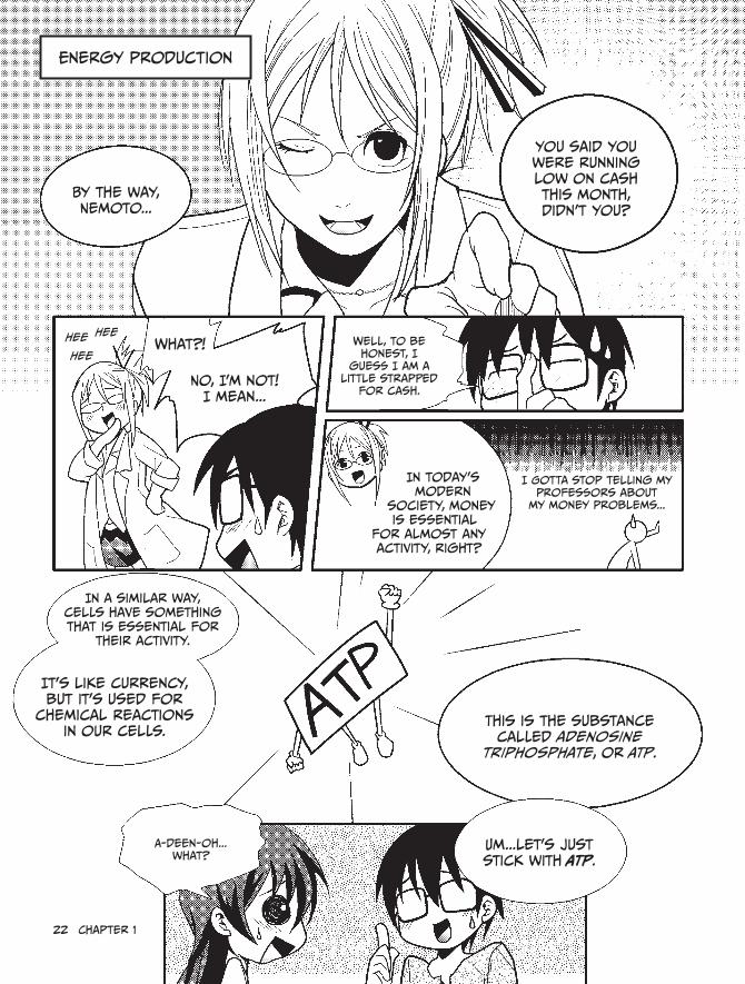
energy production
By the way, Nemoto...
you said you were running low on cash this month, didn't you?
heeWhat?!
No, i’m not! i mean...
Well, to be honest, i
guess i am a little strapped
for cash.
in today's modern
society, money is essential
for almost any activity, right?
i Gotta stop telling my professors about
my money problems...
in a similar way, cells have something that is essential for
their activity.
it's like currency, but it's used for
chemical reactions in our cells.
This is the substance called adenosine
triphosphate, or ATP.
A-deen-oh...what?
Um...let’s just stick with ATP.
hee
hee

ATP is essential for many activities!
• synthesizing proteins• Powering chemical reactions• Performing photosynthesis Protein
As you can see, ATP is essential to cells and metabolism, just like
money is essential to us!
you can’t do anything if you don’t have
money to spend...it’s so depressing.
sniff
has my misfortune
moved her to tears? Amazing!
To maintain essential cellular and metabolic processes, cells must
produce a constant supply of ATP. To do this, they require sugar content (that is, saccharides*)
and oxygen.
Gotta make more ATP!
Another day, another dollar!
it's no exaggeration to say that we eat and breathe to create ATP,
which is then used to fund the activities of proteins.
Just like working to earn a living,
isn’t it?
ATP is created by mitochondria
and proteins found in the
cytosol.
remember: ATP is the “common currency” of
energy that’s used by proteins to keep us alive.
* These saccharides are also known as carbohydrates.
$

24 chapter 1
Photosynthesis
okay...
the last topic we'll go over today is photosynthesis.
We learned about that in school!
Green plants perform
photosynthesis, right?
right!
Protein synthesis and energy production occur in the cells of all organisms...
...But photosynthesis can only occur in the cells of plants,
algae, and some bacteria.
Now, look at this diagram.
sunlight
Photosynthesis saccharide
h20

What happens inside your Body? 25
Photosynthesis is a reaction that uses
sunlight and carbon dioxide to synthesize
saccharides.
Snap
saccharides were required to create
ATP, right?
And oxygen is created as a by-product of
photosynthesis.
so do you understand why plants are so
important for living creatures like us?
saccharides and oxygen are required to create ATP, which is essential
for our bodies...
Ah!and both of those things
are produced by photosynthesis!
i totally get it!
if plants didn't perform photosynthesis, life would be so cruel.
i have no ATP.
Bingo!
one more thing to know: Photosynthesis occurs in chloroplasts, which are special organelles
found in plant cells.
Photosynthesis
saccharide
h20
Little Match girl Kumi
Uh, Kumi...
26 chapter 1
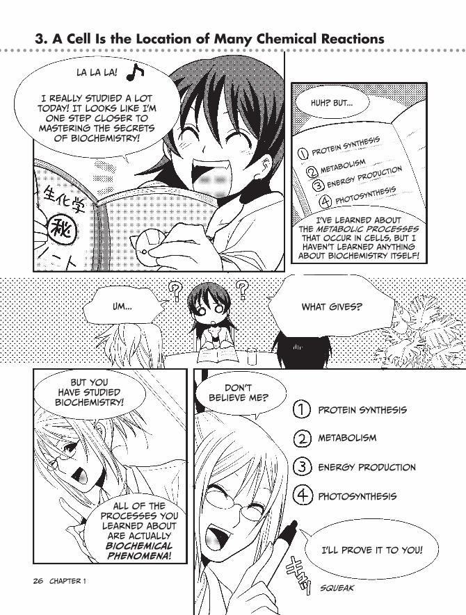
3. A Cell Is the Location of Many Chemical Reactions
La la la!
i really studied a lot today! it looks like i’m
one step closer to mastering the secrets
of biochemistry!
huh? But...
i've learned about the metabolic processes that occur in cells, but i haven’t learned anything
about biochemistry itself!
Um... What gives?
But you have studied biochemistry!
All of the processes you learned about are actually biochemical phenomena!
Don't believe me?
i'll prove it to you!
squeak
Protein synthesis
Metabolism
energy production
Photosynthesis
Protein synthesis
Metabolism
energy production
Photosynthesis
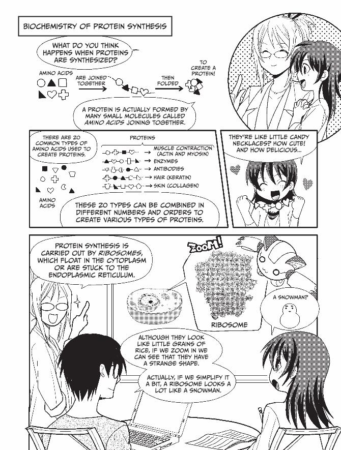
Biochemistry of protein synthesis
What do you think happens when proteins
are synthesized?
Amino acidsare joined together
then folded
to create a protein!
A protein is actually formed by many small molecules called amino acids joining together.
There are 20 common types of
amino acids used to create proteins.
Proteins
Muscle contraction (actin and myosin)
enzymes
Antibodies
hair (keratin)
skin (collagen)
Amino acids These 20 types can be combined in
different numbers and orders to create various types of proteins.
They’re like little candy necklaces? how cute! and how delicious...
Protein synthesis is carried out by ribosomes,
which float in the cytoplasm or are stuck to the
endoplasmic reticulum.
ribosome
a snowman?
Although they look like little grains of rice, if we zoom in we can see that they have
a strange shape.
Actually, if we simplify it a bit, a ribosome looks a
lot like a snowman.

28 chapter 1
how does the ribosome make
proteins?
The ribosome is the place where amino acids join
together.
oh, i see! it’s where the
candies are strung into a
necklace.
Actually, a ribosome looks more like a snowman standing on its head.
When these amino acids are "joined
together," that's a kind of chemical reaction,
right?
scattered
Joined!
chemical reaction
one amino acid
That's right! That reaction sticks two different amino acids
together. Then additional reactions pile on even more amino acids, and pretty soon you've got yourself
a protein!
remember, KUmi, biochemistry is the study of the
chemical processes that take place
inside the bodies of living
organisms...
hey! That’s exactly what Nemoto said back at my
house!
Now you’re getting it!
Amino acids are combined by
chemical reactions to form proteins...
Protein
so protein synthesis is in fact...
biochemistry!
What happens inside your Body? 29
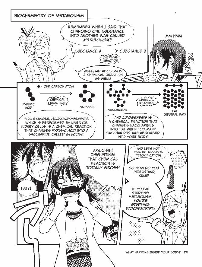
Biochemistry of metabolism
remember when i said that changing one substance into another was called
metabolism?
substance A substance B
chemical reaction
Well, metabolism is a chemical reaction
as well!
Mm hmm
Pyruvic acid
chemical reaction
Glucose
For example, gluconeogenesis, which is performed by liver or
kidney cells, is a chemical reaction that changes pyruvic acid into a
saccharide called glucose.
saccharide
chemical reaction
Fat (neutral fat)
And lipogenesis is a chemical reaction that
changes saccharides into fat when too many
saccharides are absorbed into your body.
Fat?!
Argghhh! Disgusting!
That chemical reaction is
totally gross!
and let’s not forget alcohol detoxification!
so now do you understand,
Kumi?
if you’re studying
metabolism, you’re
studying biochemistry!
= one carbon atom

Biochemistry of energy production
energy production is also a kind of
metabolism.
substance A substance B
chemical reaction
GlucosePyruvic acid
To produce energy, glucose is first broken
down into pyruvic acid in cytosol.
huh? Didn't you mention glucose and pyruvic acid earlier?
yup! This is the reverse version of gluconeogenesis, called glycolysis.
GlucosePyruvic
acid
Gluconeogenesis
Pyruvic acid
Glucose
Glycolysis is all about
breaking down saccharides!
Bing!
Glycolysis
Break it down, y'all!

i totally understand!
Glycolysis is a piece of cake!
......
Well, it may seem easy at first, But the process is actually a little more complicated than that.
chemical reactions
Glucose Pyruvic acidenergy
cytoplasm Mitochondria
Wow! lots of chemical reactions...
yup, breaking down glucose is trickier
than you thought, eh?
Mitochondria use pyruvic acid and
oxygen to create energy (ATP).
This energy production is a complex process that consists
of many chemical reactions occurring simultaneously in
different places.
Pyruvic Acid
oxygen (o2)
Mitochondria are busy little
guys, aren't they?
chemical reactions
Krebs
eTs
ooh!
ooh!

32 chapter 1
Biochemistry of photosynthesis
Finally, let's look at photosynthesis
in plants.
A complex chemical reaction occurs
when light strikes chloroplasts in a
plant's cells.
carbon dioxide is used as a raw material by that chemical
reaction to create saccharides such as glucose...
Light
chemical reaction
Glucose
chloroplast
so, Kumi, what have you noticed about
all of these cellular processes? Do they
have anything in common?
huh? um...Well...They’re all chemical
reactions?
you got it!
Kumi gets a gold star!
h20
Protein synthesis
Metabolism
energy production
Photosynthesis

if there’s one thing you should get out of today’s lesson...
It’s that all of the processes
that occur in our cells are chemical
reactions!
Countless chemical reactions are taking
place inside you, even as we speak!
chem
ical
reac
tions
Wow...
That's kind of scary.
Not only that, but they are happening
unbelievably fast—in the blink of an eye!
Tight regulation of these processes
ensures that everything occurs
in the proper order, which is essential
to cell life.
it's amazing to be alive...
And the fact that all of this is
going on inside such teeny, tiny cells...it’s mind-blowing!
Biochemistry really is
interesting!okay!
Let's end today's lesson here!

34 chapter 1
robocat output
Well, it’s been a long day. you must
be worn out.
definitely. But it was
really very fascinating! Thank you so much!
see you later!
...Nemoto?
squeeze
Are you sure your mind is on biochemistry...
...and not on Kumi?
censored

What? No way! i just want to get people interested
in biochemistry, that’s all!
whatever you say...
But i think you two have some “chemistry” of
your own.
That’s crazy! i’m just an innocent scientist!
i would never–okay, Nemoto, say no more. i understand.
i haven’t seen a boy this head-over-heels in
years.
There’s only one thing i can do!
Just call me... the professor
of love! ♪

36 chapter 1
In this section, we’ll explain some technical terms that you need to know to study biochemistry.
carbon
First, we’ll examine an extremely important chemical element in biochemistry—carbon.Carbon is the element identified by chemical symbol C and possessing the atomic
number 6 and an atomic weight of 12.0107. It’s the primary component of all known life, which is why people sometimes refer to Earth’s organisms as “carbon-based life.” Carbon is the backbone of all organic compounds, and the bodies of living organisms are made almost entirely out of these compounds. Carbon is ideal as a backbone for complex organic mol-ecules such as biopolymers, because it forms four stable bonds, which is an unusually high number for an element. Proteins, lipids, saccharides, nucleic acids, and vitamins are all built with carbon as a framework.
Although carbon is common on Earth—in the biosphere, lithosphere, atmosphere, and hydrosphere—there is a finite amount of it, so it’s recycled and reused. Over time, a carbon atom passes through air, soil, rocks, and living creatures via biogeochemical cycles. The car-bon in your body today may have once been inside a dinosaur!
chemical bonds
When carbon combines with other elements, such as oxygen, hydrogen, or nitrogen, dif-ferent chemical compounds are produced. Except for certain gases, like helium and argon, almost all chemical substances are composed of molecules, two or more atoms attached via a chemical bond. For example, a water molecule (H2O) is created when two hydrogen atoms (H) and one oxygen atom (O) join together.
There are several different types of chemical bonds. Some examples include: covalent bonds, in which electrons are shared between a pair of atoms, ionic bonds, in which oppo-sitely-charged atoms are attracted to one another, and metallic bonds, in which a pool of electrons swirl around numerous metal atoms.
The four stable bonds that carbon forms are all covalent bonds.
Biopolymers
Biopolymers are extremely important molecules to the study of biochemistry.Biopolymer is a generic term for large, modular organic molecules. Modular means
“assembled from repeating units,” like the beads of a necklace. Proteins, lipids, nucleic acid, and polysaccharides are all biopolymers. Because they tend to be especially large molecules, biopolymers can form complex structures, which makes them very useful in advanced sys-tems such as cells.
Biopolymers can form these complex chains because they’re more than simple beads. Let’s consider proteins, for example. Imagine a protein as a necklace made from a variety of different LEGO blocks that can all connect to one another. Since you can twist the necklace
4. Fundamental Biochemistry Knowledge

What happens inside your Body? 37
easily, it doesn’t matter whether the blocks are close together or far apart, but the individual properties of each block result in some connecting better than others. If this necklace was a mile long, imagine the many strange and complex forms you could build. This isn’t precisely how proteins function, but you get the idea.
enzymes
Since biochemistry explains life from a chemical point of view, it is vital to understand how chemical reactions work, and enzymes are essential to these reactions. Enzymes are pro-teins that act as catalysts—that is, they increase the rate of chemical reactions. An enzyme catalyzes nearly every chemical reaction that occurs in an organism.
In a chemical reaction catalyzed by an enzyme, the substance that the enzyme acts upon is called the substrate. The new substance that’s formed during the reaction is called the product. The activity of an enzyme is affected by the environment inside the organism (temperature, pH, and other factors), the availability of the substrate, and, in some cases, the concentration of the product.
Although almost all enzymes are proteins, it has recently been discovered that a spe-cial type of ribonucleic acid (RNA) can act as a catalyst in certain chemical reactions. This is called an RNA enzyme, or a ribozyme.
oxidation-reduction
Enzymes are broadly classified into six types, which will be introduced in detail in Chapter 4. Oxidation-reduction is one of the most important enzyme reactions, in which electrons are exchanged between two substances. If electrons are lost, the substance is oxidized, and if electrons are gained, the substance is reduced. Normally, when one substance is oxidized, another substance is reduced, so oxidation and reduction are said to occur simultaneously.
The movement of hydrogen ions (H+, aka protons) often accompanies the exchange of electrons in an organism, and NADPH, NADH, and similar compounds (which we’ll discuss in Chapter 2) work as reducing agents on other substances.
respiration
In Chapter 2, we will examine respiration. In the broadest sense, respiration is the process of obtaining energy by breaking down large compounds, but this only gives us a vague sense of the meaning.
More specifically, when respiration occurs, an organic substance (for example, the car-bohydrates that make up spaghetti) is broken down into simple, inorganic components, like carbon dioxide (CO2) and water (H2O). Energy is produced when electrons are transferred between molecules (oxidation-reduction), along a sort of factory line, until they reach oxy-gen (O2). This process is known as internal respiration or cellular respiration.
The oxygen we mentioned above is very important in respiration. It comes from the air that we breathe, and carbon dioxide is produced as a waste product of cellular respiration. When we use our lungs to inhale oxygen and exhale carbon dioxide, it’s known as external respiration.

38 chapter 1
Metabolism
The processes that alter an organism’s chemical substances are called metabolism. Broadly speaking, metabolism can be divided into substance metabolism and energy metabolism. However, since these two types occur together during metabolism, the distinction isn’t very clear. In this book, when we refer to metabolism, you may assume that we mean substance metabolism.
Substance metabolism This refers to the changes to substances that occur in an organism, including the chemical reactions that are catalyzed by enzymes. More specifically, a reaction that breaks down a complex substance into simpler substances is called catabolism, and, conversely, a reaction that synthesizes a more complex substance is called anabolism.
Energy metabolism This refers to the energy that’s gained or lost through anabolic and catabolic processes within an organism, including reactions in which the energy created via respiration or photosynthesis is stored as ATP and other high-energy intermediates.
2Photosynthesis and respiration

ecosystems and the biogeochemical cycle
* central Park
Wow!This food is so delicious! And this park is so
beautiful!
eating Professor Kurosaka's home
cooking is so awesome!
it really is delicious.
i brought a lot, so eat as much
as you want.
it's so nice to eat lunch under a clear
blue sky, surrounded by trees...
That's right!
Which is why we need to protect the global environment now and in the future.
1. Ecosystems and Cycles
*
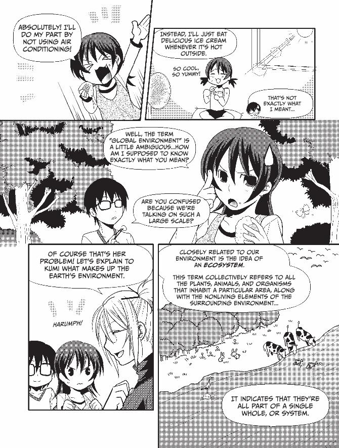
Absolutely! i'll do my part by not using air conditioning!
instead, i'll just eat delicious ice cream whenever it's hot
outside.
so cool, so yummy!
That's not exactly what
i meant...
well, the term "global environment" is a little ambiguous...how am i supposed to know exactly what you mean?
Are you confused because we’re
talking on such a large scale?
of course that’s her problem! Let’s explain to Kumi what makes up the earth’s environment.
Harumph!
closely related to our environment is the idea of
an ecosystem.
This term collectively refers to all the plants, animals, and organisms
that inhabit a particular area, along with the nonliving elements of the
surrounding environment...
it indicates that they're all part of a single whole, or system.

42 chapter 2
since all life on earth is
interconnected through natural
cycles...
...it's not an exaggeration to consider the global environment as its own
massive ecosystem.
Well, i'll be...
But how can such a vast ecosystem
be maintained?
The food chain is important—that indicates
which organisms eat which other organisms, but...
...here we need to consider how an ecosystem comes about chemically.
We are talking about biochemistry,
after all!
everything is linked through the biogeochemical
cycle.
biogeochemical cycle
it's...something that cycles around
and around?you got it!
Let's take a closer look at biogeochemical
cycles.

Photosynthesis and respiration 43
What is the biogeochemical cycle?
The elements of the global ecosystem, including the food chain, respiration, and photosynthesis, are all part of a worldwide cycle known as the biogeochemical cycle.
When one organism, like a bear, eats another organism, such as a fish, the material that made up the fish becomes part of the bear.
A very important substance in this transfer is carbon (C).
photosynthesislight
eating
carbon from the starch of potatoes (c)
carbon used as fuel and raw material for the body
Look at the diagram above. If Kumi eats a potato, the carbon from that potato enters her body.
And when I breathe, the carbon that was inside my body becomes carbon dioxide (CO2) and is expelled from my body.
That’s right! Then the carbon that left your body is captured by plants through photosynthesis and is transformed into the carbon that makes up starch (which is a type of saccharide).
Then that potato is eaten by Kumi (or a cow or some other hungry creature), and the carbon is returned once more to the body of an animal.
respiration

44 chapter 2
Since Kumi eats beef, the carbon can also move from the cow to Kumi.
Mmm...I love a tasty cheeseburger!So carbon really moves around a lot, doesn’t it? Is this movement the
biogeochemical cycle?
Yep! The entire Earth is carrying out this cycle on a gigantic scale.
carbon Dioxide
photosynthesisrespiratio
n
other human activities
predation
burying
fossil fuel
consumption
Wow! So the rice, potatoes, and apples that I eat were originally related to carbon dixiode that somebody else exhaled.
And it’s not just carbon. Hydrogen (H), oxygen (O), nitrogen (N), and sulfur (S) cycle around the Earth as well, going from organism to organism, getting emit-ted into the atmosphere, dissolved into the ocean, or buried deep underground.
When this cycle works smoothly, the ecosystem and the global environment are healthy.

Photosynthesis and respiration 45
H2N COOH
H
R
C
Next I’ll explain the carbon cycle in a little more detail.
carbon cycle
Umm, carbon...? I know we’ve talked a little about it, but I still don't understand what it is.
No problem! Nemoto, can you review some basic facts about carbon for Kumi?
Sure. Carbon is one of the most important chemical elements to living creatures like us.
Carbon is at the center of amino acids, which are the building blocks of proteins. It’s also the element that creates the framework of saccharides and lipids, and it’s a vital part of DNA.
it's here.
it's also often here.
you'll even find some here.
You see? C is essential to all of them.
Amino Acid Glucose
Fatty Acid
C
C
C
C
CC
H
H
H
H
H2OH
OH
OH
OH
H
O
HO
H
H H H H
HHHH
HHHH
HHHH
COOH
C C C C
CCCC

46 chapter 2
H
H HH
C
You’re right. It looks like we’d be in real trouble without any C. It really is important!
When carbon leaves an organism’s body, it can bond with two oxygen atoms to become carbon dioxide (CO2) or bond with four hydrogen atoms to become methane (CH4).
Also, it can accumulate deep underground and, over long periods of time, it will become crude oil, coal, and, in some cases, even diamonds.
O = C = O
Carbon dioxide Methane
Oil! Diamonds! Wow, carbon is really valuable stuff!
That’s true, but remember: It’s not all about excitement and riches. The way carbon circulates is extremely important. Disrupting this balance could make carbon dioxide concentration on Earth steadily increase...
Which would be a serious problem, wouldn’t it?
Oh man, that sounds like it would be a total bummer...
Well, think of it this way: If you can understand the Earth as a single circulating system and your own body as a system as well, then you’ll realize the impor-tance of a healthy balance and the dangers of disrupting that balance. This knowledge can lead to insights into the pursuit of health and beauty!

Photosynthesis and respiration 47
The pursuit of health and beauty? Now you’ve got my attention!
(Wow, Professor Kurosaka is really good at motivating Kumi...)
Let’s look at this figure again.
Look at j on the left. This is the flow of carbon in which plants pull carbon dioxide from the atmosphere during photosynthesis and use it as a raw material for creating saccharides.
Now look at k on the right. This is the flow of carbon in which those saccharides are used by a living creature, and through this process, they become carbon dioxide once more and are returned to the atmosphere via respiration.
Let’s talk more about these two flows. Now that we’re finished with lunch, we can really sink our teeth into the subject!
Super cool!
light
eating
carbon from the starch of potatoes
carbon used as fuel and raw material for the body
photosynthesis respiration

48 chapter 2
2. Let’s Talk Photosynthesis
At the base of the ecosystem, plants, algae, and some bacteria supply food to all living crea-tures. The majority of them use a process called photosynthesis, in which saccharides and oxygen (O2) are created from carbon dioxide by splitting atoms of water, using energy from the Sun. Saccharides are also known as carbohydrates and are vital to living creatures like us.
That’s why plants are called producers. In contrast, we animals are called consumers.Photosynthesis is important not just because it creates saccharides. It also maintains a
steady, balanced concentration of carbon dioxide in the atmosphere, and it produces oxygen, which living animals require to survive.
As you can imagine, deforestation by humans greatly reduces the number of “ producers,” upsetting the delicate balance of the biogeochemical cycle and potentially threatening “ consumers” (like us!).
Now, let’s take a closer look at the way in which plants use sunlight to create saccharides.
The importance of plants
sunlight photo-synthesis
saccharides = carbohydrates
here
you go!photosynthesis
carbo-
hydrates

Photosynthesis and respiration 49
This image (courtesy of RoboCat) shows green sacks, called chloroplasts, within a plant cell.Inside a chloroplast, structures shaped like very thin pouches are stacked in multiple
layers to form peculiar structures. Each of these flat pouches is called a thylakoid, and a stack of multiple thylakoids is called a granum.
The thylakoid membrane is a bilayer composed primarily of phospholipids, just like in cell membranes.
Now let’s look at the surface of the thylakoid membrane.
Do you see the groups of tiny grains that appear to be embedded in the surface of the thylakoid? Each of these is a collection of molecules called chlorophyll, as well as various proteins that aid in photosynthesis.
Chlorophyll molecules absorb sunlight. However, they don’t absorb the entire spectrum; they reflect the green portion of sunlight back outside, which is why plants appear green to us.
chloroplast structure
chloroplast
outer membrane (lipid bilayer)
inner membrane (lipid bilayer)
Granum
Thylakoid
chloroplast structure
collection of chlorophyll molecules
Thylakoid
Granum
Thylakoid structure

Photosynthesis—the Photophosphorylation reaction
yay!!Well, since we were able to
find such a nice gazebo...
Why don't we continue our lessons out here in the
park?Um, are you sure the lab wouldn’t
be better?
But we're going to discuss photosynthesis!
Don't you think it's more fitting to study under a blue sky than to shut ourselves
off in the laboratory?
i guess...
oh, i already learned about photosynthesis! humans can't
do it—only plants!
They use sunlight and carbon dioxide to create oxygen and
saccharides, right?
Photosynthesis
saccharide = carbohydrate
yes, That's correct!
But do you know how plants produce oxygen and
saccharides from sunlight? Do you know how the process of
photosynthesis works?
Urk

Photosynthesis and respiration 51
The two most important reactions
in photosynthesis are photophosphorylation
and carbon dioxide fixation.
Photophosphorylation is the creation of energy
using sunlight.
carbon dioxide fixation uses that energy to
synthesize saccharides.
sunlight required!
Photophos-
phorylation
carbon
dioxide
fixation
saccha- ride
First, we'll learn about photo-
phosphorylation.
Let's investigate where photo-
phosphorylation is performed inside a chloroplast.
We can use robocat to dive in and explore the inside of a
plant!
This is an image looking down at a thylakoid inside a
chloroplast.
see the collections of bumps?
ping!
errr...
each of these is an aggregation of molecules called chlorophyll and various proteins.
Dozens of chlorophyll molecules are contained in each group, and each of these molecules absorbs
sunlight like a little satellite dish.

52 chapter 2
Now let's zoom in on the thylakoid
membrane.
chlorophyll and protein
complexFlow of
electrons
Photosystem ii cytochrome b6-f complex
Photosystem i ATP synthase
Thylakoid bilayer
membrane
This chlorophyll and protein complex
appears as if it’s embedded in the middle
of the thylakoid membrane.
it sure does! But wait...what are those other weirdly shaped things?
like these...
Those are the part of the electron transport
chain. each of those weird-looking shapes
is a complex consisting of several proteins gathered together.
They are essential
for photo-synthesis.
eeeek! First we were talking about photosynthesis in plants,
and now all of a sudden electrons are in the mix?
My mind is officially boggled...

Well, as you probably know, everything around us is made up of atoms. every atom has a
center called a nucleus...
When sunlight strikes chlorophyll,
the molecule is "excited" by the light energy.
i'll explain it in detail!!
yikes!
Light
electron
Nucleus
* Actually, electrons fly off only from the central reaction center chlorophyll. The other chlorophyll molecules act as amplifiers to feed energy into the reaction center, which enables the electrons to detach.
When this happens, one of the electrons in the molecule ends
up flying off.*
The free electron is then delivered to
another protein complex near the chlorophyll.
This other protein complex is also embedded in the thylakoid membrane.
remember the collection of weird
shapes earlier?
Ah! i get it now!since the electrons are
passed along to another complex, it's called an
"electron transport chain!"yeah!
That electron is replenished by breaking down water molecules,
and o2 is produced as a result.
oh! so a plant can't perform photosynthesis
if it doesn't have water.
and a group of electrons
revolving around this nucleus.

54 chapter 2
The protein complexes do more than transport electrons in a
predetermined order.
a molecule called NADPH is synthesized along the way, and ATP is created at
the end of the chain.
you mentioned ATP before, but i think this is the first time i've heard of NADPh...
NADPh consists of an electron and a
proton (hydrogen ion) adhered to a molecule
called a hydrogen acceptor. it is created
by NADPh reductase.
Proton
Toss 'em
in here!NADPh can be thought of as
a temporary storehouse for an electron and a proton, which
are required by the carbon dioxide fixation reaction we
talked about earlier.*
The energy that’s generated is just a "flow of electrons," and, in that
sense, it's just like the electricity you use for your household appliances.
Breakfast
time!
The electron transport chain also creates a "flow of electrons." substances like ATP are
synthesized by this electron flow.
Now let's look at the way ATP is
created by photo-phosphorylation!
Fwip
* in other words, NADP + is "reduced," and NADPh is created. (see page 37 for an explanation of reduction.)

e- stands for this electron.
** A force which causes a substance to naturally flow from a high con-centration to a low concentration.
oh, i get it! When i see it laid out step-by-step, it makes way
more sense!
Because of this "electron flow," NADPh and ATP are
produced!
chlo
ro
- phyll
Photosystem ii
Photosystem i ATP synthase
Thylakoid lumen
cytochrome b6-f
sunlight strikes the chlorophyll.*
sTeP 1 (Photosystem ii)
The light energy causes an excited state in the
chlorophyll so an electron e- is emitted and passed along. At
the same time, a proton h+ accumulates in the
thylakoid lumen.
sTeP 2
The electron (e- ) and A proton are captured by NAPD+, and NADPh is
produced.
sTeP 3 (Photosystem i)
When the protons (h+ ) that were collected in the thylakoid lumen are about to leave the
thylakoid according to the concentration gradient,**
they pass into ATP synthase.At this time, ATP is
synthesized from ADP.
sTeP 4
* Photosystem i also has chlorophyll. energy is received here as well, and electrons that were transported from Photosystem ii are once again excited.
light
light
chlo
ro
- phyll
Photosystem ii
Photosystem i ATP synthase
Thylakoid lumen
cytochrome b6-f
Photosystem ii
Photosystem i ATP synthase
Thylakoid lumen
cytochrome b6-f
light
chlo
ro
- phyll
chlo
ro
- phyll
Photosystem ii
Photosystem i ATP synthase
Thylakoid lumen
cytochrome b6-f
light

56 chapter 2
The important thing here is the reaction that
creates this ATP, called phosphorylation.
phosphorylation? What kind of
reaction is that?
Well, look...here's the structure of ADP
(adenosine diphosphate).
two phosphate
groups
ADP, which has two phosphate groups,
becomes ATP (adenosine triphosphate) if one additional phosphate
group is attached.
one more phosphate
group
The reaction that attaches this new
phosphate group is phosphorylation!
oh, so that must be where the name
"photophosphorylation" comes from!
remember, ATP is also the "common currency" used in reactions other
than photosynthesis. simply put, it's important chemical energy!
Let me summarize photophosphorylation
for you!
lightit's a reaction that uses
to create ATP from ADP by phosphorylation
so ATP is synthesized by the flow of electrons triggered by sunlight...
i understand! Light energy
turns into chemical energy!
C C C C COO

Photosynthesis—carbon dioxide fixation
Photophosphorylation carbon fixation
That's all for photo-phosphorylation.
Just remember: sunlight is
always required!
Next, we’ll study carbon fixation. o-okay!
The final steps of photo-phosphorylation in which ATP
and NADPh are synthesized will occur even if light is no longer present. This is known as a light-
independent reaction.*
Then does carbon dioxide fixation happen even at night or on a cloudy day?
Actually, no. it just means that light isn't needed at
that particular station. The reaction itself occurs in the daytime because NADPh
is required and is not manufactured at night.
carbon dioxide fixation occurs in the stroma, which is the central part of the chloroplast, rather than in the thylakoid membrane.
see? right here.
Gotcha.
stroma
* This light-independent reaction is also known as the Calvin Cycle or the Reductive Pentose Phosphate Cycle.

58 chapter 2
carbon fixation is a reaction that uses chemical energy, stored as ATP, to create saccharides, using the
carbon dioxide in the air as a raw material.
Saccharides
That's the carbon dioxide that's produced by humans and other animals, isn’t it?
And the reaction also needs the chemical energy that was created
by photophosphorylation.
carbon fixation
First, co2 bonds with a substance
called ribulose-1, 5-bisphosphate to
form two molecules of 3-phosphoglycerate,
which have three carbon atoms apiece.
The chemical energy of ATP and the reducing
power of NADPh are used to create two molecules
of glyceraldehyde 3-phosphate from this 3-phosphoglycerate.
And this glyceraldehyde 3-phosphate is used to produce glucose, fructose, and other
saccharides.
saccharides
saccharides
(ribulose-1, 5-bisphosphate)
(3-phosphoglycerate) (1,3-bisphosphoglycerate) (glyceraldehyde 3-phosphate)
Bonding of carbon dioxide Use of chemical energy
so carbon dioxide is bound first,
and then chemical energy is used.

But this "glucose" that’s made at the end...
what is it exactly? relax, Kumi.
Glucose* is a basic saccharide that's
very common in living organisms and in starchy
foods especially.
so
confusing!
oh, wow! rice, noodles, potatoes...
they're all created by this amazing process!
That's right!Plants string glucose together like beads to form starch, or they convert glucose into sweeter saccharides,
such as sucrose** or fructose.
This stored material becomes the most
delicious and nutritious parts of plants, providing
us with foods like rice, wheat, potatoes, and fruit!
if plants didn't perform photosynthesis, all of those
foods would disappear...
...and we'd lose their essential nutrients. Not
to mention yummy treats!
GASP
so now do you understand the importance of
photosynthesis?
yes! From now on, i'll be grateful for every single grain of rice
i eat!
Actually, i could go for some
rice right now...
* More precisely, the saccharide created by photosynthesis is glucose 1-phosphate.
** Plants synthesize sucrose from glucose to make them more appealing for animals to eat.

60 chapter 2
3. Respiration
What is a carbohydrate?
having class outdoors was fun!
Now, are you guys up for a
little tea break?yeah!
Umm...That reminds me...
there's still something i don't quite understand.
sugar is a saccharide, which is the same as a carbohydrate,
right?
yes. carbohydrates are saccharides and
so is the sugar known as "table sugar" or
"blood sugar."
Argh! That's the part i don't get! rice is a carbohydrate,
so it's basically made of sugar. But then why
isn't it sweet?!
saccharide =
carbohydrate
= sugar
What's different about cake that makes it so sweet and delicious?
right?!

That's a great observation, Kumi.
Take a look at this. When we say
"saccharides," we're actually talking about a number of different
sugars.
Table sugar (sucrose)
Fructose
Lactose
starchoh, i see...
so each type of saccharide is
different. shall we take a closer look?
Saccharide time!
yeah!
one important type of saccharide is glucose. it has both an open-chain form and a
cyclic (or ring) form.
This diagram represents what's known as α-D-glucose. if the rightmost hydroxyl group and hydrogen in the cyclic form trade places around their carbon, it's
called β-D-glucose.
open-chain
formcyclic form
Look at the open-chain form. six carbon atoms
(c) are vertically aligned, and a hydrogen (h) and a
hydroxyl group (oh or ho) are attached to each of
the lower five.
This is one of the basic components of a
monosaccharide.*
The top carbon atom has the form of an
* see page 83 for more information on monosaccharides.
aldehyde group .( )

62 chapter 2
sugar dissolves easily, right? it can travel
throughout the body via the bloodstream because hydroxyl groups blend
well with water.
clearly, the presence of oh is an important
component of sugar, but we also need to know
where on the saccharide the oh is located.
really? Why?
Glucose Galactose
Well, if the oh attached to the
fourth carbon is on the left side...
...it will no longer be glucose but rather a different type of saccharide called
galactose.
it becomes something different
depending on whether it’s attached on the right or left...that's
so weird.
Monosaccharides, when in an actual
body, take the cyclic form more
often than the open-chain form.
Glucose is also shaped like:
more often than:
how'd she say that?
slosh
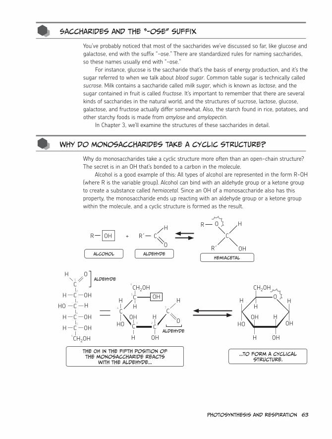
Photosynthesis and respiration 63
saccharides and the “-ose” suffix
You’ve probably noticed that most of the saccharides we’ve discussed so far, like glucose and galactose, end with the suffix “-ose.” There are standardized rules for naming saccharides, so these names usually end with “-ose.”
For instance, glucose is the saccharide that’s the basis of energy production, and it’s the sugar referred to when we talk about blood sugar. Common table sugar is technically called sucrose. Milk contains a saccharide called milk sugar, which is known as lactose, and the sugar contained in fruit is called fructose. It’s important to remember that there are several kinds of saccharides in the natural world, and the structures of sucrose, lactose, glucose, galactose, and fructose actually differ somewhat. Also, the starch found in rice, potatoes, and other starchy foods is made from amylose and amylopectin.
In Chapter 3, we’ll examine the structures of these saccharides in detail.
Why do monosaccharides take a cyclic structure?
Why do monosaccharides take a cyclic structure more often than an open-chain structure? The secret is in an OH that’s bonded to a carbon in the molecule.
Alcohol is a good example of this: All types of alcohol are represented in the form R-OH (where R is the variable group). Alcohol can bind with an aldehyde group or a ketone group to create a substance called hemiacetal. Since an OH of a monosaccharide also has this property, the monosaccharide ends up reacting with an aldehyde group or a ketone group within the molecule, and a cyclic structure is formed as the result.
R OH +H
OC
OH
H
OHHOOH H
HHO
H
CH2OH
OHH
HOOH H
HH
CH2OH
OH
OH
C
C
C C
C
O
H
C
H O
OH
CH2OH
OH
OH
1
6
1
2
6
5
4
3
C H
C
C
C
H
HO
H
H
2
5
4
3
R´ C
Alcohol Aldehydehemiacetal
R´
R O H
Aldehyde
Aldehyde
The oh in the fifth position of the monosaccharide reacts
with the aldehyde...
...to form a cyclical structure.
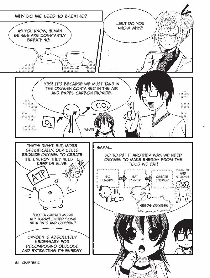
64 chapter 2
As you know, human beings are constantly
breathing...
...but do you know why?
yes! it's because we must take in the oxygen contained in the air
and expel carbon dioxide.
Wha?!
That's right. But, more specifically, our cells
require oxygen to create the energy they need to
keep us alive.
"Gotta create more ATP today! i need some nutrients and oxygen!"
oxygen is absolutely necessary for
decomposing glucose and extracting its energy.
so to put it another way, we need oxygen to make energy from the
food we eat!
hmmm...
so hungry...
eat dinner
create energy
healthy and
strong!
needs oxygen
Why Do We Need to Breathe?

When we take in oxygen and expel
carbon dioxide, it's called respiration.
however, the reaction in which cells absorb oxygen, break
down glucose to create energy, and emit carbon dioxide is also
called respiration.
Gimme some oxygen!
Gimme some glucose!
you want energy?
Wait, they're both called respiration? i'm going to mix them up
for sure...
Well, to distinguish them, we call the process of breathing external respiration and the process inside
of cells internal (or cellular) respiration.
external respiration
internal respiration
That's right. From now on when we say "respiration,"
we'll be referring to internal respiration.
internal! Got that?
so we'll be studying the reaction inside of cells, not the kind of respiration i'm
doing right now.
inhale
exhale

respiration is A reaction That Breaks Down Glucose To create energy
saccharides that are created by plants through photosynthesis
are stored in the form of starch, which we animals eat.
energy
organisms like us break down starch into glucose, which is used to make ATP, with the assistance of the oxygen that we breathe in.
That's internal respiration!
here, take a look at this. it's the general formula
for respiration.
Glucose oxygen Water energycarbon dioxide
Water
oh, that makes it crystal clear!
Glucose and oxygen are consumed, and
carbon dioxide, water, and energy
are produced.
Now, let's take a closer look at this reaction.
you can think of respiration as having three
stages...
And here they are!
Glycolysis
citric acid cycle*
electron transport chain
* This is also called the Krebs cycle or the TCA cycle.
MmM,
yummy
Glucose

cell
Mitochondria
Glucose Glycolysiscitric acid
cycle
electron transport
chain
38 molecules
of
(cytoplasm) (Mitochondria)
j Glycolysis is performed in the cytoplasm. The k citric acid cycle is performed in the mitochondrial matrix, and the l electron transport chain is located within the inner membrane of the mitochondria.
Through this process, up to 38 ATP molecules are created from 1 glucose molecule.
hmmm... This is all kinda over my head.
Well then, how about this?
Ta-da!
duhhPyruvate
Glycolysis, which doesn’t need oxygen
citric acid cycle, going round and round
high-tech electron
transport chain
That's a little bit more my speed.
hee hee
But what are NADh and FADh2? This is the first i've heard of them. Are
they related to the NADPh that's produced by photosynthesis?
Pyruvate
hang on, let's not get ahead of ourselves. The
flow of these substances is extremely important, so let's just take it one step
at a time!

68 chapter 2
Stage 1: Glucose decomposition by Glycolysis
First we'll look at
glycolysis!
We love you, glycolysis!
To start, a cell absorbs glucose and breaks it down in the cytoplasm, which is the gooey liquid that holds the
cell's organelles.
Zoom
cytoplasm
one molecule of glucose is decomposed into two molecules of pyruvate
(or lactate*).
Glycolysis
Glucose Pyruvate
Glucose is made up of six carbon atoms, while
pyruvate has three.
oh, i get it. To turn one molecule of glucose into pyruvate, it's split exactly
in two. Nice and neat!
Pyruvate
Glucose
* Lactate is created from pyruvate when oxygen isn't available.

Well, not quite.it's a little more
complicated...
Actually, glycolysis needs 10 chemical
reactions to turn that one glucose molecule
into two pyruvate molecules.
to create lactate, one additional
reaction is performed.
it looks like this:
Glucose
one molecule
Glucose 6-phosphate
Fructose 6-phosphate
Fructose 1,6-bisphosphate
2-Phosphoglycerate 3-Phospho- glycerate
1,3-Bisphospho- glycerate
Glycer- aldehyde
3-phosphate
Dihydroxy- acetone
phosphate
Phosphoenol- pyruvate
Pyruvate Lactate = one carbon molecule
two molecules
Are you kidding me? it looks like a game of chutes and Ladders gone horribly wrong!
AHH!yes, it is a long
process. since four ATP molecules are
synthesized and two ATP molecules
are consumed...
two ATP molecules are created as the final balance.
Ta-da!

only two molecules of ATP
are made after that whole process?
That's insane!
Maybe not insane but definitely
inefficient.
say what?!
That's a good point. Glycolysis is the
most primitive energy production pathway that organisms have.
That's why it's so inefficient!
Aw, poor little guy.
But the fact that glycolysis requires no oxygen is a notable
feature! When oxygen runs out, two molecules of ATP can still be synthesized from one
molecule of glucose.
We don't need no stinking oxygen!
This made it an extremely important energy
production pathway for organisms in earlier eras, when there was
less oxygen in the atmosphere. it's also a vital pathway for anaerobic
organisms* living today.
so there must be a more efficient method of
producing energy than glycolysis, right?
Purrr
That's correct! As time passed, organisms
developed two other pathways that could
create ATP much more efficiently, this time
using oxygen.
These are the citric acid cycle
and electron transport
chain, which we will discuss right now!
* An anaerobic organism is one that doesn’t require oxygen for survival.
duuuh...
Fructose 6-phosphate
Fructose 1,6-bisphosphate
Phosphoenol- pyruvate
Pyruvate
two molecules
Glycer- aldehyde
phosphat
1,3-Bispho

Photosynthesis and respiration 71
The second stage is the citric acid
cycle!
Stage 2: citric acid cycle (aka TcA cycle)
you rule!
Actively using oxygen to produce ATP is an important role of a
single mitochondrion organelle.
cell
Mitochondrion
When pyruvate, which was created in the cytoplasm by breaking down glucose, is
taken into a mitochondrion...
Pyruvate
Glycolysis citric acid cycle
it will be sucked into the vortex of the citric acid cycle.
“The vortex of the citric acid cycle”?
yay!
dizzydizzy

PyruvateAcetyl-coA
Look at this:
oxaloacetate citrate
Malate cis-Aconitate
Fumarateisocitrate
succinate
succinyl-coA
α-ketoglutarate
each represents a single carbon molecule. The carbon
of co A isn't shown. see page 85 for a description of co A.
Waaah!My head is spinning!
Kumi, you've got vortex
eyes!
Do i have to remember all
of this?
j Pyruvate becomes Acetyl-coA, and k oxaloacetate and acetyl condense to form the
6-carbon citrate. Later, l two co2 molecules are released, and one molecule of ATP is produced, along with n four molecules of NADh and o one
molecule of FADh2.
No, not quite. Just try to remember these key steps:*
it's okay if you can't remember
which "-ate" word is which.
whew
* each molecule of glucose is converted into two molecules of pyruvate, so two molecules of ATP, eight molecules of NADh, and two molecules of FADh2 are created in the citric acid cycle.
As you can see, after pyruvate changes to Acetyl-coA and then to
citrate, the citrate sequentially transforms into various substances
inside the mitochondrion. then it finally returns to being
citrate once more.
Pyruvate
Acetyl-coA
citrate
Around... ...and
around
since the reactions circle around and around, it's called a cycle. Get it?

Although only two new molecules of ATP
are created in this cycle...
other important substances
are produced as well.
They are the coenzymes NADH and FADH2 !
By the way... While NADPH is used
in photosynthesis......in respiration, it's just NADH.
i see...
These coenzymes create a huge amount of ATP in the electron transport chain that
follows.
citric Acid cycle electron Transport chain
so...the coenzymes NADh and FADh2 continue on to
the next stage.
But what do they do to produce so much ATP?
i'll explain that next!

Stage 3: Mass production of energy by the electron transport chain
At last we've made it to the third stage:
The electron transport chain!
Woo! Last
but not
least!
i'm the greatest!
The electron transport chain...
consists of several protein complexes that are embedded like islands within the inner membrane
of mitochondria.
hmm...i think i've seen this somewhere
before...
This collection, taken as a set, is one electron transport
chain.
inner membrane
intermembrane space
Matrix
outer membrane
intermembrane space
Matrix
Proteins and coenzymes of the electron transport chain
inner membrane of mitochondria

Photosynthesis and respiration 75
i remember! it was photosynthesis! The electron transport chain also appeared
there, right?
yes, But the constituent protein
complexes are different for
photosyntehsis.
While the flow of electrons is the same, each of those "weird shapes" is different,
and they all perform different tasks too.
electron
transport chain
of photosynthesisyup, those shapes look
different alright!
Now, let's get back to NADh and FADh2.
Four molecules of NADh and one molecule of FADh2 are created from one molecule
of pyruvate.
glucose
Pyruvate
Pyruvate
citric acid
cycle
citric acid
cycle
When converting from one molecule of glucose, eight molecules of NADh and two
molecules of FADh2 are created by the citric acid cycle alone.
NADh and FADh2 are synthesized in the citric acid cycle when hydrogen atoms
are delivered to both NAD+ and FAD. since a hydrogen atom consists of a
single proton and a single electron...
...this means that a proton and an electron are deposited in "temporary custody" of
NAD+ or FAD.*
hydrogen atom = proton + electron
depositing for storage!
Proton Proton
in photosynthesis, a proton and an electron
were deposited into NADP to form NADPh.
The protons and electrons that were deposited in NAD+ and FAD
will then exhibit their power in the electron
transport chain.
* in other words, NAD+ or FAD is "reduced," and NADh or FADh2 is created. (see page 37 for an explanation of reduction.)

Proton Proton
electron transport chain
What happens to the electrons (e-) and protons (h+) that were "deposited" in NAD+ and FAD?
Let's take a look!
Alright!
intermembrane space
complex i
Matrix
electron flow cytochrome c
complex iii
ATP synthase
complex iV
inner
mem
brane o
f
mito
cho
ndria
An electron directly enters the electron transport chain and, as you might guess from
the name, is transported between several protein complexes.
protons collected in the inter-membrane
space
While this electron (e-) is successively transferred, the proteins undergo various changes. As a
result, protons (h+) move from the mitochondrial matrix to the intermembrane space (between the inner and outer membranes). Three "pumps" push these protons into the intermembrane space.
interesting...
co Q
co Q
intermembrane space
complex i
Matrix
complex iii
ATP synthase
complex iV
cytochrome c inner
mem
brane o
f
mito
cho
ndria

intermembrane space
complex i
Matrix
complex iii
ATP synthase
complex iV
When this occurs, a force called The concentration gradient is
generated. This means that when the concentration of the protons (h+) in the intermembrane space becomes
higher than in the matrix, the protons try to flow toward the matrix.
The concentration gradient is a force which causes a
substance to naturally flow from a higher concentration to a lower concentration.
the proton passes
through a gate.
An electron transport chain in satisfactory condition has a "gate" through which the protons can move into the matrix.
off you go, protons!
co Q
co Q
intermembrane space
complex i
Matrix
complex iii
ATP synthase
complex iV
That gate is the ATP synthase. When a proton passes through it, one molecule of ATP is produced.
Therefore, 30 molecules of ATP are created from 10 molecules of NADh (of which two molecules are produced via glycolysis), and 4 molecules of ATP are created from 2 molecules of FADh2.
We get these numbers because the electrons originating from NADh cause the "proton pumps" to work at all three locations, but the
electrons from FADh2 cause them to function at only two locations.
bond with
oxygen
And finally the electron and the proton that were used bond with
oxygen (oxygen is required here! ) to produce water.
We get it!
inner
mem
brane o
f
mito
cho
ndria
cytochrome c
cytochrome c inner
mem
brane o
f
mito
cho
ndria

78 chapter 2
And that...
Ahem
...concludes my explanation of
respiration.hooray!
remember, if we express this respiration reaction in a chemical formula, we get the following,
which we saw earlier.
it's much more meaningful after learning the details. it feels like
we finished a long journey...
glucose oxygen Water Watercarbon dioxide
energy
By the way, if we replace the “38 ATP molecules” with "light energy" and reverse the arrow’s direction what
do you think we'll get?
Whoa! it's photosynthesis! Amazing!
glucose oxygen Water Watercarbon dioxide
light energy
Through this process, starting with 1 molecule of glucose...
glucose glycolysiscitric acid
cycle
electron transport
chain
A total of molecules are produced from 1 glucose.
Glycolysis, the citric acid cycle, and the electron transport chain combined create 38 molecules of ATP, which becomes the energy for the activity of our cells.*
* The NADh produced during glycolysis cannot enter mitochondria on its own. instead, it passes its electrons to a "shuttle" called glycerol 3-phosphate. Glycerol 3-phosphate passes its e- to FAD+ in the mitochondrial membrane, and FADh2 is created. When FADh2 deposits its e- into the electron transport chain, one step in the chain is skipped, and one less ATP is formed (two less per glucose), so the net ATP produced is 36.

conclusion
scribble
scribble
we can summarize the interrelationships of photosynthesis and
respiration as follows:
light
photophos-phorylation
carbon dioxide fixation
glycolysis
citric acid
cycle
electron transport
chain
saccharidesaccharide
photosynthesis
carbon dioxide is used by photosynthesis to make saccharides and oxygen.
respiration
saccharides and oxygen are used by respiration, and carbon dioxide is created as
a by-product.
By keeping these two processes well-balanced globally, the ecosystem is
maintained.
photo-synthesis
respi-ration
so if we look at deforestation from a biochemical point of
view, it's easy to see why it's so dangerous!
The worldwide balance could
be ruined...
And farmers won't be able to grow any more delicious food.
Uh, yeah.

80 chapter 2
Wow, it's already dark
outside.
yikes!
oh my, we got so wrapped up in studying that we didn't even notice
the whole day passing by!
scheme!
it's dangerous for a girl to walk
home alone in the dark...
Nemoto, you should escort
her home! right? right! Then it's
settled!
Whaaa?!
Um...yeah! That's right! i'll escort you home! Going out at night is scary and dangerous. i'll just take you back
to your place...
No, don't worry
about it! it's okay, really!
My dad is actually coming
to pick me up.
oblivious

oh! Nemoto, do you want us to give you a ride?
so close! What a tragedy...
No thanks... i'll be fine.
i've got to figure out a way
to get these two together...
AHA!
Kumi, we should swap cell phone numbers just in case i need to contact
you for some reason.
o-of course!
Wow, i got Professor
Kurosaka's phone number. incredible!
Thanks, professor!
He he he

82 chapter 2
O P PP
Adenosine
Adenine
3 phosphate groups
The energy that keeps
these two P s bonded
is stored here.ribose
Adenosine triphosphate (ATP)
hydrolysis
When the outermost phosphate
is detached to form adenosine
diphosphate (ADP) and inorganic
phosphate (Pi)...
7.3 kcal (31 kJ) of energy is emitted.
7.3 kcal (31 kJ) of energy is used...
to create adenosine triphosphate
(ATP) from adenosine diphosphate
(ADP) and inorganic phosphate (Pi).
O +P P
2 phosphate groups
Adenosine Diphosphate (ADP)
Pi
inorganic phosphate
P
4. ATP—The Common Currency of Energy
Plants and animals use cellular respiration to turn the sugar created by photosynthesis into potential energy, mainly in the form of adenosine triphosphate (ATP). ATP is often called the “common currency” of energy because it’s used by almost every living thing: bacteria, plants—even complex organisms like Tom Cruise. However, individual molecules of ATP are not exchanged between organisms, so how does it operate like currency?
As you can see below, ATP has three phosphate groups attached to adenosine. When the outermost phosphate group is detached and becomes adenosine diphosphate (ADP) and inorganic phosphate (Pi), 7.3 kcal (31 kJ) per mole of energy is released. If ATP is hydrolyzed in a test tube, the surrounding water is warmed by this energy, but in an actual cell that energy is used when an enzyme catalyzes a chemical reaction, a muscle moves, or a neural signal is transmitted.

Photosynthesis and respiration 83
5. Types of Monosaccharides
Aldoses and Ketoses
We already saw that one of the basic forms of a saccharide (monosaccharide) has its first carbon forming an aldehyde group (see page 61 for more details). Another form of mono-saccharide has its second carbon forming a ketone group.
Monosaccharides that have an aldehyde group are called aldoses, and monosaccharides with a ketone group are called ketoses.
Some examples of aldoses are glucose and galactose, and the most well-known ketose is fructose. (For more details about fructose, see Chapter 3.)
O
OHHO
OH
OH
CH2OH
CH2OH
O
OH
HO
OH
CH2OH
H
H
HH
H
H
H
HC
C
C
C
C
C
C
C
C
aldehyde group ketone
group
Glucose (an Aldose)
Fructose (a ketose)
Pyranose and furanose
Earlier we learned that when certain monosaccharides, like glucose, take a cyclic structure, they resemble a hexagon. A monosaccharide that takes this form of a six-membered ring, made up of five carbons and one oxygen, is called a pyranose. However, there are some cases in which a monosaccharide will resemble a pentagon, made up of four carbons and one oxygen. This is called a furanose.
Although glucose normally takes the form of pyranose, in extremely rare cases it becomes furanose. To distinguish the two, the former is called glucopyranose, and the latter is called glucofuranose.
Fructose can also become a pyranose or furanose when it takes a cyclic structure, and these forms are called fructopyranose and fructofuranose, respectively.
CH2OH
OH
HO
H
H
OH
OH
H
OH H
CH2OH
O
H
H
H
OH
OH
HOCH
OH H
glucopyranose Glucofuranose

84 chapter 2
D-form and L-form
Monosaccharides, such as glucose, can exist as D-form or L-form isomers. All monosaccha-rides that appear in this book are in D-form.
To tell the difference between the two forms, first find the asymmetric carbon. This is the carbon for which the four bonded substances all differ—in glucose, it’s the fifth one. When the OH connected to this asymmetric carbon is on the right side in the structural formula of the open-chain form, this monosaccharide is the D-form. When the OH is on the left side, it’s the L-form. Using this as a basis, the H and OH that are bonded to the car-bons at the second through fifth positions of the D-form for glucose are all reversed in the L-form.
The fact that the H and OH are reversed for all carbons from the second to the fifth positions is important. If, for example, only the H and OH at the fourth position of glucose are reversed, a different monosaccharide called galactose is formed (see page 62).
Note that most monosaccharides that exist in the natural world are known to be of the D-form.
O
OH
HO
OH
CH2OH
OH
O
OH
HO
CH2OH
HO
HO
H
H
H
H C
C
C
C
C
H
H
C
C
C
C
C
H
H
H
H
D-form L-form
D-glucose L-glucose

Photosynthesis and respiration 85
6. What Is CoA?
When pyruvate, which is created by glycolysis, enters into the citric acid cycle, it becomes a substance called acetyl-CoA. But what does CoA mean?
CoA stands for coenzyme A. Its structural formula is shown below. CoA is a substance in which two phosphates in a row are bonded to the fifth carbon of adenosine triphosphate, and a vitamin called pantothenic acid, as well as 2-mercaptoethylamine, are also bonded. Within the CoA, shown in the shaded part of the figure below, is the phosphopantetheine group, which works as a “carrier” because it transports the acetyl group (aka the carbo-hydrate chain of fatty acids). Acetyl-CoA has an acetyl group bonded at the front of this intimidating-looking molecule.
H3C C
O
S CH2 CH2 N
H
C
O
~ O
P
HS CH2O
O OH
HH H
H
NH2
N
N
N
N
O¯
O¯
OP
CH2 CH2
H
N C
O
CH2 CH2
H
N C
O
CH
CH3OH
C
CH3
CH2 O
O
O
O
OPP
O¯ O¯
5´
3´
Acetyl-coA
if an acetyl group is attached
here...
2-Mercaptoethylamine Pantothenic acid
Phosphopantetheine group
Adenosine triphosphate
structure of coA
Another protein that works similarly to CoA is ACP (acylcarrier protein). ACP, which we’ll examine more thoroughly in Chapter 3, is also a “carrier.” And like CoA, it has a phosphop-antetheine group but at a different location. The phosphopantetheine group is bonded to the serine (a type of amino acid) of ACP rather than to adenosine triphosphate.
Since it’s called a coenzyme, CoA plays the role of assisting in chemical reactions neces-sary in the procession of a metabolic pathway.


3Biochemistry in our
everyday Lives

88 chapter 3
What are lipids?
1. Lipids and Cholesterol
yawn
i slept like a rock.
Now i'm ready for some serious studying!
hey! An email from Professor Kurosaka!
cool! Wait...what's this?
fwip
I'm terribly sorry, but something important came up and I won't be able to make it today.
Why don't you and Nemoto try to figure out the answers to the following questions? You can report back to me later.
1. Is cholesterol really bad? 2. Why do you gain weight if you overeat? 3. What is blood type? 4. Why does fruit get sweeter as it ripens? 5. Why are mochi rice cakes springy?
These are mysteries that can only be solved using biochemistry! Since there's no need for you to come to the university today, just think about them leisurely while enjoying yourselves at home, okay?
Aw, that's too bad...
But these mysteries seem
interesting!
okay! We'll write a report so good, we'll knock the Professor's socks off!
We'll blow her mind!
Two hours later—h e L L o
Therefore, i'll be
teaching today...
Mumble
Alright, alright! Let's solve these mysteries!
i wonder what happened to the professor so
suddenly. i hope i can teach this stuff
properly...
okay then!
i'm a little worried, but there's no turning
back now. i'll just have to do things
my own way!
Mumble

First up:
1. Is cholesterol really bad?
Let's find out.
Yeah!
cholesterol is oil or fat, right? i think it's definitely a bad thing.
i know my dad is always going on and on about his
cholesterol levels...
cholesterol
= bad!
Fat is unhealthy, and we don't
need it. My goal is zero percent
body fat!
Goal: Lose 5 lbs!
That's not really true,
Kumi.
For your sake, we'd better study lipids next.
Although we learned about saccharides in Professor Kurosaka's lessons, we
also need to learn about two more things: lipids
and proteins. These are the three major nutrients.
Well then, Let's talk about lipids!
Ahem
What? i'm just worried about fat.
What are lipids? Are they different
from fat?
Dad
Down with the pounds!

Biochemistry in our everyday Lives 91
Well...
Tap
Lipids
Neutral fat
• Neutral lipid
• Phospholipid
• Glycolipid
• steroid
it's like this:
There are various types of lipids, such as neutral
lipids, phospholipids, glycolipids, and steroids.
Lipid is a term typically used in biochemistry, while fat is a word usually used in
nutritional science.
For our purposes, the meaning is
exactly the same. in other words,
lipid = fat.
But normally, when we're talking about a diet and say "fat"...
...we mean neutral fat/
neutral lipids.
That's confusing, though, so let's
just use the word "lipid"
from now on.
since lipid is a generic term used to refer
to various biomolecules, it's difficult to
define, but...
An important property of lipids is that they don't dissolve readily
in water but do dissolve in
organic solvents.*
Lipid
organic solvents?
More specifically, organic solvents are liquids consisting of organic compounds with carbon atoms in
their structures.
Alcohol is another example.
one example is acetone, which is often used in nail polish remover.
* There are exceptions, though: some glycolipids do dissolve in water.
Dad
Tap

92 chapter 3
Now let's discuss these different lipid
types, one by one.
Neutral fat
• Neutral lipid
First, we have neutral
lipids!
As i mentioned earlier, a neutral
lipid is the substance that we normally
call "fat."
Grrrrrrrrrr
Fat
Neutral lipids are formed from
two substances: glycerol and fatty
acids.
Among the neutral lipids inside our bodies, the most common is triacylglycerol,
which consists of one glycerol molecule
and three fatty acids, combined like this:
GlycerolFatty acid
Triacylglycerol
Neutral lipid
Triacylglycerol will often pop
up in discussions about lipids.
Aha! My enemy is triacylglycerol! i'll never forget the name of this abominable foe!
There's also monoacylglycerol,
which has just one fatty acid combined with one
glycerol...
and diacylglycerol, in which two fatty acids are combined with one
glycerol.
Moving right along...
shake

Next up are phospholipids!
• Phospholipid
Phospholipids have a structure in which one
of the three fatty acids of a neutral lipid is
replaced by a chemical compound containing
phosphoric acid.
one of these things is not like the others.
By the way, Kumi, we already talked
about phospholipids once, remember?
Umm...not really...
Guuuh
remember when we
talked about the cell
membrane?
it consists mainly of phospholipids!
Phospholipids have a property called
amphipathicity.
Because of this, a two-layer membrane can be created with the hydrophobic part facing inward and the hydrophilic
part facing outward.
phospholipid
phosphoric acid
hydrophilic
Fatty acid hydrophobic
hydrophilic phospho-lipid
hydrophobic
Variable polar head*
hydrophilic means it readily mixes with water.
hydrophobic means it does not readily mix with water. Amphipathic means that it's made from both of these
kinds of substances.
* A phospholipid can have a variable polar group as its head. A phospholipid that is based on glycerol is called a glycerophospholipid. sphingophospholipids have sphingosine heads.
The two fatty acid parts are hydrophobic, while the chemical compound part containing phosphoric
acid is hydrophilic.

94 chapter 3
And now, glycolipids!
• Glycolipid
Glycolipids are lipids that contain a saccharide as a
component.
There are various types of glycolipids, such as sphingoglycolipids and
glyceroglycolipids.
saccha-ride
Galactocerebroside (a type of
sphingoglycolipid)
sphingosine
Galactose
Fatty acid
Phospholipids and glycolipids also
include fatty acids, just like neutral
lipids do.
Lipids
Neutral fat
• Neutral lipid
• Phospholipid
• Glycolipid
• steroid
hmm...fatty acids are contained in a bunch of different lipids,
aren't they?
These contain fatty acids.
That's right! most lipids contain fatty acids.
They're the stars of the show here,
actually.
stars?! yeah right!
i'd like to put their lights out...
Kumi, the fatty acids you hate so much are actually very
important.Let's take a
closer look.

Biochemistry in our everyday Lives 95
Fatty acids
Fatty acids are a source of energy, and they can become phospholipids, which are a raw material for creating cell membranes. If there were no fatty acids, humans could not live.
Wow, really? That’s surprising! And I thought they were the enemy.
First, let’s look at the structure of fatty acids. Although they can be constructed by connecting a few to several dozen carbon (C) atoms together, the fatty acids that are in our bodies always contain 12 to 20 carbon atoms.
At the farthest end of that long chain (which is called a hydrocarbon chain) is a structure called a carboxyl group (-COOH).
Since only hydrogen (H) atoms are attached to each of the carbon atoms, the fatty acid does not mix easily with water. It lacks hydroxyl groups (OH), which saccharides have (see page 61 for information about saccharides).
Oh, I get it, like oil and water. So tough to mix!
Some fatty acids are made in our bodies. For instance, excess carbohydrates are converted into palmitic acid. Two fatty acids, linoleic and α-linolenic acid, are essen-tial, which means that they are necessary for good health and cannot be synthesized by humans. On the other hand, stearic acid and arachidonic acid cannot be synthe-sized but are not essential to good health either. All these fatty acids contain over 16 Cs!
H3CC
H
COH
O
(CH3(CH2)12COOH)H C
H
H
C
H
H C
H
H
C
H
H C
H
H
C
H
H C
H
H
C
H
H C
H
H
C
H
H C
H
Hcarboxyl
group
Fatty acid

96 chapter 3
Yikes! So many Cs...it’s like a nightmare report card!
Certain fatty acids have double bonds between the carbon atoms in the middle of the molecule, as you can see in this figure.
Palmitic acid
Stearic acid
Linoleic acid
α-linolenic acid
Arachidonic acid
CH3(CH2)14COOH
CH3(CH2)16COOH
CH3(CH2)4(CH=CHCH2)2(CH2)6COOH
CH3CH2(CH=CHCH2)3(CH2)6COOH
CH3(CH2)4(CH=CHCH2)4(CH2)2COOH
When there is a double bond inside the molecule, we write it like this.
C
H H
HH
C
A carbon atom has four “arms,” and usually a separate atom bonds
with each arm.
however, in some cases, two arms are used to bond a
carbon with another atom. This is called a double bond.
C
H H
H
C
H
Carbons that are double bonded are called unsaturated carbons, and fatty acids that have unsaturated carbons are called unsaturated fatty acids.
Unsaturated fatty acids don’t solidify as easily, remaining as liquid at lower temperatures than saturated fatty acids, so they are often included as a component of cell membranes (that is, phospholipids) for which flexibility is important.
So...if there are a lot of double bonds, fatty acids are harder to solidify?
That’s right. Double bonds can create kinks, which prevent the unsaturated fatty acids from forming a stable solid. This means that the melting point of fatty acids differs significantly depending on the number of carbon atoms and the number of double bonds between those carbon atoms.

Biochemistry in our everyday Lives 97
cholesterol is a type of steroid
Okay, before we get too off track, let's get back to cholesterol.
Yeah, that’s right! I’m pretty sure I understand lipids and fatty acids, but what exactly is cholesterol?
Cholesterol is also a type of lipid, but it has the following form:
Three hexagons
one pentagon
HOThere is a c at each corner.
The three hexagons and one pentagon combined, as shown in the above figure, are called a steroidal skeleton, and a lipid that has this basic form is called a steroid.
Well...I guess that means that steroids are a type of fat! Weird!
Lipids
• Neutral lipid
Neutral fat
• Phospholipid
• Glycolipid
• steroid
H3CH H
CC
CC
CC
CC
CC
C
CC

98 chapter 3
cholesterol’s job
I understand that cholesterol is a type of steroid, but what exactly is a steroid? All I know is that bodybuilders use them to get super buff.
Well, when we talk about “steroids,” it often brings to mind images of pharmaceuti-cals and intimidating pectoral muscles, but there are actually a number of steroids that exist in our bodies normally, like cholesterol.
For example, there is a type of hormone called a steroid hormone. The most famous of these are the sex hormones—the indispensible hormones that make males males and females females.
Sex hormones, eh? Whatever they are, I’m just glad they made me a girl. Boys are totally gross!
Um...whatever you say, Kumi.Anyway, testosterone, which is a sex hormone produced mainly in a male’s
testicles, is actually created with cholesterol as a raw material.Progesterone, another sex hormone, is produced in a female’s ovaries or
placenta and is also created from cholesterol.
OH
O O
C=0
CH3
Testosterone Progesterone

Biochemistry in our everyday Lives 99
Vitamin D is also a type of steroid created from cholesterol. Since Vitamin D is pro-duced when ultraviolet rays strike our skin, it’s very important for human beings to be exposed to the sun.
cholesterol 7-Dehydrocholesterol
Ultraviolet (UV) ray
Vitamin D3
HO OH
CH2
In addition, cholesterol plays other very important roles, such as being a raw material of bile acid,* which is required for digestion and the absorption of fat in the small intestine.
Wow! Cholesterol has a ton of important jobs! I had no idea...
That’s right. When people who pay attention to their health (like your father) talk about cholesterol, usually a negative image, like arteriosclerosis or obesity, comes to mind, but cholesterol is actually an extremely important substance for our bodies.
Jeez, now I’m really confused. Why does such a vital substance make us think of illness, unhealthiness, or being overweight?
Don’t worry, we’ll unravel that mystery soon enough...
* Bile acid is created in the liver, stored in the gallbladder, and secreted to the duodenum.
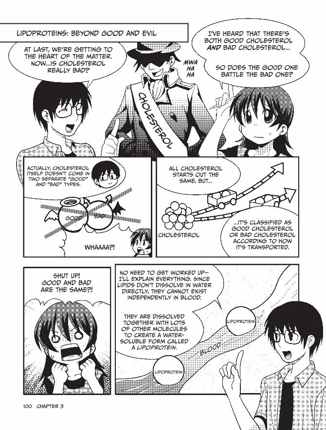
100 chapter 3
Lipoproteins: Beyond Good and evil
At last, we're getting to the heart of the matter.
Now...is cholesterol really bad?
i've heard that there's both good cholesterol and bad cholesterol...
Mwa ha ha
Actually, cholesterol itself doesn't come in two separate "good"
and "bad" types.
Good Bad
Whaaaa?!
All cholesterol starts out the
same, but...
...it's classified as good cholesterol
or bad cholesterol according to how it's transported.
cholesterol
shut up! Good and bad are the same?!
No need to get worked up—i'll explain everything. since lipids don't dissolve in water
directly, they cannot exist independently in blood.
They are dissolved together with lots of other molecules to create a water-
soluble form called a lipoprotein.
lipoprotein
lipoprotein
blood
so does the good one battle the bad one?
cho
leste
rol

Biochemistry in our everyday Lives 101
A lipoprotein is a relatively large
spherical clump that has a neutral lipid or a cholesterol ester*
inside it, neither of which mixes readily
with water.
its surface is covered with amphipathic
phospholipids and a protein called
apoprotein......and free
cholesterol.**
it looks kinda gross to me...
Splish
Splash
cholesterol ester
Phospholipid
Free cholesterol
Apoprotein
Lipoprotein
Lipoproteins can be classified into
several types
Specific gravity refers to the entire
lipoprotein particle in that, if there are more
neutral lipids than proteins, the specific gravity decreases. if there are more
proteins than neutral lipids, the specific gravity increases.
according to specific gravity, as you can see in this figure.
Neutral
lipidProtein
chylomicron (very low-density lipoprotein)
(low-density lipoprotein)
(high-density lipoprotein)
VLDL LDL hDL
Low specific gravity
high specific gravity
• More neutral lipids
• Fewer proteins
• Fewer neutral lipids
• more proteins
Let's focus on LDL and
hDL for now.
i see...
* cholesterol ester is cholesterol with a fatty acid attached to it.
** A free cholesterol is a cholesterol without a fatty acid attached to it.

102 chapter 3
LDL, which carries a lot of
cholesterol, has the role of transporting
cholesterol to the tissues of
the body.
on the other hand, hDL, which carries a small amount
of cholesterol, has the role of transporting
cholesterol from these tissues back
to the liver.Liver Peripheral
tissuecholesterol
cholesterol
blood vessel
Bad
hehe, you can never have enough
cholesterol!
Good
Liver Body tissues
oh no...a battle of good versus evil!
Ack!
i keep things tidy by bringing the excess back to the liver.
so LDL is called "bad" because it distributes cholesterol
throughout the body, and hDL is called "good" because it removes cholesterol from peripheral tissues and brings it back
to the liver.
Good Bad
i finally get it! The cholesterol itself
is all the same.
But it's regarded as "good" or "bad" based on the type
of lipoprotein that's transporting it! Bad
smack
Good

Biochemistry in our everyday Lives 103
What is arteriosclerosis?
Now that you have a better understanding of cholesterol, imagine what would happen if the density of LDL (bad) in the blood increases while the density of HDL (good) decreases.
Hmmm, I guess cholesterol would be transported into the body’s tissues faster than it could be removed, and it would pile up in the blood vessels.
Right. Cholesterol will accumulate on the walls of blood vessels, the lumens of the blood vessels will get narrower, and the flow of blood will be obstructed. This is called arteriosclerosis.
Normal blood vessel
cholesterol gets
deposited.
immune cells are recruited, and the
lumen steadily gets narrower.
Cholesterol can lead to death?! Not to be taken lightly, I guess...
The blood vessels get thicker and harder, and as the symptoms advance, it can lead to sickness or even death.
Let’s look at a typical type of arteriosclerosis called atherosclerosis. Scientists believe the process goes something like this: First, LDL cholesterol is deposited on the inner wall of a damaged blood vessel. This cholesterol is then eaten by phagocytes,* such as macrophages, and bulging cells filled with fat, called foam cells, accumulate.
When this happens, the smooth muscle cells that create the walls of a blood vessel also end up changing significantly, getting harder and thicker.
* A phagocyte is a gluttonous kind of white blood cell that eats just about anything. They’re an essential part of the immune system.

104 chapter 3
Macrophage
LDL
oxidized LDL is eaten by
macrophages.
Blood vessel wall
Foam cells
Foam cells accumulate, smooth muscle cells grow rapidly, and blood vessel
walls get thicker!
Misshapen smooth muscle cells
bulge
Stretch
Wow, that artery is even more clogged than the pipes in my bathtub. But at least some hair in the drain won’t kill me!
Don’t be so sure. The clog in my tub practically has a mind of its own. It’s a real jungle down there!
Whoa, gross! TMI, Nemoto!
Er...sorry. Anyway, you can see some of the illnesses that arteriosclerosis can cause below.
Brain cerebral infarction (stroke) cerebral hemorrhage
heart Angina pectoris Myocardial infarction

Biochemistry in our everyday Lives 105
The important thing is the
balance of hDL and LDL.
in other words, it's best to absorb a
moderate amount of cholesterol—not too much and not
too little.
i wonder if my dad knows that.
A Proper knowledge of
both metabolism and diet is important!
it's okay to eat foods like mayonnaise in moderation.
Who puts mayonnaise on
pancakes anyway?spluuurt
yummy
?mystery
1 is cholesterol really bad?
Cholesterol, which is transported through the blood via lipoproteins, can be both good (HDL) and bad (LDL).
The cholesterol in LDL is transported to the body’s peripheral tissues, and the cholesterol in HDL is transported from peripheral tissues to the liver. The balance of these is very important.
Cholesterol is an important substance that makes hormones, but if too much is absorbed, it can result in arteriosclerosis, which can lead to serious illnesses and even death.
That’s everything!
tsk
tsk

106 chapter 3
2. Biochemistry of Obesity—Why Is Fat Stored?
The next mystery is one that i really worry about!
2. Why do you gain weight if you overeat?
Being significantly overweight is actually
a medical condition called obesity.
To put it simply, to be obese means that an excess of
fat has been stored in your body.
All right! Let's talk about what obesity means biochemically.
Um...
When saccharides, lipids, and proteins
are ingested, energy (ATP) is created
from them.
ingested and expended energy
saccha ride
Protein
Lipid
1 mole of ATP molecules contains
approximately 7.3 kcal of energy.
The total energy that is created from the
saccharides, lipids, and proteins that were eaten* is called the ingested energy.
* More accurately, since some of these particles of food are directly excreted after they are eaten, ingested energy is the total energy that is created from the saccharides, lipids, and proteins that were actually absorbed by the body.

Biochemistry in our everyday Lives 107
so the more you eat, the more
ingested energy increases.
on the other hand, as long as our bodies
are alive, we are always using
energy.
That's true! even when i'm sleeping at night or just
watching TV...
...my body is still consuming the minimum required energy. This consumption is called my basal metabolism,
right?
That's right! And of course if you
exercise, you expend much more energy.
The total energy used is called
expended energy.
rah!
rah!
Urng
ing
ested e
nerg
y
expended e
nerg
y
energy stored as fat
if expended energy is less than ingested
energy...
that leftover energy is stored in the body in the
form of fat.
Whew!
Huff
Puff
Wheeze
i've got to keep active if i want to keep those pounds off.

108 chapter 3
Animals preserve fat
Animals have a mechanism for preserving a fixed level of fat in the body. If they overeat and fat accumulates, that triggers a signal that tells the brain to reduce the amount that is eaten. Conversely, if the amout of fat is reduced, causing the animal to get hungry, it will eat until the fat level returns to its original amount.
Interesting! I guess that instinct must come from animals living in the wild, where life is severe. If it gets too fat, it won’t be able to catch food, and if it gets too skinny, it could starve and die.
That’s right. For example, the protein hormone insulin causes glucose in the blood to be absorbed into muscle or adipose tissue and stored as glycogen or fat. The result is that it lowers the blood sugar level. (Insulin is also used to treat diabetes.)
Blood vessel
= a saccharide (glucose)
Muscle
Fat
high blood sugar level
Dropping blood sugar level
Oh, so “blood sugar level” is just the concentration of glucose in the blood! And if that glucose is absorbed into muscle or fat, the concentration in the blood will drop.
Also, the cell membranes of nerve cells in the hypothalamus (a special part of the brain) contain a protein called an insulin receptor.*
* Insulin receptors actually exist in the cell membranes of many types of cells in the body, not just nerve cells.

Biochemistry in our everyday Lives 109
So insulin regulates the eating behavior and fat level of an animal via the hypothalamus.
insulin
Brainhypothalamus
suppresses hunger
insulin Binds
insulin receptors are embedded in the
cell wall.
The urge to eat is suppressed by the nervous system when insulin binds with the insulin receptors in the hypothalamus.
The experiments scientists have done to prove this may surprise you—for instance, lab mice that were prevented from producing insulin receptors ended up becoming really obese.
Wow! Instructions go to my brain to keep me from eating too much? That’s crazy—it’s like my own body is playing a trick on me!
There’s also a protein called leptin, which is only created in adipose tissue, that can notify the brain of an accumulation of fat via leptin receptors in the hypothalamus, much like insulin receptors. This also suppresses hunger.
I see. So both insulin and leptin are important proteins for controlling how much we want to eat.

110 chapter 3
That’s right. And scientists have done experiments with leptin too—mice with deformed leptin genes end up just as tubby as the mice without insulin receptors.
That’s awful! I wonder why scientists are always picking on mice, anyway...
If we compare the leptin concentration in the blood of nonobese people versus obese people, they both have a value that is proportional to the amount of fat they carry.
The amount of leptin created by people who overeat is no different than that of people who eat normally. Although the creation of leptin is linked with the suppression of eating in nonobese people, this is not the case in obese people. In other words, obese people are thought to have a resistance to leptin.
Getting
pretty full... NeeD. MORE.
CHIPS!
chips chips
Leptin
Leptin working normally
Leptin not working normally
What a nightmare! If our insulin or leptin is ineffective, we’ll eat and eat, and our appetite will never be satisfied...
There are also a number of other substances that help balance our fat levels and appetite, which we’ll talk about next!

Biochemistry in our everyday Lives 111
excess saccharides become fat!
so obesity is the result of a significant
breakdown of the metabolic homeostatic*
balance.
it's a result of fat being excessively accumulated.
in short...
you've eaten too much and stored
too much fat.
Now let's study the biochemical processes
that are involved...
...when fat accumulates!
There are two main ways that fat is
accumulated.
Lipid saccharide
(1) when ingested lipids are directly accumulated
as fat...
and (2) when saccharides
are converted to fat.
Lipidsaccharide
ew
* Homeostasis is the mechanism in which substances are continually exchanged with the outside world to keep an internal environment unchanged.
What
happened?!
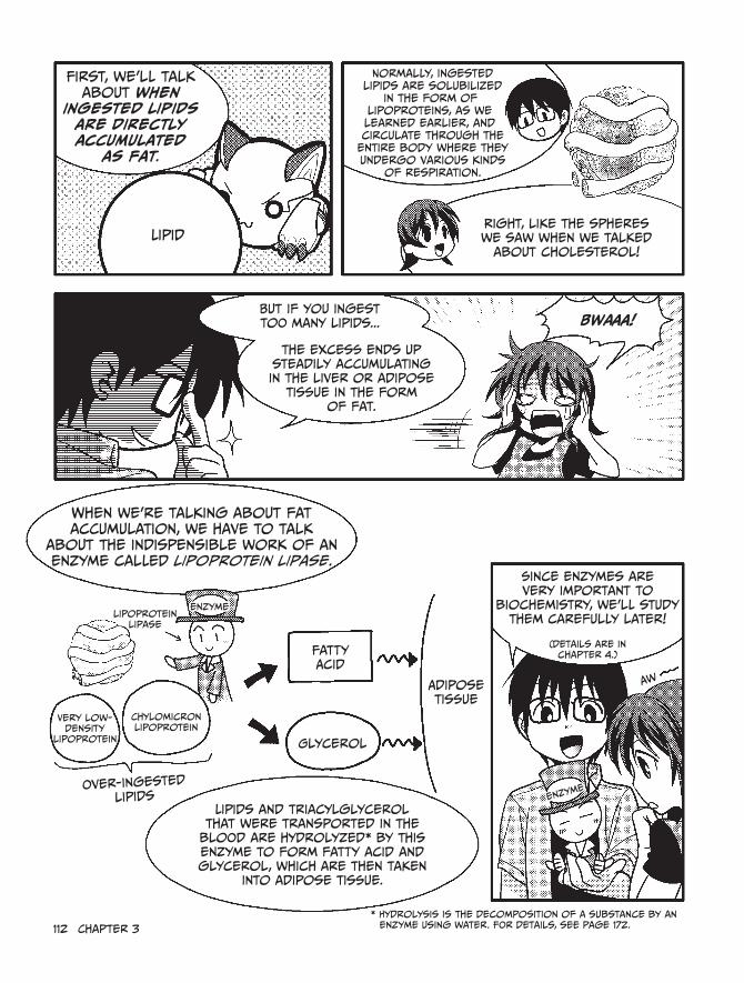
112 chapter 3
First, we'll talk about when
ingested lipids are directly accumulated
as fat.
Lipid
But if you ingest too many lipids...
Normally, ingested lipids are solubilized
in the form of lipoproteins, as we learned earlier, and circulate through the
entire body where they undergo various kinds
of respiration.
right, like the spheres we saw when we talked
about cholesterol!
the excess ends up steadily accumulating in the liver or adipose
tissue in the form of fat.
BWaaA!
When we're talking about fat accumulation, we have to talk
about the indispensible work of an enzyme called lipoprotein lipase.
lipoprotein lipase
Very low-density
lipoprotein
chylomicron lipoprotein
Fatty Acid
Glycerol
Adipose tissue
over-ingested
lipidsLipids and triacylglycerol
that were transported in the blood are hydrolyzed* by this enzyme to form fatty acid and glycerol, which are then taken
into adipose tissue.
since enzymes are very important to
biochemistry, we'll study them carefully later!
(details are in chapter 4.)
Aw
enzyme
* hydrolysis is the decomposition of a substance by an enzyme using water. For details, see page 172.
enzyme

There are four main steps to the process that turns
ingested lipids into fat.
Let's take a look!
Triacylglycerol is transported in the blood by very low-density lipoprotein (VLDL) that was created in the liver and by chylomicron that was absorbed and created in the small intestine.
Endothelial cells, which line blood vessels, have an enzyme called lipoprotein lipase on their surfaces. When triacylglycerol is trans-ported through the capillaries in adipose tissue, muscles, and other tissues, it encounters lipoprotein lipase and is hydrolyzed into fatty acids and glycerol.
liversmall
intestine
VLDL and chylomicron
VLDL chylomicron
triacylglycerol
lipoprotein lipase
fatty acid glycerol
endothelial cell
Blood vessel
fatty acid glycerol
Fat cell
energy source
recombined and stored in triacylglycerol
Break it down!
enzyme
Those fatty acids and glycerol are captured in adipose tissue.
The captured fatty acids and glycerol are recombined and stored in triacylglycerol.
And when this happens, fat steadily increases!
Ooh

ingesting too much triacylglycerol
causes weight gain...
so i won't eat any of it!
No lipids at all! Then i definitely won't get fat,
right?
Scheme!
That's taking things a little too far. remember, your
favorite foods contain lots of saccharides too, not just lipids.
oh yeah, fat is also made from saccharides!
if you eat too much ramen or
spaghetti, you'll be ingesting too many saccharides, and
you'll gain weight. There's really no shortcut here. A little
balance and moderation is all you need!
in other words, if we ingest too
many saccharides...
...our bodies convert them into fat and store that
fat in adipose tissue or in the liver.
hmm, you must be right. This magazine says i should watch
my carb intake.
lipid
saccharide
Metabolism
convert into fat
Directly accumulates
Fat
excess saccharides
Turn into fat!
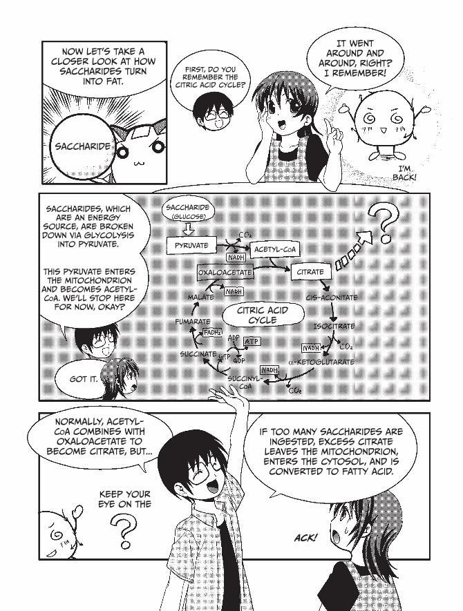
Now let's take a closer look at how
saccharides turn into fat.
saccharide
First, do you remember the
citric acid cycle?
it went around and
around, right? i remember!
i'm back!
saccharides, which are an energy
source, are broken down via glycolysis
into pyruvate.
This pyruvate enters the mitochondrion
and becomes acetyl-coA. We'll stop here
for now, okay?
saccharide(glucose)
PyruvateAcetyl-coA
citrateoxaloacetate
malate cis-Aconitate
Fumarateisocitrate
succinate
citric acid cycle
Got it. succinyl-coA
α-ketoglutarate
Normally, acetyl-coA combines with oxaloacetate to
become citrate, but...
if too many saccharides are ingested, excess citrate
leaves the mitochondrion, enters the cytosol, and is converted to fatty acid.
Ack!
Keep your eye on the

116 chapter 3
When too many saccharides are
ingested, citrate exits into the cytoplasm,
is broken back down into acetyl-coA, and
becomes the substance called malonyl-CoA.
Palmitic AcidGlucose
cytoplasm
since glycolysis is still processing saccharide, the malonyl-coA, acetyl-coa, and palmitic acid will accumulate, won't they?
Pyruvate
Malonyl-coA
Acetyl-coA
Acetyl-coA
citrate
oxaloacetate
Ooooh
citrate Does not enter the
citric acid cycle!
mitochondria
That's right, and palmitic acid is a fatty acid!
if Malonyl-coA is produced, it ends up being directly
converted into a fatty acid.
Eep!
you should also be careful about how much table sugar (fructose) you eat.
Fructose promotes fatty acid synthesis in the liver and
is one of the contributing factors to obesity.
Woe is me...
Betrayed by
sweet, sweet sugar...

Now let's backtrack a little and enlarge the
area from the last
page. Palmitic Acid is synthesized from malonyl-coA by several enzymatic
reactions. Let's take a look!
Acetyl-coA
Acetyl-AcP Malonyl-coA
Malonyl-AcP
represents the number of carbon atoms
other than in coA and AcP.
Acetoacetyl-AcP
D-3-hydroxybutyryl-AcP
trans-2-butenoyl-AcP
Butyryl-AcP
Palmitoyl-AcP
palmitic acid
These six steps are repeated six times. Two carbon atoms are added to the fatty acid
chain each time.
so when malonyl-coA is formed,* this cycle keeps going until fatty acid is
produced.
soon, i'll be fatty acid!
* The synthesis of malonyl-coA is the rate-limiting reaction in fatty acid synthesis. in other words, the formation of new fatty acids depends on how quickly malonyl-coA is formed from acetyl-coA.

118 chapter 3
When fat is used as an energy source
Okay, now I understand how fat is produced, but what I really want to know is how to get rid of the fat I’m producing.
To lose weight, you just have to steadily use up your stored fat.To do this, you need to remember one important thing: When both saccharides
and lipids are present, the saccharides will be used as an energy source first.If you eat foods containing lots of saccharides and lipids, your blood sugar level
will rise. The saccharides are used first for energy production, and the lipids are stored in adipose tissue.
However, when the saccharides are used up and your blood sugar level decreases, the stored lipids will gradually start getting metabolized. So when your hungry stomach growls, lipids are actively being consumed.
After saccharides are used up... lipids are used.
puffpuff so
hungry...
saccharide blood vessel Now it’s our turn!
Fat
Hmm, I guess that means I’ve got to exercise for quite a while before those pounds start to burn off.

Biochemistry in our everyday Lives 119
Lipase
Fat
triacylglycerol
fatty acid
β-oxidation
Glycerol
hydrolase
Fatty acid is released into the blood and transported to the various organs and muscles of the body, where it undergoes a chemical reaction called β-oxidation (that’s beta oxidation).
Triacylglycerol
adipose tissue
hormone-sensitive lipase
Fatty acid
Fatty acid
glycerol
glycerol
Fatty acid glycerol
endothelial cell
hydrolase
blood vessel
Through blood vessels to organs and muscles
β-0xidation
Now let’s talk about how fat is metabolized.First, the fat in adipose tissue, triacylglycerol, is broken down into fatty acid
and glycerol by an enzyme called hydrolase. Hydrolase is a hormone-sensitive lipase—note that this is different from a lipoprotein lipase, which we talked about on page 112.

120 chapter 3
Um...what in the world is β-oxidation?
Relax, I’m getting to it! Take a look at this diagram.
cytoplasm
Fatty Acid
Acyl-coA
eight molecules of Acetyl-coA
Acyl-carnitine
Palmitic Acid (c 16 )
This process is repeated many times.
Acyl-coA again
Mitochondria There are two fewer carbon
atoms in Acyl-coa.
Acetyl-coA
citrate
citric acid cycle
β-oxidation
Fatty acid becomes acetyl-CoA through β-oxidation. Remember when we talked about acetyl-CoA earlier? (See page 115.)
It’s the stuff that appears in the first step of the citric acid cycle, right?
j
k
l

Biochemistry in our everyday Lives 121
You got it!First, the fatty acid is activated by the attachment of CoA. This fatty acid–CoA
compound is called acyl-CoA. A carnitine is then added to form acyl-carnitine, which is shuttled into the cell. It then enters the mitochondrion and is broken down into acetyl-CoA.
Fatty acids are long carbon chains (10+), but acetyl-CoA has only two carbon atoms.
The fatty acid is broken down by β-oxidation so that one molecule of acetyl-CoA (two carbon atoms) is detached each time. The process is called β-oxidation because the CoA is attached to the second-to-last carbon atom of the fatty acid (the β carbon).
Ultimately, all carbon atoms in the fatty acid will become acetyl-CoA. The following figure shows the β-oxidation of palmitic acid, which has 16 carbon atoms.
Palmitic acid with 16 carbon atoms
16
14
12
10
8
6
4
Acetyl-coA with two carbon atoms
2

122 chapter 3
The β-oxidation cycle is repeated seven times to make eight molecules of acetyl-CoA!
Right, and since they’re already in the mitochondrion, they can directly enter the citric acid cycle there to create ATP.
So when you’re burning fat and dieting, this crazy process is breaking down fatty acids inside your body. Wow!
Earlier, I told you how palmitic acid is produced from malonyl-CoA during fatty acid synthesis. When this palmitic acid is broken down, a total of 129 molecules of ATP are produced through the TCA cycle and electron transport chain.
Wait...if I remember correctly, weren’t 38 molecules of ATP the most that could be created from 1 molecule of glucose? 129 is an awful lot, isn’t it?
Glucose (saccharide)
Palmitic acid (fatty acid)
Yup, that’s true. Fatty acid is a very efficient storage material.
Hmmm. That must be why it takes so much energy to burn it away! Biochemically speaking, dieting is a lot of work...

Biochemistry in our everyday Lives 123
?mystery
2 Why do you gain weight if you overeat?
When expended energy is less than ingested energy, the body stores the excess ingested energy as fat.
There are two main ways that fat is formed. One is the process in which ingested lipids (that is, triacylglycerol) are directly accumulated as fat. The other converts saccharides into fat.
Fat is an efficient energy storage material.
Fatty acid is great at storing
energy.
it’s incredible, really. it keeps
us alive!
Fat
But it can be awfully difficult to use up all the extra fat you’ve
stored...
Puff
Wheeze
For a wild animal trying not to starve, fat is really important. But for modern people who worry about
their weight, it’s a little more complicated than that...
sigh
That’s everything!

124 chapter 3
3. What Is Blood Type?
The third mystery is: What is blood type? This should be fun!
Even if we totally avoid the science of blood types, they can still be an interesting way to classify people. For example, in Japan it’s common to tell someone’s fortune based on their blood type.
That’s right! A Type A person is often said to be calm, composed, and serious.
Incidentally, I am an unconventional, do-it-my-own-way Type B!
My father is a power-hungry Type O, and my mother is a sensitive and moody Type AB.
Hmm, that makes sense. Children of parents with Type O and Type AB should be either Type A or Type B but not Type O or Type AB. So if you were to have a sibling, he or she would be Type A or Type B.
If I had a younger brother who was Type A, my family would have people of all four blood types.
But it’s a little strange, isn’t it? Members of one family can have different blood types, but people from completely different families can have the same blood type!
What exactly is blood type, anyway?
Blood Type
Dad
Type oKumi
Mom
sibling?!
Type AB
Type B
Type A

Biochemistry in our everyday Lives 125
3. What Is Blood Type?
how is blood type determined?
Kumi, you’ve probably learned about red blood cells in school, right?
Yeah! They’re the cells that give blood its color. They look something like this:
You got it. Blood type is determined by the type of saccharide molecules that protrude from the surface of red blood cells. The surface of many cells, including red blood cells, is covered by a sugar coating (glycocalyx) made up of saccharides.
Jeez, we’re back to saccharides again? Though I do like the sound of this “sugar coating”...
Remember the lipid bilayer of the cell membrane? Proteins are embedded in various spots in that bilayer, and saccharide molecules are often attached to their outer surfaces. Collectively, these saccharide molecules make up the cell’s sugar coating.
saccharide
protein
surface of red blood cell

126 chapter 3
That’s right. There are actually over 100 different antigens that can coat red blood cells and be used to classify blood types, so many different blood group systems are possible. The most famous is the ABO blood group system, which was discovered in 1900 by the Austrian immunologist Karl Landsteiner.
That’s the system that we use now, right? With Type A, Type B, Type AB, and Type O?
Right again! The ABO blood group system is based on three types of saccharide molecules present on the surface of red blood cells. Each of these has a structure called a sugar chain, which consists of several monosaccharides connected together.
Three types of “anemones” waving around down there, huh?
Three different saccharide
types make up the sugar chain.
sugar chain
sugar coating
red blood cell
How strange! They look almost like little sea anemones or something, reaching out from the bottom of the ocean. And they’re all over our blood cells?

Biochemistry in our everyday Lives 127
If we look at the left side of the diagrams below, we can see that the tip for each type of sugar chain is different.
For the sugar chain of Type A blood, the tip is GalNAc.
GalNAc Gal
Fuc
GlcNAc Protein or lipid
For the sugar chain of Type B blood, the tip is Gal.
Gal Gal
Fuc
GlcNAc
Gal
Fuc
GlcNAc
For the sugar chain of Type 0 blood, there is no tip!
Names of saccharides
GalNAc
Gal
Fuc
GlcNAc
: N-acetylgalactosamine
: Galactose
: Fucose
: N-acetylglucosamine
Oooh, so that’s where those three types come from!
That’s right! And people with Type AB blood have sugar chains of both Type A and Type B.
Wow, that’s all there is to it? Differences in sugar chains? How weird...
Protein or lipid
Protein or lipid

128 chapter 3
But wait...why does one person become Type A while another becomes Type B? What decides which blood type a person will have?
Well, blood type is determined by a certain gene, and an enzyme is created by that gene! (We’ll come back to this later, but you can skip ahead to page 169 for more details, if you’d like.)
Again with the enzymes! You mentioned them when we discussed fat, and now again when we’re talking about blood types? They must be really important.
Definitely. We’ll have to get the professor to tell us more about enzymes when we see her again later.
Yeah! By the way, is it possible that the differences in these sugar chains could actually be related to a person’s personality? Can you really tell a person’s fortune from their blood type?
Hmm, good question. I guess if you went out on a limb, you could say that the gene that determines blood type might also have some kind of influence on nerve cells. But claiming that that might actually influence one’s personality is a pretty big leap of logic, and there’s no scientific evidence to support it.
That’s a relief. I’d hate to think that my entire personality is controlled by a bunch of dinky little sugar chains.
I don’t care what fortune-tellers say—there’s no one in the world with a personality like yours, Kumi...
Biochemistry in our everyday Lives 129
?mystery
3 What is blood type?
The four blood types (A, B, AB, and O) are based on the ABO blood group system.
The ABO blood group system classifies types accord-ing to the differences between certain sugar chains on the surface of red blood cells.
So far, no real evidence has been found to support the idea that the differences in sugar chains affect personality, so don’t let those fortune-tellers boss you around!
That covers it!
still, blood type fortune-telling is
kind of fun...
Let’s see, you’re Type A, right? For your luck this month, it says...
you will have an unexpected chance to get close to the object of your affection.
however, your serious nature will get in the way, and it won’t go as you had hoped. your love life this month will be very stormy!
ha ha ha, good thing there’s no
scientific foundation for that, right?!
Ta-da!
* Fortune-Telling by Blood Type
*
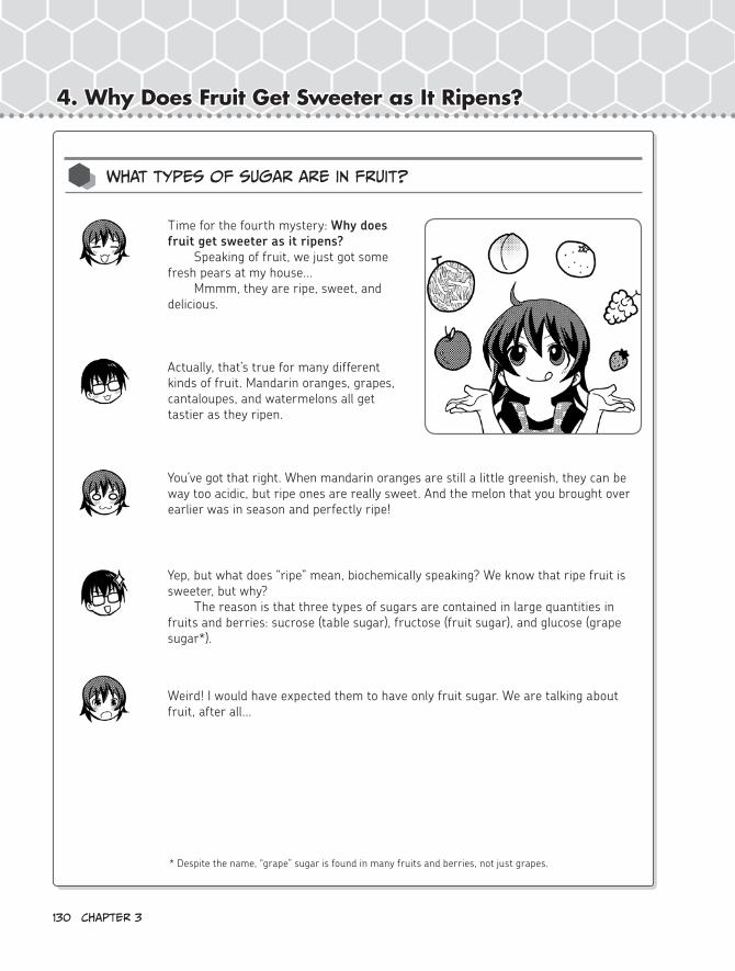
130 chapter 3
4. Why Does Fruit Get Sweeter as It Ripens?
Time for the fourth mystery: Why does fruit get sweeter as it ripens?
Speaking of fruit, we just got some fresh pears at my house...
Mmmm, they are ripe, sweet, and delicious.
Actually, that’s true for many different kinds of fruit. Mandarin oranges, grapes, cantaloupes, and watermelons all get tastier as they ripen.
You’ve got that right. When mandarin oranges are still a little greenish, they can be way too acidic, but ripe ones are really sweet. And the melon that you brought over earlier was in season and perfectly ripe!
Yep, but what does “ripe” mean, biochemically speaking? We know that ripe fruit is sweeter, but why?
The reason is that three types of sugars are contained in large quantities in fruits and berries: sucrose (table sugar), fructose (fruit sugar), and glucose (grape sugar*).
Weird! I would have expected them to have only fruit sugar. We are talking about fruit, after all...
What types of sugar are in fruit?
* Despite the name, “grape” sugar is found in many fruits and berries, not just grapes.

Biochemistry in our everyday Lives 131
4. Why Does Fruit Get Sweeter as It Ripens?
Earlier we said that sucrose, glucose, and fructose all have different structures. (See page 63 for details.)
Right! There are a bunch of different kinds of saccharides, and I love eating all of them.
Well, now it’s time to learn more about those different saccharides!The basic unit of saccharides is called a monosaccharide, which is formed by at
least three carbon atoms connected together.Glucose and fructose are monosaccharides that consist of six carbon atoms. If
two or more monosaccharides are attached, they become an oligosaccharide.Although sucrose is an oligosaccharide, since it’s formed by connecting only two
monosaccharides, it’s called a disaccharide. Here’s what these saccharides look like:
Oh, so sucrose is made up of one glucose and one fructose connected together!
Monosaccharides, oligosaccharides, and polysaccharides
Glucose Fructose sucrose
CH2OH
CH
HO
OHC
C C
CH
H
H
OH
OH
OH
CH2OHO OH
CC
CCOH
OH
HH
H
CH2OH
CH2OH
CH2OH
CH2OH
C
C C
C C
C C
C C
O
OO
H
H
H
HH
H
HOH
OH
OH
OH
HO

132 chapter 3
That's right! There are also saccharides made of many monosaccharides, which form extremely long molecules or complex branching structures. These are called polysaccharides.
Can you think of any polysaccharides that we might be familiar with?
Oh! We talked about potatoes and rice earlier, and they were chock-full of saccharides. So...starch is a polysaccharide, right?
That’s right. Starch consists of many glucose monosaccharides connected together. Plants store glucose in this form after it’s created during photosynthesis.
Glucose
starch
Our bodies (and those of animals) also contain a “storage material” like starch. It’s called glycogen, and it’s produced mainly by the liver or muscles by connecting together excess glucose molecules to store them for later.
Oh yeah, I remember you mentioning glycogen earlier, but now it actually makes sense!
Glucose
glycogen
Other types of polysaccharides include cellulose and chitin. Cellulose is the main component of plant cell walls, and chitin is the main component of the hard shells of crustaceans, like shrimp and crabs. Mushrooms also use chitin as a structural material.

Biochemistry in our everyday Lives 133
Now, let’s get back to fruit. Fruits, such as mandarin oranges and melons, get sweeter and more delicious as they ripen. Why is that?
how fruits become sweet
Hmm. When I was shopping for strawberries and mandarin oranges at the supermarket earlier, there was a sign that said, “Sugar Content: 11–12%.”
I suppose if fruit gets sweeter as it ripens, that just means that the sugar content has increased, right? Biochemically speaking, we must be talking about a change in the saccharides.
You got it. So...let’s talk about saccharides!Take a look at the graphs below. Before they’re ripe, citrus fruits, such as
mandarin oranges, contain roughly an equal amount of glucose, fructose, and sucrose. But as they ripen, the relative amount of sucrose steadily increases. In a Japanese pear, all three types increase.
When fruit ripens, polysaccharides, like starch, are broken down into mono saccharides, like glucose, and the activity of sucrose-phosphate synthase, an enzyme that synthe-sizes sucrose in fruit, increases while the activity of invertase, which breaks down sucrose, decreases.
Saccharides (mg/g fresh weight)
sucrose
Fructose
Glucose
sucrose
Fructose
Glucose
MonthMonth
100 50
Sept
Saccharides (mg/g fresh weight)
Mandarin orange Japanese Pear
Source: Saburo Ito, Editor, Science of Fruit, Asakura Publishing Co., Ltd. (1991)
FebJanDecNovOct SeptAugJulyJune

134 chapter 3
sucrose
Glucose
fructose
fructose 6-phosphate
UDP-glucose
Oh...so sucrose-phosphate synthase is what makes sucrose by combining glucose and fructose .
j invertase (low activity)
k sucrose-phosphate synthase
So does that mean that a fruit gets sweeter as more sucrose is produced?
Well, glucose, fructose, and sucrose are all sweet. Of the three, fructose is the sweetest, followed by sucrose, and then glucose.
Wow!
Fructose 2
sucrose 1.4
Glucose 1
Degrees of sweetness, when the sweetness of glucose
is considered to be 1.
So as fructose or sucrose increases, the fruit gets sweeter and becomes ripe. For example, citrus fruits are best picked in winter, after their sucrose content increases and they’re at their most sweet and delicious. For Japanese pears, on the other hand, the difference in the amounts of these sugars is more striking; as the fruit ripens, the fructose and sucrose both increase suddenly, as polysaccharides are broken down.
Similar to citrus fruits, the sweetness of melons depends mostly on sucrose content, and they’ll be most delicious when sucrose levels are highest.
When you look at the graphs on the previous page, you can see that the fructose or sucrose increases, so the fruit gets really sweet in the harvesting season.

Biochemistry in our everyday Lives 135
?mystery
4 Why does fruit become sweet?
Fruit contain three types of sweet saccharides: sucrose, fructose, and glucose.
As fruit ripens, enzymes become active, changing the amounts of the three saccharides.
Fructose and sucrose are sweeter than glucose, so fruit becomes sweeter as the amount of fructose or sucrose increases!
Mystery sooolved!
Fruit becomes sweeter and more delicious because of fructose and
sucrose.*
fructose
sucrose
it makes perfect sense. More sugar means more
delicious!
But now that i know why fruit is sweet, i’ll enjoy eating it
even more.
Whoa, down girl!
Drooooool
* Additionally, substances such as sorbitol, xylose, and organic acids are also related to the sweetness or acidity of fruit.

136 chapter 3
5. Why Are Mochi Rice Cakes Springy?
The final mystery is: Why are mochi rice cakes springy?
I love mochi, so this is my kind of mystery.
First, let’s talk about how mochi is made. Did you know that mochi is made from special rice, which is springier than nor-mal rice?
Of course I know that! I’ve got serious mochi-making experience. I’ve even participated in a traditional “mochi-pounding” ceremony!
But what makes mochi rice springier than normal rice?
It’s all because of the structural differences of the starches that make up the rice.Rice is 75% starch, so slight variances in its starch composition can seriously
affect its physical properties.As you can see in the figure below, “normal,” non-glutinous rice contains the
two starches amylose and amylopectin. Amylose makes up about 17 to 22% of the starch, and the rest is amylopectin.
However, mochi rice starch only contains amylopectin.
Differences Between Normal rice and Mochi rice
Non-glutinous rice
Mochi riceamylose
Amylopectin
17–22% 100%
composition of the starch contained in rice

Biochemistry in our everyday Lives 137
5. Why Are Mochi Rice Cakes Springy?
Okay, the graph makes perfect sense, but what does that actually mean?
Just hang on, it should become clear in a second.So...amylose and amylopectin are both made up of glucose molecules connected
together to form polysaccharides.
In other words, they’re both made up of the same raw materials!
That’s right. The only real difference is the way that their glucose molecules are connected together.
So the “mystery of springy mochi” is actually the “mystery of monosaccharides connecting in different ways to form polysaccharides and oligosaccharides.” I guess the name’s not quite as catchy, but it’s still a very interesting subject.
Aha! So the secret of mochi’s springiness is hidden in how the glucose connects together!
Glucose
Amylose structure
Glucose
Amylopectin structure

138 chapter 3
So we know that the difference between these two starches is the way in which their glucose molecules are connected. Specifically, the shape of amylose is straight, and the shape of amylopectin is branched.
The difference between Amylose and amylopectin
Amylose is formed when glucose molecules are connected in a straight line via a method called the α(1→4) glycosidic bond.
However, in amylopectin, there are places where glucose molecules are connected via another process called the α(1→6) glycosidic bond.
Straight? Branched? I have no idea what you’re talking about...
Oh, the glucose molecules are lined up in a row. That makes sense.
Amylose
O
O O
OO
O O
α(1→4) glycosidic bond
amylopectin
O
O O O
O
O
O
O O O O O
O O O O
CH2OH CH2OH CH2OH
CH2OHCH2OHCH2OHCH2OH CH2
α(1→6) glycosidic bond

Biochemistry in our everyday Lives 139
You got it, Kumi. These two types of bonds are what give amylopectin its more complex, branched structure and amylose its straightforward, linked structure.
Because of this branched structure, when amylopectin is viewed from a distance, it has a distinct “fringed” shape.
So since this “fringed” kind of starch is more elastic than the straight kind, mochi made with this special rice is springy and delicious!
By the way, even in normal, non-glutinous rice, the stickiness varies according to the amount of amylose it contains.
Oh, I see. That bond connects the molecules vertically! That must be what causes amylopectin to branch off in different directions.
I totally understand! Man, I thought this stuff was supposed to be difficult.
Mochi rice, which is mostly made up of amylopectin, becomes very viscous when cooked because the branches stick together. In other words, it feels very springy and stretchy.
Amyloseamylopectin

140 chapter 3
Since we’ve come this far, let’s take this opportunity to learn what the numbers mean in the two types of bonds we discussed: α(1→4) and α(1→6).
What do the numbers mean iN α(1→4) and α(1→6)?
This may seem a little out of the blue, but...do you like baseball?
Numbering the bases on the field makes the game much easier to understand, right?
Yeah, I was wondering about that, but I was kind of afraid to ask...
Um, I guess so. My dad is a big fan, so I see a lot of games on TV.
It would make the announcer’s life difficult, that’s for sure.
If they weren’t numbered, it would make describing a game a lot trickier. For instance, we’d have to call third base “the base to the catcher’s left.” And I don’t even want to think about how we’d talk about a triple play!
We learned that glucose and fructose have six carbon atoms each, right?Numbers have been assigned to each of those atoms just like the bases in
baseball!
3rd base
2nd base 1st
base
home plate

Biochemistry in our everyday Lives 141
Wait, I think I get it!The numbers in the α(1→4) glycosidic bond and the α(1→6) glycosidic bond
refer to those numbers!
Okay, so the α(1→6) glycosidic bond means that the first carbon of one glucose is attached to the sixth carbon of the next glucose in exactly the same way, right?
Yup, but when the carbon atoms at the first and sixth positions are attached, the glucose molecules can’t be side-by-side in a straight line.
Look carefully at the carbon atoms (C) in the following figure. This is the ring structure of glucose, and the numbers 1 through 6 have been assigned as shown.
Right you are! The α(1→4) glycosidic bond means that the first carbon of one glucose is attached via a glycosidic bond to the fourth carbon of the neighboring glucose.*
CH2OH
HO
OCH
OHC
CC
CH
H
H
H
OH
OH
j
kl
n
o
OO O O
OHOOH
j j
h+ and oh- detach and combine to
form h2o.
α(1→4) glycosidic bond
* The glucose molecules are simplified throughout this section in order to highlight the relevant bonds.

142 chapter 3
Oh! So that’s why a branch happens at that point, and that’s what makes it springy. We’re really getting to the bottom of things! ♪
Beta? What’s the difference?
Beta means no starch! When glucose molecules are connected via β(1→4) glycosidic bonds, the polysaccharide cellulose is formed rather than starch. This is the main component for creating cell walls in plants, and it’s also a type of dietary fiber.
They can connect only as you see in this diagram—vertically, rather than horizontally.
By the way, there’s another type of bond that connects carbons in glucose. It’s called the β(1→4) glycosidic bond.
Oh yeah! I read about dietary fiber in the latest issue of Dieter’s Digest. It’s difficult to digest, so it just passes right through the body. Heh heh.
That’s correct. But dietary fiber also includes substances that easily dissolve in water, such as hemicellulose or pectin, and these are easily digested.
Weird! Are they energy sources for us too?
O
j
O
CH2o
O OO
O O O Oj j j
O
O
Branchα(1→6) glycosidic bond

Biochemistry in our everyday Lives 143
Why not?
Hey, the parts that are connected are different, aren’t they? They have a strange shape, almost like the letter N.
The β(1→4) glycosidic bond differs from the α(1→4) glycosidic bond in that the positions of the hydrogen atom (H) and the hydroxyl group (OH) are flipped around their carbon. This creates a bond that is N-shaped rather than U-shaped, like the α(1→4) glycosidic bond.
They are, but let’s get back to cellulose, okay? There’s an enzyme contained in our saliva called α-amylase, which can break down starches, like rice, into pieces, but α-amylase cannot break down cellulose.
Well, look at the β(1→4) glycosidic bond shown in the following figure.
Yeah, no offense to the β type, but its connection seems twisted and totally weird!
The Greek letters α and β represent the position of the hydroxyl group (OH) on carbon 1. When OH is on the bottom, as shown on the left in the above figure, it is the α type. When it’s at the top, it is the β type.
j j j
O
O
O
O O
O
β(1→4) glycosidic bond
j
O
j
OH
OH
OH
H
α type β type
carbon at position 1
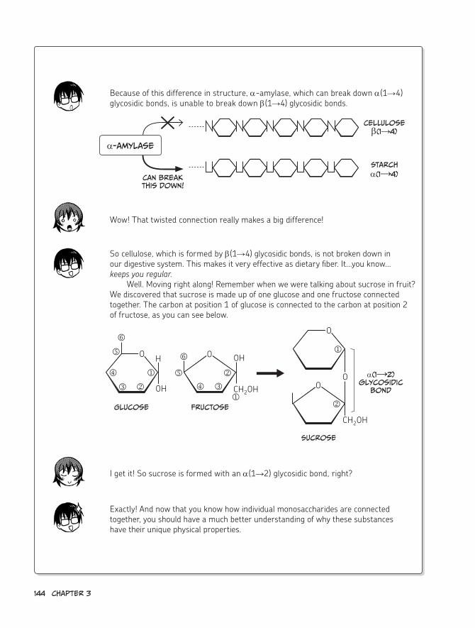
144 chapter 3
Wow! That twisted connection really makes a big difference!
I get it! So sucrose is formed with an α(1→2) glycosidic bond, right?
Exactly! And now that you know how individual monosaccharides are connected together, you should have a much better understanding of why these substances have their unique physical properties.
Because of this difference in structure, α-amylase, which can break down α(1→4) glycosidic bonds, is unable to break down β(1→4) glycosidic bonds.
So cellulose, which is formed by β(1→4) glycosidic bonds, is not broken down in our digestive system. This makes it very effective as dietary fiber. It...you know...keeps you regular.
Well. Moving right along! Remember when we were talking about sucrose in fruit? We discovered that sucrose is made up of one glucose and one fructose connected together. The carbon at position 1 of glucose is connected to the carbon at position 2 of fructose, as you can see below.
α-amylase
can break this down!
cellulose β(1→4)
starch
α(1→4)
Glucose Fructose
sucrose
α(1→2) glycosidic
bondCH2OH
O H
OHj
kl
n
o
O
O
OO
OH
j
l
kn
o
CH2OH
j
k
Biochemistry in our everyday Lives 145
?mystery
5 Why are mochi rice cakes springy?
The secret of why mochi rice cakes are springy is in the structure of the starch in mochi rice.
The starch of mochi rice contains only amylopectin and does not contain any amylose.
Since amylopectin uses a connection method called the α(1→6) glycosidic bond, it is a large, branched polysaccharide. It becomes springy because of this branched structure. ♪
We did it again!
Now we also have a better understanding of the connection methods used in cellulose and sucrose, as well as in
mochi rice cakes.
Plus we understand the
meaning of α(1→4) and α(1→6)!
And now i'll think of delicious mochi
whenever i see baseball on TV.
tee heeMwa ha

146 chapter 3
Well, we solved all the professor's mysteries!
oh yeah!
Wooooo ooooo!
Today was a lot of fun. Plus we got a lot done!
(And we were able to be together...)
My teaching methods may not be as great as the professor's, but—
That's not true!
Today's lesson was easy to understand
and even pretty interesting!
in fact, Nemoto, i...
Guh?

...i thought you were incredible!
SWooN
Well, it was nothing...
really...
okay! Let's write up our report right
away!
melt
thunk
*
* Kurosaka L
abs
i wonder how my little lovebirds are
doing.
Nemoto's so lucky i'm doing this for him! Without my help, they'd
never end up...
together...
FOREVER!Squee!


4enzymes Are the Keys to
chemical reactions

1. Enzymes and Proteins
hmm...wonderful! This report is
really amazing, you guys!
hehe...it’s No big deal.
Don't be modest—you
did great work!
it was all thanks to
you, Nemoto!No, uh, i mean...
Tee hee
Blush
you both seem to have grown a little from
this experience.
ohAh!

Grown a little?
Are you saying i've gotten
fatter?!Kumi...
Uh, well, i guess we
should start the lesson.
you haven't grown at all,
i promise.
oops
The roles of proteins
so far, of the three nutrients,
we've learned about saccharides and lipids.
Protein
saccharide lipid
Today, i'll talk about the remaining one: proteins. you can't really study
biochemistry without knowing some basics about proteins. They're that important!

152 chapter 4
Do you remember when we talked about protein
synthesis in our very first lesson?
yeah!
Proteins have very important roles in keeping our cells
alive.
Protein synthesis
Metabolism
energy production
Photosynthesis
What important roles do proteins play in our bodies?
SQUEAK
SQUEAK
Let's list the major ones.
j Build, repair, and move body tissuesk Organize the form of cells and regulate cell movementl Create and support the structures between cells, such
as collagen Exchange information between the interior and exterior
of cellsn Advance chemical reactionso Protect the body by attacking foreign invadersp Transport substances like oxygen, which is carried
through the blood by hemoglobin
Main roles of Proteins
And these are just the main roles!
Proteins have many other roles, too.
human bodies are thought to contain a minimum of
20,000 types of proteins and possibly as many as 200,000!
Ahh!
seriously? That's a lot of proteins!
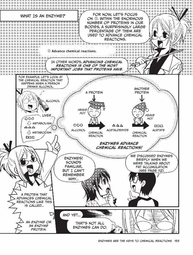
enzymes Are the Keys to chemical reactions 153
What is an enzyme? For now, let's focus on n. Within the enormous number of proteins in our
bodies, a surprisingly large percentage of them are
used to advance chemical reactions.
n Advance chemical reactions.
in other words, advancing chemical reactions is one of the most
important jobs that proteins have.
For example, let's look at the chemical reaction that
happens when a person drinks alcohol.
alcohol
A proteinAnother protein
Heave ho!
Heave ho!
metabolism
metabolismalcohol
chemical reaction
chemical reaction
Acetaldehyde Acetate
Liver
enzymes! sounds familiar,
but i can't remember
why...
We discussed enzymes briefly when we
were talking about fat accumulation
(see page 112).
A protein that advances chemical reactions like this
is called...
An enzyme or an enzyme protein.
And yet...
That's not all enzymes can do.
Enzymes Advance chemical reactions!

154 chapter 4
Bioche
mistry
Let's learn about all the important work
that enzymes do!
We're talking about biochemistry,
after all...
...so substances that advance
chemical reactions must be vital.
enzyme
enzymeenzyme
enzyme
enzyme
Jumping right into the enzyme
discussion might be biting off more than we can chew, so let's start
small and learn about the structure of
proteins.
Urp
yeah, okay!
Proteins are formed from amino acids
Well, Nemoto, this should be a no-brainer for you. Do you know
the origin of the word "protein"?
hmmm...i think so.
it's derived from the Greek word "proteios," which
means "first place" or "leading person."
in other words, you
can tell how important
proteins are just from the name!
right as always, Nemoto. ♪
you're so smart...
Aw, shucks

Proteins are formed by connecting together many relatively small molecules
called amino acids.
hey, i remember those! They look like beads strung
together.
Amino acids
connect
ProteinFold
Up
Amino Acid
one amino acid has two arms
Amino acid
Amino acids are connected like this.
This is called a peptide bond. (For more details, see
page 158.)
Glycine Alanine Valine Leucine
Isoleucine Methionine Proline Phenylalanine
right! Amino acids are
the building blocks of proteins.
Many types of amino
acids exist in the real world, but...
...only 20 types of amino acids have
been determined to be used in proteins.
AsparagineThreonineSerine
Glutamine Tyrosine Cysteine Lysine
Glutamic acidAspartic acidHistidineArginine
Tryptophan
These are the
"beads" from
earlier.
And this is the basic structure of amino acids!
i see.
This part is common to all amino acids.
This part is different for each amino acid.
Amphoteric ion
There are 20 varieties of . remember that— it's important!
since there are 20 varieties of , there
are also 20 types of amino acids.
And...
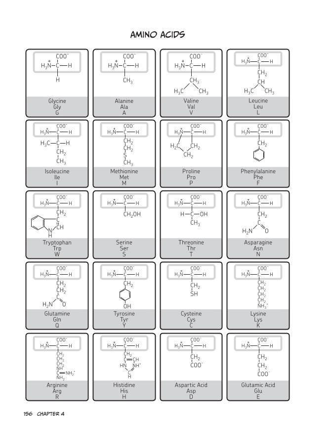
156 chapter 4
Amino Acids
C H
H
H3N+
COO−
GlycineGlyG
C HH3N+
COO−
CH3
AlanineAlaA
C HH3N+
COO−
CH3
CH3H3C
ValineValV
C HH3N+ COO−
CH2
CHCH3H3C
LeucineLeuL
IsoleucineIleI
C HH3N+ COO−
C HH3CCH2
CH3
MethionineMetM
C HH3N+ COO−
CH2CH2
CH3
S
ProlineProP
C HH3N+ COO−
H2C CH2
CH2
PhenylalaninePheF
C HH3N+ COO−
CH2
TryptophanTrpW
C HH3N+ COO−
CCH2
CH
SerineSerS
C HH3N+ COO−
CH2OH
ThreonineThrT
C HH3N+ COO−
C OHHCH3
AsparagineAsnN
N H
C HH3N+ COO−
CH2N
CH2
O
GlutamineGlnQ
C HH3N+ COO−
CH2N
CH2
OTyrosine
TyrY
C HH3N+ COO−
CH2
OHCysteine
CysC
C HH3N+ COO−
SHCH2
LysineLysK
CH2
C HH3N+ COO−
NH3+
CH2
CH2
CH2
CH2
ArginineArgR
C HH3N+ COO−
NH2
CH2
NHCH2
CH2
HistidineHisH
C HH3N+ COO−
NH+
CH2
C H
HNC
Aspartic AcidAspD
C HH3N+ COO−
COO-CH2
Glutamic AcidGluE
C HH3N+ COO−
CH2
C NH2+
CHCH2
COO-

...here are the structural formulas
of those 20 amino acids!
hey, relax! you don't have to
memorize them all right away!
Swoon
The types of proteins that can be
created are determined solely
by these 20 amino acids.
Different orders and
different amounts of
these acids produce
different proteins!
hmm, i guess even with only 20 kinds
of beads, i can make a ton of different
necklaces by combining them in different ways.
Jangle
Jangle
True, but amino acids differ from beads.
For one, you can't just string together a bunch
of amino acids and expect to get a protein.
Whaaa?
For a protein to function, it must be folded into an appropriate shape,
And it has to be folded in the right
order as well.
Let's take a closer look!

158 chapter 4
Primary structure of a Protein
First, a long amino acid chain is formed by connecting amino acids one at a time with peptide bonds. Remember earlier when we said that amino acids are joined together via chemical reactions to form proteins? This chemical reaction, which creates the peptide bonds, is called the peptidyl transfer.
This reaction occurs inside ribosomes, which are protein synthesis apparatuses con-tained in the cytoplasm and attached to the rough endoplasmic reticulum. We'll talk more about ribosomes in Chapter 5.
The long amino acid chain connected together by peptide bonds is called a polypeptide chain. This amino acid string is called the primary structure of the protein.
Amino Acid Polypeptide chain
synthesized amino acid chain
peptide bond
Primary structure
in on the process in which amino acids are
connected by peptide bonds!

enzymes Are the Keys to chemical reactions 159
secondary structure of a Protein
Each of the 20 amino acids has a characteristic part (indicated by [R] on page 155) that is unique. This is called the side chain.
Since a number of difference forces can act on these side chains, such as hydrogen bonds and hydrophobic and electrostatic interactions, neighboring amino acids are attracted to or repelled by each other in particular ways, which results in a characteristic, local three-dimensional structure. This is called the secondary structure of the protein.
There are a number of different secondary structures, such as an α-helix, in which a part of the polypeptide chain forms a spiral, and a β-sheet, which takes a folded planar shape.
α-helix β-sheet
Primary structure secondary structure

160 chapter 4
Tertiary structure of a Protein
Even when the polypeptide chain takes its secondary structure, it’s not yet a fully folded, functional protein.
To become functional, the polypeptide chain has to take on a specific three-dimensional shape, which is determined by the interactions of the amino acid side chains. This shape is called the tertiary structure of the protein.
For example, myoglobin, which is shown in the following figure, is a type of protein that exists in the muscles of animals. This protein is formed from eight α-helices surrounding an iron-containing heme, which binds oxygen.
Tertiary structure
heme
A
B
cD
e
F
G
h
Myoglobin consists of the eight α-helices indicated by the letters A through h.

enzymes Are the Keys to chemical reactions 161
Quaternary structure of a protein and subunits
Many proteins are able to operate as a protein or as an enzyme at the tertiary structure stage. However, some proteins create an aggregation in which multiple polypeptide chains that have taken tertiary structures are assembled together into a large functional unit.
For example, our red blood cells contain many iron-binding proteins called hemoglobin, which are used to transport oxygen. Hemoglobin is formed by assembling four polypep-tide chains called globin (two each of two types, α and β, which are indicated below as α1, α2, β1, and β2). An enzyme such as RNA polymerase II, which creates RNA in our cells, is formed by assembling 12 polypeptide chains.
This state is called the quaternary structure, and each of the polypeptide chains used to create the quaternary structure is called a subunit.
α1
α2
β2
β1
α1 , α2 , β1 ,
and β2 are each "subunits."
since the subunits of hemoglobin are very similar in structure to those in myoglobin, hemoglobin in this figure
has been drawn with the same structure as on page 160. however, the structures are actually somewhat different.
Quaternary structure

162 chapter 4
2. An Enzyme’s Job
substrates and enzymes
At last, it’s time to talk about
enzymes—
The keys to chemical reactions!
OKay!
The first important thing to know is that a particular enzyme can only work with a certain “partner”
material.
enzyme
The enzyme’s “partner”
For example, Pepsin, which is a digestive enzyme that breaks down
proteins in the stomach...
Protein
...will only break down protein and will never
break down DNA.
The digestive enzyme
α-amylase, which is contained in
saliva...
...will only break down starch and will not
break down fat.
starch
Fat
enzyme substrateenzyme-substrate
complexreaction product
enzymeanother
substancecomplex cannot
be formed
reaction does not occur
The “partner substance” that an enzyme works with is called the substrate.
The fact that the substrate is determined by the enzyme is
called substrate specificity.

For the α-amylase in our saliva...
...the substrate is starch, and the reaction
product (that is, the material produced when starch is broken down by α-amylase) is a type
of saccharide.
Depending on the number of glucose
molecules it contains, this saccharide is
known as
“maltose,” “maltotriose,” “limit dextrin,”
or a number of other names.
it looks something like this:
α-amylase cuts up
starch into pieces?
scissors!
enzymeenzyme
α-amylase
snip snip
Finished!
enzymeit snips the starch like a pair of scissors!
α-amylase starch
enzyme-substrate complex
enzyme
enzyme
enzyme
enzyme
enzyme
Reaction Products: maltose,
maltotriose, limit dextrin, and so on...
For some enzymes, the substrates are
determined very strictly.
others have broader substrate specificities, so they’re more laid
back about the materials they’ll interact with.
strict enzyme
relaxed enzyme
My way or the highway.
Whatever, man!
Let’s take a closer look
at these two types.

164 chapter 4
strict enzyme? relaxed enzyme?
Some enzymes are “strict,” which means they only act on very specific substrates. Other enzymes are more “relaxed” and act on a broad range of substrates.
Strict and relaxed? Sounds like the difference between my mom and my dad...
There are enzymes that can act on substances that are similar or closely related to their substrates. Many examples of these enzymes are seen in the digestive system—for example, the protein catabolism enzymes.
Remember, there are a huge number of different proteins. If enzymes were too specific, there would have to be a separate enzyme to break down every single kind of protein! Things would get pretty unwieldy.
Since proteins are so complex, the enzymes that break them down had to become a bit more flexible.
That’s right! Protein catabolism enzymes (the enzymes that break down proteins) often have a certain degree of leeway in the substrates they can interact with.
only glycine only alanine
and histidine
Strict!Strict!

enzymes Are the Keys to chemical reactions 165
For example, one of the protein catabolism enzymes secreted from the pancreas, carboxypeptidase, detaches amino acids sequentially from the end of a protein.
Carboxypeptidase comes in various types, including A, B, C, and Y. Carboxypepti-dase A, for example, can detach almost any amino acid from the C-terminal end of a protein, but it doesn’t work well on amino acids with bulky or aromatic R-groups, like arginine, lysine, and proline.
Proline Arginine Lysine
N-terminal
c-terminal side of proline
c-terminal side of Arginine
c-terminal side of lysine
c-terminal
carboxy-peptidase A
i’ll detach anything other than arginine,
lysine, or proline.
Relaaaaxed
There are 20 types of amino acids that make up proteins, right? So even though carboxypeptidase A can’t deal with arginine, lysine, and proline, there are still 17 types it can deal with. It seems pretty flexible!
That’s right. It has some serious leeway in the substrates it can interact with.However, there are also some “strict” protein catabolism enzymes. For example,
trypsin cuts only through the C-terminal side of arginine and lysine.
Arginine Lysine
Trypsin
c-terminal side of Arginine
c-terminal side of lysine
Strict!

enzyme classifications
Although our bodies contain a huge variety of enzymes,
they can be divided into a smaller number of general
groups based on shared characteristics.
enzyme
enzyme
enzymeenzyme
Mmm...just like dividing foods into groups
based on their characteristics!
Meats?
Veggies?starches?
Desserts!
enzymes are classified by their different types of catalytic reactions.
They are divided into six major
groups.
each enzyme is assigned a number called the EC Number (Enzyme Commission
Number),* which looks like this:
ec a.b.c.d (where a, b, c, and d are numbers)
a is 1 to 6 since it represents six major groups.
b and c represent the reaction patterns of each group in more detail.
d is an enzyme-specific number.Fill in the
numbers here.
By the way, ec numbers
are the same worldwide.
Group Number (1 to 6)
reaction patterns in more detail
characteristic number
That’s not so hard!
* A newly discovered enzyme is assigned an ec number and system name as well as a recommended name according to standards determined by the enzyme commission of the international Union of Biochemistry and Molecular Biology (iUBMB).

enzymes Are the Keys to chemical reactions 167
Now for the six groups!
Catalytic Reaction Types of Enzymes
EC 1. OxidoreductasesEC 2. TransferasesEC 3. HydrolasesEC 4. LyasesEC 5. IsomerasesEC 6. Ligases
Today, we’ll introduce ec 2
(Transferases) and ec 3 (hydrolases) as representatives of these catalytic
reaction types.
Alright, let’s have them make their
entrance! Wha?
Today’s guests are...
Transferase and Hydrolase! Ta -da a a a a h !
Transferasehydrolase
functional Group
Transferase hydrolase
Now let’s find out what kind of enzymes these
groups contain and what jobs they do.

Transferases
An enzyme for transferring a part of one molecule to another is called a
transferase.
Transferase
Transferase
Transferase is the generic name for enzymes that
transfer a specific clump of atoms (functional group)
to a chemical compound other than water.
represented by EC 2.X.X.X
Special delivery!
Functional Group
For example, this thymidylate synthase
(ec 2.1.1.45) is a transferase:
5,10-Methylene tetrahydrofolate
TransferaseUracil
Thymine
Methyl group
-ch3Important!
The methyl group (-ch3) that was taken from a substance
called 5,10-methylene tetrahydrofolate is attached to uracil,
changing it into thymine.
Uracil and thymine are ingredients of nucleic
acid, but we’ll get to those later (see
chapter 5).
i see...

enzymes Are the Keys to chemical reactions 169
Glucosyltransferase determines blood type
Remember when we solved the blood type mystery (in Chapter 3)?When we were trying to figure out what determines blood type, we discovered
that it was the work of an enzyme.
I remember!The ABO blood group system is classified according to the differences between
the “sugar chains” on the surface of red blood cells. A sugar chain is a collection of monosaccharides connected together!
And the differences between those chains are the monosaccharides at the tips, right?
Yeah! For people with Type A blood, that monosaccharide is N-acetylgalactosamine (GalNAc); for people with Type B blood, it’s galactose (Gal); and for people with Type O blood, there is no monosaccharide at the tip.
(After writing such a detailed report for the professor, I’ll probably remember that for the rest of my life...)
That’s correct. The particular monosaccharide that’s attached to the tip of a sugar chain (or the lack of one) is determined by a certain gene.
Genes are like the “blueprints” for proteins. (Remember: An enzyme is a type of protein.)
So what really determines blood type is the gene that creates glycosyltransferase, which is the enzyme that attaches a particular monosaccharide to the tip of a sugar chain found on the surface of the red blood cell.
So glycosyltransferase attaches a certain monosaccharide, and the type of that monosaccharide determines blood type?
That’s right. Look again at the structure of the sugar chain of each blood type.

170 chapter 4
For the sugar chain of people with Type A blood, the tip is GalNAc.
GalNAc Gal
Fuc
GlcNAc Protein or lipid
For the sugar chain of people with Type B blood, the tip is Gal.
Gal Gal
Fuc
GlcNAc
Gal
Fuc
GlcNAc
For the sugar chain of people with Type 0 blood, there is no tip.
monosaccharide Names
GalNAc
Gal
Fuc
GlcNAc
: N-acetylgalactosamine
: Galactose
: Fucose
: N-acetylglucosamine
Let’s review it again. The only difference in these three types is the monosaccharide at the very tip.
For people with Type A blood, it’s N-acetylgalactosamine. For people with Type B blood, it’s galactose. For people with Type O blood, it’s nothing!
Yeah, yeah, I’ve got it.
So, the sugar chain that Type O people have is the “prototype,” or the minimal sugar chain. People with A and B types have the O type chain plus something extra.
If someone has the transferase gene that attaches N-acetylgalactosamine, his or her blood type will be Type A! But if someone has the transferase gene that attaches galactose, his or her blood type will be Type B!
Protein or lipid
Protein or lipid

enzymes Are the Keys to chemical reactions 171
Type o
Type BType A
Transferase
That all makes sense, but I still don’t quite understand what’s going on with people with Type O blood. Why don’t they get monosaccharides on the end of their sugar chains? Seems unfair!
Type O people also have the genes associated with glycosyltransferases, but since there is no enzyme activity for the proteins created by those genes, no saccharide is attached at the tips. The activity may have been lost because of a genetic mutation during the evolutionary process.
A mutation? Cool!
So to wrap it all up: The ABO blood group system is nothing but the result of differences in glycosyltransferase genes.
Nemoto, you must have a genetic mutation that gives you an oversized brain. How else could you possibly know so much?!
Gyuh...
Transferase
Transferase

172 chapter 4
hydrolases
As the name suggests, a
hydrolase is an enzyme that uses
water to break down its substrate.
hydrolases are represented by
EC 3.X.X.X.
remember, a water molecule is made up of two hydrogens and one
oxygen: H2 O.
A hydrolase splits this into H and OH.
hydrolase
it then inserts these into a substrate to break it down into two parts!
hydrolase Break it up, guys!
it’s like breaking up a fight!
or separating two people in
love...
if you think of the parts of a substrate as
two people holding hands, hydrolase separates them by making one hold h and
the other hold oh.
hydrolase is what breaks down the common currency
of energy (ATP)!
α-amylase, which breaks down starch, and pepsin,
which breaks down protein...
...are both hydrolases!
Prote
in
starch
Now, let’s examine the work of α-amylase (ec 3.2.1.1)!
The α-amylase in our saliva breaks down starches,
such as rice.

Glucoseα(1→4) glycosidic bond
Glucose Glucose Glucose
starch is composed of glucose units
connected together in a form called a α(1→4)
glycosidic bond.
starch is divided into pieces by α-amylase. The α(1→4) glycosidic bonds are randomly cut via a method called hydrolysis!
α(1→4) glycosidic bond α-amylase
starch
it’s chopped into pieces to make saccharides of
different lengths!
randomly cuts
α(1→4) glycosidic bonds
This is hydrolysis.
Now let’s look closer at the chemical reaction
of hydrolysis.
you can see here that the hydrolase α-amylase uses one
water molecule to break a bond in
the starch.
My spit is powerful
stuff...
α-amylase
α(1→4) glycosidic bondBroken apart using
h and oh
We talked about glycosidic bonds when we learned about mochi rice cakes, remember?
(see page 140.)

174 chapter 4
3. Using Graphs to Understand Enzymes
Next we’re going to use graphs to help us do some
basic calculations.
i hope everybody’s as excited as i am!
Nooooo! Graphs?!
calculations?! i can’t do it! i’m doomed.
i was afraid of this...
Don’t worry, Kumi. it’s not as hard as you think.
Graphing Data is vital to the study of biochemistry, as well as to most other
types of science.Measuring the reaction of an
enzyme and graphing its numerical values makes it possible to
understand the reaction in new ways and advance research.
The graphs you hate so much might help us cure
cancer one day!
That does sound important, but it doesn’t mean i have to like it!
Graphs are useful to track
weight loss too, you know...
Pssst
yeah! i’ll give it my best shot!
That’s the spirit!

enzymes Are the Keys to chemical reactions 175
Why are enzymes important for chemical reactions?
A substance that expedites a chemical reaction is called a catalyst. An enzyme, which is a type of catalyst, is also known as a biological catalyst.
Although an enzyme is required to make a chemical reaction advance efficiently, it is not neccesarily required for the chemical reaction to occur.
Most chemical reactions in the body will occur eventually even without enzymes, but many would take an overwhelmingly long time. Complex chemical reactions, like glycolysis, may never reach completion without a catalyst.
Enzymes are important because, for an organism that must maintain specific condi-tions and produce energy to stay alive, the chemical reactions that occur inside its body must be as efficient as possible. For living organisms as a whole, it would be disastrous if reactions took too long.
if there were no enzymes...
There would be no life as we know it!
But because enzymes exist...
everyone’s alive and happy!
Now we’ll use some simple graphs and formulas to study the essential qualities of chemical reactions that rely on enzymes, and we’ll learn why enzymes are so meaningful to those reactions.

176 chapter 4
What is Activation energy?
A fixed amount of energy is required for a chemical reaction to proceed smoothly. This
is called activation energy.
Take a look at this graph, which shows the progress of an individual chemical reaction.
Am
ount o
f e
nerg
y
reaction Progress
Activation energy
: substrate
: reaction product
here, chemical substrate, substrate A,
is transformed via a chemical reaction to produce reaction
product B.
For the reaction to produce B from A, the activation energy has to be added to the mix.
since A and B are different substances, they have different amounts of energy.
The activation energy does not affect the
difference between the energy values of A and B.
Am
ount o
f
energ
y
Look at the energy values of A and B!
it’s almost like the substrate has to climb over a really high wall to escape imprisonment and transform into the
reaction product.
climb a wall? scary...
Uh...
The highest part of this wall is called the activation barrier or
energy barrier.
K-shunck
yikes!

enzymes bring down the “wall”
so what advantage do you suppose
there is...
For an enzyme to participate in this kind of chemical
reaction? enzyme to the rescue!
i guess it’s like... an enzyme lowers
the wall to make the climb easier.
Going
down!
on a basic level, that’s exactly right! For example, let’s imagine
you’re a substrate, Kumi.
To reach your destination and transform into a reaction product, you need to leap over
an intimidating six-foot wall, But that’s just too high!
Then along comes Mr. enzyme, who smashes the wall down
to size. Now the six-foot wall is a two-foot wall, and you hop over without a care
in the world. Get it?
No problemo!
That enzyme is such a
sweetheart!
reaction progress
Activation barrier
Am
ount o
f e
nerg
y
reaction Progress
high
Low
No enzyme
With enzyme
This graph summarizes things nicely. As you
can see, if the enzyme is present, the chemical reaction occurs more
easily, because the activation energy is
lowered.
There are also chemical reactions in which the enzyme
changes the mechanism of the reaction.
...we will assume that the presence of the
enzyme only lowers the activation energy
of the chemical reaction.
i get the job done!
But for the purpose of this discussion...

178 chapter 4
Maximum reaction rateThe ability of an enzyme to
act on the substrate to create the reaction product
is called enzyme activity.
The speed of this reaction is called the reaction rate.
enzyme power!
substrate
reaction product
enzyme activity
Measuring this “activity” is vital for checking the properties of an
enzyme.
And taking these measurements can be
a little bit tricky. oh no...tricky?!
relax, you’ll be fine as long as you’re careful. There are
two key concepts here:
u Maximum reaction ratev Michaelis constant
The maximum reaction rate is the speed of the reaction when each of the enzymes in the reaction
solution is combined with a substrate. in other words, it’s
the reaction’s speed when all of the enzymes are working.
This is represented by Vmax.*
Wha-what?
I’m completely lost...
* The V stands for “velocity,” which is just another word for “rate.”

What exactly do you mean when you say “each of the enzymes is
combined with a substrate”?
okay, imagine a place where
everyone sweeps up fallen leaves.
if additional brooms arrive at a place where there are
not enough brooms and some people are standing
around bored...
...Leaf cleaning will speed up, since those people will also be able to help out, right?
Broom
But if everyone is already sweeping with a broom, leaf cleaning would not speed
up no matter how many new brooms arrive.
Do you see the connection?
i think so.
Wham
if some new substrate is added when there are enzymes just
hanging around doing nothing, the entire reaction will speed up because those enzyme molecules
will begin to work.
however, if all of the enzymes are already working, the
reaction will not speed up even if there is new substrate added.
enzyme
substrate
so if you think of the brooms as the substrate and the people as the enzyme, then you can see what we mean by the substrate concentration. here, substrate concentration is the x-axis, and the number of
enzymes is fixed (doesn’t change).
you can see the Vmax line, which is the fastest rate at which the reaction can take place.
Notice that when the rate reaches this line, the substrate concentration can get higher and higher without affecting the reaction rate.
Will not increase above this
Maximum reaction rate Vmax
reactio
n r
ate (V)
substrate concentration (S)

180 chapter 4
The Michaelis-Menten equation and the Michaelis constant
in 1913, two biochemists, Leonor Michaelis and Maud Menten, proposed a basic equation that represents the
relationship between the reaction rate and the substrate concentration.
Named after these two scientists, the equation is called the Michaelis-Menten equation.
ohhh...i think i’m getting a migraine.
calm down, Kumi.
Yeow
Now comes the important part! in deriving this complex equation,
Michaelis defined a numeric value called the Michaelis constant (Km ), which is the substrate concentration when the initial
rate* of the reaction is half of Vmax.
* The initial rate is the rate at the beginning of the reaction, when the substrate concentration and reaction speed are still linearly related.
reactio
n r
ate (V)
substrate concentration (S)
IMPORTANT!
: reaction rate
: substrate concentration before enzyme is added

enzymes Are the Keys to chemical reactions 181
Km is extremely important in checking the
properties of an enzyme.
since this value is unique to each enzyme, you can measure it to determine how that enzyme
works relative to the substrate.
This is called the enzyme’s affinity for its substrate.
the higher the affinity, The smaller the value of Km.
if the Km value is small, that means that the reaction rate reaches half its maximum at a
lower substrate concentration, which shows us that the enzyme is
doing its job efficiently.
Km value of enzyme A
enzyme A with high affinity
Km value of enzyme B
enzyme B with low affinity
substrate concentration (S)
i see...it makes sense when we compare
the line for enzyme A with the line for B!
The reaction speed for A rises way faster than B.
That’s right, since enzyme A has a higher affinity.
reactio
n r
ate (V)

Let’s calculate Vmax and Km!
Now, let’s try to figure out the Vmax and Km values of a particular enzyme!
We’ll use DNA polymerase, which is an enzyme for
synthesizing DNA, as an example.
i hope i don’t screw this up...
Nucleotides, the building blocks of DNA, will be the substrate in this example.
Let's say we add DNA polymerase to six different solutions
with the following substrate concentrations:*
Then we let the reaction run for 60 minutes at a temperature of 37°c.
Let’s assume this experiment produced the following measurements:
These results show the concentration of reaction product formed over the
course of an hour, so we can divide these values by 1 hour to turn them into reaction rates.**
substrate concentration
0 µM1 µM2 µM4 µM10 µM20 µM
substrate concentration
producedreaction product
0 µM → 0 pmol
1 µM → 9 pmol
2 µM → 15 pmol
4 µM → 22 pmol
10 µM → 35 pmol
20 µM → 43 pmol
Now, let's graph these results!
We'll let the x-axis (horizontal axis) be the substrate concentration (µM) and the y-axis be the
reaction rate (pMol).
reactio
n r
ate
(pM
ol/h
our)
substrate concentration (µM)
When the substrate concentration is 0 µM, the measured result is
0 pmol, so...
oookay!
* Actually, we’d also need to add template DNA, magnesium ions, and other elements, but let's keep things simple!
** For example, if we start with a substrate concentration of 1 µM, our reaction rate is 9 pmol per 1 hour, or, simply, 9 pmol/hour.

is this right?reactio
n r
ate
(pm
ol/h
our)
substrate concentration (µM)
* it's rare to get a smooth curve like this from real-world measurements. This is just the ideal shape.
you got it. Nice curve, Kumi!*
Although we'll use this data to figure out Vmax and Km for
DNA polymerase...
we can't actually determine those from this curve!
WHAT?!
To get the values we need, we have to create a graph called a
Lineweaver-Burk reciprocal plot.
Line...weaver...
what?
Those are just the names of the guys who came
up with it.
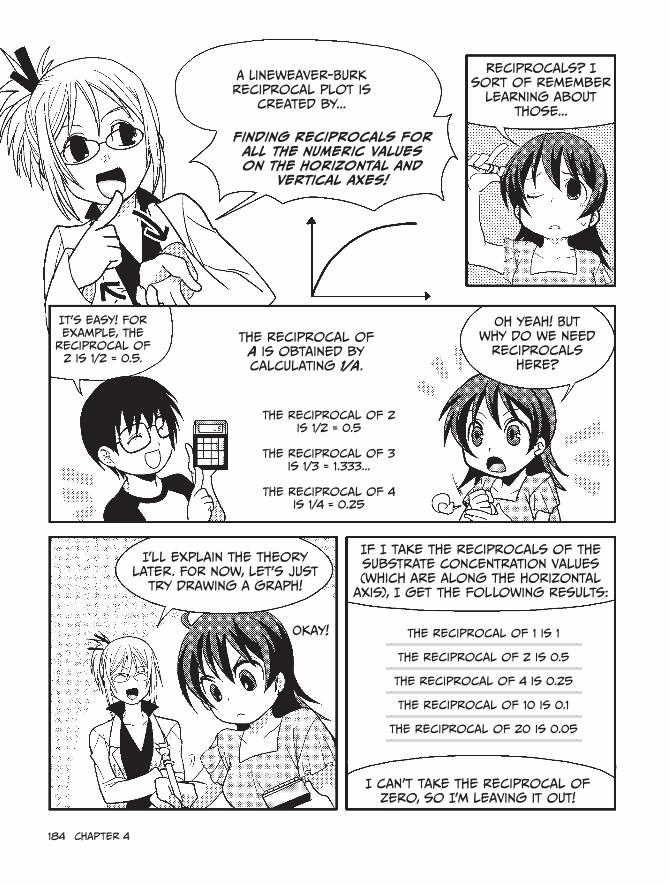
184 chapter 4
A Lineweaver-Burk reciprocal plot is
created by...
Finding reciprocals for all the numeric values on the horizontal and
vertical axes!
reciprocals? i sort of remember
learning about those...
it's easy! For example, the
reciprocal of 2 is 1/2 = 0.5.
The reciprocal of a is obtained by calculating 1/a.
oh yeah! But why do we need
reciprocals here?
i'll explain the theory later. For now, let's just
try drawing a graph!
if i take the reciprocals of the substrate concentration values (which are along the horizontal
axis), i get the following results:
The reciprocal of 2 is 1/2 = 0.5
The reciprocal of 3
is 1/3 = 1.333...
The reciprocal of 4 is 1/4 = 0.25
okay!
i can't take the reciprocal of zero, so i'm leaving it out!
The reciprocal of 1 is 1
The reciprocal of 2 is 0.5
The reciprocal of 4 is 0.25
The reciprocal of 10 is 0.1
The reciprocal of 20 is 0.05

Now take the reciprocals of the
vertical axis results and plot those
values.Good thing i brought my calculator
today!
The values on the vertical axis (other than zero) were 9, 15, 22, 35, and 43, so:
The reciprocal of 9 is 1/9, and 1 ÷ 9 = 0.111. The reciprocal of 15 is 1/15,
and 1 ÷ 15 = 0.066.
Now let me just work through the rest...
The reciprocal of 9 is approximately 0.111
The reciprocal of 15 is approximately 0.066
The reciprocal of 22 is approximately 0.045
The reciprocal of 35 is approximately 0.028
The reciprocal of 43 is approximately 0.023
All Done! ...huh?!
recip
ro
cal o
f
reactio
n r
ate (pM
ol/h
our)
reciprocal of substrate concentration (µM)
how strange... it's a straight line!
Great job!
Now i'll explain the theory behind this, so
you can understand why we take reciprocals.

186 chapter 4
Why do we take reciprocals?
Yes, why do we take reciprocals? It’s a mystery, isn’t it? Heh heh heh...
Don’t look at me! Math isn’t really my thing.
Don’t worry. We’ll solve the mystery together. ♪First, focus on Vmax. In the following graph, you can see that as the substrate
concentration values get larger, the reaction rate measurements approach Vmax.
45
35
15
25
20
40
30
10
5
015 25201050
Reac
tion
rate
(pm
ol/h
our)
Substrate concentration (µM)
You’re right. As the substrate concentration increases, the reaction rate approaches its limit, Vmax, which is the maximum rate.
If we keep taking measurements like this, we’ll be able to figure out Vmax, 1/2 Vmax, and Km, right?
In theory, that’s true—we could just take a bunch of measurements, and eventually we’d approach Vmax, but in practice, it’s a bit more difficult than that.
Huh? But why?

enzymes Are the Keys to chemical reactions 187
As you get closer to Vmax, the reaction rate measurements start to bunch up close together.
hmmm...
Substrate concentration (µM)
Vmax
Eventually, the measurements are very close together, and the results just aren’t precise enough to show us exactly where the rate maxes out.
Doesn’t it seem like the rate just keeps climbing a tiny bit at a time but never gets to Vmax?
Yeah, it does, but there has to be a way to get around this problem, right?
Of course. We just have to change our way of thinking! For instance, suppose we let the substrate concentration increase more and more, all the way to...infinity!
Hmm...if the substrate concentration were infinite, the measurement result would have to be...Vmax!
Reac
tion
rate
(pm
ol/h
our)
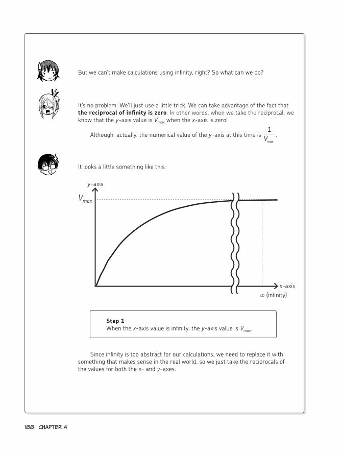
188 chapter 4
But we can’t make calculations using infinity, right? So what can we do?
It’s no problem. We’ll just use a little trick. We can take advantage of the fact that the reciprocal of infinity is zero. In other words, when we take the reciprocal, we know that the y-axis value is Vmax when the x-axis is zero!
Although, actually, the numerical value of the y-axis at this time is 1
Vmax.
It looks a little something like this:
x-axis
y-axis
Vmax
∞ (infinity)
Step 1When the x-axis value is infinity, the y-axis value is Vmax.
Since infinity is too abstract for our calculations, we need to replace it with something that makes sense in the real world, so we just take the reciprocals of the values for both the x- and y-axes.

enzymes Are the Keys to chemical reactions 189
Step 3
When the x-axis value is 0, the y-axis value is 1
Vmax
.
Luckily, we know that the reciprocal of infinity is zero.
If we create a graph for this, we can get a definite numerical value for Vmax.
I can’t believe it, but...that actually makes sense! Maybe graphs aren’t quite as evil as I thought they were...
Now that you understand the theory behind the calculations, don’t you feel like your life is richer? Like you understand the universe just a little bit better?
Now let’s return to the straight line graph you created earlier and try to obtain the value of Vmax.
Yeah, let’s do it! Graphs and reciprocals are child’s play for the likes of me!
Step 2
When the x-axis value is 1° , the y-axis value is
1Vmax
.∞

Take a look at this graph. When the x-axis value is 0, the y-axis
value is approximately 0.019. That means that, in this reaction, the Vmax value of DNA polymerase is
1/0.019, or about 52.6.
in other words, the DNA polymerase can
react with nucleotides to synthesize about
52.6 pmol of new DNA* per hour.**
We did it! We got Vmax!
recip
ro
cal o
f
reactio
n r
ate (pM
ol/h
our)
reciprocal of substrate concentration (1/µM)
slope
hang on, Kumi. you’re celebrating a bit too soon. remember, this graph also lets us calculate the exact
value of Km!
if you look carefully at this reciprocal plot, you can see that the slope of the straight line is
represented by
i hope you remember your
algebra because we’re diving
right in!
** remember, the DNA polymerase reaction time was 60 minutes.
* This value can vary according to the measurement conditions or the type of DNA polymerase. 52.6 pMol/hour is just an example.

enzymes Are the Keys to chemical reactions 191
Let’s use the Michaelis-Menten equation to figure out Km and Vmax!
Let’s do it!
A linear function? i learned about those in middle school. No problem! (i think...)
That’s right! The graph of a linear function is just a straight line, so
this makes our job much easier because...
No matter how far we extend the line, our formula will
always work!
If we take the reciprocal 1v , we get...
In other words, we can draw the graph of the straight line y = ax + b, where
Slope a
x
y
vV SS Kmax
m
=+[ ]
[ ]0
0
1
1 1
0
0
0
0 0vS KV S
SV S
KV S
VKV
m
max max
m
max
max
m
max
=+
= +
= + ×
[ ][ ]
[ ][ ] [ ]
[SS]0
yv
xS
aKV
bV
m
max
max
:
:[ ]
:
:
1
1
1
0

192 chapter 4
To get Vmax, we simply set x to 0, which leaves us with y = 1/Vmax. in other words, we find where the graph crosses the y-axis, take the reciprocal of that value,
and get Vmax.
Next up—Km. We already know that the slope is Km/Vmax,
so we can simply solve the linear equation for x when
y = 0 and get x = -1/Km. Then we just multiply by -1, take the reciprocal, and
we’ve got Km!
slope
recip
ro
cal o
f r
eactio
n
rate (pM
ol/h
our)
reciprocal of substrate concentration (µM)
i get it. it’s just a little bit of substitution and a little bit of algebra!
Kid’s stuff!
so in our example, when y is 0, x is
about -0.15.
This means that, in this reaction, the Km value of DNA
polymerase is 1/0.15, or about 6.67 µM.
We did it! Vmax and Km were
no match for us!
The Lineweaver-Burk reciprocal plot is used frequently in enzyme research. once you
understand these basic steps, you’re well on your way to becoming a great scientist!
Let’s see if i can remember... First, i take the reciprocals of the reaction values, and then i plot them to get a
straight line.
Then with a little bit of simple algebra,
i can figure out Vmax and Km.
Lineweaver and Burk, eat your hearts out!
-

enzymes Are the Keys to chemical reactions 193
4. Enzymes and Inhibitors
inhibitor
substrate can no longer bind with
enzyme!substrate
Active site of enzyme*
Why in the world do we have to use equations and graphs—which are no fun at all—to get the values of Vmax and Km?
One reason is to demonstrate that enzyme reactions are extremely precise and predict-able. They proceed in strict accordance with chemical and mathematical laws.
More importantly, for scholars doing enzyme research or research concerning the world surrounding enzymes, the values of Vmax and Km are very useful.
One of these areas of research deals with the relationship between enzymes and inhibitors. An inhibitor is a substance that affects the binding of an enzyme and substrate, or affects the enzyme itself. As a result, it inhibits the enzyme’s activity.
Many inhibitors are created artificially and used for enzyme research. Since they inhibit enzymes, they are often toxic to living organisms. However, they can also be used in positive ways—to kill cancer cells, for example.
Inhibitors also exist in the natural world. In that case, they’re known as “cellular” enzyme inhibitors rather than “medicinal” inhibitors. For example, some inhibitors are cre-ated directly in an organism’s cells and play important roles in regulating enzyme reactions. Plants can create inhibitors as well. The seeds of certain legumes, for example, contain cel-lular enzyme inhibitors such as α-amylase inhibitors or trypsin inhibitors. These are known as anti-nutritional factors. Scientists believe that these inhibitors might be part of the plant’s natural defense system, which helps protect them against predation.
If the structure of an inhibitor is similar to that of the substrate, it can bond quite eas-ily with the enzyme. However, since the shape of an inhibitor is subtly different from that of the substrate, no reaction will occur, and once the enzyme has already bonded with another substance, it isn’t able to bond with its substrate. Actually, there are several different types of enzyme inhibition, and we can tell which is occuring by using the Michaelis-Menten equation.
Two of the types of enzyme inhibition are competitive inhibition and non-competitive inhibition.
Competitive inhibition occurs when a substance that is very similar to the substrate bonds with the enzyme, inhibiting the enzyme reaction.
* The active site is the part where the substrate bonds to the enzyme in order to undergo a catalytic action.

194 chapter 4
Since the enzymes remain active, the maximum reaction rate Vmax is not affected, but the Km value will rise, since some of the enzymes accidentally bind to inhibitors. With inhibi-tors getting in the way, it takes longer for enzymes to bind to substrates, producing the same result as if a lower substrate concentration was present.
If a competitive inhibitor is added, the slope of the straight line will be greater on a Lineweaver-Burk reciprocal plot, as shown below. Note that the intersection with the y-axis, 1/Vmax, doesn’t change.
x
ycompetitive
inhibition
Although the intersection with the y-axis doesn’t change, the intersection with the x-axis does. This means that if you add an unknown inhibitor to an enzyme reaction, take measurements, graph the result, and see that 1/Km has increased, you know it’s a competi-tive inhibitor.
Non-competitive inhibition is when an inhibitor bonds at a part of the enzyme unrelated to the substrate bonding site to inhibit the enzyme reaction.
inhibitor
substrate
enzyme
The active site is empty, but the enzyme’s shape is changed by the bound inhibitor, preventing the
reaction from occurring smoothly.
if the slope
increases and 1
changesKm
1
Vmax
-1
Km

enzymes Are the Keys to chemical reactions 195
In this case, the inhibitor doesn’t block the substrate—it takes the enzymes out of the game. Km is unaffected because it depends on substrate concentration. Instead, the maxi-mum reaction rate will steadily decrease as the amount of inhibitor increases. The same thing would happen if the enzyme concentration decreased.
This means that when a non-competitive inhibitor is added, the slope of the straight line will again be greater on a Lineweaver-Burk reciprocal plot, as shown below. But unlike competitive inhibition, the intersection with the y-axis will change, while the inter section with the x-axis will stay the same.
x
y if the slope
increases and 1
Vmax
changes
Non-competitive
inhibition
Since inhibitors significantly affect enzyme reactions, they are often used in research for elucidating enzyme structures or reaction mechanisms. They are also used in cancer research by applying small amounts to inhibit enzymes that benefit the cancer cells.
1
Vmax
-1
Km
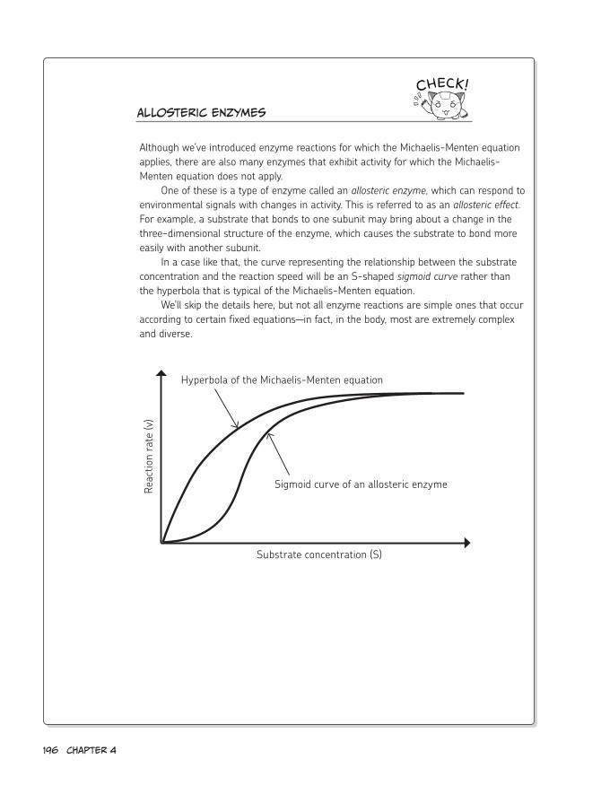
196 chapter 4
Allosteric enzymes
Although we’ve introduced enzyme reactions for which the Michaelis-Menten equation applies, there are also many enzymes that exhibit activity for which the Michaelis- Menten equation does not apply.
One of these is a type of enzyme called an allosteric enzyme, which can respond to environmental signals with changes in activity. This is referred to as an allosteric effect. For example, a substrate that bonds to one subunit may bring about a change in the three-dimensional structure of the enzyme, which causes the substrate to bond more easily with another subunit.
In a case like that, the curve representing the relationship between the substrate concentration and the reaction speed will be an S-shaped sigmoid curve rather than the hyperbola that is typical of the Michaelis-Menten equation.
We’ll skip the details here, but not all enzyme reactions are simple ones that occur according to certain fixed equations—in fact, in the body, most are extremely complex and diverse.
Hyperbola of the Michaelis-Menten equation
Sigmoid curve of an allosteric enzymeReac
tion
rate
(v)
Substrate concentration (S)

enzymes Are the Keys to chemical reactions 197
Today’s lesson was pretty
tough. how are you feeling? Good!
The calculations started to get kind of tricky toward
the end, but i’m happy that i managed to
stick it out!
Swish
Stretch
Plus, i found out that
enzymes are really nice little guys!
huh?
They lower difficult-to-climb walls and give us the boost
we need to hop over!
Well, it doesn’t quite work that way in real life,
of course.
Thanks!
No problemo
Swish

198 chapter 4
But now that you mention it, i agree that enzymes are helpful little
guys.
When there’s an impenetrable barrier between a certain boy and the girl he likes...
Heh
...that barrier can be brought down with the help of a kindly
enzyme!
This enzyme’s role is to
accelerate the relationship
between these two people.
snicker
hey, Professor Kurosaka! Maybe we should talk about the next lesson!
What the...?
sigh...i suppose so. The next lesson will be the last, so you better
prepare yourselves! Especially you,
Nemoto.
The last lesson? That’s a little sad...
Huff
Puff
Heh
Heh

5Molecular Biology and the
Biochemistry of Nucleic acids

200 chapter 5
Morning! Breakfast time, i hope? sure is. i'm
making it now.
i'm starving!
it's good to see you eating
breakfast again, Kumi.
your father’s right! i hope you're done with all that fasting nonsense. A growing girl needs a good
breakfast!
scarf
Om nom

Well, now i know better. i've got to eat so my body can
create ATP!
i'm through with stupid fad diets.
A-T-P ?
Besides, after the final lesson
today...
For all your hard work, you've
earned these— my foolproof,
scientific dieting secrets!
Thank you, professor!
...i'll know all the secrets of a
healthy diet!
Kumi's imagination
Down the hatch!
Bwa ha ha
ha ha ha ha
Om nom nom
okay, let's get started!
yeah!
Krebs
University

202 chapter 5
1. What Is Nucleic Acid?
Nucleic Acid Basics
We've already talked about certain biochemical substances, like proteins, lipids, and saccharides, but there's one
more important substance that we haven't covered yet—nucleic acids.
As you might guess from the name, nucleic acids are acidic substances found in the nucleus
of a cell.
We looked at the nucleus of a cell earlier (see chapter 1), with a little help from robocat. We called it a "DNA
warehouse."
yeah! it's big, round, and seems really important. Perfect for our
last lesson!
in short, nucleic acids are the
genetic material of a cell, or
the substances that store the
information known as genes.
Ah, i know this! DNA is the language of
genes, right?
Nucleic acid
That's right, but DNA isn't totally
made up of genes, you know.
That's true. Genes are a part of DNA—the part that
acts as the blueprints for proteins. Proteins are created based
on the information encoded in the sequence of nucleic
acids (how they are lined up along the dNA).*
Gene Gene
* genes also contain the "blueprints" for rNA.

recipe
(see page 19.)
protein
prote
in
Ah, i remember! Genes are written in the DNA inside
the nucleus!
And proteins are created based on those blueprints.
Mm hm!
There are actually two types of
nucleic acid: DNA ( deoxyribonucleic
acid ) and RNA ( ribonucleic acid ).
DNA
rNA
rNA and DNA are both necessary for the creation of proteins.
(contains genes)
Transcription Translation
Protein
i already know a little bit about
DNA...
...But rNA is a total Mystery
to me!
Don’t worry! rNA isn't quite as well
known as DNA...
That’s right! Now we’ll talk about that process
in more detail.
but it was recently found to be an extremely important substance to
living organisms like us.
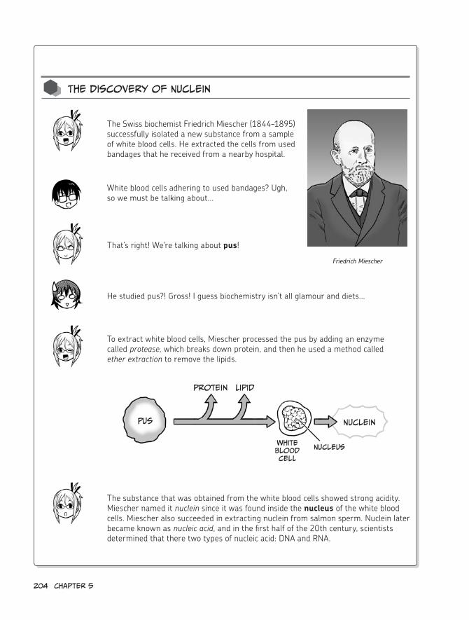
204 chapter 5
The Discovery of Nuclein
The Swiss biochemist Friedrich Miescher (1844–1895) successfully isolated a new substance from a sample of white blood cells. He extracted the cells from used bandages that he received from a nearby hospital.
White blood cells adhering to used bandages? Ugh, so we must be talking about...
To extract white blood cells, Miescher processed the pus by adding an enzyme called protease, which breaks down protein, and then he used a method called ether extraction to remove the lipids.
The substance that was obtained from the white blood cells showed strong acidity. Miescher named it nuclein since it was found inside the nucleus of the white blood cells. Miescher also succeeded in extracting nuclein from salmon sperm. Nuclein later became known as nucleic acid, and in the first half of the 20th century, scientists determined that there two types of nucleic acid: DNA and RNA.
That’s right! We're talking about pus!
He studied pus?! Gross! I guess biochemistry isn’t all glamour and diets...
Friedrich Miescher
Pus
protein Lipid
White Blood cell
Nucleus
Nuclein

Nucleic acid and nucleotides
Now let's examine the structure of
nucleic acid!
Nucleic acid consists of a long chain of substances called
nucleotides* connected together.
i see.
* Nucleotides also appeared when we discussed graphs in chapter 4.
A nucleotide is formed by bonding a base, a phosphate, and a pentose (five-carbon sugar), as follows:
Pentose Nucleotide
Base
ribose
1 to 3 phosphates
Base
Phosphate
orribonucleotide
Base
Deoxyribose
Deoxyribonucleotide
1 to 3 phosphates
Uhh... And if just a base and a pentose bond (without a phosphate), the
result is called a nucleoside.
Base Base

206 chapter 5
There are five types of bases that form nucleic acid: adenine (A), guanine (G), cytosine (C ), Uracil ( U ),
and Thymine (T ).
The four types used in DNA are A, G, C, and T. The four types used in rNA
are A, G, C, and U.
Base
Adenine (A) Guanine (G)
cytosine (c) Uracil (U) Thymine (T)
A and G are called purine bases, while c, U, and T are called
pyrimidine bases.
Purine has one hexagonal ring and
one pentagonal ring.
Pyrimidine has only one hexagonal ring.
purine
pyrimidine
i can remember that because Apple and Ginger are the best, most purely
delicious flan flavors!

Molecular Biology and the Biochemistry of Nucleic Acids 207
This diagram shows a Nucleotide structure
with adenine (A) as its base.
This is called adenosine
monophosphate or adenosine- 5'-Phosphate.
Adenin
e
Adeno
sin
e
Phosphate
Adenosine Monophosphate
if the pentose in this nucleotide was deoxyribose instead of ribose, then we
would have deoxyadenosine monophosphate.
saccharide (Pentose)
or
ribose Deoxyribose
for DNA, deoxyribose is used for the pentose, and for rNA, ribose is used.
Deoxyribose, ribose...ooohh, i'm getting really confused.
For now, just remember that
deoxyribose is used in DNA and ribose is used in rNA. We'll go into more detail later.

By the way, adenosine triphosphate has a similar structure but with
three phosphates attached.
hey, it’s our old friend ATP, the common currency
of energy!
That's right! Not only is ATP the common currency of energy, it's
also a building block of rNA.
Wow! ATP is even more important than i thought!
look! if we swap out the base in adenosine monophosphate with the other four kinds of bases, we get the following
nucleotides:
• If the base is guanine (G), we get guanosine monophosphate and deoxyguanosine monophosphate, for RNA and DNA respectively.
• If it’s cytosine (C), we get cytidine monophosphate and deoxycytidine monophosphate.
• If it’s thymine (T), we get deoxythymidine monophosphate.*
• If it’s uracil (U), we get uridine monophosphate and deoxyuridine monophosphate.**
* Since the deoxy form of thymine is the most prevalent, the deoxy prefix is often left out.
** Although uracil is not a base that’s normally in DNA, the deoxy type does exists.
O P PP
Adenosine
Adenine
i see! There are two types of pentoses and five types of bases. if the base changes,
the nucleotide also changes, right? it’s the same with udon and soba!
curry
Depending on what we combine it with, it can turn into soba and egg, tempura
soba, and so on...
slurp
Don't forget the green onions and seven-spice
powder!

Molecular Biology and the Biochemistry of Nucleic Acids 209
Nucleic acid consists of nucleotides connected in a long, linear chain. The car-bons at the 3´ position and the 5´ position of each pentose link to a phosphate, forming a bridge between individual nucleotides. This is called a polynucleotide.
Base complementarity and DNA structure
Looks kind of like a comb!
P A
C
T
G
P P
P
P
P
or U
3´
5´
4́
2´
1́
3´
5´
4́
2´
1́
3´
5´
4́
2´
1́
3´
5´
4́
2´
1́
OH
3´
5´
3´ end
5´ end
The bases form shapes that protrude like the teeth of a comb on one side of the polynucleotide. For DNA, two polynucleotide strands form a double strand by connecting these bases, which creates a helix structure.
Polynucleotide
* For the nucleotide saccharides, ´ (prime) is appended to the numbers given to each carbon—for example, 1́ , 2 ,́ and so on—to distinguish them from the numbers assigned to the carbons in the nucleotide base.
The five carbons are numbered 1́ to 5 .́*
Deoxyribose
3´
5´
4́
2´
1́
O
OP
O
O
O
H
H
H
CH2
H H
OP
O
O
O
H
H
H
CH2
H H
CH3HN
NO
T
O
A
NH2
N
N N
N
5´ end (since it's the end of the 5´ side)
NH2
N
NO
C
OP
O
O
O
H
H
H
CH2
H H
OH
G
O
HN
N N
N
H2N
3´
5´
3´ end (since it's the end of the 3´ side)
OO P
O
P
O
OP
OO
H
H
H
CH2
H H
-O -O -O-O
-O
-O
-O

210 chapter 5
Oh! Those comb-teeth-looking parts stick together and become the rungs in the middle!
Right. In other words, a “pair” is created via hydrogen bonds between two bases, so that the polynucleotides form a double strand. The pairings are always faithful: A pairs with T (with two hydrogen bonds), and G pairs with C (with three hydrogen bonds).
Always these combinations
hydrogen bond
A
C
T
G
P
P
P
3´
5´
2´
1́
3´
5´
4́
2´
1́
3´
5´
4́
2´
1́
3´
5´
4́
2´
1́
T
A
P
G
P
C
P
3´
5´
4́
2´
1́
3´
5´
4́
2´
1́
3´
5´
4́
2´
1́
3´
5´
4́
2´
1́
if the two combs are
joined...
...and twisted into a helix...
...we get DNA!
A G
T c
4́

Molecular Biology and the Biochemistry of Nucleic Acids 211
This pairing configuration is called complementarity. Because of this comple-mentarity, DNA can be replicated—in other words, the two-stranded poly-nucleotide chain can be separated into individual strands, and those strands can be used as templates to build new strands. The result of this replication is two DNA molecules that have the same arrangement of bases.
if T (thymine) is the template, the
base that will join the new strand is
A (adenine).
it will have the
complementary
base sequence.
DNA
A template is like the mold that you pour batter into to make cupcakes.
I get it! If you have a mold, they’re easy to replicate.
If you know what the sequence of the template strand is, you’ll know the sequence of a new strand that uses that tempate as a mold!
That's right. The biggest difference between DNA and other biopolymers (proteins, saccharides, and lipids) is that ability to replicate easily.

212 chapter 5
That’s...totally cool!
The first person to isolate an enzyme involved in DNA replication—called DNA polymerase I—was the American biochemist Arthur Kornberg (1918–2007).
cell DNA is replicated
cleanly divided
Arthur Kornberg
DNA polymerase was the enzyme we used when we obtained Vmax and Km. Do you remember?
Yeah! That’s the enzyme that replicates DNA? Enzymes really do a lot, don’t they?
In 1956, Kornberg extracted an active enzyme used for synthesizing DNA from a liquid in which E. coli was pulverized. This enzyme could be used to artificially synthesize DNA in a test tube, which created a great sensation in the scientific world at the time.
That discovery was seriously important.
DNA replication and the enzyme DNA Polymerase
So why is DNA replicated? Since DNA contains our genes, it must be replicated whenever cells divide and multiply.
Genes are passed on from parents to children, as well as from cell to cell. When a cell divides, the same genes must be inherited by the two new cells, so the DNA must be replicated before the cell splits.

Molecular Biology and the Biochemistry of Nucleic Acids 213
It sure was! In fact, Kornberg was awarded the Nobel Prize in Physiology or Medicine in 1959.
DNA polymerase is officially called “DNA-dependent DNA polymerase.” This means that a DNA polymerase enzyme requires the presence of an existing DNA template molecule in order to create a new DNA molecule.
DNA
polymerase
I get it. Since the pairs are complementary, DNA polymerase steadily creates the chain by referring to the template.
The chemical reaction that DNA polymerase catalyzes is shown below.
O-O P
O
O
O
H
CH2
HO
Base
O
OO P
O
O
O
H
CH2
O
OO P
O
O
OCH2
5´
Phosphodiester bond
P P
3' end
OO P
O
P
O
OP
OO
H
CH2-O
-O -O -OBase
HO
connects to the next deoxyribonucleotide
phosphate 3´
5´
in this reaction, a nucleotide with three phosphates attached is used as the substrate.

214 chapter 5
In other words, DNA polymerase catalyzes the attachment of the next deoxyribonucleotide phosphate to the 3´ end of a new DNA strand. This attachment is called a phosphodiester bond.
The nucleic acids that belong together are joined firmly by the “matchmaker” DNA polymerase.
That’s right! Ha ha ha...
...
rNA structure
On the other hand, RNA usually exists as a single strand. The enzyme RNA polymerase actually uses DNA as a template when it binds ribonucleotides together into a new RNA strand.
Oh, that reminds me...earlier you said that RNA is an extremely important substance, but you never said why!
Just hold your horses, Kumi. We’ll get into that soon (on page 220, to be exact).
Got it!
DNA rNA
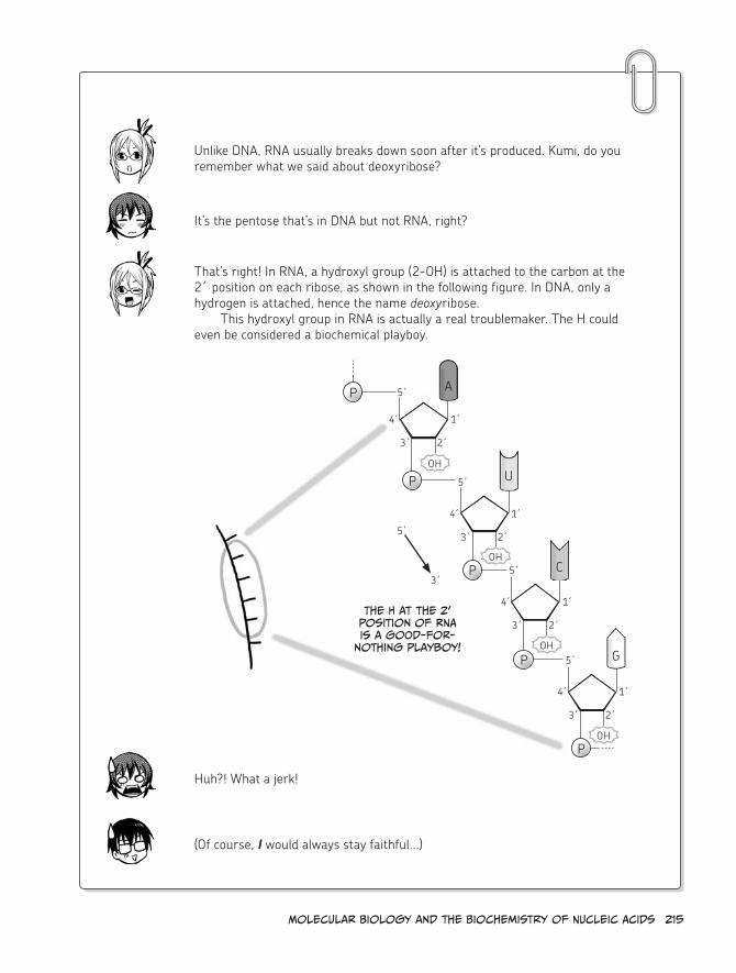
Molecular Biology and the Biochemistry of Nucleic Acids 215
Unlike DNA, RNA usually breaks down soon after it’s produced. Kumi, do you remember what we said about deoxyribose?
It’s the pentose that’s in DNA but not RNA, right?
That’s right! In RNA, a hydroxyl group (2-OH) is attached to the carbon at the 2´ position on each ribose, as shown in the following figure. In DNA, only a hydrogen is attached, hence the name deoxyribose.
This hydroxyl group in RNA is actually a real troublemaker. The H could even be considered a biochemical playboy.
A
C
U
G
P
P
P
3´
5´
4́
2´
1́
3´
5´
4́
2´
1́
3´
5´
4́
2´
1́
3´
5´
4́
2´
1́
OH
OH
OH
OH
P
P
3´
5´
The h at the 2’ position of rNA is a good-for-
nothing playboy!
Huh?! What a jerk!
(Of course, I would always stay faithful...)

216 chapter 5
Because of the existence of this hydroxyl group, RNA is known to undergo autolysis (a self-inflicted decomposition) due to a phenomenon known as base catalysis.
So the 2 -́OH proton is extracted by a base?
That’s right. Then the lone oxygen goes looking for a new partner to bond with.
Actually, after the proton is extracted, the oxygen (O-), which carries a nega-tive charge, will bond with the phosphate (P) in the phosphodiester bond at the neighboring 3´ position (that is, the important bond connecting two ribonucleotides).
If you think of the O and the H of a hydroxyl group as a couple, then H is the “playboy” that can be tempted by bases that exist around the RNA.
It’s important to note that these aren’t the A, U, C, and G bases we’ve been talking about so far. In this context, a base is a substance that can accept a proton (H+), such as a hydroxide ion (OH-). Bases can be thought of as the chemical opposite of acids, because they neutralize each other.
Autolysis!

Molecular Biology and the Biochemistry of Nucleic Acids 217
As a result, the phosphodiester bond breaks apart, dividing the RNA chain.
It’s all a little sordid, isn’t it?
Since RNA frequently undergoes this kind of autolysis, it’s considered “unstable” and is not very suitable for storing genetic information. DNA is far more stable than RNA since it does not contain the 2 -́OH. Therefore, trustworthy DNA is the best for this purpose. A different but still very important role is assigned to RNA instead.
We’ll learn about that role next!
O
3´
5´
4́
2´
1́
O
O OH
P OO-
O
O
3´
5´
4́
2´
1́
O OH
phosphodiester bond
Base
:OH-
Attack!
O
3´
5´
4́
2´
1́
O
O O
PO-O
Divided into
pieces
O
3´
5´
4́
2´
1́
HO
O OH
What?!
hey!

218 chapter 5
2. Nucleic Acid and Genes
DNA is the Language of genes
People say kids look like their parents because of genes.
Dad
Do i look like mine?
Afraid so!
Mom
Genes are the “blueprints” that guide
the creation of rNA and proteins. They are
stored in DNA.
Genes are in DNA.
DNA is formed by lining up bases, right? Well, this
arrangement of bases is actually very important.
it’s important because the arrangement itself is the blueprint of proteins!

2. Nucleic Acid and Genes
in other words, a gene is just an arrangement of bases in a certain
section of DNA. This arrangement of bases is called the
base sequence.*
Wow...
For us humans...
...only approximately 1.5% of the DNA in a cell acts as the blueprints for proteins.**
only 1.5%?! That’s insane!
Gene
Gene
Gene
The remaining 98.5% contains elements that regulate the work of genes, as well
as a small part made up of introns.***
Also, there are many parts that are believed to have been transferred
in for some reason during the course of our evolution.
so there are still mysteries to be
solved, eh?
even now, there are many sequences of DNA that we
don’t really understand at all.
* in this book, we’ll generally say that genes are “written” (in the form of a base sequence) in DNA.
** Actually, the exon part of genes is considered to be 1.5% of the whole. (see page 227.)
*** We’ll examine introns and exons further on page 227.

220 chapter 5
rNA has several jobs
Nucleus
cytoplasm
so DNA acts as the blueprint for proteins
and is found in the nucleus of a cell.
it’s actually trapped there and
can never escape to the cytoplasm!*
Kumi, do you remember ribosomes, which create proteins?
errr...
Hee hee
Pop quiz!
Although they look like specks of salt from a distance, when
we zoom in they look more like little snowmen all jumbled
together.
They adhere to the endoplasmic reticulum or float in the cytoplasm.
(see page 27.)
yep! ribosomes, which synthesize proteins, are
in the cytoplasm...
But DNA can’t leave the nucleus...
Nucleus
cytoplasm
DNA
cell
ribosome
Oh no!
We’ve got to get
those two together!
* The chloroplasts (in photosynthetic cells) and mitochondria floating in the cytoplasm contain their own characteristic DNA.

i guess DNA can’t exactly send the blueprints to the
ribosome in a letter...
Alack! yon handsome ribosome will
never learn of my affections...
how in the world can the blueprints
get outside of the nucleus?
hmmm...
Squawk?!
That’s where rNA saves the day!
Gasp!
you don’t say!
rNA reads and transcribes the blueprints written in the DNA and carries
that information to the ribosomes, where proteins will be created!
coooool! That really is an important role!
replication
Transcription
Translation
Protein
Actually, there are several different
types of rNA.
The types of rNA that play a role in protein
synthesis are mrNA, rrNA, and trNA.

222 chapter 5
mrNA
The enzyme called RNA polymerase enables genes that are written in the DNA—that is, their base sequences—to be used as templates to synthesize RNA with complementary base sequences.
This process is called transcription (it can also be referred to as RNA synthesis), and the synthesized RNA is called messenger RNA (mRNA).
For example, take a look at the gene shown in the following figure.
Gene
rNA synthesis direction
sense strand
Antisense strand
mrNA
5´
3´
3´
5´
5´
3´
DNA is double stranded, and, for each gene, only one side of the DNA is mean-ingful (that is, its base sequence becomes the blueprint). This is the sense strand for that gene. The other strand, which is complementary to the sense strand, is called the antisense strand.
That makes...sense.
Since mRNA is synthesized using the antisense strand as a template, the base sequence of the RNA that’s produced is the same as that of the sense strand. However, where the base is T (thymine) in the sense strand, there will be a U (uracil) in the mRNA.

Molecular Biology and the Biochemistry of Nucleic Acids 223
The synthesized mRNA is called precursor mRNA. It undergoes a series of chemical reactions and other processing* to become mature mRNA, which is then transported from the nucleus to the cytoplasm and is soon delivered to a ribosome.
So you can see why we call it a “messenger”: it’s transporting vital information out of the stronghold of the nucleus.
DNANucleus
cell
mrNA mrNA ribosome
Nuclear Pore
DNA strand is unwound.
Genetic information
is copied to mrNA.
mrNA is transported outside of the nucleus to the cytoplasm through a nuclear pore and is soon delivered to a ribosome!
* This process includes splicing, which removes intron parts (see page 227), and the addition of a 5´ “cap” and a poly-A tail.
rrNA and trNA
A ribosome is like a big protein-making machine consisting of several types of RNA called ribosomal RNA (rRNA) and several dozen types of ribosomal proteins.
They’re not as large as mitochondria or chloroplasts, though.
They may not look like much, but ribosomes are pretty darned impressive!
ribosomal proteins
ribosome!rrNA

224 chapter 5
Another type of RNA, which transports amino acids, is called transfer RNA (tRNA). The tRNA matches the base sequences of the delivered mRNA with the amino acids needed to build the coded proteins.
It should be pretty obvious where transfer RNA gets its name: it transfers amino acids.
The mRNA base sequence is a code for an amino acid sequence. More specifi-cally, a three-base sequence forms the code for one amino acid. This three-base sequence is called a codon.
There are unique tRNA molecules for each codon. In other words, only a tRNA with the proper base sequence—called an anticodon—that is complementary to the mRNA’s codon can adhere at the site of the ribosome.*
The role of rRNA is to connect the amino acids that were transported via tRNA into long chains. Amino acids are connected according to the mRNA base sequence (that is, the codon sequence) to create proteins in accordance with the base sequence (the genes, or “blueprint”) of the original DNA.
So in other words, genetic information is conveyed like this: DNA RNA protein.
A CU G A A U UG
codon codon codon
* The type of amino acid that is transported is also determined by the anticodon.
ribosome (rrNA)
mrNAmrNA
trNA
Anticodon
codon
Amino acid
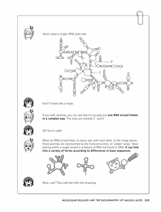
Molecular Biology and the Biochemistry of Nucleic Acids 225
Here’s what a single rRNA looks like:
Huh? It looks like a maze.
If you look carefully, you can see that it’s actually just one RNA strand folded in a complex way. The ends are marked 3´ and 5 .́
Oh! You’re right!
Wow, cool! They look like little line drawings.
When an RNA strand folds, its bases pair with each other. In the image above, those pairings are represented by the transverse lines, or “ladder rungs.” Base pairing within a single strand is a feature of RNA not found in DNA: It can fold into a variety of forms according to differences in base sequences.

226 chapter 5
ribozymes
To summarize, RNA breaks down quickly, but it can do things that DNA can’t. RNA exists both in the nucleus and the cytoplasm, and it can take various forms depending on the base sequence.
Huh!
So they combined the words “RNA (ribonucleic acid)” and “enzyme” to coin the term ribozyme.
Definitely. RNA researchers have long believed that RNA probably has other important jobs besides copying genes and interacting with ribosomes.
Ribozymes, which were independently discovered at the beginning of the 1980s by the American microbiologist Thomas Cech and the Canadian molecu-lar biologist Sidney Altman, foretold later developments in RNA research. You learned earlier that one very important job of proteins is their work as enzymes (see page 153), but Cech and Altman actually discovered that RNA has the flex-ibility to work as an enzyme as well.
It’s a very flexible molecule, isn’t it?
enzyme
rNA enzyme
ribonucleic
acid enzyme ribozyme
rNA

Molecular Biology and the Biochemistry of Nucleic Acids 227
Many of the ribozymes that have been discovered or artificially created so far are involved in building or cleaving RNA or DNA.
Cech discovered that part of RNA itself catalyzes the chemical reaction to remove sections that are meaningless as protein blueprints (introns) and to join the sections that are meaningful (exons). This is referred to as self-splicing* and is fairly rare.
Usually, splicing involves an enzyme known as spliceosome, which is a large complex made of RNA and proteins.
* The genes of eukaryotes (like humans) are divided into several exons by non-coding base sequences called introns. The introns must be removed from the mRNA before translation, in a reaction known as splicing.
Splicing involves several hydroxyl groups (-OH) of RNA, such as the 2 -́OH or the 3 -́OH.
Hey, it made a loop!
The discovery of ribozymes caused RNA to be viewed as a multifaceted molecule that does a variety of tasks, and RNA research advanced rapidly.
As the 21st century began, this research showed that RNA has other various roles and that many different forms of RNA exist in cells. RNA research is truly in full bloom now, and there are high expectations for future research.
intron
exon
2’-oh attack!
3’-oh attack!
eukaryotic mrNA splicing removes the intron ( ) parts.
228 chapter 5
3. Biochemistry and Molecular Biology
Nowadays—especially in cities—there are fewer and fewer opportunities for exploring nature and really getting your hands dirty. In a world where a mustard-stained tie is the greatest potential tragedy, most people can’t even imagine trekking out into the wilderness to really dig through the (often filthy!) wonders of the natural world.
But humans, of course, are products of nature. Long before the miracles of instant spot remover and nectarine-scented hand sanitizer, we lived like other animals, surrounded by the great outdoors.
Although biochemistry is a discipline that attempts to view the incredible phenomena of life through the lens of science, the raw materials of that research are the living organisms themselves—again, products of nature.
Once, during my graduate student days, I went to the mountains in order to collect certain plants that contain large amounts of the proteins that I was researching.
I drove with a younger graduate student to some nearby mountains, and we proceeded along a road that was thickly overgrown with plants. When we got to a place where the car could no longer pass, we continued on foot to find the plant called Phytolacca Americana (American pokeweed).
We brought the pokeweed back to the laboratory, washed off the dirt, and used kitchen knives and scissors to cut them up so that we could extract the target proteins from them.
I would also routinely visit a nearby meatpacking plant to obtain an organ called the thymus from recently slaughtered cows. (The thymus is usually discarded, so they were happy to give them away free of charge.) I would use scissors to cut up the organ and store the little pieces in the laboratory’s refrigerator as experimental samples. This was necessary to extract the research target protein (DNA polymerase).
In this way, biochemistry originally developed based on a methodology of extracting (isolating, purifying, etc.) chemical substances from organic raw materials and checking their chemical properties.
In contrast, molecular biology is a discipline that tries to elucidate the phenomena of life by using biopolymers such as DNA and proteins.
Simply speaking, if molecular biology just deals with DNA and RNA or prepares an environment for artificially creating proteins (by using E. coli, for example), it is unnecessary to use flesh-and-blood, organic raw materials such as a cow’s thymus or plant materials.
In a manner of speaking, the data obtained in these artificial lab environments is “digital.” Molecular biology is practiced using high-tech equipment and cutting-edge tech-nologies, and molecular biologists are rarely caked in mud or smeared with animal blood...
As a result, some biochemists refer to their own research as “dirty work.” I think that when they do this, it’s somehow self-deprecating.
The Dirty Job of a Biochemist

Molecular Biology and the Biochemistry of Nucleic Acids 229
However, it is an undeniable fact that the accumulation of knowledge from that kind of dirty work built the foundation of molecular biology in the first place. Although many young molecular biologists have never once used flesh-and-blood, organic materials other than E. coli, cell cultures, or experimental animals, biochemistry and molecular biology have long been bound together and remain so even today. This fact should never be forgotten.
early Biochemistry and Molecular Biology
In 1897, German biochemist Eduard Buchner made the revolutionary discovery that fer-mentation occurred in an extract created from yeast cells, which contained yeast protein but no living yeast cells.
Prior to that, people believed that the chemical reaction called fermentation, which is characteristic of living organisms, could not happen without the presence of living cells. However, this idea was utterly obliterated by Buchner’s discovery.
Because of this, the theory of vitalism, in which the phenomena of life occur only because of a characteristic force (spirit or life energy) of living organisms, practically dis-appeared, and the foundation was built for research on chemical reactions occurring in living organisms in test tubes. In other words, biochemistry was born.
Since Buchner found that an actual living organism need not be present, it would not be an exaggeration to say that this was the discovery that paved the way for the arrival of molecular biology.
As a clearer understanding of the chemistry of life developed, it became apparent that certain mechanisms common in all living organisms form the basic foundation of life. Some of these mechanisms, for example, are that DNA is used as the genetic language, that the basic theory (central dogma) behind the reading of genes to create proteins is consistent, and that the same proteins often do the same kinds of work.
Because these mechanisms are universal among living organisms, it was important to develop methodologies for studying DNA, which serves as the blueprint for proteins, and for elucidating the function of proteins.
Development of recombinant DNA Techniques
In 1972, American molecular biologist Paul Berg performed the first successful recombinant DNA experiment in the world by artificially manipulating DNA in a test tube to create a DNA sequence that did not exist in the natural world.
A method for easily decoding the base sequences of DNA was developed by English biochemist Frederick Sanger in 1977, and a method for copying (or amplifying) DNA was developed by American molecular biologist Kary Mullis in 1985. After these discoveries, recombinant DNA experimental techniques advanced rapidly.

230 chapter 5
Recombinant DNA techniques developed after it had been clearly demonstrated that DNA is the genetic language (or, in other words, that the information referred to as genes is written in the form of base sequences in DNA). Molecular biologists imagined that if they only had the DNA base sequences and if they only created an environment in which proteins could be created from those base sequences, they could elucidate the phenomena of life, which can be thought of as the totality of chemical reactions in which proteins are involved.
For example, if genes from an external source are introduced into a simple, easy-to-manipulate organism for which these mechanisms are well understood (such as E. coli ) to create proteins within that organism, a large quantity of proteins can be obtained all at once without ever having to perform any more “dirty work.”
Generally speaking, this idea can be expressed as: Explain the DNA sequences! Explain life!
Molecular biology has developed with this aim. The methodology used to achieve this was based on recombinant DNA techniques.
returning to Biochemistry
After the Human Genome Project (an international cooperative research project aiming to sequence the genetic information of humans) was completed in 2003, researchers turned their attention from DNA back towards proteins and RNA.
The post-genome era, or post-sequencing era, arrived.No matter how much DNA steals the limelight, no matter how its handling techniques
develop, and no matter what anyone says, when life phenomena are viewed as a collection of “chemical reactions,” the only things at work there are proteins and RNA.
This is because, even if all of the base sequences of human DNA (that is, the entire genome) are known, the data is meaningless unless the roles of the proteins and RNA that are made from DNA are understood.
Currently, if the amino acid sequences of many proteins are known and the role they play is also understood, then the work of unknown proteins can be predicted to a certain degree based on only amino acid information.
However, the role of unknown proteins must be verified by using biochemical tech-niques. No matter how many molecular biological techniques (such as DNA recombination) are used to research the activity of proteins to elucidate their roles, the question of whether or not those proteins really do those jobs inside natural cells will remain unanswered. In a manner of speaking, this is the same as the idiomatic expression “you can’t see the forest for the trees.” As long as the research target is a biological substance, biochemistry remains an absolutely necessary and important academic discipline.

Molecular Biology and the Biochemistry of Nucleic Acids 231
The origin of the cell
No doubt you’ve heard the phrase “origin of life.” We’re not going to tackle the intimidating question of what life actually is here. Instead, we’re concerned with the first living organisms or, in other words, “the origin of cells.”
So how did cells originate on Earth?By now, you should understand that a cell is the location of many vital chemical reac-
tions in living organisms and that cells can divide and multiply. These are two major traits that define life.
Chemical reactions that transform one substance into another substance, which are performed inside of cells, and networks of many of these chemical reactions, which occur across many cells, are collectively referred to as metabolism.
Cells must constantly undergo metabolism in order to produce the energy required to maintain their organization and to reproduce.
In addition to metabolism, reproduction, also called self-replication, is a major char-acteristic of living organisms. Unicellular organisms can often simply replicate their cellular contents and split in two; this process is called binary fission. For multicellular organisms, reproduction is more complex and involves specialized reproductive cells, which initiate the development of offspring.
(Although the way in which cells replicate themselves can certainly be called self- replication, the expression is a little awkward. Let’s just call it plain old “replication” from now on.)
Cells perform both metabolism and replication, both of which are extremely important when considering the origin of life. But which came first, metabolism or replication?
This is one of the most difficult questions confronting scientists who are conduct-ing research concerning the origin of life. When the “pouch wrapped in a membrane” (the cell) originated, what had been occurring inside that enclosure? Some scholars think that the “pouch” had been performing metabolism after many diverse molecules had collected together. Some time later, the “pouch” absorbed molecules capable of causing replication, so the entire assembly became able to divide and multiply. This idea is known as metabo-lism first.
Other scholars think that the original “pouch” had enclosed replicating molecules and had been dividing. Later, it became able to perform metabolism, and it eventually obtained a more advanced replication method, causing it to evolve into a cell. This idea is known as replication first.
Many other scholars think that posing the question of “which came first” is nonsense and that metabolism and replication coevolved cooperatively.
In any case, the biochemical process of metabolism is a very large-scale phenomenon.

232 chapter 5
4. Conducting Biochemistry Experiments
It was mentioned on page 230 that the functions of proteins can be verified by using bio-chemical techniques, but what kinds of experiments do biochemists perform?
There are various experimental methods depending on the field of research, and not each and every one will be presented here. Instead, I will introduce several experimental methods that I have used myself.
column chromatography
Column chromatography is an experimental method for separating substances that have the same property from a mixture of substances. For example, we can collect just proteins with certain properties from the liquid extract of American pokeweed, which was introduced earlier, or from the liquid obtained after a cow thymus was blended in a juicer. Special resins are packed in a long, narrow tube made of glass or some other material. Some substances adhere to those resins and are trapped, allowing for the collection of only those substances that do not adhere.
Various types of chromatography, such as ion exchange chromatography, gel filtration chromatography, and affinity chromatography, can be used, depending on the type of resin or target protein. As an example, here we will look at a method for purifying the enzyme DNA polymerase α from the thymus of a calf.
As shown below, the cells are smashed by grinding up the calf thymus, and a solution with a high salt (sodium chloride) concentration is used to extract proteins and other mol-ecules. This extract (the liquid in the flask) passes through the large glass tube (called the column) in which the ion exchange resin has been packed. The proteins are broadly divided into those that are trapped in the column and those that pass through the ion exchange resin.
Liquified calf thymus
Approximately 1.5 Mcolumn
roughly “fractionates” the proteins according to ion exchange chromatography
1) compounds that pass through the column
2) compounds that are absorbed by resin in the column
compounds that flow out with low salt concentration
compounds that flow out with middle salt concentration
compounds that flow out with high salt concentration
DNA polymerase α is here!
sample 1

Molecular Biology and the Biochemistry of Nucleic Acids 233
The substances that pass through the ion exchange resin are collected in test tubes, but the substances that are absorbed are separated from the resin and collected in test tubes later by adding a liquid with a higher salt concentration. DNA polymerase α adheres to the column and can be retrieved by adding a liquid with a salt concentration of 0.5 M (moles per liter). (This is “sample 1.”)
The figure below shows the method of purifying DNA polymerase α from this “sample 1.”
This technique, called affinity chromatography, uses a small glass tube packed with a resin combined with an antibody (a type of protein that’s created by the immune system) that can only bond with DNA polymerase α. When sample 1 is passed directly through this resin, it’s divided into two parts—one that is absorbed and one that passes through. DNA polymerase α, which is absorbed, can be retrieved by running a solution with an extremely high concentration (3.2 M) of magnesium chloride through the resin. Since the part that’s retrieved here is almost 100% DNA polymerase α, it has been “purified” at this time.
In this way, DNA polymerase α can be efficiently purified by using a combination of ion exchange chromatography and affinity chromatography.
electrophoresis and a Western Blot
This is an experimental method for isolating a specific protein, recognizing what type of protein is in a sample, or checking the size of a target protein. Electrophoresis is a method in which a sample is “loaded” on top of a thin gelatinous slab (gel) and a current passes through it to cause the sample to move through the gel. SDS-polyacrylamide gel electro-phoresis (left side of the following figure), which separates proteins based on molecular size, is commonly used. After the proteins are separated, they are detected by reacting them with a special reagent, such as a dye or a fluorescent marker.
A western blot (right side of the following figure) is a method of transferring the pro-teins that are in the gel onto a thin membrane, while retaining the positions that they were in after they were separated, and then detecting a specific protein on the membrane by adding an “antibody” that only reacts with that protein. Lectin blotting, which is described next, is an application of a western blot.
sample 1
Approximately 20 cm
column
1) compounds that pass through the column
2) compounds that are absorbed by resin in the column
“Purify” DNA polymerase α from sample 1
according to affinity chromatography
compounds that flow out with sodium chloride (Nacl)
compounds that flow out with magnesium chloride (mgcl2)
DNA polymerase α is here!
Purified!

234 chapter 5
Lectin Blotting
Lectins are proteins that can bond specifically to certain sugar chains. Because lectins will bond differently according to the type of sugar chain, lectins can be used to identify the type of sugar chain that is bonded to a protein. A method similar to a western blot can be used after proteins are transferred onto a membrane. Various lectins are mixed with the proteins, and only the lectins that reacted are detected. This lets us identify the types of sugar chains that exist in the proteins that were transferred onto the membrane. This is called lectin blotting.
The following figure shows an experiment in which a lectin called wheat germ agglu-tinin (WGA) identifies a sugar chain to which the saccharide called N-acetylglucosamine (GlcNAc) is attached.
In this lectin blot performed for the rough protein fraction of the starfish oocyte, it is apparent that two large bands are glowing brightly. WGA was the lectin used here.
(use the reagent called sDs)
Proteins are separated according to size.
Add the protein sample.
–
+
Polyacrylamide gel (thickness: approximately 1 mm; height: several cm to
several dozen cm)
Pass a current through.
–
+
smaller proteins move farther down.
These marks that appear when proteins are detected are
called protein bands.
sDs-polyacrylamide gel electrophoresis
Western Blot
Thin membrane, nitrocellulose membrane,
or PVDF membrane
Attach gel to membrane as described
above, and apply electricity to transfer the
proteins in the gel to the membrane
(blotting).
–+
Perform experiments on this membrane using various methods
(Like Lectin blotting, for example).
Prepare the gel so that it carries a negative charge ( – ).

Molecular Biology and the Biochemistry of Nucleic Acids 235
centrifugation
Like column chromatography, centrifugation is an experimental method that is performed to separate organelles, proteins, and other biological molecules that have the same density from a mixture of substances with differing densities. The sample solution is placed in a test tube, and the test tube then spins around the centrifuge axis at a very high speed. To handle small molecules, like proteins, ultracentrifugation may also be performed, which spins the test tube at tens of thousands of revolutions per minute. DNA and other polymers can also be isolated in this way.
Transferred protein bands
Membrane
sugar chains are attached.
When viewed directly from the side...
Lectin
When using the property of a lectin that reacts
to GlCNAC...
GlcNAc
Membrane
the reagent that bonded to the lectin glows. only proteins that
have sugar chains with GlCNAC attached will be detected.
Bingo!
j All proteins are stained with cBB.k only proteins possessing sugar
chains with GlCNAC attached are stained because of WGA (arrows).
l These proteins are stained with concanavalin A, which is another lectin.
(Photos provided by: Mitsutaka ogawa, Nagahama institute of Bio-science and Technology)
Mixture of various substances
app
ro
xim
ately
40 mm
Place the Micro test tube in the centrifuge...
which revolves at a
high speed.
substance A
substance B
small container such as a micro test tube centrifuge isolated samples

236 chapter 5
enzyme reaction Measurement
There are various methods of measuring enzyme activity, such as using radioisotopes to measure the amount of a reaction product produced or using a change in color to measure the reaction product formed when a substrate is acted on by an enzyme.
I will explain a method of using radioisotopes to measure the enzyme activity of DNA polymerase and a method of using a color reaction to measure the enzyme activity of α-amylase.
DNA Polymerase Activity Measurement Method
First, place the following in a mirco test tube: the solution for measuring activity (with pH optimized and buffered for DNA polymerase), DNA polymerase, the DNA that is to be the template, the nucleotide that is to be the raw material, and the magnesium chloride co-factor. Add the nucleotide that contains the radioisotope, and let it react at 37° C for a fixed interval.
When this is done, the nucleotides that contain the radioisotopes are captured in the new DNA strands that have been synthesized by the DNA polymerase. The unreacted nucleotides are removed, and the synthesized DNA strands are placed in a small radioiso-tope bottle for measurement in a device called a liquid scintillation counter, which measures radioisotopes. Since higher enzyme activity means more radioisotopes were captured in the DNA, higher numeric values indicate higher enzyme activity.
Micro test tube radioisotope
(3h)radioisotopes are captured in synthesized DNA.
Unreacted nucleotides are removed. sample is placed
in the liquid scintillation
counter.DNA polymerase
nucleotideTemplate DNA
MeasurementDNA polymerase is reacted with nucleotides that
contain radioisotopes.
small radioisotope measurement bottle
DNA polymerase activity measurement method

Molecular Biology and the Biochemistry of Nucleic Acids 237
starch
color reaction
When starch is present, it’s colored blue-violet when it reacts with
iodine-potassium iodide.
As the starch is broken down by α-amylase, the color
steadily changes from blue-violet to violet to red to
orange to pale orange and finally becomes colorless.α-amylase
iodine solution is
added. reaction progresses
α-amylase activity measurement method
α-amylase Activity Measurement Method
An α-amylase solution (such as saliva) is added to a solution in which starch has been dissolved inside a test tube. If an iodine solution is immediately added to this, before any of the starch has been broken down, the starch will react with the iodine and produce a blue-violet color. However, as time passes, the starch is broken down by the α-amylase, and the color steadily changes (blue-violet violet red orange pale orange). Even-tually, when all the starch has been broken down, the solution will become colorless. The enzyme activity of α-amylase can be measured by using a spectrophotometer to quantify the appearance of its color as a numeric value.


epilogue 239
*
* Projection r
oom
Well, that's the end of our lessons!
you two did a great job!
it's All thanks to you, professor!
wham
Um...by the way, Professor...
Now that we've finished our last
lesson...
The time has come!
Teach me the ultimate secrets of dieting!

silly girl—you've had the "secrets"
of dieting this entire time!
Wha-?
Look...
it's all right here in your
report.
tap
This graph is the key!
ing
ested e
nerg
y
expended e
nerg
y
energy stored as fat
if you expend more calories than you ingest, you'll lose
weight.
eat moderately and exercise frequently!
Although there are endless fad
diets out there, it all really boils down to that!

And make sure you get a good balance of
nutrients, of course!
you've learned that proteins, saccharides,
and lipids are all important to your body. you must realize by now that dieting by starving yourself is complete
nonsense, right?
right, Kumi? right?!
...oh dear.
Guuuuuuh
Stagger
Stagger
Well...i guess i learned some
valuable things.
But...
Who am i kidding? i love food too much to watch what i eat!
i'll get fat for sure!calm down, Kumi!
hmm, i wonder...
Poor Kumi
Waaaahhh

242 epilogue
i'll eat cake and pizza until i'm huge!
i'll turn into a bloated, snack-
scarfing manatee!
Kumi, you're being ridiculous. you've got a long way to go before
you're as big as a manatee.
if anything, you'd be more like a midsized
sea lion...
Doomed!
...or maybe a small bear
preparing for hibernation, covered in winter fat.
heh heh heh
hey!
Fatty!
squirm
squirm
hey, Professor...
What?
But you know...if you think about it, chuBBy animals
are almost always the cutest, so don't worry!
Ha ha ha ha
sob
sob
sob

epilogue 243
Give Kumi a break!
can't you see she's too sensitive to stand
up to this kind of treatment?
sniff
oh yeah? if you have something to say, you'd better
spill it!
i will!
First of all, i don't think Kumi's fat at all.
Goal: lose 5 lbs!
Down with
the pounds!
But even if she was fat, she'd still
be beautiful!

244 epilogue
she's beautiful when she's stuffing
her face...
she's beautiful when she's poring over her studies...
And she's extra beautiful in her
bathing suit!
in fact...
she's one of the most beautiful girls
i've ever met! i'd even go as far as
saying that...
Kumi is a total dreamboat!
yum
Hmmm...

Ne-
Nemoto?
Well duuuuh! you think i don't know
that?
huh?
Kumi is a cutie patootie!
But...that "sea lion" crack...
...and the "fat little bear"...

246 epilogue
Just a little reverse
psychology!
Worked like a charm, too. it's about time Nemoto told you
how he felt! ♪
yes, this lesson was a huge
success, if i do say so myself.
A trap...
Kumi, you studied hard and learned some important
things about your body, right?
Definitely. No more fad diets for me.
And you also learned some
other important information...hehe.
blush
wink

epilogue 247
so now that we're through with our lessons, how about
we go grab a bite to eat?
Nemoto's treat, of course.
But...but...i'm still totally broke!
oh, i'm sure a smart kid like you can figure something out.
i'll go get ready!...
Um...Nemoto...
yes!

248 epilogue
Thanks! For everything!
!
And now...
Let 's go stuff our
faces!

A
A (adenine), 206ABO blood group system, 126acetyl-CoA, 72, 85, 115–117,
120–122activation energy, 176acyl-carnitine, 121acyl-CoA, 120–121adenine (A), 206adenosine diphosphate (ADP), 82adenosine monophosphate
(AMP), 207adenosine triphosphate (ATP),
22–23, 82, 106affinity chromatography, 233affinity of enzymes, 181alcohol, 20, 153aldoses, 83allosteric enzymes, 196–199α-amylase, 143, 144, 162, 163α-amylase activity measurement
method, 237amino acids
in carbon cycle, 45overview, 154–157primary structure of, 158protein synthesis and, 27–28quaternary structure of, 161secondary structure of, 159tertiary structure of, 160
AMP (adenosine monophosphate), 207
amphipathicity, 93amylopectin structure, 137–139amylose structure, 137–139angina pectoris, 104anticodon, 224anti-nutritional factors, 193antisense strand, 222arteriosclerosis, 103–104
atoms, 53ATP (adenosine triphosphate),
22–23, 82, 106autolysis, 216
B
bad cholesterol, 100, 105basal metabolism, 107base catalysis, 216base complementarity, 209–211base sequence, 219ß-oxidation, 119–120ß type, 143binary fission, 231biochemistry, definition of, 6, 8biochemists, 228–229biogeochemical cycle, 40–44biological catalyst, 175biopolymers, 36–37, 228blood
ABO group system, 126glucosyltransferase, 169–171types of, 124–129, 169, 170
blood sugar, 60, 63, 108breathing, 64–65
c
C (carbon). See carbon (C)C (cytosine), 206, 208carbohydrates
monosaccharides and cyclic structure, 63
overview, 60–62plants and, 48saccharides and -ose suffix, 63
carbon (C)biogeochemical cycle and, 43cycle of, 45–47fixation of, 57fundamentals of, 36
carbon bonds, 36carbon dioxide
in carbon cycle, 46fixation of, 51, 57–59in photosynthesis, 32
carboxyl group, 95catalysts, 175cells
chemical reactions in, 26–35interior of, 18–25membrane of, 16origin of, 231structure of, 14–17
cellular respiration, 37, 65cellulose, 132, 142centrifugation, 235cerebral hemorrhage, 104cerebral infarction, 104chemical bonds, 140–144
covalent, 36double, 96glycosidic, 138, 141–143, 173iconic, 36metallic, 36peptide, 155, 158phosphodiester, 214
chitin, 132chlorophyll, 49–55chloroplasts
overview, 17photosynthesis and, 32structure of, 49
cholesterollipoproteins
arteriosclerosis, 103–104good and bad cholesterol,
105overview, 102
purpose of, 98–99as steroid, 97
index

250 index
cholesterol ester, 101chromatography of columns,
232–233citric acid cycle (TCA cycle), 67,
71–73, 115CoA (coenzyme A), 85
acetyl-CoA, 72, 85, 115–117, 120–122
acyl-CoA, 120–121malonyl-CoA, 116–117succinyl-CoA, 72
column chromatography, 232–233
competitive inhibition, 193complementarity
configurations, 211concentration gradient force, 77consumers, 48covalent bonds, 36cycles
biogeochemical, 40–44carbon, 45–47citric acid, 71–73
cyclic structure, 61, 63, 141cytidine monophosphate, 208cytoplasm, 16cytosine (C), 206, 208cytosol, 16, 30
D
deoxyadenosine monophosphate, 207
deoxycytidine monophosphate, 208
deoxyguanosine monophosphate, 208
deoxyribonucleic acid (DNA), 17, 202–203, 218–219
deoxyribose, 207deoxythymidine
monophosphate, 208deoxyuridine monophosphate, 208detoxification, 21D-form isomers, 84diacylglycerol, 92dietary fiber, 142digestion, 7
DNA (deoxyribonucleic acid), 17, 202–203, 218–219
DNA polymerase I, 212DNA polymerase activity mea-
surement method, 236double bonds, 96
e
EC Number (Enzyme Commission Number), 166
ecosystemsbiogeochemical cycle, 40–44carbon cycle, 45–47
electron transport chain, 52–55, 67, 74–78
electrophoresis, 233–234endoplasmic reticulum, 16, 17endothelial cells, 113energy
ATP, 82creation of
citric acid cycle, 71–73electron transport chain,
74–78glucose decomposition by
glycolysis, 68–70overview, 66–67
fat used as, 118–122ingested and expended,
106–110metabolism, 38
Enzyme Commission Number (EC Number), 166
enzymesclassifications of, 166–167DNA polymerase, 212–214fundamentals of, 37hydrolases, 172–173inhibitors and
allosteric enzymes, 196–199
overview, 193–195lipoprotein lipase, 112proteins and
amino acids, 154–161overview, 150–151,
153–154roles of, 151–152
reaction measurement ofα-amylase activity
method, 237DNA polymerase activity
method, 236overview, 236
substrates and, 162–165transferases
determining blood type, 169–171
overview, 168using graphs to understand
activation energy, 176calculating Vmax and Km,
182–192chemical reactions,
175, 177maximum reaction
rate, 178Michaelis-Menten equation
and Michaelis constant, 180–181
overview, 174essential fatty acids, 95ether extraction, 204eukaryotes, 227exons, 219, 227expended energy, 106–110experiments
centrifugation, 235column chromatography,
232–233electrophoresis and western
blots, 233–234enzyme reaction measurement
α-amylase activity measurement method, 237
DNA polymerase activity measurement method, 236
overview, 236lectin blotting, 234–235overview, 232
external respiration, 37, 65

index 251
F
fatingested and expended energy,
106–110overview, 106saccharides
fat used as energy source, 118–122
overeating and weight gain, 123
overview, 111–117fatty acids, 45, 95–96foam cells, 103free cholesterol, 101fructofuranose, 83fructopyranose, 83fructose, 59, 61, 116fruit
becoming sweet, 133–135saccharides, 131–132sugar in, 130
furanose, 83
G
G (guanine), 206, 208galactose, 62genes
DNA, 17, 202–203, 218–219exons, 219, 227introns, 219, 223, 227overview, 19RNA. See RNA
(ribonucleic acid)genetic material of cells, 202global environment, 40–41globin, 161glucofuranose, 83gluconeogenesis, 29–30glucose
in carbon cycle, 45composition of, 62cyclic (ring) form, 61decomposition by glycolysis,
68–70in energy production, 30in food, 59open-chain form, 61
glucosyltransferase, 169–171glycocalyx, 125glycogen, 132glycolipids, 94glycolysis
in energy production, 30glucose decomposition by,
68–70in respiration, 67
glycosidic bond, 138, 141–143, 173
glycosyltransferase, 169golgi apparatus, 16, 17good cholesterol, 100, 105granums, 49graphs
activation energy, 176chemical reactions, 175, 177maximum reaction rate, 178Michaelis-Menten equation,
180–181, 191–192overview, 174reciprocals, 186–190
guanine (G), 206, 208guanosine monophosphate, 208
h
HDL (high-density lipoproteins), 101–105
heme, 160hemiacetal, 63hemoglobin, 161high-density lipoproteins (HDL),
101–105hydrocarbon chain, 95hydrogen acceptor, 54hydrolases, 119, 172–173hydrolysis, 173hydrophilic, 16, 93hydrophobic, 16, 93
i
ingested energy, 106–110ingested lipids, 112inhibition
competitive, 193non-competitive, 194
inhibitorsallosteric enzymes, 196–199overview, 193–195
insulin, 108internal respiration, 37, 65introns, 219, 223, 227invertase, 133, 134ionic bonds, 36
K
ketoses, 83Krebs cycle (citric acid cycle), 67,
71–73, 115
L
lactose, 61LDL (low-density lipoproteins),
101–105lectin blotting, 234–235leptin, 109, 110L-form isomers, 84Lineweaver-Burk reciprocal plot,
183–184, 192lipids
cholesterol, 97–99fatty acids, 95–96lipoproteins
arteriosclerosis, 103–104good and bad
cholesterol, 105overview, 100–102
overview, 88–90types of, 91–94
lipogenesis, 29lipoprotein lipase enzyme, 112lipoproteins
arteriosclerosis, 103–104good (HDL) and bad (LDL)
cholesterol, 105overview, 100–102
liquid scintillation counter, 236low-density lipoproteins (LDL),
101–105lysosome, 16–17

252 index
M
macrophages, 103malonyl-CoA, 116–117maximum reaction rate, 178–179messenger RNA (mRNA),
222–223metabolism, 38, 107, 231metabolism first theory, 231metallic bonds, 36methane, 46Michaelis constant, 180–181Michaelis-Menten equation,
180–181, 191–192, 196Miescher, Friedrich, 204milk sugar, 63mitochondria, 16–17, 31, 116molecular biology
and biochemistry, 228–230origin of cells, 231recombinant DNA techniques,
229–230monoacylglycerol, 92monosaccharides
aldoses and ketoses, 83D-form and L-form, 84pyranose and furanose, 83in respiration, 62–63structure of, 131–132
mRNA (messenger RNA), 222–223
multicellular organisms, 15myocardial infarction, 104
N
NADH molecule, 54–55, 75NADPH molecule, 54–55neutral fat, 91neutral lipids, 91non-competitive inhibition,
194–195nucleic acids. See also nucleotides
conducting experimentscentrifugation, 235column chromatography,
232–233electrophoresis and
western blots, 233–234
enzyme reaction measurement, 236–237
lectin blotting, 234–235overview, 232
genesDNA, 218–219RNA, 220–227
molecular biologyand biochemistry, 228–230origin of cells, 231recombinant DNA
techniques, 229–230nuclein, 204overview, 199–203
nuclein, 204nucleosides, 205nucleotides
base complementarity and DNA structure, 209–211
DNA replication and enzyme DNA polymerase, 212–214
overview, 205–208RNA structure, 214–217
nucleus, 16, 17, 204
o
obesityingested and expended energy,
106–110overview, 106saccharides
fat used as energy source, 118–122
overeating and weight gain, 123
overview, 111–117oligosaccharides, 131–132, 137open-chain glucose form, 61organelles, 16-ose suffix, 63overeating, 123oxidation-reduction, 37oxidized, defined, 37oxygen, 77, 216
P
palmitic acid, 116pepsin, 162peptide bonds, 155, 158peptidyl transfer, 158phagocyte, 103phosphate, 205phosphodiester bond, 214phospholipids, 16, 93, 95phosphopantetheine group, 85phosphorylation, 56
photophosphorylation reaction, 50–56
photosynthesiscarbon dioxide fixation, 57–59chloroplast structure, 49overview, 79–81photophosphorylation reaction,
50–56plants, 48
photosystem I, 55photosystem II, 55Phytolacca americana plant, 228pokeweed, 228polynucleotides, 209–210polypeptide chain, 158polysaccharides, 131–132, 137post-genome era, 230precursor mRNA, 223primary structure, 158producers, 48product, defined, 37progesterone, 98protease, 204protein catabolism enzymes,
164, 165proteins
amino acidsoverview, 154–157primary structure of, 158quaternary structure of, 161secondary structure of, 159tertiary structure of, 160
created by cells, 18overview, 150–151, 153–154roles of, 151–152

index 253
proteios, 154pyranose, 83pyrimidine bases, 206pyruvate, 68, 72pyruvic acid, 29–30
Q
quaternary structure, 161
r
rate-limiting reaction, 117reaction rate, 178reciprocals, 186–190recombinant DNA techniques,
229–230reduced, defined, 37reducing agents, 37replication first theory, 231replication of DNA, 211, 212–214reproduction, 231respiration
breathing, 64–65carbohydrates
monosaccharides and cyclic structure, 63
overview, 60–62saccharides and -ose
suffix, 63energy creation by
citric acid cycle, 71–73electron transport chain,
74–78glucose decomposition by
glycolysis, 68–70overview, 66–67
fundamentals of, 37overview, 79–81
ribonucleic acid (RNA). See RNA (ribonucleic acid)
ribosomal RNA (rRNA), 223–225ribosomes
in cells, 16–19defined, 37in protein synthesis, 27–28
ribozymes, 226–227rice starch, 136–137ring structure of glucose, 61, 141
RNA (ribonucleic acid)in creation of proteins, 203mRNA, 222–223overview, 220–221ribozymes, 226–227rRNA and tRNA, 223–225self-splicing, 227structure of, 214–217
rRNA (ribosomal RNA), 223–225
s
saccharidesaldoses and ketoses, 83in carbon fixation, 58D-form and L-form
isomers, 84excess, transformation into fat,
111–117in fruits, 133–135-ose suffix and, 63overeating and weight
gain, 123photosynthesis and, 25pyranose and furanose, 83types of, 131–132using fat as energy source,
118–122secondary structure, 159self-replication, 231sense strand, 222side chain, 159sigmoid curve, 196specific gravity, 101spliceosome, 227splicing, 223starch, 66, 132, 136steroids, 97
steroid hormone, 98steroidal skeleton, 97
stroma, 57substance metabolism, 38substrates, 37, 162–165subunit, 161succinyl-CoA, 72sucrose, 59, 61–63sucrose-phosphate synthase,
133–134
sugar, 60–61, 116, 130sugar chain, 126
T
T (thymine), 206, 208TCA cycle (citric acid cycle), 67,
71–73, 115templates, DNA replication, 211tertiary structure, 160testosterone, 98thylakoid membrane, 52thylakoids, 49thymine (T), 206, 208thymus, 228transcription, 222transferases
glucosyltransferase and blood type, 169–171
overview, 168transfer RNA (tRNA), 223–225triacylglycerol, 92, 113–114tRNA (transfer RNA), 223–225
U
U (uracil), 206, 208unicellular microorganisms, 15unsaturated carbons, 96unsaturated fatty acids, 96uracil (U), 206, 208uridine monophosphate, 208
V
very low-density lipoproteins (VLDL), 113
vitalism theory, 229Vitamin D, 99
W
weight gain, 123western blots, 233–234wine, 21

Notes

Notes

Notes

Notes

About the AuthorMasaharu Takemura, PhD, is currently an Associate Professor at the Tokyo University of Science. His specialties are molecular biology and biology education.
Production Team for the Japanese edition
Production: Office Sawa Email: [email protected]
Established in 2006, Office Sawa has produced numerous practical documents and advertisements in the fields of medicine, personal computers, and education. Office Sawa specializes in manuals, reference books, or sales promotional materials that frequently use instructional text and manga.
Scenario: Sawako Sawada
Artist: Kikuyaro
DTP: Office Sawa

how This Book Was MadeThe Manga Guide series is a co-publication of No Starch Press and Ohmsha, Ltd. of Tokyo, Japan, one of Japan’s oldest and most respected scientific and technical book publishers. Each title in the best-selling Manga Guide series is the product of the combined work of a manga illustrator, scenario writer, and expert scientist or mathematician. Once each title is translated into English, we rewrite and edit the translation as necessary and have an expert review each volume. The result is the English version you hold in your hands.
More Manga GuidesFind more Manga Guides at your favorite bookstore, and learn more about the series at http://www.nostarch.com/manga.

Updates
Visit http://www.nostarch.com/mg_biochem.htm for updates, errata, and other information.
The Manga Guide to Biochemistry was laid out in Adobe InDesign. The fonts are CCMeanwhile and Chevin.
This book was printed and bound at Malloy Incorporated in Ann Arbor, Michigan. The paper is Glatfelter Spring Forge 60# Smooth, which is certified by the Sustainable Forestry Initiative (SFI).
We’ll be fine as long as we expend more
than we take in!
urp
Too bad it doesn’t work that way with money...

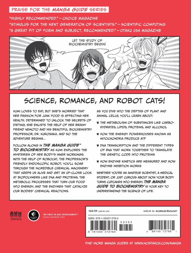
PRaise for the manga guide series
“Highly Recommended.” — Choice Magazine
“Stimulus for the next generation of scientists.” — Scientific computing
“A great fit of FOrm and subject. Recommended.” — Otaku USA Magazine
Science, Romance, and Robot Cats!Kumi loves to eat, but she’s worried that
her passion for junk food is affecting her
health. Determined to unlock the secrets of
dieting, she enlists the help of her brainy
friend Nemoto and his beautiful biochemistry
professor, Dr. Kurosaka. And so the
adventure begins…
Follow along in The Manga Guide™ to Biochemistry as Kumi explores the
mysteries of her body’s inner workings.
With the help of RoboCat, the professor’s
friendly endoscopic robot, you’ll soar
through the incredible chemical machinery
that keeps us alive and get an up-close look
at biopolymers like DNA and proteins, the
metabolic processes that turn our food
into energy, and the enzymes that catalyze
our bodies’ chemical reactions.
As you dive into the depths of plant and
animal cells, you’ll learn about:
The metabolism of substances like carbo-
hydrates, lipids, proteins, and alcohol
How the energy powerhouses known as
mitochondria produce ATP
DNA transcription and the different types
of RNA that work together to translate
the genetic code into proteins
How enzyme kinetics are measured and how
enzyme inhibition works
Whether you’re an amateur scientist, a medical
student, or just curious about how your body
turns cupcakes into energy, The Manga Guide to Biochemistry is your key to
understanding the science of life.
Find more Manga Guides at www.nostarch.com/manga
$24.95 ($25.95 CDN) SHelve iN: Science/BioloGy
THe M
ANGA G
UiD
e™ TO
BiO
CHeM
iSTRY
TakeM
ura
kik
uyaro
off
i ce S
aw
a
TH E F I N EST I N G E E K E NTE RTA I N M E NT™www.nostarch.com
BIOCHEMISTRYBiOCHeMiSTRYMasaharu TakemuraKikuyaroOffice sawa
The Manga Guide™ to comics
inside!