mandibular fractures in children under 3 years: a rare ... · acidente doméstico sofreu trauma em...
TRANSCRIPT
r e v p o r t e s t o m a t o l m e d d e n t c i r m a x i l o f a c . 2 0 1 3;54(3):166–170
Revista Portuguesa de Estomatologia,Medicina Dentária e Cirurgia Maxilofacial
www.elsev ier .p t /spemd
Clinical case
Mandibular fractures in children under 3 years: A rare casereport
Renato Maranoa,∗, Patrício de Oliveira Netoa, Keiko Oliveira Sakugawab,Liliane S.S. Zanetti c, Márcio de Moraesa
a Piracicaba Dental School (UNICAMP), Piracicaba, São Paulo, Brazilb Universidade Federal do Espírito Santo (UFES), Vitória, Espírito Santo, Brazilc Faculdades Integradas Espírito-Santenses (FAESA), Vitória, Espírito Santo, Brazil
a r t i c l e i n f o
Article history:
Received 29 August 2012
Accepted 10 June 2013
Available online 4 October 2013
Keywords:
Mandibular fractures
Mandibular condyle
Internal fracture fixation
a b s t r a c t
Mandibular fractures in children are relatively rare, not only by their anatomical and phys-
iological aspects, but also by their social factor, which makes this group less exposed to
high-impact trauma. Thus, the specific pediatric treatment, through a minimally invasive
approach should also be established, avoiding future functional disorders. In our case, a
female patient, a 3-year-old accident victim was affected by a domestic symphysis frac-
ture and bilateral mandibular condyle. Surgical treatment of fractures of the mandibular
symphysis was performed two days after the trauma, under general anesthesia for reduc-
tion and fixation, employing a 1.5 titanium plate system in the midline of the mandibular
symphysis. The condylar fractures were treated conservatively, immediately using gruel and
oral therapy to avoid complications such as temporomandibular joint ankylosis. The patient
has been followed up by our staff and has no restriction in mandibular movements and no
limitation of mouth opening.
© 2012 Sociedade Portuguesa de Estomatologia e Medicina Dentária. Published by
Elsevier España, S.L. All rights reserved.
Fratura mandibular em crianca com idade menor que 3 anos: um rarorelato de caso
Palavras-chave:
Fraturas mandibulares
Côndilo mandibular
Fixacão interna de fraturas
r e s u m o
Fraturas mandibulares em criancas são relativamente raras, não apenas por seus aspectos
anatômicos e fisiológicos, mas pelo fator social, o que torna este grupo menos exposto
ao trauma de alto impacto. Assim sendo, o tratamento pediátrico específico, por meio
de uma abordagem minimamente invasiva também deve ser instituído, evitando futuros
transtornos funcionais. Neste caso, uma crianca com idade menor que 3 anos, vítima de
acidente doméstico sofreu trauma em face resultando em fratura sinfisária e em côndilo
mandibular bilateralmente. O tratamento precoce foi instituído sob anestesia geral para a
reducão e fixacão, empregando um sistema de placa de titânio 1.5 em sínfise mandibular.
Fraturas condilares foram tratadas conservadoramente, por meio de dieta líquido/pastosa e
fisioterapia, para evitar complicacões como a anquilose da articulacão temporomandibular.
∗ Corresponding author.E-mail address: [email protected] (R. Marano).
1646-2890/$ – see front matter © 2012 Sociedade Portuguesa de Estomatologia e Medicina Dentária. Published by Elsevier España, S.L. All rights reserved.
http://dx.doi.org/10.1016/j.rpemd.2013.06.002

r e v p o r t e s t o m a t o l m e d d e n t c i r m a x i l o f a c . 2 0 1 3;54(3):166–170 167
A paciente está em acompanhamento pela equipe e não apresenta restricão nos movimen-
tos mandibulares e nem limitacão de abertura bucal.
© 2012 Sociedade Portuguesa de Estomatologia e Medicina Dentária. Publicado por
Elsevier España, S.L. Todos os direitos reservados.
I
Piiiadfatctttf
tgt
mdtogohtto
eifbr
C
Afvncwtaofsfl
trauma to reduce the mandibular symphysis fracture with a1.5 titanium plate system (Neortho, Curitiba, Brazil) (Fig. 6).
ntroduction
ediatric fractures are unusual when compared with fracturesn adults. The reasons for this statement are based primar-ly on social and anatomical factors. Most often children aren protected environments, under the supervision of parentsnd thus less exposed to major trauma, occupational acci-ents or interpersonal violence, which are common causes ofacial fractures in adults.1 Regarding maxillofacial fractures,
low incidence is due to the early stage of development ofhe facial skeleton and of the sinuses, leading to a craniofa-ial disproportion. The flexibility of the facial skeleton andhe relative protection offered by existing fat in the subcu-aneous tissue around the bones of the face also contributeo reduce the incidence of fractures, especially maxillofacialractures.2,3
Approximately half of all pediatric facial fractures involvehe mandible4 and boys are more commonly affected thanirls by a ratio of 2:1 and the majority of injuries occur ineenagers.5
Although much of the relevant technology is shared,anagement of pediatric mandible fractures is substantially
ifferent from that of the adult injury. This is due primarily tohe presence of multiple tooth buds throughout the substancef the mandible, as well as to the potential injury to futurerowth. Although these issues complicate the managementf pediatric mandible fractures, these younger patients alsoave the potential for restitutional remodeling, as opposed tohe sclerotic, and functional remodeling seen in adults. All ofhis must be taken into consideration during the evaluationf and approach to these injuries.6–8
An understanding of the surgical or treatment options isssential for making informed choices to best manage thesenjuries. This paper describes a case of a pediatric mandibularracture where a conservative treatment was performed in theilateral condyles and a rigid internal fixation was done in theegion of the mandibular symphysis.9
ase report
3-year-old was the victim of a domestic accident not suf-ered from a fall (Fig. 1). The mother reported no syncope,omiting or drowsiness by the child, after discarding theeurological examination of suspected head trauma asso-iated with brain injury. On physical examination thereas local presence of the lower lip laceration, avulsion of
he lateral incisor and upper right canine, extrusive lux-tion of the maxillary central incisors, lateral dislocation
f the first molars and right and left vestibular wall boneracture in the anterior maxilla. In addition, the child hadwelling and bruising in the submentual region and mouthoor. By radiography, a bone fracture was found in theFig. 1 – Initial picture preoperative.
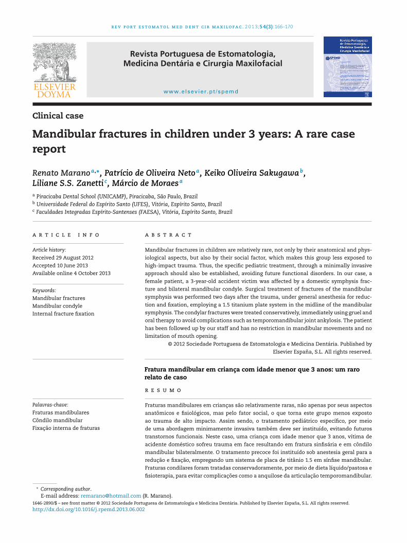
region of the mandibular symphysis and bilateral mandibu-lar condyle fracture, confirmed by physical examination(Figs. 2 and 3).
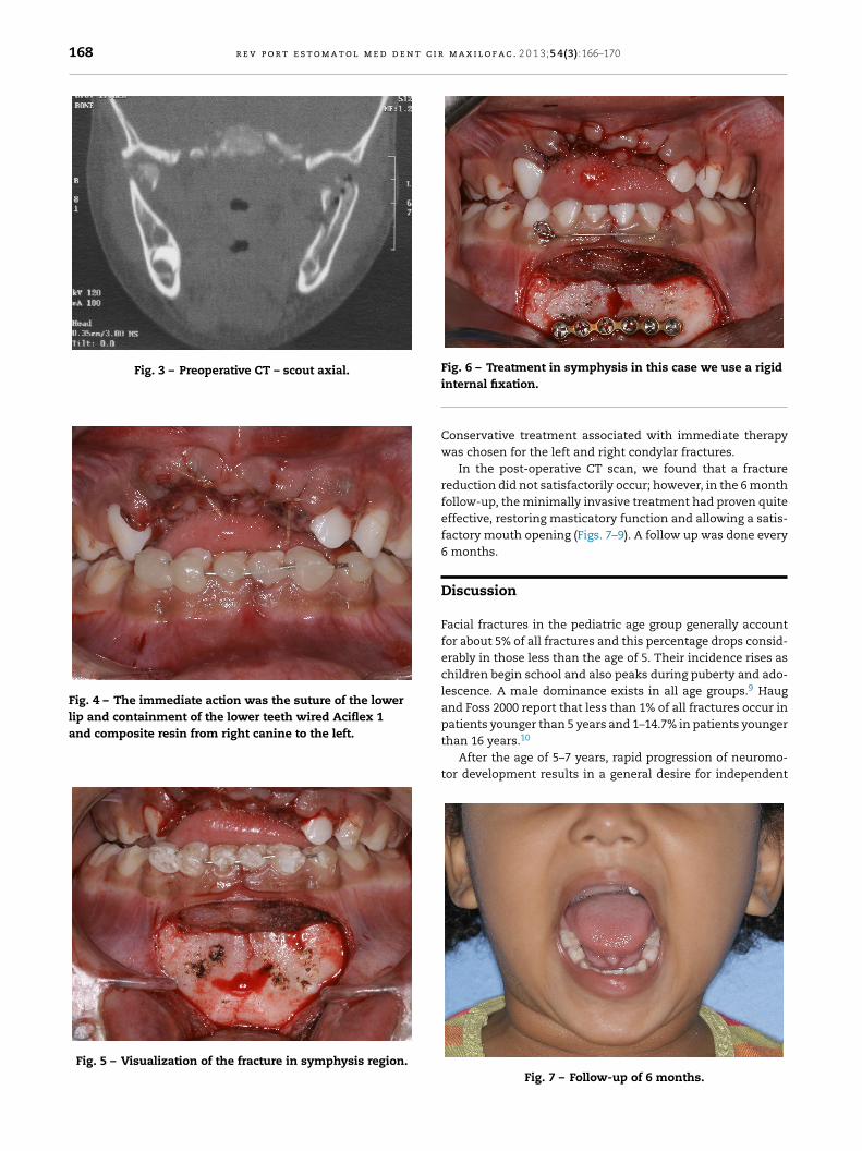
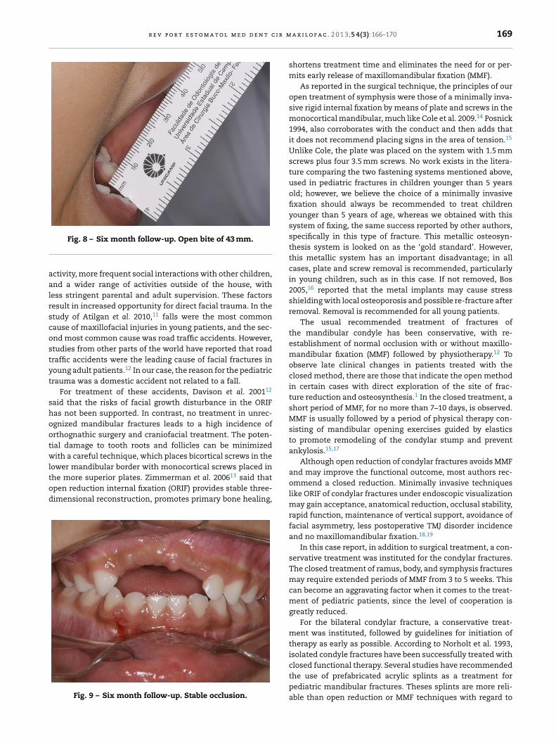
The immediate action was the suture of the lower lip andcontainment of the lower teeth with wire aciflex (Ethicon,Johnson & Johnson Medical, Brazil) and composite resin fromthe right canine to the left (Fig. 4). To perform the symphy-sis treatment in this case, we used a rigid internal fixation(Fig. 5). However, because there is a need for minimal invasive-ness, we recommended “minimally invasive” internal fixation(MIIF) where a maximum fixation becomes as important as thelowest possible level of injury. The definitive treatment wasinstituted surgery under general anesthesia, 36 h after facial
Fig. 2 – Preoperative CT – scout coronal.
168 r e v p o r t e s t o m a t o l m e d d e n t c i r m a x i l o f a c . 2 0 1 3;54(3):166–170
Fig. 3 – Preoperative CT – scout axial.
Fig. 4 – The immediate action was the suture of the lowerlip and containment of the lower teeth wired Aciflex 1and composite resin from right canine to the left.
Fig. 5 – Visualization of the fracture in symphysis region.
Fig. 6 – Treatment in symphysis in this case we use a rigid
internal fixation.Conservative treatment associated with immediate therapywas chosen for the left and right condylar fractures.
In the post-operative CT scan, we found that a fracturereduction did not satisfactorily occur; however, in the 6 monthfollow-up, the minimally invasive treatment had proven quiteeffective, restoring masticatory function and allowing a satis-factory mouth opening (Figs. 7–9). A follow up was done every6 months.
Discussion
Facial fractures in the pediatric age group generally accountfor about 5% of all fractures and this percentage drops consid-erably in those less than the age of 5. Their incidence rises aschildren begin school and also peaks during puberty and ado-lescence. A male dominance exists in all age groups.9 Haugand Foss 2000 report that less than 1% of all fractures occur inpatients younger than 5 years and 1–14.7% in patients younger
than 16 years.10After the age of 5–7 years, rapid progression of neuromo-tor development results in a general desire for independent
Fig. 7 – Follow-up of 6 months.
r e v p o r t e s t o m a t o l m e d d e n t c i r m
aalrscostyt
shootwltod
Fig. 8 – Six month follow-up. Open bite of 43 mm.
ctivity, more frequent social interactions with other children,nd a wider range of activities outside of the house, withess stringent parental and adult supervision. These factorsesult in increased opportunity for direct facial trauma. In thetudy of Atilgan et al. 2010,11 falls were the most commonause of maxillofacial injuries in young patients, and the sec-nd most common cause was road traffic accidents. However,tudies from other parts of the world have reported that roadraffic accidents were the leading cause of facial fractures inoung adult patients.12 In our case, the reason for the pediatricrauma was a domestic accident not related to a fall.
For treatment of these accidents, Davison et al. 200112
aid that the risks of facial growth disturbance in the ORIFas not been supported. In contrast, no treatment in unrec-gnized mandibular fractures leads to a high incidence ofrthognathic surgery and craniofacial treatment. The poten-ial damage to tooth roots and follicles can be minimizedith a careful technique, which places bicortical screws in the
ower mandibular border with monocortical screws placed in
he more superior plates. Zimmerman et al. 200613 said thatpen reduction internal fixation (ORIF) provides stable three-imensional reconstruction, promotes primary bone healing,Fig. 9 – Six month follow-up. Stable occlusion.
a x i l o f a c . 2 0 1 3;54(3):166–170 169
shortens treatment time and eliminates the need for or per-mits early release of maxillomandibular fixation (MMF).
As reported in the surgical technique, the principles of ouropen treatment of symphysis were those of a minimally inva-sive rigid internal fixation by means of plate and screws in themonocortical mandibular, much like Cole et al. 2009.14 Posnick1994, also corroborates with the conduct and then adds thatit does not recommend placing signs in the area of tension.15
Unlike Cole, the plate was placed on the system with 1.5 mmscrews plus four 3.5 mm screws. No work exists in the litera-ture comparing the two fastening systems mentioned above,used in pediatric fractures in children younger than 5 yearsold; however, we believe the choice of a minimally invasivefixation should always be recommended to treat childrenyounger than 5 years of age, whereas we obtained with thissystem of fixing, the same success reported by other authors,specifically in this type of fracture. This metallic osteosyn-thesis system is looked on as the ‘gold standard’. However,this metallic system has an important disadvantage; in allcases, plate and screw removal is recommended, particularlyin young children, such as in this case. If not removed, Bos2005,16 reported that the metal implants may cause stressshielding with local osteoporosis and possible re-fracture afterremoval. Removal is recommended for all young patients.
The usual recommended treatment of fractures ofthe mandibular condyle has been conservative, with re-establishment of normal occlusion with or without maxillo-mandibular fixation (MMF) followed by physiotherapy.12 Toobserve late clinical changes in patients treated with theclosed method, there are those that indicate the open methodin certain cases with direct exploration of the site of frac-ture reduction and osteosynthesis.1 In the closed treatment, ashort period of MMF, for no more than 7–10 days, is observed.MMF is usually followed by a period of physical therapy con-sisting of mandibular opening exercises guided by elasticsto promote remodeling of the condylar stump and preventankylosis.15,17
Although open reduction of condylar fractures avoids MMFand may improve the functional outcome, most authors rec-ommend a closed reduction. Minimally invasive techniqueslike ORIF of condylar fractures under endoscopic visualizationmay gain acceptance, anatomical reduction, occlusal stability,rapid function, maintenance of vertical support, avoidance offacial asymmetry, less postoperative TMJ disorder incidenceand no maxillomandibular fixation.18,19
In this case report, in addition to surgical treatment, a con-servative treatment was instituted for the condylar fractures.The closed treatment of ramus, body, and symphysis fracturesmay require extended periods of MMF from 3 to 5 weeks. Thiscan become an aggravating factor when it comes to the treat-ment of pediatric patients, since the level of cooperation isgreatly reduced.
For the bilateral condylar fracture, a conservative treat-ment was instituted, followed by guidelines for initiation oftherapy as early as possible. According to Norholt et al. 1993,isolated condyle fractures have been successfully treated with
closed functional therapy. Several studies have recommendedthe use of prefabricated acrylic splints as a treatment forpediatric mandibular fractures. Theses splints are more reli-able than open reduction or MMF techniques with regard to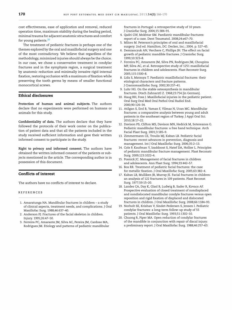
t c i r
r
1
1
1
1
1
1
1
1
1
1
170 r e v p o r t e s t o m a t o l m e d d e n
cost effectiveness, ease of application and removal, reducedoperation time, maximum stability during the healing period,minimal trauma for adjacent anatomic structures and comfortfor young patients.20
The treatment of pediatric fractures is perhaps one of thethemes explored by the oral and maxillofacial surgery and oneof the most contradictory. We believe that regardless of themethodology, minimized injuries should always be the choice.In our case, we chose a conservative treatment in condylarfractures and in the symphysis region, a surgical treatmentby anatomic reduction and minimally invasive rigid internalfixation, restoring occlusion with a maximum of fixation whilepreserving the tooth germs by means of smaller functionalmonocortical screws.
Ethical disclosures
Protection of human and animal subjects. The authorsdeclare that no experiments were performed on humans oranimals for this study.
Confidentiality of data. The authors declare that they havefollowed the protocols of their work center on the publica-tion of patient data and that all the patients included in thestudy received sufficient information and gave their writteninformed consent to participate in the study.
Right to privacy and informed consent. The authors haveobtained the written informed consent of the patients or sub-jects mentioned in the article. The corresponding author is inpossession of this document.
Conflicts of interest
The authors have no conflicts of interest to declare.
e f e r e n c e s
1. Amaratunga NA. Mandibular fractures in children – a studyof clinical aspects, treatment needs, and complications. J OralMaxillofac Surg. 1988;46:637–40.
2. Anderson PJ. Fractures of the facial skeleton in children.Injury. 1995;26:47–50.
3. Ferreira PC, Amarante JM, Silva AC, Pereira JM, Cardoso MA,Rodrigues JM. Etiology and patterns of pediatric mandibular
2
m a x i l o f a c . 2 0 1 3;54(3):166–170
fractures in Portugal: a retrospective study of 10 years.J Craniofac Surg. 2004;15:384–91.
4. Qadri GW, Mokhtar SM. Paediatric mandibular fractures:report of a case. Dent Traumatol. 2008;24:e67–70.
5. Miloro M. Peterson’s principles of oral and maxillofacialsurgery. 2nd ed. Hamilton, DC: Decker, Inc.; 2004. p. 527–45.
6. Demianczuk AN, Verchere C, Phillips JH. The effect on facialgrowth of pediatric mandible fractures. J Craniofac Surg.1999;10:323–8.
7. Ferreira PC, Amarante JM, Silva PN, Rodrigues JM, ChoupinaMP, Silva AC, et al. Retrospective study of 1251 maxillofacialfractures in children and adolescents. Plast Reconstr Surg.2005;115:1500–8.
8. Lida S, Matsuya T. Paediatric maxillofacial fractures: theiretiological characters and fracture patterns.J Craniomaxillofac Surg. 2002;30:237–41.
9. Luhr HG. On the stable osteosynthesis in mandibularfractures. Dtsch Zahnarztl Z. 1968;23:754 [in German].
0. Haug RH, Foss J. Maxillofacial injuries in the pediatric patient.Oral Surg Oral Med Oral Pathol Oral Radiol End.2000;90:126–34.
1. Atilgan S, Erol B, Yaman F, Yilmaz N, Ucan MC. Mandibularfractures: a comparative analysis between young and adultpatients in the southeast region of Turkey. J Appl Oral Sci.2010;18:17–22.
2. Davison PS, Clifton MS, Davison MN, Hedrick M, Sotereanos G.Pediatric mandibular fractures: a free hand technique. ArchFacial Plast Surg. 2001;3:185–9.
3. Zimmermann CE, Troulis MJ, Kaban LB. Pediatric facialfractures: recent advances in prevention, diagnosis andmanagement. Int J Oral Maxillofac Surg. 2006;35:2–13.
4. Cole P, Kaufman Y, Izaddoost S, Hatef DA, Hollier L. Principlesof pediatric mandibular fracture management. Plast ReconstrSurg. 2009;123:1022–4.
5. Posnick JC. Management of facial fractures in childrenand adolescents. Ann Plast Surg. 1994;33:442–57.
6. Bos RR. Treatment of pediatric facial fractures: the casefor metallic fixation. J Oral Maxillofac Surg. 2005;63:382–4.
7. Kaban LB, Mulliken JB, Murray JE. Facial fractures in children:an analysis of 122 fractures in 109 patients. Plast ReconstSurg. 1977;59:15–20.
8. Landes CA, Day K, Glasl B, Ludwig B, Sader R, Kovacs AF.Prospective evaluation of closed treatment of nondisplacedand nondislocated mandibular condyle fractures versus openreposition and rigid fixation of displaced and dislocatedfractures in children. J Oral Maxillofac Surg. 2008;66:1184–93.
9. Norholt SE, Krishan V, Sindet-Pedersen S, Jensen I. Pediatriccondylar fractures: a long-term follow-up study of 55
patients. J Oral Maxillofac Surg. 1993;51:1302–10.0. Chuong R, Piper MA. Open reduction of condylar fracturesof the mandible in conjunction with repair of discal injury:a preliminary report. J Oral Maxillofac Surg. 1988;46:257–63.