management of the emergent airway - university of texas ... · pdf filemanagement of the...
TRANSCRIPT
Benjamin Walton, M.D.
Faculty Advisor: Michael Underbrink, M.D. The
University of Texas Medical Branch
Department of Otolaryngology
Grand Rounds Presentation
March 31, 2011
Management of the Emergent Airway
Table of Contents
I. The Difficult Airway
Evaluation
II. Non-surgical Options
III. Surgical Options
IV. Controversies
V. Management of the Pediatric Patient
The Difficult Airway
Problematic ventilation using a face mask
Inability to deliver necessary tidal volume via face
mask utilizing nasal or oral airway
Incomplete laryngoscopic visualization
Cormack and Lehane grade 3 or 4
Difficult Intubation with standard airway equipment
Requiring external laryngeal manipulation
Greater than 3 attempts at intubation
Requiring nonstandard equipment
Evaluation of the Airway
Elective Versus Urgent
Adult Versus Pediatric
Patient Status
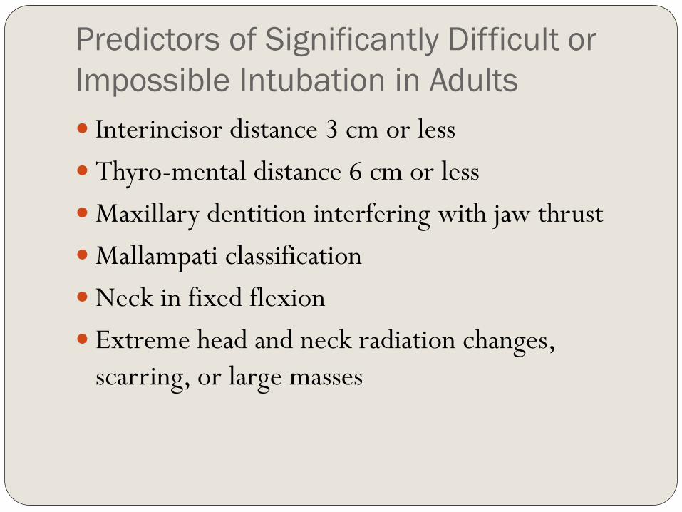
Predictors of Significantly Difficult or
Impossible Intubation in Adults
Interincisor distance 3 cm or less
Thyro-mental distance 6 cm or less
Maxillary dentition interfering with jaw thrust
Mallampati classification
Neck in fixed flexion
Extreme head and neck radiation changes,
scarring, or large masses
Airway Management Team
Anesthesiologist
Otolaryngologist
Emergency Physician
Respiratory Therapist
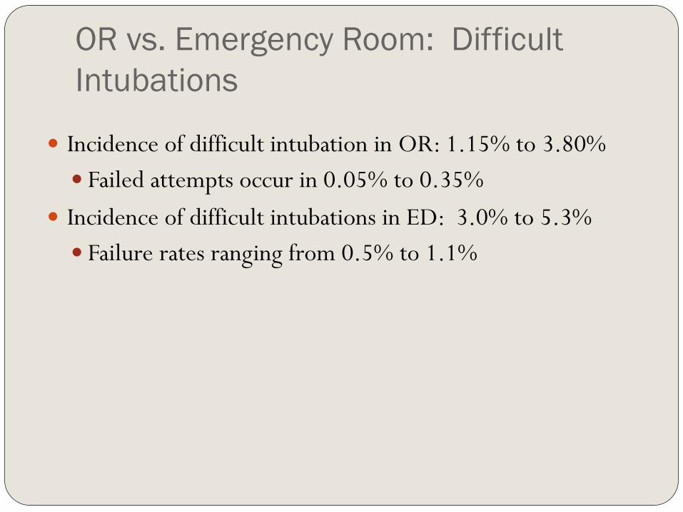
OR vs. Emergency Room: Difficult
Intubations
Incidence of difficult intubation in OR: 1.15% to 3.80%
Failed attempts occur in 0.05% to 0.35%
Incidence of difficult intubations in ED: 3.0% to 5.3%
Failure rates ranging from 0.5% to 1.1%
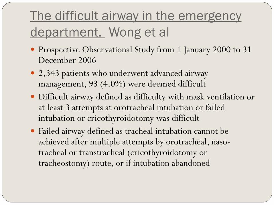
The difficult airway in the emergency
department. Wong et al
Prospective Observational Study from 1 January 2000 to 31 December 2006
2,343 patients who underwent advanced airway management, 93 (4.0%) were deemed difficult
Difficult airway defined as difficulty with mask ventilation or at least 3 attempts at orotracheal intubation or failed intubation or cricothyroidotomy was difficult
Failed airway defined as tracheal intubation cannot be achieved after multiple attempts by orotracheal, naso-tracheal or transtracheal (cricothyroidotomy or tracheostomy) route, or if intubation abandoned
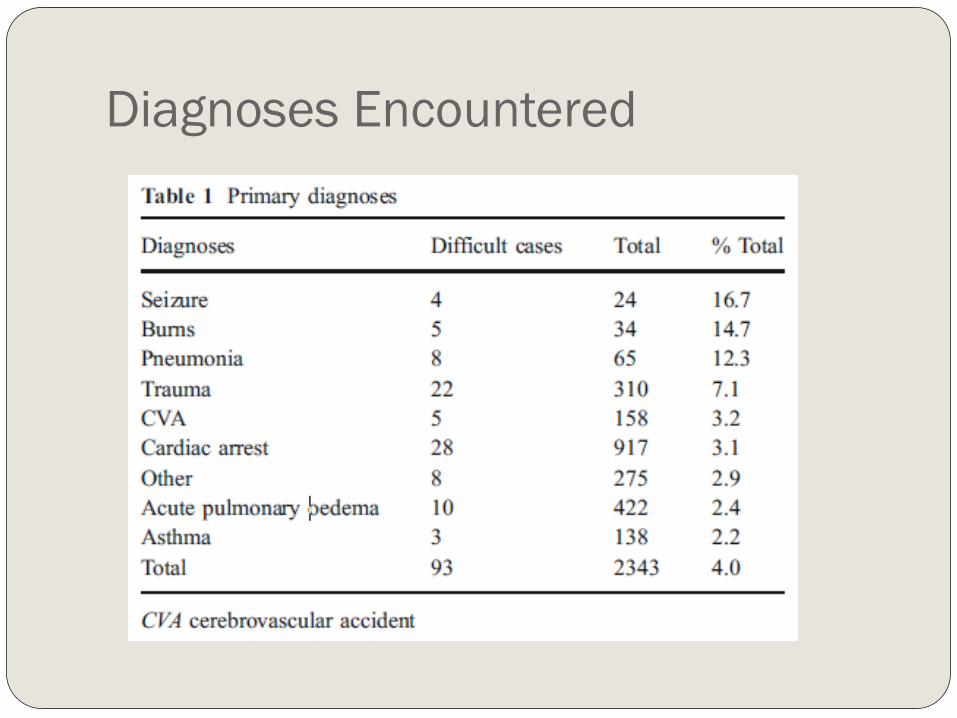
Diagnoses Encountered
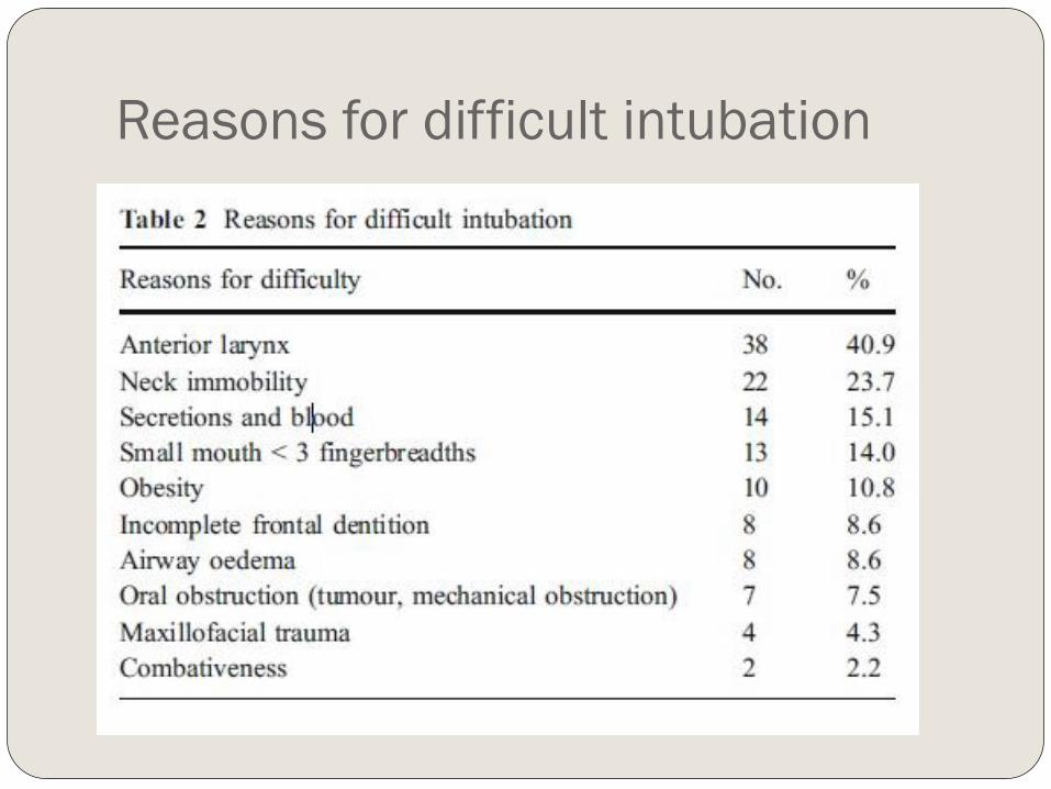
Reasons for difficult intubation
Results (continued)
Mean number of attempts at intubation for
difficult airway group was 3.6 compared to 1.2
for all patients
Surgical airway rate was 0.3%
Difficult intubation rate: 4%
Failed intubation rate: 0.7%
Causes of a Difficult Airway
Trauma
Midface
Mandible
Neck
Bleeding into airway
Caustic ingestion
Thermal burns
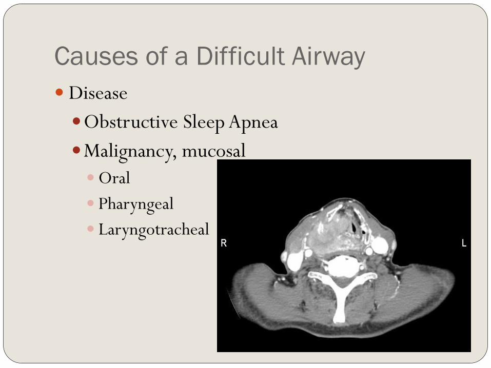
Causes of a Difficult Airway
Disease
Obstructive Sleep Apnea
Malignancy, mucosal
Oral
Pharyngeal
Laryngotracheal
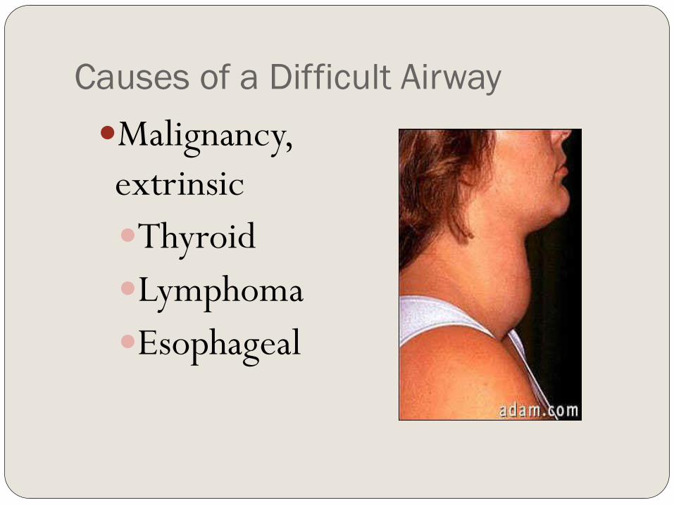
Causes of a Difficult Airway
Malignancy,
extrinsic
Thyroid
Lymphoma
Esophageal
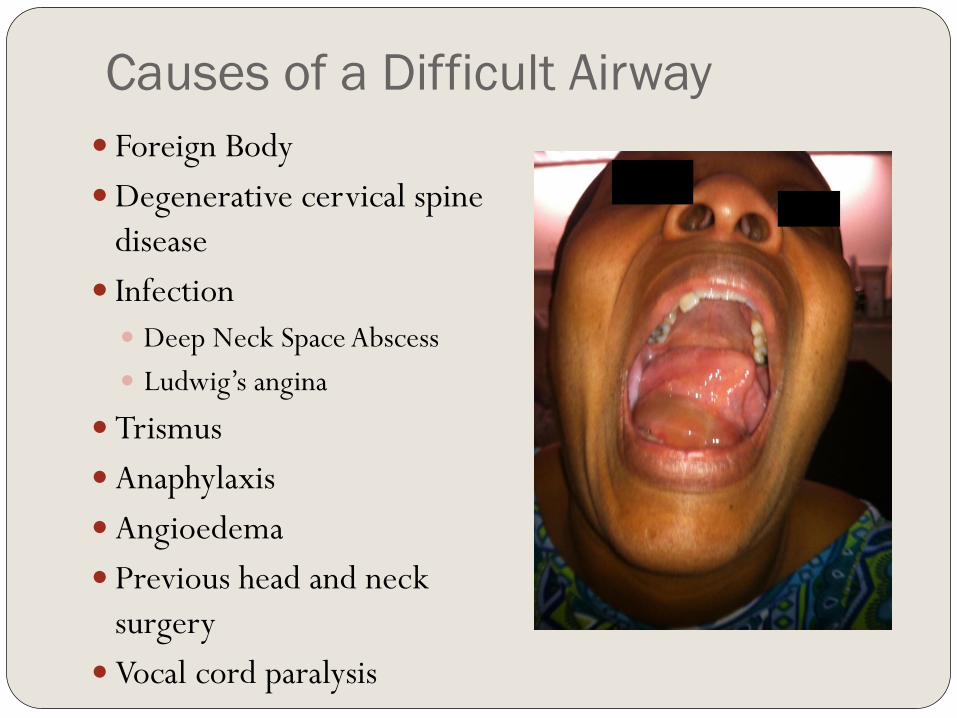
Causes of a Difficult Airway
Foreign Body
Degenerative cervical spine
disease
Infection
Deep Neck Space Abscess
Ludwig’s angina
Trismus
Anaphylaxis
Angioedema
Previous head and neck
surgery
Vocal cord paralysis
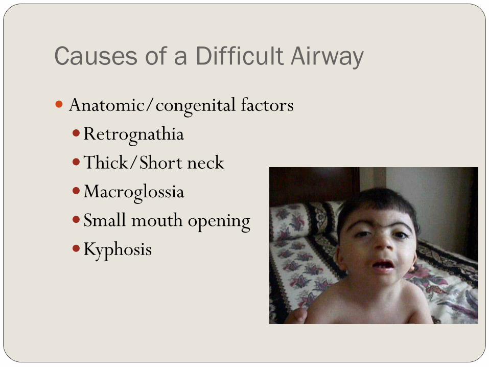
Causes of a Difficult Airway
Anatomic/congenital factors
Retrognathia
Thick/Short neck
Macroglossia
Small mouth opening
Kyphosis
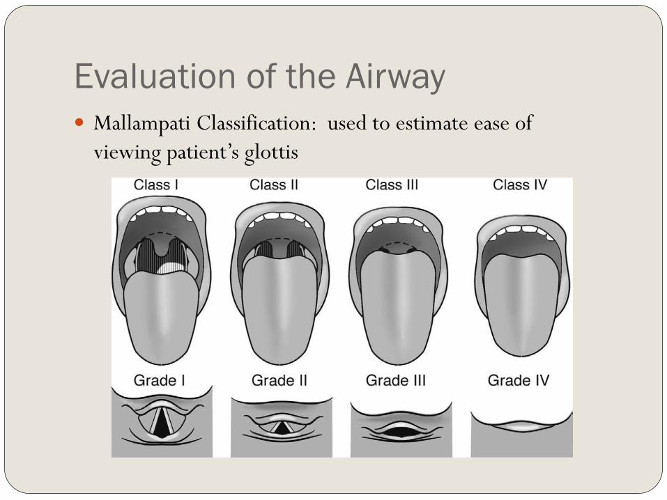
Evaluation of the Airway
Mallampati Classification: used to estimate ease of
viewing patient’s glottis
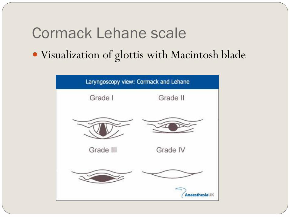
Cormack Lehane scale
Visualization of glottis with Macintosh blade
Questions I Need to Ask Myself
Can I perform effective mask ventilation?
Incidence of difficult mask ventilation in adult patients in OR is
1.4-5%
Defined as inability to maintain O2 sat >90% with 100%
inspired O2 or to prevent or reverse signs of hypoventilation
with positive-pressure mask ventilation
Can I safely intubate this patient?
Although tracheal intubation is the ultimate goal in airway
management, the ability to provide effective mask-ventilation is
life-saving (1)
Prediction and Outcomes of Impossible
Mask Ventilation
Kheterpal et al. reviewed 50,000 patients undergoing
airway management in the operating room
Impossible mask ventilation in 0.15% of cases
Predictors included previous neck radiation, male sex,
OSA, Mallampati class III or IV, and presence of a beard
25% (19/77) of patients with impossible mask
ventilation were difficult to intubate.
2 of those patients required surgical airways
Pre-oxygenation
Healthy adults breathing room air will develop
oxygen desaturation (Sp02 <90%) with 2
minutes of apnea
Preoxygenation with 100% oxygen can maintain
oxygen saturation above 90% for more than 6
minutes
4 vital-capacity breaths in 30 seconds or 8 vital
capacity breaths in 60 seconds provide adequate
pre-oxygenation
Monitoring During Anesthesia
American Society of Anesthesiology
Standards for Basic Anesthetic Monitoring
released in 1986
Continual monitoring of oxygen,
ventilation, circulation, temperature
Intermittent (no less frequent than every 5
minutes) measurement of arterial blood
pressure and heart rate

Anesthetic Pharmacology
Induction Agents
IV induction agents have quick onset, produce
unconsciousness within 1-2 minutes
Thiopental and Propofol: negative inotropic effect and
produces apnea along with unconsciousness
Etomidate: less effects on hemodynamics, potential
adrenal suppression and myoclonic activity
Ketamine: does not produce apnea with administration,
can give IM, can cause tachycardia and hypertension
along with exaggerated secretions
Volatile Anesthetic Agents
Used for maintenance in most instances
Halothane- nonflammable, alkane, lacks bronchoirritant
effects, highly soluble in blood and fatty tissues, negative
inotropic effects on cardiac muscles
Enflurane and Isoflurane: nonflammable, pungent odor,
produce more intrinsic respiratory depression, less
cardiac effects (Enflurane now rarely used due to risk of
renal toxicity and seizure)
Sevoflurane and Desflurane: low lipid solubility, cause
little myocardial depression, Desflurane has
bronchoirritative properties
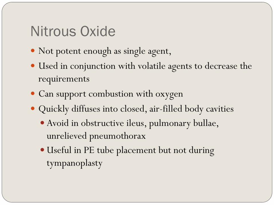
Nitrous Oxide
Not potent enough as single agent,
Used in conjunction with volatile agents to decrease the
requirements
Can support combustion with oxygen
Quickly diffuses into closed, air-filled body cavities
Avoid in obstructive ileus, pulmonary bullae,
unrelieved pneumothorax
Useful in PE tube placement but not during
tympanoplasty
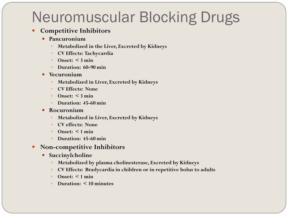
Neuromuscular Blocking Drugs Competitive Inhibitors
Pancuronium Metabolized in the Liver, Excreted by Kidneys
CV Effects: Tachycardia
Onset: < 3 min
Duration: 60-90 min
Vecuronium Metabolized in Liver, Excreted by Kidneys
CV Effects: None
Onset: < 3 min
Duration: 45-60 min
Rocuronium Metabolized in Liver, Excreted by Kidneys
CV effects: None
Onset: < 1 min
Duration: 45-60 min
Non-competitive Inhibitors Succinylcholine
Metabolized by plasma cholinesterase, Excreted by Kidneys
CV Effects: Bradycardia in children or in repetitive bolus to adults
Onset: < 1 min
Duration: < 10 minutes
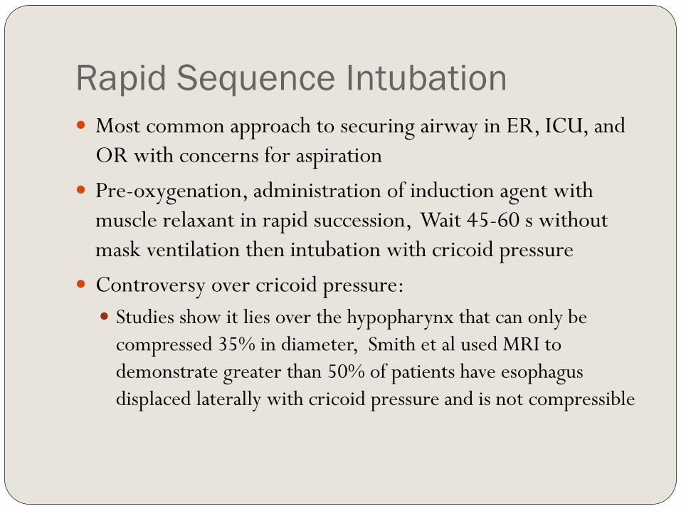
Rapid Sequence Intubation
Most common approach to securing airway in ER, ICU, and
OR with concerns for aspiration
Pre-oxygenation, administration of induction agent with
muscle relaxant in rapid succession, Wait 45-60 s without
mask ventilation then intubation with cricoid pressure
Controversy over cricoid pressure:
Studies show it lies over the hypopharynx that can only be
compressed 35% in diameter, Smith et al used MRI to
demonstrate greater than 50% of patients have esophagus
displaced laterally with cricoid pressure and is not compressible
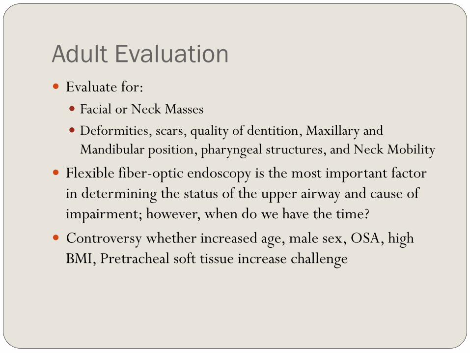
Adult Evaluation
Evaluate for:
Facial or Neck Masses
Deformities, scars, quality of dentition, Maxillary and
Mandibular position, pharyngeal structures, and Neck Mobility
Flexible fiber-optic endoscopy is the most important factor
in determining the status of the upper airway and cause of
impairment; however, when do we have the time?
Controversy whether increased age, male sex, OSA, high
BMI, Pretracheal soft tissue increase challenge
Pediatric Evaluation
Noisy breathing during exercise, at rest or when feeding
Previous surgeries or Intubations
Neck pain, fever, recent upper respiratory infections
Birth Trauma
Congenital Anomalies
Difficult Airway Algorithm
Accepted by the American Society of Anesthesiologists
4 main steps in the process of developing the strategy for
acquiring control of the airway
(1) Assess the likelihood and clinical impact of basic
management problems Difficult ventilation
Difficult intubation
Difficulty with Patient Cooperation or Consent
Difficult Tracheostomy
Algorithm continued (2) Actively pursue opportunities to deliver supplemental
oxygen throughout the process of difficult airway management
(3) Consider the relative merits and feasibility of basic management choices
Awake intubation vs. Intubation Attempts After Induction of General Anesthesia
Non-invasive Technique for Initial Approach to Intubation vs. Invasive Technique for Initial Approach to Intubation
Preservation of Spontaneous Ventilation vs. Ablation of Spontaneous Ventilation
(4) Develop primary and alternative strategies
Scenario 1 You decide that the best approach is an awake intubation
Decision is between approaching the airway by non-invasive
intubation or invasive airway access (tracheostomy vs.
cricothyroidotomy)
You succeed !!!
Hooray, you have confirmed ventilation,
tracheal intubation, or LMA placement
with exhaled CO2
Or -- You Have Failed
(1) Consider cancelling the case
(2) Consider feasibility of other options:
face mask or LMA, local anesthesia
infiltration or regional nerve block (not
really an option for many of our cases)
(3) Invasive Airway Access
Scenario 2
Intubation Attempts After Induction of
General Anesthesia
Major decision tree based on ability to
adequately ventilate through face mask
Tube is in the trachea
Congrats, you are done. Go on your coffee break then
20 minutes later, go on your lunch break, then read the
paper for 30 minutes while various beeps go off. Take
out a syringe and then push some saline into the IV.
Complain for a while about the length of the OR case
and why you wish your stocks were doing better. Flirt
with the scrub nurse.
Initial Intubation Attempt Unsuccessful
From here on out consider:
Calling for Help,
Return to Spontaneous Ventilation,
Awakening the Patient
Attempt Face Mask Ventilation
If adequate, head toward non-emergent
pathway
Alternative Approaches to Intubation
Using different laryngoscope blades
LMA as an intubation conduit (with or without
fiberoptic guidance)
Fiberoptic Intubation
Intubating stylet or tube changer
Light wand
Retrograde Intubation
Blind oral or nasal intubation
Failing after Multiple Attempts
Invasive Airway Access
Consider other options
Awaken the patient
If Face Mask Ventilation Not Adequate
First Consider or Attempt to place LMA
If the LMA is adequate then you can attempt alternative
approaches to intubation
If LMA is not adequate or not feasible:
Call for Help
Emergency Non-Invasive Airway Ventilation
Rigid Bronchoscopy
Esophageal-Tracheal Combitube
Transtracheal jet ventilation
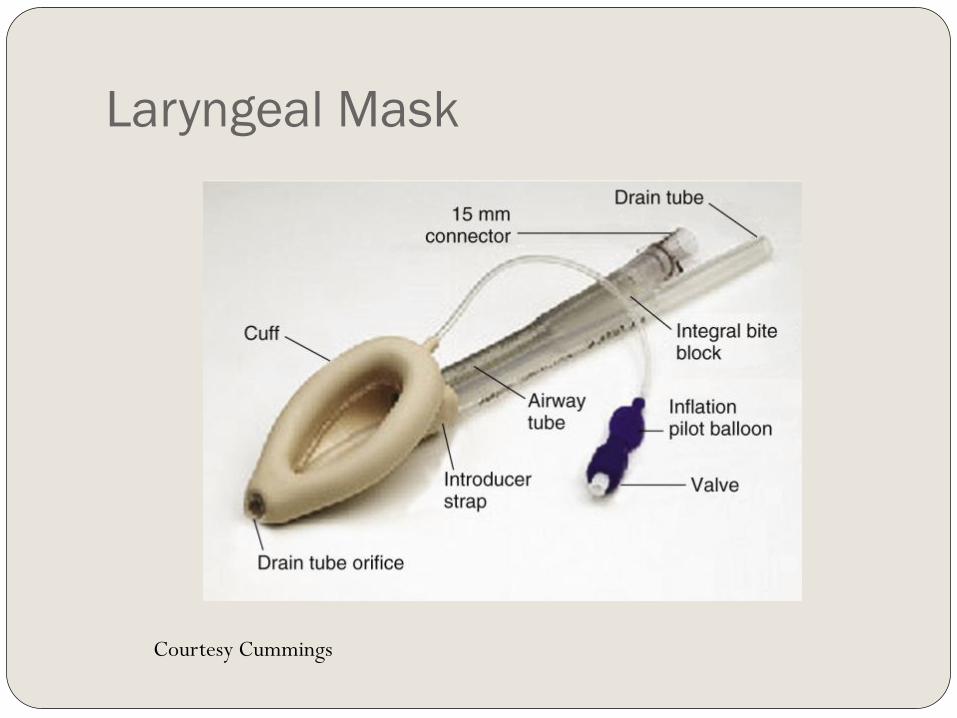
Laryngeal Mask
Courtesy Cummings
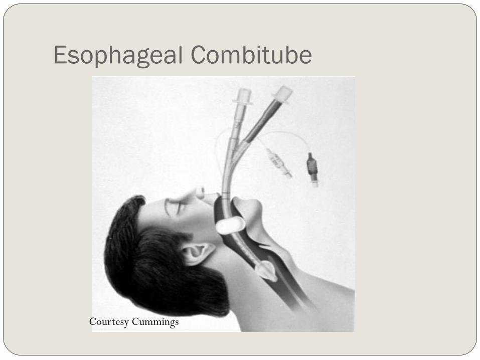
Esophageal Combitube
Courtesy Cummings
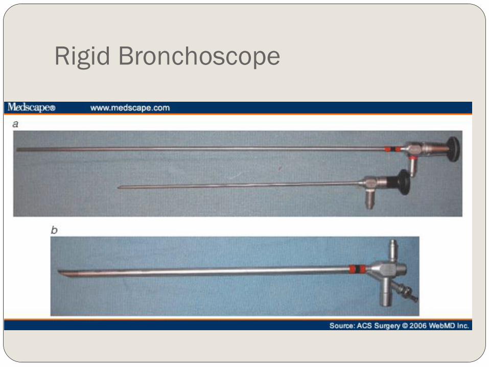
Rigid Bronchoscope
If Emergency Non-invasive Ventilation
is Successful
Consider invasive airway access
Consider feasibility of other options as
discussed before
Awaken the Patient
If Emergency Non-Invasive Ventilation
is Unsuccessful
Emergency Invasive Airway Access
That would be us.
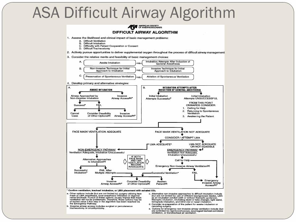
ASA Difficult Airway Algorithm
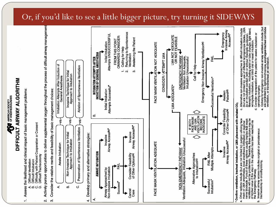
Or, if you’d like to see a little bigger picture, try turning it SIDEWAYS
Non-surgical Options
Face Mask Ventilation
Endotracheal Intubation
LMA
Combitube
Fiberoptic Nasotracheal Intubation
Face Mask Ventilation
Essential element to airway management
Used during induction and as a rescue technique
during failed attempts
More difficult in bearded patients
Using a nasal trumpet or oral airway may assist in
ventilation
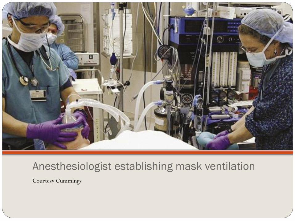
Anesthesiologist establishing mask ventilation Courtesy Cummings
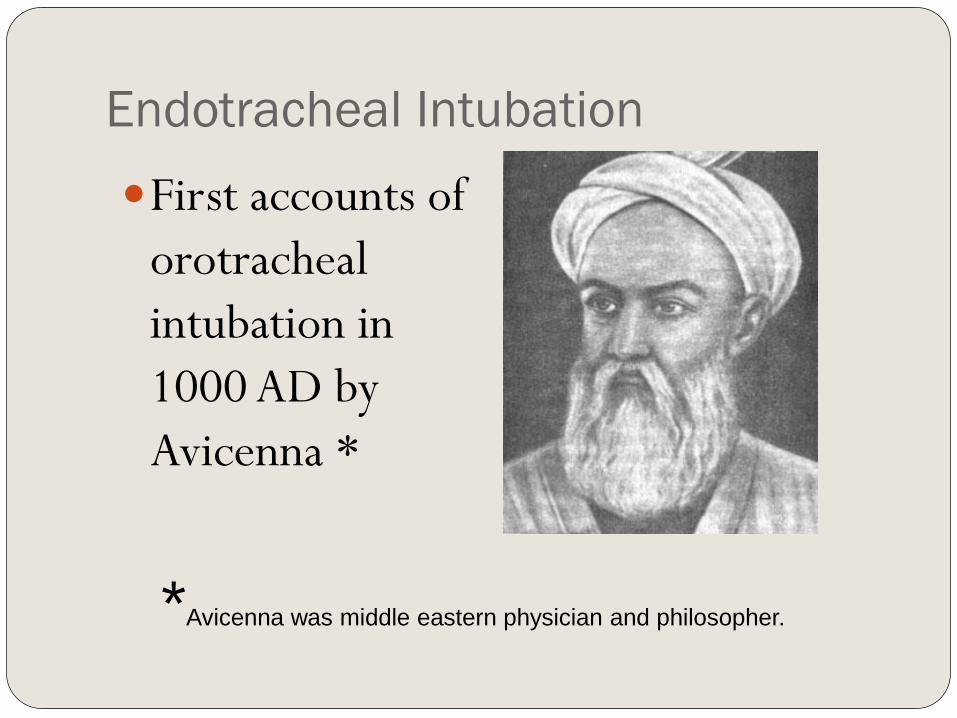
Endotracheal Intubation
First accounts of
orotracheal
intubation in
1000 AD by
Avicenna *
*Avicenna was middle eastern physician and philosopher.
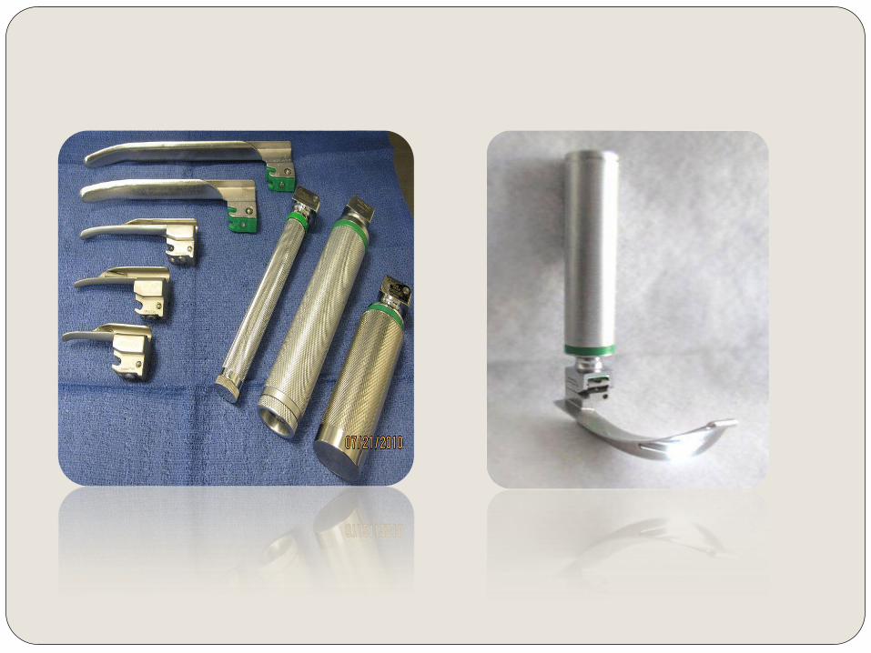
Oroendotracheal intubation
Utilizes either Macintosh or Miller Blade
Macintosh is a curved laryngoscope that slides under the vallecula
and lifts the entire larynx anteriorly or ventrally to expose the
glottis
Miller is a straight laryngoscope and is placed under the epiglottis
where it sits in the petiole of the epiglottis lifting the larynx
anteriorly to view the glottis
If not successful on initial attempt, 3 maneuvers are helpful
Place patient in modified Jackson’s position
Apply external laryngeal pressure
Maneuver Laryngoscope
After 3 failed attempts, proceed with alternative procedures
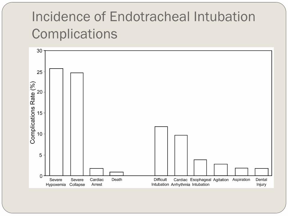
Incidence of Endotracheal Intubation
Complications
LMA
Initially introduced in the U.S in early 1990s
First utilized for elective face mask cases requiring
general anesthesia
Has revolutionized the algorithm for difficult airway
management
Is fast and easy to place, even by inexperience personnel
Does not offer full protection of airway from aspiration
Can intubate patient through LMA
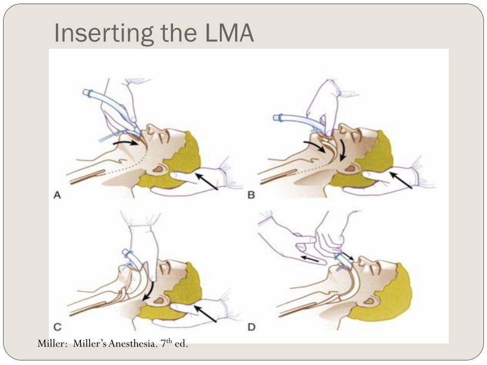
Inserting the LMA
Miller: Miller’s Anesthesia. 7th ed.
Combitube
Widely used in prehospital care
Closed distal end designed for passage into esophagus
with inflatable cuff providing seal
Proximal seal achieved with facemask or oropharyngeal
cuff
Holes in tube between proximal seal and distal cuff
deliver gases to laryngopharynx
Has second open-ended tube that can function as
tracheal tube if inserted into trachea
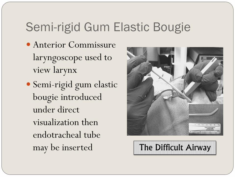
Semi-rigid Gum Elastic Bougie
Anterior Commissure
laryngoscope used to
view larynx
Semi-rigid gum elastic
bougie introduced
under direct
visualization then
endotracheal tube
may be inserted The Difficult Airway
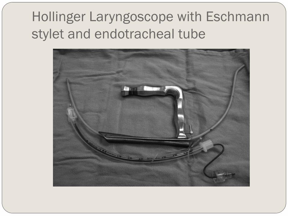
Hollinger Laryngoscope with Eschmann
stylet and endotracheal tube
Nasotracheal Intubation
Fiberoptic flexible bronchoscope is passed transnasally
after passing through endotracheal tube
Endoscope introduced into the subglottic trachea then
endotracheal tube passed over the endoscope
Contraindications include history or possible basal skull
fracture
Can cause damage to nasal mucosa creating epistaxis and
can tear mucosa of posterior nasopharynx passing
submucosally
Retrograde Intubation
Percutaneously passing narrow flexible
guide into the trachea from site below the
vocal cords, advancing guide out of mouth
or nose
Endotracheal tube then passed over the
guide into the upper part of the trachea
Can perform either oral or nasal intubation
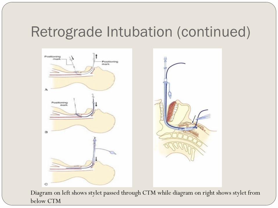
Retrograde Intubation (continued)
Diagram on left shows stylet passed through CTM while diagram on right shows stylet from
below CTM
Retrograde Intubation Invasive and takes time
Complications: Minor bleeding Subcutaneous emphysema Pneumomediastinum Infection
Contraindications: Coagulopathy Inability to identify landmarks Laryngeal disease Local infection
Surgical Management of the Difficult Airway
Awake Tracheostomy
Emergency Cricothyroidotomy
Transtracheal Needle Ventilation
Emergency Tracheostomy
Awake Tracheostomy
When intubation is deemed impractical
Best performed in a controlled environment
Requires clear communication among surgeon,
anesthesiologist, nurses, and technicians
Place patient where anesthesia can have ready access to
airway and to improve primary and accessory respiratory
muscle function
Under local anesthesia with minimal sedation to keep patient
comfortable
Emergency Cricothyroidotomy
Relatively simple, fast and with lower
perioperative complication rate
Cricothyroid Membrane only separated from skin
by subcutaneous fat, anterior cervical fascia and
strap muscles laterally
Vocal cords are approximately 1 cm above
Cricothyroid membrane
Contraindications to Cricothyroidotomy
Age less than 10 years of age
Severe neck trauma with inability to palpate
landmarks
Expanding neck hematoma
Preexisting laryngeal disease with subglottic
extension
Planned urgent awake tracheostomy preferred
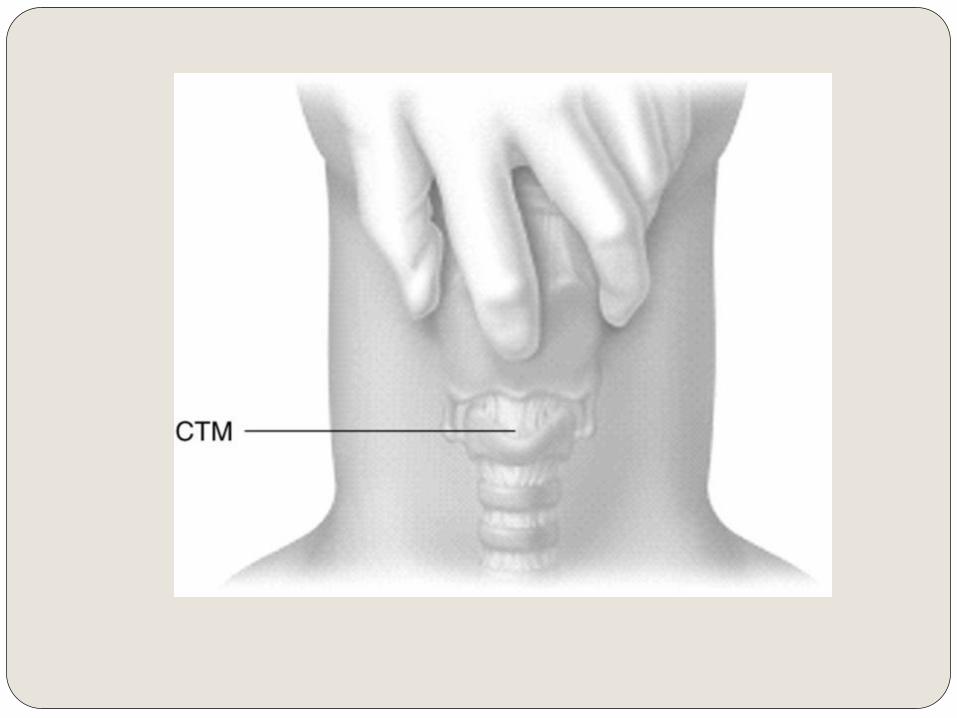
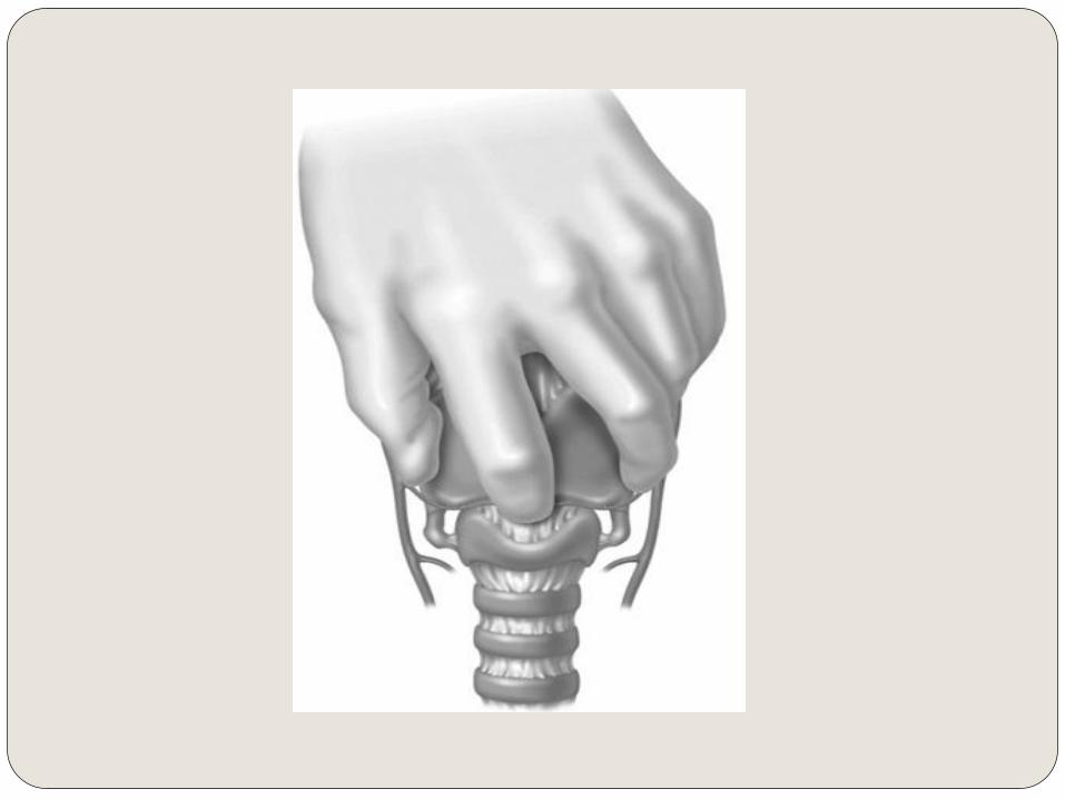
Procedure
Palpate the landmarks
Stabilize upper airway with nondominant hand is single
most important factor in successful outcome
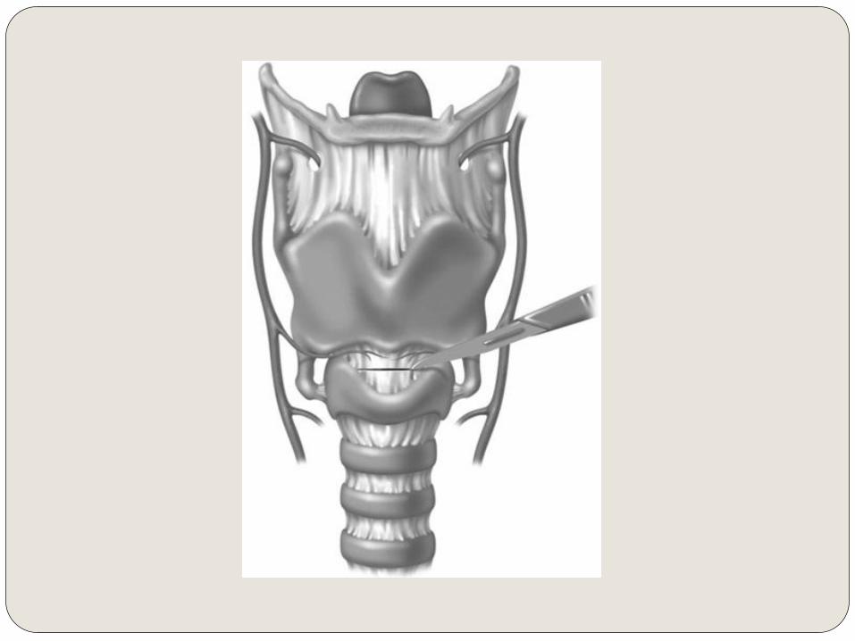
Perform midline vertical skin incision
Palpate membrane through incision and enter with a
horizontal incision at lower edge of membrane
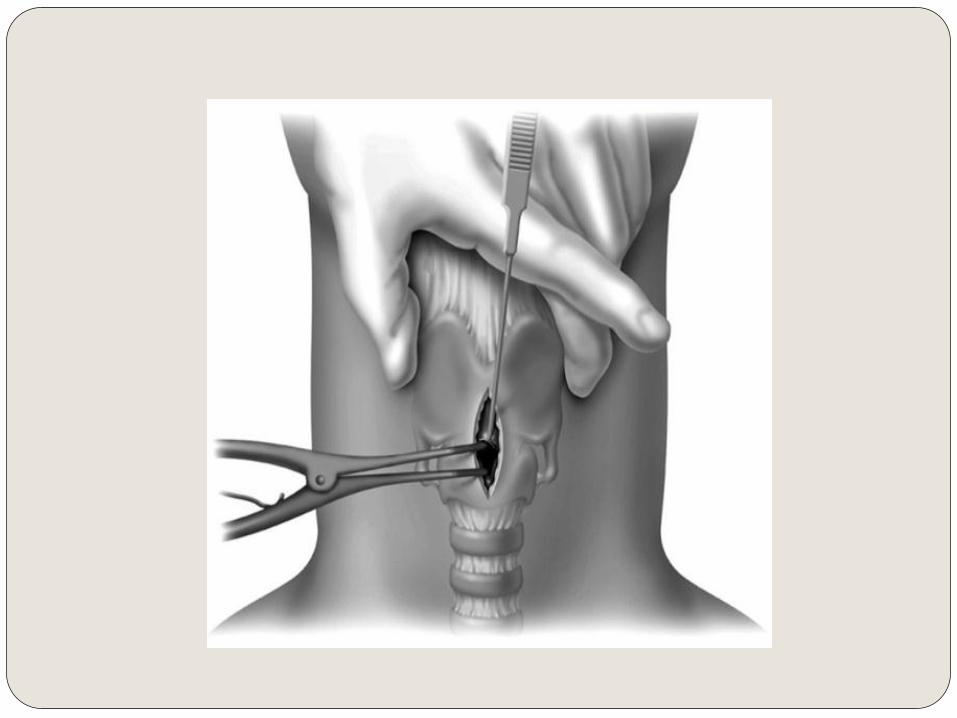
Dilate with a hemostat or Kelly clamp
Place small (5.0) endotracheal tube or tracheostomy
tube
Transtracheal Needle Ventilation
Equipment:
100% oxygen at 50 psi
Large-bore needle and cannula (14 G)
Luer-lok connector
Puncture either trachea or Cricothyroid Membrane with
saline-filled syringe in midline at 30-degree caudal
direction
Patient can be oxygenated for 30 minutes to 2 hours
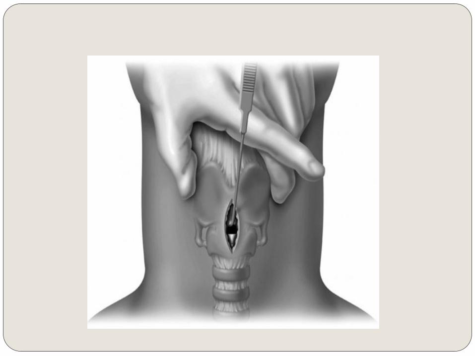
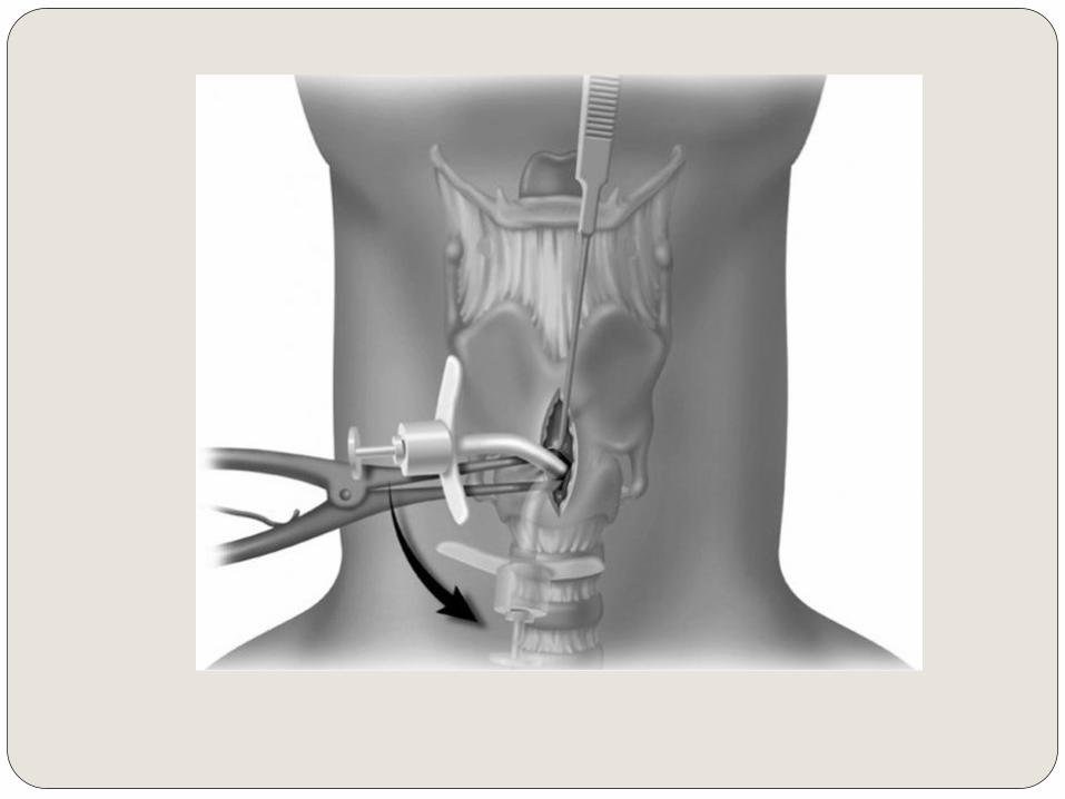
Emergency Tracheostomy “Slash Trach”
Since anoxia causes death in 4-5 minutes, so must perform within 2 or 3 minutes
Perform through a vertical incision
Stabilize the larynx with non-dominant hand
Vertical incision through skin, platysma and subcutaneous tissue and thyroid isthmus
Use index finger of non-dominant finger to help palpate and dissect
Make vertical incision at second or third tracheal ring
If available, use reinforced endotracheal tube
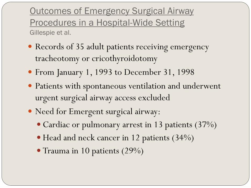
Outcomes of Emergency Surgical Airway
Procedures in a Hospital-Wide Setting Gillespie et al.
Records of 35 adult patients receiving emergency
tracheotomy or cricothyroidotomy
From January 1, 1993 to December 31, 1998
Patients with spontaneous ventilation and underwent
urgent surgical airway access excluded
Need for Emergent surgical airway:
Cardiac or pulmonary arrest in 13 patients (37%)
Head and neck cancer in 12 patients (34%)
Trauma in 10 patients (29%)
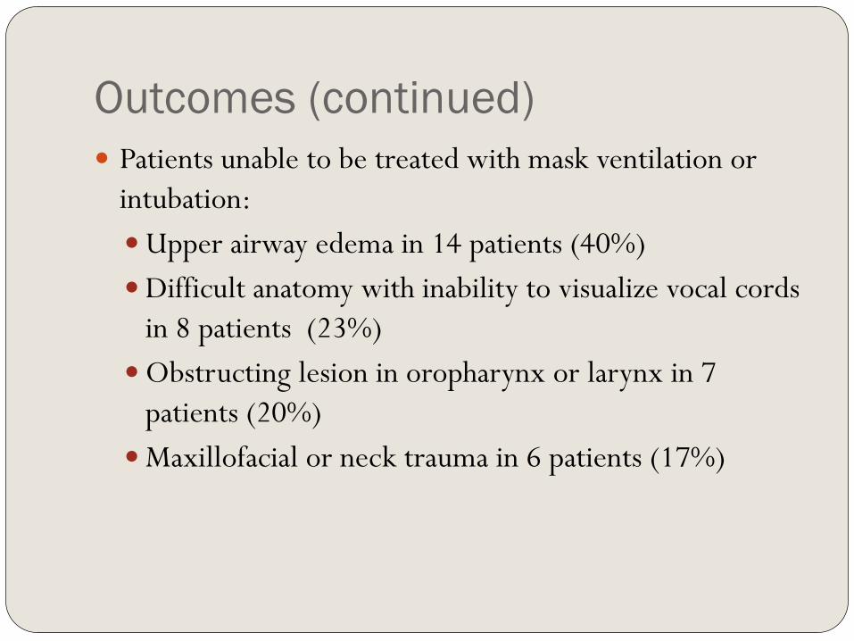
Outcomes (continued)
Patients unable to be treated with mask ventilation or
intubation:
Upper airway edema in 14 patients (40%)
Difficult anatomy with inability to visualize vocal cords
in 8 patients (23%)
Obstructing lesion in oropharynx or larynx in 7
patients (20%)
Maxillofacial or neck trauma in 6 patients (17%)
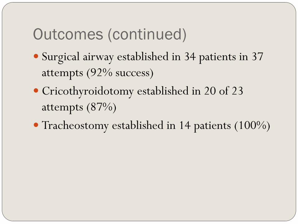
Outcomes (continued)
Surgical airway established in 34 patients in 37
attempts (92% success)
Cricothyroidotomy established in 20 of 23
attempts (87%)
Tracheostomy established in 14 patients (100%)
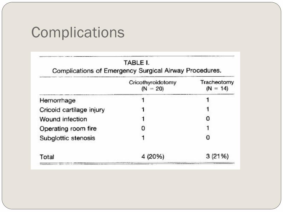
Complications
Complications (continued)
Less known about role of tracheotomy in emergency
situation
In study, effective 100% of attempts
Emergency cricothyroidotomy associated with 13% to
40% complication rate
Elective Tracheostomy has complication rate of 15%,
complication rate of emergent tracheostomy thought to
be 2 to 5 times higher
In present study, equivalent complication rates between
both procedures
Controversy in Cricothyroidotomy Principal long-term morbidity is subglottic stenosis
Intense debate since publication of Chevalier Jackson’s article on tracheostomy in 1921 discouraging the use of elective cricothyroidotomy due to high rate of subglottic stenosis High rate in study secondary to use of metal tracheostomy tubes in
pediatric patients with chronic inflammatory disease of laryngotracheal framework
Elective cricothyroidotomy shown to have low rate of subglottic stenosis (<1%); Avoid in patients treated with intubation for more than 7 days
True incidence of subglottic and tracheal stenosis is difficult to determine as many patients lost to follow-up
Converting from Cricothyroidotomy to
Tracheostomy
Controversy surrounds whether to expeditiously convert emergency
cricothyroidotomy to tracheostomy
Conversion advocated by many authors to decrease incidence of
subglottic stenosis
DeLaurier et al questioned need to convert, 0 of 11 patients they
studied who underwent cricothyroidotomy developed stenosis
2 of the 9 patients who underwent conversion developed tracheal
granulation tissue secondary to tracheostomy tube
Conversion did not prevent the only case of cricothyroidotomy-related
subglottic stenosis
Trauma patients decannulated after average of 3 days and had lower
rate of complication than patients undergoing conversion to
tracheostomy
When to consider conversion
Patients requiring operative exploration for
hemorrhage or other complications including
suspected laryngeal cartilage injury
Patients requiring long-term airway maintenance
or mechanical ventilation
Management of the Pediatric Patient
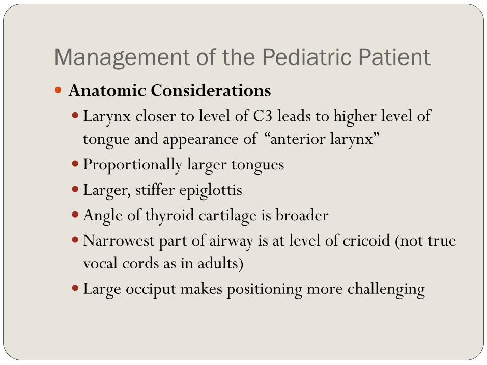
Anatomic Considerations
Larynx closer to level of C3 leads to higher level of
tongue and appearance of “anterior larynx”
Proportionally larger tongues
Larger, stiffer epiglottis
Angle of thyroid cartilage is broader
Narrowest part of airway is at level of cricoid (not true
vocal cords as in adults)
Large occiput makes positioning more challenging
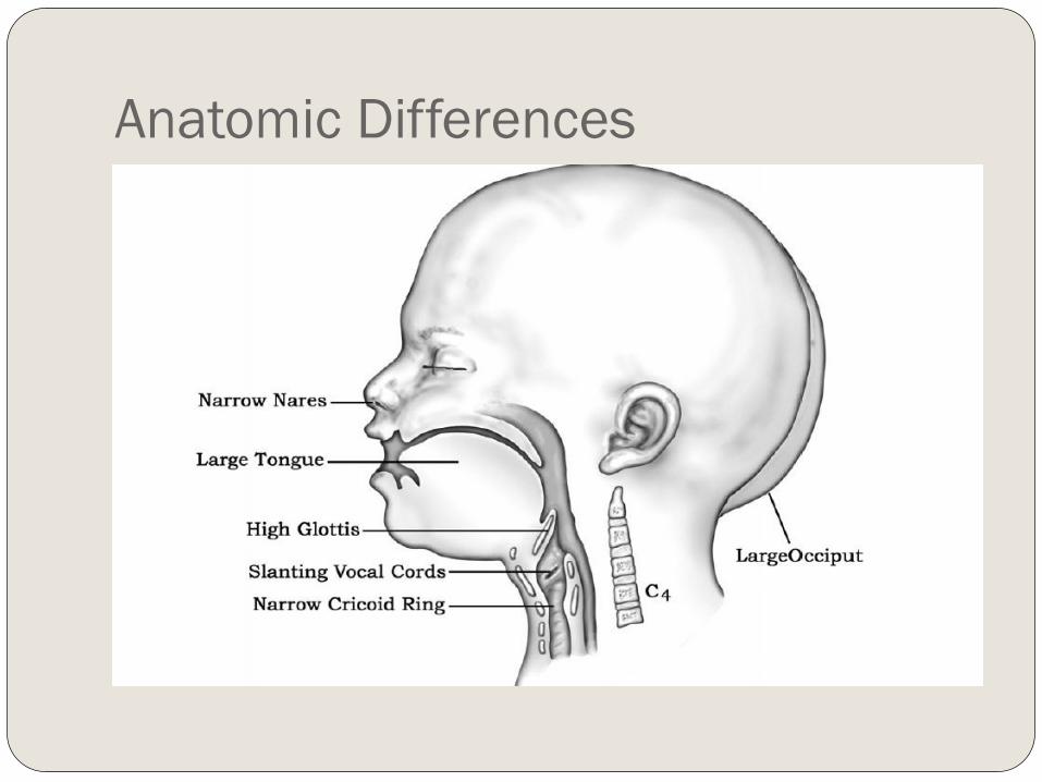
Anatomic Differences
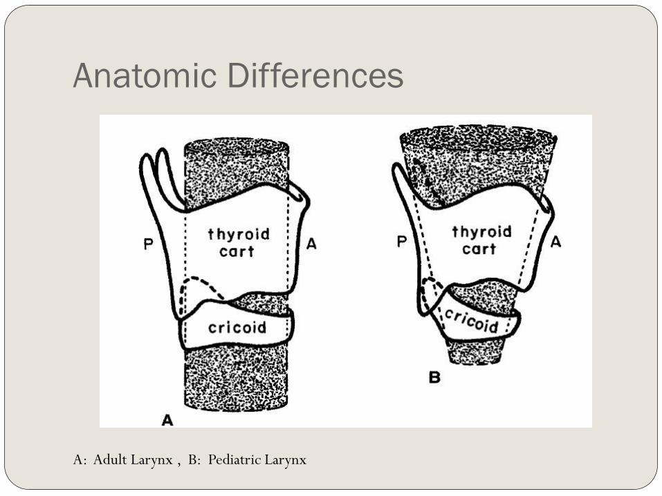
Anatomic Differences
A: Adult Larynx , B: Pediatric Larynx
General Considerations in Pediatric
Airway Management
Resistance to flow is inversely proportional to
radius of the lumen to the fourth power, makes
similar changes in edema more substantial in
pediatrics
Estimate endotracheal tubes using formula (Age
+ 16)/4 = ETT size
Pre-operative medications should be given that
have minimal respiratory depressant effects, such
as Midazolam
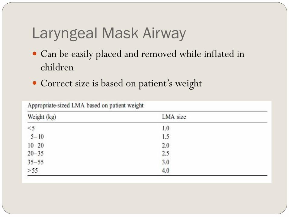
Laryngeal Mask Airway
Can be easily placed and removed while inflated in
children
Correct size is based on patient’s weight
Light Wand
Rigid fiber optic stylet with light at its tip
Blind Technique for intubation
Does not depend on good mouth opening or extension
of the neck
Can be simpler and quicker than fiberoptic intubation,
can succeed even with bleeding in airway
Requires room lights dimmed
Difficult to use when anatomy is distorted and laryngeal
structures not midline
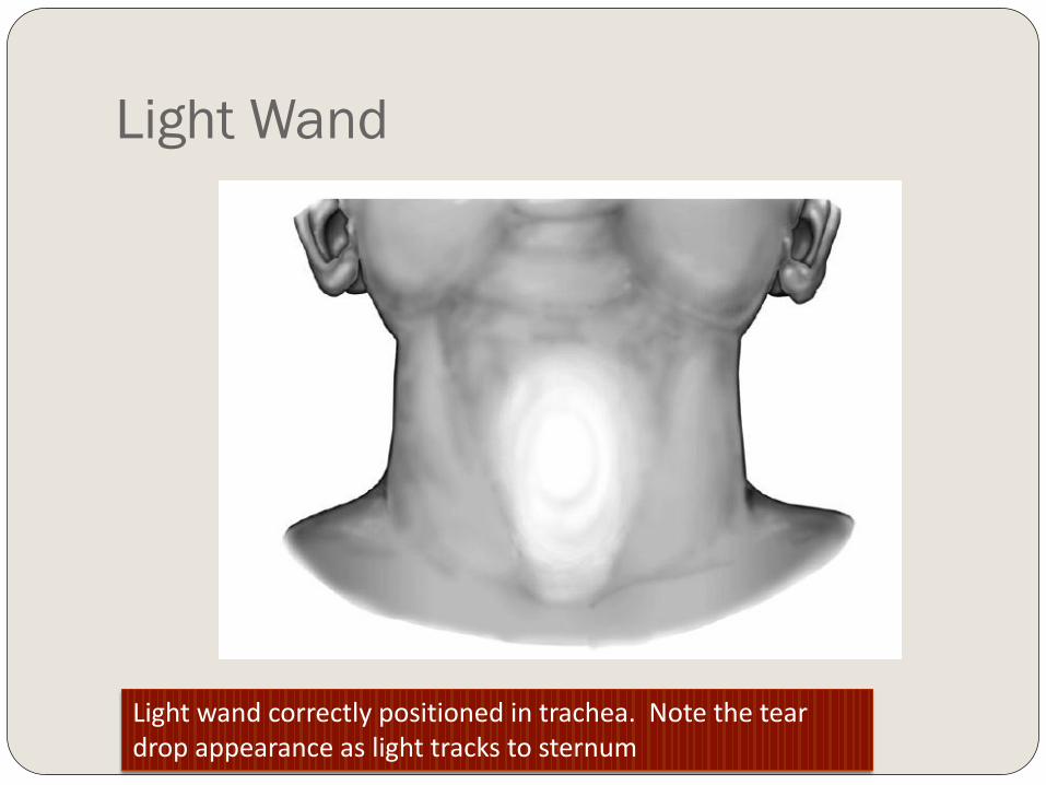
Light Wand
Light wand correctly positioned in trachea. Note the tear drop appearance as light tracks to sternum
Flexible Fiberoptic Bronchoscope
Allows operator to see around corners
Advantageous in patients with poor mouth opening,
limited neck movement or other conditions that make
direct laryngoscopy difficult
Bleeding and secretions cane make it difficult to use
Keep child anesthetized but breathing spontaneously at
100% oxygen
LMA is good adjunct in difficult intubation
Craniofacial Dysmorphologies Pierre-Robin Sequence
Treacher-Collins Syndrome (mandibulofacial dysostosis)
Goldenhar Syndrome (oculoauriculovertebral dysplasia)
Hallerman-Streiff Syndrome (oculomanidbulodyscephaly)
Mobius Syndrome
Cornelia de Lange Syndrome
Macroglossia with glycogen storage disease
Cystic Hygroma of the tongue
Fetal Alcohol Syndrome
Cherubism (familial osseous dysplasia)
Carpenter Syndrome
Crouzon disease
Freeman-Sheldon Syndrome
Treacher-Collins Syndrome
Patients have maxillary, mandibular, and zygomatic
hypoplasia
Mask ventilation and tracheal intubation are difficult,
impossible if there are TMJ abnormalities
Sedated fiberoptic intubation or laryngeal mask
placement is helpful
Generally intubation improves as patients grow older
Many patients require surgical airway
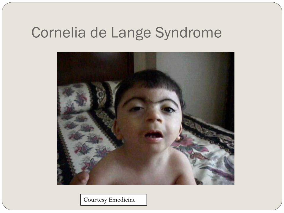
Cornelia de Lange Syndrome
Courtesy Emedicine
Cornelia De Lange Syndrome
Microcephaly
Confluent eyebrows
Underdeveloped orbital arches
Long philtrum
High arched palate and overt or submucous cleft palate
Micrognathia
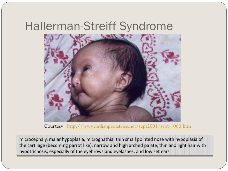
Hallerman-Streiff Syndrome
Courtesy: http://www.indianpediatrics.net/sept2001/sept-1060.htm
microcephaly, malar hypoplasia, micrognathia, thin small pointed nose with hypoplasia of the cartilage (becoming parrot like), narrow and high arched palate, thin and light hair with hypotrichosis, especially of the eyebrows and eyelashes, and low set ears
Hallerman-Streif Syndrome
Microcephaly,
Malar hypoplasia,
Micrognathia,
Thin small pointed nose with hypoplasia of the cartilage
(becoming parrot like),
Narrow and high arched palate
Thin and light hair with hypotrichosis, especially of the
eyebrows and eyelashes
Low set ears
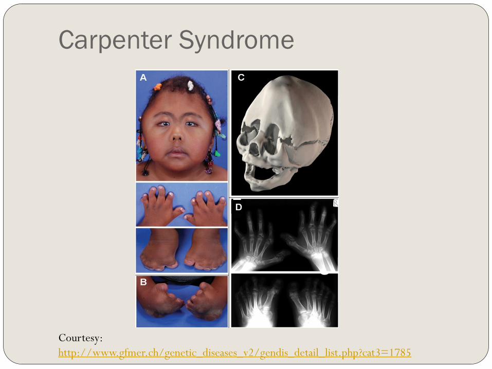
Carpenter Syndrome
Courtesy:
http://www.gfmer.ch/genetic_diseases_v2/gendis_detail_list.php?cat3=1785
Carpenter Syndrome
Autosomal Recessive Condition
Craniosynostosis
Many fusions of sutures
Soft tissue syndactyly of hands and feet always
present
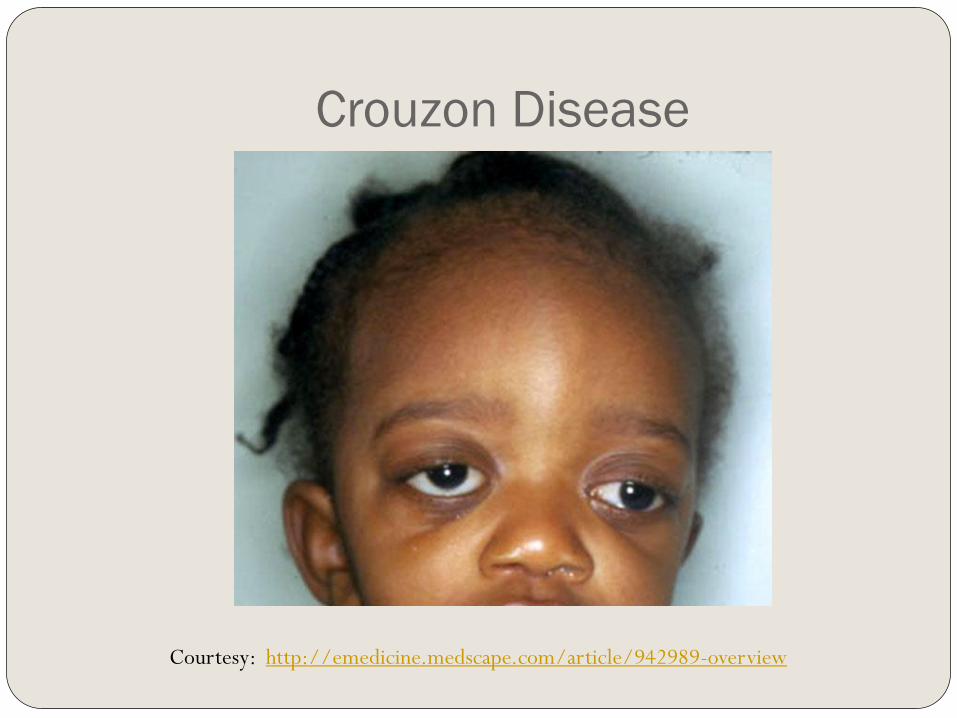
Crouzon Disease
Courtesy: http://emedicine.medscape.com/article/942989-overview
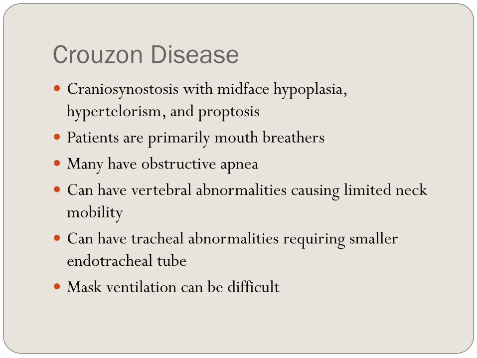
Crouzon Disease
Craniosynostosis with midface hypoplasia,
hypertelorism, and proptosis
Patients are primarily mouth breathers
Many have obstructive apnea
Can have vertebral abnormalities causing limited neck
mobility
Can have tracheal abnormalities requiring smaller
endotracheal tube
Mask ventilation can be difficult
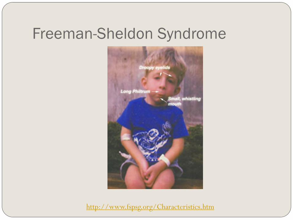
Freeman-Sheldon Syndrome
http://www.fspsg.org/Characteristics.htm
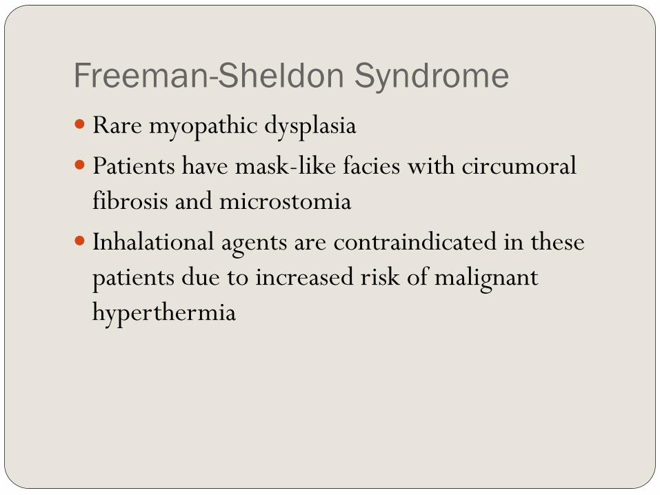
Freeman-Sheldon Syndrome
Rare myopathic dysplasia
Patients have mask-like facies with circumoral
fibrosis and microstomia
Inhalational agents are contraindicated in these
patients due to increased risk of malignant
hyperthermia
Inflexibility or Instability of Larynx
Down Syndrome
Juvenile Rheumatoid Arthritis
Goldenhar Syndrome
Klippel-Feil Syndrome
Laryngeal Anomalies Congenital Lesions
Cricoid Ring Stenosis
Subglottic Stenosis
Laryngeal Webs
Laryngeal Cysts
Laryngoceles
Subglottic Hemangiomas
Bilateral True Vocal Cord Paralysis
Infectious Disease Epiglottitis
Respiratory Papillomatosis
Croup
Traumatic Injuries Foreign Bodies
Chemical or Thermal Burns
Conclusions Very few difficult airway scenarios require a surgical airway
It is best to have several back-up plans and then back-up plans for
those back-up plans
Know where your equipment is and how to use it
The LMA has revolutionized airway management and can create
time for more permanent solutions
Pediatric patients have differing anatomy from adults that can be
more challenging
Otolaryngologists have many special skills and tools that are
invaluable in the management of the difficult and emergent airway
References Tekin M, Bodurtha J. “Cornelia De Lange Syndrome.” Emedicine.
http://emedicine.medscape.com/article/9427 92-overview. Site visited 3/29/2011.
Issaivanan M, Virdi V. “Dyscephalia Mandibulo-Oculo-Facialis”. Indian Pediatrics. 2001; 38: 1060.
“Carpenter Syndrome” http://www.gfmer.ch/genetic diseases_v2/gendis_detail_list.php?cat3=1785. Site visited on 3/29/2011
Kinsman SL, Johnston, MV. “Craniosynostosis” Kliegman: Nelson Textbook of Pediatrics, 18th ed. 2007: Philadelphia, PA.
Gudzenko V, Bittner EA, Schmidt UH. “Emergency Airway Management.” Respiratory Care. (2010) 55, 1026-1035.
Katos, MG, Goldenbery D. “Emergency Cricothyrotomy.” Operative Techniques in Otolaryngology. (2007) 18, 110-114.
Ondik MP, Kmatian S, Carr MM. “Management of the difficult airway in the pediatric patient.” Operative Techniques in Otolaryngology. (2007) 18, 121-126.
Gillespie MB, Eisele DW. “Outcomes of Emergency Surgical Airway Procedures in a Hopsital-Wide Setting.” Laryngoscope. (1999) 109, 1766-1769.
Infosino A. “Pediatric upper airway and congenital anomalies.” Anesthesiology Clin N Am. (2002) 20, 747-766.
Nargozian C. “The airway in patients with craniofacial abnormalities.” Pediatric Anesthesia. (2004) 14, 53-59.
References Goldstein BJ, Goldenberg D. “The difficult airway: Implications for the otolaryngologist-head
and neck surgeon.” Operative Techniques in Otolaryngology. (2007) 18, 72-76.
Wong E, Ng Y. “The difficult airway in the emergency department.” Int J Emerg Med. (2008)
1, 107-111.
Liess BD, Scheidt TD, Templer JW. “The Difficult Airway”. Otolaryngol Clin N Am. (2008) 41,
567-580.
Bruce IA, Rothera MP. “Upper airway obstruction in children.” Pediatric Anesthesia. (2009)
19, 88-99.
Sofferman RA, Greene CM. “Complex Upper Airway Problems”. Head & Neck Surgery –
Otolaryngology. Bailey BJ and Johnson JT, 2006; Philadelphia, PA.
Weissler MC, Couch ME. “Tracheotomy and Intubation.” Head & Neck Surgery –
Otolaryngology. Bailey BJ and Johnson JT, 2006; Philadelphia, PA.
Bhatti NI. “Surgical Management of the Difficult Adult Airway.” .” Flint: Cummings
Otolaryngology: Head & Neck Surgery, 5th ed. 2010.
Mark L, Herzer K, Akst S, Michelson J. “General Considerations of Anesthesia and Management
of the Difficult Airway.” .” Flint: Cummings Otolaryngology: Head & Neck Surgery, 5th ed.
2010.
Henderson J. “Airway Management in Adults.” Miller: Miller’s Anesthesia, 7th ed. 2007.