management of skin and soft-tissue infections in the...
TRANSCRIPT

Management of Skin and Soft-TissueInfections in the Emergency Department
Fredrick M. Abrahamian, DO, FACEPa,b,*,David A. Talan, MD, FACEP, FAAEM, FIDSAa,b,c,
Gregory J. Moran, MD, FACEP, FAAEMa,b,c
aDavid Geffen School of Medicine at the University of California Los Angeles,
Los Angeles, CA, USAbDepartment of Emergency Medicine, Olive View–University of California Los Angeles
Medical Center, Sylmar, CA, USAcDepartment of Medicine, Division of Infectious Diseases, Olive View–University of California
Los Angeles Medical Center, Sylmar, CA, USA
Skin and soft-tissue infections (SSTIs) are among the most commoninfections encountered by emergency physicians. The spectrum of diseaseseverity as seen by emergency physicians is wide, and can range frommild, uncomplicated cellulitis to cutaneous abscesses and necrotizing SSTIs.Infections can include acute, recurrent, and chronic wounds, and commu-nity-associated and health care-associated infections in immunocompetentor immunocompromised hosts.
Because of the nature of their practice, emergency physicians encounterpatients on a daily basis with a wide variety of mechanisms that lead toSSTIs. These can include animal and human bites, gunshot wounds, illicitdrug injection, work-related injuries, pressure sores, and iatrogenic injuriescaused by procedures (eg, intravenous lines). Similarly, infectious disease(ID) specialists see a variety of SSTIs in their practice; however, for theID specialist the spectrum of disease is more skewed toward patients witha complicated course, those who had multiple treatment failures, and recur-rent, severe, or rare and unusual infections. Unlike ID specialists, emergencyphysicians more frequently have to initiate empiric antibiotics because of theabsence of culture and susceptibility results, more often have to consider life
Infect Dis Clin N Am 22 (2008) 89–116
* Corresponding author. Department of Emergency Medicine, Olive View–University of
California Los Angeles Medical Center, 14445 Olive View Drive, North Annex, Sylmar, CA
91342-1438.
E-mail address: [email protected] (F.M. Abrahamian).
0891-5520/08/$ - see front matter � 2008 Elsevier Inc. All rights reserved.
doi:10.1016/j.idc.2007.12.001 id.theclinics.com

90 ABRAHAMIAN et al
and limb-threatening SSTIs, and commonly perform minor surgical care ofabscesses and infected wounds.
This article is written from the perspective of the evaluation and initial man-agement of SSTIs in the emergency department (ED). It highlights themanage-ment pitfalls and clinical dilemmas pertinent to emergency physicians that arenot often encountered by ID specialists. Emphasis is placed on the utility ofwound and blood cultures, disposition,methicillin-resistantStaphylococcus au-reus (MRSA) infections, animal and human bites, and necrotizing SSTIs.
Classification
Numerous classification schemes have been proposed for SSTIs. Eachclassification divides SSTIs based on specific variables, such as infectionof normal skin (primary skin infection) versus infection complicatinga chronic skin disorder (secondary infection), acute versus chronic, localizedversus diffuse, and nonnecrotizing versus necrotizing infections. Anotherproposed classification system divides SSTIs based on the severity of localand systemic signs, and symptoms of infection and the presence ofcomorbidities [1].
Themost commonly used classification systemforSSTIs is basedon thepres-ence or absence of complicating factors (ie, uncomplicated versus complicatedinfections) [2,3]. This classification system is used by the pharmaceutical indus-try in the design of registration studies for new antimicrobials.
The Center for Drug Evaluation and Research (CDER) at the UnitedStates Food and Drug Administration (FDA) has proposed a series of guid-ance documents for the pharmaceutical industry in development of antimi-crobial drugs for the treatment of SSTIs [3]. According to CDER, theuncomplicated category of SSTI includes simple abscesses, impetiginouslesions, furuncles, and cellulitis. The complicated category includes infec-tions involving deeper skin structures, those requiring formal surgicalinterventions (eg, infected ulcers, burns, major abscesses), and the presenceof a significant underlying medical condition that complicates the responseto treatment. Infections involving anaerobic or gram-negative organisms(eg, rectal abscesses) are considered complicated [3]. Table 1 depicts exam-ples of skin infections based on the uncomplicated versus complicatedclassification scheme [2,3].
The most recent practice guidelines developed by the Infectious DiseaseSociety of America (IDSA) for the diagnosis and management of SSTIsdo not categorize skin infections based on the uncomplicated versus compli-cated terminology [4]. The guidelines are written in reference to specificdisease entities (eg, cellulitis, abscess, necrotizing infection), mechanism ofinjury (eg, animal bites, human bites, infection associated with animalcontact, surgical site infection), or host factors (eg, infections in the immu-nocompromised patient) [4]. Unfortunately, these guidelines were writtenjust as community-associated MRSA (CA-MRSA) was emerging as an

Table 1
Examples of skin and soft-tissue infections based on the uncomplicated versus complicated
classification scheme
Uncomplicated infections Complicated infections
Cellulitis Traumatic wound infection
Erysipelas Bite-related wound infection
Abscess, folliculitis, furunculosis Postoperative wound infection
Impetigo, ecthyma Secondary infection of a diseased skin (eg, eczema)
Diabetic foot infection
Venous stasis ulcers, infected pressure sores
Perianal skin infection
Necrotizing infection
Myonecrosis
Data from DiNubile MJ, Lipsky BA. Complicated infections of skin and skin structures:
when the infection is more than skin deep. J Antimicrob Chemother 2004;53(Suppl 2):ii37–50.
91MANAGEMENT OF SKIN AND SOFT-TISSUE INFECTIONS
important cause of SSTI. The management of SSTIs has changed consider-ably in the last few years and some of these changes are not reflected in theIDSA guidelines.
Microbiology
The microbiology of SSTIs is dependent on numerous factors, such as thehost, the environment (eg, community- versus health care-associated), themechanism of injury, and the duration and severity of illness. Knowledgeof these variables is imperative in the selection of an initial empiric antimi-crobial regimen.
In general, S. aureus, and to a lesser extent streptococci, are by far themost common causes of both uncomplicated and complicated SSTIs, witha few notable exceptions, such as animal and human bite-wound infections[5,6]. Polymicrobial infections with gram-negative and anaerobic organismsare typically seen in complicated infections [2,5–8].
The most important new development in the era of SSTI is the emergenceof CA-MRSA, a phenomenon largely recognized and studied in ED popu-lations. CA-MRSA infections have become a global emerging health prob-lem. Most infections are noninvasive [9], involve skin and soft-tissuestructures, and present as purulent skin infections [10–12]. The spectrumof skin infections caused by CA-MRSA is wide and can range from simplecutaneous abscesses to fulminant necrotizing fasciitis [12,13].
In the United States, the most common strain of MRSA associated withcommunity infections is USA300, and less commonly USA400 andUSA1000 [12,14]. Compared with health care-associated strains, CA-MRSA strains more frequently produce toxins and appear to be morevirulent. The Panton-Valentine leukocidin genes, which produce cytotoxinsthat cause tissue necrosis, damage host cell membranes, and leukocyte

92 ABRAHAMIAN et al
destruction, are commonly seen with CA-MRSA and rarely with healthcare-associated MRSA (HA-MRSA) strains [15–19]. Novel peptides havebeen recently identified that are cytotoxic to leukocytes and may be impor-tant virulence factors of CA-MRSA [20].
In a recent multicenter, prospective, prevalence study involving adult sub-jects with purulent SSTIs, MRSA was isolated from 59% of subjects (n ¼422; range: 15% to 74%). In this study, MRSA was isolated from 61% ofabscesses, 53% of infected wounds, and 47% of cellulitis associated withpurulent exudate. Methicillin-susceptible S. aureus (MSSA) was isolatedfrom 14% of abscesses, 21% of infected wounds, and 34% of cellulitis asso-ciated with purulent exudate. Streptococcal species was isolated from 7% ofabscesses, 9% of infected wounds, and 13% of cellulitis associated with pu-rulent exudate [12]. Although it is clear that MRSA is now a common causeof SSTI overall, it is not clear to what extent it is now responsible for all pos-sible subtypes of SSTI, such as diabetic foot infections or bite-woundinfections.
In one investigation of ED patients with SSTI, historical and clinicalfeatures associated with MRSA compared with other bacteria include a his-tory of close contact with similar infection (odds ratio [OR], 3.4; 95%confidence index or CI, 1.5–8.1; P!.05), prior history of MRSA infection(OR, 3.3; 95% CI, 1.2–10.1; P!.05), reported ‘‘spider bite’’ (OR, 2.8; 95%CI, 1.5–5.3; P!.05), received antibiotics in the past 1 month (OR, 2.4; 95%CI, 1.4–4.1; P!.05), and abscess (OR, 1.8; 95% CI, 1.0–3.1; P!.05) [12].A common pitfall is to assume one can reliably exclude MRSA based onthe absence of risk factors. However, even in the absence of risk factors,CA-MRSA is still prevalent, and was present in 48% of ED SSTI patientsin one investigation [12]. There are no clinical or epidemiologic risk factorsthat can exclude MRSA etiology and reliably distinguish between patientsinfected with CA-MRSA and other organisms [11,12,21,22].
Another common mistake is to assume one can also reliably differentiateCA-MRSA and HA-MRSA based on epidemiologic factors. The epidemio-logic definition of HA-MRSA infection includes MRSA identified after 48hours of hospital admission, or identified in patients with a history ofsurgery, hospitalization, dialysis, or residence in a long-term care facilitywithin the last year, an indwelling catheter or other invasive medical devices,and previous history of MRSA infection or colonization [9,23,24]. Theexclusive use of this definition can underestimate the true prevalence ofCA-MRSA infection. In the ED-based study by Moran and colleagues[12], almost all MRSA isolated from SSTIs had molecular characteristicsof CA-MRSA, even though more than 25% of subjects met the epidemio-logic definition of HA-MRSA.
Another pitfall is to assume that pure cellulitis is exclusively caused bystreptococci, as suggested by the recent IDSA guidelines [4]. Investigationsinto the etiology of cellulitis without drainage are limited by the lack of spec-imen availability. Studies using tissue biopsies and aspirate specimens, and

93MANAGEMENT OF SKIN AND SOFT-TISSUE INFECTIONS
relying on conventional and nonconventional identification methods, suchas immunofluorescence and serologic testing, suggest that S. aureusdandto much lesser extent streptococcidare the most common pathogens impli-cated in the pathogenesis of cellulitis [25–32]. With the emergence ofCA-MRSA, the role of this pathogen in these difficult-to-study infectionsis uncertain. In the ED-based study by Moran and colleagues, among cellu-litis cases accompanied by purulent drainage, MRSA was found in 47%[12]. Thus, it is reasonable to assume CA-MRSA has a role in these infec-tions. Also, it has become evident that many occult cutaneous abscesses,which are now predominantly caused by CA-MRSA, are misdiagnosed ascellulitis on clinical evaluation, potentially leading to treatment failure,and their detection is greatly enhanced with the use of bedside soft-tissueultrasonography [33,34].
Necrotizing fasciitis and myonecrosis are typically caused by infectionwith Group A streptococcus, Clostridium perfringens, or, most commonly,aerobic and anaerobic organisms as part of a polymicrobial infection thatmay include S. aureus. In case series, CA-MRSA has recently been describedas a predominantly monomicrobial cause of necrotizing fasciitis [35,36]. Aretrospective review of patients presenting with necrotizing fasciitis between2000 and 2006 indicated that MRSA was the most common pathogen,accounting for one-third of the organisms isolated [37].
Other important causes of SSTI, including dog, cat, and human bite in-fections have been investigated among ED patients. Bite-wound infectionsare often mixed aerobic and anaerobic infections, with some unique patho-gens transmitted by the biter’s oral flora. In a study of 107 subjects with dogand cat bite infections, mixed aerobic and anaerobic infections were presentin 56% of all wounds (dogs: 48%; cats: 63%) [5]. Pasteurella species was themost common pathogen isolated from both dog (50%) and cat (75%) bitesand was commonly isolated from abscesses and puncture wounds. The mostcommon pasteurella strain isolated from infected dog bites was P. canis(26%), while in infected cat bites it was P. multocida subspecies multocida(54%). Streptococci were seen in 46% of both types of bite infections.S. aureus (all MSSA) was only isolated in 20% of dog bite infections and4% of cat bite infections, suggesting animal oral flora dominates humanskin flora, especially for deep puncture wounds. Anaerobes (eg, Fusobacte-rium nucleatum, Bacteroides tectum, Porphyromonas species, Prevotella hep-arinolytica) were usually present as mixed infections with aerobic organisms.
Similar to animal bites, human bites are also frequently mixed aerobicand anaerobic infections. In a study of 50 subjects with infected humanbites, 54% of wounds were mixed aerobic and anaerobic infection withorganisms [6]. The most common organisms recovered from human bitewounds included Streptococcus species (84%; S. anginosus was the mostcommon pathogen, isolated in 52% of cases), S. aureus (30%), Eikenellacorrodens (30%), Prevotella species (36%), Fusobacterium species (34%),and Veillonella species (24%) [6].

94 ABRAHAMIAN et al
Mixed aerobic and anaerobic bacteria are also commonly observed inSSTIs among injecting-drug users (IDUs). A comparative microbiologicstudy of cutaneous abscesses among IDUs and non-IDUs found a greaterfrequency of mixed aerobic and anaerobic infections in IDUs. The preva-lence of oral anaerobic organisms was higher among IDUs. In additionto S. aureus, which was found in 50% of IDU abscess cases, Streptococcusmilleri, F. nucleatum, Prevotella species, Peptostreptococcus micros, Actino-myces odontolyticus, and Veillonella species were found [38].
Clinical presentation
The clinical manifestations of SSTIs are variable and depends on factorssuch as the host, the infecting organism, and the inciting event. CA-MRSASSTIs commonly present as a spontaneous abscess [10–12]. The patient of-ten attributes the infection to a ‘‘spider bite.’’ Misclassification of a deep ab-scess as cellulitis is a common pitfall. Physicians often attribute treatmentfailure of ‘‘cellulitis’’ to antimicrobial resistance and change to a different,or often multiple, antibiotic regimen. The presence of an underlying deepabscess should be considered in patients with cellulitis who ‘‘fail’’ initial an-timicrobial therapy. Treatment failure may be caused by an undrained ab-scess that was missed on the initial presentation, rather than inadequateantimicrobial therapy. Bedside ultrasound is more frequently available inthe ED and is recommended for evaluation of all suspicious SSTIs.
Some wounds may be associated with injuries to deeper structures, suchas bones, joints, tendons, and neurovascular structures. Animal and humanbite injuries that appear to be minor may have underlying joint and otherstructural injuries. Clenched fist injuries (‘‘fight bites’’) are particularlyprone to underlying bone or metacarpal joint involvement that can bemissed on initial presentation. Cats, with their sharp thin teeth, oftenproduce small deep puncture wounds that seem to be trivial, but furtherevaluation may reveal joint or cortical injuries. Pain out of proportion tothe wound and physical findings should raise suspicion for joint or bonyinjuries.
Different terms and classifications have been used to describe varioustypes of necrotizing SSTIs (eg, synergistic necrotizing cellulitis, necrotizingfasciitis, streptococcal myonecrosis, gas gangrene). Although various factorsmay distinguish each type of infection, it is currently recommended todescribe these generally as ‘‘necrotizing soft-tissue infections’’ or necrotizingSSTI, because their initial treatment, which emphasizes early surgical inter-vention and broad-spectrum antimicrobials, is the same [39].
Patients with necrotizing SSTI most often present with severe pain (oftenout of proportion to physical findings) and rapidly advancing indurationand tenderness that extends beyond the area of erythema. These patients ap-pear ill and most often have abnormal vital signs on presentation. However,the recognition of a necrotizing SSTI, especially in the early stages, is
95MANAGEMENT OF SKIN AND SOFT-TISSUE INFECTIONS
extremely difficult. Patients present in variable stages in the spectrum of theillness. In a retrospective review of 89 patients with necrotizing fasciitis, only13 (15%) patients had the diagnosis of necrotizing fasciitis at the time of ad-mission [35]. At the time of the presentation, patients can be afebrile [36,40]and have minimal pain at the site of infection [36]. The cutaneous findingsearly in the course of disease may be lacking or are nonspecific. In a retro-spective review of patients with necrotizing SSTIs, the most specific signs,crepitus and blistering, were absent in 63% (n ¼ 189) and 76% (n ¼ 190)of patients, respectively [40]. In 15% of patients (n ¼ 170), none of the spe-cific findings, (ie, crepitus, blistering, or radiographic evidence of soft-tissuegas) were present [40]. Lymphangitis and lymphadenopathy are typically ab-sent [41].
Recent reports (mostly case reports and case series) of necrotizing SSTIscaused by CA-MRSA have revealed that this type of necrotizing SSTI pres-ents more subacutely and is a more benign syndrome than necrotizing infec-tions caused by other etiologies [13,42–44]. In a retrospective study of 14patients with necrotizing SSTIs, 57% of patients had a preoperative diagno-sis of abscess, and necrotizing infection was not suspected [13]. In this series,the onset of disease was often subacute (average 6 days; range, 3–21), andeven in the presence of serious complications (eg, need for reconstructivesurgery and prolonged stay in the intensive care unit), none of the patientsdied [13]. The 100% survival rate in this series, compared with the approx-imately 70% survival in most necrotizing SSTI series [36,40], suggests thatperhaps some of these may be a different type of necrotizing infection. Largeabscesses that can be caused by CA-MRSA are sometimes found to havenecrotic tissue, even though they are not associated with the high mortalityof what has been traditionally considered to be a necrotizing infection.
Laboratory evaluation and imaging
Microbiologic studies, such as wound cultures and blood cultures, can beof value in selected patients with SSTIs; however, their routine use in allSSTI is debated [45]. Historically, routine wound cultures were rarelyperformed because of the predictable infection etiology and the associatedantimicrobial susceptibility patterns. In the current era of increased preva-lence of SSTIs caused by CA-MRSA, and varying antimicrobial suscepti-bility patternsdeven among CA-MRSA strainsdmore physicians areperforming wound cultures to assess the etiology of the infection and guidefurther antimicrobial therapy.
The decision to do any diagnostic test, including wound or blood cultures,depends on its clinical utility and the likelihood that the result will changemanagement. The majority of patients with CA-MRSA infection will presentwith simple, uncomplicated abscess [12]. Although the additional benefit ofCA-MRSA active antibiotics requires further study, based on the current

96 ABRAHAMIAN et al
available evidence, the management of uncomplicated abscesses, even thosecaused by CA-MRSA, is solely incision and drainage, which is associatedwith a cure rate of 85% to 90% [12,46–48]. Therefore, it is unlikely thatculture and susceptibility testing would be helpful in the patient’s care.
Wound cultures should be reserved for patients that have a greaterchance of treatment failure, such as patients with complicated infections,immunocompromising conditions, moderate-to-severe illness, refractory orrecurrent infections, and those that have failed prior surgical therapy.Wound cultures are also appropriate for patients who will be treated withan antibiotic with variable activity against the presumed pathogen (eg,CA-MRSA and clindamycin). Cultures should be obtained for all patientsadmitted to the hospital, both to ensure that the empiric regimen has activityagainst the pathogen, and to allow appropriate narrowing of the antimicro-bial spectrum (eg, switch vancomycin to oxacillin if MSSA is identified).
Although pathogen prevalence rates and susceptibility patterns can besurmised from the selected group of patients who require cultures, broaderlocal surveillance can help to determine optimal empiric therapy for futurepatients. However, patients should be spared the expense of tests for whichthey receive no direct benefit, and costs of surveillance testing should beborne by public health departments or health care systems. At this pointin time, CA-MRSA appears to be a frequent and endemic cause of SSTI;however, its antimicrobial susceptibility pattern appears to be changing.
As with wound cultures, the utility of blood cultures in all types ofSSTIs is also debated. A typical case of a cellulitis deemed appropriatefor outpatient antimicrobial therapy is unlikely to be associated with bac-teremia. Even for hospitalized patients with community-acquired cellulitis,bacteremia is an uncommon finding [49], and discordant antibiotic ther-apy is rare. In a study of 757 hospitalized subjects with community-acquired cellulitis [49], blood cultures were performed on 553 subjects.A ‘‘true’’ pathogen (ie, not a contaminant) was isolated from the bloodcultures in only 11 cases (2%) [49]. In order of decreasing frequency, iso-lated organisms included: Group G streptococcus (5), Group A strepto-coccus (3), S. aureus (1), Vibrio vulnificus (1), and Morganella morganii(1). Univariate regression analysis revealed that an age greater than 45years, short duration of symptoms before presentation, temperaturehigher or equal to 38.5�C, and white blood cell (WBC) count greaterthan 13,300/mm3 at admission were predictive of bacteremia. Initial em-piric antimicrobial therapy was concordant in all patients with bacter-emia. For the two patients with gram-negative bacteremia, there wassufficient historical information to suspect an infection with an unusualorganism. One patient with V. vulnificus bacteremia was a 69-year-oldmale, with a history of fish bone injury to his right hand second digit,who developed cellulitis and chills 6 hours after the injury. Another pa-tient with M. morganii bacteremia was a 50-year-old female with pastmedical history of noninsulin-dependent diabetes mellitus and end-stage

97MANAGEMENT OF SKIN AND SOFT-TISSUE INFECTIONS
renal disease, receiving hemodialysis that was temporality administeredthrough an indwelling central catheter. Contaminated samples were al-most twice as common as true-positive blood cultures. False-positiveblood culture results have the potential for introducing inappropriatetreatment and can increase length of hospital stay, resource use, and costs[50].
The latest IDSA SSTI guidelines [4] mention the low yield of bloodcultures; however, they fail to clearly recommend which patients wouldbenefit from them. Other sources recommend reserving blood cultures forpatients with signs and symptoms of systemic toxicity, severe infec-tions, those with chills and high-grade fever, elderly patients with acute on-set of illness, significant leukocytosis, and immunocompromised patients[1,4,49,51]. However, most patients that are considered for hospitalizationhave some to all of these loosely defined criteria.
Blood cultures are only helpful if the results would identify the need fora change in a patient’s therapy such that it would improve clinical outcomes.ID specialists should reconsider the need for routine blood cultures in EDpatients who are to be admitted to the hospital with SSTI, especially ifthe pathogen can be isolated from the infected wound or abscess. Instancesin which blood cultures could be of clinical utility can include patients withSSTI who present with severe sepsis or septic shock [52], are at risk for anunusual infection requiring unusual therapy, have immunocompromisingconditions, and for whom the finding of bacteremia would have importantimplications, such as patients at risk for endovascular infection (eg, thosewith prosthetic heart valves).
Other diagnostic tests to investigate the microbiologic etiology of SSTIshave included cultures from needle aspirations of inflamed skin (either at thepoint of maximal inflammation or the leading edge) and tissue punchbiopsies. The positive yields of these techniques are variable and, overall,low [25–28,53], and as a result are not recommended.
Tissue biopsy and frozen section examination has been advocated forpatients with suspected necrotizing SSTI [54,55]. However, correct interpre-tation of this test requires an experienced pathologist (not often available atall times), and the process can result in a delay in surgical intervention. Mostsurgeons prefer to explore the site of infection directly in the operatingroom. Rapid streptococcal antigen testing of the wound (which is onlyapproved for pharyngeal infections) in the ED has been reported to provideearly identification of streptococcal toxic shock syndrome [56]. Serologictests for streptococcal infection are not helpful in the ED because meaning-ful interpretation of these tests requires paired acute and convalescent titers.
Basic hematologic studies and serum chemistries are commonly per-formed on patients requiring hospital admission. These tests can aid inassessing the severity of disease, reveal organ dysfunction, and may exposeunderlying medical conditions (eg, anemia, renal disease). The most recentIDSA SSTI guidelines recommend obtaining these tests in patients with

98 ABRAHAMIAN et al
signs and symptoms of systemic toxicity (eg, fever, hypothermia, tachycar-dia, and hypotension) [4].
A diagnostic scoring system based on laboratory tests has been proposedas a tool for distinguishing necrotizing SSTI from other SSTIs [57]. TheLaboratory Risk Indicator for Necrotizing Fasciitis (LRINEC) score isdepicted in Table 2. The score assigns points based on the level of variouslaboratory variables that include C-reactive protein, total WBC count,serum sodium, creatinine, glucose, and hemoglobin. The maximum cumula-tive score is 13. A score greater than or equal to 6 had a positive predictivevalue of 92% (95% CI, 84.3–96.0) and negative predictive value of 96%(95% CI, 92.6–97.9) for necrotizing SSTI. The probability of necrotizingSSTI increased to more than 75% when the LRINEC score was greaterthan or equal to 8 (Table 3). The authors advocate using this tool as anadjunct in the management of SSTI. However, the ability of the LRINECscore to detect early cases of necrotizing SSTI is unproven.
The derivation of the LRINEC score was based on a retrospective obser-vational study of subjects divided into developmental or derivation (n ¼314) and validation (n ¼ 140) cohorts [57]. Because of the retrospectivenature of the study, for both groups (developmental and validation cohorts)the diagnosis of necrotizing SSTI and nonnecrotizing SSTI was alreadymade. Unfortunately, the utility of the LRINEC score is potentially limited
Table 2
Laboratory Risk Indicator for Necrotizing Fasciitis score
Laboratory parameter (units) LRINEC points
C-reactive protein (mg/L)
!150 0
R150 4
Total white blood cell count (mm3)
!15 0
15–25 1
O25 2
Hemoglobin (g/dL)
O13.5 0
11–13.5 1
!11 2
Sodium (mmol/L)
R135 0
!135 2
Creatinine (mg/dL)
%1.6 0
O1.6 2
Glucose (mg/dL)
%180 0
O180 1
Modified from Wong CH, Khin LW, Heng KS, et al. The LRINEC (Laboratory Risk Indi-
cator for Necrotizing Fasciitis) score: a tool for distinguishing necrotizing fasciitis from other
soft- tissue infections. Crit Care Med 2004;32(7):1535–41; with permission.
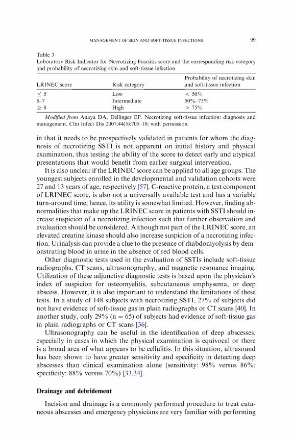
Table 3
Laboratory Risk Indicator for Necrotizing Fasciitis score and the corresponding risk category
and probability of necrotizing skin and soft-tissue infection
LRINEC score Risk category
Probability of necrotizing skin
and soft-tissue infection
% 5 Low ! 50%
6–7 Intermediate 50%–75%
R 8 High O 75%
Modified from Anaya DA, Dellinger EP. Necrotizing soft-tissue infection: diagnosis and
management. Clin Infect Dis 2007;44(5):705–10; with permission.
99MANAGEMENT OF SKIN AND SOFT-TISSUE INFECTIONS
in that it needs to be prospectively validated in patients for whom the diag-nosis of necrotizing SSTI is not apparent on initial history and physicalexamination, thus testing the ability of the score to detect early and atypicalpresentations that would benefit from earlier surgical intervention.
It is also unclear if the LRINEC score can be applied to all age groups. Theyoungest subjects enrolled in the developmental and validation cohorts were27 and 13 years of age, respectively [57]. C-reactive protein, a test componentof LRINEC score, is also not a universally available test and has a variableturn-around time; hence, its utility is somewhat limited. However, finding ab-normalities that make up the LRINEC score in patients with SSTI should in-crease suspicion of a necrotizing infection such that further observation andevaluation should be considered. Although not part of the LRINEC score, anelevated creatine kinase should also increase suspicion of a necrotizing infec-tion. Urinalysis can provide a clue to the presence of rhabdomyolysis by dem-onstrating blood in urine in the absence of red blood cells.
Other diagnostic tests used in the evaluation of SSTIs include soft-tissueradiographs, CT scans, ultrasonography, and magnetic resonance imaging.Utilization of these adjunctive diagnostic tests is based upon the physician’sindex of suspicion for osteomyelitis, subcutaneous emphysema, or deepabscess. However, it is also important to understand the limitations of thesetests. In a study of 148 subjects with necrotizing SSTI, 27% of subjects didnot have evidence of soft-tissue gas in plain radiographs or CT scans [40]. Inanother study, only 29% (n ¼ 65) of subjects had evidence of soft-tissue gasin plain radiographs or CT scans [36].
Ultrasonography can be useful in the identification of deep abscesses,especially in cases in which the physical examination is equivocal or thereis a broad area of what appears to be cellulitis. In this situation, ultrasoundhas been shown to have greater sensitivity and specificity in detecting deepabscesses than clinical examination alone (sensitivity: 98% versus 86%;specificity: 88% versus 70%) [33,34].
Drainage and debridement
Incision and drainage is a commonly performed procedure to treat cuta-neous abscesses and emergency physicians are very familiar with performing

100 ABRAHAMIAN et al
this procedure. A step-by-step, descriptive video clip of how to perform in-cision and drainage can be viewed at http://content.nejm.org/cgi/content/short/357/19/e20 [58].
The initial step in performing an incision and drainage is to prepare thesite for incision and remove any dirt or debris. The infected area is furthercleaned with a skin-cleansing agent (eg, chlorhexidine, povidone iodine, orisopropyl alcohol). Although this procedure is not considered sterile, thephysician should attempt to keep the area as clean as possible and devoidof unnecessary contamination.
The area is anesthetized with preservative-free 1% to 2% lidocaine. Injec-tion of lidocaine within the abscess cavity has not been shown to altermicrobiologic data [59]. After 2 to 5 minutes, when the onset of anesthesiahas occurred, using a number 11 scalpel, a straight incision is made over thearea of maximal fluctuance. The length of the incision will depend on thesize of the abscess; however, in general, the incision ought to be made largeenough to promote adequate drainage. After obtaining culture material (ifindicated) and the initial decompression of purulent material, the abscesscavity is probed thoroughly using a curved or straight hemostat. This actionwill further release any pockets of purulent material and break any remain-ing loculations and adhesions. All necrotic and devitalized tissue is alsodebrided.
To remove all loosened purulent and necrotic material, the abscess cavityis irrigated with sterile saline, using an 18-gauge angiocatheter attached toa 10-mL syringe. Although it is recommended [58], the additional clinicalutility of irrigating the abscess cavity is unknown.
After thorough drainage and removal of purulent material and any devi-talized tissue, the abscess cavity is loosely packed using 1⁄4 - or
1⁄2 -inch plainpacking strips. In order to ensure that the incision site will remain open andallow for continued drainage, 1 cm to 2 cm of the packing material is leftextending outside of the wound. The last step involves covering the woundwith absorbent 4 � 4 gauze dressing. Patients are instructed to return withina few days for removal of the packing material, or can be instructed tochange the packing themselves at home. Some patients will need a prescrip-tion of narcotic analgesic for dressing changes.
Antimicrobial therapy
The choice of initial empiric antimicrobial therapy is dependent uponprediction of the most likely microbiologic etiology and local antimicrobialsusceptibility patterns. Table 4 depicts recommended initial empiric antimi-crobial therapy options for commonly encountered SSTIs in the ED.
The mechanism of injury is crucial to predicting the bacterial etiology,which is especially important if organisms are involved that require specifictreatment. Examples of acute infections in which mechanism of injury is keyinclude dog and cat bite infections (Pasteurella species), human bite

101MANAGEMENT OF SKIN AND SOFT-TISSUE INFECTIONS
infections (Eikenella species), salt-water (Vibrio species) and fresh-water(Aeromonas species) exposure.
CA-MRSA is the most common cause of purulent SSTIs in most areas ofthe United States [12]. It demonstrates variable susceptibility patterns tocommonly used agents, such as clindamycin and tetracyclines. In vitrosusceptibility patterns of CA-MRSA to a variety of antimicrobial agentsare depicted in Table 5 [12,21,23,60]. Antibiotics that have adequate activityagainst CA-MRSA include trimethoprim/sulfamethoxazole (TMP/SMX),rifampin, vancomycin, linezolid (Zyvox), daptomycin (Cubicin), tigecycline(Tygacil), and quinupristin-dalfopristin (Synercid) [12,21,23,60–68].
Currently, the clinical trials for off-patent antibiotics for uncomplicatedSSTIs are being planned with support of the National Institutes of Health.Other potential future MRSA antimicrobials that are currently in variousphases of clinical trials for SSTI include novel glycopeptides, such as dalba-vancin (administered once weekly), telavancin, oritavancin (LY333328), cef-tobiprole (a cephalosporin with anti-MRSA activity), and Iclaprim(formerly AR-100, Ro 48-2622; a specific and selective microbial dihydrofo-late reductase inhibitor) [69–73].
The most common type of SSTI associated with CA-MRSA is an abscess[10–12,47,60]. Although further study is needed to determine if antibioticslead to additional benefit for CA-MRSA abscesses, it appears that 85%to 90% of patients do well with a treatment of incision and drainage alone[12,46–48]. In a recent observational study of 492 adult patients with CA-MRSA SSTIs, the use of an inactive antimicrobial agent was found to bean independent predictor of treatment failure (adjusted OR, 2.80; 95%CI, 1.26–6.22; P ¼ .01) [60]. However, of the 45 patients who experiencedtreatment failure, 38 (84%) were attributed to the need for additionalincision and drainage.
In an observational study of 69 children with culture-proven CA-MRSAabscesses, a patient with an abscess greater than or equal to 5 cm in diameterwas more likely to require subsequent hospitalization with incision anddrainage alone [46]. Based on this study, The Sanford Guide recommendsinstituting antibiotics for patients with abscesses greater than or equal to5 cm in diameter [74]. Unfortunately, these findings derive from observa-tional data in which the incision and drainage procedure was not standard-ized. In the authors’ experience, the most common reason abscesses failinitial management is inadequate incision and drainage, irrespective of theactivity of the antibiotic against CA-MRSA, and inadequate drainage ismore likely with larger abscesses if there is not careful attention to propertechnique.
In the absence of large randomized trials conducted in the area of CA-MRSA, current recommendations on antimicrobial therapy of CA-MRSASSTIs are based on expert opinion [24]. In conjunction with surgical drain-age, the IDSA SSTI guidelines recommend the addition of systemic antimi-crobial agents in patients with cutaneous abscesses in the following

Table 4
Recommended initial empiric antimicrobial therapy options for commonly encountered skin
and soft-tissue infections in the emergency department
Infection Therapeutic setting Initial empiric antimicrobial therapy
Mild cellulitis Outpatient therapy � TMP/SMX DS (160/800 mg)
1–2 tablets bid plus Cephalexin
500 mg qid
� Clindamycin 300 mg qid
� Minocycline 100 mg bid
(first dose, 200 mg)
� Doxycycline 100 mg bid
Abscess with mild
cellulitis
Outpatient therapy � Incision and drainage (no need
for antimicrobial therapy).
� Use of outpatient antimicrobial
therapy is recommended in patients
with multiple lesions,
immunocompromised state, those
with evidence of mild systemic toxicity
(eg, fever), recurrent infections, those
who have failed initial surgical therapy
(after exclusion of inadequate
drainage or deeper abscess),
and abscesses that cannot be
completely drained.
TMP/SMX DS (160/800 mg)
1–2 tablets bid
Clindamycin 300 mg qid
Minocycline 100 mg bid (first
dose, 200 mg)
Doxycycline 100 mg bid
Moderate-to-severe
cellulitis or abscess
Inpatient parenteral
therapy
� Clindamycin 600 mg–900 mg
IV q 8 h
Monotherapy with clindamycin
is preferred only for moderate
infections.
� Vancomycin 1 g IV q 12 h �cefazolin 1 g IV q 6 h
May replace cefazolin with nafcillin
or oxacillin 1 g–2 g every 4 hours.
Addition of b-lactam agent may
provide enhanced activity against
MSSA or streptococci (preferred
for patients suspected to be
bacteremic with S. aureus).
Diabetic foot infection Outpatient therapy � Clindamycin 300 mg qid plus
Ciprofloxacin 500 mg bid
� Amoxicillin/clavulanate 875/125 mg
bid � TMP/SMX DS (160/800 mg)
1–2 tablets bid
(continued on next page)
102 ABRAHAMIAN et al

Table 4 (continued )
Infection Therapeutic setting Initial empiric antimicrobial therapy
Diabetic foot infection Inpatient parenteral
therapy
� Ceftriaxone 1 g IV q 24 h plus
metronidazole 500 mg IV q
6–8 h � Vancomycin 1 g IV q 12 h
� Ertapenem 1 g IV q 24 h �vancomycin 1 g IV q 12 h
� Tigecycline 50 mg IV q 12 h
(first dose, 100 mg IV)
Dog, cat, and human
bites
Outpatient therapy � Amoxicillin/clavulanate
875/125 mg bid
� Moxifloxacin 400 mg daily
� Clindamycin 300 mg qid plus
Ciprofloxacin 500 mg bid
Dog, cat, and human
bites
Inpatient parenteral
therapy
� Ampicillin/sulbactam
1.5 g–3 g IV q 6 h
� Moxifloxacin 400 mg IV qd
Necrotizing soft-tissue
infections
� Vancomycin 1 g IV q 12 h plus
Clindamycin 900 mg IV q 8 h
plus Piperacillin/ tazobactam
3.375 g IV q 6 h
May replace vancomycin with
daptomycin 4mg/kg–6mg/kg IV qd.
Clindamycin (or alternatively
linezolid, see below) is
recommended because of its
ability to inhibit toxin production.
May substitute piperacillin/
tazobactam with imipenem
or meropenem.
� Linezolid 600 mg IV q 12 h
plus Piperacillin/tazobactam
3.375 g IV q 6 h
Recommended dosages are for a non-pregnant 70-kg adult with a normal renal and hepatic
function. Antimicrobial therapy should be initiated based on knowledge of local susceptibility
patterns and adjusted once culture and susceptibility data are known.
Abbreviations: DS, double-strength; IV, intravenous; TMP/SMX, trimethoprim-
sulfamethoxazole.
103MANAGEMENT OF SKIN AND SOFT-TISSUE INFECTIONS
situations: multiple lesions, cutaneous gangrene, immunocompromisedstate, extensive surrounding cellulitis, and those with evidence of systemictoxicity (eg, high fever) [4]. The initiation of antibiotics is also reasonablein patients requiring hospitalization (a reflection of the severity of the dis-ease), recurrent infections, those who have failed initial surgical therapy(after exclusion of inadequate drainage or deeper abscess), and in abscessesassociated with unusual pathogens (eg, human bite).
TMP/SMX is a commonly used drug in the United States for outpatientmanagement of CA-MRSA SSTIs. The Sanford Guide recommends twodouble-strength TMP/SMX tablets twice a day, with the goal of ensuring

Table 5
In vitro susceptibility patterns of community-associated methicillin-resistant Staphylococcus
aureus to a variety of antimicrobial agents
Antibiotic Moran et al [12] Miller et al [21] Naimi et al [23] Ruhe et al [60]
TMP/SMX 100% (n ¼ 217) 100% (n ¼ 120) 95% (n ¼ 106) 99% (n ¼ 322)
Rifampin 100% (n ¼ 186) 100% (n ¼ 120) 96% (n ¼ 106) 99% (n ¼ 318)
Clindamycin 95% (n ¼ 226)a 95% (n ¼ 102) 83% (n ¼ 106) 98% (n ¼ 482)b
Tetracycline 92% (n ¼ 226) 81% (n ¼ 120) 92% (n ¼ 106) 93% (n ¼ 455)
Gentamicin NT 100% (n ¼ 120) 94% (n ¼ 106) 100% (n ¼ 320)
Ciprofloxacin 60% (n ¼ 176) 15% (n ¼ 101) 79% (n ¼ 106) 73% (n ¼ 354)
Erythromycin 6% (n ¼ 226) 7% (n ¼ 120) 44% (n ¼ 106) 5% (n ¼ 23)
Vancomycin NT 100% (n ¼ 120) 100% (n ¼ 106) 100% (n ¼ 492)
Linezolid NT 100% (n ¼ 19) NT NT
Susceptibility patterns are dynamic and may vary markedly by geographic regions.
Physicians’ familiarity with the prevalence and susceptibility patterns of CA-MRSA in their
community is a crucial element in the management of CA-MRSA infections.
Abbreviations: NT, Not tested; TMP/SMX, Trimethoprim/sulfamethoxazole.a Four (approximately 2%; n ¼ 226) MRSA isolates had inducible clindamycin resistance
detected by an antimicrobial susceptibility D-zone disk diffusion test.b Two (3%; n ¼ 59) MRSA isolates had inducible clindamycin resistance detected by an
antimicrobial susceptibility D-zone disk diffusion test.
Modified from Abrahamian FM, Snyder EW. Community-associated methicillin-resistant
Staphylococcus aureus: incidence, clinical presentation, and treatment decisions. Curr Infect
Dis Rep 2007;9(5):391–7; with permission.
104 ABRAHAMIAN et al
adequate serum levels with respect to the minimum inhibitory concentration(MIC) and maximizing concentration-dependent killing [74]. Although thisrecommendation seems logical and is made to prevent under-dosing, itshould be noted that there are no prospective human trials demonstratingthe superiority of a two double-strength, compared with a one double-strength, regimen. TMP/SMX has been shown to have adequate penetrationinto experimentally made human skin blisters [75,76]; however, the samemay not apply to abscesses even with an increased dosage regimen. Mostimportantly, the issue of penetration into the abscess cavity may bea moot point if they are treated with adequate incision and drainage.Dosage increases may also lead to increased side-effects and potentiallylower patient compliance with the advocated regimen.
Rifampin, a highly active agent against CA-MRSA, is commonly used incombination with TMP/SMX or doxycycline. It should not be used alonebecause of its rapid tendency to select resistant strains [77]. The SanfordGuide recommends the addition of rifampin to TMP/SMX for patientswho have an abscess associated with fever, those with large or multipleabscesses, and in severe infections [74]. The only supporting data are froma retrospective study of CA-MRSA SSTIs, in which clinical resolutionwas achieved in all of six patients treated with a combination of TMP/SMX and rifampin, but in only 6 of 12 patients treated with double-strengthTMP/SMX [78]. Rifampin has numerous drug-drug interactions and an

105MANAGEMENT OF SKIN AND SOFT-TISSUE INFECTIONS
unpleasant side-effect profile (eg, discoloration of bodily fluids). In combina-tion with another agent, it introduces complexity and confusion regardingdosing regimens, which in turn may translate into noncompliance. A poten-tial role of rifampin, because of its greater penetration into mucosal tissue,may lie with its ability to eradicate nasal MRSA colonization and poten-tially reduce the rate of recurrence, but this has not been demonstrated inthe setting of CA-MRSA skin infections [79,80].
The activity of clindamycin against CA-MRSA is geographically vari-able, with a higher prevalence of resistance compared with TMP/SMX.However, compared with TMP/SMX, it has superior activity against S. pyo-genes and some anaerobes (eg, Peptostreptococcus), and has the capability ofinhibiting toxin production [81]. Some strains of MRSA display inducibleclindamycin resistance. Pretherapy, these strains demonstrate in vitro eryth-romycin-resistant and clindamycin-sensitive susceptibility patterns. How-ever, when exposed to clindamycin, they develop in vitro resistance toclindamycin. This trait can be detected by the double-disk diffusion assay(D test). The prevalence of such strains is geographically variable and theclinical significance is not well understood, as both clinical cure and treat-ment failures have been reported [82–87]. In a small retrospective study ofinvasive CA-MRSA infections in children, clindamycin demonstratedclinical efficacy in patients infected with clindamycin-susceptible CA-MRSA isolates [88]. However, because of lack of published experience,clindamycin is not advocated for use as a sole agent in severe infections [89].
Extended-spectrum tetracyclines, such as doxycycline and minocycline,are also reasonable choices for oral agents against CA-MRSA if localisolates display a high susceptibility rate [90,91]. Minocycline may havea slight advantage over doxycycline in its staphylococcal (MSSA) and strep-tococcal activity [74]. These drugs should be avoided in children under9 years old, pregnant patients, and nursing mothers.
Vancomycin is the most commonly used intravenous agent for the treat-ment of MRSA infections. Although vancomycin has been used for manydecades without many alternatives, its poor tissue penetration, increasingstaphylococcal MICs associated with clinical failure, and inferior clinicalefficacy, compared with antistaphylococcal b-lactams in the treatment ofMSSA infections, has raised concerns regarding its efficacy relative to neweragents [92–94]. Alternate intravenous agents with FDA approval for thetreatment of MRSA SSTIs include linezolid (Zyvox), daptomycin (Cubicin),and tigecycline (Tygacil). Quinupristin-dalfopristin (Synercid) has in vitroactivity against MRSA and an indication for treatment of complicatedSSTI; however, it does not currently have specific approval for MRSAinfections [66–68].
Linezolid has excellent in vitro activity against MRSA [62], and unlikevancomycin has the ability to suppress toxin production [81]. In a random-ized, open-label, multicenter study of complicated SSTIs, based on subgroupanalysis, linezolid outcomes were statistically superior to vancomycin at the

106 ABRAHAMIAN et al
test-of-cure visit for patients with MRSA infections [63]. However, a reanal-ysis of this study challenged the differential treatment effect based on themicroorganism [95]. Although both daptomycin and tigecycline are FDA-approved for the treatment of complicated SSTIs caused by MRSA, theassociated clinical studies are limited by the small number of subjects withdocumented MRSA SSTIs [96,97].
If indicated, empiric antimicrobial therapy for SSTIs associated withpurulence should include agents that have been shown to have adequatein vitro activity against CA-MRSA [12]. The role of CA-MRSA in nonpur-ulent SSTIs (eg, nonpurulent cellulitis) is unclear. However, in light of theemergence of CA-MRSA in purulent SSTI and the prominent role ofMSSA in previous studies of cellulitis, empiric CA-MRSA coverage isrecommended. Because streptococci have been shown to be another com-mon cause of cellulitis [25–28,53], it is reasonable to include an agent oragents with in vitro activity against CA-MRSA and S. pyogenes.
It should be noted that the clinical efficacy of TMP/SMX for SSTIs causedby Group A streptococci is unclear. In addition, the activity of TMP/SMX isnot routinely tested against S. pyogenes, and as a result, current resistancerates of S. pyogenes to TMP/SMX are unknown. Although there isevidence that TMP/SMXhas in vitro activity [98,99] and some clinical efficacyin infections caused by S. pyogenes [100,101], the most recent Centers forDisease Control and Prevention guidelines do not advocate use of this agentas monotherapy for patients presenting with cellulitis [24].
Empiric antimicrobial therapy for infected dog and cat bite woundsshould include coverage against Pasteurella, streptococci, staphylococci,and anaerobic species [5]. For infected human bites, empiric antimicrobialtherapy should include coverage against Eikenella, streptococci, staphylo-cocci, and anaerobic species [6]. S. aureus has been found in 4%, 20%,and 30% of cat, dog, and human bite infections, respectively, and nonewere MRSA [5,6]. Although these studies were done before the emergencyof CA-MRSA, at the present time colonization rates in human beingsremain low, so empiric CA-MRSA coverage is not recommended in thesetypes of infections [102].
Because of inadequate activity against Pasteurella or Eikenella species,monotherapy with first-generation cephalosporins, dicloxacillin, erythromy-cin, clindamycin, and metronidazole should be avoided [103]. The combina-tion therapy of penicillin plus a cephalosporin has been advocated for theinitial treatment of human bites [51]. However, this combination, especiallyif a first-generation cephalosporin is used, does not cover b-lactamaseproducing anaerobes [6]. Empirical regimens for marine- and freshwater-acquired infection, should cover Vibrio and Aeromonas species, respectively,with agents such as third-generation cephalosporins (eg, cefotaxime) andfluoroquinolones.
The management of necrotizing SSTI in the ED involves aggressive resus-citation, hemodynamic stabilization, initiation of antibiotics, and surgical

107MANAGEMENT OF SKIN AND SOFT-TISSUE INFECTIONS
consultation. Broad-spectrum empiric antimicrobial therapy should be initi-ated as soon as possible once the diagnosis is suspected. The spectrum ofantibiotic coverage should include gram-positive, gram-negative, andanaerobic organisms, with special attention to resistant organisms (eg,CA-MRSA) [13]. Suitable multidrug regimens include vancomycin pluspiperacillin or tazobactam plus clindamycin. Clindamycin have been shownto inhibit toxin production with streptococcal and clostridial infections[104–106], which in turn may be an important intervention in controllingthe inflammatory response. One observational study of children with inva-sive streptococcal infections found superior clinical outcomes in patientsreceiving antibiotics inhibiting protein synthesis, compared with agentsactive at the cell wall [107]. In a more recent study, clindamycin and line-zolid, both protein synthesis inhibitors at the ribosomal level, were shownto have the capability to suppress toxin production in CA-MRSA strains[81].
The optimal duration of antimicrobial therapy of SSTI is unknown and isultimately dependent on how the infection responds. Most SSTIs are treatedwith a 7 to 10 day course of antimicrobial therapy. A shorter course of 3 to5 days may be appropriate for infections associated with a drainable abscess,though this recommendation is not based on any comparative studies. Com-plicated cases, such as infections associated with osteomyelitis, an immuno-compromising condition, or diminished vascular supply may require a longerduration of therapy. In a randomized, double-blind, placebo-controlledtrial of 87 subjects with uncomplicated cellulitis, there was no significantdifference in clinical outcome at 14 and 28 days of therapy among subjectswho were treated with 5 days, compared with 10 days, of antimicrobialtherapy [108]. In a recent retrospective cohort study of 492 subjects withCA-MRSA SSTIs, subjects who had a successful outcome received antibi-otics for a median duration of 10 days (interquartile range, 7–14 days) [60].
Disposition
The majority of patients with SSTI can be treated as outpatients. Hospi-talization is indicated for patients with hemodynamic instability, alteredmental status, severe infection (including those requiring formal operativeintervention), intractable nausea and vomiting, the presence of immuno-compromising infection, failure of outpatient therapy, and poor socialsupport. The latest IDSA SSTI guidelines indicate hospitalization shouldbe considered in patients with ‘‘hypotension and/or an elevated creatininelevel, low serum bicarbonate level, elevated creatine phosphokinase level(2–3 times the upper limit of normal), marked left shift, or a C-reactiveprotein level O13 mg/L’’ [4].
Studies of risk factors for mortality, complications, and treatment fail-ures in patients with SSTIs exist [109–111]; however, only a few small studieshave attempted to determine a set of variables that may predict the need for

108 ABRAHAMIAN et al
(and benefit from) hospitalization [112]. In a study of patients with extrem-ity cellulitis, independent predictors of ‘‘need’’ for hospital admission werea history of diabetes, temperature, hand infections, induration, area greaterthan 70 cm2, and the absence of fluctuance. The need for hospitalization wasdefined as hospital stay greater than 24 hours, operative incision and drain-age, or failed outpatient management [112].
In an expert panel recommendation on the management of SSTIs [1], thepanel classified patients with SSTIs into four classes: Class I was afebrileand healthy, other than cellulitis; Class 2 was febrile and ill-appearing,but with no unstable comorbidies; Class III was toxic appearing, or withat least one unstable comorbidity or a limb-threatening infection; and ClassIV was with a sepsis syndrome or life-threatening infection (eg, necrotizingfasciitis).
For Class I patients, outpatient care on oral antimicrobials was recom-mended. For the Class IV patients, hospital admission was recommended.For both the Class II and Class III patients, a period of observation, anddepending on the outcome, either outpatient care on oral or intravenous(home or infusion center delivered) antimicrobials or hospital admissionwas recommended. The proposed classification system does not addressthe most important question as to which patients actually achieve benefitfrom hospitalization. Most EDs do not have observation units. In addition,for most patients and settings, outpatient parenteral antimicrobial therapy isnot feasible or practical.
Some EDs may have observation units that are either run by emergencyphysicians or general internists. SSTIs are among the most common reasonfor admission to an observation unit [113]. For ED physicians, it is not un-usual to encounter patients who initially present with worrisome clinicalfindings and equivocal criteria for hospital admission, but after a few simpleinterventions (eg, intravenous fluids, antipyretics) and a period of observa-tion, they become suitable for outpatient therapy. In some instances, iffeasible, patients are brought back to the ED for a few additional dosesof intravenous antibiotics, or are treated at home with intravenous antimi-crobial therapy [114,115].
For the appropriate indication, dalbavancin (administered in two doses,1 week apart [69]) may provide a potentially attractive alternative and averthospitalization for otherwise stable patients who are thought to require par-enteral antibiotic therapy or for whom noncompliance is a concern.
Infection control
Since the majority of SSTIs presenting to the ED are likely to be causedby CA-MRSA [12], it would be ideal for all these patients to be placed inprivate rooms with MRSA contact precautions until culture results areavailable. However, many EDs are currently dealing with severe overcrowd-ing issues, and isolation capacity is extremely limited. Similarly, because of

109MANAGEMENT OF SKIN AND SOFT-TISSUE INFECTIONS
a limited number of isolation beds in the hospitals, such patients often en-dure long waits for admission, which in turn results in delays in ED patientflow and disposition. Rapid real-time polymerase chain reaction assay forMRSA may allow rapid identification of these patients and in turn betterfacilitate infection control measures [116].
MRSA isolation precautions that are used in most hospitals were devel-oped in the era of hospital-associated MRSA. Their utility in the era inwhich CA-MRSA is prevalent in the community at large is unclear. How-ever, it is known that CA-MRSA strains can spread within hospitals [117].A common scenario in many facilities is that SSTI patients are admitted with-out MRSA precautions, then placed in isolation 2 days later when MRSA isconfirmed. Although this scenario is clearly suboptimal, it may be imprac-tical to admit every SSTI patient to a private room with MRSA precautions.Because it is known that MRSA is now a likely cause of SSTI, a cohortingstrategy for admitted SSTI patients may help reduce nosocomial spread[118]. Whether the infection is caused by MRSA or MSSA, standard precau-tions should be used for any patient with a purulent wound to preventexposing other patients or personnel to infected material. Gloves shouldalways be used when handling purulent material, such as when performingincision and drainage or changing dressings of infected wounds. Gownsand eye protection should be used for procedures that are likely to generatesplashes or sprays of fluids [24]. Frequent handwashing should always beencouraged in the ED.
MRSA decolonization should be considered in patients with recurrent,active MRSA infections not responding to appropriate therapy, or MRSAinfection occurring in closely-associated cohorts (eg, MRSA infection ina family) [24]. Although the efficacy of commonly prescribed decolonizationregimens in the prevention of recurrent CA-MRSA skin infections has notbeen studied, commonly prescribed agents for the purpose of decolonizationin the United States includes mupirocin 2% nasal ointment plus chlorhexi-dine 4% skin cleanser. In a randomized controlled trail, treatment withtopical mupirocin, chlorhexidine gluconate washes, oral rifampin, and doxy-cycline for 7 days was safe and effective in eradicating MRSA colonizationin hospitalized patients for at least 3 months [80].
Proper decolonization practices require obtaining cultures from multiplebody sites (eg, nares, axilla, groin), performing special susceptibility testing(eg, mupirocin) [119], and educating and possibly treating the patient’s fam-ily or other close contacts [24]. EDs may not be the optimal place to initiatethese interventions. However, many patients do not have access to primarycare physicians and EDs are the only site for their medical care. Initiatinga decolonization regimen, in selected cases, without obtaining cultures orspecial susceptibility testing and not involving the family is not uncommon.Although this practice may not be the optimal or preferred way, it is oftenthe only opportunity to initiate a potential therapy for patients who havelimited amount of resources and poor follow-up capability.
110 ABRAHAMIAN et al
Summary
The most important new development in the area of SSTI is the increasedprevalence of CA-MRSA, a phenomenon largely recognized and studied inED populations. There are no clinical or epidemiologic risk factors that canreliably excludeMRSA. The emergence of CA-MRSA has made a significantimpact in the empiric treatment of SSTIs.
The misclassification of a deep abscess as cellulitis is a common pitfall.The presence of an underlying deep abscess should be considered in patientswith cellulitis who fail initial antimicrobial therapy. Treatment failure maybe caused by an undrained abscess that was missed on the initial presenta-tion, rather than inadequate antimicrobial therapy. Ultrasonography can beuseful in the identification of deep abscesses, especially in cases in which thephysical examination is equivocal or there is a broad area of what appears tobe cellulitis. Wound and blood cultures can be of value in selected patientswith SSTIs; however, their routine performance, clinical utility, and cost-effectiveness in all types of SSTIs are debatable.
The recognition of a necrotizing SSTI, especially in the early stages, isextremely difficult. The utility of the LRINEC score is limited in that it needsto be prospectively validated in patients for whom the diagnosis of necrotizingSSTI is not apparent on initial history and physical examination. Protein syn-thesis inhibitors (eg, clindamycin, linezolid) have a potential role in the man-agement of necrotizing SSTIs. The majority of patients with SSTI can betreated as outpatients. Prospectively derived sets of variables that maypredict the need for (and benefit from) hospitalization in patients with SSTIsare insufficient at this time, and further studies in this area are indicated.
References
[1] Eron LJ, Lipsky BA, LowDE, et al. Managing skin and soft tissue infections: expert panel
recommendations on key decision points. J Antimicrob Chemother 2003;52(Suppl 1):
i3–17.
[2] DiNubile MJ, Lipsky BA. Complicated infections of skin and skin structures: when the
infection is more than skin deep. J Antimicrob Chemother 2004;53(Suppl 2):ii37–50.
[3] Center for Drug Evaluation and Research (CDER). Uncomplicated and complicated skin
and skin structure infections: developing antimicrobial drugs for treatment. Guidance for
industry. Available at: http://www.fda.gov/cder/guidance/2566dft.pdf. Accessed Novem-
ber 20, 2007.
[4] Stevens DL, Bisno AL, Chambers HF, et al. Practice guidelines for the diagnosis and
management of skin and soft-tissue infections. Clin Infect Dis 2005;41(10):1373–406.
[5] Talan DA, Citron DM, Abrahamian FM, et al. Bacteriologic analysis of infected dog and
cat bites. N Engl J Med 1999;340(2):85–92.
[6] Talan DA, Abrahamian FM, Moran GJ, et al. Clinical presentation and bacteriologic
analysis of infected human bites in patients presenting to emergency departments. Clin
Infect Dis 2003;37(11):1481–9.
[7] Moet GJ, Jones RN, Biedenbach DJ, et al. Contemporary causes of skin and soft tissue
infections in North America, Latin America, and Europe: report from the SENTRY

111MANAGEMENT OF SKIN AND SOFT-TISSUE INFECTIONS
Antimicrobial Surveillance Program (1998–2004). Diagn Microbiol Infect Dis 2007;57(1):
7–13.
[8] Rennie RP, Lones RN, Mutnick AH, et al. Occurrence and antimicrobial susceptibility
patterns of pathogens isolated from skin and soft tissue infections: report from the
SENTRY Antimicrobial Surveillance Program (United States and Canada, 2000). Diagn
Microbiol Infect Dis 2003;45(4):287–93.
[9] Klevens RM, Morrison MA, Nadle J, et al. Invasive methicillin-resistant Staphylococcus
aureus infections in the United States. J Am Med Assoc 2007;298(15):1763–71.
[10] Frazee BW, Lynn J, Charlebois ED, et al. High prevalence of methicillin-resistant Staphy-
lococcus aureus in emergency department skin and soft tissue infections. Ann Emerg Med
2005;45(3):311–20.
[11] MoranGJ,Amii RN,AbrahamianFM, et al.Methicillin-resistantStaphylococcus aureus in
community-acquired skin infections. Emerg Infect Dis 2005;11(6):928–30.
[12] Moran GJ, Krishnadasan A, Gorwitz RJ, et al. Methicillin-resistant S. aureus infections
among patients in the emergency department. N Engl J Med 2006;355(7):666–74.
[13] Miller LG, Perdreau-Remington F, Rieg G, et al. Necrotizing fasciitis caused by commu-
nity-associated methicillin-resistant Staphylococcus aureus in Los Angeles. N Engl J Med
2005;352(14):1445–53.
[14] Diep BA, Gill SR, Chang RF, et al. Complete genome sequence of USA300, an epidemic
clone of community-acquired methicillin-resistant Staphylococcus aureus. Lancet 2006;
367(9512):731–9.
[15] Voyich JM, OttoM,Mathema B, et al. Is Panton-Valentine leukocidin the major virulence
determinant in community-associated methicillin-resistant Staphylococcus aureus disease?
J Infect Dis 2006;194(12):1761–70.
[16] Lina G, Piemont Y, Godail-Gamot F, et al. Involvement of Panton-Valentine leukocidin-
producingStaphylococcus aureus in primary skin infections and pneumonia. Clin InfectDis
1999;29(5):1128–32.
[17] Vandenesch F, Naimi T, Enright MC, et al. Community-acquired methicillin-resistant
Staphylococcus aureus carrying Panton-Valentine leukocidin genes: worldwide emergence.
Emerg Infect Dis 2003;9(8):978–84.
[18] Gillet Y, Issartel B, Vanhems P, et al. Association between Staphylococcus aureus strains
carrying gene for Panton-Valentine leukocidin and highly lethal necrotizing pneumonia
in young immunocompetent patients. Lancet 2002;359(9308):753–9.
[19] Labandeira-Rey M, Couzon F, Boisset S, et al. Staphylococcus aureus Panton-Valentine
leukocidin causes necrotizing pneumonia. Science 2007;315(5815):1130–3.
[20] Wang R, Braughton KR, Kretschmer D, et al. Identification of novel cytolytic peptides as
key virulence determinants for community-associated MRSA. Nat Med 2007;13(12):
1510–4.
[21] Miller LG, Perdreau-Remington F, Bayer AS, et al. Clinical and epidemiologic character-
istics cannot distinguish community-associated methicillin-resistant Staphylococcus aureus
infection from methicillin-susceptible S. aureus infection: a prospective investigation. Clin
Infect Dis 2007;44(4):471–82.
[22] Skiest DJ, Brown K, Cooper TW, et al. Prospective comparison of methicillin-susceptible
and methicillin-resistant community-associated Staphylococcus aureus infections in hospi-
talized patients. J Infect 2007;54(5):427–34.
[23] Naimi TS, LeDell KH, Como-Sabetti K, et al. Comparison of community- and health
care-associated methicillin-resistant Staphylococcus aureus infection. J Am Med Assoc
2003;290(22):2976–84.
[24] Gorwitz RJ, Jernigan DB, Powers JH, et al, and Participants in the CDC-Convened
Experts’ Meeting on Management of MRSA in the Community. Strategies for clinical
management of MRSA in the community. Summary of an experts’ meeting convened by
the Centers for Disease Control and Prevention, 2006. Available at: http://www.cdc.gov/
ncidod/dhqp/ar_mrsa_ca.html. Accessed November 20, 2007.

112 ABRAHAMIAN et al
[25] Hook EW,Hooton TM,Horton CA, et al. Microbiologic evaluation of cutaneous cellulitis
in adults. Arch Intern Med 1986;146(2):295–7.
[26] Sigurdsson AF, Gudmundsson S. The etiology of bacterial cellulitis as determined by
fine-needle aspiration. Scand J Infect Dis 1989;21(5):537–42.
[27] SachsMK. The optimum use of needle aspiration in the bacteriologic diagnosis of cellulitis
in adults. Arch Intern Med 1990;150(9):1907–12.
[28] Duvanel T, Auckenthaler R, Rohner P, et al. Quantitative cultures of biopsy specimens
from cutaneous cellulitis. Arch Intern Med 1989;149(2):293–6.
[29] Bernard P, Bedane C, Mounier M, et al. Streptococcal cause of erysipelas and cellulitis in
adults. A microbiologic study using a direct immunofluorescence technique. Arch Derma-
tol 1989;125(6):779–82.
[30] Chartier C, Grosshans E. Erysipelas. Int J Dermatol 1990;29(7):459–67.
[31] Eriksson B, Jorup-Ronstrom C, Karkkonen K, et al. Erysipelas: clinical and bacteriologic
spectrum and serological aspects. Clin Infect Dis 1996;23(5):1091–8.
[32] Kielhofner MA, Brown B, Dall L. Influence of underlying disease process on the utility of
cellulitis needle aspirates. Arch Intern Med 1988;148(11):2451–2.
[33] Squire BT, Fox JC, Anderson C. ABSCESS: applied bedside sonography for convenient
evaluation of superficial soft tissue infections. Acad Emerg Med 2005;12(7):601–6.
[34] Tayal VS, Hasan N, Norton HJ, et al. The effect of soft-tissue ultrasound on the man-
agement of cellulitis in the emergency department. Acad Emerg Med 2006;13(4):384–8.
[35] WongCH,ChangHC, Pasupathy S, et al.Necrotizing fasciitis: clinical presentation,micro-
biology, and determinants of mortality. J Bone Joint Surg Am 2003;85-A(8):1454–60.
[36] McHenry CR, Piotrowski JJ, Petrinic D, et al. Determinants of mortality for necrotizing
soft-tissue infections. Ann Surg 1995;221(5):558–65.
[37] Elhabash S, Lee L, FarrowB, et al. Characteristics andmicrobiology of patients presenting
with necrotizing fasciitis. Presented at the Association of VA Surgeons 31st Annual Meet-
ing. Little Rock: Arkansas; May 10–12, 2007.
[38] Summanen PH, Talan DA, Strong C, et al. Bacteriology of skin and soft-tissue infections:
comparison of infections in intravenous drug users and individuals with no history of intra-
venous drug use. Clin Infect Dis 1995;20(Suppl 2):S279–82.
[39] Anaya DA, Dellinger EP. Necrotizing soft-tissue infection: diagnosis and management.
Clin Infect Dis 2007;44(5):705–10.
[40] Elliott DC, Kufera JA, Myers RA. Necrotizing soft tissue infections. Risk factors for
mortality and strategies for management. Ann Surg 1996;224(5):672–83.
[41] Green RJ, Dafoe DC, Raffin TA. Necrotizing fasciitis. Chest 1996;110(1):219–29.
[42] Pannaraj PS,HultenKG,Gonzalez BE, et al. Infective pyomyositis andmyositis in children
in the era of community-acquired, methicillin-resistant Staphylococcus aureus infection.
Clin Infect Dis 2006;43(8):953–60.
[43] DehorityW,Wang E, Vernon PS, et al. Community-associated methicillin-resistant Staph-
ylococcus aureus necrotizing fasciitis in a neonate. Pediatr Infect Dis J 2006;25(11):1080–1.
[44] Wong CH, Tan SH, Kurup A, et al. Recurrent necrotizing fasciitis caused by methicillin-
resistant Staphylococcus aureus. Eur J Clin Microbiol Infect Dis 2004;23(12):909–11.
[45] Abrahamian FM, Shroff SD. Use of routine wound cultures to evaluate cutaneous
abscesses for community-associated methicillin-resistant Staphylococcus aureus. Ann
Emerg Med 2007;50(1):66–7.
[46] Lee MC, Rios AM, Aten MF, et al. Management and outcome of children with skin and
soft tissue abscesses caused by community-acquired methicillin-resistant Staphylococcus
aureus. Pediatr Infect Dis J 2004;23(2):123–7.
[47] Miller LG, Quan C, Shay A, et al. A prospective investigation of outcomes after hospital
discharge for endemic, community-acquired methicillin-resistant and -susceptible Staphy-
lococcus aureus skin infection. Clin Infect Dis 2007;44(4):483–92.
[48] Rajendran PM, Young D, Maurer T, et al. Randomized, double-blind, placebo-controlled
trial of cephalexin for treatment of uncomplicated skin abscesses in a population at risk for
113MANAGEMENT OF SKIN AND SOFT-TISSUE INFECTIONS
community-acquired methicillin-resistant Staphylococcus aureus infection. Antimicrobial
Agents Chemother 2007;51(11):4044–8.
[49] Perl B, Gottehrer NP, RavehD, et al. Cost-effectiveness of blood cultures for adult patients
with cellulitis. Clin Infect Dis 1999;29(6):1483–8.
[50] Bates DW,Goldman L, Lee TH. Contaminant blood cultures and resource utilization. The
true consequences of false-positive results. J Am Med Assoc 1991;265(3):365–9.
[51] Swartz MN. Cellulitis. N Engl J Med 2004;350(9):904–12.
[52] NguyenHB,Rivers EP,AbrahamianFM, et al. Severe sepsis and septic shock: review of the
literature and emergency department management guidelines. Ann EmergMed 2006;48(1):
28–54.
[53] Newell PM, Norden CW. Value of needle aspiration in bacteriologic diagnosis of cellulitis
in adults. J Clin Microbiol 1988;26(3):401–4.
[54] Stamenkovic I, Lew PD. Early recognition of potentially fatal necrotizing fasciitis. The use
of frozen-section biopsy. N Engl J Med 1984;310(26):1689–93.
[55] Majeski J, Majeski E. Necrotizing fasciitis: improved survival with early recognition by
tissue biopsy and aggressive surgical treatment. South Med J 1997;90(11):1065–8.
[56] Ault MJ, Geiderman J, Sokolov R. Rapid identification of group A streptococcus as the
cause of necrotizing fasciitis. Ann Emerg Med 1996;28(2):227–30.
[57] Wong CH, Khin LW, Heng KS, et al. The LRINEC (Laboratory Risk Indicator for
Necrotizing Fasciitis) score: a tool for distinguishing necrotizing fasciitis from other soft
tissue infections. Crit Care Med 2004;32(7):1535–41.
[58] FitchMT,MantheyDE,McGinnis HD, et al. Abscess incision and drainage. NEngl JMed
2007;357(19):e20.
[59] Berg JO,Mossner BK, SkovMN, et al. Antibacterial properties of EMLA and lidocaine in
wound tissue biopsies for culturing. Wound Repair Regen 2006;14(5):581–5.
[60] Ruhe JJ, SmithN, BradsherRW, et al. Community-onsetmethicillin-resistantStaphylococ-
cus aureus skin and soft-tissue infections: impact of antimicrobial therapy on outcome.
Clin Infect Dis 2007;44(6):777–84.
[61] Fridkin SK, Hageman JC, Morrison M, et al. Methicillin-resistant Staphylococcus aureus
disease in three communities. N Engl J Med 2005;352(14):1436–44.
[62] Betriu C, Redondo M, Boloix A, et al. Comparative activity of linezolid and other new
agents against methicillin-resistant Staphylococcus aureus and teicoplanin-intermediate
coagulase-negative staphylococci. J Antimicrob Chemother 2001;48(6):911–3.
[63] Weigelt J, Itani K, Stevens D, et al. Linezolid versus vancomycin in treatment of compli-
cated skin and soft tissue infections. Antimicrobial Agents Chemother 2005;49(6):2260–6.
[64] Carpenter CF, Chambers HF.Daptomycin: another novel agent for treating infections due
to drug-resistant gram-positive pathogens. Clin Infect Dis 2004;38(7):994–1000.
[65] Stein GE, Craig WA. Tigecycline: a critical analysis. Clin Infect Dis 2006;43(4):518–24.
[66] Jones RN, Ballow CH, Acar J, et al. World-wide evaluation of quinupristin-dalfopristin
(Q/D) (Synercid): Antimicrobial activity directed against resistant Gram-positive patho-
gens: Report from the global SMART study. Presented at the 39th Interscience Conference
on Antimicrobial Agents and Chemotherapy, San Francisco, USA, 1999. Abstract 1262.
p. 263. American Society for Microbiology, Washington, D.C.
[67] Drew RH, Perfect JR, Srinath L, et al. Treatment of methicillin-resistant Staphylococcus
aureus infections with quinupristin-dalfopristin in patients intolerant of or failing prior
therapy. J Antimicrob Chemother 2000;46(5):775–84.
[68] Nichols RL, Graham DR, Barriere SL, et al. Treatment of hospitalized patients with
complicatedGram-positive skin and skin structure infections: two randomized, multicenter
studies of quinupristin/dalfopristin versus cefazolin, oxacillin or vancomycin. Synercid
Skin and Skin Structure Infection Group. J Antimicrob Chemother 1999;44(2):263–73.
[69] Jauregui LE, Babazadeh S, Seltzer E, et al. Randomized, double-blind comparison of
once-weekly dalbavancin versus twice-daily linezolid therapy for the treatment of compli-
cated skin and skin structure infections. Clin Infect Dis 2005;41(10):1407–15.

114 ABRAHAMIAN et al
[70] Saravolatz LD, Pawlak J, Johnson LB. Comparative activity of telavancin against isolates
of community-associated methicillin-resistant Staphylococcus aureus. J Antimicrob
Chemother 2007;60(2):406–9.
[71] Van Bambeke F, Van Laethem Y, Courvalin P, et al. Glycopeptide antibiotics: from
conventional molecules to new derivatives. Drugs 2004;64(9):913–36.
[72] RouseMS, Steckelberg JM, Patel R. In vitro activity of ceftobiprole, daptomycin, linezolid,
and vancomycin against methicillin-resistant staphylococci associated with endocarditis
and bone and joint infection. Diagn Microbiol Infect Dis 2007;58(3):363–5.
[73] Arpida. Intravenous iclaprim. Available at: http://www.arpida.ch/index.php?MenuID¼13&UserID¼1&ContentID¼65. Accessed November, 2007.
[74] Gilbert DN, Moellering RC, Eliopoulos GM, et al. The Sanford Guide to antimicrobial
therapy. 37th edition. Sperryville (VA): Antimicrobial Therapy, Inc; 2007.
[75] Bruun JN, Ostby N, Bredesen JE, et al. Sulfonamide and trimethoprim concentrations in
human serum and skin blister fluid. Antimicrobial Agents Chemother 1981;19(1):82–5.
[76] Nowak A, KadykowM, Klimowicz A. Penetration of trimethoprim and sulfamethoxazole
into skin blister fluid. Eur J Clin Pharmacol 1983;25(6):825–7.
[77] Strausbaugh LJ, Jacobson C, Sewell DL, et al. Antimicrobial therapy for methicillin-
resistant Staphylococcus aureus colonization in residents and staff of a Veterans Affairs
nursing home care unit. Infect Control Hosp Epidemiol 1992;13(3):151–9.
[78] Iyer S, Jones DH. Community-acquired methicillin-resistant Staphylococcus aureus skin
infection: a retrospective analysis of clinical presentation and treatment of a local outbreak.
J Am Acad Dermatol 2004;50(6):854–8.
[79] Yu VL, Goetz A, Wagener M, et al. Staphylococcus aureus nasal carriage and infection in
patientsonhemodialysis.Efficacyof antibiotic prophylaxis.NEngl JMed1986;315(2):91–6.
[80] Simor AE, Phillips E, McGeer A, et al. Randomized controlled trial of chlorhexidine
gluconate for washing, intranasal mupirocin, and rifampin and doxycycline versus no treat-
ment for the eradication of methicillin-resistant Staphylococcus aureus colonization. Clin
Infect Dis 2007;44(2):178–85.
[81] Stevens DL, Ma Y, Salmi DB, et al. Impact of antibiotics on expression of virulence-
associated exotoxin genes in methicillin-sensitive and methicillin-resistant Staphylococcus
aureus. J Infect Dis 2007;195(2):202–11.
[82] Siberry GK, Tekle T, Carroll K, et al. Failure of clindamycin treatment of methicillin-
resistant Staphylococcus aureus expressing inducible clindamycin resistance in vitro. Clin
Infect Dis 2003;37(9):1257–60.
[83] Lewis JS 2nd, Jorgensen JH. Inducible clindamycin resistance in staphylococci: should
clinicians and microbiologists be concerned? Clin Infect Dis 2005;40(2):280–5.
[84] Frank AL,Marcinak JF, Mangat PD, et al. Clindamycin treatment of methicillin-resistant
Staphylococcus aureus infections in children. Pediatr Infect Dis J 2002;21(6):530–4.
[85] Panagea S, Perry JD, Gould FK. Should clindamycin be used as treatment of patients with
infections caused by erythromycin-resistant staphylococci? J Antimicrob Chemother 1999;
44(4):581–2.
[86] Rao GG. Should clindamycin be used in treatment of patients with infections caused by
erythromycin-resistant staphylococci? J Antimicrob Chemother 2000;45(5):715–6.
[87] Drinkovic D, Fuller ER, Shore KP, et al. Clindamycin treatment of Staphylococcus aureus
expressing inducible clindamycin resistance. J Antimicrob Chemother 2001;48(2):315–6.
[88] Martinez-Aguilar G, Hammerman WA, Mason EO, et al. Clindamycin treatment of
invasive infections caused by community-acquired, methicillin-resistant and methicillin-
susceptible Staphylococcus aureus in children. Pediatr Infect Dis J 2003;22(7):593–8.
[89] Daum RS. Skin and soft-tissue infections caused by methicillin-resistant Staphylococcus
aureus. N Engl J Med 2007;357(4):380–90.
[90] Ruhe JJ, Monson T, Bradsher RW, et al. Use of long-acting tetracyclines for methicillin-
resistant Staphylococcus aureus infections: case series and review of the literature. Clin
Infect Dis 2005;40(10):1429–34.

115MANAGEMENT OF SKIN AND SOFT-TISSUE INFECTIONS
[91] Ruhe JJ, Menon A. Tetracyclines as an oral treatment option for patients with community
onset skin and soft tissue infections caused by methicillin-resistant Staphylococcus aureus.
Antimicrobial Agents Chemother 2007;51(9):3298–303.
[92] Deresinski S. Vancomycin and Staphylococcus aureusdan antibiotic enters obsolescence.
Clin Infect Dis 2007;44(12):1543–8.
[93] Mohr JF, Murray BE. Vancomycin is not obsolete for the treatment of infection caused by
methicillin-resistant Staphylococcus aureus. Clin Infect Dis 2007;44(12):1536–42.
[94] KimSH,KimKH,KimHB, et al. Outcomeof vancomycin treatment in patientswithmeth-
icillin-susceptible Staphylococcus aureus bacteremia. Antimicrobial Agents Chemother
2008;52(1):192–7.
[95] Kalil AC, Puumala S, Stoner J. Is linezolid superior to vancomycin for complicated skin
and soft tissue infections due to methicillin-resistant Staphylococcus aureus? Antimicrobial
Agents Chemother 2006;50(5):1910.
[96] Arbeit RD,MakiD, Tally FP, et al. The safety and efficacy of daptomycin for the treatment
of complicated skin and skin-structure infections. Clin Infect Dis 2004;38(12):1673–81.
[97] Ellis-Grosse EJ, Babinchak T, Dartois N, et al. The efficacy and safety of tigecycline in the
treatment of skin and skin-structure infections: results of 2 double-blind phase 3 compari-
son studies with vancomycin-aztreonam. Clin Infect Dis 2005;41(Suppl 5):S341–53.
[98] Eliopoulos GM, Wennersten CB. In vitro activity of trimethoprim alone compared with
trimethoprim-sulfamethoxazole and other antimicrobials against bacterial species associ-
ated with upper respiratory tract infections. Diagn Microbiol Infect Dis 1997;29(1):33–8.
[99] Bushby SR. Trimethoprim-sulfamethoxazole: in vitro microbiological aspects. J Infect Dis
1973;128(Suppl):442–62.
[100] Trickett PC,Dineen P,MogabgabW.Clinical experience: respiratory tract. Trimethoprim-
sulfamethoxazole versus penicillin G in the treatment of group A beta-hemolytic strepto-
coccal pharyngitis and tonsillitis. J Infect Dis 1973;128(Suppl):693–5.
[101] Quick CA. Comparison of penicillin and trimethoprim-sulfamethoxazole in the treatment
of ear, nose and throat infections. Can Med Assoc J 1975;112(13 Spec No):83–6.
[102] Loeffler A, Boag AK, Sung J, et al. Prevalence of methicillin-resistant Staphylococcus
aureus among staff and pets in a small animal referral hospital in the UK. J Antimicrob
Chemother 2005;56(4):692–7.
[103] Abrahamian FM,Goldstein EJC. Bites. In: Gorbach S, Bartlett JG, BlacklowNR, editors.
Infectious diseases. 3rd edition. Maryland: Williams & Wilkins; 2004. p. 1440–6.
[104] Stevens DL, Bryant AE, Hackett SP. Antibiotic effects on bacterial viability, toxin produc-
tion, and host response. Clin Infect Dis 1995;20(Suppl 2):S154–7.
[105] StevensDL,MaierKA,Mitten JE. Effect of antibiotics on toxin production and viability of
Clostridium perfringens. Antimicrobial Agents Chemother 1987;31(2):213–8.
[106] Stevens DL, Gibbons AE, Bergstrom R, et al. The Eagle effect revisited: efficacy of clinda-
mycin, erythromycin, and penicillin in the treatment of streptococcal myositis. J Infect Dis
1988;158(1):23–8.
[107] Zimbelman J, Palmer A, Todd J. Improved outcome of clindamycin compared with
beta-lactam antibiotic treatment for invasive Streptococcus pyogenes infection. Pediatr
Infect Dis J 1999;18(12):1096–100.
[108] Hepburn MJ, Dooley DP, Skidmore PJ, et al. Comparison of short-course (5 days) and
standard (10 days) treatment for uncomplicated cellulitis. Arch Intern Med 2004;164(15):
1669–74.
[109] Carratala J, Roson B, Fernandez-Sabe N, et al. Factors associated with complications and
mortality in adult patients hospitalized for infectious cellulitis. Eur J Clin Microbiol Infect
Dis 2003;22(3):151–7.
[110] Benfield T, Jensen JS, Nordestgaard BG. Influence of diabetes and hyperglycemia on
infectious disease hospitalization and outcome. Diabetologia 2007;50(3):549–54.
[111] Murray HE, Stiell IG, Wells GA. Defining treatment failure in ED patients with cellulitis.
Acad Emerg Med 2001;8(5):478.

116 ABRAHAMIAN et al
[112] Diercks DB, Kuppermann N, Derlet RW, et al. Derivation and validation of a model for
the need of hospital admission in patients with extremity cellulitis. Acad Emerg Med
2000;7(5):562.
[113] Roberts R. Management of patients with infectious diseases in an emergency department
observation unit. Emerg Med Clin North Am 2001;19(1):187–207.
[114] Corwin P, Toop L, McGeoch G, et al. Randomized controlled trial of intravenous antibi-
otic treatment for cellulitis at home compared with hospital. BMJ 2005;330(7483):129.
[115] Donald M, Marlow N, Swinburn E, et al. Emergency department management of home
intravenous antibiotic therapy for cellulitis. Emerg Med J 2005;22(10):715–7.
[116] Huletsky A, Lebel P, Picard FJ, et al. Identification of methicillin-resistant Staphylococcus
aureus carriage in less than 1 hour during a hospital surveillance program. Clin Infect Dis
2005;40(7):976–81.
[117] Seybold U, Kourbatova EV, Johnson JG, et al. Emergence of community-associated
methicillin-resistant Staphylococcus aureus USA300 genotype as a major cause of health
care-associated blood stream infections. Clin Infect Dis 2006;42(5):647–56.
[118] Siegel JD, Rhinehart E, Jackson M, et al. Management of Multidrug-Resistant
Organisms in Healthcare Settings, 2006. Available at: www.cdc.gov/ncidod/dhqp/pdf/ar/
mdroGuideline2006.pdf. Accessed November 20, 2007.
[119] Jones JC, Rogers TJ, Brookmeyer P, et al. Mupirocin resistance in patients colonized with
methicillin-resistant Staphylococcus aureus in a surgical intensive care unit. Clin Infect Dis
2007;45(5):541–7.