management of obstetric emergencies y5.ppt
DESCRIPTION
O>RANSCRIPT

Management of obstetric emergencies in primary care
Dr Shuhaila AhmadJabatan Obstetrik &
GinekologiFakulti Perubatan
Universiti Kebangsaan Malaysia
14/12/2005

Scope of the lecture: Definition of condition considered to
be an obstetric emergency Understanding the significance Identifying these conditions Instituting appropriate management Issues regarding these conditions Conclusion

Definition Emergency:
An sudden serious change in a patient’s condition which requires immediate medical or surgical intervention
Obstetrics: A branch of medicine that concerns
management of women during pregnancy, chldbirth and puerperium.
Taber’s cyclopedic medical dictionary
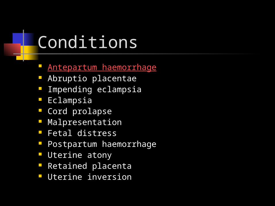
Conditions Antepartum haemorrhage Abruptio placentae Impending eclampsia Eclampsia Cord prolapse Malpresentation Fetal distress Postpartum haemorrhage Uterine atony Retained placenta Uterine inversion

Case 1
32 year old Gravida 3 Para 2 at 34 weeks of gestation went to see a GP for PV bleeding
1. What is your diagnosis?2. Outline your immediate and
subsequent management?

Antepartum haemorrhage
Defined as : “ any bleeding from the genital
tract from 22 weeks of gestation until before delivery”
Below 22 weeks of gestation is considered as threatened miscarriage.

Possible causes: Placenta praevia Abruptio placenta Local causes Vasa praevia

Initial management Unpredictable and patient can
deteriorate rapidly Aim:
To resuscitate and stabilize the patient To reassess patient and to make a
diagnosis Consider delivery or conservative
management

Flow chart of managementInitial assessment
Severe bleeding/shock mild/moderate
Consider delivery history/examination/investigation
Salvageable/non-salvagable diagnosis & management

Issues: Vaginal examination
Best to avoid until diagnosis of placenta praevia is excluded
Rhesus negative mother Anti D antibodies must be given
within 72 hours Role of Kleihauer test
To calculate proper dosage of antiD Ab

Conclusion for APH Be sure of the diagnosis Initial assessment improves
patient’s outcome Aim to prevent further
deterioration of patient Transfer patient to an appropriate
hospital for further management

Case 2 A 19 year old single school drop-out in
her first pregnancy complained of severe abdominal pain after being punched in the stomach by her boyfriend.
She was pale and tachycardic. The uterus was about 28 weeks size, tender and hard. No fetal heart activity was detected. Diagnosis?

Abruptio placenta Detachment of a normally sited
placenta before the delivery of the fetus
Incidence : 0.5 – 1.8% 2 types:
Revealed : 65 – 80% Concealed : 20 -35% - most dangerous

Aetiology Unknown except for direct trauma to
the uterus. Known association with:
Cigarette smoking (decidual necrosis) Sudden decompression of uterus-
polyhydramnios Placental abnormality Hypertension ?Folic acid deficiency

Outline your management if you are the attending doctor.
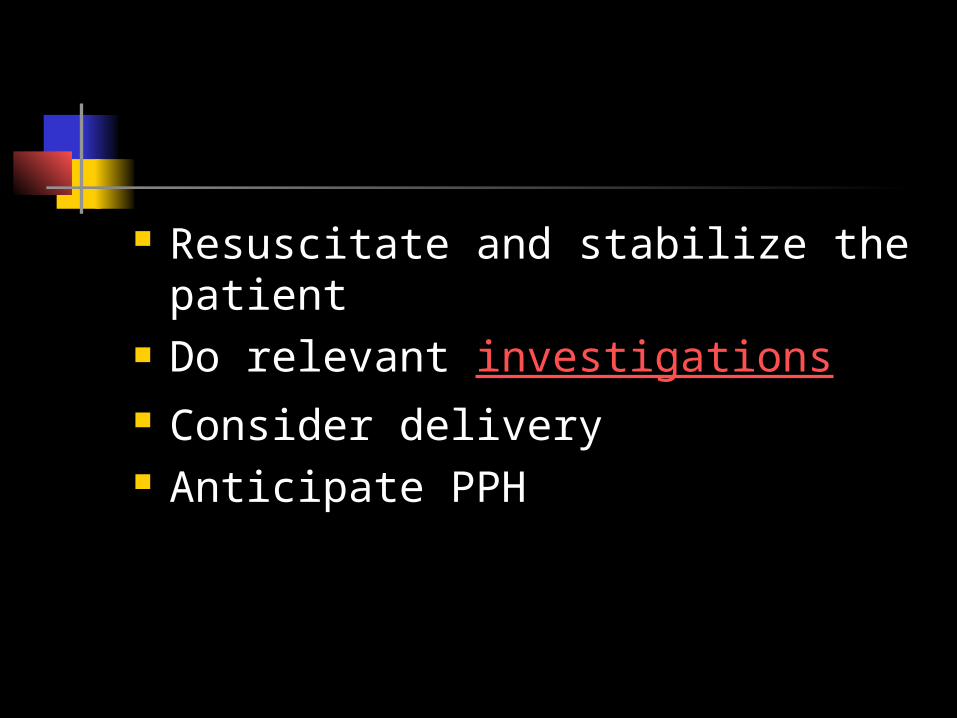
Resuscitate and stabilize the patient
Do relevant investigations Consider delivery Anticipate PPH

Investigation FBC – Hb & platelet count Coagulation profile / bleeding time ? role of ultrasound scan

Management : Active resuscitation Need to correct DIVC Consider delivery in most cases.
SVD should be chosen if there is fetal death
If baby is alive, CS is reported to improve fetal outcome.

Issues : Complications :
Maternal Fetal

Maternal complications Maternal mortality Hypovolumic shock DIVC Acute renal failure Postpartum haemorrhage Recurrence
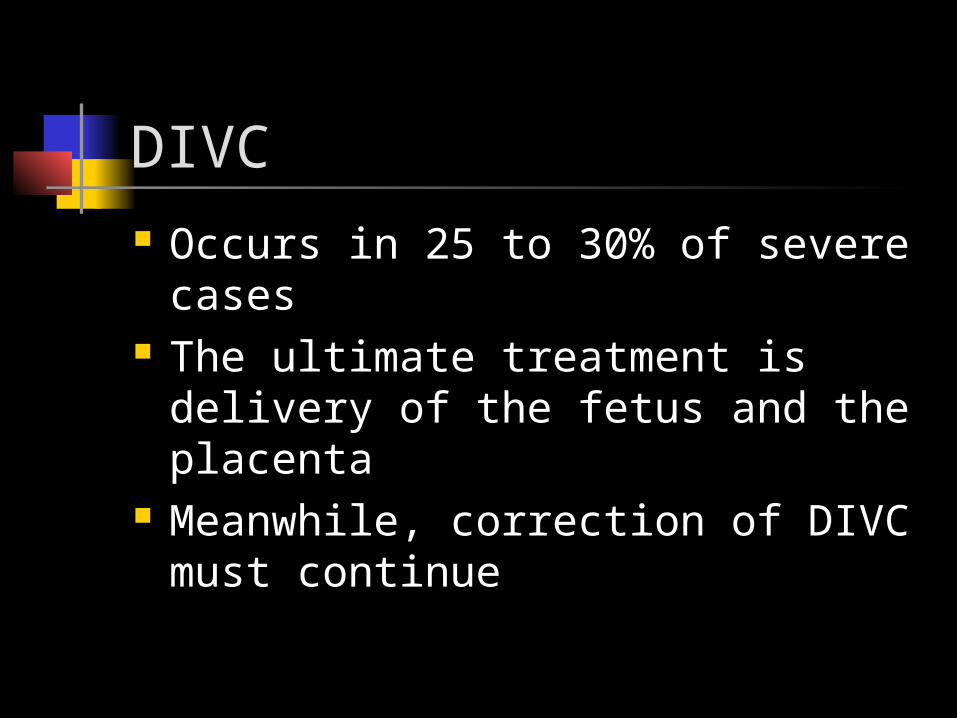
DIVC Occurs in 25 to 30% of severe
cases The ultimate treatment is delivery
of the fetus and the placenta Meanwhile, correction of DIVC
must continue
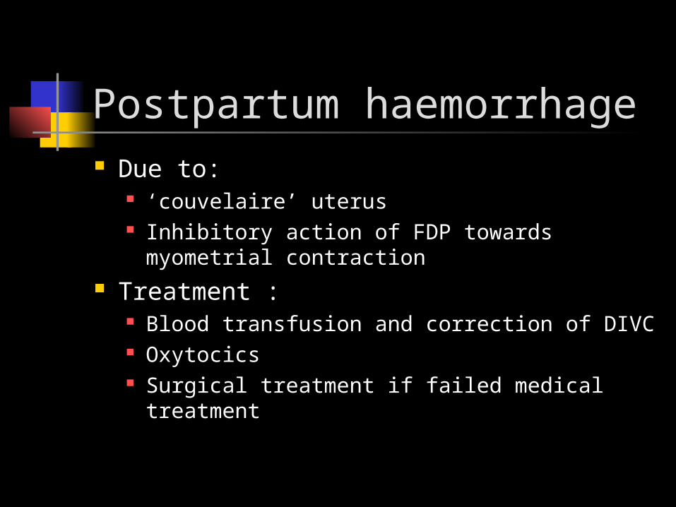
Postpartum haemorrhage Due to:
‘couvelaire’ uterus Inhibitory action of FDP towards myometrial
contraction Treatment :
Blood transfusion and correction of DIVC Oxytocics Surgical treatment if failed medical
treatment

Recurrence risks: One episode :
8 to 17%
Two or more episodes: 25 to 30%

Fetal complications Perinatal mortality Prematurity Intrauterine growth retardation Fetal anemia and transient
coagulopathy

Perinatal mortality 4.4% to 68% depending on:
Severity of abruptio placenta Timing of delivery Gestation of the pregnancy Neonatal facilities

Conclusion Mainly a clinical diagnosis Ultrasound has a limited role DIVC sets in fast Appropriate resuscitative
measures and timely delivery greatly influence both maternal and fetal outcome.

Case 3
21 year old primigravida at 36 weeks has refused hospital admission despite having persistent headache and blurring of vision since yesterday.
Her blood pressure is 170/95mmHg and urine albumin 3+.Easy diagnosis,yes?

Eclampsia / Impending eclampsia Eclampsia :
Occurrence of convulsion in a women whose condition meets the criteria of preeclampsia and not caused by coincidental neurological disease
Impending eclampsia Is there such a condition? Preeclampsia with mainly
neurological symptoms

Significance of this condition Hypertensive disorder is the
second leading cause of maternal mortality in Malaysia Eclampsia encompasses one-third of
the reason
Is it preventable?
If you are the attending doctor in the Pusat Kesihatan, outline your initial management.

Management: Aim:
To treat convulsion To prevent convulsion To control the blood pressure To transfer to hospital with facilities
for: Ceasarean section Blood transfusion Intensive care for mother and newborn

Treat convulsion Magnesium sulphate
Treatment of choice Deep IM injection of 5 gm to each
buttocks (total 10 gm) Intravenous slow bolus 4 gm
Diazepam Intravenous or intramuscular bolus 10
mg

Prevent convulsion Magnesium sulphate
Intramuscular 5 mg in alternate buttocks every 4 hourly for 24 hours
Intravenous infusion : 1 to 2 mg/hour for 24 hours
Diazepam Intravenous infusion of 40mg in 500
ml of normal saline at 10 dpm.

To control blood pressure Nifedipine 10 mg slow release Intramuscular Hydrallazine 2.5-5.0
mg every 20 minutes Intravenous Hydrallazine infusion Intravenous and intramuscular
Labetolol Monitor BP every 15 minutes

Transfer to hospital Accompanied preferably by a doctor Keep patient in left lateral position Insert an airway Give oxygen via nasal
prong/ventimask Insert Foley’s catheter Monitor urine output and vital signs

If you are the receiving doctor in the hospital, how would you further manage this patient?

Consider delivery! Role of Dexamethasone Timing of delivery Method of delivery
CS is the preferred method if delivery is not imminent
Prophylactic instrumental delivery may be feasible if patient is already in second stage of labour

How long Mg SO4 should be infused?
Should the antihypertensive be continued postpartum?
What is the critical period during the postpartum where patients could develop eclampsia?

Conclusion The initial management of patient
influences the outcome of the patient Institution of anticonvulsant
(Magnesium sulphate) has proven to reduce complications especially maternal deaths
Eclampsia could be prevented by early detection of PIH/preeclampsia.

A Gravida 4 Para 3 at 38 weeks gestation has been in the ward for further management of unstable lie.
As the house officer on call, you are called by the staff nurse to review this patient as she has started leaking. Will you attend to the patient urgently?

Definition of cord prolapse Cord presentation:
Umbilical cord is the lowermost part of the fetus present in lower segment with intact amniotic membrane
Cord prolapse: As above without intact amniotic
membrane

Significance of this condition It causes acute severe fetal distress It is preventable in many cases:
should be able to identify possible conditions which may predispose :
Abnormal lie Multiple pregnancies Grand multiparae Preterm labours Polyhydramnios Obstetric manipulation ( forceps delivery )

Management Do not panic Reposition patient:
Traditional knee-chest position (facing downwards)
Steep tredenlenburg position with left lateral tilt
Replace umbilical cord into the vagina and place a warm pad over the introitus

How to prevent cord compression ? Manually elevate the presenting
part ( placing a gloved hand in the vagina)
Vago in 1970 : Inflating the bladder with 500 to 700
ml of normal saline

Urgent delivery Paediatric standby to resuscitate
the baby

Issues Timing :
Better outcome if interval between cord prolapse and delivery is short ( less than 15 minutes)
Deliveries: Mainly caesarean section Instrumental deliveries is possible if
deem fast and easy.
A Gravida 3 Para 2 at 38 weeks of gestation with uncontrolled GDM is currently in labour. What problems would anticipate in
this labour?

Shoulder dystocia Definition:
Difficulty in delivering the anterior shoulder after the head
The anterior shoulder is stuck behind the symphisis pubis
Shoulder dystocia cannot be predicted

You are hoping she will end up with secondary arrest of labour but she managed to reach second stage of labour. Most unfortunately, your registrar was called off urgently for a ruptured ectopic pregnancy and you are instructed to conduct the delivery.
To your dismay, she pushed and the head was out but it pressed hard against the perineum.

Management Shout for help Aim:
To widen the pelvic inlet To rotate the shoulder to a bigger
diameter of the pelvic inlet To reduce the diameter between the
shoulders ( fracture both clavicles) Symphisectomy


After delivery of the fetus and placenta, there is torrential bleeding from the internal os.
The patient becomes pale and hypotensive.
Diagnosis, please?

Definition Blood loss of more than 500 ml from
the genital tract following delivery of the fetus
Primary : within the first 24 hours
Secondary : Excessive blood loss after the first 24 hours

Significance of this condition The major cause of maternal
mortality in Malaysia So important, KKM has national
guidelines and conducted echo-training nationwide. any difference, anybody ?
It is a preventable condition

Problem arising: Underestimation of blood loss
Delayed intervention
Managed by most junior staff

Causes Uterine atony (79-90%) Retained placenta / cotyledons Trauma :
Uterine rupture Broad ligament haematoma Cervical tears Vaginal tears / haematoma Vulval tears / haematoma

Treatment Aim:
Active resuscitation according to the degree of hypovolumic shock
Treat the cause Prevent complications such as:
DIVC Renal failure Sheehan syndrome

Summary of management options: Prevention:
Identify high risk patients Active management of third stage of
labour General management:
Active resuscitation Oxytocics Look for possible cause and treat

When does a low risk patient becomes a high risk patient? Prolonged labour Precipitated labour Instrumental or difficult deliveries Unsuspected abruptio placenta Retained placenta Uterine inversion

Modalities to arrest bleeding Oxytocics
Ergometrine Syntocinon Syntometrine (IM only) Haemabate (IM or intramyometrial)
Best to be given as intravenous route

Modalities to arrest bleeding Uterine massage:
Rub at the fundus of the uterus To ensure contraction To expel blood and blood clots
Blood clots in uterine cavity prevents effective uterine contraction


Modalities to arrest bleeding Bimanual uterine compression
To oppose the anterior and posterior wall together
Reduce potential areas of bleeding Temporary measure


Modalities to arrest bleeding Aortic compression
To arrest bleeding by reducing the perfusion via the common iliac arteries
Have to be released intermittently to prevent ischaemia of lower limbs


Uterine atony Failure of uterus to contract which
results in excessive bleeding Possible cause:
Overdistension of uterus Grandmultiparae High dose / prolonged oxytocin infusion Precipitous or prolonged labour Abruptio placenta General anesthesia

Treatment Massage the fundus of uterus
continuously Oxytocics given as sequentially or
together Examine placenta for completeness Early blood transfusion enhances uterine
contractions Bimanual compression of the uterus Aortic compression

Surgical treatment A last resort:
Conservative Internal iliac ligation B-Lynch suturing Occlusion of uterine and ovarian arteries Uterine arteries embolization
Aggressive hysterectomy

A junior medical student was conducting his first delivery. The baby was out and the staffnurse who was assisting him left him for a while.
Unsupervised, he decided to perform CCT before there was any sign of placental separation
What would be the consequence of his action?

Uterine inversion Complete / incomplete:
Depends whether fundus has passed through the cervix
Acute: Occurs within the first 24 hours post partum
Subacute: Occurs after the first 24 hours but before 4
weeks Chronic:
Presents more than 4 weeks postpartum Extremely rare

Risk factors Fundally sited placenta Overdistension of uterus Oxytocic use Incorrect technique in third stage
of labour

How to diagnose? Severe pain In shock Mass protuding in the vagina Indentation in the fundus of the
uterus

Treatment Alleviate pain
Parenteral analgesia (opiate) Correction of the inversion
Manual under regional / general anaesthesia O’Sullivan hydrostatic correction Combined abdominal-vaginal approach
Antibiotic coverage Ensure continuous uterine contraction

Manual replacement Should be attempted first Oxytocic should be deferred first Placenta should not be detached
prior to correction The first portion out should be
replaced last With general anaesthesia, halothane
further relaxes the uterus


References: Managing Complications in Pregnancy and Childbirth:
A guide for midwives and doctors.WHO 2003 James DK,Steer PJ,Weiner CP,Gonik B eds.High risk
pregnancy:Management options. 2nd ed. W.B Saunders. James M, Timothy D, Robert F, Micheal R
eds.Obstetrics and Gynaecology: a problem solving approach.1st ed. W.B Saunders.
Training manual on Hypertensive disorders in pregnancy.National Technical Committee Confidential enquiries into Maternal Death. Ministry of Health Malaysia 2003
Taber’s Cyclopedic medical dictionary. 15th eds. F.A Davis

Thank you for not sleeping!!