management of an iatrogenic injury in a crossed ectopic ... · we pr esent a unique case of a dual...
TRANSCRIPT
Korean Journal of UrologyⒸ The Korean Urological Association, 2014 554 Korean J Urol 2014;55:554-556
http://crossmark.crossref.org/dialog/?doi=10.4111/kju.2014.55.8.554&domain=pdf&date_stamp=2014-08-16
www.kjurology.orghttp://dx.doi.org/10.4111/kju.2014.55.8.554
Case Report
Management of an Iatrogenic Injury in a Crossed Ectopic Kidney Without FusionTarun Jindal, Mir Reza Kamal, Satyadip Mukherjee, Soumendra Nath Mandal, Dilip KarmakarDepartment of Urology, Calcutta National Medical College, Kolkata, India
Crossed renal ectopia is a condition in which a kidney is located on the side opposite of its ureteral insertion. Ninety percent of crossed ectopic kidneys are fused to their ipsilateral uncrossed renal unit. Crossed renal ectopia without fusion is rare, with only 62 patients reported in the literature to date. These kidneys may suffer iatrogenic injury during an unrelated surgical intervention. The injury, unless self-limiting, may neces-sitate the removal of the ectopic kidney. We present a unique case of a dual injury, renal as well as ureteric, in a crossed ectopic kidney without fusion that was successfully man-aged without surgical excision.
Keywords: Acute renal injury; Congenital abnormalities; Urogenital abnormalities
This is an Open Access article distributed under the terms of the Creative Commons Attribution Non-Commercial License (http://creativecommons.org/licenses/by-nc/3.0) which permits unrestricted non-commercial use, distribution, and reproduction in any medium, provided the original work is properly cited.
Article History:received 22 May, 2012accepted 11 October, 2012
Corresponding Author:Tarun JindalDepartment of Urology, Calcutta National Medical College, 92-B, First Floor, Jhowtala Road, Kolkata-700017, IndiaTEL: +91-9674444929FAX: +91-9674444929,E-mail: [email protected]
INTRODUCTION
Crossed renal ectopia without fusion is a rare condition. These kidneys may suffer iatrogenic injury during an un-related surgical intervention. The injury, unless self-limit-ing, may necessitate the removal of the ectopic kidney [1]. We present a unique case of a dual injury, renal as well as ureteric, in a crossed ectopic kidney without fusion that was successfully managed without surgical excision.
CASE REPORT
A 40-year-old male patient was admitted to the Department of Urology, Calcutta National Medical College, with a complaint of clear fluid discharge from an intra-abdominal drain placed on the right side of his lower abdomen. He had a history of an open appendicectomy done at a primary health care center 3 weeks previously for clin-ically suspected acute appendicitis. According to the oper-ating surgeon’s brief, there was a mass in the right iliac fossa. The appendix was inflamed and was densely adhered to the mass. The surgeon was able to dissect out the appen-dix with difficulty. During the dissection, the mass sus-tained an injury that resulted in clear discharge through it. The surgeon suspected a ureteral injury but was unable to repair it; hence, he inserted a drain and referred the pa-
tient to us for further management.On examination, the patient was anxious but his clinical
parameters, hemogram, renal function tests, and serum bi-ochemistry were normal. The patient had no significant medical or surgical history apart from the present episode. There was an output of approximately 500 to 600 mL per day from the abdominal drain. Analysis of the drain fluid revealed a high creatinine level, which suggested that the output was urine. Ultrasound examination of the abdomen could not delineate the left kidney. The right kidney and ureter were normal on the intravenous pyelogram (IVP) but the left kidney was not seen in its anatomical position. On careful examination of the IVP, a faint shadow with areas of contrast excretion could be seen on the right side, especially in late films at the level of the fourth and fifth lumbar vertebrae (Fig. 1A). Considering the possibility of crossed renal ectopia, contrast enhanced computed tomog-raphy (CECT) was performed, which confirmed the diag-nosis (Fig. 1B). The ectopic left kidney was small and showed excretion of contrast with evidence of hydrone-phrosis. The two renal units were not fused with each other. Cystoscopy revealed a normally placed left ureteric orifice. A retrograde pyelogram (RGP) on the left side showed a hy-dronephrotic kidney with distorted pelvicalyceal system. There was extravasation of the contrast from the kidney. There was also an area of narrowing in the ureter sugges-
Korean J Urol 2014;55:554-556
Injury in Crossed Ectopic Kidney 555
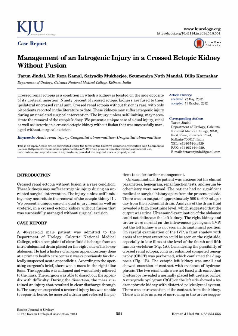
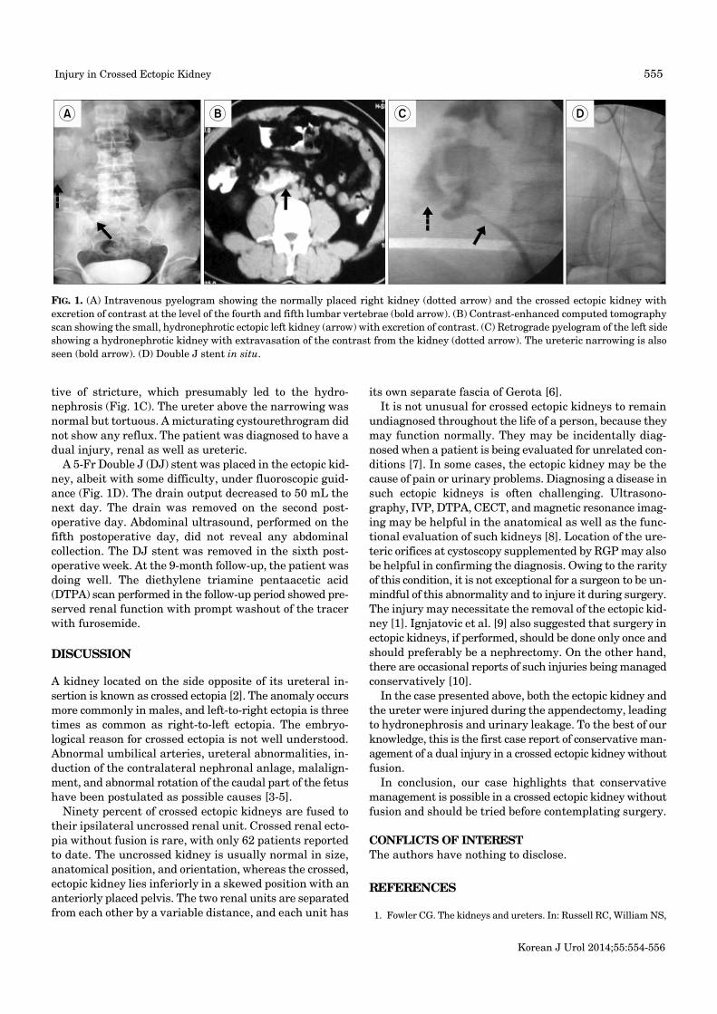
FIG. 1. (A) Intravenous pyelogram showing the normally placed right kidney (dotted arrow) and the crossed ectopic kidney with excretion of contrast at the level of the fourth and fifth lumbar vertebrae (bold arrow). (B) Contrast-enhanced computed tomography scan showing the small, hydronephrotic ectopic left kidney (arrow) with excretion of contrast. (C) Retrograde pyelogram of the left side showing a hydronephrotic kidney with extravasation of the contrast from the kidney (dotted arrow). The ureteric narrowing is also seen (bold arrow). (D) Double J stent in situ.
tive of stricture, which presumably led to the hydro-nephrosis (Fig. 1C). The ureter above the narrowing was normal but tortuous. A micturating cystourethrogram did not show any reflux. The patient was diagnosed to have a dual injury, renal as well as ureteric.
A 5-Fr Double J (DJ) stent was placed in the ectopic kid-ney, albeit with some difficulty, under fluoroscopic guid-ance (Fig. 1D). The drain output decreased to 50 mL the next day. The drain was removed on the second post-operative day. Abdominal ultrasound, performed on the fifth postoperative day, did not reveal any abdominal collection. The DJ stent was removed in the sixth post-operative week. At the 9-month follow-up, the patient was doing well. The diethylene triamine pentaacetic acid (DTPA) scan performed in the follow-up period showed pre-served renal function with prompt washout of the tracer with furosemide.
DISCUSSION
A kidney located on the side opposite of its ureteral in-sertion is known as crossed ectopia [2]. The anomaly occurs more commonly in males, and left-to-right ectopia is three times as common as right-to-left ectopia. The embryo-logical reason for crossed ectopia is not well understood. Abnormal umbilical arteries, ureteral abnormalities, in-duction of the contralateral nephronal anlage, malalign-ment, and abnormal rotation of the caudal part of the fetus have been postulated as possible causes [3-5].
Ninety percent of crossed ectopic kidneys are fused to their ipsilateral uncrossed renal unit. Crossed renal ecto-pia without fusion is rare, with only 62 patients reported to date. The uncrossed kidney is usually normal in size, anatomical position, and orientation, whereas the crossed, ectopic kidney lies inferiorly in a skewed position with an anteriorly placed pelvis. The two renal units are separated from each other by a variable distance, and each unit has
its own separate fascia of Gerota [6]. It is not unusual for crossed ectopic kidneys to remain
undiagnosed throughout the life of a person, because they may function normally. They may be incidentally diag-nosed when a patient is being evaluated for unrelated con-ditions [7]. In some cases, the ectopic kidney may be the cause of pain or urinary problems. Diagnosing a disease in such ectopic kidneys is often challenging. Ultrasono-graphy, IVP, DTPA, CECT, and magnetic resonance imag-ing may be helpful in the anatomical as well as the func-tional evaluation of such kidneys [8]. Location of the ure-teric orifices at cystoscopy supplemented by RGP may also be helpful in confirming the diagnosis. Owing to the rarity of this condition, it is not exceptional for a surgeon to be un-mindful of this abnormality and to injure it during surgery. The injury may necessitate the removal of the ectopic kid-ney [1]. Ignjatovic et al. [9] also suggested that surgery in ectopic kidneys, if performed, should be done only once and should preferably be a nephrectomy. On the other hand, there are occasional reports of such injuries being managed conservatively [10].
In the case presented above, both the ectopic kidney and the ureter were injured during the appendectomy, leading to hydronephrosis and urinary leakage. To the best of our knowledge, this is the first case report of conservative man-agement of a dual injury in a crossed ectopic kidney without fusion.
In conclusion, our case highlights that conservative management is possible in a crossed ectopic kidney without fusion and should be tried before contemplating surgery.
CONFLICTS OF INTERESTThe authors have nothing to disclose.
REFERENCES
1. Fowler CG. The kidneys and ureters. In: Russell RC, William NS,

Korean J Urol 2014;55:554-556
556 Jindal et al
Bulstrode CJ, editors. Baily & Love's short practice of surgery. 24th ed. London: Arnold; 2004. p. 1321-33.
2. McDonald JH, Mcclellan DS. Crossed renal ectopia. Am J Surg 1957;93:995-1002.
3. Winram RG, Ward-McQuaid JN. Crossed renal ectopia without fusion. Can Med Assoc J 1959;81:481-3.
4. Cook WA, Stephens FD. Fused kidneys: morphologic study and theory of embryogenesis. Birth Defects Orig Artic Ser 1977;13:327-40.
5. Alexander JC, King KB, Fromm CS. Congenital solitary kidney with crossed ureter. J Urol 1950;64:230-4.
6. Bauer SB. Anomalies of the upper urinary tract. In: Wein AJ, Kavoussi LR, Novick AC, Partin AW, Peters CA, editors. Campbell-Walsh urology. 9th ed. Philadelphia: Saunders; 2007. p. 3269-304.
7. Felzenberg J, Nasrallah PF. Crossed renal ectopia without fusion associated with hydronephrosis in an infant. Urology 1991;38: 450-2.
8. Yano H, Konagai N, Maeda M, Itoh M, Kuwabara A, Kudou T, et al. Abdominal aortic aneurysm associated with crossed renal ecto-pia without fusion: case report and literature review. J Vasc Surg 2003;37:1098-102.
9. Ignjatovic I, Stojkovic I, Dinic LJ, Jovanovic M, Ivanovic D. Bilateral fused pelvic kidney ectopia with a single draining ureter solved by craniolateral displacement of the kidney and Boari-modified bladder-calyceal anastomosis with bladder augmentation. Int J Urol 2007;14:552-4.
10. Becker AB, Baig MB, Becker AM. Conservative management of a grade V injury to an ectopic pelvic kidney following blunt trauma to the lower abdomen: a case report. J Med Case Rep 2010;4:224.