management of acute asthma - rcp london
TRANSCRIPT

Management of Acute Asthma
Professor Ruth GreenGlenfield Hospital, Leicester
RCP BTS Acute Respiratory Medicine on the Medical Take
29.1.20

Management of acute asthma:
• Assessment• Management guidelines • Drugs and evidence• Quality standards• Tips and pitfalls• An important differential diagnosis• Why patients still die from asthma


Remember: if you discharge the patient they may not see a health care professional again until the next crisis.
You have a responsibility to:Educate Prescribe inhaled corticosteroid

Patient NT 19 yr old female
• Unemployed, smokes 20 cpd and occasional Cannabis
• Lives intermittently with grandmother and friends• Asthma diagnosed age 17, symptoms for “years”• PMH: Eczema• on prn salbutamol and “that brown one”• 3 previous presentations to ED in 4 months via 999
– given nebulisers and discharged

Assessment on admission
• 1/52 history of increasing breathlessness and cough
• On arrival: distressed, unable to complete sentences
• RR 30, pulse 120• PEF 100 (best 350)• ABGs: pH 7.26, PC02 6.04, PO2 13.21 (FiO2 0.4)• Blood eosinophils 0.47 x 10⁹/L

Severity assessment
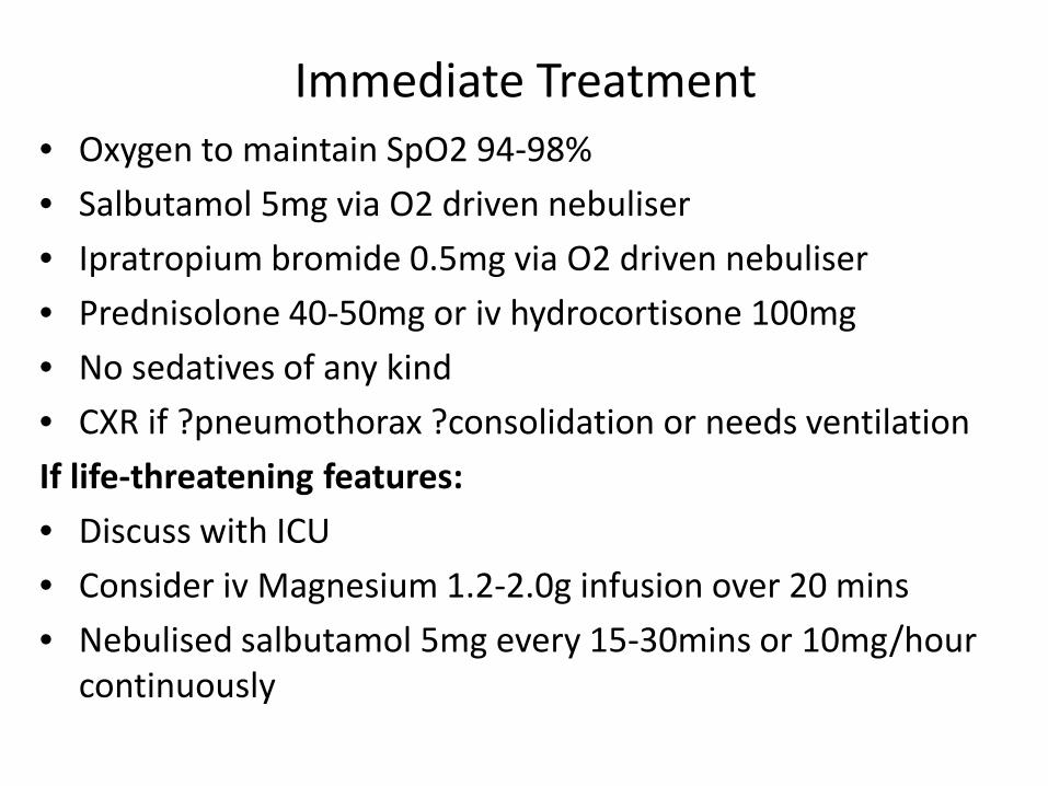
Immediate Treatment• Oxygen to maintain SpO2 94-98%• Salbutamol 5mg via O2 driven nebuliser• Ipratropium bromide 0.5mg via O2 driven nebuliser• Prednisolone 40-50mg or iv hydrocortisone 100mg• No sedatives of any kind• CXR if ?pneumothorax ?consolidation or needs ventilationIf life-threatening features:• Discuss with ICU• Consider iv Magnesium 1.2-2.0g infusion over 20 mins• Nebulised salbutamol 5mg every 15-30mins or 10mg/hour
continuously

Severity assessment

NT: management
• Documented life threatening asthma• Oxygen 40% initially, O2 sat>94%• Hydrocortisone 100mg iv• Nebulised salbutamol 5mg and ipratropium
0.5mg• Iv magnesium 1.2g• Call ICU

Monitoring• Repeat PEF 15-30 mins after Rx started• Oximetry target sats 94-98%• Repeat ABG at one hour if paO2 <8 kPA (unless
Spo2>92%); or paCO2 N or raised or patient deteriorates
• PEF chart qds pre and post 𝛽𝛽2 agonist
• Transfer to ICU with a doctor able to intubate if:– Deteriorating PEF, persistent hypoxia or hypercapnia– Exhaustion / reduced GCS– Poor respiratory effort or respiratory arrest

NT: Scenario
• ICU comment:
“ We currently do not have an ICU bed. Please give iv aminophylline and if no better start NIV on the respiratory ward……”
• What do you do?

Drugs and evidence• Iv magnesium
– Pooled cochrane review shows possible benefit from iv (not inhaled) Magnesium sulfate
– Prevents 7 admissions per 100 patients with acute asthma not responding to nebs and steroids in ED
– Probably safe• Iv aminophylline
– Still widely used but evidence base is limited– Risk of side-effects 20 vomiting, 15 arrythmias or palpitations
per 100 patients– some patients report benefit– Use in guidelines appears historic
• Iv salbutamol– No convincing evidence of benefit, less commonly used (adults)

NIV and asthma• Controversial• Potential uses:
– Pre-oxygenation and whilst preparing to intubate– To avoid intubation
• Rationale/advantages:– Mechanical intubation in asthma is difficult and risks
dynamic hyperinflation, barotrauma and may need use of neuromuscular blockers which increase risk of steroid myopathy
• Risks:– May delay intubation– Usual risks of NIV eg local pressure sores, aspiration etc

NIV and asthma• Evidence
– Lack of high level evidence, no large RCTs– 2013 Cochrane review 5 trials, 206 patients, inconclusive– Some support in observational studies and case series
• Which patients, where and how?– Only on ICU and if intubation not imminently needed but
where there are life threatening features– Contraindicated if reduced consciousness, vomiting,
agitation, profuse secretions, significant haemodynamic instability
– Suggested PEEP 3-5cmH20 (low); iPAP 7-15 cm H20, adjust to target RR<25. High inspiratory flow rate, low I:E ratio (ie 1:5) and prolonged expiratory time
– Monitor very carefully, treat aggressively and intubate if they deteriorate

NT: Scenario• ICU comment:
“ We currently do not have an ICU bed. Please give iv aminophylline and if no better start NIV on the respiratory ward……”
• What do you do?
Trial of iv aminophylline appropriate but not for NIV on the ward – insist on ICU review

Patient NT: further Rx
• Continued wheeze and tiring despite treatment, becoming exhausted
• transferred to ICU with anaesthetic StR• Intubated: high airway pressures• Iv hydrocortisone and aminophylline
continued• Improves within 48 hours and is extubated• Transfer back to respiratory ward
You are called by ward nurse
• Patient wishes to go home.
• What assessments do you make?
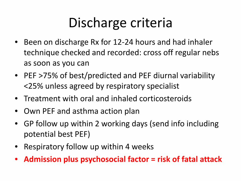
Discharge criteria• Been on discharge Rx for 12-24 hours and had inhaler
technique checked and recorded: cross off regular nebs as soon as you can
• PEF >75% of best/predicted and PEF diurnal variability <25% unless agreed by respiratory specialist
• Treatment with oral and inhaled corticosteroids• Own PEF and asthma action plan• GP follow up within 2 working days (send info including
potential best PEF)• Respiratory follow up within 4 weeks• Admission plus psychosocial factor = risk of fatal attack

Asthma care bundles
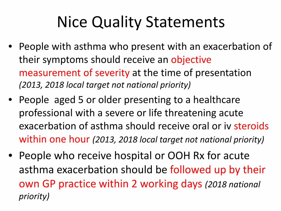
Nice Quality Statements• People with asthma who present with an exacerbation of
their symptoms should receive an objective measurement of severity at the time of presentation (2013, 2018 local target not national priority)
• People aged 5 or older presenting to a healthcare professional with a severe or life threatening acute exacerbation of asthma should receive oral or iv steroids within one hour (2013, 2018 local target not national priority)
• People who receive hospital or OOH Rx for acute asthma exacerbation should be followed up by their own GP practice within 2 working days (2018 national priority)

Tips and pitfalls• Always look at the FBC eosinophil count• Patients with high eosinophils may need prolonged
course of oral prednisolone especially if recurrent attacks
• Always suspect non-adherence to ICS• Always suspect poor inhaler technique• If you are not sure whether this is genuine acute severe
asthma always treat it as such – but document your thoughts
• If you are not sure of the severity of the attack, always assume it is life threatening until proved otherwise

• Most admissions are due to one of the following:– New or unsuspected diagnosis– Suboptimal Rx of known asthma usually due to poor
adherence with ICS but sometimes due to poor primary care prescribing
– Incorrect diagnosis (more to follow)– Psychosocial issues– Co-morbidity– Abnormal illness behaviour eg secondary gain, opiate
dependance
Any asthma admission is a “red flag”: most patients are never admitted

Differential diagnosis of acute severe asthma
• Inducible laryngeal obstruction (Vocal cord dysfunction)
• Dysfunctional breathlessness• COPD• Lower respiratory tract infection• Bronchiectasis• Inhaled foreign body (or drugs)• Heart failure• Lung cancer

KD: History
• 45 year old male, previously fit and well• Lichen Sclerosis• No previous respiratory illness• January 2014: sudden onset SOB, neck
constriction, wheeze. Diagnosis: asthma – treated as acute severe episode
• Multiple admissions: Not responding to treatment with inhaled or oral corticosteroids
• Apparent salbutamol induced bronchospasm

baseline
Post salbutamol

3+ Tight chest and severe change in voice Induced sputum results:
Neutrophils 91%Eosinophils 0%

Inducible Laryngeal Obstruction (ILO):
• “an inappropriate, transient, reversible narrowing of the larynx in response to external triggers”
• Normal: vocal cords move away from the midline during inspiration and slightly back towards the midline during expiration. In ILO, the vocal cords move paradoxically toward the midline during inspiration or excessively so during expiration, resulting in airflow obstruction
• Variable symptoms include: laryngeal tickle, pain, a choking/strangled sensation, cough and dysphonia , episodic (often sudden) episodes of inspiratory (sometimes expiratory) dyspnoea, wheeze and stridor

ILO: pathophysiology• Pathophysiology poorly understood and no consensus
agreed.
• Early literature focuses on psychogenic / emotional reasons for ILO/ VCD
• More recently co-existing organic presentations have been highlighted including: upper airway sensitivity, laryngeal irritants, laryngo-pharyngeal reflux, asthma, post nasal drip.
• Variable triggers: emotional stress, exposure to cold air, exercise, smoke, coughing, laughing, talking, viruses, reflux, allergens, food and drink, forced respiratory manoeuvres

ILO: making the diagnosis• Gold standard: paradoxical movement of the vocal cords at
nasendoscopy (classically anterior 2/3 adduction, posterior diamond shaped chink on inspiration). Cords need to be visualised when symptomatic – challenges (breathing, phonation, topical trigger, exercise)
Normal AbductionAnterior adduction
Posterior “chink”
Listen over the trachea: wheeze here with clear lungs very suggestive: if in doubt treat as acute severe asthma but document your doubt – helps later

• Gold standard: paradoxical movement of the vocal cords at nasendoscopy (classically anterior 2/3 adduction, posterior diamond shaped chink on inspiration). Cords need to be visualised when symptomatic – challenges (breathing, phonation, topical trigger, exercise)
• Flow-volume loops: often poorly tolerated/non-reproducible -may show inspiratory loop truncation (extra-thoracic obstruction); ↑MEF 50%/MIF 50% ratio ↓MVV; ↑ Raw
• Emerging alternative tests: Questionnaires (eg VCDQ) impulse oscillometry, airway fluoroscopy, plethysmography, CT with 3D reconstruction of larynx and colour doppler ultrasound imaging of vocal cord movement (not fully evaluated)
• Exclude alternative/additional pathologies especially asthma

ILO: Treatment:• No Randomised controlled trials• MDT: SLT, physician, physiotherapist, clinical psychologist,
ENT surgeon.• Patient education critical• Identification and avoidance of triggers (eg treat GOR)• Psychological therapy• Physiotherapy if dysfunctional breathing• Other Rx anecdotal benefit but trials needed (Botox,
surgery, inspiratory muscle training)
2017 ILO: official joint ERS/ELS statement Task Force Report

Back to asthma: Patient KS
• 17 year old female under severe asthma clinic• Severe eosinophilic asthma with poor
symptom control and recurrent severe exacerbations
• Known poor adherence to treatment• Previous near-fatal attack (respiratory arrest
on holiday age 12)• Advised to take regular oral steroids but
adverse effect on mood

Final admission• Attended GP urgent care centre : Worsening SOB and wheeze despite
4 days of Prednisolone 40mg. Given 2.5mg neb salbutamol, EWS 5 -refer ED resus.
• ED 19:18 Used whole salbutamol inhaler at home. RR24 PEF330 HR 125 (Best PEF 370-390) sats 100% on neb, generalised wheeze
• Plan: nebulisers iv access, 200mg iv hydrocortisone (given 19:00), bloods, CXR (no penumothorax), review. Also given 1g iv meropenemand 1g iv paracetamol
• ED review 20:05 - walked to toilet, no wheezing
• ED review 20:55 Improved: RR 18, sats 97% on air PF 380 (normal for her) Imp: good response Plan: advised send to EDU and aim home later but patient unhappy as in the past she has deteriorated after discharge, would like overnight admission therefore plan to admit

ED asthma pathway

• ED review 22:50 (SpR) “Patient remained stable, last neb 19:30. I spoke to her again and she is happy to go home and will stay with her friend. I advised staying in EDU for observation but she wanted to go home. Patient has full capacity.”
• Patient self discharged and went to friend's house. Parents not aware of admission or discharge

15 hours later: 999 EMAS to ED • 999 call 13:01 Cardiac arrest at friend’s house - witnessed collapse, vomiting,
friend started CPR
• Arrival at ED: asystolic arrest:– A: bag and valve ventilation– B: reduced AE very difficult to bag– C: cardiac arrest
• Intubated, 4Hs and 4Ts addressed, bilateral needle thoracotomies performed -right sided pneumothorax 20F drain inserted then changed to 28F. Given salbutamol, hydrocortisone, aminophylline
• ROSC 14:21 (down time 52 minutes)
• Bronchoscopy and manual chest compression performed by ITU cons
• Imp: Cardiac arrest secondary to tension pneumothorax and bronchospasm
• Tragically died 18 hours later: cause of death 1(a) tension pneumothorax (b) acute severe asthma

Asthma still killsNational Review of Asthma Deaths published May 2014• 2/3 of deaths could be
prevented with better routine care
• Room for improvement in care of 83% of those who died
• Particular issues in children
2017: young patients still dying due to inadequate asthma care
Lessons are not being learnt
7 KEY PREVENTABLE FACTORS LINKED TO
ASTHMA DEATHS

Over use of rescue 𝛽𝛽2-agonist
Under use of inhaled corticosteroid
No objective assessment of control
No Personalised Asthma Action Plan (PAAP)
Missed opportunity for review after acute attack
Psychosocial factors
Triggers not avoided
• Good control is salbutamol <twice per WEEK• Review if >12 salbutamol inhalers/12 months
• Review prescribing, inhaler technique, adherence
• Assess control each visit (RCP 3Qs, Peak flow): ACT if not controlled
• PAAP saves lives : give to all patients and review every visit
• Refer to specialist if >2 courses prednisolone/year • Follow up within 48 hours after admission/ED/OOH
• Address smoking / adherence / psychological factors / obesity / comorbidity / safeguarding
• Address smoking, allergen exposure• Avoid NSAIDs and Beta-blockers
2
3
4
5
6
7
1
WHY DOES ASTHMA STILL KILL?

National quality improvement priorities for 2019/20
• Ensure 90% of patients are assessed for asthma severity including measurement of PEF within one hour
• Ensure 90% of patients receive a respiratory specialist review before discharge
• Ensure 90% of patients receive systemic steroids within one hour of arrival (unless given at home)