management of a patient with transverse mandibular … · etiology and diagnosis ... management of...
TRANSCRIPT
Address for correspondence:
B. Delvallez31D rue Vaneau, 35000 [email protected]
DOI: 10.1051/odfen/2013206 J Dentofacial Anom Orthod 2013;16:306� RODF / EDP Sciences
1
Article received: 02:2013.Accepted for publication: 03:2013.
C L I N I C A L C A S E
Management of a patient withtransverse mandibular dyssymmetry
Benjamin DELVALLEZ, François GUYOMARD, Olivier SOREL
INTRODUCTION
Cases of dyssymmetries of the basalbone and/or of the dentoalveolar archesare complicated to manage. Orthognathicsurgery is often a necessary means forachieving treatment objectives.
This report presents the management of acase of a skeletal Class II associated with amandibular dyssymmetry and a mild oto-mandibular syndrome.
Article available at http://www.jdao-journal.org or http://dx.doi.org/10.1051/odfen/2013206

ETIOLOGY AND DIAGNOSIS
A young 16 year old patient namedDorian was referred to us in Novem-ber, 2006 with delayed dental erup-tion, especially of the lower canines.
He was diagnosed with:– Facial: a right-left facial asymmetry
with a protruding left ear, and adeviation of the chin (Fig. 1). His
forced smile hardly shows hisupper incisors. His profile shows aslightly retruded mandible and thecomparative size of the lower thirdof the face is diminished, thenasolabial angle is slightly closedand the cervical-mental distance isdiminished.
CL
IN
IC
AL
CA
SE
Figure 1Extraoral photographs face atrepose (a), smile (b), profile (c)at the start of treatment.
BERNARD DELVALLEZ, FRANÇOIS GUYOMARD, OLIVIER SOREL
2 Delvallez B., Guyomard F., Sorel O. Management of a patient with transverse mandibular dyssymmetry

– Skeletal: a narrow maxilla, a weakskeletal class II with a slightlyreceded chin (ANB 4�), a mandibu-lar asymmetry and a reducedanterior vertical facial height(SN/GoGn 16�).
– Dentoalveolar: lingualized maxillaryand mandibular teeth, overallretrusion of the mandibular archand a maxillary protrusion (I/Na6 mm).
– Dental relationships: an Angle classII with right subdivision of 2 mm, anincisal overjet of 7 mm, lowerincisors completely hidden and se-vere dentomaxillary discrepancy.The interincisal midlines are notdeviated. But the mandibular inter-incisal midline does not align withthe mandibular basal midline. Thepatient also has malformed max-illary first molars.
CL
IN
IC
AL
CA
SE
Figure 2Initial panoramic xray (a), lateral cephalometric xray (b), Delaire analysis on xray 10 months after the start oftreatment.
MANAGEMENT OF A PATIENT WITH TRANSVERSE MANDIBULAR DYSSYMMETRY
J Dentofacial Anom Orthod 2013;16:306. 3

TREATMENT OBJECTIVES
The solution to this bone disorderwill be maxillary and mandibularorthognathic surgery. A Lefort 1 low-ering of the maxilla, a slight advance-ment of the body of the mandible anda mandibular derotation with saggitalosteotomies of the ascending rami. Asignificant dentoalveolar correctionwill be required in order to establishalignment between the mandibularinterincisal midline and the mandibularbasal bone.
The dentoalveolar problem will beresolved by alveolar expansion andnot by extractions even though therewere long and lively debates beforeand during treatment. The choice tonot extract was motivated by thepossibility of increasing the width ofthe alveolus and inclining the mandib-ular incisors labially. An intrusion ofthe incisors and canines should beperformed but the incisor overjet will
CL
IN
IC
AL
CA
SE
Figure 3Initial Steiner analysis on lateral cephalometric xray 10
months after the start of treatment.
Figure 4Facial AP xray.
BERNARD DELVALLEZ, FRANÇOIS GUYOMARD, OLIVIER SOREL
4 Delvallez B., Guyomard F., Sorel O. Management of a patient with transverse mandibular dyssymmetry

CL
IN
IC
AL
CA
SE
Figure 5Initial intraoral photographs.
Figure 6Initial intraoral photographs at the start of treatment.
MANAGEMENT OF A PATIENT WITH TRANSVERSE MANDIBULAR DYSSYMMETRY
J Dentofacial Anom Orthod 2013;16:306. 5

largely be resolved by the orthog-nathic surgery.
The incisor overjet should be re-duced because it is far greater thanthe necessary mandibular advance-ment.
TREATMENT STEPS
Treatment was started once all thepermanent teeth had erupted, or 10months after the orthognathic evalua-tion (October 2007).
Alignment and maxillary levelingwere achieved after 8 months oftreatment. Mandibular alignment aswell as the traction of 33 weresuccessfully treated two years afterthe beginning of therapy. Slow man-dibular and maxillary alveolar expan-sion was accomplished (1 ½ years).
Mandibular leveling was not com-pletely achieved in order to obtain apost-surgical bilateral posterior open-ing with the objective of extruding theposterior sectors to resolve the verti-cal discrepancy.
It took a long time to establish thecoordination of the arches. The pre-surgical treatment lasted for 3 years(November 2010). The post-surgicaltreatment lasted for 6 months (end ofJune 2011).
We observed a worsening of theprofile during the pre-surgical prepara-tion (Fig. 8). The smile remainedunchanged. The smile will be im-proved by lowering the maxillary.
An analysis of the lateral cephalo-metric xray (Fig. 9) shows a labialinclination of the maxillary incisors of
10� (I/Na 36�) and of the mandibularincisor of 6� (i/Na 27�).
A superimposition clearly demon-strates the movements that wereachieved (Fig. 10). A partial correctionof the overjet was not established by atrue intrusion of the mandibular inci-sors but rather by an extrusion of themaxillary and mandibular molars and alabial inclination of the maxillary andmandibular incisors.
The AP head film clearly shows thealignment of the mandibular incisorswith the mandibular basal midline(Fig. 11).
Surgery was performed to lower themaxilla with a clockwise rotation andadvancement of the body of themandible more to the right than tothe left. The surgeon left a minorposterior open bite. Treatment wasresumed 6 months after the bi-max-illary orthognathic surgery.
The surgeon did not achieve asgreat a mandibular derotation as we
CL
IN
IC
AL
CA
SE
Figure 7Intraoral photograph at the start of traction on 33.
BERNARD DELVALLEZ, FRANÇOIS GUYOMARD, OLIVIER SOREL
6 Delvallez B., Guyomard F., Sorel O. Management of a patient with transverse mandibular dyssymmetry

CL
IN
IC
AL
CA
SE
Figure 8Pre-surgical facial photographs, face at repose (a) smile (b), and profile (c).
Figure 9Pre-surgical panoramic xray (a), pre-surgical lateral cephalometric xray (b), and pre-surgical Steiner analysis (c).
MANAGEMENT OF A PATIENT WITH TRANSVERSE MANDIBULAR DYSSYMMETRY
J Dentofacial Anom Orthod 2013;16:306. 7

CL
IN
IC
AL
CA
SE
Figure 10Pre-surgical superimposition according to the LVD method: Overall A and mandibular B.
Figure 11Pre- and post-surgical facial AP xrays.
BERNARD DELVALLEZ, FRANÇOIS GUYOMARD, OLIVIER SOREL
8 Delvallez B., Guyomard F., Sorel O. Management of a patient with transverse mandibular dyssymmetry

CL
IN
IC
AL
CA
SE
Figure 12Pre-surgical intraoral photographs.
Figure 13Post-surgical intraoral photographs.
MANAGEMENT OF A PATIENT WITH TRANSVERSE MANDIBULAR DYSSYMMETRY
J Dentofacial Anom Orthod 2013;16:306. 9

had expected (Fig. 15), because hedecided that he would have had tomove the facial chin too far to thepatient’s left.
The finishing touches consisted ofreducing the 3 mm post-surgical inter-cisal midline discrepancy and achiev-ing posterior occlusion.
We can see in Figure 14 that theteeth appear in the smile, the vertical
dimension of the lower third of theface has improved and the profile isstraighter.
A post-surgical Steiner Analysisshows an improvement of the ANB(2�), a surgical reduction of the labialinclination of the maxillary incisors of6� (I/Na 30�) (Fig. 15). The inclination ofthe mandibular incisors is almostidentical.
CL
IN
IC
AL
CA
SE
Figure 14Facial photographs face at repose (a), Smile (b), three-quarter facial smile (c), and profile (d).
BERNARD DELVALLEZ, FRANÇOIS GUYOMARD, OLIVIER SOREL
10 Delvallez B., Guyomard F., Sorel O. Management of a patient with transverse mandibular dyssymmetry

CL
IN
IC
AL
CA
SE
Figure 15Facial profile at the end of treatment.
Figure 16Panoramic xray (a), lateral cephalometic xray (b) Steiner analysis (c) post-surgical.
MANAGEMENT OF A PATIENT WITH TRANSVERSE MANDIBULAR DYSSYMMETRY
J Dentofacial Anom Orthod 2013;16:306. 11

DISCUSSION
The stability of the treatment al-ways has to be assessed. In fact,increases in intercanine and intermolardistances were required. There is stilla mesiolingual rotation of 36 butfortunately this allows for efficientinterlocking of 36 and 26 (since thecoronal shape of 26 is very unusual).
There is still a slight discrepancy of theinterincisal midlines of 1.5 mm. Thediastema between 23 and 22 hasbeen filled by two esthetic restora-tions, on the mesial of 22 and on thedistal of 23. The inclination of themaxillary canines is different (23 ismore corono-buccally inclined) with an
CL
IN
IC
AL
CA
SE
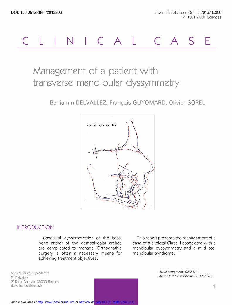
Figure 17Overall superimposition at the end of treatment: initial tracing in black, pre-surgical tracingin blue and post-surgical tracing in red.
BERNARD DELVALLEZ, FRANÇOIS GUYOMARD, OLIVIER SOREL
12 Delvallez B., Guyomard F., Sorel O. Management of a patient with transverse mandibular dyssymmetry

CL
IN
IC
AL
CA
SE
Figure 18Post-treatment introral photographs.
Figure 19Intraoral photographs 1 and 1/2 years after treatment.
MANAGEMENT OF A PATIENT WITH TRANSVERSE MANDIBULAR DYSSYMMETRY
J Dentofacial Anom Orthod 2013;16:306. 13

AFMP slightly greater than on the left.Therefore, we performed a slightcoronoplasty on the palatal face of13. There is left and right canineguidance. Due to the length of pre-surgical treatment (that exhausted themotivation of our 20 year old patient)and the appearance of white spots(synonymous with poor oral hygiene),he asked us to curtail the finishingtouches because he was concerned
about whether the risk-benefit rela-tionship was a positive one. In spite ofthis, the esthetics, the Class 1 staticocclusion of the right and left molarsand canines and the dynamic occlu-sion are entirely correct. The vestibu-lar periodontium of the mandibularincisors does not seem to be wea-kened or harmed by the treatment(verification by probing, radiographyand palpation).
CONCLUSION
Because the management of thetherapy was tridimensional, we had to
make casts of several models andassemble three articulated montages
CL
IN
IC
AL
CA
SE
Figure 20Facial photographs 1 and 1/2 years after the end of treatment.
BERNARD DELVALLEZ, FRANÇOIS GUYOMARD, OLIVIER SOREL
14 Delvallez B., Guyomard F., Sorel O. Management of a patient with transverse mandibular dyssymmetry

in order to situate all the componentsin the three dimensions of space. Thecommunication and team work invol-ving the motivated patient, the max-i l lo fac ia l surgeon, the denta lconsultants and the students incharge of the patient, were the keyto the success of the active treatment.Practitioners will have to carefully
monitor the stability of the treatment.We recently took photographs 1½years after the end of treatment(January 2012) and the stability seemscorrect (Fig. 19, 20).
Special thanks to Dr. Le Strat and toProfessor Guyomard.
REFERENCES FOR CONSULTATION
1. Casteigt J, Faure J, Labarrere H, Treil J. Symbiose chirurgico-occlusoorthodontiquedans les dysmorphies maxillofaciales. EMC 23-499-A-10, 2006, Elsevier SAS.
2. Cheong YW, Lo LJ. Facial asymmetry: etiology, evaluation, and management. ChangGung Med J 2011 34(4):341-51.
3. Garcia C, La chirurgie orthognathique du futur. Orthod Fr 2008;79:67–77.4. Dahan J, Troubles d’attitudes mandibulaires. 23-472-A-10, 2002, Editions Scientifiques
et Medicales Elsevier SAS.5. Guyot L, Brignol L, Thiery G, Cheynet F, Chossegros C, Gola R. Traitement chirurgical
des retromandibulies par avancee mandibulaire par cale cartilagineuse retrocondy-lienne. EMC 23-499-A-11 Odontologie/Orthopedie 2007 Elsevier Masson SAS.
6. Richter M, Mossaz C, Laurent F, Goudot P. Chirurgie correctrice des dysmorphiesmaxillomandibulaires. Insuffisances et exces sagittaux associes a une hauteur facialenormale. EMC 22-066-E-20, 2000. Elsevier SAS.
7. Zhang Y, Che B, Ni Y, Zhang H, Pan Y, Wang L, Ma J. Three-dimensional condylarpositions and forms associated with different anteroposterior skeletal patterns andfacial asymmetry in Chinese adolescents. Acta Odontol Scand, 2013 ; DOI : 10.3109/00016357.2012.757359
CL
IN
IC
AL
CA
SE
MANAGEMENT OF A PATIENT WITH TRANSVERSE MANDIBULAR DYSSYMMETRY
J Dentofacial Anom Orthod 2013;16:306. 15