mammography accreditation program … document is copyright protected by the american college of...
TRANSCRIPT

This document is copyright protected by the American College of Radiology. Any attempt to reproduce, copy, modify, alter or otherwise change or use this document without the express written permission of the American College of Radiology is prohibited.
Page 1 of 23
S:\Marketing\Web Content Migration\Documents\Accred\New Mammo Docs\Mammography Accreditation Program Requirements.docx Revised: 5/2/12
Mammography Accreditation Program Requirements
OVERVIEW ........................................................................................................................................................................... 2
PERSONNEL QUALIFICATIONS ..................................................................................................................................... 3
INTERPRETING PHYSICIAN .................................................................................................................................................... 3 RADIOLOGIC TECHNOLOGIST ............................................................................................................................................... 4 MEDICAL PHYSICIST ............................................................................................................................................................ 4
EQUIPMENT ......................................................................................................................................................................... 5
QUALITY CONTROL .......................................................................................................................................................... 7
MAMMOGRAPHY EQUIPMENT EVALUATIONS ....................................................................................................................... 8 ANNUAL MEDICAL PHYSICIST’S SURVEY ............................................................................................................................. 8 RADIOLOGIC TECHNOLOGIST QUALITY CONTROL TESTS ..................................................................................................... 9
QUALITY ASSURANCE/MEDICAL AUDIT .................................................................................................................... 9
REPORTING ........................................................................................................................................................................ 10
RECORD KEEPING ........................................................................................................................................................... 10
CONSUMER COMPLAINTS ............................................................................................................................................. 11
FACILITY CLOSURE ........................................................................................................................................................ 12
APPLYING FOR ACCREDITATION .............................................................................................................................. 13
NEW FACILITIES ................................................................................................................................................................. 13 NEW UNITS ........................................................................................................................................................................ 15 MOBILE MAMMOGRAPHY .................................................................................................................................................. 16
ACCREDITATION TESTING ........................................................................................................................................... 17
CLINICAL IMAGES .............................................................................................................................................................. 17 MAMMOGRAPHIC IMAGE IDENTIFICATION ......................................................................................................................... 17 PHANTOM IMAGE AND DOSE .............................................................................................................................................. 19
FINAL REPORTS ................................................................................................................................................................ 19
ACCREDITATION RENEWAL ........................................................................................................................................ 20
ACCREDITATION FEES ................................................................................................................................................... 20
ANNUAL UPDATES ........................................................................................................................................................... 21
VALIDATION ...................................................................................................................................................................... 21
FILM CHECKS ..................................................................................................................................................................... 21 ON-SITE SURVEYS .............................................................................................................................................................. 21
FOR ADDITIONAL INFORMATION .............................................................................................................................. 22
ACR PRACTICE GUIDELINES AND TECHNICAL STANDARDS ............................................................................ 22
REFERENCES ..................................................................................................................................................................... 22

This document is copyright protected by the American College of Radiology. Any attempt to reproduce, copy, modify, alter or otherwise change or use this document without the express written permission of the American College of Radiology is prohibited.
Page 2 of 23
S:\Marketing\Web Content Migration\Documents\Accred\New Mammo Docs\Mammography Accreditation Program Requirements.docx Revised: 5/2/12
Overview
The American College of Radiology’s (ACR) Mammography Accreditation Program provides facilities with peer review and constructive feedback on staff qualifications, equipment, quality control (QC), quality assurance, image quality, and radiation dose. Developed in 1987 by the ACR Task Force on Breast Cancer, the accreditation program is the country’s first and the largest for mammography. The program is directed by radiologists and medical physicists on the Committee on Mammography Accreditation of the ACR Commission on Quality and Safety. As an ACR peer-review program, all reports, documentation, correspondence (including e-mail) between the ACR and the facility undergoing accreditation, and any information provided by the facility to evaluate its imaging services is privileged and confidential under the Code of Virginia 8.01-581.17. (The facility name, address, phone number, and listed contact personnel are not considered privileged and confidential).
The success of the ACR program in improving the quality of mammography motivated the U.S. Congress to model the provisions of its 1992 Mammography Quality Standards Act (MQSA) after the ACR’s Mammography Accreditation Program. MQSA requires that all mammography facilities in the United States (including those that examine Medicare or private pay patients and those that are only screening or diagnostic) be:
Accredited by an approved body Certified by the U.S. Department of Health and Human Services (HHS), and Inspected by the HHS (or a state agency acting on behalf of the HHS)
The U.S. Food and Drug Administration’s (FDA) MQSA regulations1 define a mammography facility as “a hospital, outpatient department, clinic, radiology practice, mobile unit, office of a physician, or other facility that conducts mammography activities.” The following MQSA milestones impacted all mammography facilities:
October 1, 1994 - all mammography facilities in the U.S. were required to be MQSA-certified; a facility without a current MQSA certificate performing mammography after this date is practicing unlawfully and may be subject to FDA sanctions and fines
October 28, 1997 - FDA published the Quality Mammography Standards; Final Rule April 28, 1999 - the Final Rule went into effect October 28, 2002 - more stringent equipment regulations went into effect
The FDA has approved the ACR as an accrediting body for both screen-film and designated full-field digital mammography (FFDM) systems. (See the FDA’s Digital Accreditation webpage for a current list of accrediting bodies approved by the FDA to accredit specific units and models of FFDM systems.) Facilities with other FFDM manufacturers and/or models must apply to and be approved by the FDA in order to legally use them to examine patients.
The accreditation program requirements outlined in this document are identical or equivalent to those in the FDA Final Rules. ACR recommendations for improved quality are clearly designated as such but are not required to meet these recommendations in order to be accredited.

This document is copyright protected by the American College of Radiology. Any attempt to reproduce, copy, modify, alter or otherwise change or use this document without the express written permission of the American College of Radiology is prohibited.
Page 3 of 23
S:\Marketing\Web Content Migration\Documents\Accred\New Mammo Docs\Mammography Accreditation Program Requirements.docx Revised: 5/2/12
Personnel Qualifications
All interpreting physicians, radiologic technologists, and medical physicists (including part-time and locum tenens staff) must have documentation showing that they meet minimum MQSA qualifications. The FDA allows some flexibility in subject areas that may be counted toward the required mammography continuing education. (See the FDA’s Policy Guidance Health System2 for more information.) If personnel do not meet continuing education or continuing experience requirements, the facility needs to be aware of the FDA’s requirements and guidance for re-establishing qualifications2.
Interpreting Physician
All physicians interpreting mammograms must meet the following MQSA-required qualifications:
Qualifications Board Certified in Radiology Not Board Certified in Radiology Initial Licensed to practice medicine
AND Certified in diagnostic radiology by:
- American Board of Radiology, or - American Osteopathic Board of Radiology, or - Royal College of Physicians and Surgeons of
Canada AND
Initial experience: - Interpreted 240 examinations in any 6 months
within the last 2 years of residency (if board certified at 1st possible opportunity), or
- Interpreted 240 examinations within the 6 months immediately prior to the physician’s qualification date (if not board certified at 1st possible opportunity)
Licensed to practice medicine AND
Documented training in mammography interpretation, radiation physics, radiation effects and radiation protection: - 2 months (if initially qualified before
April 28, 1999), or - 3 months (if initially qualified after
April 28, 1999) AND
Initial experience: interpreted 240 examinations within the 6 months immediately prior to the physician’s qualification date
AND Category 1 CME in mammography (at least 15 of which must have been acquired in the 3
years immediately prior to the physician’s qualification date): - 40 hours (if initially qualified before April 28, 1999), or - 60 hours (if initially qualified after April 28, 1999)
AND 8 hours of training in a mammographic modality (e.g., digital) before beginning to use that
modality Continuing Experience
Interpret 960 mammographic examinations over a 24-month period
Continuing Education
15 Category I CME in mammography in a 36-month period
The ACR and the FDA require that each facility identify a lead interpreting physician who will be responsible for ensuring that all MQSA-required activities are carried out. The ACR Practice Guideline for the Performance of Screening and Diagnostic Mammography specifies that screening mammography may be performed without a physician in attendance. The Committee on Mammography Accreditation believes that adequate supervision can be maintained at off-site facilities through quarterly professional feedback. The facility should maintain a log of these interactions signed by the radiologist. This review should include:

This document is copyright protected by the American College of Radiology. Any attempt to reproduce, copy, modify, alter or otherwise change or use this document without the express written permission of the American College of Radiology is prohibited.
Page 4 of 23
S:\Marketing\Web Content Migration\Documents\Accred\New Mammo Docs\Mammography Accreditation Program Requirements.docx Revised: 5/2/12
Clinical image quality Quality assurance procedures QC documentation, and A determination that safe operating procedures are used
Radiologic Technologist
All radiologic technologists performing mammography must meet the MQSA-required qualifications in the table below. The ACR also recommends that technologists performing mammography hold the American Registry of Radiologic Technologists (ARRT) post-primary certification in mammography.
Qualifications Qualified Before April 28, 1999 Qualified After April 28, 1999 Initial Certified by:
- American Registry of Radiologic Technologists (ARRT), or
- American Registry of Clinical Radiologic Technologists, or
Licensed to perform general radiographic procedures in a state
AND 40 hours of training in mammography
Certified by: - American Registry of Radiologic
Technologists (ARRT), or - American Registry of Clinical Radiologic
Technologists, or Licensed to perform general radiographic
procedures in a state AND
40 hours of training in mammography including: - Training in breast anatomy and
physiology, positioning and compression, QA/QC techniques, and imaging of patients with breast implants, and
- 25 mammography examinations under direct supervision of an appropriate MQSA-qualified individual
AND 8 hours of training in using a mammographic modality (e.g., digital), before beginning to use
that modality independently Continuing Experience
Perform 200 mammographic examinations over a 24-month period
Continuing Education
15 Category 1 CEUs in mammography in a 36-month period
Medical Physicist
All medical physicists conducting mammography equipment evaluations and annual surveys must meet the following MQSA-required qualifications:

This document is copyright protected by the American College of Radiology. Any attempt to reproduce, copy, modify, alter or otherwise change or use this document without the express written permission of the American College of Radiology is prohibited.
Page 5 of 23
S:\Marketing\Web Content Migration\Documents\Accred\New Mammo Docs\Mammography Accreditation Program Requirements.docx Revised: 5/2/12
Qualifications Master’s Degree or Higher Bachelor’s Degree
(must be qualified before April 28, 1999) Initial Master’s degree or higher in a physical
science, and 20 semester hours of physics, and 20 contact hours of training in conducting
surveys of mammography facilities, and Experience in conducting surveys of 10
units and 1 facility
Bachelor’s degree in a physical science, and
Qualified under the Interim Regulations, and
10 semester hours of physics, and 40 contact hours of training in conducting
surveys of mammography facilities, and Experience in conducting surveys of 20
units and 1 facility AND
Certified in Diagnostic Radiological Physics, Diagnostic Medical Physics, Imaging Physics, or Radiological Physics by: - American Board of Radiology, or - American Board of Medical Physics. or
Licensed or approved by a state AND
8 hours of training in a mammographic modality (e.g., digital) before surveying units of that modality
Continuing Experience
Survey 2 mammography facilities and 6 mammography units over a 24-month period
Continuing Education
15 CME/CEUs in mammography in a 36-month period
Equipment
Mammography equipment must meet all FDA requirements. The following table summarizes section 900.12(b) of the FDA equipment regulations and applies to both screen-film and FFDM as indicated:
Feature FDA Equipment Requirement Applies To Motion of tube-image receptor assembly
The assembly shall be capable of being fixed in any position where it is designed to operate. Once fixed in any such position, it shall not undergo unintended motion.
S-F & FFDM
This mechanism shall not fail in the event of power interruption. S-F & FFDM
Image receptor sizes
Systems using screen-film image receptors shall provide, at a minimum, for operation with image receptors of 18 x 24 cm and 24 x 30 cm.
S-F
Systems using screen-film image receptors shall be equipped with moving grids matched to all image receptor sizes provided.
S-F
Systems used for magnification procedures shall be capable of operation with the grid removed from between the source and image receptor.
S-F & FFDM
Beam limitation and light fields
All systems shall have beam limiting devices that allow the useful beam to extend to or beyond the chest wall edge of the image receptor.
S-F & FFDM
For any mammography system with a light beam that passes through the X-ray beam-limiting device, the light shall provide an average illumination of not less than 160 lux (15 ft-candles) at 100 cm or the maximum source-image receptor distance (SID), whichever is less.
S-F & FFDM (except Fischer)
Magni-fication
Systems used to perform noninterventional problem-solving procedures shall have radiographic magnification capability available for use by the operator.
S-F & FFDM

This document is copyright protected by the American College of Radiology. Any attempt to reproduce, copy, modify, alter or otherwise change or use this document without the express written permission of the American College of Radiology is prohibited.
Page 6 of 23
S:\Marketing\Web Content Migration\Documents\Accred\New Mammo Docs\Mammography Accreditation Program Requirements.docx Revised: 5/2/12
Feature FDA Equipment Requirement Applies To Systems used for magnification procedures shall provide, at a minimum, at least one magnification value within the range of 1.4 to 2.0.
S-F & FFDM
Focal spot selection
When more than one focal spot is provided, the system shall indicate, prior to exposure, which focal spot is selected.
S-F & FFDM
When more than one target material is provided, the system shall indicate, prior to exposure, the preselected target material.
S-F & FFDM
When the target material and/or focal spot is selected by a system algorithm that is based on the exposure or on a test exposure, the system shall display, after the exposure, the target material and/or focal spot actually used during the exposure.
S-F & FFDM
Application of compres-sion
Each system shall provide an initial power-driven compression activated by hands-free controls operable from both sides of the patient.
S-F & FFDM
Each system shall provide fine adjustment compression controls operable from both sides of the patient.
S-F & FFDM
Compres-sion paddle
Systems shall be equipped with different sized compression paddles that match the sizes of all full-field image receptors provided for the system.
S-F & FFDM
The compression paddle shall be flat and parallel to the breast support table and shall not deflect from parallel by more than 1.0 cm at any point on the surface of the compression paddle when compression is applied.
S-F & FFDM (except Fischer)
Equipment intended by the manufacturer’s design to not be flat and parallel to the breast support table during compression shall meet the manufacturer’s design specifications and maintenance requirements.
S-F & FFDM
The chest wall edge of the compression paddle shall be straight and parallel to the edge of the image receptor.
S-F & FFDM
The chest wall edge may be bent upward to allow for patient comfort but shall not appear on the image.
S-F & FFDM
Technique factor selection and display
Manual selection of mAs or at least one of its component parts (mA and/or time) shall be available.
S-F & FFDM
The technique factors (kVp and either mA and seconds or mAs) to be used during an exposure shall be indicated before the exposure begins, except when AEC is used, in which case the technique factors that are set prior to the exposure shall be indicated.
S-F & FFDM
Following AEC mode use, the system shall indicate the actual kVp and mAs (or mA and time) used during the exposure.
S-F & FFDM
Automatic exposure control
Each screen-film system shall provide an AEC mode that is operable in all combinations of equipment configuration provided, e.g., grid, non-grid; magnification, nonmagnification; and various target-filter combinations.
S-F
The positioning or selection of the detector shall permit flexibility in the placement of the detector under the target tissue. The size and the available positions of the detector shall be clearly indicated at the X-ray input surface of the breast compression paddle. The selected position of the detector shall be clearly indicated.
S-F
The system shall provide means for the operator to vary the selected optical density from the normal (zero) setting.
S-F
X-ray film The facility shall use X-ray film for mammography that has been designated by the film manufacturer as appropriate for mammography.
S-F
Intensifying screens
The facility shall use intensifying screens for mammography that have been designated by the screen manufacturer as appropriate for mammography and shall use film that is matched to the screen’s spectral output as specified by the manufacturer.
S-F

This document is copyright protected by the American College of Radiology. Any attempt to reproduce, copy, modify, alter or otherwise change or use this document without the express written permission of the American College of Radiology is prohibited.
Page 7 of 23
S:\Marketing\Web Content Migration\Documents\Accred\New Mammo Docs\Mammography Accreditation Program Requirements.docx Revised: 5/2/12
Feature FDA Equipment Requirement Applies To Film processing solutions
For processing mammography films, the facility shall use chemical solutions that are capable of developing the films used by the facility in a manner equivalent to the minimum requirements specified by the film manufacturer.
S-F
Lighting The facility shall make special lights for film illumination, i.e., hot-lights, capable of producing light levels greater than that provided by the view box, available to the interpreting physicians.
S-F & FFDM (for hardcopy comparison)
Film masking devices
Facilities shall ensure that film masking devices that can limit the illuminated area to a region equal to or smaller than the exposed portion of the film are available to all interpreting physicians interpreting for the facility.
S-F & FFDM (for hardcopy comparison)
In addition, facilities with FFDM systems should note the following:
The FDA requires that the facility be able to provide the medical institution, physician, healthcare provider, patient, or patient’s representative with hardcopy films of final interpretation quality. Consequently, the facility must have access to a film printer (either on-site or at a third party.) The printer must exist and be tested by a qualified medical physicist according to the FFDM unit manufacturer’s QC manual before the facility performs mammography on patients. The facility must also include information and QC data for the printer in its accreditation application as it does for film processors.
The FDA recommends that only monitors and printers specifically cleared for FFDM use by FDA’s Office of Device Evaluation (ODE) be used. However, a facility may legally use others.
See the FDA's Policy Guidance Help System2 for more information. The ACR Practice Guideline for Determinants of Image Quality in Digital Mammography contains further guidance for FFDM systems.
Quality Control
Compliance with minimum frequencies and performance of QC tests as defined by the FDA’s Final Rule is a requirement for accreditation. Although not required for accreditation, the ACR recommends that facilities with screen-film systems follow the procedures and performance criteria outlined in the 1999 ACR Mammography Quality Control Manual3.
The FDA requires that facilities with FFDM systems comply with a QC program that is substantially the same as that recommended by the image receptor manufacturer. In general, QC technologists and medical physicists should refer to the most recent version of their FFDM unit’s QC manual for the specific tests, frequencies, and performance criteria.
This requirement to follow the FFDM image receptor manufacturer’s QC also applies to review workstations (monitors) and film printers. However, the FDA allows some flexibility for workstation/printer QC depending on the device’s clearance from the FDA’s ODE for FFDM use. Facilities should check with their workstation/printer manufacturer for their device's FDA clearance status.

This document is copyright protected by the American College of Radiology. Any attempt to reproduce, copy, modify, alter or otherwise change or use this document without the express written permission of the American College of Radiology is prohibited.
Page 8 of 23
S:\Marketing\Web Content Migration\Documents\Accred\New Mammo Docs\Mammography Accreditation Program Requirements.docx Revised: 5/2/12
Workstations/printers approved by the FDA's ODE for FFDM - FDA considers the workstation/printer's QC manual to be "substantially the same" as that of the image receptor manufacturer and the facility may follow it for QC
Workstations/printers not approved by the FDA's ODE for FFDM - the facility must follow the QC manual provided by the image receptor manufacturer (the facility should check with the image receptor manufacturer for their required tests)
Each facility must submit documentation of compliance for all QC tests as part of the application process. Both screen-film and FFDM versions of the required forms may be downloaded from the ACR website.
Mammography Equipment Evaluations
The FDA requires a mammography facility to have its medical physicist perform a Mammography Equipment Evaluation when a new unit is installed. This applies to both screen-film and FFDM units and includes previously owned units, accredited units moved from one site to another site, and new units replacing previously accredited units. This Equipment Evaluation must determine whether the new unit meets both the FDA’s:
Equipment requirements (section 900.12(b)), and Quality assurance requirements (section 900.12(e))
The Mammography Equipment Evaluation results must be submitted to the ACR on the MQSA Requirements for Mammography Equipment Checklist and the Medical Physicist Mammography QC Test Summary, which may be downloaded from the ACR website. All problems must be corrected before the facility may apply for accreditation of the unit and before the new unit is put into service for mammography.
Annual Medical Physicist’s Survey
The medical physicist must perform the QC tests listed in the table below for screen-film system and manufacture specified QC for FFDM system when equipment is installed and at least annually thereafter. The medical physicist should provide a written report of the survey’s findings to responsible physician(s) and professional(s) responsible for service of the equipment. Although FDA regulations allow the medical physicist 30 days from the date of the survey to send a report, a 30-day delay in getting the report to the facility allows the facility no time to take corrective actions. The ACR strongly recommends that written reports be submitted in time to give facilities the opportunity to take corrective actions in case of an adverse finding.
Annual Medical Physicist’s Survey Screen-Film Tests Required by MQSA
Timeframe for Corrective Action
Mammographic Unit Assembly Evaluation Within 30 days of the test date
Collimation Assessment Within 30 days of the test date
Evaluation of System Resolution Within 30 days of the test date
AEC System Performance Within 30 days of the test date
Uniformity of Screen Speed Within 30 days of the test date
Artifact Evaluation Within 30 days of the test date
Image Quality Evaluation Immediately
kVp Accuracy and Reproducibility Within 30 days of the test date
Beam Quality Assessment Within 30 days of the test date

This document is copyright protected by the American College of Radiology. Any attempt to reproduce, copy, modify, alter or otherwise change or use this document without the express written permission of the American College of Radiology is prohibited.
Page 9 of 23
S:\Marketing\Web Content Migration\Documents\Accred\New Mammo Docs\Mammography Accreditation Program Requirements.docx Revised: 5/2/12
Annual Medical Physicist’s Survey Screen-Film Tests Required by MQSA
Timeframe for Corrective Action
Breast Exposure and AEC Reproducibility Within 30 days of the test date
Average Glandular Dose Immediately
Radiation Output Rate Within 30 days of the test date
Measurement of Viewbox Luminance and Room Illuminance
The medical physicist must summarize the Annual Medical Physicist’s Survey results on the Medical Physicist Mammography QC Test Summary form, which may be downloaded from the ACR website.
Radiologic Technologist Quality Control Tests
A QC program must be implemented for all mammography systems and should be established with the assistance of a medical physicist. A designated QC technologist must perform the FDA-required QC tests listed in the table below for screen-film systems and manufacturer specified QC for FFDM system at the specified minimum frequencies. If any QC parameter falls outside of the control limits, corrective action must be taken. A medical physicist should be available to assist in prescribing corrective actions for unresolved problems.
Radiologic Technologist Screen-Film QC Test
Minimum Frequency
Required by MQSA
Timeframe for Corrective Action
Darkroom Cleanliness Daily
Processor Quality Control Daily Immediately
Mobile Unit QC Daily Immediately
Screen Cleanliness Weekly
Viewboxes and Viewing Conditions Weekly
Phantom Images Weekly Immediately
Visual Checklist Monthly
Repeat Analysis Quarterly Within 30 days of the test date
Analysis of Fixer Retention in Film Quarterly Within 30 days of the test date
Darkroom Fog Semi-annually Immediately
Screen-Film Contact Semi-annually Immediately
Compression Semi-annually Immediately
Both screen-film and FFDM Mammography Quality Control Checklists and screen-film QC forms are available for the technologist on the ACR website.
Quality Assurance/Medical Audit
Each facility must establish and maintain a mammography medical outcomes audit program to follow up positive mammography assessments and to correlate pathology results with the interpreting physician’s findings. The ACR strongly recommends that facilities use the Breast Imaging Reporting and Data System (BI-RADS®)4 final assessment codes and terminology for reporting and tracking of mammography outcomes. (However, there is no requirement that the mammography facilities currently accredited by the ACR, nor those applying for accreditation in the future, purchase any ACR products in order to obtain or maintain their ACR accreditation.) The BI-RADS® Atlas also contains guidance on monitoring outcomes and conducting a mammography audit.

This document is copyright protected by the American College of Radiology. Any attempt to reproduce, copy, modify, alter or otherwise change or use this document without the express written permission of the American College of Radiology is prohibited.
Page 10 of 23
S:\Marketing\Web Content Migration\Documents\Accred\New Mammo Docs\Mammography Accreditation Program Requirements.docx Revised: 5/2/12
Reporting
The interpreting physician must prepare a written report containing the results of each examination in addition to the following information:
The name of the patient and an additional patient identifier The date of the examination The name of the examination’s interpreting physician, and An overall final assessment
The ACR strongly recommends that the final assessment be categorized into one of the FDA-approved BI-RADS® final assessment categories:
BI-RADS® Category Overall Final Assessment 1 Negative 2 Benign Finding(s) 3 Probably Benign Finding – Initial Short-Term Follow-Up Suggested 4 Suspicious Abnormality – Biopsy Should Be Considered 5 Highly Suggestive of Malignancy – Appropriate Action Should Be Taken 6 Known Biopsy-Proven Malignancy – Appropriate Action Should Be Taken
In cases where no final assessment can be assigned due to incomplete work-up, the ACR recommends that BI-RADS® Category 0 (Incomplete: Need Additional Imaging Evaluation and/or Prior Mammograms for Comparison) be used along with reasons why no assessment can be made. Excerpted text from the BI-RADS® Atlas is available on the ACR website.
This written report, signed by the interpreting physician, must be provided to the patient’s health care provider within 30 days of the examination date. If the assessment is “suspicious” or “highly suggestive of malignancy” reasonable attempts must be made to communicate this to the health care provider (or designee) as soon as possible. FDA guidance recommends no more than 3 business days.
In addition, the facility must send a written summary of the mammography report to the patient in terms easily understandable by a lay person (i.e., lay letter) within 30 days of the examination date. If the assessment is “suspicious” or “highly suggestive of malignancy” reasonable attempts must be made to communicate this to the patient as soon as possible: FDA guidance recommends no more than 5 business days. This applies to every patient who receives a mammogram, not only self-referred patients. The intent of this law is to address women’s concerns about breakdowns in communication that prevent timely and appropriate diagnosis and treatment of breast disease. Sample lay letters are available on the ACR website. If the patient is self-referred and has not named a health care provider, the facility must also send a copy of the written report to the patient.
Record Keeping
Current mammograms and records must be kept by the facility:

This document is copyright protected by the American College of Radiology. Any attempt to reproduce, copy, modify, alter or otherwise change or use this document without the express written permission of the American College of Radiology is prohibited.
Page 11 of 23
S:\Marketing\Web Content Migration\Documents\Accred\New Mammo Docs\Mammography Accreditation Program Requirements.docx Revised: 5/2/12
For at least 5 years, or At least 10 years if no additional mammograms of the patient are performed at the facility, or Longer if mandated by state or local law, or Until a request is made by or on behalf of the patient, to permanently or temporarily transfer
her records to a medical institution, her physician or health care provider, or to the patient herself
For FFDM, either the original or lossless compressed full field digital data or hardcopy films of final interpretation quality must be maintained.
The FDA requires that each mammography facility permanently or temporarily transfer the original mammograms and copies of the patient’s reports to the patient or to her medical institution, physician, or health care provider upon request or on behalf of the patient. For FFDM, the facility must be able to provide hardcopy films of final interpretation quality. The ACR will assist patients calling to report that their mammography facility will not transfer original images by calling the mammography facility to inform them about the FDA requirement for original film transfer. If the facility continues to refuse to transfer the images, ACR staff will refer the patient to appropriate staff at the FDA.
If the facility continues to refuse to transfer the images, it is cause for a “Serious Complaint” according to the FDA’s criterion, i.e. a serious adverse event is one “for which a facility fails to take appropriate corrective action in a timely manner.” The ACR will initiate an investigation of the facility.
Consumer Complaints
ACR-accredited facilities must:
Have a written and documented system for collecting and resolving consumer complaints Maintain a record of each serious complaint received by the facility for at least 3 years from the
date the complaint was received Provide the consumer with directions for filing serious complaints with the ACR if the facility
is unable to resolve a serious complaint to the consumer’s satisfaction, and Report unresolved serious complaints to the ACR as soon as possible
A serious complaint is defined by the FDA as “a report of a serious adverse event, which means an event that significantly compromises clinical outcomes or one for which a facility fails to take appropriate corrective action in a timely manner.”
In addition, consumers may directly contact the ACR to report a serious complaint if they feel that their concerns have not been adequately addressed by the facility. All serious consumer complaints must be submitted to the ACR in writing and include the:
Consumer’s name, address and telephone number Consumer’s signature (if reported by the consumer) Name and location of the ACR-accredited facility where the mammogram was performed Description of the complaint
Serious Adverse Events - Examples 1. Poor image quality
2. Missed cancers
3. The use of personnel that do not meet FDA qualifications
4. Failure to send the mammography reports or lay summaries within 30 days

This document is copyright protected by the American College of Radiology. Any attempt to reproduce, copy, modify, alter or otherwise change or use this document without the express written permission of the American College of Radiology is prohibited.
Page 12 of 23
S:\Marketing\Web Content Migration\Documents\Accred\New Mammo Docs\Mammography Accreditation Program Requirements.docx Revised: 5/2/12
Copies of any supporting documentation that would be helpful in addressing the complaint
The ACR will:
Acknowledge the receipt of the complaint by letter to the patient (or facility) Obtain a signed Serious Consumer Complaint Inquiry Release Authorization from the patient Request a response from the facility in writing Provide a summary of its resolution to the patient Forward unresolved complaints to the FDA as appropriate
Consumer complaints may be faxed, e-mailed, or mailed to:
Although all consumer complaints are important, not all complaints submitted to the ACR are of a serious nature. These include patient waiting times, uncomfortable compression, and excessive fees charged by facilities for transferring films. The ACR will acknowledge the receipt of each complaint and advise the patient which complaints can best be addressed directly by the facility. ACR staff will provide the patient with copies of FDA regulation and guidance supporting their complaint and alternative sources of assistance as appropriate. The ACR will not follow up any complaint that is submitted verbally or anonymously.
Facility Closure
If a facility ceases mammography and closes its doors, the FDA continues to hold the facility responsible for assuring that there is a mechanism to release the films to the appropriate entity when requested. Before a facility permanently stops performing mammography, it should do the following:
Inform the ACR that it will no longer be performing mammography, Notify its State radiation control program, and Arrange transfer of each patient’s medical record (original mammography images and reports)
to the mammography facility where the patient will be receiving future care, the patient’s referring physician or health care provider, or the patient.
The facility should make reasonable attempts to inform its former patients of how they can obtain their mammography records. (Facilities should check with state or local agencies to determine if their requirements are more stringent.) Radiology practices and other medical facilities that still see patients but have permanently stopped performing mammography, may choose to keep the patients’ medical records rather than transfer them to another facility (unless the patient requests such a transfer).
If the third option is not viable, facilities must assure that there is a mechanism to release the films to the appropriate entity when requested and that former patients are made aware of that mechanism. For instance a facility could store the medical records in a hospital, if appropriate, or make arrangements to warehouse the records. If no one else is willing to accept the records, the facility remains responsible for them.
Director, Breast Imaging Accreditation Programs American College of Radiology
1891 Preston White Drive Reston, VA 20191-4397
Fax: (703) 648-9176 [email protected]
This document is copyright protected by the American College of Radiology. Any attempt to reproduce, copy, modify, alter or otherwise change or use this document without the express written permission of the American College of Radiology is prohibited.
Page 13 of 23
S:\Marketing\Web Content Migration\Documents\Accred\New Mammo Docs\Mammography Accreditation Program Requirements.docx Revised: 5/2/12
Once the facility ceases operation, both the ACR certificate and the MQSA certificate should be removed from display.
Applying for Accreditation
New Facilities
Before a new mammography facility may legally perform mammography on anyone (including volunteers) it must apply for accreditation of all their mammography units and receive a provisional MQSA certificate from the FDA (or state certifying body). This applies to both screen-film and FFDM systems.
The following mammography sites must apply separately for accreditation and obtain unique facility MAP IDs and MQSA certificate numbers:
A new site, not associated with an existing mammography facility, opening with new equipment and personnel
A new site associated with an existing mammography facility, opening at a separate physical location with new equipment
A new site at a separate physical location, with a previously-accredited unit from a sister site (the sister site remains in operation with other units under its current MQSA certificate)
A site at the same physical location as a previously accredited and certified facility opening under new ownership and with new personnel and equipment
The flowchart below summarizes the MQSA certification and ACR mammography accreditation process. The facility is responsible for taking the following steps to obtain an MQSA certificate:
Scheduling the medical physicist’s survey at least 1 week before the “applications” training to allow time for corrective action of potentially non-compliant tests (contact the medical physicist early, especially if he/she is a consultant)
Completing an Entry Application to the ACR that includes: Basic facility, equipment, and personnel information The medical physicist’s Mammography Equipment Evaluation summary showing that all FDA-
required tests have passed Legal agreements with appropriate signatures The appropriate application fee Sending the complete Entry Application to the ACR with sufficient lead time and via a
traceable method If the application is complete and all FDA requirements are met, the ACR notifies the FDA (or
state certifying body) to fax the facility a 6-month provisional MQSA certificate The facility should expect to receive its provisional MQSA certificate within 4 business days of
the day the ACR receives all required application material Scheduling the manufacturer’s “applications” training accordingly (the facility must have
this provisional MQSA certificate before imaging human subjects as part of this training)
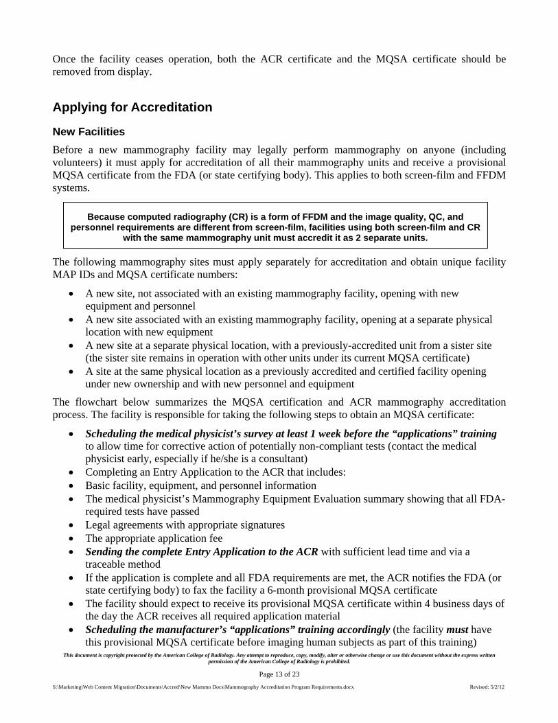
Because computed radiography (CR) is a form of FFDM and the image quality, QC, and personnel requirements are different from screen-film, facilities using both screen-film and CR
with the same mammography unit must accredit it as 2 separate units.

This document is copyright protected by the American College of Radiology. Any attempt to reproduce, copy, modify, alter or otherwise change or use this document without the express written permission of the American College of Radiology is prohibited.
Page 14 of 23
S:\Marketing\Web Content Migration\Documents\Accred\New Mammo Docs\Mammography Accreditation Program Requirements.docx Revised: 5/2/12
Contacting their state radiation control agency for any state-specific requirements for new facilities
Soon after the facility receives their provisional MQSA certificate, the ACR will send the following:
Facility Completes Entry Application
ACR Reviews Entry Application; Sends Facility Full Application
Facility Completes Full Application & Returns to ACR
ACR Reviews Full Application
Clinical Image Review
Phantom Image Review
ACR Writes Final Report
orFacility/Unit
Deficiency (1st)Facility/Unit
Passes
Facility Repeats,
Appeals or Withdraws
ACR Notifies FDA
FDA Sends Facility 3-yr Certificate(facility may continue mammography)
Facility Renews Accreditation in 3 Years
ACR Notifies FDA
FDA Sends Facility a 6-mo Provisional Certificate
(facility may do mammography)
New facilities only

This document is copyright protected by the American College of Radiology. Any attempt to reproduce, copy, modify, alter or otherwise change or use this document without the express written permission of the American College of Radiology is prohibited.
Page 15 of 23
S:\Marketing\Web Content Migration\Documents\Accred\New Mammo Docs\Mammography Accreditation Program Requirements.docx Revised: 5/2/12
An application to obtain information on Policies and procedures Reporting mechanisms Patient outcome data Personnel (radiologists, medical physicists and radiologic technologists) QC results Screen-film or FFDM testing materials to assess Clinical image quality Phantom image quality Dose
Facilities must send all application and testing materials to the ACR within 45 calendar days of the date the ACR sent the full application to the facility.
New Units
A currently accredited and certified facility installing a new unit (or a previously used or accredited unit that is new to the facility) should contact the ACR for appropriate instructions. The type of accreditation options available will depend on how much time the facility has left on its current MQSA certificate when the new unit is installed:
Over 13 months: New Unit Addendum - a facility with other fully accredited units may choose to complete a
New Unit Addendum and pay a reduced fee for accrediting the new unit (since it will not receive a full, 3-year certification). A facility with a new unit(s) applying mid-cycle in this manner need submit only testing for the new unit (not a full application). Once accreditation is approved for the new unit, its expiration date will be the same as the other units at the facility.
Reinstate – the ACR recommends that a facility with only 1 unit choose to give up its current MQSA certificate and reinstate the facility with the new unit. The facility must submit a New Unit Reinstatement Application at the full fee and will receive a 6-month provisional MQSA certificate. Once approved, the facility will receive a full 3-year accreditation from the date of approval.
Less than 13 months: Early Renewal - the ACR will instruct the facility to begin early renewal on all units at the full
renewal fee. Once approved, the facility will have a new expiration date that is 3 years from the old expiration date; thus, the facility will not lose any time from its current MQSA certificate.
A facility with a current MQSA certificate may legally begin examining patients with the new unit after they have sent the complete application (with their medical physicist’s passing Equipment Evaluation results) to the ACR. Once approved, the ACR will notify the FDA that an accreditation application has been accepted for the new unit. These facilities are not required to wait for a response from the ACR to begin clinical use of the new unit since they are operating with a current MQSA certificate. However, if this is the facility’s first FFDM unit, the Center for Medicare and Medicaid Services (CMS) will not reimburse for digital examinations until the FDA has received notification
A facility must have an MQSA certificate in order to legally perform mammography.

This document is copyright protected by the American College of Radiology. Any attempt to reproduce, copy, modify, alter or otherwise change or use this document without the express written permission of the American College of Radiology is prohibited.
Page 16 of 23
S:\Marketing\Web Content Migration\Documents\Accred\New Mammo Docs\Mammography Accreditation Program Requirements.docx Revised: 5/2/12
that the facility has applied for accreditation of a FFDM unit. In order to ensure CMS payment, the facility must:
Fax the application materials with the Equipment Evaluation results to the ACR, and After 3 business days, call the ACR to confirm that the new unit information was transmitted to
the FDA
In addition, facilities should contact their state radiation control agencies for any state-specific requirements for new units.
Mobile Mammography
Facilities with mobile units must call the ACR for specific instructions on accreditation. Because mobile practice settings vary significantly, the FDA has approved the ACR to provide some flexibility in accrediting mobile units. In general, mobile units may apply for accreditation as follows:
Scenario I: A mobile unit operating under a fixed, accredited, and certified facility (utilizing the same personnel) may either
Accredit as an additional unit under the facility’s MQSA certification, or Accredit and certify as an entirely separate facility If the fixed facility chooses to accredit the mobile unit under its existing MQSA certificate, the
mobile unit must be present at the fixed site during MQSA inspections. Scenario II: A mobile unit owned and operated by a single entity must designate a single lead
interpreting physician in order to accredit and certify as a single facility. Scenario III: A mobile unit owned by a single entity servicing multiple sites with different
lead interpreting physicians must accredit and certify as separate facilities.
Mobile Scenario Examples
Mobile Unit Owner
Mobile Service Site
Accreditation/ Certification Location Mammo Service Provider/Owner
Lead MD
I. Mobile unit owned and operated by a single, fixed accredited and certified facility; services 3 sites
A Hospital 1. B Hospital A Hospital Dr. Q Option 1: Accredit the mobile unit under the fixed facility’s (A Hospital) MQSA certification or
Option 2: Accredit and certify the mobile unit as a separate facility
2. C Nursing Home
A Hospital Dr. Q
3. D Health Center
A Hospital Dr. Q
II. Independent mobile service owned and operated by a single entity; services 3 sites
E Mobile Co.
1. F Hospital E Mobile Co. Dr. S Option 1: Accredit and certify the mobile unit as 1 facility (E Mobile Co.) or
Option 2: Accredit and certify the mobile unit as 3 separate facilities
2. G Hospital E Mobile Co. Dr. S 3. H Hospital E Mobile Co. Dr. S
III. Independent mobile service owned by a single entity; services 5 sites, with 3 different lead interpreting physicians
I Mobile Co. 1. J Hospital J Hospital Dr. U Accredit and certify as 1 facility (either I Mobile Co. or J Hospital)
2. K Hospital K Hospital Dr. V Option 1: Accredit and certify as 1 facility; only 1 owner may be designated (either I Mobile Co., K Hospital or L Hospital) or
Option 2: Accredit and certify the mobile unit as 2 separate facilities (K Hospital and L Hospital)
3. L Hospital L Hospital Dr. V
4. M Hospital M Hospital Dr. W Accredit and certify as 1 facility; only 1 owner may be designated (either I Mobile Co. or M Hospital) 5. N Nursing
Home M Hospital Dr. W

This document is copyright protected by the American College of Radiology. Any attempt to reproduce, copy, modify, alter or otherwise change or use this document without the express written permission of the American College of Radiology is prohibited.
Page 17 of 23
S:\Marketing\Web Content Migration\Documents\Accred\New Mammo Docs\Mammography Accreditation Program Requirements.docx Revised: 5/2/12
Accreditation Testing
Clinical Images
The facility must submit 2 clinical cases (1 from a patient with fatty breasts; 1 from a patient with dense breasts) to be evaluated as part of accreditation. At least 2 ACR radiologist reviewers will score 8 parameters on each case. See the “Clinical Image Evaluation” section of the 1999 ACR Mammography Quality Control Manual for examples. The submitted images must meet the following criteria:
The cases must be “negative” (BI-RADS® Assessment Category 1) and not “benign” (Category
2) or “incomplete” (Category 0). If the facility only performs diagnostic exams and cannot submit “negative” images, they should call the ACR for assistance.
The cases must be examples of the facility’s best work. The images must be from actual patients and must have been
formally interpreted. They may not be from models or volunteers. The entire breast must be imaged in a single exposure on each
projection. Digital images must be on hardcopy and must be as close to “true
size” as possible (i.e., with no minification or magnification). Both screen-film and digital images must be labeled with the MQSA-required identification
information. The ACR reviewers will evaluate this. The lead interpreting physician must review and approve the hardcopy clinical images.
Mammographic Image Identification
Images are an important part of the medical record. One of the requirements for clinical images is correct labeling to include patient identification. The ACR understands that as providers, facilities are subject to the Health Insurance Portability and Accountability Act of 1996 (HIPAA) and that is why the ACR executes a HIPAA business associate agreement (BAA) with facilities. This agreement allows the collection of patient information in the performance of ACR accreditation activities which are specifically mentioned in the HIPAA regulations. If the facility has a BAA with ACR, they are covered under HIPAA. If not, contact the ACR to obtain an agreement for signature.
The MQSA-required identification below applies to both screen-film and FFDM images.
Clinical Image Review Parameters
1. Positioning
2. Compression
3. Exposure level
4. Sharpness
5. Contrast
6. Noise
7. Artifacts
8. Exam identification

This document is copyright protected by the American College of Radiology. Any attempt to reproduce, copy, modify, alter or otherwise change or use this document without the express written permission of the American College of Radiology is prohibited.
Page 18 of 23
S:\Marketing\Web Content Migration\Documents\Accred\New Mammo Docs\Mammography Accreditation Program Requirements.docx Revised: 5/2/12
MQSA-Required Mammographic Image Identification 1. Name of patient (first and last)
2. Additional patient identifier (e.g., medical record number or social security number; date of birth is less desirable)
3. Date of examination
4. Standardized view and laterality codes placed on the image in a position near the axilla
5. Facility name and location (must include city, state, and zip code)
6. Technologist identification
7. Cassette/screen identification
8. Mammography unit identification, if more than one unit in the facility
The label which contains the patient and facility information outlined in the table must be permanent.
Radiopaque markers or identification indicating laterality (R/L) and projection/view (MLO, CC) must be placed near the aspect of the breast closest to the axilla. If radiopaque markers are used, they should be placed on the cassette holder so that they can be read directly from overhead; they should not be so large as to be distracting but large enough to be clearly read. Standardized abbreviations for mammography views (see the 1999 ACR Mammography Quality Control Manual) must be employed to eliminate confusion from one facility to another.
The technologist who performed the examination must be identified on the image. Technologist identifiers, such as unique initials, should be placed either on a designated location in the patient ID area or with radiopaque letters on the cassette holder. The facility should maintain a log of the technologists and their identifying initials.
Cassette/screen identification is usually designated by an Arabic numeral written or pressed on the screen. This is used to identify screens with artifacts or defects.
A mammography unit number (usually a Roman numeral) is required if there is more than one unit at the facility.
Except for view and laterality, labels should be placed as far as possible from the breast so as not to distract from evaluating the breast image. Collimating close to the surface of the breast is not recommended because light transmitted through clear areas of the film adversely affects film viewing. Collimating close to the edge of the breast does not significantly improve image contrast since there is virtually no X-ray scatter from air. Therefore, collimation should be to the edge of the film so that as much of the film as possible will be exposed.
The ACR also recommends the following identification on each image:

This document is copyright protected by the American College of Radiology. Any attempt to reproduce, copy, modify, alter or otherwise change or use this document without the express written permission of the American College of Radiology is prohibited.
Page 19 of 23
S:\Marketing\Web Content Migration\Documents\Accred\New Mammo Docs\Mammography Accreditation Program Requirements.docx Revised: 5/2/12
Identification Advantages/Discussion Strongly recommended
A “flash card” patient ID system
More permanent than stick-on labels
Information reproduces on copy films
ID should fit squarely in its designated space, near the edge of the film
Not acceptable if any information is illegible, does not fit, or is lopsided, causing cut-off of information
Recommended Separate date stickers Allows for the date to be easily read with overhead light
May be color-coded by year to facilitate the sorting of examinations
Technical factors Include target-filter combination, kVp, mAs, exposure time, compression force, compressed breast thickness and degree of obliquity
Phantom Image and Dose
The facility must submit an image of an ACR-approved phantom for review. The phantom simulates a 4.2 cm compressed breast of average density and has a wax insert containing the fibers, specks, and mass sizes outlined in the table below. At least 2 ACR medical physicists will score the phantom image; the 4 largest fibers, the 3 largest speck groups, and the 3 largest masses must be visualized (after artifacts are subtracted) in order to pass. The ACR evaluation criteria are outlined in the 1999 ACR Mammography Quality Control Manual3.
Test Object # Fibers (mm diameter) Specks (mm diameter) Masses (mm thick)1 1.56* 0.54* 2.00*
2 1.12* 0.40* 1.00*
3 0.89* 0.32* 0.75*
4 0.75* 0.24 0.50
5 0.54 0.16 0.25
6 0.40 *Must be visible to pass
The facility must purchase the phantom directly from the manufacturer. At this time, the ACR has approved the following phantoms for use in the Mammography Accreditation Program:
Computerized Imaging
Reference Systems, Inc. Gammex, Inc. Fluke Biomedical, RMS Model # CIRS Model 015 Gammex Model 156 Nuclear Associates Model 18-220
Phone # (800) 617-1177 or (757) 855-2765
(800) GAMMEX-1 (800) 850-4608
Website www.cirsinc.com www.gammex.com www.flukebiomedical.com/rms
Radiation dose will be evaluated using a dosimeter provided by the ACR. The dosimeter must be exposed on the phantom at the same time the accreditation image is produced. The average glandular dose may not exceed 300 mrad (3 mGy).
Final Reports
Within approximately 60 days of receiving the testing materials, the ACR will issue a final report to the lead interpreting physician that includes specific assessments and recommendations for improvement. The facility’s original images will also be returned with the report.

This document is copyright protected by the American College of Radiology. Any attempt to reproduce, copy, modify, alter or otherwise change or use this document without the express written permission of the American College of Radiology is prohibited.
Page 20 of 23
S:\Marketing\Web Content Migration\Documents\Accred\New Mammo Docs\Mammography Accreditation Program Requirements.docx Revised: 5/2/12
When the facility’s unit passes accreditation:
The ACR will send the facility a 3-year accreditation certificate and decal for each unit, notify the FDA of the unit’s approval, and list the accredited facility on the ACR website.
The FDA (or state certifying body) will mail the facility a full, 3-year MQSA certificate.
Because the FDA and states certify facilities rather than units, each accredited unit must have the same expiration date, regardless of when it was accredited. The MQSA certificate will have the same expiration date as the ACR accreditation expiration.
If a facility does not pass accreditation:
The ACR’s final report will provide specific recommendations for improvement. If this is a unit’s first attempt, the facility should take corrective action on its own. If this is a unit’s second attempt, the ACR must notify the FDA of its failure. In these cases, the
facility must submit documentation of corrective action in order to reinstate. Facilities may appeal any denial of accreditation.
Facility options after unsuccessful attempts at accreditation are outlined in the table below:
Attempt Accreditation Result Facility Options First
NOT GRANTED
First deficiency
Facility may continue performing mammography with unit as long as they have a valid MQSA certificate
REPEAT not acceptable area(s) (only if more than 60 days on MQSA certificate),
REINSTATE by retesting all areas (if 60 days or less on MQSA certificate),
APPEAL decision on original images, or
WITHDRAW
Second NOT GRANTED
Second deficiency = first failure
ACR strongly recommends facility cease performing mammography with unit
REINSTATE by retesting all areas (with corrective action),
APPEAL decision on original images (may not operate until the appeal is complete), or
WITHDRAW
Third
NOT GRANTED
Third deficiency = second failure
ACR strongly recommends facility cease performing mammography with unit
REINSTATE after participating in Scheduled On-Site Survey,
APPEAL decision on original images (may not operate until the appeal is complete), or
WITHDRAW
Accreditation Renewal
The ACR will mail a renewal application to the accredited facility’s lead interpreting physician approximately 8 months prior to the expiration of ACR accreditation. The renewal application process is essentially the same as the process for new facilities.
Accreditation Fees
The following mammography accreditation fees have been approved by the FDA. Facilities must submit the appropriate fee with their application. All fees are non-refundable and subject to change without notice.

This document is copyright protected by the American College of Radiology. Any attempt to reproduce, copy, modify, alter or otherwise change or use this document without the express written permission of the American College of Radiology is prohibited.
Page 21 of 23
S:\Marketing\Web Content Migration\Documents\Accred\New Mammo Docs\Mammography Accreditation Program Requirements.docx Revised: 5/2/12
Cycle Fees Accreditation (Initial cycle and renewal) $1475 for the first unit
$1300 for each additional unit
Repeat $700
Reinstate/Corrective Action Plan $1475 for the first unit
$1300 for each additional unit
Additional units (mid-cycle) $850 per unit
Replacement Certificate $65 per certificate
Replacement Dosimeter $70 per dosimeter
Annual Updates
The ACR will mail each facility an annual update package to complete and return each year after they are accredited. Facilities must identify changes in address and certain personnel as part of this annual update.
Validation
Film Checks
Under MQSA, the ACR is required to conduct “random clinical image reviews of a sample of facilities to monitor and assess their compliance with standards established by the body for accreditation.” The ACR uses this review as an opportunity to provide facilities with mid-cycle educational feedback on image quality. The review is conducted by means of a mailed validation film check of approximately 300 randomly selected facilities each year. The ACR validation film check evaluates the following:
Clinical image quality Phantom image quality Dose Quality control
The ACR recognizes that the clinical images selected for this evaluation may be drawn from a relatively small sample of films in relation to the total number of mammograms performed at the facility. Furthermore, variations in image quality may be attributed to the natural anatomical differences present in the female population. Reviewers will take this into consideration when evaluating validation film check images. The ACR will provide a report when the review is complete.
On-Site Surveys
The FDA also requires the ACR to conduct on-site surveys of a random sample of accredited facilities. Any facility chosen for an on-site survey will be notified in advance. The survey team will include ACR radiologist and medical physicist reviewers and an ACR staff technologist. During this survey, the ACR team will review the facility’s:
This document is copyright protected by the American College of Radiology. Any attempt to reproduce, copy, modify, alter or otherwise change or use this document without the express written permission of the American College of Radiology is prohibited.
Page 22 of 23
S:\Marketing\Web Content Migration\Documents\Accred\New Mammo Docs\Mammography Accreditation Program Requirements.docx Revised: 5/2/12
Quality assurance program Mammography policies and procedures Personnel qualifications, and Clinical images and mammography reports
In addition, the site visit team will work with the facility’s staff to acquire and evaluate a phantom image and a dose assessment. These site visits offer facility staff an excellent opportunity for personal interaction with experts in the field as well as providing validation of facility accreditation information.
For Additional Information
For further information about the ACR Mammography Accreditation Program, downloadable accreditation program forms and Frequently Asked Questions, log on to the ACR web site at www.acr.org, click on “Accreditation” then click on “Mammography”. Also, check out the ACR's Breast Imaging Resources page at www.acr.org/Breast-Imaging for the latest information about the ACR’s breast imaging accreditation programs (including the Breast Imaging Centers of Excellence initiative) as well as breast imaging information in general. To contact the ACR Mammography Accreditation Program office by phone, dial (800) 227-6440.
ACR Practice Guidelines and Technical Standards
The following ACR Practice Guidelines and Technical Standards provide additional guidance for facilities performing mammography.
1. ACR Practice Guideline for the Performance of Screening and Diagnostic Mammography
2. ACR Practice Guideline for Determinants of Image Quality in Digital Mammography
3. ACR Practice Guideline for Imaging Pregnant or Potentially Pregnant Adolescents and Womenwith Ionizing Radiation
4. ACR Practice Guideline for Communication of Diagnostic Imaging Findings
5. ACR Position Statement: Quality Control and Improvement, Safety, Infection Control, and PatientEducation Concerns
References
1. Food and Drug Administration. Mammography Quality Standards; Final Rule. Available at:http://www.fda.gov/Radiation-EmittingProducts/MammographyQualityStandardsActandProgram/Regulations/ucm110906.htm
2. Food and Drug Administration. Mammography Policy Guidance Help System. Available at:http://www.fda.gov/Radiation-EmittingProducts/MammographyQualityStandardsActandProgram/Guidance/PolicyGuidanceHelpSystem/default.htm
3. Hendrick RE, Bassett L, Botsco MA, et al. Mammography Quality Control Manual. Reston, Va:American College of Radiology; 1999.

This document is copyright protected by the American College of Radiology. Any attempt to reproduce, copy, modify, alter or otherwise change or use this document without the express written permission of the American College of Radiology is prohibited.
Page 23 of 23
S:\Marketing\Web Content Migration\Documents\Accred\New Mammo Docs\Mammography Accreditation Program Requirements.docx Revised: 5/2/12
4. D'Orsi CJ, Bassett LW, Berg WA, et al: Breast Imaging Reporting and Data System: ACR BI-RADS-Mammography (ed 4), Reston, VA, American College of Radiology, 2003.
5. Destouet JM, Bassett LW, Yaffe MJ, et al: The ACR’s Mammography Accreditation Program: ten years of experience since MQSA. Am Coll Radiol 2005; 2:585-594.