mammalian eak-7 activates alternative mtor...
TRANSCRIPT

SC I ENCE ADVANCES | R E S EARCH ART I C L E
CELL B IOLOGY
1Department of Biologic and Materials Sciences, University of Michigan, Ann Arbor,MI 48105, USA. 2Biointerfaces Institute, University of Michigan, Ann Arbor, MI 48105,USA. 3Section of Periodontics, University of California, Los Angeles, Los Angeles, CA90095, USA.*Corresponding author. Email: [email protected]
Nguyen et al., Sci. Adv. 2018;4 : eaao5838 9 May 2018
Copyright © 2018
The Authors, some
rights reserved;
exclusive licensee
American Association
for the Advancement
of Science. No claim to
originalU.S. Government
Works. Distributed
under a Creative
Commons Attribution
NonCommercial
License 4.0 (CC BY-NC).
Dow
nloaded fro
Mammalian EAK-7 activates alternative mTOR signalingto regulate cell proliferation and migrationJoe Truong Nguyen,1,2 Connor Ray,1,2 Alexandra Lucienne Fox,1,2 Daniela Baccelli Mendonça,1,2
Jin Koo Kim,1,2,3 Paul H. Krebsbach1,2,3*
Nematode EAK-7 (enhancer-of-akt-1-7) regulates dauer formation and controls life span; however, the functionof the human ortholog mammalian EAK-7 (mEAK-7) is unknown. We report that mEAK-7 activates an alternativemechanistic/mammalian target of rapamycin (mTOR) signaling pathway in human cells, in which mEAK-7 inter-acts with mTOR at the lysosome to facilitate S6K2 activation and 4E-BP1 repression. Despite interacting withmTOR and mammalian lethal with SEC13 protein 8 (mLST8), mEAK-7 does not interact with other mTOR complex1 (mTORC1) or mTOR complex 2 (mTORC2) components; however, it is essential for mTOR signaling at the lysosome.This phenomenon is distinguished by S6 and 4E-BP1 activity in response to nutrient stimulation. Conventional S6K1phosphorylation is uncoupled from S6 phosphorylation in response to mEAK-7 knockdown. mEAK-7 recruits mTORto the lysosome, a crucial compartment for mTOR activation. Loss of mEAK-7 results in a marked decrease in lyso-somal localization of mTOR, whereas overexpression of mEAK-7 results in enhanced lysosomal localization of mTOR.Deletion of the carboxyl terminus of mEAK-7 significantly decreases mTOR interaction. mEAK-7 knockdown de-creases cell proliferation and migration, whereas overexpression of mEAK-7 enhances these cellular effects.Constitutively activated S6K rescues mTOR signaling in mEAK-7–knocked down cells. Thus, mEAK-7 activates analternative mTOR signaling pathway through S6K2 and 4E-BP1 to regulate cell proliferation and migration.
hm
on August 31, 2018ttp://advances.sciencem
ag.org/
INTRODUCTIONEvolution demonstrates that fundamental signaling pathways in eu-karyotes are conserved through orthologous and paralogous genes. InCaenorhabditis elegans, EAK-7 (enhancer-of-akt-1-7) negatively regu-lates DAF-16/FoxO and functions in parallel with insulin receptorsignaling/Akt to affect nematode dauer formation and life span (1).However, how EAK-7 imparts these effects inmammals is unknown.In humans, insulin receptor signaling also controls diverse signalingcascades related to cell growth, proliferation, and survival (2). One ofthese vital metabolic signaling cascades is the mechanistic/mammaliantarget of the rapamycin (mTOR) signaling pathway. On an expedition-ary search for novel antibiotic compounds in the South Pacific, rapamy-cin was discovered on Rapa Nui and was found to block yeast growthand to have strong immunosuppressive effects in mammals (3). Rapa-mycin was subsequently shown to form a complex with FKBP12,which resulted in a gain of function that inhibited signal transductionpathways required for cell growth and proliferation (4). TOR/mTORwas identified as the target of the rapamycin-FKBP12 inhibitorycomplex responsible for repressing protein production and cell me-tabolism in yeast (5) and eukaryotes (6). Since then, laboratories acrossthe world have demonstrated the essential role of mTOR signaling ineukaryotic development and disease in response to nutrient sensing (3).
TOR/mTORwas identified as amember of the phosphatidylinositol-3kinase–related kinase family (7). mTOR signaling diverges into two knowncomplexes: mTOR complex 1 (mTORC1) and mTOR complex 2(mTORC2) in mammals (3) as well as TOR1 and TOR2 in yeast (8).Both complexes act at the lysosome, an essential cellular compartmentfor mTOR signaling, but govern different cellular processes. Upstreamof mTORC1, the tuberous sclerosis complex integrates biologic inputssuch as low energy levels and growth factor activation (9). However, for
mTORC1 to be fully activated, it must be recruited to the lysosomethrough amino acid signaling via the Rag guanosine triphosphatase(GTPase) Rag A or B, which dimerizes with either Rag C or D (10). Inthe amino acid–starved state, mTOR is diffuse within the cell, but aminoacid stimulation is sufficient to allow the Ragulator-Rag complex to re-cruitmTOR to the lysosome (11). Thus, the culmination of these nutrientsignals allows for Rheb GTPase to activate mTOR at the lysosome (12).
We focused on mTORC1 signaling because it integrates metabolicprocesses to affect macromolecular biosynthesis, growth, and proteinsynthesis (3). Dysregulation of the aforementioned mechanisms pro-motes cancer formation and progression, and aberrant mTORC1 sig-naling is implicated in thepathogenesis of humandisease (3). Independentreports reveal that human EAK-7 mRNA is overexpressed in diseasessuch as hepatocellular carcinoma (13) and lymph node–positive breastcancers (14). Although these findings are intriguing, their relevance re-mains unknown and requires further study. BecausemTOR is an essentialeffector formanyof these important cellular contexts and functionswithinthe insulin receptor signaling pathway, we hypothesized that the humanortholog of EAK-7, termed mammalian EAK-7 (mEAK-7), could po-tentially affect mTOR signaling in human cells.
RESULTSmEAK-7 is an evolutionarily conserved proteinBioinformatics databases were analyzed to gain insight into the molec-ular functions ofmEAK-7, also known through genomic and proteomicstudies as KIAA1609 (15), LOC57707 (16), or TLDC1 (TBC/LysM-associateddomain-containing1) (17).Algorithmic analysis demonstratedthat the amino acid identity of mEAK-7 and EAK-7 is 89% similaracross eukaryotes (Fig. 1A and fig. S1A) (18), suggesting that mEAK-7is conserved across eukaryotes.
mEAK-7 contains two known domains: the TLD (TBC-containingand LysM-associated domain) and the N-myristoylation motif (Fig.1B). TLD domain–containing proteins confer neuroprotection againstoxidative stress through unknown mechanisms (19). Computational
1 of 15

SC I ENCE ADVANCES | R E S EARCH ART I C L E
on August 31, 2018
http://advances.sciencemag.org/
Dow
nloaded from
F H1975
siRNA: Ctl 1 2 3
mEAK-7
S6
(Ser240/244) p-S6mEAK-7
MDA-MB-231H1975 H1299 HG
MDA-MB-231H1975 H1299
CtlsiRNA:
4E-BP1
(Ser65) p–4E-BP1
mEAK-7Ctl Ctl
H1975
mEAK-7
(Ser240/244) p-S6
S6
Amino acids:Insulin:
siRNA:
H1299MDA-MB-231I
J
mEAK-7Insulin:
siRNA:
S6(Ser240/244) p-S6
(Thr389) p-S6K1
S6K1S6K2
Amino acids:
H1975 H1299MDA-MB-231
HEK-293T
Ctl 1 2
mEAK-7
siRNA:
S6
(Ser240/244) p-S6mEAK-7
Ctl 1 2
mEAK-7
Ctl 1 2
mEAK-7
GAPDHGAPDH
GAPDH
GAPDH
GAPDH
mEAK-7 mEAK-7 mEAK-7
(Thr37/46) p–4E-BP1
(Thr70) p–4E-BP1
50 kDa
26 kDa
26 kDa
37 kDa
50 kDa
26 kDa
26 kDa
37 kDa
HEK-293TCtl mEAK-7
(Thr389) p-S6K1
S6K1
HEK-293T
50 kDa
26 kDa
26 kDa
37 kDa
70 kDa65 kDa
70 kDa65 kDa
50 kDa
26 kDa
26 kDa
37 kDa
70 kDa65 kDa
70 kDa65 kDa
60 kDa
50 kDa
17 kDa
37 kDa
17 kDa
17 kDa
17 kDa
ASpeciesHumanMouseFrogZebrafishNematodeFruit flyAverage
B
TLD
N-myristoylationmEAK-7
CmEAK-7GAPDH
50 kDa
N terminus C terminus
37 kDa
% Amino acid conservation D
E
mEAK-7Control––
+– +
–++
1 2––
+– +
–++ –
–+– +
–++
mEAK-7Control––
+– +
–++
1 2––
+– +
–++ –
–+– +
–++
mEAK-7Control––
+– +
–++
1 2––
+– +
–++ –
–+– +
–++
mEAK-7Control––
+– +
–++
1 2––
+– +
–++ –
–+– +
–++
Control mEAK-7 S6K1 S6K2
–+ + +
1 10 –+ + +
1 10 –+ + +
1 10 –+ + +
1 10
Control mEAK-7 S6K1 S6K2
–+ + +
1 10 –+ + +
1 10 –+ + +
1 10 –+ + +
1 10
Control mEAK-7 S6K1 S6K2
–+ + +
1 10 –+ + +
1 10 –+ + +
1 10 –+ + +
1 10
Control mEAK-7 S6K1 S6K2
–+ + +
1 10 –+ + +
1 10 –+ + +
1 10 –+ + +
1 10
H1 hESC undifferentiated
H1 hESC endoderm
H1 hESC meso
derm
H1 hESC ectoderm
H1 hESC embryoid
Human fibroblast
#2
Human fibroblast
#4
UM-SCC-1
H1975
UM-SCC-17A
H1299
MDA-MB-231
BT474
89798284848485
Score
mEAK-7 mEAK-7LAMP1 LAMP1LAMP2 LAMP2
LAMP1/mEAK-7 LAMP2/mEAK-7 LAMP1/LAMP2
DAPI LAMP1HA–mEAK-7HA–mEAK-7 LAMP2DAPI
Exogenous mEAK-7 colocalization
Endogenous mEAK-7 colocalization
Fig. 1. mEAK-7 is a lysosomal protein, conserved across eukaryotes, and is required formTOR signaling in human cells. (A) Comparison of eukaryoticmEAK-7 orthologs.(B) Diagramdepicting themEAK-7N-myristoylationmotif and the TLD (TBC/LysM-associated) domain. (C) Immunoblot screen of human cell lines to detectmEAK-7 protein.(D) Confocal microscopy analysis of H1299 cells stably expressing HA–mEAK-7WT for HA and LAMP2 or LAMP1. Scale bars, 10 mm. (E) Analysis of endogenous colocalizationof mEAK-7 and lysosomal markers in H1299 cells. Scale bars, 25 mm. (F) H1975 cells were treated with control (Ctl) or three unique mEAK-7 siRNAs to assess S6 phospho-rylation. (G) H1975, MDA-MB-231, and H1299 cells were treated with control (Ctl) or two unique mEAK-7 siRNAs to assess S6 phosphorylation. (H) Cells were treated withcontrol (Ctl) or mEAK-7 #1 siRNA to assess 4E-BP1 phosphorylation. (I) Cells were treated with control or two unique mEAK-7 siRNAs. Next, cells were starved in DMEM−AAs
for 2 hours, and amino acids, insulin, or both were reintroduced for 30min. (J) Cells were treated with control, mEAK-7 #1, S6K1, and S6K2 siRNA. Next, cells were starved inDMEM+AAs for 2 hours, and insulin (1 and 10 mM) were reintroduced for 30 min. All experiments were repeated at least three times. Glyceraldehyde-3-phosphate de-hydrogenase (GAPDH) was used for loading controls. hESC, human embryonic stem cells.
Nguyen et al., Sci. Adv. 2018;4 : eaao5838 9 May 2018 2 of 15

SC I ENCE ADVANCES | R E S EARCH ART I C L E
on August 31, 2018
http://advances.sciencemag.org/
Dow
nloaded from
analysis predicts that mEAK-7 is an enzyme that folds into a/b + bsheets (20). The crystal structure of the TLDc domain of oxidationresistance protein 2 from zebrafish reveals that two antiparallel b sheetsform a central b-sandwich surrounded by two helices and two one-turnhelices (21). The N-myristoylation motif irreversibly attaches myristateto anchor proteins to lipid bilayers or endomembrane compartments.Despite this information, the functional relevance of these domains isnot known for mEAK-7.
To investigate the molecular function of mEAK-7, we verified anantibody that detects endogenous mEAK-7 in human cells (fig. S1B),and we identified cells that express endogenous mEAK-7 protein(Fig. 1C and fig. S1C). mEAK-7 protein was detected in UM-SCC-1,H1975, MDA-MB-231, H1299, HCC1937, MDA-MB-436, SUM149,MDA-MB-468, UM-SCC-10A, UM-SCC-11A, UM-SCC-17B, andUM-SCC-81B (Fig. 1C and fig. S1C). Through this limited human cellscreen, we detected mEAK-7 in many human cell lines.
mEAK-7 is anchored at the lysosomal membranemEAK-7 has been identified in membrane-bound/organelle fractions(22) and lysosomal fractions (16), but definitive evidence of the precisecellular compartment where mEAK-7 resides has not yet been demon-strated. By generatingH1299 cell lines with stably expressed C-terminalhemagglutinin (HA)–tagged mEAK-7 (HA–mEAK-7WT) and costainingwith antibodies that recognize compartment-specific proteins, wedetermined that HA–mEAK-7WT strongly colocalizes with lysosomal-associatedmembraneprotein–2 (LAMP2, lysosome), LAMP1 (lysosome),and, to a lesser extent, the plasmamembrane (Fig. 1D). Green fluorescentprotein–tagged EAK-7 in nematodes exhibited fluorescence in the plasmamembrane of the pharynx, nervous system, intestine, body wall muscle,hypodermis, vulva, and a group of cells near the anus (1). However,subcellular localization at the lysosome has not been demonstratedin nematodes.
We observed little to no colocalization of HA–mEAK-7WT in theendosome (fig. S2A), mitochondria (fig. S2B), endoplasmic reticulum(fig. S2C), and Golgi complex (fig. S2D). However, overexpression ofexogenous protein can sometimes result in nonspecific targeting to ran-dom cell compartments. Thus, we validated an antibody that targetsendogenous mEAK-7 to determine the physiological localizationwithin cells (fig. S3, A and B). We demonstrated that endogenousmEAK-7 strongly colocalizes with endogenous LAMP1 and LAMP2(Fig. 1E). These data illustrate that mEAK-7 is principally a lysosomalprotein, an essential cellular compartment for mTORC1 signaling (23).
mEAK-7 supports mTORC1 signaling in responseto nutrientsmTORC1 localizes to the lysosome in response to nutrient stimulation,and this process is required for mTOR function (12). Further insightsguiding our hypothesis that mEAK-7 may be an effector of mTORC1signaling include the observations that nematode EAK-7 functionswithin the insulin receptor signaling pathway (1), mEAK-7 is primarilya lysosomal protein (Fig. 1, D and E), andmTORC1 is the core complexfor this signaling pathway at the lysosome (23).
To test the extent to which mEAK-7 functions in mTORC1 sig-naling, we treated H1975 cells with three unique mEAK-7 small inter-fering RNAs (siRNAs) for 48 hours in 10% serum-containingDulbecco’sminimum essential medium (DMEM+serum). mEAK-7 knockdownsubstantially decreased (Ser240/244) phospho (p)–S6 levels, an indicatorof activated mTORC1 signaling (24), revealing mEAK-7 functions inmTORC1 signaling under DMEM+serum conditions (Fig. 1F). To deter-
Nguyen et al., Sci. Adv. 2018;4 : eaao5838 9 May 2018
mine whether this was a universal phenomenon, we treated H1975,MDA-MB-231, and H1299 cells with two unique mEAK-7 siRNAs,which resulted in acutely diminished (Ser240/244) p-S6 levels (Fig. 1G).Finally,H1299 cells were treatedwithmEAK-7 siRNA, starved of serumfor 2 hours in DMEM-containing amino acids (DMEM+AAs), and re-introduced to serum for 24 hours.mEAK-7–knocked downH1299 cellsfailed to activate and sustain (Ser240/244) p-S6 levels in response to serumstimulation (fig. S4A). Together, these data indicate thatmEAK-7 is im-portant for basal-level and serum-mediated mTORC1 signaling inmEAK-7+ cells.
mTORC1 regulates cap-dependent protein translation by phos-phorylating the eukaryotic translation initiation factor 4E-bindingprotein 1 (4E-BP1) at Thr37/46, which primes the 4E-BP1 phospho-rylation site at Ser65 and Thr70, and allows 4E-BP1 detachment fromeukaryotic translation initiation factor 4E (eIF4E) (25). BecausemEAK-7 supports S6 phosphorylation through mTOR, we soughtto assess the functional status of 4E-BP1, a major target of mTOR.To test the effects of mEAK-7 on 4E-BP1 phosphorylation, we treatedH1975, MDA-MB-231, H1299, and human embryonic kidney (HEK)–293T cells with control ormEAK-7 siRNA for 48 hours in DMEM+serum.mEAK-7 knockdown appreciably decreased (Ser65) p–4E-BP1, (Thr37/46)p–4E-BP1, and (Thr70) p–4E-BP1 levels (Fig. 1H). mEAK-7–knockeddown H1299 cells also failed to activate and sustain (Ser65) p–4E-BP1levels in response to serum stimulation (fig. S4A). Thus, data suggest thatmEAK-7 is capable of regulating both S6 and 4E-BP1, two primarymarkers for mTORC1 signaling.
mTORC1 signaling is stimulated amino acids and/or insulin at thelysosome (12). To address the possibility that mEAK-7 regulatesmTORC1 signaling in response to specific nutrients, we starved cellsfor 2 hours in custom-manufactured DMEM lacking amino acids(DMEM−AAs). Subsequently, cells were collected as a starved controlor collected after reintroduction of amino acids, insulin, or both. Con-trol siRNA–treated H1975, MDA-MB-231, H1299, and HEK-293T cellsincreasedmEAK-7protein levels that correlatedwith increased (Ser240/244)p-S6 levels in response to all nutrient conditions (Fig. 1I and fig. S5).mEAK-7 protein levels also increased after serum reintroduction at dif-ferent time points following serum starvation in H1299 cells (fig. S4A).Thus, serum, amino acids, and insulin increase mEAK-7 protein levels.
H1975, MDA-MB-231, H1299, and HEK-293T cells treated withmEAK-7 siRNA demonstrated reduced (Ser240/244) p-S6 levels underall conditions (Fig. 1I and fig. S5). In addition,mEAK-7–knocked downH1299 cells displayed an impaired ability to activate and sustain (Ser65)p–4E-BP1 levels in response to amino acid and insulin stimulation overtime (fig. S4B). Together, these data suggest that mEAK-7 can regulatemTORC1 signaling in response to serum, amino acids, and insulin andthat mEAK-7 protein is influenced by nutrient stimulation. AlthoughHEK-293T cells, a widely used cell line to study mTOR signaling,exhibits comparatively low mEAK-7 protein levels (fig. S4C), mEAK-7knockdown still led to a significant reduction in mTOR signaling, asdemonstrated by S6 and 4E-BP1 phosphorylation.
mEAK-7 functions through S6K2 rather than S6K1Upon further examination of mEAK-7 function in mTORC1 sig-naling, we obtained evidence that was unexpectedly contrary to ourinitial hypothesis. After knocking down mEAK-7, we discoveredthat whereas (Ser240/244) p-S6 levels were decreased, (Thr389) p-S6K1levels were increased (Fig. 1I and figs. S4, A and D, and S5). Because(Thr389) p-S6K1 is used as a reliable indicator of mTORC1 signaling,these findings appear to uncouple S6K1 activity from (Ser240/244) p-S6
3 of 15

SC I ENCE ADVANCES | R E S EARCH ART I C L E
on August 31, 2018
http://advances.sciencemag.org/
Dow
nloaded from
levels in certain contexts. HEK-293T cells demonstrated typical(Thr389) p-S6K1 regulation in response to amino acid and/or insulinstimulation, whereas H1975, MDA-MB-231, and H1299 cells exhibitedaberrantly functioning S6K1 (Fig. 1I and fig. S5). Thus, an alternativekinase may exist to compensate for dysregulated S6K1 activity inH1975, MDA-MB-231, and H1299 cells.
To investigate this perceived molecular anomaly, we examinedS6K2, an understudied target of mTORC1. S6K1 is a prominent targetof mTOR, but mTOR also targets S6K2, a closely related homolog ofS6K1 (26). It is believed that the role of S6K2 is redundant to S6K1,but emerging evidence suggests that these kinases also have distinctfunctions. S6K1−/− cells are capable of regulating (Ser240/244) p-S6 levels,whereas S6K2−/− cells fail to regulate (Ser240/244) p-S6 levels, demon-strating that S6K1 may not always be the primary kinase linked to(Ser240/244) p-S6 levels (27). Furthermore, S6K2 knockout mice andS6K2 siRNA–treated cells exhibit increased S6K1 function, demonstratedby a stark increase in (Thr389) p-S6K1 levels (28). These data suggest thatS6K2may play a vital role inmTOR signaling. Therefore, we investigatedthe extent to which S6K1 and S6K2 may be linked to (Ser240/244) p-S6levels in mEAK-7+ cells.
To elucidate the roles of S6K1 and S6K2, we analyzed (Ser240/244)p-S6 levels in response to insulin stimulation after knockdown ofmEAK-7, S6K1, or S6K2. Cells were starved for 2 hours in DMEM+AAs
without serum and subsequently introduced to insulin at 1 or 10 mMfor 30min. InH1975 andMDA-MB-231 cells,mEAK-7 or S6K2 knock-down markedly reduced (Ser240/244) p-S6 levels, but S6K1 knockdownhad a lesser effect (Fig. 1J and fig. S6, A and B). We observed thatmEAK-7 or S6K2 knockdown markedly increased (Thr389) p-S6K1levels, which suggests the uncoupling of S6K1 on (Ser240/244) p-S6 levelsin some cell contexts (Fig. 1J and fig. S6, A and B).
In contrast, S6K1 affects (Ser240/244) p-S6 levels to a greater degree inH1299 and HEK-293T cells, although mEAK-7 or S6K2 knockdownsubstantially abrogated (Ser240/244) p-S6 levels (Fig. 1J and fig. S6, Cand D). These findings suggest that most mEAK-7+ cell lines functionprimarily through S6K2, rather than S6K1, to activate mTOR signaling.In addition, differential levels of mEAK-7 protein (fig. S4C) were notpredictive of whether cell lines will favor S6K2 over S6K1 in mTORC1-mediated signaling. These findings are consistent with reports that S6K1and S6 phosphorylation are not exclusively linked and that S6K2 hasadditional biological roles in eukaryotes (28).
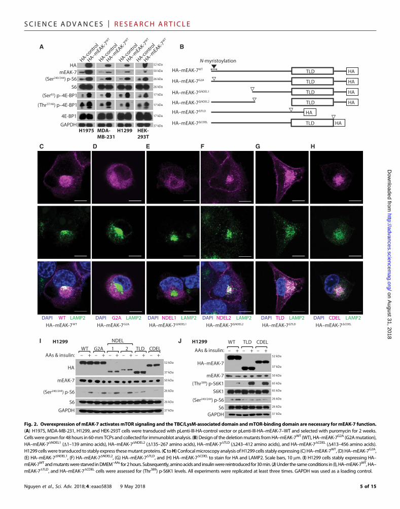
Molecular analysis of mEAK-7 demonstrates the role ofthe TLD domain and C terminus as crucial regulatorsof mEAK-7 functionTo rule out the possibility that siRNA-mediated knockdown ofmEAK-7nonspecifically influences mTORC1 signaling, we transduced H1975,MDA-MB-231, H1299, and HEK-293T cells with pLenti-III-HA-control or pLenti-III-HA(C-terminus)–mEAK-7WT lentivirus and selectedthese cells with puromycin (1 mg/ml) for 2weeks.Wedemonstrated thatoverexpression of HA–mEAK-7 activatedmTORC1 signaling in H1975,MDA-MB-231, H1299, and HEK-293T cells (Fig. 2A). Thus, bothknockdown and overexpression studies demonstrated that mEAK-7 isan essential component of mTORC1 signaling in mEAK-7+ cells.
Next, we investigated themolecular domains necessary formEAK-7function. To assess themEAK-7 domains essential formTOR signaling,we generated several mutants and transduced them with lentivirusinto cells that expressed endogenous mEAK-7 (Fig. 2B). We comparedHA–mEAK-7WT with HA–mEAK-7 mutants for LAMP2 colocaliza-tion at the lysosome (Fig. 2, C to H).We also investigated differential
Nguyen et al., Sci. Adv. 2018;4 : eaao5838 9 May 2018
overexpression effects of wild type (WT) andmutants onmTOR sig-naling stimulated by amino acids and/or insulin (Fig. 2, I and J). HA–mEAK-7WT colocalized with LAMP2 (Fig. 2C), amino acids, and insulinstimulation successfully induced (Ser240/244) p-S6 in WT overexpres-sing H1299 cells (Fig. 2I).
HA–mEAK-7G2A, a mutant with a point mutation that replacesthe first glycine residue with alanine within theN-myristoylation motif,failed to anchor to the lysosome (Fig. 2D). However, amino acids andinsulin stimulation induced (Ser240/244) p-S6, possibly because of en-dogenous mEAK-7 function (Fig. 2I). Deletion of amino acids 1 to 139(HA–mEAK-7DNDEL1) also led to a lysosomal anchorage defect (Fig.2E), due to the loss of theN-myristoylationmotif, but did not significantlyalter endogenousmTORC1 signaling (Fig. 2I).HA–mEAK-7DNDEL2 co-localized with LAMP2 (Fig. 2F) and also did not significantly alterendogenous mTORC1 signaling (Fig. 2I).
Although both HA–mEAK-7DTLD and HA–mEAK-7DCDEL localizeat the lysosome (Fig. 2, G andH), stable expression of either HA–mEAK-7DTLD or HA–mEAK-7DCDEL inhibited the induction of (Ser240/244) p-S6levels by amino acids and insulin (Fig. 2, I and J), and resulted in increased(Thr389) p-S6K1 levels (Fig. 2J). Although it is unclear how these mutantsaffect endogenous mEAK-7 function to impair mTOR signaling, theseresults demonstrate that the TLD domain and C terminus are necessaryfor mEAK-7–mediated mTOR function.
mEAK-7 recruits mTOR to the lysosome undernutrient-deprived and nutrient-rich conditionsmTOR signaling components are translocated to the lysosome in re-sponse to nutrient stimulation, and this shuttling is necessary to activatemTORC1 signaling (3). Because mEAK-7 is predominantly lysosomal,we posited a role for mEAK-7 in targeting mTOR to the lysosome. Todetermine the role of mEAK-7 in lysosomal localization of mTOR,H1299 cells were treated with control or mEAK-7 siRNA for 48 hours.Subsequently, cells were starved in DMEM−AAs for 1 hour, and aminoacids and insulinwere reintroduced for 30min.We found thatmEAK-7knockdown impaired mTOR localization to the lysosome (Fig. 3A),confirming that mEAK-7 is important for mTOR localization. Al-though low levels of mTOR remained capable of migrating to thelysosome after mEAK-7 knockdown, this might have been due toresidual mEAK-7 still expressed because siRNA treatment is not 100%effective and because other major regulators of mTOR, such as the RagGTPases, have been shown to recruit mTOR to the lysosome (11).
Further analysis demonstrated that the expression of mTORC1/2components was not altered after mEAK-7 siRNA treatment (Fig.3B). H1299 cells treated with mEAK-7 siRNA demonstrated a statis-tically significant decrease in mTOR/LAMP2 colocalization under thestarved condition (Fig. 3C). Under the nutrient-replenished condition,H1299 cells treated with mEAK-7 siRNA also exhibited a statisticallysignificant decrease inmTOR/LAMP2 colocalization (Fig. 3C). To sub-stantiate this finding, we performed the reciprocal experiment by over-expressingHA–mEAK-7. HA–mEAK-7 overexpression inH1299 cellsresulted in a statistically significant increase in mTOR/LAMP2 colo-calization in the absence of nutrients (Fig. 3, D and E). In addition,reintroduction of nutrients in control cells resulted in a significant en-hancement of the colocalization of mTOR/LAMP2, and HA–mEAK-7overexpression increasedmTOR/LAMP2 colocalization in the presenceof nutrients (Fig. 3, D and E). Further analysis demonstrated thatnutrient reintroduction did not result in a statistically significant changeof HA–mEAK-7/LAMP2 colocalization (Fig. 3F). We then hypothe-sized that endogenous mEAK-7 would colocalize with endogenous
4 of 15
SC I ENCE ADVANCES | R E S EARCH ART I C L E
on August 31, 2018
http://advances.sciencemag.org/
Dow
nloaded from
HA–mEAK-7 TLD
HA–mEAK-7 CDEL
HA–mEAK-7WT
HA–mEAK-7G2A
HA–mEAK-7 NDEL1
HA–mEAK-7 NDEL2
ED
A
WT LAMP2DAPI TLD LAMP2DAPI CDEL LAMP2DAPI
C
G2A LAMP2DAPI NDEL1 LAMP2DAPI
B
NDEL2 LAMP2DAPIHA–mEAK-7G2A HA–mEAK-7 NDEL1 HA–mEAK-7 NDEL2
HA-contro
l
HA–mEAK-7W
T
HA-contro
l
HA–mEAK-7W
T
HA-contro
l
HA–mEAK-7W
T
HAmEAK-7
(Ser240/244) p-S6
S6
(Ser65) p–4E-BP1
4E-BP1
H1975 MDA-MB-231
H1299 HEK-293T
HA-contro
l
HA–mEAK-7W
T
GAPDH
HA–mEAK-7WT
F HG
HA–mEAK-7 TLD HA–mEAK-7 CDEL
52 kDa
26 kDa
26 kDa
37 kDa
17 kDa
17 kDa
50 kDa
(Thr37/46) p–4E-BP1 17 kDa
HA
HATLD
HATLD
HATLD
HATLD
HATLD
N-myristoylation
I
HA
(Ser240/244) p-S6
S6
AAs & insulin: – + – + – + – + – + – +WT G2A 1 2 TLD CDEL
NDEL
HA–mEAK-7
WT
AAs & insulin:
S6(Ser240/244) p-S6
S6K1(Thr389) p-S6K1
– + – + – +
TLD CDELJ
GAPDHGAPDH
mEAK-7
52 kDa
37 kDa
26 kDa
26 kDa
37 kDa
52 kDa
37 kDa
26 kDa
26 kDa
37 kDa
50 kDa
65 kDa
65 kDa
mEAK-7 50 kDa
H1299 H1299
Fig. 2. Overexpression ofmEAK-7 activates mTOR signaling and the TBC/LysM-associated domain andmTOR-binding domain are necessary formEAK-7 function.(A) H1975, MDA-MB-231, H1299, and HEK-293T cells were transduced with pLenti-III-HA-control vector or pLenti-III-HA–mEAK-7–WT and selected with puromycin for 2 weeks.Cellswere grown for 48hours in 60-mmTCPs and collected for immunoblot analysis. (B) Design of the deletionmutants fromHA–mEAK-7WT (WT), HA–mEAK-7G2A (G2Amutation),HA–mEAK-7DNDEL1 (D1–139 amino acids), HA–mEAK-7DNDEL2 (D135–267 amino acids), HA–mEAK-7DTLD (D243–412 amino acids), and HA–mEAK-7DCDEL (D413–456 amino acids).H1299 cellswere transduced to stably express thesemutant proteins. (C toH) Confocalmicroscopy analysis of H1299 cells stably expressing (C) HA–mEAK-7WT, (D) HA–mEAK-7G2A,(E) HA–mEAK-7DNDEL1, (F) HA–mEAK-7DNDEL2, (G) HA–mEAK-7DTLD, and (H) HA–mEAK-7DCDEL to stain for HA and LAMP2. Scale bars, 10 mm. (I) H1299 cells stably expressing HA–mEAK-7WTandmutantswere starved inDMEM−AAs for2hours. Subsequently, aminoacidsand insulinwere reintroduced for30min. (J)Under thesameconditions in (I), HA–mEAK-7WT,HA–mEAK-7DTLD, and HA–mEAK-7DCDEL cells were assessed for (Thr389) p-S6K1 levels. All experiments were replicated at least three times. GAPDH was used as a loading control.
Nguyen et al., Sci. Adv. 2018;4 : eaao5838 9 May 2018 5 of 15

SC I ENCE ADVANCES | R E S EARCH ART I C L E
on August 31, 2018
http://advances.sciencemag.org/
Dow
nloaded from
Control siRNA mEAK-7 siRNA
DAPI LAMP2 mTOR
A
D HA-control HA–mEAK-7
Starved1 hour
HA–mEAK-7
+
GAPDH
mEAK-7siRNA:
DAPI LAMP2 mTOR
DAPI LAMP2 mTOR DAPI LAMP2 mTOR DAPI LAMP2 HA
Starved1 hour
Starved1 hour
Starved1 hour
Starved1 hour
+ AAs &insulin30 min
+ AAs &insulin30 min
+ AAs &insulin30 min
+ AAs &insulin30 min
+ AAs &insulin30 min
0
10
20
30
40
50
60
70
mTO
R/LA
MP2
% c
oloc
aliz
atio
n
n.s.*
*****
P < 0.05
siRNA control:siRNA mEAK-7:
Starvation:AAs & insulin:
HA-control:HA–mEAK-7:
Starvation:AAs & insulin: – –
++ + +
0102030405060708090
100n.s.
¥
***
mTO
R/LA
MP2
% c
oloc
aliz
atio
n
HA–mEAK-7:Starvation:
AAs & insulin:
++–+ +
+
0102030405060708090
100n.s.
HA
–mEA
K-7/
LAM
P2%
col
ocal
izat
ion
Ctl mEAK-7
mLST8PRAS40
raptor
mTOR
B
C
FE
H1299
DEPTOR
mTORC2
mTORC1
rictorSin1
*
50 kDa
289 kDa
200 kDa
78 kDa
150 kDa
40 kDa
37 kDa
48 kDa
37 kDa
–
+ –
–+++
++
+ +
–– + +– –++
+ ––– + +
+
Fig. 3. mEAK-7 is required for lysosomal localization of mTOR. (A) H1299 cells were treated with control or mEAK-7 siRNA for 48 hours in 10% DMEM+serum. Subsequently,200,000 cells were transferred to two-well glass chamber slides and allowed to settle for 24 hours. H1299 cells were then starved in DMEM−AAs for 1 hour, and amino acids andinsulinwere reintroduced for 30min. (B) Immunoblot analysis ofH1299 cells treatedwith control ormEAK-7 siRNAtoassess the expressionofmEAK-7 andmTORcomplexproteinsafter mEAK-7 knockdown. (C) Statistical analysis of colocalization of mTOR and LAMP2 for Fig. 3A. (D) A total of 200,000 normal H1299 cells or H1299 cells stably expressing HA–mEAK-7 were seeded onto two-well glass chamber slides and allowed to settle for 24 hours. Cells were then starved in DMEM−AAs for 1 hour, and amino acids and insulin werereintroduced for 30min. (E and F) Statistical analysis of colocalization ofmTOR and LAMP2 or HA–mEAK-7 and LAMP2 for Fig. 3D. Oil magnification, ×100. Cells were processed todetect 4′,6-diamidino-2-phenylindole (DAPI) (DNA), LAMP2 (lysosomal marker), mTOR, and HA (mEAK-7). *P < 0.01, **P < 0.001, ***P < 0.0001, ‡P < 0.00001, §P < 0.000001, WP <0.0000001, ¥P < 0.00000001, ∑P < 0.000000001. Scale bars, 25 mm. n.s., not significant.
Nguyen et al., Sci. Adv. 2018;4 : eaao5838 9 May 2018 6 of 15
SC I ENCE ADVANCES | R E S EARCH ART I C L E
on August 31, 2018
http://advances.sciencemag.org/
Dow
nloaded from
mTOR in response to nutrient stimulation because amino acids recruitmTOR to the lysosome.H1299 cells were nutrient-starved for 1 hour andstimulated with amino acids, insulin, or both for 1 hour. EndogenousmEAK-7 and endogenous mTOR strongly colocalized in response tonutrient stimulation (fig. S7, A to E).
We hypothesized that mEAK-7 could directly affect mTOR kinasefunction, possibly as an adaptor protein. mTOR interaction with itscomplex components is known to be sensitive under different bufferconditions (29). To rule out the possibility of nonspecific or artificialinteractions due to an abundance of exogenously produced proteinand buffer-dependent conditions, we harvested either H1299 or HA–mEAK-7–expressing H1299 cells, in either NP-40 or CHAPS buffer.Thus, we assessed the potential for interaction between these two pro-teins using a coimmunoprecipitation assay. Coimmunoprecipitation ofexogenous HA–mEAK-7WT confirmed significant interaction with en-dogenous mTOR, compared to an immunoglobulin G (IgG) controlunder serum-containing conditions (Fig. 4A). In addition, endoge-nous mEAK-7 interacted with endogenous mTOR (Fig. 4B). Finally,knockdown of mEAK-7 diminished the interaction of endogenousmEAK-7 with endogenous mTOR and mammalian lethal with SEC13protein 8 (mLST8) (Fig. 4C and fig. S8A). However, mEAK-7 failedto interact with regulatory associated protein of mTOR (raptor) orrapamycin-insensitive companion ofmTOR (rictor), key components ofmTORC1 and mTORC2, respectively. Intriguingly, exogenous HA–mEAK-7WT also interactedwithmTORandmLST8but did not interactwith rictor, Sin1, raptor, proline-rich AKT1 substrate 1 (PRAS40), orDEPTOR (Fig. 4D). These findings suggest the possibility of an alter-native mTOR complex that is yet to be identified in mammalian cells.
To assess the nutrient dependency of this interaction, we starvedHA–mEAK-7WT cells for 2 hours and reintroduced amino acids, insulin,or both for 30min. All lysates for immunoprecipitation were collected inNP-40 lysis buffer, unless noted otherwise. Under these conditions, ex-ogenousHA–mEAK-7WT and endogenousmTOR interacted under thestarved condition, and this interaction was increased by nutrient stim-ulation (Fig. 4E and fig. S8B). To further demonstrate the validity ofthese nutrient-dependent interactions, data support that endogenousmEAK-7 also strongly interacts with endogenous mTOR under theamino acid and insulin or amino acid and serum conditions (Fig. 4Fand fig. S8C). In addition, coimmunoprecipitation of endogenousmTOR to detect exogenous HA–mEAK-7WT also confirmed that thisinteraction increased under nutrient stimulation (Fig. 4G and fig. S8D).Therefore, data suggest that both exogenous and endogenous mEAK-7are capable of interacting with mTOR.
To determine themolecular domain necessary for mEAK-7 interac-tion with mTOR, we performed coimmunoprecipitation in cells stablyexpressing HA–mEAK-7WT, HA–mEAK-7DTLD, and HA–mEAK-7DCDEL, because these domains are necessary for the activation ofmTORC1 signaling in mEAK-7+ cells (Fig. 2, I and J). We found thatthe C-terminal protein region is necessary for the interaction of ex-ogenousHA–mEAK-7WTandendogenousmTOR(Fig. 4Hand fig. S8E),so this region of the C terminus was termed the mTOR-binding (MTB)domain. Other mEAK-7 mutants were also capable of interacting withendogenous mTOR, suggesting that maintaining an intact MTB domainis sufficient formTORbinding (fig. S7, F andG). In addition, overexpres-sion of HA–mEAK-7DTLD and HA–mEAK-7DCDEL increased (Thr389)p-S6K1 levels (Fig. 4H). These findings suggest that the interaction ofmEAK-7 and mTOR diverts mTOR targeting from S6K1 to S6K2,whereas loss of mEAK-7 diverts mTOR targeting from S6K2 to S6K1,resulting in increased (Thr389) p-S6K1 levels.
Nguyen et al., Sci. Adv. 2018;4 : eaao5838 9 May 2018
We hypothesized that mEAK-7 regulates mTORC1 signalingthrough S6K2 because (Thr389) p-S6K1 levels were not linked to itsdownstream target, (Ser240/244) p-S6 (Figs. 1, I and J, and 2J, and fig. S4,A and D). To assess this possibility, we transiently transfected H1299cells with pcDNA3-HA-S6K2-WT and either control or mEAK-7 siRNAand immunoprecipitated HA-S6K2. mEAK-7 knockdown considerablydecreased the interaction between endogenous mTOR and HA-S6K2(Fig. 4I and fig. S8F). To ensure mEAK-7 specificity in this interaction,we used two mEAK-7 siRNAs, and both demonstrated a substantialdecrease in HA-S6K2 interaction with mTOR (Fig. 4J and fig. S8G).These outcomes suggest that mEAK-7 supports the interaction of S6K2and mTOR.
Next, we hypothesized that mEAK-7 may also influence S6K1 andmTOR interaction. To test this, we transiently transfected H1299 cellswithpRK7-HA-S6K1-WTand control ormEAK-7 siRNAand immuno-precipitated HA-S6K1. mEAK-7 knockdown increased the interactionof exogenousHA-S6K1with endogenousmTOR (Fig. 4K and fig. S8H).Subsequently, two different mEAK-7 siRNAs confirmed that theseenhanced interactions were the result of mEAK-7 knockdown (Fig. 4Land fig. S8I). Thus, we demonstrate that mEAK-7 intricately controlsmTOR interaction with both S6K2 and S6K1.
Although these data support the necessity of mEAK-7 for the inter-action ofmTORwith S6K2, they do not provide direct evidence of S6K2function. To our knowledge, an antibody specific to (Thr388) p-S6K2does not exist. However, the amino acid sequences of the hydrophobicmotifs of the S6Ks (Fig. 4M) are nearly identical. Given this similarity,we predicted that the monoclonal antibody against (Thr389) p-S6K1[Cell Signaling Technology (CST), clone 108D2] would reveal the rela-tive phosphorylation status of S6K2, because it was designed to targetthe mTOR-targeting hydrophobic motif, and this is a strategy used byother groups (30). Because (Thr389) p-S6K1 levels are a common readoutof S6K1 kinase activity and mTORC1 functionality, we expected that(Thr388) p-S6K2 levels would also indicate S6K2 kinase activity in thiscontext. In addition, the mass of S6K1 and S6K2 are different in thatS6K1 is 65 to 70 kDa and S6K2 is 60 kDa; therefore, immuno-precipitation would yield detectable phosphorylation differences of theconcentrated kinase. To determine the phosphorylation status of S6K2by mEAK-7, we transiently transfected H1299 cells with pcDNA3-HA-S6K2-WT and either control or mEAK-7 siRNA, starved of nutri-ents, and we reintroduced DMEM+serum for 30 min. HA-S6K2 was thenimmunoprecipitated and probed with the 108D2 antibody. Data suggestthat mEAK-7 is required for the interaction of mTOR with S6K2 in re-sponse to serum and regulates (Thr388) p-S6K2 levels, as demonstratedby a loss of S6K2 phosphorylation in response to mEAK-7 knockdown(Fig. 4M). Furthermore, loss of mEAK-7 diminished S6K2-mediatedphosphorylationof S6 in response to serumstimulation (Fig. 4M).There-fore, mEAK-7 is required for S6K2 activity in these cells.
Finally, to demonstrate the extent to which mEAK-7 regulates 4E-BP1 and eIF4E interaction, we treatedH1299 cells with either control ormEAK-7 siRNA and immunoprecipitated eIF4E. We discovered thatmEAK-7 knockdown enhanced binding of 4E-BP1 to eIF4E (Fig. 4Nand fig. S8J). Thus, these data establish mEAK-7 as a novel effector ofmTOR signaling that regulates both S6K2 activity and 4E-BP1 activity.
mEAK-7 supports cell proliferation and migrationAfter demonstrating that mEAK-7 supports mTOR signaling, we hy-pothesized that mEAK-7 was essential for critical cellular functionsgoverned bymTOR.mTOR signaling is important for regulation of cellnumber (3). To elucidate the influence of mEAK-7 on proliferation,
7 of 15

SC I ENCE ADVANCES | R E S EARCH ART I C L E
on August 31, 2018
http://advances.sciencemag.org/
Dow
nloaded from
A
HA–mEAK-7
mTOR
IP
HAIgGWCL
H1299 cells stably expressing HA–mEAK-7
mTOR
HA–mEAK-7
GAPDH
mTORHA–mEAK-7
GAPDH
IP: m
TOR
WCL
Amino acids:Insulin :
H1299 cells stably expressing HA–mEAK-7
B
H1299 cells stably expressing HA–mEAK-7
mTORHA–mEAK-7
GAPDHmTOR
HA–mEAK-7GAPDH
IP: H
AW
CL
Amino acids:Insulin:
––
+ –++
F
H1299 cells transientlytransfected with HA-S6K2
H1299 cells transientlytransfected with HA-S6K2
I
mTORHA-S6K2
mTOR
HA-S6K2
4E-BP1
IP: H
AW
CL
siRNA: Ctl 1 2
mEAK-7
mTOR
HA–mEAK-7
mTOR
HA–mEAK-7
S6K1
(Thr389) p-S6K1
IP: H
AW
CL
WT
TLDCDEL
H1299 cells stably expressing HA mutants
H1299 cells transientlytransfected with HA-S6K1
H1299 cells transientlytransfected with HA-S6K1
K
S6K1:S6K2:
LSESANQVFLGFTYVAPSVLLSESANQAFLGFTYVAPSVL* * * * * * * * * * * * * * * * * *. *
FxxFT/SYmTOR hydrophobic motif:
H1299 cells transientlytransfected with HA-S6K2
(Thr388) p-S6K2HA-S6K2
+siRNA: Ctl mEAK-7
Serum: –
mEAK-7GAPDH
IP: H
AW
CL
(Ser240/244) p-S6S6
mTOR
mTOR
M
HA-S6K2mTOR
GAPDH
mEAK-7(Ser240/244) p-S6
IP
HAIgGWCLsiRNA control:
siRNA mEAK-7: siRNA mEAK-7:– +HA-S6K1
mTOR
GAPDH
mEAK-7(Ser240/244) p-S6
S6
GAPDH
GAPDH
HA-S6K2
mEAK-7(Thr37/46)
p–4E-BP1
H1299 cellsN
J
S6
60 kDa
mTORHA-S6K1
mTORHA-S6K1
4E-BP1
IP: H
AW
CL
siRNA: Ctl 1 2
mEAK-7
GAPDH
mEAK-7(Thr37/46)
p–4E-BP1
289 kDa
52 kDa
289 kDa
52 kDa
289 kDa
52 kDa
37 kDa
37 kDa
289 kDa
52 kDa
289 kDa
52 kDa
37 kDa
37 kDa
289 kDa
52 kDa
289 kDa
52 kDa
37 kDa
37 kDa
65 kDa70 kDa
65 kDa70 kDa
37 kDa
289 kDa
60 kDa
50 kDa
37 kDa
26 kDa
26 kDa
289 kDa
65 kDa
50 kDa
37 kDa
26 kDa
26 kDa
289 kDa
60 kDa
289 kDa
60 kDa
50 kDa
37 kDa
17 kDa
17 kDa
289 kDa
65 kDa
289 kDa
65 kDa
50 kDa
37 kDa
17 kDa
17 kDa
289 kDa
60 kDa
26 kDa
26 kDa
60 kDa
26 kDa
50 kDa
37 kDa
289 kDa
IP
HAIgGWCLsiRNA control:
siRNA:
4E-BP1
mEAK-7GAPDH
Ctl mEAK-7
4E-BP1
eIF4E
eIF4E
50 kDa
37 kDa
17 kDa
17 kDa
25 kDa
25 kDa
IP: e
IF4E
WCL
mTOR
mEAK-7
mTORmEAK-7
IP: m
EAK-
7W
CL
289 kDa
50 kDa
50 kDa
289 kDa
InsulinAmino acids: +
Serum: +H1299 cells
GmTOR
mEAK-7 50 kDa
289 kDa
IP
mEAK-7
IgGWCL
289 kDa
50 kDa
37 kDa
mLST8
raptor 150 kDa
mTOR
mEAK-7GAPDH
Control
mEAK-7
Control
mEAK-7IP:mEAK-7WCL
siRNA:
rictor 200 kDa
H1299 cells
H1299 cells
IP: H
AW
CL
raptor
PRAS40
mLST8
HA–mEAK-7
PRAS40mLST8
HA–mEAK-7
Ctl
raptor
mTOR
mTOR
C
DEPTOR
DEPTOR
GAPDH
HA–mEAK-7
289 kDa
289 kDa
37 kDa
52 kDa
48 kDa
48 kDa
52 kDa
40 kDa
40 kDa
37 kDa
37 kDa
150 kDa
150 kDa
rictor
rictor
Sin1
Sin1
200 kDa
200 kDa
78 kDa74 kDa
78 kDa74 kDa
mTO
RC2m
TORC1
mTO
RC2m
TORC1
H1299 cells stably expressing HA–mEAK-7
ED
GAPDH 37 kDa
GAPDH 37 kDa
37 kDa
IgG L.C. 25 kDa
CHAPS
CHAPS
CHAPS
CHAPS
H
+–
– +
– +
––
+ –++
– +
– +
–+ –+–+
– +– + – +
–+ –+–+
––––++ ––
– – –+ +
L
Fig. 4. mEAK-7 interacts withmTOR through theMTB domain and is required for S6K2 activity. (A) HA–mEAK-7WT cells immunoprecipitated (IP) with goat IgG or goat anti-HA. (B) H1299cells, inCHAPS, immunoprecipitatedwith anti–mEAK-7.WCL,whole-cell lysate. (C) H1299 cells transfectedwith control ormEAK-7 siRNA, inCHAPS, immunoprecipitatedwith anti–mEAK-7. L.C., light chain. (D) Normal H1299 or HA–mEAK-7WT cells, in CHAPS, immunoprecipitated with anti-HA. (E) HA–mEAK-7WT cells starved in DMEM−AAs for 2 hours,nutrient-stimulated for 30min, and immunoprecipitatedwith anti-HA. (F) H1299 cells starved in DMEM−AAs for 2 hours, nutrient-stimulated for 60min, and immunoprecipitatedwithanti–mEAK-7. (G) Conditionsmimicked in (E) and immunoprecipitatedwith anti-mTORantibody. (H) HA–mEAK-7WT,HA–mEAK-7DTLD, andHA–mEAK-7DCDEL cells immunoprecipitatedwith anti-HA. (I and J) H1299 cells transfected with pcDNA3-HA-S6K2-WT and control, mEAK-7 #1, or mEAK-7 #2 siRNA and immunoprecipitated with anti-HA. (K and L) H1299 cellstransfected with pRK7-HA-S6K1-WT and control, mEAK-7 #1, or mEAK-7 #2 siRNA and immunoprecipitated with anti-HA. (M) mTOR targeting hydrophobic motif. H1299 cells trans-fected with pcDNA3-HA-S6K2-WT and control or mEAK-7 #1 siRNA. Cells starved in DMEM−AAs for 2 hours, 10% serum–stimulated, and immunoprecipitated with anti-HA. (N) H1299cells transfected with control or mEAK-7 siRNA and immunoprecipitated with anti-eIF4E. Experiments were repeated three times. GAPDH was a loading control.
Nguyen et al., Sci. Adv. 2018;4 : eaao5838 9 May 2018 8 of 15

SC I ENCE ADVANCES | R E S EARCH ART I C L E
on August 31, 2018
http://advances.sciencemag.org/
Dow
nloaded from
B
A
C
F
G
H1975To
tal c
ell n
umbe
r (10
6 )
0
1
2
3
4
5
Time (days)0 3 5
siRNA controlsiRNA mEAK-7 §
MDA-MB-231
Tota
l cel
l num
ber (
106 )
01234567
***
‡
H1299
Tota
l cel
l num
ber (
106 )
§
***
H1975
Cell
inde
x
0
0.5
1.0
2.0
1.5
0 12 24 36 48Hours
**
***
***
MDA-MB-231
****
*
*
*****
M MDA-MB-231 H1299H1975
Day 0 Day 2
Cont
rol
mEA
K-7si
RNA
N
Day 0 Day 2
IE
Day 0 Day 2
Cell proliferation Cell migration
Scratch wound assay
D
Tota
l cel
l num
ber (
106 )
012345678
HEK-293T
‡
*
H
H1975
MDA-MB-231
H1299
HEK-293T
0123456
P < 0.05
***
***
§
*
***
P < 0.05
P < 0.05
Time (days)0 3 5
0
1
2
3
4
5
Time (days)0 3 5
0
1
2
3
4
Time (days)0 3 5
0123456
Time (days)0 3 5To
tal c
ell n
umbe
r (10
6 )To
tal c
ell n
umbe
r (10
6 )
Time (days)0 3 5
0123456789
Tota
l cel
l num
ber (
106 )
Time (days)0 3 5
HA-controlHA–mEAK-7
Tota
l cel
l num
ber (
106 )
Time (days)0 3 5
J
K
L
O
0
4
6
8
2Cell
inde
x
0
4
10
6
8
2
Cell
inde
x
0 12 24 36 48Hours
H1299
0 12 24 36 48Hours
siRNA controlsiRNA mEAK-7
siRNA controlsiRNA mEAK-7
siRNA controlsiRNA mEAK-7
siRNA controlsiRNA mEAK-7
siRNA controlsiRNA mEAK-7
siRNA controlsiRNA mEAK-7
HA-controlHA–mEAK-7
HA-controlHA–mEAK-7
HA-controlHA–mEAK-7
Cell
inde
x
0
1
2
4
3
0 12 24 36 48Hours
**
**
‡siRNA controlsiRNA mEAK-7
P
Cont
rol
mEA
K-7si
RNA
Cont
rol
mEA
K-7si
RNA
HEK-293T
Day 2
HEK-293T
Cont
rol
mEA
K-7si
RNA
0 Day
Fig. 5. mEAK-7 is essential for cell proliferation and cell migration. (A to D) (A) H1975 (n = 13), (B) MDA-MB-231 (n = 9), (C) H1299 (n = 8), and (D) HEK-293T (n = 6) cellstreatedwith control or mEAK-7 #1 siRNA. A total of 200,000 cells were transferred to 100-mmTCPs and counted at days 3 and 5. (E toH) (E) H1975 (n = 6), (F) MDA-MB-231 (n = 6),(G) H1299 (n=6), and (H) HEK-293T (n=6) cellswere transducedwithpLenti-III-HA-control vector or pLenti-III-HA–mEAK-7–WT.A total of 200,000 cellswere transferred to 100-mmTCPs and counted at days 3 and 5. (I to L) (I) H1975 (n= 6), (J) MDA-MB-231 (n= 5), (K) H1299 (n= 5), and (L) HEK-293T (n = 6) cells were treatedwith control ormEAK-7 #1 siRNA. Atotal of 50,000 cells were transferred to CIM 16-well plates, and real-time analysis was performed for 48 hours using an ACEA Biosciences RCTADP instrument. (M to P) (M) H1975,(N) MDA-MB-231, (O) H1299, and (P) HEK-293T cells were treated with control or mEAK-7 siRNA. A total of 1,500,000 cells were transferred into 35-mm TCPs. The following day, ascratch was created down themiddle, and pictures were taken at 0 and 48 hours. Scale bars, 125 mm. Data are represented as means ± SEM. Statistical significance denoted: *P <0.01, **P < 0.001, ***P < 0.0001, ‡P < 0.00001, §P < 0.000001, WP < 0.0000001, ¥P < 0.00000001.
Nguyen et al., Sci. Adv. 2018;4 : eaao5838 9 May 2018 9 of 15

SC I ENCE ADVANCES | R E S EARCH ART I C L E
on August 31, 2018
http://advances.sciencemag.org/
Dow
nloaded from
cells were treated with either control or mEAK-7 siRNA and countedafter 3 and 5 days. In H1975 (Fig. 5A), MDA-MB-231 (Fig. 5B), H1299(Fig. 5C), andHEK-293T cells (Fig. 5D), treatment withmEAK-7 siRNAresulted in a significant reduction in cell proliferation. Further, annexinVstaining or acridine orange–propridium iodide (AO-PI) staining demon-strated no difference in cell death after mEAK-7 knockdown (fig. S9,A to C). Previous reports corroborate the finding that the loss of TLDdomain–containing proteins (19) or single knockdownof S6K2withoutan apoptotic stimulator (31) does not result in significant levels of celldeath. Because of the low expression of mEAK-7 in some human cells,we hypothesized thatmEAK-7 overexpressionwould promote cell pro-liferation. Thus, to test the effect ofmEAK-7 overexpression on cell pro-liferation,we transducedH1975,MDA-MB-231,H1299, andHEK-293Tcells with pLenti-GIII-CMV-control-HA and pLenti-GIII-CMV–mEAK-7–HA. Overexpression of HA–mEAK-7 in H1975 (Fig. 5E), MDA-MB-231 (Fig. 5F), H1299 (Fig. 5G), and HEK-293T (Fig. 5H) significantlyenhanced cell proliferation at days 3 and 5. Thus, we concluded thatmEAK-7 is vital for cell proliferation in mEAK-7+ cells.
mTORC1 signaling has substantial control over cell migration andmetastasis, with the 4E-BP1–eIF4E axis regulatingmTOR-sensitive mi-gration and invasion genes (32). Given the role ofmEAK-7 inmTORsignaling, we investigated the impact of mEAK-7 on cell migration.H1975, MDA-MB-231, H1299, and HEK-293T cells were treated witheither control or mEAK-7 siRNA and seeded into CIM-plates, whichuse xCELLigence technology to quantify cell migration in real time,collecting hundreds of data points through a dimensionless cell-indexparameter. Cells must pass through a pore embedded in a gold-platedelectric grid, creating electrical impedance and registering a signal forreal-time, quantifiable cellmigration.We proceeded to conduct statisticalanalyses at select time points of 12, 24, 36, and 48 hours. Treatment ofH1975 (Fig. 5I), MDA-MB-231 (Fig. 5J), H1299 (Fig. 5K), and HEK-293T (Fig. 5L) cellswithmEAK-7 siRNAresulted in statistically significantreductions of real-time cell migration at 24, 36, and 48 hours. In addition,scratch wound assay analysis of H1975 (Fig. 5M), MDA-MB-231 (Fig.5N), H1299 (Fig. 5O), and HEK-293T (Fig. 5P) cells treated withmEAK-7 siRNAresulted in amarkeddefect ofwound closure after 2 days,demonstrating that mEAK-7 is essential for cell migration in these cells.
The S6 kinases have differential functions mediatedthrough mEAK-7Data from several sources suggest that S6 kinases play redundant rolesbecause of their high homology, but recent evidence reveals that theindependent role of S6K2 remains undetermined (28). Genome-wideassessment of S6K1 and S6K2 in human tumors and in vitro silencingof these kinases demonstrate that their targets are different from eachother and that S6K2more closelymirrors eIF4E function (33). S6K2 hasalso been shown to be an essential regulator of cell proliferation, due toits involvement with heterogeneous ribonucleoprotein F (34). Previousreports demonstrate that S6 is essential for mammalian cell prolifera-tion and that S6K1 controls eukaryotic size (35). Given that mEAK-7regulates S6K2 function, we hypothesized that mEAK-7 also regulatesS6K2-mediated cell proliferation.
To determine the extent to which S6K2 regulates cell proliferationand to compare its functional role in other mTOR targets, we treatedH1975 cells with control, S6K1, S6K2, or eIF4E siRNA for 48 hours inDMEM+serum (Fig. 6A). Cells were then seeded into new tissue cultureplates andwere counted after 3 and 5 days. Treatment with S6K1, S6K2,and eIF4E siRNA resulted in a significant reduction of cell proliferationat days 3 and 5 (Fig. 6B). Treatment with S6K2 or eIF4E siRNAs signif-
Nguyen et al., Sci. Adv. 2018;4 : eaao5838 9 May 2018
icantly reduced cell proliferation at day 5, compared to S6K1 siRNA.However, H1975 cells treated with S6K2 siRNA, compared to eIF4EsiRNA, did not result in a statistically significant reduction, suggestingthat S6K2 functions similarly to eIF4E with regard to cell proliferation,as the literature reports (33). These results demonstrate that S6K2 isessential for cell proliferation under these conditions.
mTOR signaling also controls cell size in eukaryotes (36). Becausewe found that mEAK-7 is a positive activator of mTOR signaling, wesought to determine the role of mEAK-7 in regulating cell size. H1975,MDA-MB-231, and H1299 cells were treated with control or mEAK-7siRNA for 48 hours, seeded onto new tissue culture plates, and pro-cessed at day 3. Cells were analyzed on a Beckman Coulter CyAn 5 flowcytometer for forward scatter. H1975 (fig. S9D),MDA-MB-231 (fig. S9E),and H1299 (fig. S9F) cells treated with mEAK-7 siRNA resulted in anincrease in cell size, suggesting that dysfunctional S6K1 activity resultsin aberrant cell size regulation after mEAK-7 knockdown. Under thesame conditions, cell size was also assessed using the Logos BiosystemsLunaCell Counter.H1975 (Fig. 6C),MDA-MB-231 (Fig. 6D), andH1299(Fig. 6E) cells treated with mEAK-7 siRNA resulted in a significant in-crease in cell size. These data demonstrate that whereas the loss of mEAK-7resulted in decreased downstream mTOR signaling, aberrant activation ofS6K1 leads to dysregulation of cell size. In addition, H1975 cells were treatedwith control, S6K1, or S6K2 siRNA and then analyzed via forward scatter inflowcytometry.S6K1knockdownreducedcell size,whereasS6K2knockdowndemonstrated limited change in cell size (Fig. 6F).
Because overexpression of pcDNA3-HA-S6K2-WT or pRK7-HA-S6K1-WT was not sufficient to rescue mTOR signaling in H1299 cellstreated with mEAK-7 siRNA (Fig. 4, I to M), we posited that this ob-stacle could be overcome by transfecting cells with constitutively ac-tivated forms of S6K1 or S6K2. To determine whether this couldrescue mEAK-7 knockdown effects, we treated cells with control siRNA,mEAK-7 siRNA, mEAK-7 siRNA + pRK7-HA-S6K1-F5A-E389-deltaCT (50 kDa—deletion that results in a truncated kinase) (cS6K1)plasmid, or mEAK-7 siRNA + pcDNA3-HA-S6K2-E388-D3E (60 kDa)(cS6K2) plasmid. Concomitant knockdownofmEAK-7 andoverexpres-sion of cS6K1 and cS6K2 in H1299, H1975, andMDA-MB-231 cells re-sulted in rescue of (Ser240/244) p-S6 levels (Fig. 6G). Knockdown ofmEAK-7 and overexpression of cS6K1 or cS6K2 resulted in partial res-cue of cell proliferation defects (Fig. 6H). Thus, we demonstrate thatmEAK-7 functions upstream of S6K2 and promotes S6K2-mediatedsignaling and proliferation.
DISCUSSIONHere, we determined that mEAK-7 is an important, evolutionarilyconserved, lysosomal protein that activatesmTOR signaling in responseto nutrient stimulation in many cell types (Fig. 6I). We provide mech-anistic insight for a novel protein that is required for serum-, aminoacid–, and insulin-mediated mTOR signaling in human cells. We alsodemonstrate that mEAK-7 is necessary for S6K2 function by regulatingS6K2-mTOR interaction and 4E-BP1–eIF4E interaction and by sup-porting cell proliferation and cellmigration inmEAK-7+ cells.We iden-tifiedmEAK-7 as an essential interacting protein ofmTOR andmLST8,but not of other mTORC1 components (raptor, DEPTOR, or PRAS40)and mTORC2 components (rictor, DEPTOR, or Sin1). mEAK-7 inter-acts withmTOR through theMTB domain (Fig. 6J). mEAK-7 regulatesthe mTORC1 signaling at the lysosome and is a key player for mTORrecruitment to the lysosome. Thus, we determined that mEAK-7 func-tions as an essential component of mTOR signaling to regulate S6K2
10 of 15

SC I ENCE ADVANCES | R E S EARCH ART I C L E
on August 31, 2018
http://advances.sciencemag.org/
Dow
nloaded from
eIF4E
Cell size
Cell proliferation
Cell migrationCell proliferation
mEAK-7
RagA/C S6K1
S6K2
4E-BP1
Raptor
???
mTOR
mLST8
mTOR
mLST8S6K1
S6K2
4E-BP1
mEAK-7
LysosomemTOR
mLST8
RagA/C
Raptor
–Amino acids +Amino acids
+Insulin
–Insulin I J
TLD
mEAK-7
N terminus C terminus
mTOR-binding domainMTB
A
Tota
l cel
l num
ber (
106 )
0
1
2
3
4
5
Time (days)0 3 5
siRNA controlsiRNA S6K1siRNA S6K2siRNA eIF4E
**
**
n.s.
**
*****
S6K1
S6K2
eIF4e
GAPDH
siRNA: Control
S6K1S6K2
eIF4E
H1975
C
B
H1975 MDA-MB-231 H1299
20
22
24*
Cell
size
(μM
)
18
20
22
24
26 **
Cell
size
(μM
)
ED
18
2022
24
26
siRNA Control
siRNA mEAK-7
Cell
size
(μM
)
0
50
100
150
200
250
0K 20K 40K 60K 80K 100K
siRNAS6K1
siRNAcontrol
siRNAS6K2
F
60 kDa
65 kDa
25 kDa
37 kDa
§
Cell
coun
t
Forward scatter
siRNA Control
siRNA mEAK-7
siRNA Control
siRNA mEAK-7
H1975
G
mEAK-7
HA
(Ser240/244) p-S6
S6
siRNA control:
H Rescue experimentRescue experiment
H1299 H1975 MDA-MB-231GAPDH
50 kDa
60 kDa(Thr389) p-S6K1CST clone: 108D2
26 kDa
26 kDa
37 kDa
50 kDa
60 kDa
50 kDa
siRNA mEAK-7:cS6K1:cS6K2: –
–
+–
––
–+
–
–+
–
++
+
–
––
+–
––
–+
–
–+
–
++
+
–
––
+–
––
–+
–
–+
–
++
+
–
siRNA controlsiRNA mEAK-7siRNA mEAK-7 + cS6K1siRNA mEAK-7 + cS6K2
Tota
l cel
l num
ber (
106 )
0
1
23
45
6
7
Time (days)0 3 5
***
n.s.
*
****
n.s.
**
H1299
–Amino acids +Amino acidsssssss
Insulin receptorN-myristoylation
Fig. 6. Overexpression of constitutively activated S6K2 or S6K1 is capable of rescuing cell defects due tomEAK-7 knockdown. (A) H1975 cells were treatedwith control,S6K1, S6K2, or eIF4E siRNA. (B) From (A), 500,000 cells were transferred to 100-mm TCPs and counted at days 3 and 5. (C to E) (C) H1975 (n = 13), (D) MDA-MB-231 (n = 9), and (E)H1299 (n= 8) cells were treatedwith control ormEAK-7 siRNA. A total of 500,000 cells were transferred to 100-mmTCPs, and cell sizewas analyzed at day 3with AO-PI staining viaLogos Biosystems (LB). (F) H1975 cellswere treatedwith control, S6K1, or S6K2 siRNAandanalyzed for forward scatter via flowcytometry. (G) H1299, H1975, andMDA-MB-231 cellswere transiently transfected with control siRNA, mEAK-7 siRNA, mEAK-7 siRNA + pRK7-HA-S6K1-F5A-E389-deltaCT plasmid, or mEAK-7 siRNA + pcDNA3-HA-S6K2-E388-D3Eplasmid. (H) A total of 500,000 H1299 cells treated as described in (G) were transferred to 100-mm TCPs and counted at days 3 and 5 via LB. (I) Diagram depicting mEAK-7function on mTOR complex formation for S6K2. (J) Summary of mEAK-7 domains: N-myristoylation motif, TLD domain, and MTB domain. Data are represented as means ± SEM.Statistical significance denoted: *P < 0.01, **P < 0.001, ***P < 0.0001, ‡P < 0.00001, §P < 0.000001. GAPDH was used as a loading control.
Nguyen et al., Sci. Adv. 2018;4 : eaao5838 9 May 2018 11 of 15

SC I ENCE ADVANCES | R E S EARCH ART I C L E
on August 31, 2018
http://advances.sciencemag.org/
Dow
nloaded from
and 4E-BP1, through a potentially alternative pathway to the canonicalmTORC1 or mTORC2 models.
Since the discovery of rapamycin, decades of research have con-tributed to understanding themechanism by whichmTOR is regulatedin response to nutrients and stress (3). The two best-known complexesthat contain mTOR are mTORC1 and mTORC2. mTORC1 is com-posed of raptor, mLST8 or GbL, PRAS40, and DEPTOR (3). mTORC2is composed ofmLST8, rictor,mammalian stress-activated protein kinaseinteracting protein 1, Protor, DEPTOR, and Tti1 and Tel2 (3).
With the finding that mEAK-7 is an interacting and functioningpartner ofmTORandmLST8,weposited thatmEAK-7may formanovelcomplex to regulate the specificity of S6K2 interaction with mTOR.4E-BP1 binding to eIF4E is also affected by the loss of mEAK-7, andour data suggest that a potential new complex may, in certain con-texts, regulate this mTOR-mediated function as well. Our evidencesuggests that mEAK-7 functions at the level of mTOR as a coordinatorfor S6K2 and 4E-BP1, but the downstream partners that mediate thisprocess remain unknown.
It has been theorized that additional mTOR complexes may com-plement mTORC1 and mTORC2 in mammalians. Astrocytes of thecentral nervous system provide one such example of how mTOR mayfunction in a cell type–dependentmanner. In this context, GIT1 functionsas an interacting partner ofmTOR that does not associate with either rap-tor or rictor (37). This finding is intriguing because it demonstrates thatcell type specificitymay dictate themolecular landscape that allows for fullmTOR regulation and activation. mEAK-7 may have eluded previousmTOR immunoprecipitation and mass spectrometry analyses becauseit is found to a limited extent in human cells (Fig. 1C and figs. S1Cand S4C). It is also unknown where mEAK-7 is expressed during de-velopment. The interaction between mEAK-7 and mTOR, as well asthe associated influence on S6K2 and 4E-BP1 functions, suggest thatthere aremoremTORcomponents yet to be identified thatmay interactin a cell type– or context-dependent manner (Fig. 6I).
Because we did not screen mEAK-7 in all human cell types, furtherinvestigation of mEAK-7 in other physiological contexts is essential forunderstanding how mEAK-7 functions in human development or dis-ease. We provide molecular insight demonstrating that mEAK-7supports the interaction of S6K2 and mTOR, but the full complementof interacting partners is yet to be determined. In addition, evolutionarydifferences arose between mEAK-7 and nematode EAK-7. NematodeEAK-7 functions in parallel to Akt signaling to regulate DAF-16 (humanFoxO) during development and life span (1). We demonstrate thatmEAK-7 is essential for mTOR signaling to regulate S6K2 and 4E-BP1, but nematodes only have one S6 kinase, RSKS-1. Thus, it is unclearhow, or whether, EAK-7 regulates TOR signaling in nematodes.
One of the challenges in studying mTOR in mammalian systems isthe difficulty of discerning tissue or organ-specific functions, becauseknockout of major components of mTORC1 or mTORC2 results inembryonic lethality (38). S6K1 is the best-studied target of mTOR ineukaryotes. In mice, loss of S6K1 reduces weight and improves insulinsensitivity with high-fat diets (HFDs) (39), whereas mice with a stan-dard diet (SD) are glucose-intolerant, hypoinsulinemic, and have re-duced b cell size (40). Intriguingly, loss of both S6K1 and S6K2reverses the deleterious effects of S6K1 knockout mice, restores glucosetolerance under an SD, and further improves glucose tolerance withan HFD (41).
Because S6K2 has been studied to a lesser extent in the scientificcommunity, much less is known about the importance of S6K2 indevelopment, metabolism, and disease. Compared to WT mice, S6K1
Nguyen et al., Sci. Adv. 2018;4 : eaao5838 9 May 2018
null mice are much smaller, S6K2 null mice are slightly larger, anddouble-knockout animals result in perinatal lethality (27). Recent datasuggest that S6K2 may not be a purely redundant kinase to S6K1 be-cause S6K1 null mice demonstrate higher S6K2 expression in the liver,muscles, thymus, and brain and because S6K2 remains responsive torapamycin-mediated inhibition of S6 phosphorylation in S6K1 nullmice (42). Thus, S6K2was largely neglected, and tissue-specific func-tions of S6K2 are now beginning to be understood. The loss of S6K2resulted in higher basal levels of insulin in plasma, 2.5×more b cellmasswith an SD, and improved glucose tolerance, as well as enhanced insulinsensitivity with an HFD (43). In addition, single S6K2 knockout en-hances ketone body production and increases peroxisome proliferator–activated receptor a activity in the liver, and S6K1 knockout mice arecapable of maintaining (Ser240/244) p-S6 levels, whereas S6K2 knockoutmice are not, aswe have demonstrated (30). Thus, elucidating the role ofmEAK-7–mediated regulation of S6K2 and mTOR signaling in mam-mals will further our understanding of diseases where hyperactivationof mTOR signaling occurs through aberrant S6K2 activity.
MATERIALS AND METHODSCell linesH1299, H1975,MDA-MB-231, andHEK-293T cell lines were obtainedfromAmericanTypeCultureCollection.Other cell lysateswere donationsand used as part of the initial cell screen. For Fig. 1C and fig. S1C lysates,the donor laboratories verified the cell lysates and hold the validationpaperwork. Cell lysates derived fromour laboratory are as follows: humanembryonic stem cell line H1 (undifferentiated), H1 endoderm (differ-entiated to pancreatic progenitors), H1mesoderm (cardiac progenitors),H1 ectoderm (neuronal progenitors), H1 embryoid body, and humangingival fibroblasts (two different patients). The following cell lines weredonated:T.Carey:UM-SCC-1,UM-SCC-10A,UM-SCC-11A,UM-SCC-14A, UM-SCC-17A, UM-SCC-17B, UM-SCC-74A, UM-SCC-74B, andUM-SCC-81B; M. Cohen: H1975 and H1299; S. Takayama: MDA-MB-231; and M. S. Wicha: BT474, HCC1937, MDA-MB-436, SK-BR-3,SUM149, SUM159, T4D7, and MDA-MB-468.
Cell cultureFor cell culture, cell lines were grown in DMEM [Thermo Fisher Scien-tific (TFS), catalog #11995-073], without antibiotics/antimycotics andsupplemented with a concentration of 10% fetal bovine serum (FBS;TFS, catalog #10437-036, lot #1399413) at 37°C in a 5.0% CO2 incuba-tor. Cells were grown in Falcon Tissue Culture Treated Flasks T-75[Fisher Scientific (FS), catalog #13-680-65] until 75% confluent and splitwith 0.25% trypsin-EDTA (TFS, catalog #25200-056) for 5 min in the37°C cell incubator. Cells were washed 1× with phosphate-buffered sa-line (PBS) and resuspended in 10% FBS containing DMEM. Cells werecountedwith the LUNAAutomatedCell Counter [LB, catalog #L10001]using LUNACell Counting Slides (LB, catalog #L12003) andAO-PI dye(LB, catalog #F23001).Starvation protocolCells were starved in DMEM−AAs (TFS, catalog #ME120086L1) for50 min or 2 hours and then stimulated with amino acids (normalDMEM), insulin (1 or 10 mM; Sigma-Aldrich, catalog #I9278-5ML),or both for 30 or 60 min.
siRNA or plasmid transfectionCells were seeded at a density of 500,000 cells per 60-mm tissue cultureplates (TCP) and grown for 24 hours. For siRNA transfection, we
12 of 15

SC I ENCE ADVANCES | R E S EARCH ART I C L E
on August 31, 2018
http://advances.sciencemag.org/
Dow
nloaded from
incubated Lipofectamine RNAiMAXTransfection Reagent (TFS, catalog#13778-150) within Opti-MEM I Reduced SerumMedium (TFS, catalog#31985-070), and 100 nM siRNA was incorporated before being intro-duced into cells at 100 nM concentration. For plasmid transfection, weusedFuGENE6TransfectionReagent (Promega, catalog #E2691) intoOpti-MEM I Reduced Serum Medium, and 2 mg of plasmid was incorporatedbefore being introduced into cells. For dual transfection, we added bothsolutions. mEAK-7 siRNAs are identified as KIAA1609 or TLDC1: forsiRNA mEAK-7 #1 (TFS, ID #s33640), #2 (TFS, ID #HSS126697), #3(TFS, ID #HSS126699). We also used the following siRNAs: mEAK-7(TFS, ID #s33641, ID #s33642, and ID #HSS126698), S6K1 (TFS, ID#s12282), S6K2 (TFS, ID #s12287), eIF4E (TFS, ID #s4578), and con-trol (TFS, catalog #4390843). Plasmids were purchased from Addgene.pRK7-HA-S6K1-WT was a gift from J. Blenis (Addgene, #8984).pcDNA3-S6K2-WT was a gift from J. Blenis (Addgene, #17729). pRK7-HA-S6K1-F5A-E389-deltaCT was a gift from J. Blenis (Addgene, #8990).pcDNA3-S6K2-E388-D3E was a gift from J. Blenis (Addgene, #17731).
Immunoblot analysisCells were collected with a cell scraper and lysed in cold NP-40 lysisbuffer (50 mM tris, 150 mMNaCl, and 1.0% NP-40 at pH 8.0). Proteinlysate (50 mg) was separated in Novex WedgeWell 4-20% tris-glycinegels (TFS; catalog #XP04205BOX) or 10% tris-glycine gels (TFS, cat-alog #XP00105BOX). Proteins were transferred to polyvinylidene di-fluoridemembranes, incubated with primary antibodies overnight at4°C, and then incubated with secondary antibodies at room tempera-ture for 1 hour.Membraneswere incubatedwith SuperSignalWest PicoChemiluminescent Substrate (TFS, catalog #34078) or Femto (TFS,catalog #34095). Primary antibodies were as follows: mouse mono-clonal antibody against mEAK-7 (KIAA1609) was obtained fromOrigene Technologies (clone OTI12B1, formerly 12B1, catalog#TA501037). All antibodies from CST were as follows: glycer-aldehyde-3-phosphate dehydrogenase (catalog #2118S), (Ser240/244)p-S6 ribosomal protein (2215S), S6 ribosomal protein (2217S),(Thr389) p-p70 S6 kinase (9234S), S6K1 (2708S), S6K2 (14130S),mTOR (2983S), HA-tag mouse (2367S), HA-tag rabbit (3724S),(Ser65) p–4E-BP1 (9451S), (Thr37/46) p–4E-BP1 (9459S), (Thr70) p–4E-BP1 (13396S), mLST8 (3274S), raptor (2280S), PRAS40 (2691S),DEPTOR/DEPDC6 (11816S), rictor (2114S), Sin1 (12860S), 4E-BP1(9452S), and eIF4E (2067S). Antibodies p-S6, S6, and 4E-BP1were usedat 1:3000 dilution and the remainder at 1:1000 dilution in 5% bovineserum albumin (BSA) in 1× tris-buffered saline with Tween-20 (TBST)buffer with 0.04% sodium azide. Secondary antibodies for immunoblotanalysis: 1:4000 dilution for an a-mouse IgG horseradish peroxidase(HRP) conjugate (Promega, catalog #W4021) and 1:7500 dilution foran a-rabbit IgG HRP conjugate (Promega, catalog #W4011).
Molecular cloning of HA–mEAK-7WT into mutantsGibsonAssembly reaction (NewEnglandBiolabs, catalog #E2611S)wasused to generatemutant constructs in accordance to themanufacturer’sinstructions. In table S1, we outline the list of primers used to producethe different mutant plasmids and extended materials and methods.pLenti-GIII-CMV–mEAK-7–HA (ABM Inc., catalog #LV198982)was used as backbone. All mutants were confirmed by DNA sequencing.For WT and mutant constructs, 10× lentiviral supernatant was preparedby the Vector Core at the University of Michigan. H1299 cells wereinfected with 1× virus and selected with puromycin (1 mg/ml) in10%DMEM+serum. After 2 weeks, cells were used for immunoblot anal-ysis and confocal microscopy.
Nguyen et al., Sci. Adv. 2018;4 : eaao5838 9 May 2018
Immunoprecipitation analysisAfter siRNA and/or plasmid transfection, cells were harvested in 1%NP-40 lysis buffer or CHAPS lysis buffer [FIVEphoton Biochemicals(FB), catalog #CIB-1] supplemented with protease inhibitors (FB, catalog#PI-1) and phosphatase inhibitors (FB, catalog #PIC1). Proteins (250 mg)were used for immunoprecipitation reactions, and 25 mg of proteins wasused for whole-cell lysate analysis. Antibodies (2 mg) and 40 ml of Immu-noCruz IP/WB Optima C agarose beads [Santa Cruz Biotechnology(SCB), catalog #sc-45040] were incubated in 1 ml of PBS for 1 hour at4°C. Separately, 250 mg of proteins was incubated with 50 ml of Pre-clearingMatrix C (SCB, catalog #sc-45054) inCHAPS buffer for 1 hour.After washing, the antibody-bead conjugate was incubated with pre-cleared protein lysate for 1.5 hours at 4°C, and precipitated beads werewashed three times with PBS. Antibodies for immunoprecipitation were asfollows: goat anti-HAepitope tag (NovusBiologicals, catalog #NB600-362),goat IgG (SCB, catalog #sc-2028),mTOR (CST, catalog #2983S),mEAK-7(SCB, catalog #sc-247321), and eIF4E (SCB, catalog #sc-271480).
Cell immunofluorescence analysisA total of 250,000 cells were seeded into a two-well Nunc Lab-Tek IIChamber Slide System (FS, catalog #12-565-5) for 24 hours. Cellswere starved for 50 min in DMEM−AAs and stimulated with aminoacids or 1 mM insulin for 30 min. Then, cells were fixed with Z-Fixsolution (Anatech LTD, catalog #170) for 10 min at room temperature,washed three times in PBS, and incubated with the following: un-masking solution (PBS, 2N HCl, and 0.5% Triton X-100) for 10 min,quenching solution (TBS and0.1% sodiumborohydride) for 10min, per-meabilization solution (PBS and 0.02% Triton X-100) for 10 min,and 5% BSA for 1 hour. Cells were incubated overnight at 4°C withprimary antibody. Next, slides were washed with PBS and incubatedin secondary antibodies for 1 hour at room temperature. We used aNikon Ti Eclipse confocal microscope (×100 magnification) to cap-ture images. We captured images with or without 3× digital zoom,1/32 frames/s, 1024 × 1024 image capture, 1.2 airy units, 2× line aver-aging, appropriate voltage, and power settings optimized per antibody.No modification was done, except image sizing reduction for figurepreparation. Quantitative analyses were completed via Imaris softwarefor confocal images with calculation of colocalization as percentages.Identical threshold settings captured images across three to five individualfields (10 to 15 cells) per condition, with the data representing at leastthree independent experiments. Primary antibodies for immuno-fluorescencewere as follows:mouseHA-tag (CST, catalog #2367S), rabbitHA-tag (CST, catalog #3724S), LAMP1 (CST, catalog #9091S), LAMP2(SCB, catalog #sc-18822), mEAK-7 (SCB, catalog #sc-247321), EEA1 en-dosome (CST, catalog #3288S), AIFmitochondria (CST, catalog #5318S),PDI endoplasmic reticulum (CST, catalog #3501S), RCAS-1 Golgi com-plex (CST, catalog #12290S), and mTOR (1:1000) (CST, catalog #2983S).All antibodies were used at 1:1500 with a working volume of 1.5ml in 5%BSA in PBS, unless noted otherwise. Secondary antibodies for immuno-fluorescence were as follows: donkey anti-mouse IgG Alexa Fluor 488(TFS, catalog #R37114), donkey anti-rabbit IgGAlexaFluor 594 (TFS, cat-alog #A-21207), and donkey anti-goat IgG Alexa Fluor 647 (TFS, catalog#A-21447). All antibodies were used at a concentration of 1:1500 with aworkingvolumeof1.5ml in5%BSA inPBS.DAPI stainwasused forDNA.
Cell proliferation, size, migration, apoptosis, and scratchwound assay analysisFor cell proliferation and size assay, cells were processedwith the LUNAAutomated Cell Counter (LB, catalog #L10001) using LUNA Cell
13 of 15
SC I ENCE ADVANCES | R E S EARCH ART I C L E
http://advances.sciencemD
ownloaded from
Counting Slides (LB, catalog #L12003) and AO-PI dye (LB, catalog#F23001). After siRNA/plasmid transfection or lentiviral transduction,200,000 cells were seeded in 100-mm TCPs for 3- and 5-day analysis.All cell size data were processed at day 3. Cell size was consistent acrossmultiple platforms for analysis. For cell migration assay, cells were seededat a density of 50,000 cells per well in CIM-plate 16 (ACEA Biosciences,catalog #05665817001). Real-time capture of cell migration was per-formed over 48 hours on the xCELLigence System, RTCA DP Instru-ment (ACEA Biosciences, catalog #00380601050) and processed byRTCA Software 2.0. This technology is a widely used and validated tool,with more than 1200 publications (https://aceabio.com/publications/).Cells must pass through a pore embedded in a gold-plated electric grid,creating electrical impedance and registering a signal for real-time,quantifiable cell migration. As cells migrate through a small pore, theypass over a gold grid, which causes electrical impedance that is registeredin real time as cellular migration. To prepare the data, we plotted the cellindex every 12 hours.We can distribute all data points upon request. Forscratch wound assays, cells were seeded at a density of 1,500,000 cells in35-mm TCPs. After 24 hours, cell plates were linearly scratched with apipette tip, and images representative of wound healing were capturedafter 48 hours. For cell size and death analysis via flow cytometry, cellswere analyzed for forward scatter with the BeckmanCoulter CyAn flowcytometer at the University of Michigan Flow Cytometry Core. Cellswere analyzed by annexin V/PI staining with annexin V (TFS, catalog#A13199) and PI (Sigma-Aldrich, catalog #25535-16-4), according tothe manufacturer’s protocols.
Statistical analysisCell proliferation,migration, and sizewere analyzed via paired Student’st test. Immunoblot and immunoprecipitation assays were repeated atleast three times in all cell lines.
on August 31, 2018
ag.org/
SUPPLEMENTARY MATERIALSSupplementary material for this article is available at http://advances.sciencemag.org/cgi/content/full/4/5/eaao5838/DC1fig. S1. T-coffee analysis of mEAK-7 in eukaryotes, validation of human mEAK-7 antibody, andan expanded cell screen for mEAK-7 protein.fig. S2. Extended immunofluorescence analysis of HA–mEAK-7WT in other cellularcompartments.fig. S3. Validation of mEAK-7 antibody for immunofluorescence analysis of endogenous mEAK-7.fig. S4. mEAK-7 regulates serum-mediated activation of mTORC1 signaling and knockdown ofmEAK-7 results in increased (Thr389) p-S6K1 levels.fig. S5. Densitometry analysis of Fig. 1I.fig. S6. Densitometry analysis of Fig. 1J.fig. S7. Immunofluorescence analysis of endogenous mEAK-7 colocalizing with endogenousmTOR in response to nutrients and immunoprecipitation analysis of HA–mEAK-7 mutants formTOR interaction.fig. S8. Densitometry analysis of Fig. 4.fig. S9. Knockdown of mEAK-7 does not result in enhanced cell apoptosis but increases cell size.table S1. Cloning primers.Extended Materials and Methods for cloning.
REFERENCES AND NOTES1. H. Alam, T. W. Williams, K. J. Dumas, C. Guo, S. Yoshina, S. Mitani, P. J. Hu, EAK-7 controls
development and life span by regulating nuclear DAF-16/FoxO activity. Cell Metab. 12,30–41 (2010).
2. M. Shimobayashi, M. N. Hall, Making new contacts: The mTOR network in metabolismand signalling crosstalk. Nat. Rev. Mol. Cell Biol. 15, 155–162 (2014).
3. R. A. Saxton, D. M. Sabatini, mTOR signaling in growth, metabolism, and disease. Cell 168,960–976 (2017).
4. J. Chung, C. J. Kuo, G. R. Crabtree, J. Blenis, Rapamycin-FKBP specifically blocksgrowth-dependent activation of and signaling by the 70 kd S6 protein kinases. Cell 69,1227–1236 (1992).
Nguyen et al., Sci. Adv. 2018;4 : eaao5838 9 May 2018
5. J. Heitman, N. R. Movva, M. N. Hall, Targets for cell cycle arrest by theimmunosuppressant rapamycin in yeast. Science 253, 905–909 (1991).
6. D. M. Sabatini, H. Erdjument-Bromage, M. Lui, P. Tempst, S. H. Snyder, RAFT1:A mammalian protein that binds to FKBP12 in a rapamycin-dependent fashion and ishomologous to yeast TORs. Cell 78, 35–43 (1994).
7. J. Kunz, R. Henriquez, U. Schneider, M. Deuter-Reinhard, N. R. Movva, M. N. Hall,Target of rapamycin in yeast, TOR2, is an essential phosphatidylinositol kinase homologrequired for G1 progression. Cell 73, 585–596 (1993).
8. S. B. Helliwell, P. Wagner, J. Kunz, M. Deuter-Reinhard, R. Henriquez, M. N. Hall, TOR1 andTOR2 are structurally and functionally similar but not identical phosphatidylinositolkinase homologues in yeast. Mol. Biol. Cell 5, 105–118 (1994).
9. J. Huang, B. D. Manning, The TSC1-TSC2 complex: A molecular switchboard controllingcell growth. Biochem. J. 412, 179–190 (2008).
10. Y. Sancak, T. R. Peterson, Y. D. Shaul, R. A. Lindquist, C. C. Thoreen, L. Bar-Peled,D. M. Sabatini, The Rag GTPases bind raptor and mediate amino acid signaling tomTORC1. Science 320, 1496–1501 (2008).
11. Y. Sancak, L. Bar-Peled, R. Zoncu, A. L. Markhard, S. Nada, D. M. Sabatini, Ragulator-Ragcomplex targets mTORC1 to the lysosomal surface and is necessary for its activationby amino acids. Cell 141, 290–303 (2010).
12. C. Betz, M. N. Hall, Where is mTOR and what is it doing there? J. Cell Biol. 203, 563–574(2013).
13. P. Riou, R. Saffroy, J. Comoy, M. Gross-Goupil, J.-P. Thiéry, J.-F. Emile, D. Azoulay,D. Piatier-Tonneau, A. Lemoine, B. Debuire, Investigation in liver tissues and cell lines ofthe transcription of 13 genes mapping to the 16q24 region that are frequently deleted inhepatocellular carcinoma. Clin. Cancer Res. 8, 3178–3186 (2002).
14. R. E. Ellsworth, L. A. Field, B. Love, J. L. Kane, J. A. Hooke, C. D. Shriver, Differentialgene expression in primary breast tumors associated with lymph node metastasis.Int. J. Breast Cancer 2011, 142763 (2011).
15. T. Nagase, R. Kikuno, M. Nakayama, M. Hirosawa, O. Ohara, Prediction of the codingsequences of unidentified human genes. XVIII. The complete sequences of 100new cDNA clones from brain which code for large proteins in vitro. DNA Res. 7, 273–281(2000).
16. B. Schröder, C. Wrocklage, C. Pan, R. Jäger, B. Kösters, H. Schäfer, H.-P. Elsässer, M. Mann,A. Hasilik, Integral and associated lysosomal membrane proteins. Traffic 8, 1676–1686(2007).
17. K. A. Gray, B. Yates, R. L. Seal, M. W. Wright, E. A. Bruford, Genenames.org: The HGNCresources in 2015. Nucleic Acids Res. 43, D1079–D1085 (2015).
18. C. Notredame, D. G. Higgins, J. Heringa, T-Coffee: A novel method for fast and accuratemultiple sequence alignment. J. Mol. Biol. 302, 205–217 (2000).
19. M. J. Finelli, L. Sanchez-Pulido, K. X. Liu, K. E. Davies, P. L. Oliver, The evolutionarilyconserved Tre2/Bub2/Cdc16 (TBC), lysin motif (LysM), domain catalytic (TLDc) domain isneuroprotective against oxidative stress. J. Biol. Chem. 291, 2751–2763 (2016).
20. T. Doerks, R. R. Copley, J. Schultz, C. P. Ponting, P. Bork, Systematic identificationof novel protein domain families associated with nuclear functions. Genome Res. 12,47–56 (2002).
21. M. Blaise, H. M. A. B. Alsarraf, J. E. M. M. Wong, S. R. Midtgaard, F. Laroche, L. Schack,H. Spaink, J. Stougaard, S. Thirup, Crystal structure of the TLDc domain of oxidationresistance protein 2 from zebrafish. Proteins 80, 1694–1698 (2012).
22. K. Moriya, K. Nagatoshi, Y. Noriyasu, T. Okamura, E. Takamitsu, T. Suzuki, T. Utsumi, ProteinN-myristoylation plays a critical role in the endoplasmic reticulum morphological changeinduced by overexpression of protein Lunapark, an integral membrane protein of theendoplasmic reticulum. PLOS ONE 8, e78235 (2013).
23. C. Settembre, A. Fraldi, D. L. Medina, A. Ballabio, Signals from the lysosome: A controlcentre for cellular clearance and energy metabolism. Nat. Rev. Mol. Cell Biol. 14, 283–296(2013).
24. P. P. Roux, D. Shahbazian, H. Vu, M. K. Holz, M. S. Cohen, J. Taunton, N. Sonenberg,J. Blenis, RAS/ERK signaling promotes site-specific ribosomal protein S6 phosphorylationvia RSK and stimulates cap-dependent translation. J. Biol. Chem. 282, 14056–14064(2007).
25. X. M. Ma, J. Blenis, Molecular mechanisms of mTOR-mediated translational control.Nat. Rev. Mol. Cell Biol. 10, 307–318 (2009).
26. K. K. Lee-Fruman, C. J. Kuo, J. Lippincott, N. Terada, J. Blenis, Characterization of S6K2, anovel kinase homologous to S6K1. Oncogene 18, 5108–5114 (1999).
27. M. Pende, S. H. Um, V. Mieulet, M. Sticker, V. L. Goss, J. Mestan, M. Mueller, S. Fumagalli,S. C. Kozma, G. Thomas, S6K1−/−/S6K2−/− mice exhibit perinatal lethality and rapamycin-sensitive 5′-terminal oligopyrimidinemRNA translation and reveal amitogen-activated proteinkinase-dependent S6 kinase pathway. Mol. Cell. Biol. 24, 3112–3124 (2004).
28. O. E. Pardo, M. J. Seckl, S6K2: The neglected S6 kinase family member. Front. Oncol. 3, 191(2013).
29. D.-H. Kim, D. D. Sarbassov, S. M. Al, J. E. King, R. R. Latek, H. Erdjument-Bromage,P. Tempst, D. M. Sabatini, mTOR interacts with raptor to form a nutrient-sensitivecomplex that signals to the cell growth machinery. Cell 110, 163–175 (2002).
14 of 15

SC I ENCE ADVANCES | R E S EARCH ART I C L E
http://advanceD
ownloaded from
30. K. Kim, S. Pyo, S. H. Um, S6 kinase 2 deficiency enhances ketone body production andincreases peroxisome proliferator-activated receptor alpha activity in the liver.Hepatology 55, 1727–1737 (2012).
31. S. Sridharan, A. Basu, S6 kinase 2 promotes breast cancer cell survival via Akt. Cancer Res.71, 2590–2599 (2011).
32. A. C. Hsieh, Y. Liu, M. P. Edlind, N. T. Ingolia, M. R. Janes, A. Sher, E. Y. Shi, C. R. Stumpf,C. Christensen, M. J. Bonham, S. Wang, P. Ren, M. Martin, K. Jessen, M. E. Feldman,J. S. Weissman, K. M. Shokat, C. Rommel, D. Ruggero, The translational landscape of mTORsignalling steers cancer initiation and metastasis. Nature 485, 55–61 (2012).
33. E. Karlsson, I. Magić, J. Bostner, C. Dyrager, F. Lysholm, A.-L. Hallbeck, O. Stål, P. Lundström,Revealing different roles of the mTOR-targets S6K1 and S6K2 in breast cancer byexpression profiling and structural analysis. PLOS ONE 10, e0145013 (2015).
34. E. T. H. Goh, O. E. Pardo, N. Michael, A. Niewiarowski, N. Totty, D. Volkova, I. R. Tsaneva,M. J. Seckl, I. Gout, Involvement of heterogeneous ribonucleoprotein F in theregulation of cell proliferation via the mammalian target of rapamycin/S6 kinase 2pathway. J. Biol. Chem. 285, 17065–17076 (2010).
35. R. J. O. Dowling, I. Topisirovic, T. Alain, M. Bidinosti, B. D. Fonseca, E. Petroulakis,X. Wang, O. Larsson, A. Selvaraj, Y. Liu, S. C. Kozma, G. Thomas, N. Sonenberg,mTORC1-mediated cell proliferation, but not cell growth, controlled by the 4E-BPs.Science 328, 1172–1176 (2010).
36. D. C. Fingar, S. Salama, C. Tsou, E. Harlow, J. Blenis, Mammalian cell size is controlled bymTOR and its downstream targets S6K1 and 4EBP1/eIF4E. Genes Dev. 16, 1472–1487 (2002).
37. L. J. Smithson, D. H. Gutmann, Proteomic analysis reveals GIT1 as a novel mTOR complexcomponent critical for mediating astrocyte survival. Genes Dev. 30, 1383–1388 (2016).
38. D. A. Guertin, D. M. Stevens, C. C. Thoreen, A. A. Burds, N. Y. Kalaany, J. Moffat,M. Brown, K. J. Fitzgerald, D. M. Sabatini, Ablation in mice of the mTORC componentsraptor, rictor, or mLST8 reveals that mTORC2 is required for signaling to Akt-FOXOand PKCa, but not S6K1. Dev. Cell 11, 859–871 (2006).
39. S. H. Um, F. Frigerio, M. Watanabe, F. Picard, M. Joaquin, M. Sticker, S. Fumagalli,P. R. Allegrini, S. C. Kozma, J. Auwerx, G. Thomas, Absence of S6K1 protects against age-and diet-induced obesity while enhancing insulin sensitivity. Nature 431, 200–205 (2004).
40. M. Pende, S. C. Kozma, M. Jaquet, V. Oorschot, R. Burcelin, Y. Le Marchand-Brustel,J. Klumperman, B. Thorens, G. Thomas, Hypoinsulinaemia, glucose intolerance anddiminished b-cell size in S6K1-deficient mice. Nature 408, 994–997 (2000).
Nguyen et al., Sci. Adv. 2018;4 : eaao5838 9 May 2018
41. T. R. Castañeda, W. Abplanalp, S. H. Um, P. T. Pfluger, B. Schrott, K. Brown, E. Grant,L. Carnevalli, S. C. Benoit, D. A. Morgan, D. Gilham, D. Y. Hui, K. Rahmouni, G. Thomas,S. C. Kozma, D. J. Clegg, M. H. Tschöp, Metabolic control by S6 kinases depends on dietarylipids. PLOS ONE 7, e32631 (2012).
42. H. Shima, M. Pende, Y. Chen, S. Fumagalli, G. Thomas, S. C. Kozma, Disruption of thep70s6k/p85s6k gene reveals a small mouse phenotype and a new functional S6 kinase.EMBO J. 17, 6649–6659 (1998).
43. K. M. Taylor, J. Bajko, M. S. Cabrera, C. Kremer, B. Meyer-Puttlitz, A. M. Schulte, S. H. Cheng,R. K. Scheule, R. J. Moreland, S6 kinase 2 deficiency improves glucose disposal in mice feda high fat diet. J. Diabetes Metab. 5, 441 (2014).
Acknowledgments: We thank T. Carey, M. Cohen, S. Takayama, and M. Wicha for cell lysates anduse of equipment. We thank E. Pedersen, A. Hawkins, and E. Lawlor’s laboratory for use of ACEABiosciences RCTA DP instrument. We thank F. Haidar and H. Amatullah for replicating keyexperiments. We thank F. Haidar for the artwork (Fig. 6I). Funding:We thank our funding sources:National Institute of Dental and Craniofacial Research (1F30DE026048-01, R01-DE016530, andT32-DE007057) and Stuart and Barbara Padnos Research Award from the Comprehensive CancerCenter at the University of Michigan. Author contributions: J.T.N., C.R., A.L.F., D.B.M., J.K.K., andP.H.K. designed experiments and analyzed results. J.T.N., C.R., and A.L.F. carried out experiments.J.T.N. and P.H.K. are responsible for the major ideas. J.T.N. and D.B.M. created deletion mutants.J.T.N. and P.H.K. wrote the paper. J.T.N., C.R., D.B.M., A.L.F., J.K.K., and P.H.K. edited the manuscript.Competing interests: The authors declare that they have no competing interests. Data andmaterials availability: All data needed to evaluate the conclusions in the paper are presentin the paper and/or the Supplementary Materials. Additional data related to this paper may berequested from the authors.
Submitted 17 September 2017Accepted 27 March 2018Published 9 May 201810.1126/sciadv.aao5838
Citation: J. T. Nguyen, C. Ray, A. L. Fox, D. B. Mendonça, J. K. Kim, P. H. Krebsbach, MammalianEAK-7 activates alternative mTOR signaling to regulate cell proliferation and migration. Sci.Adv. 4, eaao5838 (2018).
s.s
15 of 15
on August 31, 2018
ciencemag.org/

migrationMammalian EAK-7 activates alternative mTOR signaling to regulate cell proliferation and
Joe Truong Nguyen, Connor Ray, Alexandra Lucienne Fox, Daniela Baccelli Mendonça, Jin Koo Kim and Paul H. Krebsbach
DOI: 10.1126/sciadv.aao5838 (5), eaao5838.4Sci Adv
ARTICLE TOOLS http://advances.sciencemag.org/content/4/5/eaao5838
MATERIALSSUPPLEMENTARY http://advances.sciencemag.org/content/suppl/2018/05/04/4.5.eaao5838.DC1
REFERENCES
http://advances.sciencemag.org/content/4/5/eaao5838#BIBLThis article cites 43 articles, 16 of which you can access for free
PERMISSIONS http://www.sciencemag.org/help/reprints-and-permissions
Terms of ServiceUse of this article is subject to the
registered trademark of AAAS.is aScience Advances Association for the Advancement of Science. No claim to original U.S. Government Works. The title
York Avenue NW, Washington, DC 20005. 2017 © The Authors, some rights reserved; exclusive licensee American (ISSN 2375-2548) is published by the American Association for the Advancement of Science, 1200 NewScience Advances
on August 31, 2018
http://advances.sciencemag.org/
Dow
nloaded from