malignant peripheral nerve sheath tumors of the …den onset of severe epigastric pain. ct also...
TRANSCRIPT
471
The Korean Journal of Pathology 2009; 43: 471-4DOI: 10.4132/KoreanJPathol.2009.43.5.471
Malignant peripheral nerve sheath tumors (MPNSTs) have rarely been reported to occur inthe adrenal gland and all of the reported cases were associated with neurofibromatosis, phe-ochromocytoma or ganglioneuroma. We present here a case of MPNST in the bilateral adrenalglands without any history of neurofibromatosis or combined tumor. Histologic examinationshowed the tumor cells had a spindle to ovoid shape, they were arranged in sweeping fasci-cles and there were frequent mitotic figures. The immunohistochemical and ultrastructuralfeatures of the tumor are also presented. To the best of our knowledge, this is the first reportin the English medical literature about MPNSTs in the bilateral adrenal glands without anyhistory of neurofibromatosis or combined tumor.
Key Words : MPNST; Adrenal gland
Jae Hong Park Seung Yeon HaHyun Yee Cho
471
Malignant Peripheral Nerve Sheath Tumors of the Bilateral Adrenal
Glands: Are They Metachronous Primary Tumors
- A Case Report -
Most malignant peripheral nerve sheath tumors (MPNSTs)arise in association with major nerve trunks. Their most com-mon anatomic sites are the proximal portions of the upper andlower extremities and the trunk. MPNSTs have rarely been re-ported to occur in the adrenal gland and all of the reported caseswere associated with neurofibromatosis, pheochromocytoma organglioneuroma.1-4 We present here a case of MPNST occur-ring in the bilateral adrenal glands and the patient had no his-tory of neurofibromatosis or combined tumor.
CASE REPORT
A 31-year-old woman was admitted to our hospital becauseof a sudden onset of severe epigastric pain. The pain was sting-ing and continuous and it radiated to back. Her past medicalhistory and family history did not suggest any remarkable dis-ease including neurofibromatosis. The physical examinationdidn’t reveal any pigmented skin lesion, and there was no pal-pable mass or nodule found in the cervical area. However, ten-
derness and mild rebound tenderness was noted in the epigastricarea. The initial laboratory studies were nearly normal, but thehemoglobin level decreased from 12.8 g/dL on the first hospitalday to 7.9 g/dL on the fourth hospital day. A chest x-ray revealedno specific abnormality. CT showed a 4 cm-sized lobulated,slightly enhancing mass in the left adrenal gland without anyevidence of metastasis (Fig. 1A). There was a fluid collection inthe rectovaginal pouch, and this was suggestive of hemoperi-toneum. No abnormal findings were observed in the other organs,including the pancreas. A mass was noted in the left adrenalgland when performing laparotomy. There was about 300 mLof collected blood in the intraabdominal cavity. There was noevidence of metastatic disease. Left adrenalectomy was done. Theresected left adrenal gland measured 6×4.8×2.2 cm and itweighed 31 g. It revealed a 5 cm-sized ruptured tumor thatshowed a gray pink color and a soft consistency. Extensive hem-orrhage was noted around the rupture site (Fig. 2). Twelve blockswere made from the tumor. Microscopically, the tumor cells ra-nged from spindle to ovoid in shape. They were arranged in sw-eeping fascicles (Fig. 3A). Mitotic figures were frequently found
471 471
Corresponding AuthorSeung Yeon Ha, M.D.Department of Pathology, Gachon University GilHospital, 1198 Guwol-dong, Namdong-gu, Incheon405-760, KoreaTel: 032-460-3073Fax: 032-460-3073E-mail: [email protected]
Department of Pathology, GachonUniversity Gil Hospital, Incheon, Korea
Received : August 14, 2008Accepted : June 1, 2009

472 Jae Hong Park Seung Yeon Ha Hyun Yee Cho
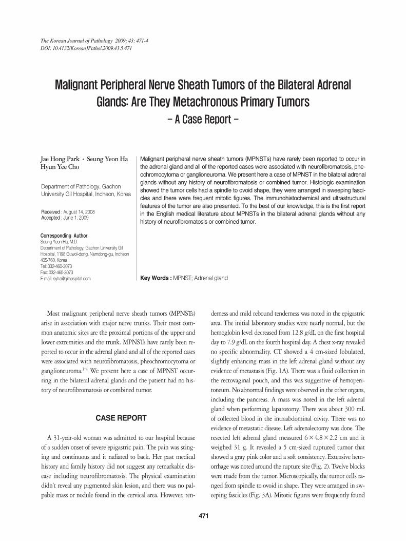
(more than 20 per 10 high power fields). On immunohistochemi-cal staining, the tumor cells were positive for S-100 protein andvimentin (Fig. 3B). No immunoreactivity for c-kit, CD34, cytok-eratin, actin, desmin, chromogranin, synaptophysin, and HMB45 was identified. The ultrastructural findings showed spindlecells with long cytoplasmic processes (Fig. 4A). The cells werefocally surrounded by a basal lamina and they were joined byprimitive junctional complexes (Fig. 4B). After discharge, thepatient did not visit the hospital for follow-up. Three years later,she was admitted to the hospital again because of a similar sud-den onset of severe epigastric pain. CT also showed a similar massin the right adrenal gland (Fig. 1B). The resected right adrenalgland measured 8×6×4 cm and it weighed 45 g. It revealeda 4 cm-sized ruptured tumor. It was similar to the previous leftadrenal tumor for the gross, microscopic and immunohistochem-ical findings (Fig. 3C, D). The patient is taking prednisolone toprevent adrenal insufficiency and she is undergoing postopera-
tive chemotherapy with vinblastine, ifosfamide, and cisplatin.The postoperative laboratory test didn’t reveal any abnormal
Fig. 3. Left adrenal tumor consists of dense fascicles of spindle cells (A). Immunohistochemically, the tumor cells are of focally positive forS-100 protein (B). Right adrenal tumor shows similar histological and immunohistochemical findings (C, D).
A B C D
Fig. 1. CT shows a lobulated mass in the left adrenal gland (A). Three years later, similar mass is found in the right adrenal gland (B).
A B
Fig. 2. Sectioned surface of the left adrenal gland shows an ill de-fined gray pink tumor. Rupture with extensive hemorrhage is noted.
MPNSTs of the Bilateral Adrenal Glands 473
findings. The T3 level was 123.66 ng/dL and the cortisol levelwas 0.62 μg/dL. Other laboratory studies for detecting endocrinediseases were not done.
DISCUSSION
We describe here a woman with unusual bilateral adrenaltumors that consisted of dense fascicles of spindled cells. Theimmunohistochemical and electron microscopic findings showedthe features of neural differentiation. We interpreted these tumorsto be MPNSTs that arose from the bilateral adrenal glands.MPNST arising from the adrenal gland is extremely rare withonly four such cases having been reported in the English litera-ture.1-4 All the reported cases were so-called composite MPNSTs,that is, MPNSTs with other neural crest-derived cell constituentssuch as pheochromocytoma and ganglioneuroma. MPNSTs com-monly occur in the setting of von Recklinghausen’s disease. Oneof the reported cases with pheochromocytoma-MPNST had a
history of von Recklinghausen’s disease.4 In our case, no combinedadrenal tumor was present and there was no family history ofvon Recklinghausen’s disease. We cannot find a previous reportof bilateral adrenal MPNSTs without a combined adrenal tumoror a family history of von Recklinghausen’s disease (Table 1). Thehistogenesis of various kinds of neuronal tumors is based on theconcept of neurocristopathy, as described by Bolande,5 which ischaracterized as the results of divergent differentiation of neuralcrest cells. In the embryonal stage, neural crest-derived cells mi-grate and differentiate into melanocytes, the adrenal medulla,root ganglia, Schwann cells, and so on. Migration disturbance,developmental arrest or maldevelopment of neural crest-derivedcells may result in various types of neoplasms. In support of thisconcept, bizarre clinical syndromes of multiple neuroendocrineadenomatosis or composite tumors of the sympathetic gangliaand adrenal medulla have been reported. Although we cannotconclude whether this is a case of bilateral primary tumors thathas adrenalotropism or a primary tumor that has metastasizedfrom one adrenal gland to the other one, we think that the caserepresents extremely rare, metachronous primary bilateral adrenalMPNSTs that originated from neural crest derivatives at thesame level of the somites for the following reasons: 1) the bilat-eral adrenal tumors were localized; they had neither extensionbeyond the midline nor distant metastasis; 2) the right adrenaltumor appeared 3 years after the left adrenalectomy; most ofthe previous reports of the adrenal MPNSTs showed metastasiswithin several months (Table 1). We think that MPNSTs in thebilateral adrenal glands without a composite tumor, like ourcase, are within the spectrum of neurocristopathy.
REFERENCES
1. Chandrasoma P, Shibata D, Radin R, Brown LP, Koss M. Malignant
peripheral nerve sheath tumor arising in an adrenal ganglioneuro-
ma in an adult male homosexual. Cancer 1986; 57: 2022-5.
2. Miettinen M, Saari A. Pheochromocytoma combined with malig-
Fig. 4. Electron microscopy reveals spindle cells with long cyto-plasmic processes containing neurofilaments (A, original magni-fication ×18,900). The cells are focally coated by basal laminae(B, original magnification ×18,900) and joined by junctional com-plexes (inset).
A B
Sex/ageLeft or Composite tumor Outcome of follow-upright or related disease (follow-up duration)
Case 11 Male/30 Left Ganglioneuroma Metastasis to retroperitoneum (4 months)Case 22 Female/38 Left Pheochromocytoma Metastasis to mesentery of small bowel (3 months)Case 33 Female/39 Left Pheochromocytoma Abdominal sarcomatosis (8 months)Case 44 Male/48 Bilateral Pheochromocytoma Died of respiratory insufficiency (3 months)
Neurofibromatosis
Table 1. Reported adrenal malignant peripheral nerve sheath tumors

nant schwannoma: unusual neoplasm of the adrenal medulla. Ultra-
struct Pathol 1988; 12: 513-27.
3. Min KW, Clemens A, Bell J, Dick H. Malignant peripheral nerve sheath
tumor and pheochromocytoma: a composite tumor of the adrenal.
Arch Pathol Lab Med 1988; 112: 266-70.
4. Sakaguchi N, Sano K, Ito M, Baba T, Fukuzawa M, Hotchi M. A case
of von Recklinghausen’s disease with bilateral pheochromocytoma-
malignant peripheral nerve sheath tumors of the adrenal and gas-
trointestinal autonomic nerve tumors. Am J Surg Pathol 1996; 20:
889-97.
5. Bolande RP. The neurocristopathies: a unifying concept of disease
arising in neural crest maldevelopment. Hum Pathol 1974; 5: 409-29.
474 Jae Hong Park Seung Yeon Ha Hyun Yee Cho